···
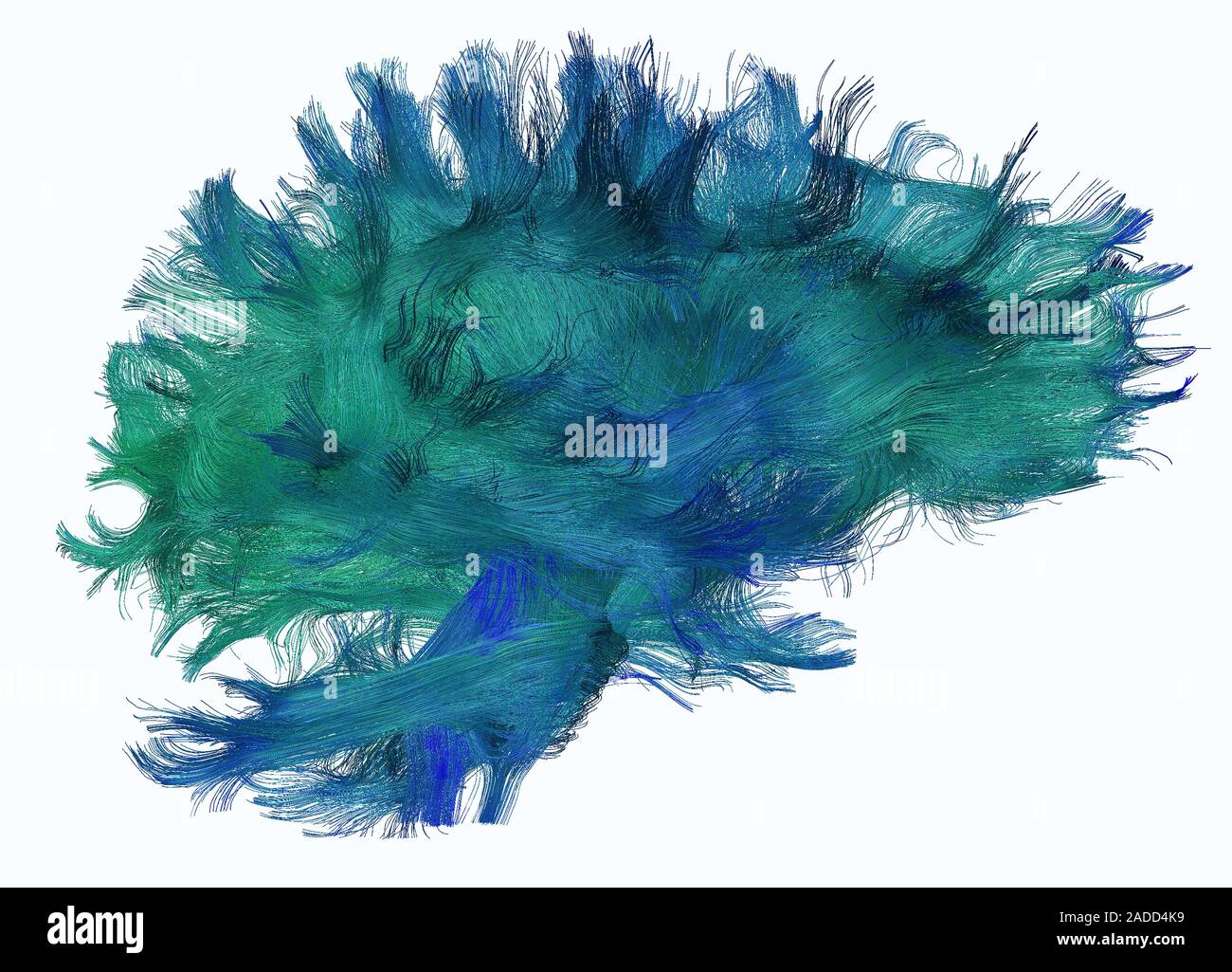
Diffusion tensor imaging (DTI) scan. A close-up look into the reconstructed white matter fibres of the brain. DTI offers a non-invasive look into how Image details File size:
49.3 MB (3.1 MB Compressed download)
Open your image file to the full size using image processing software.
Dimensions:
4892 x 3522 px | 41.4 x 29.8 cm | 16.3 x 11.7 inches | 300dpi
More information:
Diffusion tensor imaging (DTI) scan. A close-up look into the reconstructed white matter fibres of the brain. DTI offers a non-invasive look into how the white matter, or the highways of our brain, connect distant regions.
Search stock photos by tags
Similar stock images Diffusion MRI, also referred to as diffusion tensor imaging or DTI, of the human brain Stock Photo https://www.alamy.com/image-license-details/?v=1 https://www.alamy.com/stock-photo-diffusion-mri-also-referred-to-as-diffusion-tensor-imaging-or-dti-88275372.html RF F3H83T – Diffusion MRI, also referred to as diffusion tensor imaging or DTI, of the human brain Diffusion MRI, also referred to as diffusion tensor imaging or DTI, of the human brain Stock Photo https://www.alamy.com/image-license-details/?v=1 https://www.alamy.com/stock-photo-diffusion-mri-also-referred-to-as-diffusion-tensor-imaging-or-dti-88275450.html RF F3H86J – Diffusion MRI, also referred to as diffusion tensor imaging or DTI, of the human brain White matter fibres. Computer enhanced 3D diffusion spectral imaging (DSI) scan of the bundles of white matter nerve fibres in the brain. The fibres transmit nerve signals between brain regions and between the brain and the spinal cord. Diffusion spectrum imaging (DSI) is a variant of magnetic resonance imaging (MRI) in which a magnetic field maps the water contained in neuron fibers, thus mapping their criss-crossing patterns. A similar technique called diffusion tensor imaging (DTI) is also used to explore neural data of white matter fibres in the brain. Both methods allow mapping of their Stock Photo https://www.alamy.com/image-license-details/?v=1 https://www.alamy.com/stock-photo-white-matter-fibres-computer-enhanced-3d-diffusion-spectral-imaging-170920014.html RF KX2266 – White matter fibres. Computer enhanced 3D diffusion spectral imaging (DSI) scan of the bundles of white matter nerve fibres in the brain. The fibres transmit nerve signals between brain regions and between the brain and the spinal cord. Diffusion spectrum imaging (DSI) is a variant of magnetic resonance imaging (MRI) in which a magnetic field maps the water contained in neuron fibers, thus mapping their criss-crossing patterns. A similar technique called diffusion tensor imaging (DTI) is also used to explore neural data of white matter fibres in the brain. Both methods allow mapping of their This spread spectrum imaging scan, which samples 257 directions over a range of different gradient strengths, shows tractography at a triple crossover in the white matter of the parietal lobe. Stock Photo https://www.alamy.com/image-license-details/?v=1 https://www.alamy.com/this-spread-spectrum-imaging-scan-which-samples-257-directions-over-a-range-of-different-gradient-strengths-shows-tractography-at-a-triple-crossover-in-the-white-matter-of-the-parietal-lobe-image476707125.html RM 2JKFTPD – This spread spectrum imaging scan, which samples 257 directions over a range of different gradient strengths, shows tractography at a triple crossover in the white matter of the parietal lobe. 3D illustration of a human brain, titled as DTI - Diffusion Tensor Imaging. Stock Photo https://www.alamy.com/image-license-details/?v=1 https://www.alamy.com/3d-illustration-of-a-human-brain-titled-as-dti-diffusion-tensor-imaging-image600873661.html RF 2WWG4DH – 3D illustration of a human brain, titled as DTI - Diffusion Tensor Imaging. Diffusion MRI, also referred to as diffusion tensor imaging or DTI, of the human brain Stock Photo https://www.alamy.com/image-license-details/?v=1 https://www.alamy.com/stock-photo-diffusion-mri-also-referred-to-as-diffusion-tensor-imaging-or-dti-88275371.html RF F3H83R – Diffusion MRI, also referred to as diffusion tensor imaging or DTI, of the human brain Diffusion MRI, also referred to as diffusion tensor imaging or DTI, of the human brain Stock Photo https://www.alamy.com/image-license-details/?v=1 https://www.alamy.com/stock-photo-diffusion-mri-also-referred-to-as-diffusion-tensor-imaging-or-dti-88275379.html RF F3H843 – Diffusion MRI, also referred to as diffusion tensor imaging or DTI, of the human brain White matter fibres. Computer enhanced 3D diffusion spectral imaging (DSI) scan of the bundles of white matter nerve fibres in the brain. The fibres transmit nerve signals between brain regions and between the brain and the spinal cord. Diffusion spectrum imaging (DSI) is a variant of magnetic resonance imaging (MRI) in which a magnetic field maps the water contained in neuron fibers, thus mapping their criss-crossing patterns. A similar technique called diffusion tensor imaging (DTI) is also used to explore neural data of white matter fibres in the brain. Both methods allow mapping of their Stock Photo https://www.alamy.com/image-license-details/?v=1 https://www.alamy.com/stock-photo-white-matter-fibres-computer-enhanced-3d-diffusion-spectral-imaging-170920024.html RF KX226G – White matter fibres. Computer enhanced 3D diffusion spectral imaging (DSI) scan of the bundles of white matter nerve fibres in the brain. The fibres transmit nerve signals between brain regions and between the brain and the spinal cord. Diffusion spectrum imaging (DSI) is a variant of magnetic resonance imaging (MRI) in which a magnetic field maps the water contained in neuron fibers, thus mapping their criss-crossing patterns. A similar technique called diffusion tensor imaging (DTI) is also used to explore neural data of white matter fibres in the brain. Both methods allow mapping of their White matter tracts in a mouse brain acquired with diffusion tensioner imaging. The brain is viewed from below and with the front of the brain to the right. The colors represent different fiber directions. White matter integrity can be studied after brain injury using this visualization method. Stock Photo https://www.alamy.com/image-license-details/?v=1 https://www.alamy.com/white-matter-tracts-in-a-mouse-brain-acquired-with-diffusion-tensioner-imaging-the-brain-is-viewed-from-below-and-with-the-front-of-the-brain-to-the-right-the-colors-represent-different-fiber-directions-white-matter-integrity-can-be-studied-after-brain-injury-using-this-visualization-method-image476706933.html RM 2JKFTFH – White matter tracts in a mouse brain acquired with diffusion tensioner imaging. The brain is viewed from below and with the front of the brain to the right. The colors represent different fiber directions. White matter integrity can be studied after brain injury using this visualization method. Diffusion MRI, also referred to as diffusion tensor imaging or DTI, of the human brain Stock Photo https://www.alamy.com/image-license-details/?v=1 https://www.alamy.com/stock-photo-diffusion-mri-also-referred-to-as-diffusion-tensor-imaging-or-dti-88275354.html RF F3H836 – Diffusion MRI, also referred to as diffusion tensor imaging or DTI, of the human brain Diffusion MRI, also referred to as diffusion tensor imaging or DTI, of the human brain Stock Photo https://www.alamy.com/image-license-details/?v=1 https://www.alamy.com/stock-photo-diffusion-mri-also-referred-to-as-diffusion-tensor-imaging-or-dti-88275380.html RF F3H844 – Diffusion MRI, also referred to as diffusion tensor imaging or DTI, of the human brain White matter fibres. Computer enhanced 3D diffusion spectral imaging (DSI) scan of the bundles of white matter nerve fibres in the brain. The fibres transmit nerve signals between brain regions and between the brain and the spinal cord. Diffusion spectrum imaging (DSI) is a variant of magnetic resonance imaging (MRI) in which a magnetic field maps the water contained in neuron fibers, thus mapping their criss-crossing patterns. A similar technique called diffusion tensor imaging (DTI) is also used to explore neural data of white matter fibres in the brain. Both methods allow mapping of their Stock Photo https://www.alamy.com/image-license-details/?v=1 https://www.alamy.com/stock-photo-white-matter-fibres-computer-enhanced-3d-diffusion-spectral-imaging-170920026.html RF KX226J – White matter fibres. Computer enhanced 3D diffusion spectral imaging (DSI) scan of the bundles of white matter nerve fibres in the brain. The fibres transmit nerve signals between brain regions and between the brain and the spinal cord. Diffusion spectrum imaging (DSI) is a variant of magnetic resonance imaging (MRI) in which a magnetic field maps the water contained in neuron fibers, thus mapping their criss-crossing patterns. A similar technique called diffusion tensor imaging (DTI) is also used to explore neural data of white matter fibres in the brain. Both methods allow mapping of their Diffusion MRI, also referred to as diffusion tensor imaging or DTI, of the human brain Stock Photo https://www.alamy.com/image-license-details/?v=1 https://www.alamy.com/stock-photo-diffusion-mri-also-referred-to-as-diffusion-tensor-imaging-or-dti-88275350.html RF F3H832 – Diffusion MRI, also referred to as diffusion tensor imaging or DTI, of the human brain 