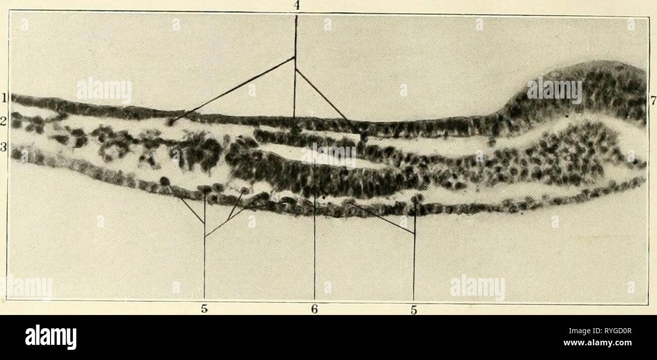
Early stages of vasculogenesis in the cat (Felis domestica) with especial reference to the mesenschymal origin of endothelium earlystagesofvas03schuuoft Year: 1914 FiS- •') Transverse sootion of lui embryo of two somites ;it the level iiuli- eated in Figure 3. Slide .5, row 1, section 0, X 300. Reduced J-. 1 Ectoderm 2 Mesoderm 3 Entoderm 4 Hypectodennal mesenchyme 5 Hypercntodermal mesenchyme 6 Coelom 7 Xeural plate Fig. (J Dorsal view of model of mesoderm ^white), mesenchyme (green) and vasofactive cells (yellow) of an embryo of four somites. Columbia collection No. 409, X 300. Reduced !
Image details
Contributor:
Bookend / Alamy Stock PhotoImage ID:
RYGD0RFile size:
5.7 MB (278.5 KB Compressed download)Releases:
Model - no | Property - noDo I need a release?Dimensions:
2043 x 979 px | 34.6 x 16.6 cm | 13.6 x 6.5 inches | 150dpiMore information:
This image is a public domain image, which means either that copyright has expired in the image or the copyright holder has waived their copyright. Alamy charges you a fee for access to the high resolution copy of the image.
This image could have imperfections as it’s either historical or reportage.
Early stages of vasculogenesis in the cat (Felis domestica) with especial reference to the mesenschymal origin of endothelium earlystagesofvas03schuuoft Year: 1914 FiS- •') Transverse sootion of lui embryo of two somites ;it the level iiuli- eated in Figure 3. Slide .5, row 1, section 0, X 300. Reduced J-. 1 Ectoderm 2 Mesoderm 3 Entoderm 4 Hypectodennal mesenchyme 5 Hypercntodermal mesenchyme 6 Coelom 7 Xeural plate Fig. (J Dorsal view of model of mesoderm ^white), mesenchyme (green) and vasofactive cells (yellow) of an embryo of four somites. Columbia collection No. 409, X 300. Reduced !, . 1 Somite 1 2 Anlages of .juxta-ncural anastomosis 3 Connection between the j uxta-nein'al anastomosis and I he :i(iila, cf. fig- ure 25 4 Site of quintal ganglion 5 Endothelial anlage opposite the second nephrotonie; cf. figures 8 and it 0 Region of cardiac coelom 60