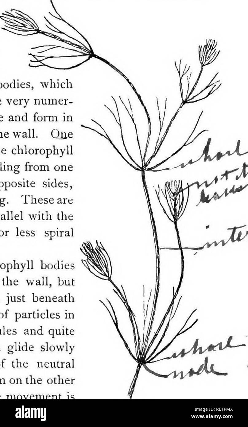
. Elementary botany. Botany. PROTOPLASM. node. These internodes are peculiar. They consist of but a single " cell," and are cylindrical, with closed ends. They are sometimes 5-10 cm. long. 19. Internode of nitella.—For the study of an internode of nitella, a small one, near the end, or the ends of one of the " leaves " is best suited, since it is more transparent. A small portion of the plant should be placed on the glass slip in water with the cover glass over a tuft of the branches near the growing end. Examined with the microscope the green chlorophyll bodies, which form

Image details
Contributor:
The Book Worm / Alamy Stock PhotoImage ID:
RE1PMXFile size:
7.1 MB (265.4 KB Compressed download)Releases:
Model - no | Property - noDo I need a release?Dimensions:
1246 x 2004 px | 21.1 x 33.9 cm | 8.3 x 13.4 inches | 150dpiMore information:
This image is a public domain image, which means either that copyright has expired in the image or the copyright holder has waived their copyright. Alamy charges you a fee for access to the high resolution copy of the image.
This image could have imperfections as it’s either historical or reportage.
. Elementary botany. Botany. PROTOPLASM. node. These internodes are peculiar. They consist of but a single " cell, " and are cylindrical, with closed ends. They are sometimes 5-10 cm. long. 19. Internode of nitella.—For the study of an internode of nitella, a small one, near the end, or the ends of one of the " leaves " is best suited, since it is more transparent. A small portion of the plant should be placed on the glass slip in water with the cover glass over a tuft of the branches near the growing end. Examined with the microscope the green chlorophyll bodies, which form oval or oblong discs, are seen to be very numer- ous. They lie quite closely side by side and form in perfect rows along the inner surface of the wall. Que peculiar feature of the arrangement of the chlorophyll bodies is that there are two lines, extending from one end of the internode to the other on opposite sides, where the chlorophyll bodies are wanting. These are known as neutral lines. They run parallel with the axis of the internode, or in a more or less spiral manner as shown in fig. 9. 20. CyclosiB in nitella.—The chlorophyll bodies are stationary on the inner surface of the wall, but if the microscope be properly focussed just beneath this layer we notice a rotary motion of particles in the protoplasm. There are small granules and quite large masses of granular matter which glide slowly along in one direction on a given side of the neutral line. If now we examine the protoplasm on the other side of the neutral line, we see that the movement is in the opposite direction. If we examine this move- ment at the end of an internode the particles are seen to glide around the end from one side of the neutral line to the other. So that when conditions are favorable, such as temperature, healthy state of the plant, etc., this gliding of the particles or apparent streaming of the proto- plasm down one side of the " cell, " and back upon the other, continues in an u