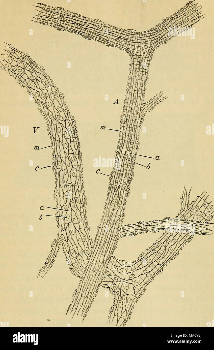
. Elementary physiology . Fig s6.-A small artery, A, and vein, J", from the subcutaneous connective tissue of the rat. treated with nitrate of silver. (,175 diameters.; ^ n endothelial cells with b, b, their nuclei ; m, m, transverse markings due to staining ' orsubstance'between the muscular fibre cells; c, c, nuclei of connective-tissue corpuscles attached to exterior of vessel. veins. In these small arteries and veins there are disposed outside the epitheloid coat (endothelium) layers of involuntary muscle fibres, which are arranged in a circular and slightly H
RMID:Image ID:MA6YEJ
Image details
Contributor:
The Bookworm Collection / Alamy Stock PhotoImage ID:
MA6YEJFile size:
14.3 MB (546.1 KB Compressed download)Releases:
Model - no | Property - noDo I need a release?Dimensions:
1806 x 2767 px | 15.3 x 23.4 cm | 6 x 9.2 inches | 300dpiMore information:
This image is a public domain image, which means either that copyright has expired in the image or the copyright holder has waived their copyright. Alamy charges you a fee for access to the high resolution copy of the image.
This image could have imperfections as it’s either historical or reportage.