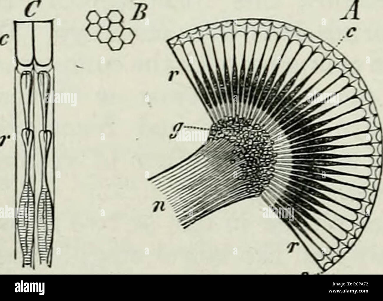
. Elements of Comparative Anatomy. 266 COlklPAEATIVE ANATOMY. an elevation of the cepHalotliorax. In tlie Pycnogonida 4 eyes occupy a similar position. On tlie other hand they are reduced to 2 in many Mites, and so also in the Tardigrada; in many parasitic Mites they have completely disappeared. The principal point in their structure is the presence of a cornea-lens, which is ordinarily very large in each, eye; behind this is a layer of cells, which represents the vitreous body, and to this the retina is attached. In the Aranea the retina is formed in two ways, the eyes directed anteriorly dif
Image details
Contributor:
Paul Fearn / Alamy Stock PhotoImage ID:
RCPA72File size:
7.1 MB (259.7 KB Compressed download)Releases:
Model - no | Property - noDo I need a release?Dimensions:
1865 x 1340 px | 31.6 x 22.7 cm | 12.4 x 8.9 inches | 150dpiMore information:
This image is a public domain image, which means either that copyright has expired in the image or the copyright holder has waived their copyright. Alamy charges you a fee for access to the high resolution copy of the image.
This image could have imperfections as it’s either historical or reportage.
. Elements of Comparative Anatomy. 266 COlklPAEATIVE ANATOMY. an elevation of the cepHalotliorax. In tlie Pycnogonida 4 eyes occupy a similar position. On tlie other hand they are reduced to 2 in many Mites, and so also in the Tardigrada; in many parasitic Mites they have completely disappeared. The principal point in their structure is the presence of a cornea-lens, which is ordinarily very large in each, eye; behind this is a layer of cells, which represents the vitreous body, and to this the retina is attached. In the Aranea the retina is formed in two ways, the eyes directed anteriorly differing in structure from those which are turned upwards. That is to say, the retinal cells of the former surround a small longitudinally bisected rod at their anterior end (Epeira). § 206. The compound eye is characterised by the above-mentioned fusion (§ 204) of a number (7-4) of retinal cells into a structure which surrounds the rhabdomâthe "retinula" (Fig. 135, C r). The eye is made up of these retinulje, each of which, is enveloped in pigment. The multifid crystalline cone lies in front of the retinula. Two of these structures are represented in Fig. C. The crystalline cones may be made out in front of the retinula, and be- hind the cornea-lenses (c). The whole arrangement is easy to under- stand, when we derive it from the simple eye. A reduction of the retinal elements of the simple eye gives rise to the retinula, and a compound eye is formed by the gradual concrescence of a number of simple eyes. Most of the Crustacea have eyes of this kind. In the Cladocera the movable eye (Fig. 136, oc) lies in a cavity roofed over by the integument. In the Lfemodipoda also the cuticular layer of the integument seems to take no part in forming the eye. On the other hand in the Phyl- lopoda we meet with a faceting of the inner surface of the cuticle covering the eye, the facets corre- sponding to the crystalline cones. In the Isopoda the compound eye still consists of a