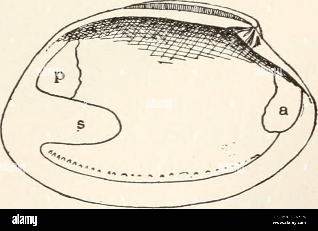
. Elements of comparative zoology. Zoology. FIG. 41.—Quahog (Venus mercenaria), with foot and siphons extended. then strong retractor muscles to draw them back are present. All of these muscles—adductors, retractors, etc. -leave their impress on the shell, so that the student, with the shell alone, may know of some •/ of the structures of the soft parts (fig. 42). Water is drawn into the mantle-cavity by means. of very minute hair-like FlG- 42.—inside of bivalve shell ing muscular impressions of a, an- StrUCtlireS (Cilia} Which terior adductor; p, posterior adduc- tor; s, siphonal muscle. cove

Image details
Contributor:
Paul Fearn / Alamy Stock PhotoImage ID:
RCNK3MFile size:
7.1 MB (171.4 KB Compressed download)Releases:
Model - no | Property - noDo I need a release?Dimensions:
1946 x 1284 px | 33 x 21.7 cm | 13 x 8.6 inches | 150dpiMore information:
This image is a public domain image, which means either that copyright has expired in the image or the copyright holder has waived their copyright. Alamy charges you a fee for access to the high resolution copy of the image.
This image could have imperfections as it’s either historical or reportage.
. Elements of comparative zoology. Zoology. FIG. 41.—Quahog (Venus mercenaria), with foot and siphons extended. then strong retractor muscles to draw them back are present. All of these muscles—adductors, retractors, etc. -leave their impress on the shell, so that the student, with the shell alone, may know of some •/ of the structures of the soft parts (fig. 42). Water is drawn into the mantle-cavity by means. of very minute hair-like FlG- 42.—inside of bivalve shell ing muscular impressions of a, an- StrUCtlireS (Cilia} Which terior adductor; p, posterior adduc- tor; s, siphonal muscle. cover the gills and other parts. These cilia are in constant motion, * and thus currents of water are produced, flowing always in one * The teacher should demonstrate this ciliary action under the compound microscope, . Please note that these images are extracted from scanned page images that may have been digitally enhanced for readability - coloration and appearance of these illustrations may not perfectly resemble the original work.. Kingsley, J. S. (John Sterling), 1854-1929. New York, H. Holt and Company