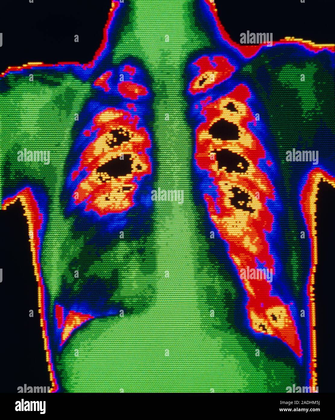
False-colour chest X-ray showing lobar pneumonia in the lobes of a patient's right lung (green & black area at bottom of lung at left of image). The d
RMID:Image ID:2ADHM5J
Image details
Contributor:
Science Photo Library / Alamy Stock PhotoImage ID:
2ADHM5JFile size:
44.1 MB (2.3 MB Compressed download)Releases:
Model - no | Property - noDo I need a release?Dimensions:
3630 x 4247 px | 30.7 x 36 cm | 12.1 x 14.2 inches | 300dpiDate taken:
12 June 1989Photographer:
Science Photo LibraryMore information:
False-colour chest X-ray showing lobar pneumonia in the lobes of a patient's right lung (green & black area at bottom of lung at left of image). The disease is caused by certain strains of the bacterium Streptococcus pneumoniae. Alveoli (the air sacs of the lung) become blocked with pus, forcing air out and causing the lung to become solidified. Here, colouring highlights the reduction in effective size of healthy lung on the left of image, compared to the normal right-side lung.