···
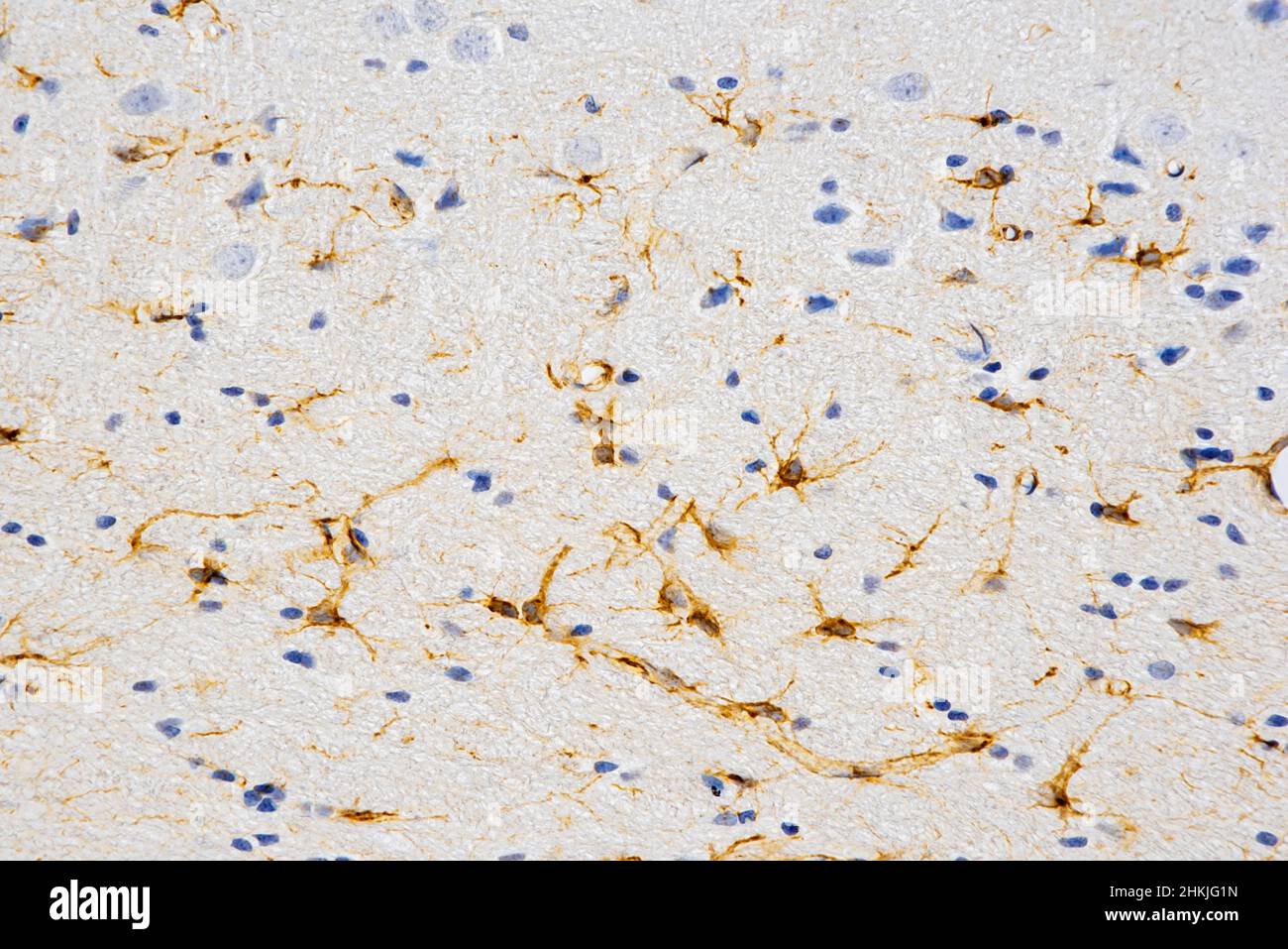
Glial cells, light micrograph Image details File size:
68.8 MB (2.3 MB Compressed download)
Open your image file to the full size using image processing software.
Dimensions:
6004 x 4008 px | 50.8 x 33.9 cm | 20 x 13.4 inches | 300dpi
Date taken:
22 November 2021
More information:
Light micrograph of glial cells. Glial cells, or neuroglia, are non-neural cells of both the central and peripheral nervous systems. They provide physical and functional support to neurons (nerve cells). Magnification: x200 when printed at 15cm wide.
Search stock photos by tags
Similar stock images Pseudounipolar neurons. Pseudounipolar neurons of rounded soma of a dorsal root ganglion.Сell body is surrounded by glia cells called satellite cells. Stock Photo https://www.alamy.com/image-license-details/?v=1 https://www.alamy.com/pseudounipolar-neurons-pseudounipolar-neurons-of-rounded-soma-of-a-dorsal-root-ganglionell-body-is-surrounded-by-glia-cells-called-satellite-cells-image471432281.html RF 2JAYGK5 – Pseudounipolar neurons. Pseudounipolar neurons of rounded soma of a dorsal root ganglion.Сell body is surrounded by glia cells called satellite cells. Light micrograph of human brain tissue showing neurons and glial cells Stock Photo https://www.alamy.com/image-license-details/?v=1 https://www.alamy.com/light-micrograph-of-human-brain-tissue-showing-neurons-and-glial-cells-image550655798.html RF 2PYTF2E – Light micrograph of human brain tissue showing neurons and glial cells Purkinje nerve cells, Light micrograph of asection through the cerebellum in the brain Stock Photo https://www.alamy.com/image-license-details/?v=1 https://www.alamy.com/stock-photo-purkinje-nerve-cells-light-micrograph-of-asection-through-the-cerebellum-21205659.html RF B6E00Y – Purkinje nerve cells, Light micrograph of asection through the cerebellum in the brain Light micrograph of human brain tissue showing neurons and glial cells Stock Photo https://www.alamy.com/image-license-details/?v=1 https://www.alamy.com/light-micrograph-of-human-brain-tissue-showing-neurons-and-glial-cells-image550655821.html RF 2PYTF39 – Light micrograph of human brain tissue showing neurons and glial cells Dorsal root ganglion. Pseudounipolar neurons of a dorsal root ganglion. Hematoxlyn and eosin stain. Magnification: x40. Stock Photo https://www.alamy.com/image-license-details/?v=1 https://www.alamy.com/dorsal-root-ganglion-pseudounipolar-neurons-of-a-dorsal-root-ganglion-hematoxlyn-and-eosin-stain-magnification-x40-image471432248.html RF 2JAYGJ0 – Dorsal root ganglion. Pseudounipolar neurons of a dorsal root ganglion. Hematoxlyn and eosin stain. Magnification: x40. Light micrograph of human brain tissue showing neurons and glial cells Stock Photo https://www.alamy.com/image-license-details/?v=1 https://www.alamy.com/light-micrograph-of-human-brain-tissue-showing-neurons-and-glial-cells-image550655807.html RF 2PYTF2R – Light micrograph of human brain tissue showing neurons and glial cells Light micrograph of human brain tissue showing neurons and glial cells Stock Photo https://www.alamy.com/image-license-details/?v=1 https://www.alamy.com/light-micrograph-of-human-brain-tissue-showing-neurons-and-glial-cells-image550655809.html RF 2PYTF2W – Light micrograph of human brain tissue showing neurons and glial cells Liquefactive necrosis of the human brain, light photomicrograph showing loss of cell outlines, accumulation of cellular debris, macrophage infiltratio Stock Photo https://www.alamy.com/image-license-details/?v=1 https://www.alamy.com/liquefactive-necrosis-of-the-human-brain-light-photomicrograph-showing-loss-of-cell-outlines-accumulation-of-cellular-debris-macrophage-infiltratio-image555053999.html RF 2R70W13 – Liquefactive necrosis of the human brain, light photomicrograph showing loss of cell outlines, accumulation of cellular debris, macrophage infiltratio Liquefactive necrosis of the human brain, light photomicrograph showing loss of cell outlines, accumulation of cellular debris, macrophage infiltratio Stock Photo https://www.alamy.com/image-license-details/?v=1 https://www.alamy.com/liquefactive-necrosis-of-the-human-brain-light-photomicrograph-showing-loss-of-cell-outlines-accumulation-of-cellular-debris-macrophage-infiltratio-image555053894.html RF 2R70TWA – Liquefactive necrosis of the human brain, light photomicrograph showing loss of cell outlines, accumulation of cellular debris, macrophage infiltratio Liquefactive necrosis of the human brain, light photomicrograph showing loss of cell outlines, accumulation of cellular debris, macrophage infiltratio Stock Photo https://www.alamy.com/image-license-details/?v=1 https://www.alamy.com/liquefactive-necrosis-of-the-human-brain-light-photomicrograph-showing-loss-of-cell-outlines-accumulation-of-cellular-debris-macrophage-infiltratio-image555053892.html RF 2R70TW8 – Liquefactive necrosis of the human brain, light photomicrograph showing loss of cell outlines, accumulation of cellular debris, macrophage infiltratio Liquefactive necrosis of the human brain, light photomicrograph showing loss of cell outlines, accumulation of cellular debris, macrophage infiltratio Stock Photo https://www.alamy.com/image-license-details/?v=1 https://www.alamy.com/liquefactive-necrosis-of-the-human-brain-light-photomicrograph-showing-loss-of-cell-outlines-accumulation-of-cellular-debris-macrophage-infiltratio-image555053909.html RF 2R70TWW – Liquefactive necrosis of the human brain, light photomicrograph showing loss of cell outlines, accumulation of cellular debris, macrophage infiltratio Liquefactive necrosis of the human brain, light photomicrograph showing loss of cell outlines, accumulation of cellular debris, macrophage infiltratio Stock Photo https://www.alamy.com/image-license-details/?v=1 https://www.alamy.com/liquefactive-necrosis-of-the-human-brain-light-photomicrograph-showing-loss-of-cell-outlines-accumulation-of-cellular-debris-macrophage-infiltratio-image555053921.html RF 2R70TX9 – Liquefactive necrosis of the human brain, light photomicrograph showing loss of cell outlines, accumulation of cellular debris, macrophage infiltratio Liquefactive necrosis of the human brain, light photomicrograph showing loss of cell outlines, accumulation of cellular debris, macrophage infiltratio Stock Photo https://www.alamy.com/image-license-details/?v=1 https://www.alamy.com/liquefactive-necrosis-of-the-human-brain-light-photomicrograph-showing-loss-of-cell-outlines-accumulation-of-cellular-debris-macrophage-infiltratio-image555053910.html RF 2R70TWX – Liquefactive necrosis of the human brain, light photomicrograph showing loss of cell outlines, accumulation of cellular debris, macrophage infiltratio 