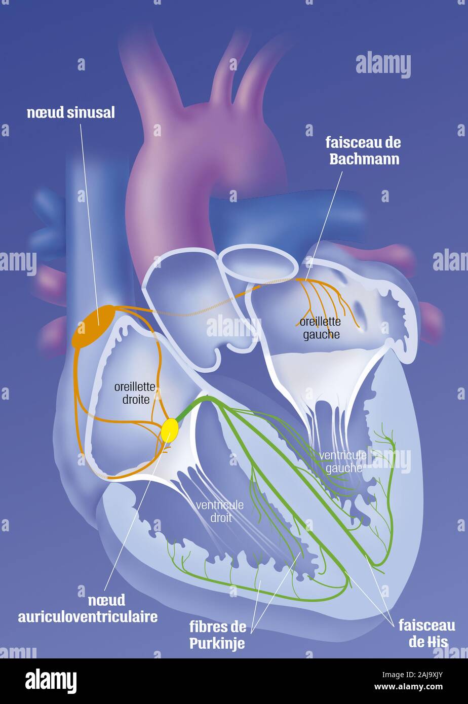
Heart and cardionector system
RMID:Image ID:2AJ9XJY
Image details
Contributor:
BSIP SA / Alamy Stock PhotoImage ID:
2AJ9XJYFile size:
39.2 MB (561.8 KB Compressed download)Releases:
Model - no | Property - noDo I need a release?Dimensions:
3122 x 4391 px | 26.4 x 37.2 cm | 10.4 x 14.6 inches | 300dpiDate taken:
30 November 2019Photographer:
WITT-DEGUILLAUME / BSIPMore information:
The heart and its nerve conduction, system cardionectors. Frontal cut of the heart showing the 4 cardiac cavities: right and left atria, separated from the right and left ventricles by the tricuspid and mitral valves. The opening of the large vessels: the aorta the pulmonary trunk and the superior and inferior vena cava. The electrical conduction of the heart starts at the sinus node of Keith and Flack (orange), spreads to the auricles and atrioventricular node (yellow), then the bundle of His and the fibers of Punkinje (green)