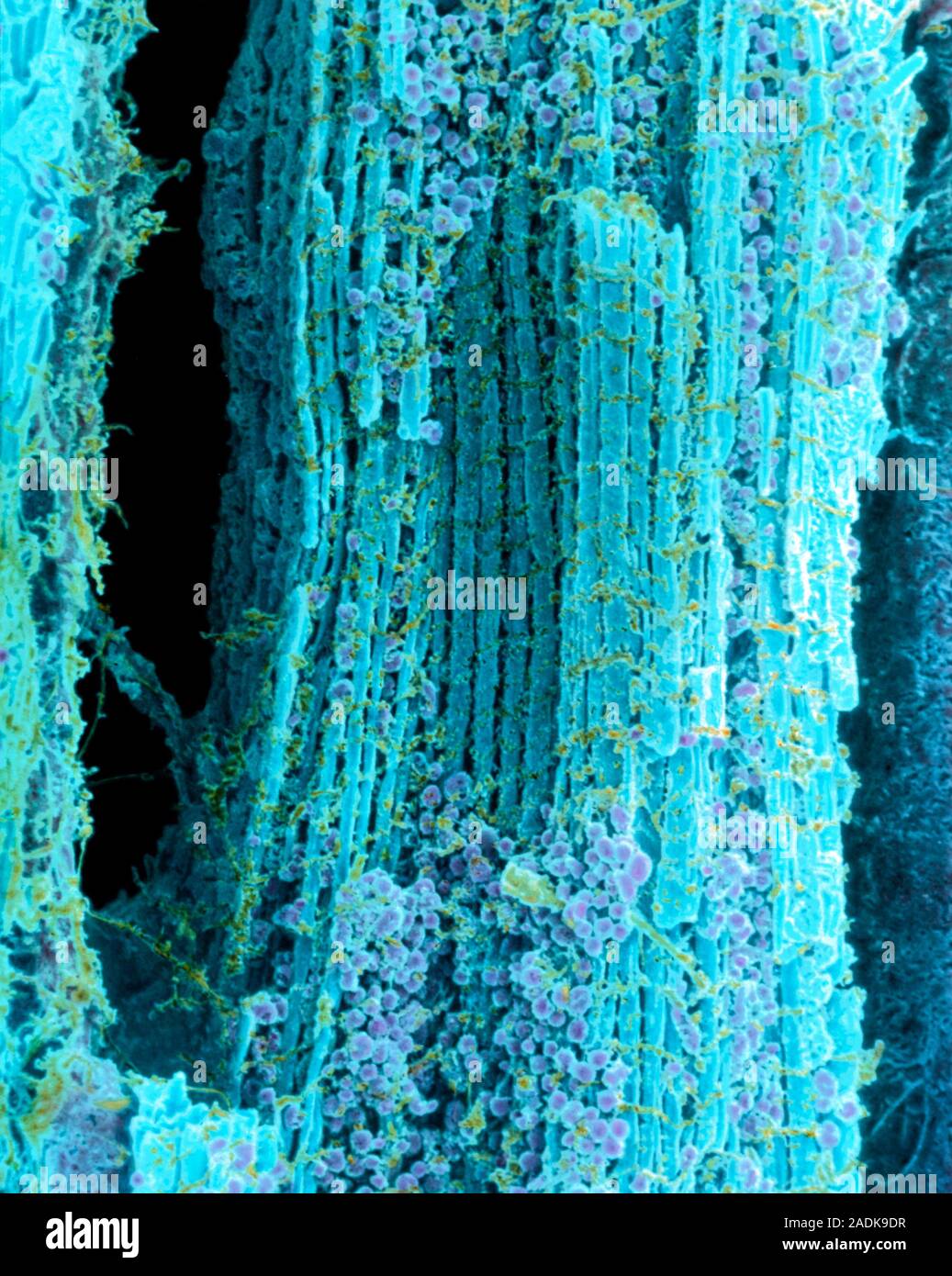
Heart muscle. Coloured scanning electron micrograph (SEM) of healthy human heart (cardiac) muscle fibres (blue). Mitochondria (mauve) supply the muscl
RMID:Image ID:2ADK9DR
Image details
Contributor:
Science Photo Library / Alamy Stock PhotoImage ID:
2ADK9DRFile size:
28.7 MB (1.3 MB Compressed download)Releases:
Model - no | Property - noDo I need a release?Dimensions:
2828 x 3543 px | 23.9 x 30 cm | 9.4 x 11.8 inches | 300dpiDate taken:
21 July 1998Photographer:
STEVE GSCHMEISSNER/SCIENCE PHOTO LIBRARYMore information:
Heart muscle. Coloured scanning electron micrograph (SEM) of healthy human heart (cardiac) muscle fibres (blue). Mitochondria (mauve) supply the muscle cells with energy. The muscle fibres, or myofibrils, are crossed by transverse tubules (orange). These tubules mark the division of the myofibril into contractile units known as sarcomeres. Cardiac muscle, which normally works subconsciously, pumps blood continuously around the body without tiring. Magnification unknown.