···
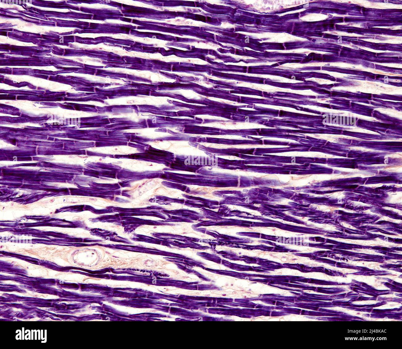
Heart myocardium, light micrograph Image details File size:
33.8 MB (2.3 MB Compressed download)
Open your image file to the full size using image processing software.
Dimensions:
3840 x 3072 px | 32.5 x 26 cm | 12.8 x 10.2 inches | 300dpi
More information:
Light micrograph of striated muscle fibres of the heart myocardium stained with Mallory's phosphotungstic haematoxylin. The striated appearance of the cardiomyocytes and the intercalated discs stand out clearly.
Search stock photos by tags
Similar stock images Cardiac muscle section under light microscope. Heart muscle, also myocardium, a vertebrate muscle. Striated muscle. Stock Photo https://www.alamy.com/image-license-details/?v=1 https://www.alamy.com/cardiac-muscle-section-under-light-microscope-heart-muscle-also-myocardium-a-vertebrate-muscle-striated-muscle-image369496566.html RF 2CD40HX – Cardiac muscle section under light microscope. Heart muscle, also myocardium, a vertebrate muscle. Striated muscle. Human cardiac muscle, light micrograph. Stock Photo https://www.alamy.com/image-license-details/?v=1 https://www.alamy.com/human-cardiac-muscle-light-micrograph-image385222629.html RF 2DAMBB1 – Human cardiac muscle, light micrograph. Human heart muscle, light micrograph. Striated cardiac muscle cells, or myocytes Stock Photo https://www.alamy.com/image-license-details/?v=1 https://www.alamy.com/human-heart-muscle-light-micrograph-striated-cardiac-muscle-cells-or-myocytes-image550655471.html RF 2PYTEJR – Human heart muscle, light micrograph. Striated cardiac muscle cells, or myocytes Cardiac muscle, light micrograph. Stock Photo https://www.alamy.com/image-license-details/?v=1 https://www.alamy.com/cardiac-muscle-light-micrograph-image385222979.html RF 2DAMBRF – Cardiac muscle, light micrograph. Histopathology of heart hypertrophy, high magnification. Photomicrograph showing hypertrophic myocardium with thick muscle fibers and enlarged and dar Stock Photo https://www.alamy.com/image-license-details/?v=1 https://www.alamy.com/histopathology-of-heart-hypertrophy-high-magnification-photomicrograph-showing-hypertrophic-myocardium-with-thick-muscle-fibers-and-enlarged-and-dar-image555071273.html RF 2R71K21 – Histopathology of heart hypertrophy, high magnification. Photomicrograph showing hypertrophic myocardium with thick muscle fibers and enlarged and dar Microscopy photography. Cardiac muscle section. Stock Photo https://www.alamy.com/image-license-details/?v=1 https://www.alamy.com/microscopy-photography-cardiac-muscle-section-image217516319.html RF PHTM7Y – Microscopy photography. Cardiac muscle section. This micrograph reveals an intranuclear inclusion body in a heart section from a patient with diphtheria-related myocarditis, 1965. In approximately 25 percent of patients with diphtheria, a toxin produced by Corynebacterium diphtheria bacteria causes inflammation of the myocardium, which is the most common and most worrisome complication. Image courtesy CDC/Dr. Martin Hicklin. Stock Photo https://www.alamy.com/image-license-details/?v=1 https://www.alamy.com/this-micrograph-reveals-an-intranuclear-inclusion-body-in-a-heart-section-from-a-patient-with-diphtheria-related-myocarditis-1965-in-approximately-25-percent-of-patients-with-diphtheria-a-toxin-produced-by-corynebacterium-diphtheria-bacteria-causes-inflammation-of-the-myocardium-which-is-the-most-common-and-most-worrisome-complication-image-courtesy-cdcdr-martin-hicklin-image244837856.html RM T69954 – This micrograph reveals an intranuclear inclusion body in a heart section from a patient with diphtheria-related myocarditis, 1965. In approximately 25 percent of patients with diphtheria, a toxin produced by Corynebacterium diphtheria bacteria causes inflammation of the myocardium, which is the most common and most worrisome complication. Image courtesy CDC/Dr. Martin Hicklin. . The Biological bulletin. Biology; Zoology; Biology; Marine Biology. 106 M. G. MARTYNOVA AND O. A. BYSTROVA pc. Figure 1. Light micrograph of semithin section through the atrium of the snail Ac/uitina fit/ica. The heart wall consists of epicardium (arrows) facing the pericardia! cavity (pc) and a myocardium (m). The endoihdial cells (arrowheads) adjoin the luminal surface of the myocardial trabeculae hi. heart lumen. Scale bar = 0.05 mm. somes, and a number of small vesicles. UCs were easy to distinguish from other nonmuscle cells in the heart, such as endothelial cells, gliointerstitial cell Stock Photo https://www.alamy.com/image-license-details/?v=1 https://www.alamy.com/the-biological-bulletin-biology-zoology-biology-marine-biology-106-m-g-martynova-and-o-a-bystrova-pc-figure-1-light-micrograph-of-semithin-section-through-the-atrium-of-the-snail-acuitina-fitica-the-heart-wall-consists-of-epicardium-arrows-facing-the-pericardia!-cavity-pc-and-a-myocardium-m-the-endoihdial-cells-arrowheads-adjoin-the-luminal-surface-of-the-myocardial-trabeculae-hi-heart-lumen-scale-bar-=-005-mm-somes-and-a-number-of-small-vesicles-ucs-were-easy-to-distinguish-from-other-nonmuscle-cells-in-the-heart-such-as-endothelial-cells-gliointerstitial-cell-image234629951.html RM RHM8W3 – . The Biological bulletin. Biology; Zoology; Biology; Marine Biology. 106 M. G. MARTYNOVA AND O. A. BYSTROVA pc. Figure 1. Light micrograph of semithin section through the atrium of the snail Ac/uitina fit/ica. The heart wall consists of epicardium (arrows) facing the pericardia! cavity (pc) and a myocardium (m). The endoihdial cells (arrowheads) adjoin the luminal surface of the myocardial trabeculae hi. heart lumen. Scale bar = 0.05 mm. somes, and a number of small vesicles. UCs were easy to distinguish from other nonmuscle cells in the heart, such as endothelial cells, gliointerstitial cell Cardiac muscle, light micrograph. Stock Photo https://www.alamy.com/image-license-details/?v=1 https://www.alamy.com/cardiac-muscle-light-micrograph-image385222904.html RF 2DAMBMT – Cardiac muscle, light micrograph. Histopathology of heart hypertrophy, high magnification. Photomicrograph showing hypertrophic myocardium with thick muscle fibers and enlarged and dar Stock Photo https://www.alamy.com/image-license-details/?v=1 https://www.alamy.com/histopathology-of-heart-hypertrophy-high-magnification-photomicrograph-showing-hypertrophic-myocardium-with-thick-muscle-fibers-and-enlarged-and-dar-image555071276.html RF 2R71K24 – Histopathology of heart hypertrophy, high magnification. Photomicrograph showing hypertrophic myocardium with thick muscle fibers and enlarged and dar Cardiac muscle, light micrograph. Stock Photo https://www.alamy.com/image-license-details/?v=1 https://www.alamy.com/cardiac-muscle-light-micrograph-image385222712.html RF 2DAMBE0 – Cardiac muscle, light micrograph. Human cardiac muscle, light micrograph Stock Photo https://www.alamy.com/image-license-details/?v=1 https://www.alamy.com/human-cardiac-muscle-light-micrograph-image574554393.html RF 2TAN5YN – Human cardiac muscle, light micrograph Histopathology of heart hypertrophy, high magnification. Photomicrograph showing hypertrophic myocardium with thick muscle fibers and enlarged and dar Stock Photo https://www.alamy.com/image-license-details/?v=1 https://www.alamy.com/histopathology-of-heart-hypertrophy-high-magnification-photomicrograph-showing-hypertrophic-myocardium-with-thick-muscle-fibers-and-enlarged-and-dar-image555071149.html RF 2R71JWH – Histopathology of heart hypertrophy, high magnification. Photomicrograph showing hypertrophic myocardium with thick muscle fibers and enlarged and dar 