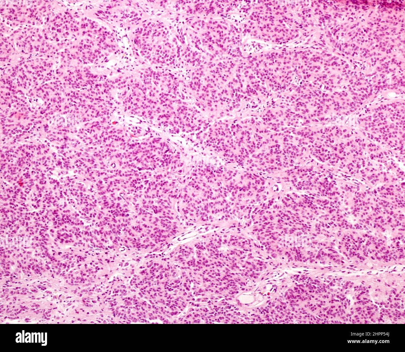
Human pineal gland lobulation, light micrograph

RMID:Image ID:2HPP54J
Image details
Contributor:
Science Photo Library / Alamy Stock PhotoImage ID:
2HPP54JFile size:
33.8 MB (1.9 MB Compressed download)Releases:
Model - no | Property - noDo I need a release?Dimensions:
3840 x 3072 px | 32.5 x 26 cm | 12.8 x 10.2 inches | 300dpiDate taken:
16 February 2022Photographer:
JOSE CALVO / SCIENCE PHOTO LIBRARYMore information:
Light micrograph of a young human pineal gland showing incipient lobulation of the parenchyma. The pineal parenchyma tends to organize itself into rounded units separated by thin connective tissue septa where the blood vessels are located. However, unlike the true lobes present in the exocrine glands, these units are not delimited but rather merge extensively with neighbouring units. Although the distribution of the parenchyma in rounded units can be seen, the lobes are still poorly defined.