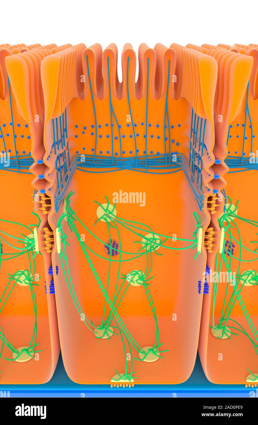
Intestinal cell junctions. Illustration of an intestinal epithelial cell, showing anchoring junctions (desmosomes, yellow; adherens, orange), gap junc
RMID:Image ID:2AD0PE9
Image details
Contributor:
Science Photo Library / Alamy Stock PhotoImage ID:
2AD0PE9File size:
51 MB (1.2 MB Compressed download)Releases:
Model - no | Property - noDo I need a release?Dimensions:
3403 x 5238 px | 28.8 x 44.3 cm | 11.3 x 17.5 inches | 300dpiDate taken:
16 March 2015Photographer:
Science Photo LibraryMore information:
Intestinal cell junctions. Illustration of an intestinal epithelial cell, showing anchoring junctions (desmosomes, yellow; adherens, orange), gap junctions (blue), tight junctions (ridged) and a hemidesmosome junction (bottom). Cytoskeletal filaments are blue and green. Across top are the microvilli that absorb nutrients from the intestinal lumen as food is digested. The various junctions shown here have a range of functions. Tight junctions form a relatively impermeable barrier. Anchoring junctions provide mechanical support. Gap junctions allows chemical and electrical communication between cells. For this artwork with labels, see C023/8827.