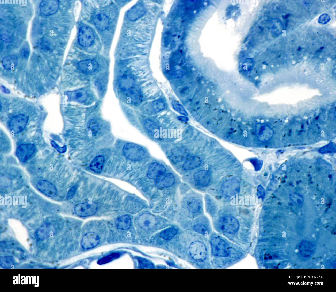
Kidney cortex, light micrograph

RMID:Image ID:2HTN76K
Image details
Contributor:
Science Photo Library / Alamy Stock PhotoImage ID:
2HTN76KFile size:
33.8 MB (1.4 MB Compressed download)Releases:
Model - no | Property - noDo I need a release?Dimensions:
3840 x 3072 px | 32.5 x 26 cm | 12.8 x 10.2 inches | 300dpiDate taken:
24 February 2022Photographer:
JOSE CALVO / SCIENCE PHOTO LIBRARYMore information:
Renal cortex, light micrograph. 0.5 micrometre thick section of material embedded in plastic, stained with toluidine blue. Two proximal convoluted tubules are seen (right); the rest are distal convoluted . The brush border and lysosomes (deep blue grains) are seen in the superior proximal convoluted tubule. The distal tubules lack these two structures, but have highly developed basal infoldings that appear as a striation perpendicular to the basement membrane. These infoldings also exist in the proximal tubule, although they are less developed.