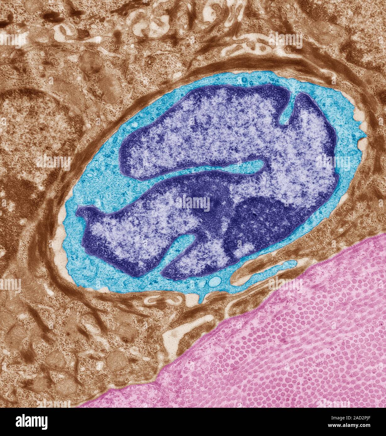
Langerhans cell. Coloured transmission electron micrograph (TEM) of a section through the skin to show a Langerhans cell (blue). The function of these

RMID:Image ID:2AD2PJF
Image details
Contributor:
Science Photo Library / Alamy Stock PhotoImage ID:
2AD2PJFFile size:
56.5 MB (3.9 MB Compressed download)Releases:
Model - no | Property - noDo I need a release?Dimensions:
4323 x 4572 px | 36.6 x 38.7 cm | 14.4 x 15.2 inches | 300dpiDate taken:
11 September 2015Photographer:
STEVE GSCHMEISSNER/SCIENCE PHOTO LIBRARYMore information:
Langerhans cell. Coloured transmission electron micrograph (TEM) of a section through the skin to show a Langerhans cell (blue). The function of these cells is to detect foreign bodies (antigens) which have penetrated the epidermis, capturing intruders and then carrying them to the lymph nodes in the dermis, where they are presented to the lymphocytes. A cellular type of immune response is then triggered, neutralising and finally eliminating the antigen. Keratinocytes (brown) surround the Langerhans cell and dermis (pink) is seen at bottom right of the image. Magnification: x7000 when printed at 10 centimetres high.