···
Light micrograph of a section through epidermis with a verruca, or plantar wart. The epidermis has an enlarged stratum granulosum (hypergranulosis, da Image details File size:
30.1 MB (1.6 MB Compressed download)
Open your image file to the full size using image processing software.
Dimensions:
3627 x 2902 px | 30.7 x 24.6 cm | 12.1 x 9.7 inches | 300dpi
Date taken:
22 October 2018
More information:
Light micrograph of a section through epidermis with a verruca, or plantar wart. The epidermis has an enlarged stratum granulosum (hypergranulosis, dark purple) and a well-developed stratum corneum (hyperkeratosis, top).
Search stock photos by tags
Similar stock images Radish root, cross section under light microscope. Transverse section through the root of the Raphanus sativus plant. Micrograph at 8X magnification. Stock Photo https://www.alamy.com/image-license-details/?v=1 https://www.alamy.com/radish-root-cross-section-under-light-microscope-transverse-section-through-the-root-of-the-raphanus-sativus-plant-micrograph-at-8x-magnification-image501670260.html RF 2M451F0 – Radish root, cross section under light microscope. Transverse section through the root of the Raphanus sativus plant. Micrograph at 8X magnification. Frog skin glands. Light micrograph of a section through the skin of an marsh frog. The large round (blue) structures are poison glands. Stock Photo https://www.alamy.com/image-license-details/?v=1 https://www.alamy.com/frog-skin-glands-light-micrograph-of-a-section-through-the-skin-of-an-marsh-frog-the-large-round-blue-structures-are-poison-glands-image471208224.html RF 2JAHAW4 – Frog skin glands. Light micrograph of a section through the skin of an marsh frog. The large round (blue) structures are poison glands. Cotton plant root, under the light microscope. Cross section through the root of Gossypium hirsutum, upland or also Mexican cotton. Stock Photo https://www.alamy.com/image-license-details/?v=1 https://www.alamy.com/cotton-plant-root-under-the-light-microscope-cross-section-through-the-root-of-gossypium-hirsutum-upland-or-also-mexican-cotton-image501626075.html RF 2M4314Y – Cotton plant root, under the light microscope. Cross section through the root of Gossypium hirsutum, upland or also Mexican cotton. Broom stem. Light micrograph (LM) of a transverse section through the stem of a common broom (Salicornia europaea) plant. Stock Photo https://www.alamy.com/image-license-details/?v=1 https://www.alamy.com/broom-stem-light-micrograph-lm-of-a-transverse-section-through-the-stem-of-a-common-broom-salicornia-europaea-plant-image395528786.html RF 2DYDW02 – Broom stem. Light micrograph (LM) of a transverse section through the stem of a common broom (Salicornia europaea) plant. The image presents Anemone sylvestris stalk in transversal cross-section, photographed through the microscope in polarized light at a magnification of Stock Photo https://www.alamy.com/image-license-details/?v=1 https://www.alamy.com/the-image-presents-anemone-sylvestris-stalk-in-transversal-cross-section-photographed-through-the-microscope-in-polarized-light-at-a-magnification-of-image564575210.html RM 2RPEHCA – The image presents Anemone sylvestris stalk in transversal cross-section, photographed through the microscope in polarized light at a magnification of . The Biological bulletin. Biology; Zoology; Biology; Marine Biology. DECAPOD CRUSTACEAN PHOTOPHORES 299. Figure 22. Light micrograph of a longitudinal section through the maxilliped of Oplophorus x/iinosHx showing photophores in which the "clear areas" (ca) of the photocytes exhibit stages of increasing osmiophi- lia. c, paracrystalline bodies; cut. surface cuticle; di, distal; do, dorsal; e, "window" epidermis cytoplasm; en. "window" epidermis nucleus; f, fibroblast; 1, ligament; p. carotenoid pigment cell processes; pn. photocyte nuclei; r. reflector pigment ce Stock Photo https://www.alamy.com/image-license-details/?v=1 https://www.alamy.com/the-biological-bulletin-biology-zoology-biology-marine-biology-decapod-crustacean-photophores-299-figure-22-light-micrograph-of-a-longitudinal-section-through-the-maxilliped-of-oplophorus-xiinoshx-showing-photophores-in-which-the-quotclear-areasquot-ca-of-the-photocytes-exhibit-stages-of-increasing-osmiophi-lia-c-paracrystalline-bodies-cut-surface-cuticle-di-distal-do-dorsal-e-quotwindowquot-epidermis-cytoplasm-en-quotwindowquot-epidermis-nucleus-f-fibroblast-1-ligament-p-carotenoid-pigment-cell-processes-pn-photocyte-nuclei-r-reflector-pigment-ce-image234616565.html RM RHKKR1 – . The Biological bulletin. Biology; Zoology; Biology; Marine Biology. DECAPOD CRUSTACEAN PHOTOPHORES 299. Figure 22. Light micrograph of a longitudinal section through the maxilliped of Oplophorus x/iinosHx showing photophores in which the "clear areas" (ca) of the photocytes exhibit stages of increasing osmiophi- lia. c, paracrystalline bodies; cut. surface cuticle; di, distal; do, dorsal; e, "window" epidermis cytoplasm; en. "window" epidermis nucleus; f, fibroblast; 1, ligament; p. carotenoid pigment cell processes; pn. photocyte nuclei; r. reflector pigment ce The image presents nettle tissues in the transversal cross-section of the stalk, photographed through the microscope in polarized light at a magnifica Stock Photo https://www.alamy.com/image-license-details/?v=1 https://www.alamy.com/the-image-presents-nettle-tissues-in-the-transversal-cross-section-of-the-stalk-photographed-through-the-microscope-in-polarized-light-at-a-magnifica-image564575212.html RM 2RPEHCC – The image presents nettle tissues in the transversal cross-section of the stalk, photographed through the microscope in polarized light at a magnifica Broom stem. Light micrograph (LM) of a transverse section through the stem of a common broom (Salicornia europaea) plant. Stock Photo https://www.alamy.com/image-license-details/?v=1 https://www.alamy.com/broom-stem-light-micrograph-lm-of-a-transverse-section-through-the-stem-of-a-common-broom-salicornia-europaea-plant-image395528799.html RF 2DYDW0F – Broom stem. Light micrograph (LM) of a transverse section through the stem of a common broom (Salicornia europaea) plant. Fingertip. Light micrograph (LM) of a section through the fingertip. The nail (orange) is at top center, with the nail root below. The nail bed is dark purple and is continous with the epithelium. The tip of the finger bone is pale pink(left center). Beneath the stratified squamous epithelium is the dermis, which contains adipose (fat) cells connective tissue and blood vessels. Magnification: x5 when printed at 10 centimetres wide. Stock Photo https://www.alamy.com/image-license-details/?v=1 https://www.alamy.com/stock-photo-fingertip-light-micrograph-lm-of-a-section-through-the-fingertip-the-122807666.html RF H3PAC2 – Fingertip. Light micrograph (LM) of a section through the fingertip. The nail (orange) is at top center, with the nail root below. The nail bed is dark purple and is continous with the epithelium. The tip of the finger bone is pale pink(left center). Beneath the stratified squamous epithelium is the dermis, which contains adipose (fat) cells connective tissue and blood vessels. Magnification: x5 when printed at 10 centimetres wide. The image presents nettle tissues in the transversal section of the stalk, photographed through the microscope in polarized light at a magnification o Stock Photo https://www.alamy.com/image-license-details/?v=1 https://www.alamy.com/the-image-presents-nettle-tissues-in-the-transversal-section-of-the-stalk-photographed-through-the-microscope-in-polarized-light-at-a-magnification-o-image564575219.html RM 2RPEHCK – The image presents nettle tissues in the transversal section of the stalk, photographed through the microscope in polarized light at a magnification o . The Biological bulletin. Biology; Zoology; Marine biology. 102 D. J. NUCKLEY ET AL.. Figure 3. Light microscopy of l-^m sections of the dorsal eye stained with toluidine blue. (A) Low magnification micrograph of a dorsal tangential section through one wing. The massed rhabdomeral segments (r) of the photoreceptor? form a continuous sheet of photosensitive membrane surrounded by corneal epidermis (e) and cornea (c). The plane of section is tilted slightly from horizontal; anterior is to the left and medial toward the top. (B) Higher magnification micrograph of a more heavily stained section c Stock Photo https://www.alamy.com/image-license-details/?v=1 https://www.alamy.com/the-biological-bulletin-biology-zoology-marine-biology-102-d-j-nuckley-et-al-figure-3-light-microscopy-of-l-m-sections-of-the-dorsal-eye-stained-with-toluidine-blue-a-low-magnification-micrograph-of-a-dorsal-tangential-section-through-one-wing-the-massed-rhabdomeral-segments-r-of-the-photoreceptor-form-a-continuous-sheet-of-photosensitive-membrane-surrounded-by-corneal-epidermis-e-and-cornea-c-the-plane-of-section-is-tilted-slightly-from-horizontal-anterior-is-to-the-left-and-medial-toward-the-top-b-higher-magnification-micrograph-of-a-more-heavily-stained-section-c-image234628294.html RM RHM6NX – . The Biological bulletin. Biology; Zoology; Marine biology. 102 D. J. NUCKLEY ET AL.. Figure 3. Light microscopy of l-^m sections of the dorsal eye stained with toluidine blue. (A) Low magnification micrograph of a dorsal tangential section through one wing. The massed rhabdomeral segments (r) of the photoreceptor? form a continuous sheet of photosensitive membrane surrounded by corneal epidermis (e) and cornea (c). The plane of section is tilted slightly from horizontal; anterior is to the left and medial toward the top. (B) Higher magnification micrograph of a more heavily stained section c The image presents oak tissues in transversal cross-section of the stalk, photographed through the microscope in polarized light at a magnification of Stock Photo https://www.alamy.com/image-license-details/?v=1 https://www.alamy.com/the-image-presents-oak-tissues-in-transversal-cross-section-of-the-stalk-photographed-through-the-microscope-in-polarized-light-at-a-magnification-of-image564575217.html RM 2RPEHCH – The image presents oak tissues in transversal cross-section of the stalk, photographed through the microscope in polarized light at a magnification of The image presents nettle tisues in the transversal cross-section of the stalk, photographed through the microscope in polarized light at a magnificat Stock Photo https://www.alamy.com/image-license-details/?v=1 https://www.alamy.com/the-image-presents-nettle-tisues-in-the-transversal-cross-section-of-the-stalk-photographed-through-the-microscope-in-polarized-light-at-a-magnificat-image564575215.html RM 2RPEHCF – The image presents nettle tisues in the transversal cross-section of the stalk, photographed through the microscope in polarized light at a magnificat 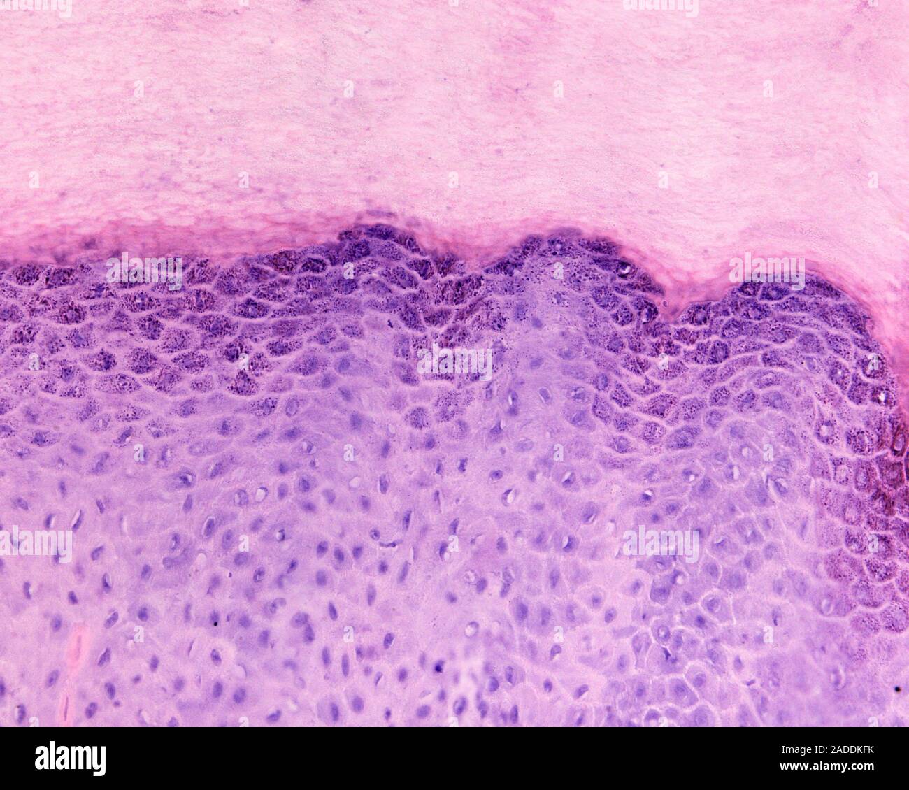