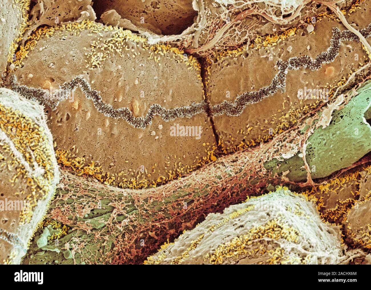
Liver tissue, coloured scanning electron micrograph (SEM). Hepatocytes (liver cells, brown) are specialised epithelial cells and make up approximately
RMID:Image ID:2ACHX6M
Image details
Contributor:
Science Photo Library / Alamy Stock PhotoImage ID:
2ACHX6MFile size:
51 MB (6.6 MB Compressed download)Releases:
Model - no | Property - noDo I need a release?Dimensions:
5000 x 3564 px | 42.3 x 30.2 cm | 16.7 x 11.9 inches | 300dpiDate taken:
26 November 2010Photographer:
THOMAS DEERINCK, NCMIR/SCIENCE PHOTO LIBRARYMore information:
Liver tissue, coloured scanning electron micrograph (SEM). Hepatocytes (liver cells, brown) are specialised epithelial cells and make up approximately 80 per cent of the mass of the liver. Also seen is a blood vessel (green) and a bile canaliculus (dark brown channel with white edges running across upper frame). The bile canaliculi transport bile, produced by the liver, to the gall bladder. Magnification: x3000 when printed at 10 centimetres wide.