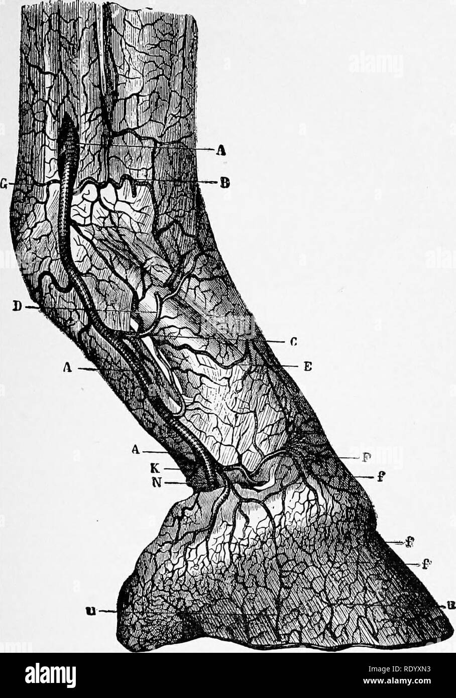
. Manual of operative veterinary surgery. Veterinary surgery. ANATOMY OF THE FOOT. 579. Fig. 480.—Arteries of the Digital Region. AAA.—Digital artery. B.—Transversal branch in front of fetlock Joint C—Per- pendicular artery of Percival. D.—Its ascending branch. E.—The descending branch. F.—Branch to form the superficial coronary circle. G.—Posterior transverse branches. K.—Artery of the plantar cushion. P.—Circumflex artery. C C—Ascending terminal branches of the digital artery. 3d, the velvety tissue or villous tunic which covers the plantar cushion at the interior face of the foot, and is th
Image details
Contributor:
The Book Worm / Alamy Stock PhotoImage ID:
RDYXN3File size:
7.1 MB (471.6 KB Compressed download)Releases:
Model - no | Property - noDo I need a release?Dimensions:
1321 x 1891 px | 22.4 x 32 cm | 8.8 x 12.6 inches | 150dpiMore information:
This image is a public domain image, which means either that copyright has expired in the image or the copyright holder has waived their copyright. Alamy charges you a fee for access to the high resolution copy of the image.
This image could have imperfections as it’s either historical or reportage.
. Manual of operative veterinary surgery. Veterinary surgery. ANATOMY OF THE FOOT. 579. Fig. 480.—Arteries of the Digital Region. AAA.—Digital artery. B.—Transversal branch in front of fetlock Joint C—Per- pendicular artery of Percival. D.—Its ascending branch. E.—The descending branch. F.—Branch to form the superficial coronary circle. G.—Posterior transverse branches. K.—Artery of the plantar cushion. P.—Circumflex artery. C C—Ascending terminal branches of the digital artery. 3d, the velvety tissue or villous tunic which covers the plantar cushion at the interior face of the foot, and is the secreting organ of the sole and frog, its surface covered with viUosities similar to those of the coronary band, and Kke them, of various sizes, are lodged in the porosities of the internal face of the sole and frog. The external parts of the foot are four in number : the wall.. Please note that these images are extracted from scanned page images that may have been digitally enhanced for readability - coloration and appearance of these illustrations may not perfectly resemble the original work.. Liautard, Alexandre Franc?ois Augustin, 1835-. New York, Sabiston & Murray