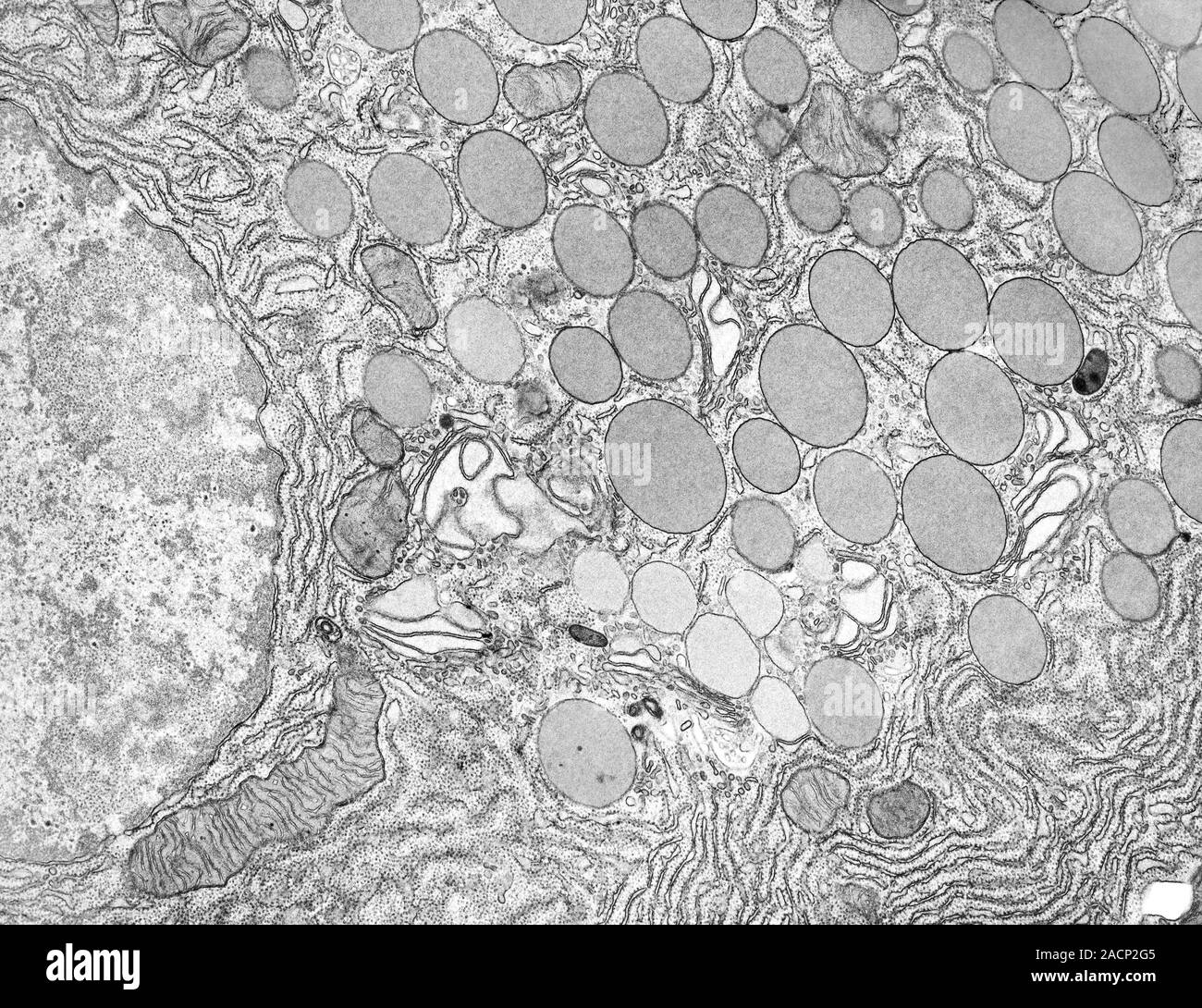
Pancreatic acinar cell. Transmission electron micrograph (TEM) of a section through an enzyme-secreting acinar cell in the human pancreas, showing par

Image details
Contributor:
Science Photo Library / Alamy Stock PhotoImage ID:
2ACP2G5File size:
50.7 MB (6.1 MB Compressed download)Releases:
Model - no | Property - noDo I need a release?Dimensions:
4800 x 3694 px | 40.6 x 31.3 cm | 16 x 12.3 inches | 300dpiDate taken:
14 February 2013Photographer:
MICROSCAPE/SCIENCE PHOTO LIBRARYMore information:
Pancreatic acinar cell. Transmission electron micrograph (TEM) of a section through an enzyme-secreting acinar cell in the human pancreas, showing part of the nucleus (round, far left), rough endoplasmic reticulum (ER, dark lines), numerous Golgi complexes (stacks of ER), and many zymogen granules (circles), which contain the inactive forms of digestive enzymes. These enzymes are released from the cell by exocytosis. They pass into the pancreatic duct system and are delivered into the duodenum in the gut, where they are converted into their active state. Various locally acting hormones arising from the gut stimulate the production of this pancreatic enzyme-rich fluid. Magnification: x10, 000 when printed 10 centimetres wide.