. Practical anatomy of the rabbit : an elementary laboratory textbook in mammalian anatomy . Rabbits; Anatomy, Comparative. The Anterior Limb. 2X1 -"â *â MediaL (b) Thedeltoideus: Acromial portion. Origin: The Acromion. Insertion: Distal portion of deltoid tuberosity. Scapular portion. Origin: Infraspinous fascia. The end of the muscle forms a curved line over the dorsal portion of the infraspinatus, leaving only a small triangular portion of '^"'"'"^ the latter exposed. Insertion: The distal portion of the mus- cle passes beneath the meta- cromion, which also serves as a p
Image details
Contributor:
The Book Worm / Alamy Stock PhotoImage ID:
RDJK6AFile size:
7.2 MB (260.6 KB Compressed download)Releases:
Model - no | Property - noDo I need a release?Dimensions:
1118 x 2236 px | 18.9 x 37.9 cm | 7.5 x 14.9 inches | 150dpiMore information:
This image is a public domain image, which means either that copyright has expired in the image or the copyright holder has waived their copyright. Alamy charges you a fee for access to the high resolution copy of the image.
This image could have imperfections as it’s either historical or reportage.
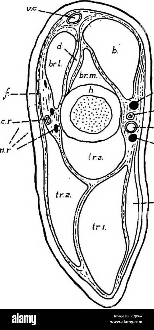
. Practical anatomy of the rabbit : an elementary laboratory textbook in mammalian anatomy . Rabbits; Anatomy, Comparative. The Anterior Limb. 2X1 -"â *â MediaL (b) Thedeltoideus: Acromial portion. Origin: The Acromion. Insertion: Distal portion of deltoid tuberosity. Scapular portion. Origin: Infraspinous fascia. The end of the muscle forms a curved line over the dorsal portion of the infraspinatus, leaving only a small triangular portion of '^"'"'"^ the latter exposed. Insertion: The distal portion of the mus- cle passes beneath the meta- cromion, which also serves as a point of attachment, and is replaced on the lateral surface of the humerus, beneath the acromial portion, by a long thin tendon, through which it is inserted. The scapular portion of the deltoideus should-be separated from the infraspinatus and divid- ed, the distal end being reflected together with the metacromion. (d) The infraspinatus. Origin: Posterior por- tion of the lateral sur- face of the scapula, in- cluding the spine. In- sertion: Greater tub- ercle of the humerus. The muscle fills the infraspinous fossa. (e) The supraspinatus. Origin: Anterior portion of the lateral surface of the scapula (supraspinous fossa), supraspinous fascia, and, to a certain extent, the subscapular fascia. Insertion: Greater tubercle of the humerus. The extent of this muscle is evident only after rem6val of the loosely attached fleshy parts of the pectorals from its surface.. Poalenor Fig. 74. Transverse section through the distal portion of the arm; semidiagrammatic; a.b., brachial artery; a.c.i., radial collateral artery; b:, biceps; br.l. and br.m., lateral and medial heads of the brachialis; d.. deltoideus (insertion); e.a.p., , ' extensor antibrachii parvus; f., bracnial fascia; h, , humerus; n.m, me_ian nerve; n.r., radial nerve; n.u., ulnar nerve; tr. i-tr.s, long, lateral, and medial heads of the triceps; v.b., brachial vein; v.c, cephalic vein.. Please note that these images are extrac