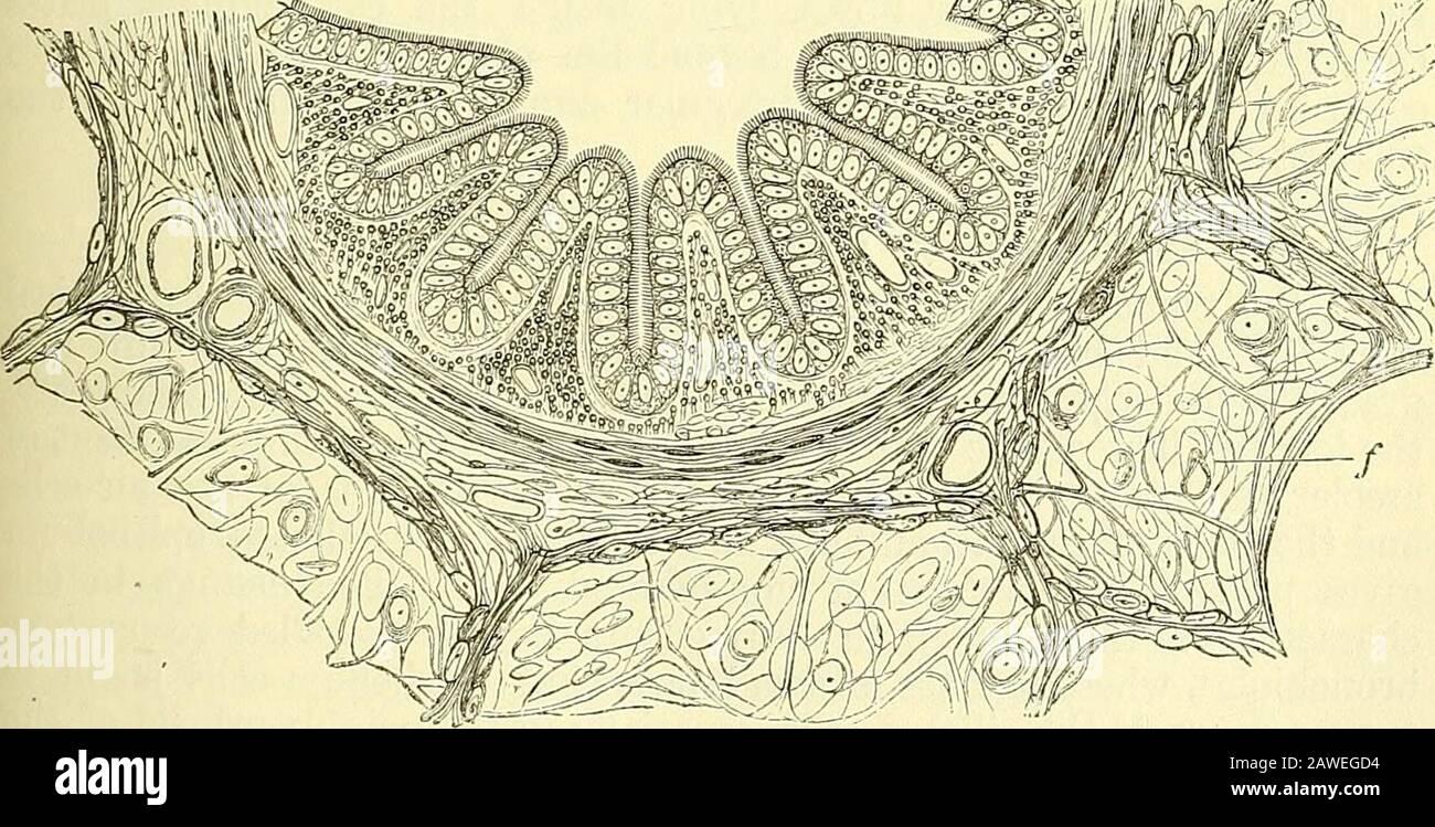
Quain's elements of anatomy . Fig. 444.—Portion of a transverse section op a bronchial tube, euman (6 mm. indiameter) (F. E. Scliiiltze). Magnified 30 diameters. a, cartilage and fibrous layer with mncoiis glands, and, in the outer part, a little fat;in the middle, the duct of a gland opens on the inner surface of the tube ; h, annularlayer of involuntary muscular fibres ; c, elastic layer, the elastic fibres in bundles whichare seen cut across; d, columnar ciliated epithelium. Within the lungs the air-tubes are not flattened behind like the bronchiand trachea, but form completely cylindrical
Image details
Contributor:
The Reading Room / Alamy Stock PhotoImage ID:
2AWEGD4File size:
7.1 MB (573.8 KB Compressed download)Releases:
Model - no | Property - noDo I need a release?Dimensions:
2219 x 1126 px | 37.6 x 19.1 cm | 14.8 x 7.5 inches | 150dpiMore information:
This image is a public domain image, which means either that copyright has expired in the image or the copyright holder has waived their copyright. Alamy charges you a fee for access to the high resolution copy of the image.
This image could have imperfections as it’s either historical or reportage.
Quain's elements of anatomy . Fig. 444.—Portion of a transverse section op a bronchial tube, euman (6 mm. indiameter) (F. E. Scliiiltze). Magnified 30 diameters. a, cartilage and fibrous layer with mncoiis glands, and, in the outer part, a little fat;in the middle, the duct of a gland opens on the inner surface of the tube ; h, annularlayer of involuntary muscular fibres ; c, elastic layer, the elastic fibres in bundles whichare seen cut across; d, columnar ciliated epithelium. Within the lungs the air-tubes are not flattened behind like the bronchiand trachea, but form completely cylindrical tubes. Hence, although Fig. 445.. Fig. 445.—Section of a small bronchial tube (4 mm. in diameter) from thepigs lung (F. E. Schultze). Magnified 240 diameters. a, fibrous layer ; &, muscular layer; c, mucous membrane in longitudinal folds, withnumerous longitudinally running elastic fibres cut across ; d, ciliated epithelium; /, sur-rounding alveoli. L L 2 616 THE LUA^GS. they contain the same elements as the larger air-passages, they are reducedgradually to a state of gTeater tenuity, but possess certain peculiaritiesof structure. Thus, the cartilages no longer appear as imperfect ringsrunning only upon the front and lateral surfaces of the air-tube, but aredisposed over all sides of the tubes in the form of irregularly shapedplates and incomplete rings of various sizes. These are most developedat the points of division of the bronchia, where they form a sharp con-cave ridge projecting inwards into the tube. They may be traced, be-coming rarer and rarer and more reduced in size, as far as bronchia onemilhmeter i