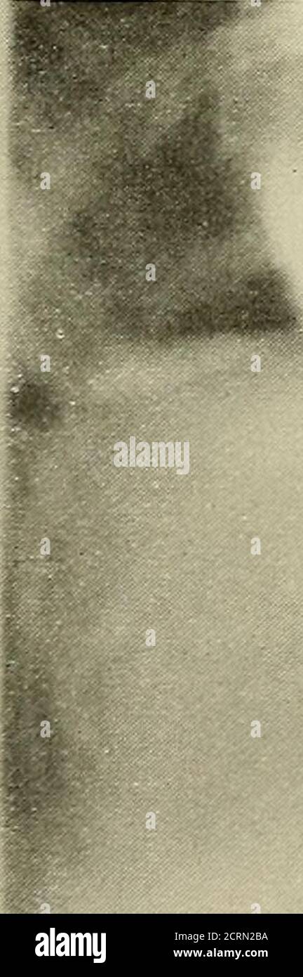
. Radiography and radio-therapeutics . PLATE LXI.—Chest showing Dilatation of (Esophagus. a, (Esophagus dilated and filled with food. b, (Esophagus empty after an interval of 3 days, showing irregular shading in mediastinum. c, Lower portion of dilated oesophagus containing bismuth food ; point of stricture is seen, and foodwhich has passed into the stomach. (ESOPHAGEAL OBSTRUCTION 317 area of the chest is occupied by an elongated shadow, extending down tothe diaphragm and terminating high up in the superior mediastinum. Iffully distended by food, it is clearly seen on the fluorescent screen,
Image details
Contributor:
Reading Room 2020 / Alamy Stock PhotoImage ID:
2CRN2BAFile size:
7.1 MB (261.1 KB Compressed download)Releases:
Model - no | Property - noDo I need a release?Dimensions:
911 x 2741 px | 7.7 x 23.2 cm | 3 x 9.1 inches | 300dpiMore information:
This image could have imperfections as it’s either historical or reportage.
. Radiography and radio-therapeutics . PLATE LXI.—Chest showing Dilatation of (Esophagus. a, (Esophagus dilated and filled with food. b, (Esophagus empty after an interval of 3 days, showing irregular shading in mediastinum. c, Lower portion of dilated oesophagus containing bismuth food ; point of stricture is seen, and foodwhich has passed into the stomach. (ESOPHAGEAL OBSTRUCTION 317 area of the chest is occupied by an elongated shadow, extending down tothe diaphragm and terminating high up in the superior mediastinum. Iffully distended by food, it is clearly seen on the fluorescent screen, and onthe right side an occasional feeble peristaltic wave can be detected. This isquite distinct from the pulsation of an aneurism of the descending aorta.It has a large wave-like motion, which is quite unmistakable for any othertype of movement. The en-largement is much more pro-nounced on the right side ofthe thorax than on the left.There is an anatomical ex-planation of this phenomenon.This type of case has beendescribed as oeso