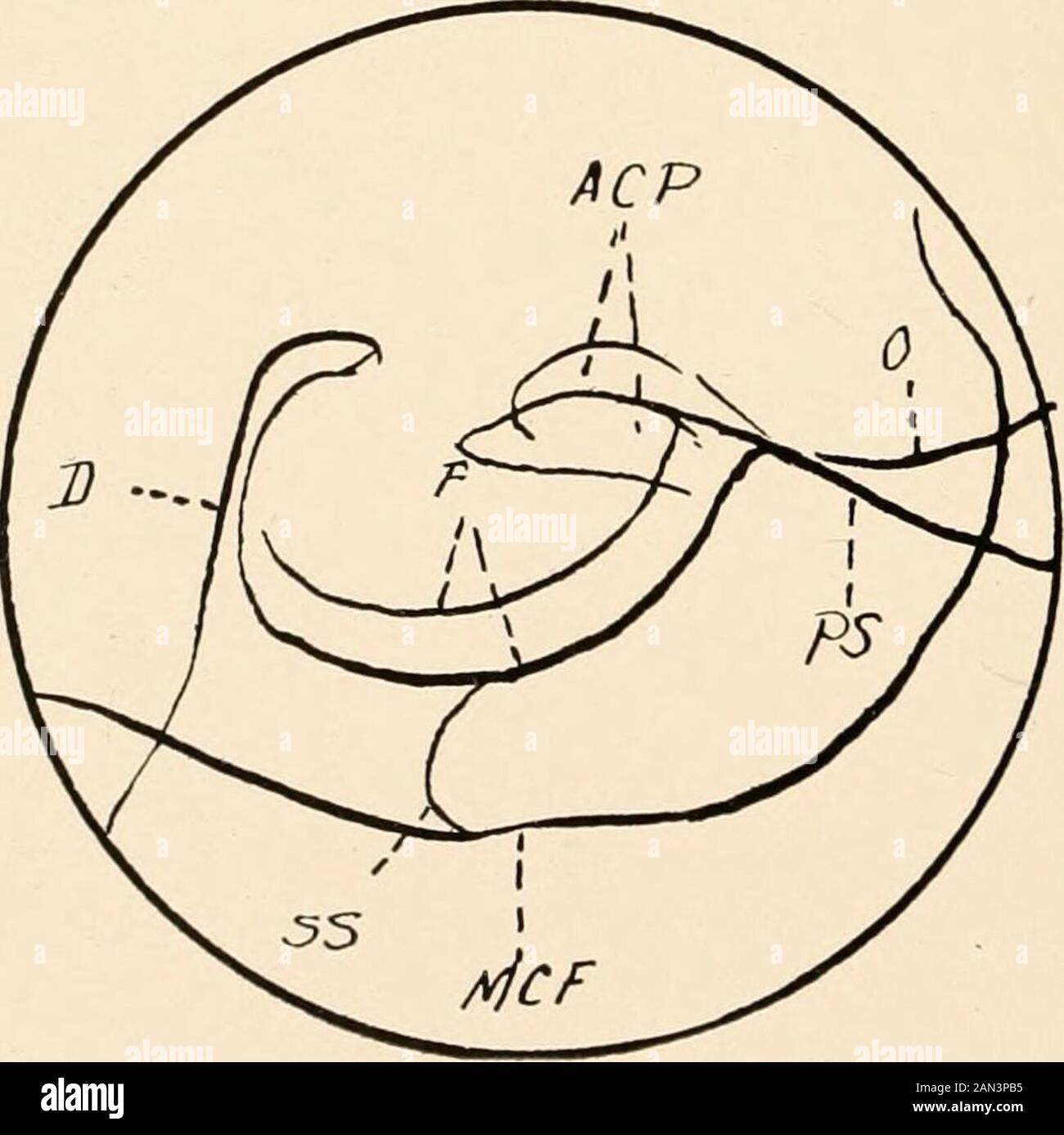
Roentgen diagnosis of diseases of the head . Fig. 60.—A sinistrodextral picture of sella erosion in a patient with a tumor ofthe hypophysis associated with acromegaly. The sella is wide and deep, the dorsumis pushed backward and the anterior clinoid processes are plump.. Fig. 61.—A sketch of Fig. (>0 showing the important features of the sella. D. Dor-sum sellae. /•. Floor of the sella. ACP. Anterior clinoid processes. O. Roof of theillit nearest to the plate. P. Planum sphenoidale. MCF. Outline of the middlecranial fossa. SS. Posterior wall of the sphenoidal sinus. Cask 8.—K. K., male, for

Image details
Contributor:
The Reading Room / Alamy Stock PhotoImage ID:
2AN3PB5File size:
7.1 MB (173.7 KB Compressed download)Releases:
Model - no | Property - noDo I need a release?Dimensions:
1582 x 1579 px | 26.8 x 26.7 cm | 10.5 x 10.5 inches | 150dpiMore information:
This image is a public domain image, which means either that copyright has expired in the image or the copyright holder has waived their copyright. Alamy charges you a fee for access to the high resolution copy of the image.
This image could have imperfections as it’s either historical or reportage.
Roentgen diagnosis of diseases of the head . Fig. 60.—A sinistrodextral picture of sella erosion in a patient with a tumor ofthe hypophysis associated with acromegaly. The sella is wide and deep, the dorsumis pushed backward and the anterior clinoid processes are plump.. Fig. 61.—A sketch of Fig. (>0 showing the important features of the sella. D. Dor-sum sellae. /•. Floor of the sella. ACP. Anterior clinoid processes. O. Roof of theillit nearest to the plate. P. Planum sphenoidale. MCF. Outline of the middlecranial fossa. SS. Posterior wall of the sphenoidal sinus. Cask 8.—K. K., male, forty-eight years old. Hands and feet wereplump. Lower jaw was larger than normal. Cirrhosis hepatis. The roentgenogram showed a moderate deepening and a considerable INTRACRANIAL DISEASES 185 widening of the sella (anteroposterior diameter 17 mm.). The dorsumsella? was plump and tipped backward. There was a striking plumpnessof the anterior clinoid processes. (See Fig. 58.) Case 9.—B., male, thirty-six years old. Enlargement of the hands forthe last fourteen years. Typical acromegaly. The roentgenogram showed extreme deepening of the sella and an ex-tremely long and thin dorsum. (See Fig. 59.) Case 10.—A. S., female, forty-four years old. In connection wi