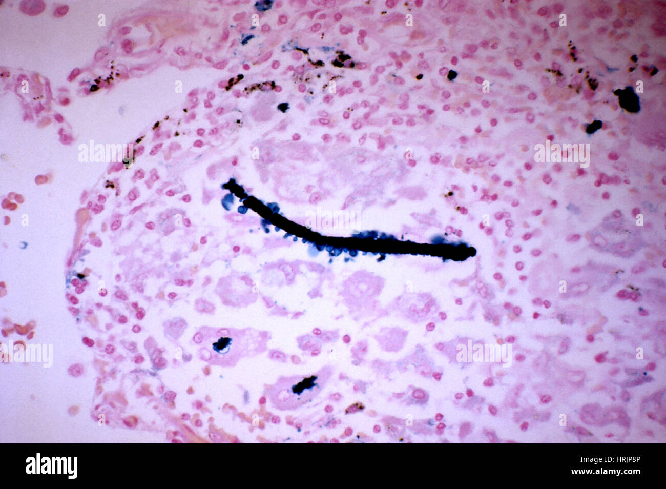
Asbestos in Lung Tissue, LM
Image details
Contributor:
Science History Images / Alamy Stock PhotoImage ID:
HRJP8PFile size:
38.5 MB (1.1 MB Compressed download)Releases:
Model - no | Property - noDo I need a release?Dimensions:
4500 x 2993 px | 38.1 x 25.3 cm | 15 x 10 inches | 300dpiPhotographer:
Photo ResearchersMore information:
This image could have imperfections as it’s either historical or reportage.
Photomicrograph revealed histopathologic changes found in a lung tissue specimen, showing a ferruginous body. A fiber of asbestos was coated by an iron-protein complex and surrounded by macrophages. The specimen was processed using a Prussian blue iron stain. Asbestos is a fibrous mineral used for insulation. Its needle-shaped fibers are indestructible once inhaled, and remain lodged in the lungs for life. In time the fibers acquire a segmented protein coat, colored brown by an iron-containing pigment derived from disintegrated red blood cells. They cause chronic irritation, development of fibrous tissue, and increased risk of cancer. Symptoms may take years to appear and the damage can be fatal. Stained with eosin and hematoxylin. Magnification unknown.