···
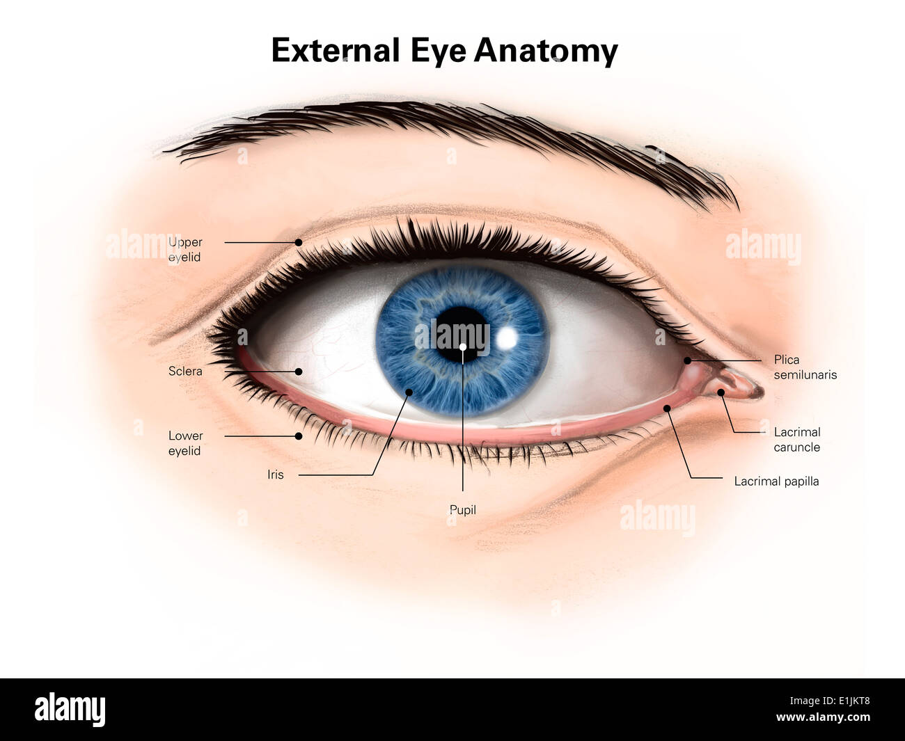
External anatomy of the human eye (with labels). Image details File size:
51.9 MB (1.1 MB Compressed download)
Open your image file to the full size using image processing software.
Dimensions:
4920 x 3690 px | 41.7 x 31.2 cm | 16.4 x 12.3 inches | 300dpi
Search stock photos by tags
Similar stock images Illustration of the eye anatomy. The structure of the eye includes three different layers. The external layer, formed by the sclera and cornea. The intermediate layer, the iris and ciliary body, extraocular muscles and choroid. The internal layer, or the sensory part of the eye, the retina and the optic nerve. Stock Photo https://www.alamy.com/image-license-details/?v=1 https://www.alamy.com/illustration-of-the-eye-anatomy-the-structure-of-the-eye-includes-three-different-layers-the-external-layer-formed-by-the-sclera-and-cornea-the-intermediate-layer-the-iris-and-ciliary-body-extraocular-muscles-and-choroid-the-internal-layer-or-the-sensory-part-of-the-eye-the-retina-and-the-optic-nerve-image628777527.html RF 2YEY847 – Illustration of the eye anatomy. The structure of the eye includes three different layers. The external layer, formed by the sclera and cornea. The intermediate layer, the iris and ciliary body, extraocular muscles and choroid. The internal layer, or the sensory part of the eye, the retina and the optic nerve. Illustration of the eye anatomy. The structure of the eye includes three different layers. The external layer, formed by the sclera and cornea. The intermediate layer, the iris and ciliary body, extraocular muscles and choroid. The internal layer, or the sensory part of the eye, the retina and the optic nerve. Stock Photo https://www.alamy.com/image-license-details/?v=1 https://www.alamy.com/illustration-of-the-eye-anatomy-the-structure-of-the-eye-includes-three-different-layers-the-external-layer-formed-by-the-sclera-and-cornea-the-intermediate-layer-the-iris-and-ciliary-body-extraocular-muscles-and-choroid-the-internal-layer-or-the-sensory-part-of-the-eye-the-retina-and-the-optic-nerve-image628777544.html RF 2YEY84T – Illustration of the eye anatomy. The structure of the eye includes three different layers. The external layer, formed by the sclera and cornea. The intermediate layer, the iris and ciliary body, extraocular muscles and choroid. The internal layer, or the sensory part of the eye, the retina and the optic nerve. Illustration of the eye anatomy. The structure of the eye includes three different layers. The external layer, formed by the sclera and cornea. The intermediate layer, the iris and ciliary body, extraocular muscles, choroid and blood vessels. The internal layer, or the sensory part of the eye, the retina and the optic nerve. Stock Photo https://www.alamy.com/image-license-details/?v=1 https://www.alamy.com/illustration-of-the-eye-anatomy-the-structure-of-the-eye-includes-three-different-layers-the-external-layer-formed-by-the-sclera-and-cornea-the-intermediate-layer-the-iris-and-ciliary-body-extraocular-muscles-choroid-and-blood-vessels-the-internal-layer-or-the-sensory-part-of-the-eye-the-retina-and-the-optic-nerve-image628777551.html RF 2YEY853 – Illustration of the eye anatomy. The structure of the eye includes three different layers. The external layer, formed by the sclera and cornea. The intermediate layer, the iris and ciliary body, extraocular muscles, choroid and blood vessels. The internal layer, or the sensory part of the eye, the retina and the optic nerve. Illustration of the eye anatomy. The structure of the eye includes three different layers. The external layer, formed by the sclera and cornea. The intermediate layer, the iris and ciliary body, extraocular muscles, choroid and blood vessels. The internal layer, or the sensory part of the eye, the retina and the optic nerve. Stock Photo https://www.alamy.com/image-license-details/?v=1 https://www.alamy.com/illustration-of-the-eye-anatomy-the-structure-of-the-eye-includes-three-different-layers-the-external-layer-formed-by-the-sclera-and-cornea-the-intermediate-layer-the-iris-and-ciliary-body-extraocular-muscles-choroid-and-blood-vessels-the-internal-layer-or-the-sensory-part-of-the-eye-the-retina-and-the-optic-nerve-image628777557.html RF 2YEY859 – Illustration of the eye anatomy. The structure of the eye includes three different layers. The external layer, formed by the sclera and cornea. The intermediate layer, the iris and ciliary body, extraocular muscles, choroid and blood vessels. The internal layer, or the sensory part of the eye, the retina and the optic nerve. Illustration of the eye anatomy. The structure of the eye includes three different layers. The external layer, formed by the sclera and cornea. The intermediate layer, the iris and ciliary body, extraocular muscles, choroid and blood vessels. The internal layer, or the sensory part of the eye, the retina and the optic nerve. Stock Photo https://www.alamy.com/image-license-details/?v=1 https://www.alamy.com/illustration-of-the-eye-anatomy-the-structure-of-the-eye-includes-three-different-layers-the-external-layer-formed-by-the-sclera-and-cornea-the-intermediate-layer-the-iris-and-ciliary-body-extraocular-muscles-choroid-and-blood-vessels-the-internal-layer-or-the-sensory-part-of-the-eye-the-retina-and-the-optic-nerve-image628777516.html RF 2YEY83T – Illustration of the eye anatomy. The structure of the eye includes three different layers. The external layer, formed by the sclera and cornea. The intermediate layer, the iris and ciliary body, extraocular muscles, choroid and blood vessels. The internal layer, or the sensory part of the eye, the retina and the optic nerve. Illustration of the eye anatomy. The structure of the eye includes three different layers. The external layer, formed by the sclera and cornea. The intermediate layer, divided into two parts: anterior (iris and ciliary body and extraocular muscles) and posterior (choroid). The internal layer, or the sensory part of the eye, the retina and the optic nerve. Stock Photo https://www.alamy.com/image-license-details/?v=1 https://www.alamy.com/illustration-of-the-eye-anatomy-the-structure-of-the-eye-includes-three-different-layers-the-external-layer-formed-by-the-sclera-and-cornea-the-intermediate-layer-divided-into-two-parts-anterior-iris-and-ciliary-body-and-extraocular-muscles-and-posterior-choroid-the-internal-layer-or-the-sensory-part-of-the-eye-the-retina-and-the-optic-nerve-image628777518.html RF 2YEY83X – Illustration of the eye anatomy. The structure of the eye includes three different layers. The external layer, formed by the sclera and cornea. The intermediate layer, divided into two parts: anterior (iris and ciliary body and extraocular muscles) and posterior (choroid). The internal layer, or the sensory part of the eye, the retina and the optic nerve. Illustration of the eye anatomy. The structure of the eye includes three different layers. The external layer, formed by the sclera and cornea. The intermediate layer, divided into two parts: anterior (iris and ciliary body and extraocular muscles) and posterior (choroid). The internal layer, or the sensory part of the eye, the retina and the optic nerve. Stock Photo https://www.alamy.com/image-license-details/?v=1 https://www.alamy.com/illustration-of-the-eye-anatomy-the-structure-of-the-eye-includes-three-different-layers-the-external-layer-formed-by-the-sclera-and-cornea-the-intermediate-layer-divided-into-two-parts-anterior-iris-and-ciliary-body-and-extraocular-muscles-and-posterior-choroid-the-internal-layer-or-the-sensory-part-of-the-eye-the-retina-and-the-optic-nerve-image628777530.html RF 2YEY84A – Illustration of the eye anatomy. The structure of the eye includes three different layers. The external layer, formed by the sclera and cornea. The intermediate layer, divided into two parts: anterior (iris and ciliary body and extraocular muscles) and posterior (choroid). The internal layer, or the sensory part of the eye, the retina and the optic nerve. Illustration of the eye anatomy. The structure of the eye includes three different layers. The external layer, formed by the sclera and cornea. The intermediate layer, divided into two parts: anterior (iris and ciliary body and extraocular muscles) and posterior (choroid). The internal layer, or the sensory part of the eye, the retina and the optic nerve. Stock Photo https://www.alamy.com/image-license-details/?v=1 https://www.alamy.com/illustration-of-the-eye-anatomy-the-structure-of-the-eye-includes-three-different-layers-the-external-layer-formed-by-the-sclera-and-cornea-the-intermediate-layer-divided-into-two-parts-anterior-iris-and-ciliary-body-and-extraocular-muscles-and-posterior-choroid-the-internal-layer-or-the-sensory-part-of-the-eye-the-retina-and-the-optic-nerve-image628777554.html RF 2YEY856 – Illustration of the eye anatomy. The structure of the eye includes three different layers. The external layer, formed by the sclera and cornea. The intermediate layer, divided into two parts: anterior (iris and ciliary body and extraocular muscles) and posterior (choroid). The internal layer, or the sensory part of the eye, the retina and the optic nerve. Illustration of the eye anatomy. The structure of the eye includes three different layers. The external layer, formed by the sclera and cornea. The intermediate layer, divided into two parts: anterior (iris and ciliary body and extraocular muscles) and posterior (choroid). The internal layer, or the sensory part of the eye, the retina and the optic nerve. Stock Photo https://www.alamy.com/image-license-details/?v=1 https://www.alamy.com/illustration-of-the-eye-anatomy-the-structure-of-the-eye-includes-three-different-layers-the-external-layer-formed-by-the-sclera-and-cornea-the-intermediate-layer-divided-into-two-parts-anterior-iris-and-ciliary-body-and-extraocular-muscles-and-posterior-choroid-the-internal-layer-or-the-sensory-part-of-the-eye-the-retina-and-the-optic-nerve-image628777532.html RF 2YEY84C – Illustration of the eye anatomy. The structure of the eye includes three different layers. The external layer, formed by the sclera and cornea. The intermediate layer, divided into two parts: anterior (iris and ciliary body and extraocular muscles) and posterior (choroid). The internal layer, or the sensory part of the eye, the retina and the optic nerve. Illustration of the eye anatomy. The structure of the eye includes three different layers. The external layer, formed by the sclera and cornea. The intermediate layer, divided into two parts: anterior (iris and ciliary body and extraocular muscles) and posterior (choroid). The internal layer, or the sensory part of the eye, the retina and the optic nerve. Stock Photo https://www.alamy.com/image-license-details/?v=1 https://www.alamy.com/illustration-of-the-eye-anatomy-the-structure-of-the-eye-includes-three-different-layers-the-external-layer-formed-by-the-sclera-and-cornea-the-intermediate-layer-divided-into-two-parts-anterior-iris-and-ciliary-body-and-extraocular-muscles-and-posterior-choroid-the-internal-layer-or-the-sensory-part-of-the-eye-the-retina-and-the-optic-nerve-image628777543.html RF 2YEY84R – Illustration of the eye anatomy. The structure of the eye includes three different layers. The external layer, formed by the sclera and cornea. The intermediate layer, divided into two parts: anterior (iris and ciliary body and extraocular muscles) and posterior (choroid). The internal layer, or the sensory part of the eye, the retina and the optic nerve. Illustration of the eye anatomy. The structure of the eye includes three different layers. The external layer, formed by the sclera and cornea. The intermediate layer, divided into two parts: anterior (iris and ciliary body and extraocular muscles) and posterior (choroid). The internal layer, or the sensory part of the eye, the retina and the optic nerve. Stock Photo https://www.alamy.com/image-license-details/?v=1 https://www.alamy.com/illustration-of-the-eye-anatomy-the-structure-of-the-eye-includes-three-different-layers-the-external-layer-formed-by-the-sclera-and-cornea-the-intermediate-layer-divided-into-two-parts-anterior-iris-and-ciliary-body-and-extraocular-muscles-and-posterior-choroid-the-internal-layer-or-the-sensory-part-of-the-eye-the-retina-and-the-optic-nerve-image628777555.html RF 2YEY857 – Illustration of the eye anatomy. The structure of the eye includes three different layers. The external layer, formed by the sclera and cornea. The intermediate layer, divided into two parts: anterior (iris and ciliary body and extraocular muscles) and posterior (choroid). The internal layer, or the sensory part of the eye, the retina and the optic nerve. Illustration of the eye anatomy. The structure of the eye includes three different layers. The external layer, formed by the sclera and cornea. The intermediate layer, divided into two parts: anterior (iris and ciliary body and extraocular muscles) and posterior (choroid). The internal layer, or the sensory part of the eye, the retina and the optic nerve. Stock Photo https://www.alamy.com/image-license-details/?v=1 https://www.alamy.com/illustration-of-the-eye-anatomy-the-structure-of-the-eye-includes-three-different-layers-the-external-layer-formed-by-the-sclera-and-cornea-the-intermediate-layer-divided-into-two-parts-anterior-iris-and-ciliary-body-and-extraocular-muscles-and-posterior-choroid-the-internal-layer-or-the-sensory-part-of-the-eye-the-retina-and-the-optic-nerve-image628777536.html RF 2YEY84G – Illustration of the eye anatomy. The structure of the eye includes three different layers. The external layer, formed by the sclera and cornea. The intermediate layer, divided into two parts: anterior (iris and ciliary body and extraocular muscles) and posterior (choroid). The internal layer, or the sensory part of the eye, the retina and the optic nerve. 