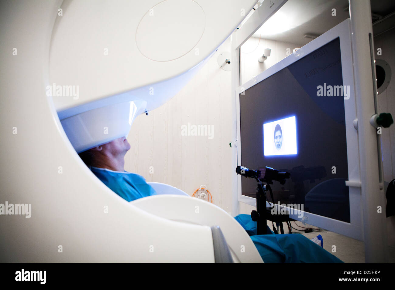
MAGNETOENCEPHALOGRAPHY

Image details
Contributor:
BSIP SA / Alamy Stock PhotoImage ID:
D25HKPFile size:
60.2 MB (3.5 MB Compressed download)Releases:
Model - yes | Property - noDo I need a release?Dimensions:
5616 x 3744 px | 47.5 x 31.7 cm | 18.7 x 12.5 inches | 300dpiDate taken:
23 November 2012Photographer:
AMELIE-BENOIST / BSIPMore information:
Reportage at the Neuroimaging research centre in Pitié Salpêtrière hospital in Paris, France. Magnetoencephalography (MEG) platform. MEG detects variations in the brain's magnetic field during various types of cerebral activity. It studies normal and abnormal brain function. Recording the magnetic field produced by neuronal currents requires ultra sensitive sensors called SQUIDs (Superconducting Quantum Interference Device). The 306 sensors spread over 102 areas allow both near and far magnetic fields to be measured, and the brain's deep structures to be 'seen'. The person receives visual stimulation (faces with different expressions) from a high resolution video projector onto a projection screen. The screen is coupled with a high speed system that follows eye movement.