33 fetal development Black & White Stock Photos
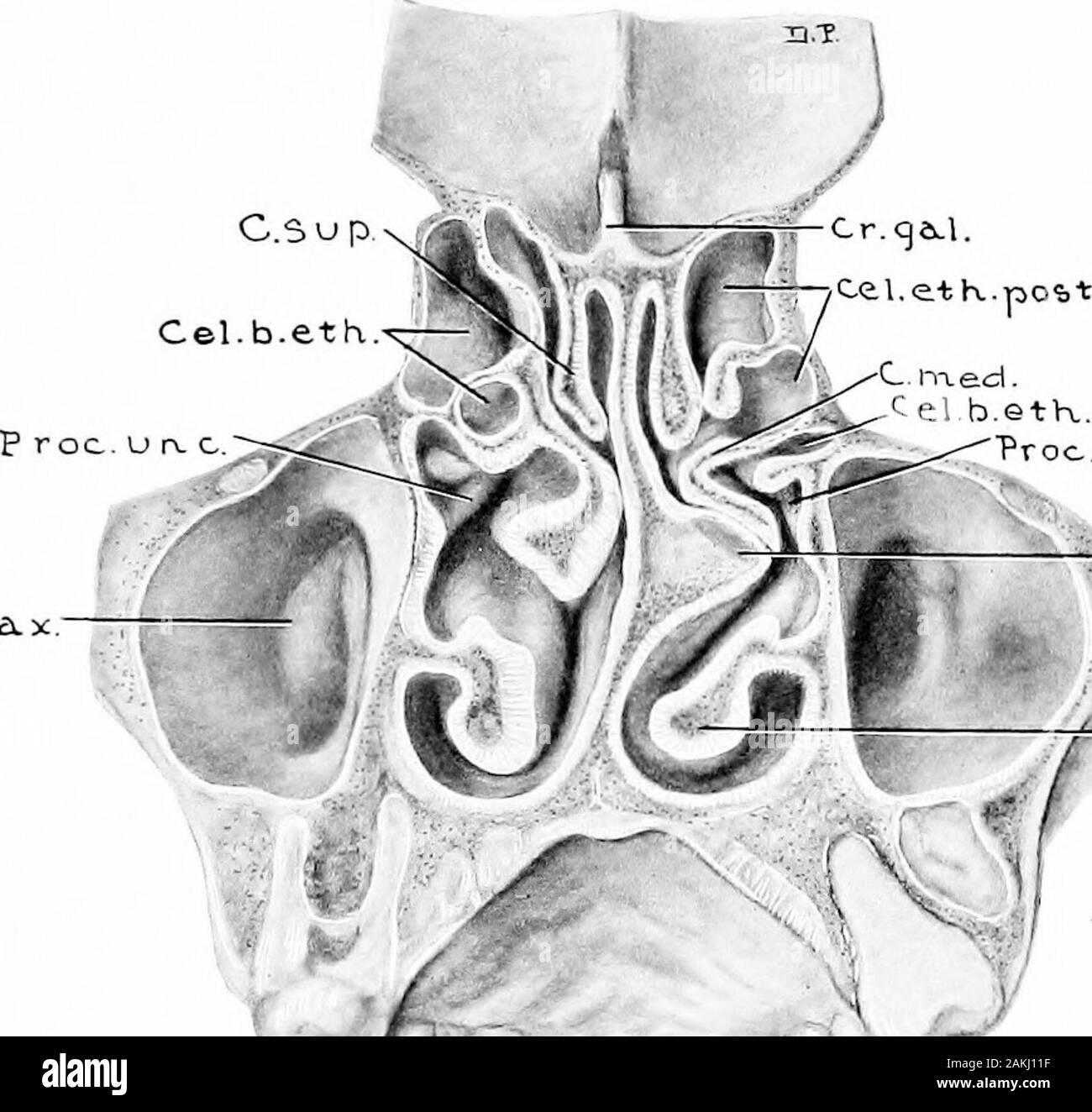
 Development and anatomy of the nasal accessory sinuses in man; observations based on two hundred and ninety lateral nasal walls, showing the various stages and types of development of the accessory sinus areas from the sixtieth day of fetal life to advanced maturity . thickness. The more constantof the two ridges is the one which forms the inferior wall ofthe above-mentioned canalis infraorbitalis (Figs. 25, 26,42, and 52). The second ridge is well marked in the major-ity of cases, and extends laterally along the roof from theposterior margin of the ostium maxillare (Figs. 33, 39, 45,and 54). Stock Photohttps://www.alamy.com/image-license-details/?v=1https://www.alamy.com/development-and-anatomy-of-the-nasal-accessory-sinuses-in-man-observations-based-on-two-hundred-and-ninety-lateral-nasal-walls-showing-the-various-stages-and-types-of-development-of-the-accessory-sinus-areas-from-the-sixtieth-day-of-fetal-life-to-advanced-maturity-thickness-the-more-constantof-the-two-ridges-is-the-one-which-forms-the-inferior-wall-ofthe-above-mentioned-canalis-infraorbitalis-figs-25-2642-and-52-the-second-ridge-is-well-marked-in-the-major-ity-of-cases-and-extends-laterally-along-the-roof-from-theposterior-margin-of-the-ostium-maxillare-figs-33-39-45and-54-image339071419.html
RM2AKJ11F–Development and anatomy of the nasal accessory sinuses in man; observations based on two hundred and ninety lateral nasal walls, showing the various stages and types of development of the accessory sinus areas from the sixtieth day of fetal life to advanced maturity . thickness. The more constantof the two ridges is the one which forms the inferior wall ofthe above-mentioned canalis infraorbitalis (Figs. 25, 26,42, and 52). The second ridge is well marked in the major-ity of cases, and extends laterally along the roof from theposterior margin of the ostium maxillare (Figs. 33, 39, 45,and 54).
 Development and anatomy of the nasal accessory sinuses in man; observations based on two hundred and ninety lateral nasal walls, showing the various stages and types of development of the accessory sinus areas from the sixtieth day of fetal life to advanced maturity . Vos pter y cjopa-L / O St.rrax.emcees.. Fig. 33.—Rpecimex From a Child Eight Years, Two Months, and TenDays Old. (Series D, No. 59.)Lateral view of frontal, ethmoidal, and maxillary areas. Note that thesinus frontalis developed from a cell having its origin from the suprabullarfurrow. The right sinus frontalis had a similar orig Stock Photohttps://www.alamy.com/image-license-details/?v=1https://www.alamy.com/development-and-anatomy-of-the-nasal-accessory-sinuses-in-man-observations-based-on-two-hundred-and-ninety-lateral-nasal-walls-showing-the-various-stages-and-types-of-development-of-the-accessory-sinus-areas-from-the-sixtieth-day-of-fetal-life-to-advanced-maturity-vos-pter-y-cjopa-l-o-strraxemcees-fig-33rpecimex-from-a-child-eight-years-two-months-and-tendays-old-series-d-no-59lateral-view-of-frontal-ethmoidal-and-maxillary-areas-note-that-thesinus-frontalis-developed-from-a-cell-having-its-origin-from-the-suprabullarfurrow-the-right-sinus-frontalis-had-a-similar-orig-image339074733.html
RM2AKJ57W–Development and anatomy of the nasal accessory sinuses in man; observations based on two hundred and ninety lateral nasal walls, showing the various stages and types of development of the accessory sinus areas from the sixtieth day of fetal life to advanced maturity . Vos pter y cjopa-L / O St.rrax.emcees.. Fig. 33.—Rpecimex From a Child Eight Years, Two Months, and TenDays Old. (Series D, No. 59.)Lateral view of frontal, ethmoidal, and maxillary areas. Note that thesinus frontalis developed from a cell having its origin from the suprabullarfurrow. The right sinus frontalis had a similar orig