Quick filters:
Abductor digiti minimi Stock Photos and Images
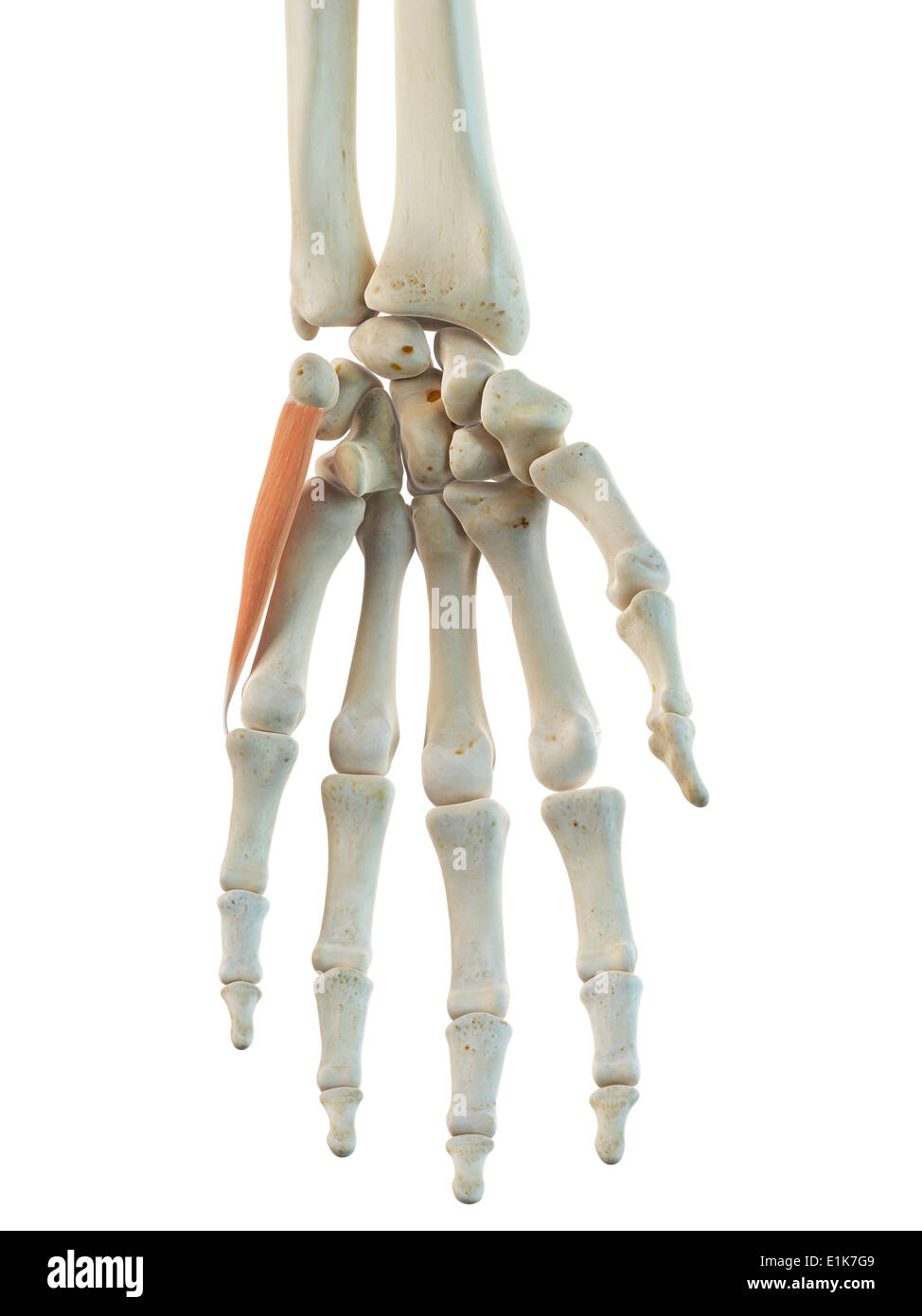 Human abductor digiti minimi muscle computer artwork. Stock Photohttps://www.alamy.com/image-license-details/?v=1https://www.alamy.com/human-abductor-digiti-minimi-muscle-computer-artwork-image69879161.html
RFE1K7G9–Human abductor digiti minimi muscle computer artwork.
 medical accurate illustration of the abductor digiti minimi Stock Photohttps://www.alamy.com/image-license-details/?v=1https://www.alamy.com/stock-photo-medical-accurate-illustration-of-the-abductor-digiti-minimi-89735509.html
RFF5YPFH–medical accurate illustration of the abductor digiti minimi
 Abductor digiti minimi muscle, illustration Stock Photohttps://www.alamy.com/image-license-details/?v=1https://www.alamy.com/abductor-digiti-minimi-muscle-illustration-image273701511.html
RFWW851Y–Abductor digiti minimi muscle, illustration
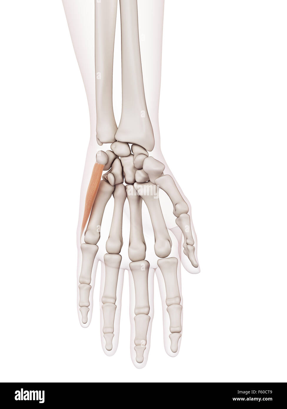 medically accurate muscle illustration of the abductor digiti minimi Stock Photohttps://www.alamy.com/image-license-details/?v=1https://www.alamy.com/stock-photo-medically-accurate-muscle-illustration-of-the-abductor-digiti-minimi-89749865.html
RFF60CT9–medically accurate muscle illustration of the abductor digiti minimi
 Illustration of the abductor digiti minimi muscle. Stock Photohttps://www.alamy.com/image-license-details/?v=1https://www.alamy.com/stock-photo-illustration-of-the-abductor-digiti-minimi-muscle-126898847.html
RFHACMNK–Illustration of the abductor digiti minimi muscle.
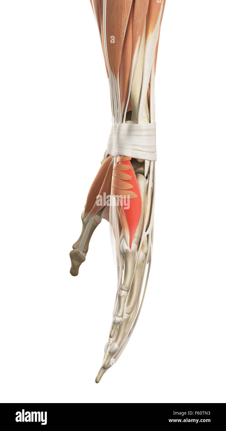 medically accurate illustration of the abductor digiti minimi Stock Photohttps://www.alamy.com/image-license-details/?v=1https://www.alamy.com/stock-photo-medically-accurate-illustration-of-the-abductor-digiti-minimi-89759183.html
RFF60TN3–medically accurate illustration of the abductor digiti minimi
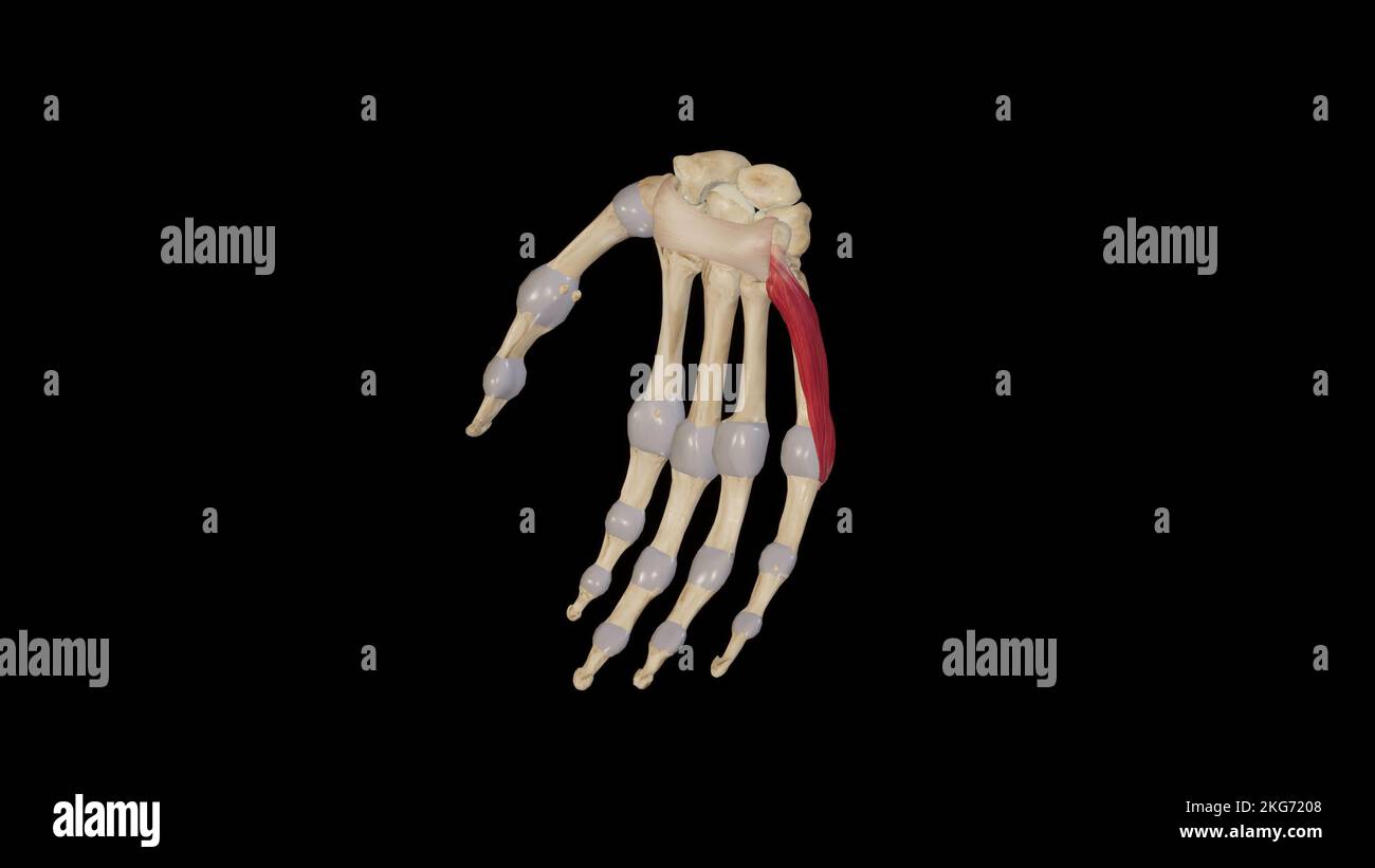 Abductor Digiti Minimi Stock Photohttps://www.alamy.com/image-license-details/?v=1https://www.alamy.com/abductor-digiti-minimi-image491880040.html
RF2KG7208–Abductor Digiti Minimi
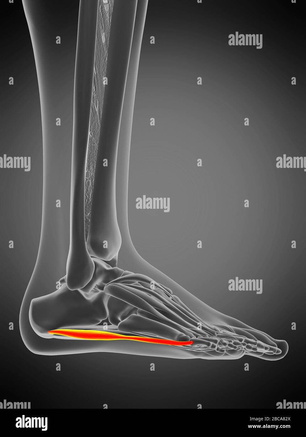 Abductor digiti minimi muscle, illustration. Stock Photohttps://www.alamy.com/image-license-details/?v=1https://www.alamy.com/abductor-digiti-minimi-muscle-illustration-image351809106.html
RF2BCA82X–Abductor digiti minimi muscle, illustration.
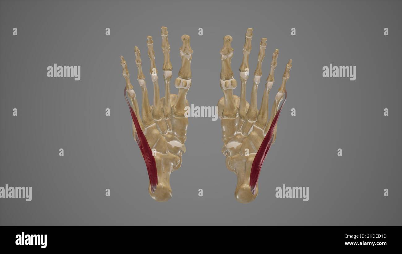 Medical Illustration of Abductor Digiti Minimi Stock Photohttps://www.alamy.com/image-license-details/?v=1https://www.alamy.com/medical-illustration-of-abductor-digiti-minimi-image490198393.html
RF2KDED1D–Medical Illustration of Abductor Digiti Minimi
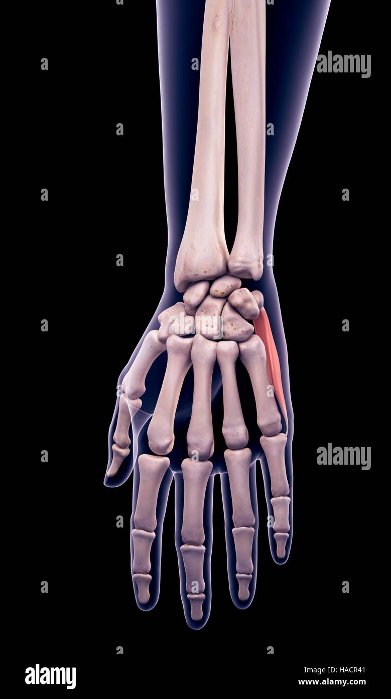 Illustration of the abductor digiti minimi muscle. Stock Photohttps://www.alamy.com/image-license-details/?v=1https://www.alamy.com/stock-photo-illustration-of-the-abductor-digiti-minimi-muscle-126900705.html
RFHACR41–Illustration of the abductor digiti minimi muscle.
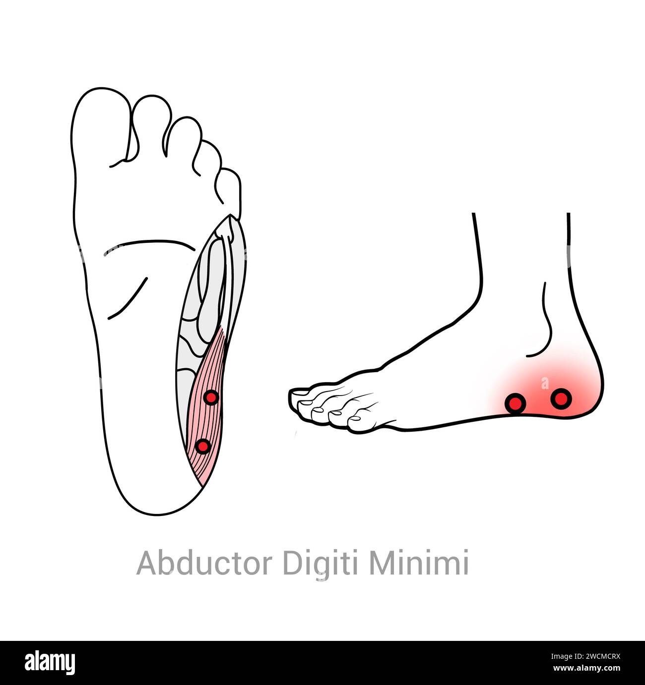 Abductor Digiti Minimi: Myofascial trigger points and associated pain locations Stock Photohttps://www.alamy.com/image-license-details/?v=1https://www.alamy.com/abductor-digiti-minimi-myofascial-trigger-points-and-associated-pain-locations-image592977502.html
RF2WCMCRX–Abductor Digiti Minimi: Myofascial trigger points and associated pain locations
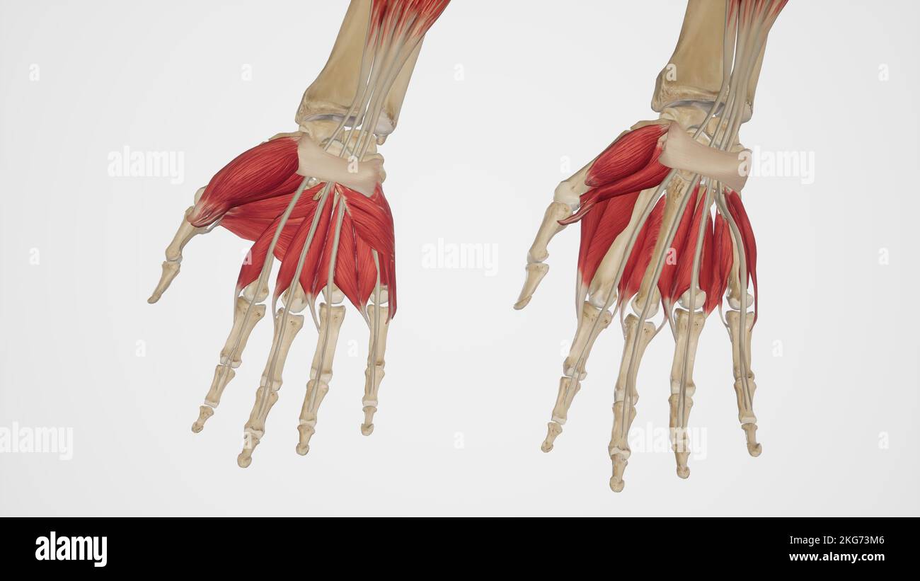 Muscles of the Hand Palmar View-Superficial and Deep Stock Photohttps://www.alamy.com/image-license-details/?v=1https://www.alamy.com/muscles-of-the-hand-palmar-view-superficial-and-deep-image491881382.html
RF2KG73M6–Muscles of the Hand Palmar View-Superficial and Deep
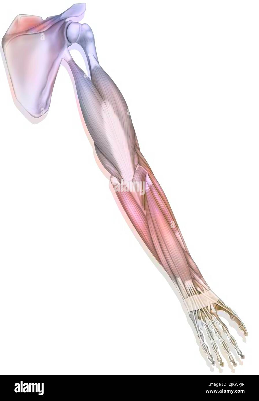 The muscles of the upper right limb in posterior view. Stock Photohttps://www.alamy.com/image-license-details/?v=1https://www.alamy.com/the-muscles-of-the-upper-right-limb-in-posterior-view-image476924975.html
RF2JKWPJR–The muscles of the upper right limb in posterior view.
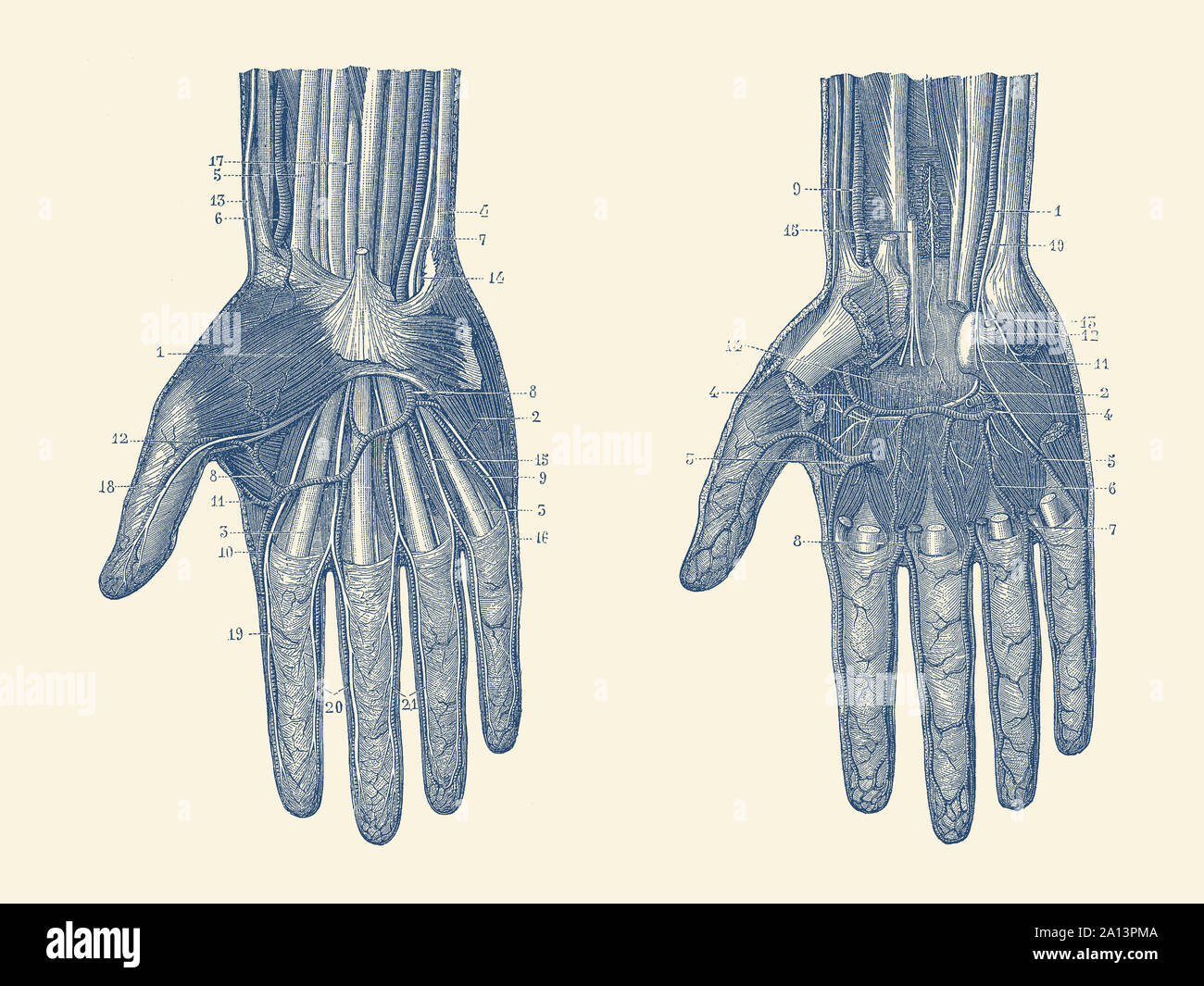 Dual view of the human hand, showcasing the muscles, bones and veins throughout. Stock Photohttps://www.alamy.com/image-license-details/?v=1https://www.alamy.com/dual-view-of-the-human-hand-showcasing-the-muscles-bones-and-veins-throughout-image327695322.html
RF2A13PMA–Dual view of the human hand, showcasing the muscles, bones and veins throughout.
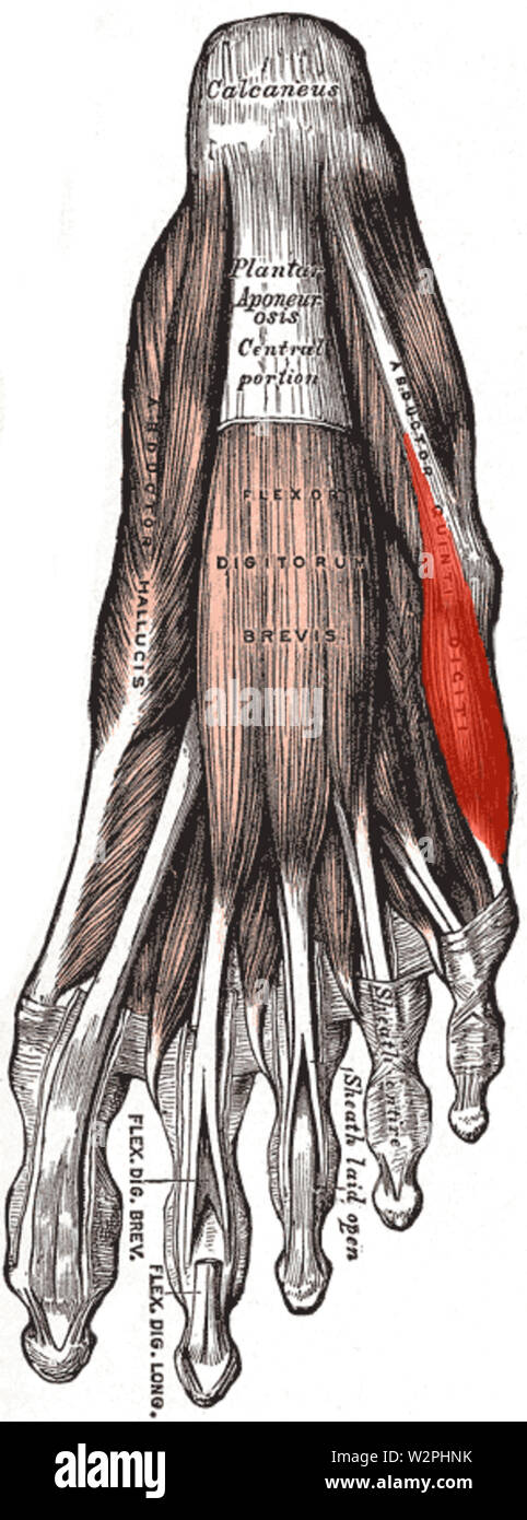 Abductor digiti minimi (foot) Stock Photohttps://www.alamy.com/image-license-details/?v=1https://www.alamy.com/abductor-digiti-minimi-foot-image259881711.html
RMW2PHNK–Abductor digiti minimi (foot)
 The muscles of the hand Stock Photohttps://www.alamy.com/image-license-details/?v=1https://www.alamy.com/stock-photo-the-muscles-of-the-hand-13166040.html
RFACJ38W–The muscles of the hand
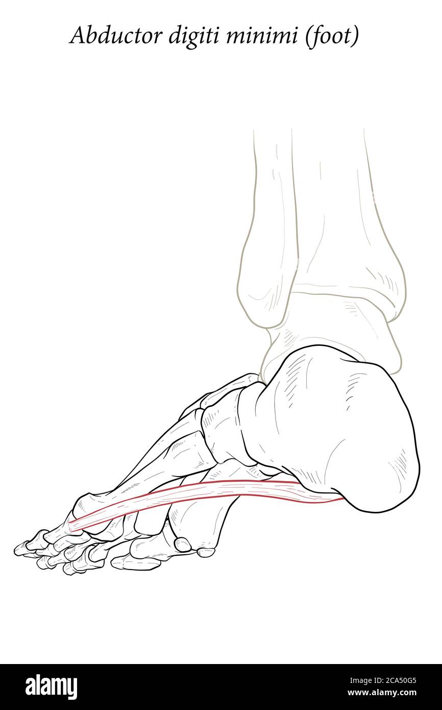 Abductor digiti minimi muscle of foot. Stock Vectorhttps://www.alamy.com/image-license-details/?v=1https://www.alamy.com/abductor-digiti-minimi-muscle-of-foot-image367674501.html
RF2CA50G5–Abductor digiti minimi muscle of foot.
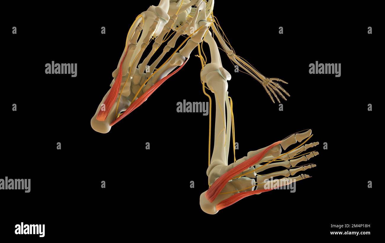 Abductor Hallucis and Digiti Minimi Leg Muscles anatomy for medical concept 3D illustration Stock Photohttps://www.alamy.com/image-license-details/?v=1https://www.alamy.com/abductor-hallucis-and-digiti-minimi-leg-muscles-anatomy-for-medical-concept-3d-illustration-image502043265.html
RF2M4P18H–Abductor Hallucis and Digiti Minimi Leg Muscles anatomy for medical concept 3D illustration
 Electro-diagnosis and electro-therapeutics : a guide for practitioners and students . c conditions in order to exclude ex-tinction of excitability) current streamers easily produceon the long finger flexors or extensors a seeming interos-seal effect. Let the beginner be on the lookout for this,and always observe whether the first phalanx is actuallyflexed and the last two are extended. The muscles of the ball of the small finger (the oppo-nens, flexor, abductor digiti minimi muscles) are ex-cited at the root of the hypothenar. They produce theeffects expressed by their names, and are not alway Stock Photohttps://www.alamy.com/image-license-details/?v=1https://www.alamy.com/electro-diagnosis-and-electro-therapeutics-a-guide-for-practitioners-and-students-c-conditions-in-order-to-exclude-ex-tinction-of-excitability-current-streamers-easily-produceon-the-long-finger-flexors-or-extensors-a-seeming-interos-seal-effect-let-the-beginner-be-on-the-lookout-for-thisand-always-observe-whether-the-first-phalanx-is-actuallyflexed-and-the-last-two-are-extended-the-muscles-of-the-ball-of-the-small-finger-the-oppo-nens-flexor-abductor-digiti-minimi-muscles-are-ex-cited-at-the-root-of-the-hypothenar-they-produce-theeffects-expressed-by-their-names-and-are-not-alway-image342704467.html
RM2AWFF17–Electro-diagnosis and electro-therapeutics : a guide for practitioners and students . c conditions in order to exclude ex-tinction of excitability) current streamers easily produceon the long finger flexors or extensors a seeming interos-seal effect. Let the beginner be on the lookout for this,and always observe whether the first phalanx is actuallyflexed and the last two are extended. The muscles of the ball of the small finger (the oppo-nens, flexor, abductor digiti minimi muscles) are ex-cited at the root of the hypothenar. They produce theeffects expressed by their names, and are not alway
 3D Illustration, Muscle is a soft tissue, Muscle cells contain proteins , producing a contraction that changes both the length and the shape of the ce Stock Photohttps://www.alamy.com/image-license-details/?v=1https://www.alamy.com/3d-illustration-muscle-is-a-soft-tissue-muscle-cells-contain-proteins-producing-a-contraction-that-changes-both-the-length-and-the-shape-of-the-ce-image395465915.html
RF2DYB0PK–3D Illustration, Muscle is a soft tissue, Muscle cells contain proteins , producing a contraction that changes both the length and the shape of the ce
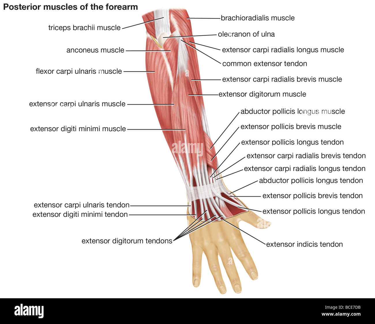 The posterior view of the muscles of the human forearm. Stock Photohttps://www.alamy.com/image-license-details/?v=1https://www.alamy.com/stock-photo-the-posterior-view-of-the-muscles-of-the-human-forearm-24899431.html
RMBCE7DB–The posterior view of the muscles of the human forearm.
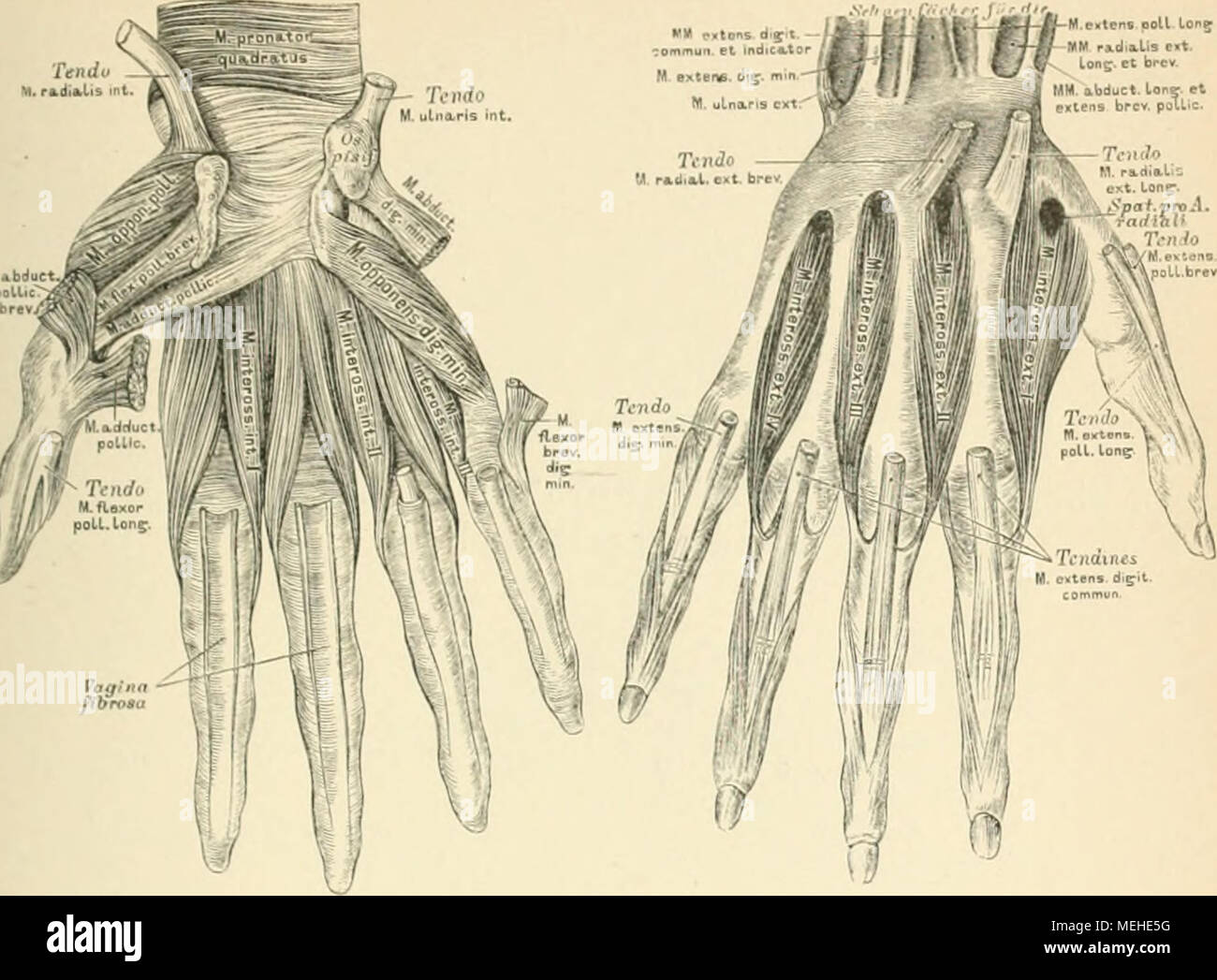 . Die descriptive und topographische Anatomie des Menschen . 313. Die Muskehl an der Hand. )514. Die Muskeln an der Hand. I>ie Muskeln des K I e i 11 l'i n gr r lia 11 i'ii s, Antithenar: M. palmaris brems (Fig. 310). [Jrspr.: Aponeurosis palmaris. [nsert.: Haul am Ulnarrande Jcr Hand. .1/. abductor digiti minimi. (Fig. -11 1 . (Jrspr.: Os pisiforme. (nsert.: Basis 1. phalangis und Apt talis des kleinen Fingers. M. ßexor brevis dig. min. (Fig. 311). [Jrspr.: Lig. carpi transvers. und Haken des Os hamaium. [nsert.: wie die des vorigen. '/. opponens dig. min. (Jrspr.: wie der des M. ßexor bre Stock Photohttps://www.alamy.com/image-license-details/?v=1https://www.alamy.com/die-descriptive-und-topographische-anatomie-des-menschen-313-die-muskehl-an-der-hand-514-die-muskeln-an-der-hand-igtie-muskeln-des-k-i-e-i-11-li-n-gr-r-lia-11-iii-s-antithenar-m-palmaris-brems-fig-310-jrspr-aponeurosis-palmaris-nsert-haul-am-ulnarrande-jcr-hand-1-abductor-digiti-minimi-fig-11-1-jrspr-os-pisiforme-nsert-basis-1-phalangis-und-apt-talis-des-kleinen-fingers-m-exor-brevis-dig-min-fig-311-jrspr-lig-carpi-transvers-und-haken-des-os-hamaium-nsert-wie-die-des-vorigen-opponens-dig-min-jrspr-wie-der-des-m-exor-bre-image181093180.html
RMMEHE5G–. Die descriptive und topographische Anatomie des Menschen . 313. Die Muskehl an der Hand. )514. Die Muskeln an der Hand. I>ie Muskeln des K I e i 11 l'i n gr r lia 11 i'ii s, Antithenar: M. palmaris brems (Fig. 310). [Jrspr.: Aponeurosis palmaris. [nsert.: Haul am Ulnarrande Jcr Hand. .1/. abductor digiti minimi. (Fig. -11 1 . (Jrspr.: Os pisiforme. (nsert.: Basis 1. phalangis und Apt talis des kleinen Fingers. M. ßexor brevis dig. min. (Fig. 311). [Jrspr.: Lig. carpi transvers. und Haken des Os hamaium. [nsert.: wie die des vorigen. '/. opponens dig. min. (Jrspr.: wie der des M. ßexor bre
 . The development of the human body : a manual of human embryology. Embryology; Embryo, Non-Mammalian. Fig. 128.—Transverse sections through (A) the crus and (B) the foot, showing the arrangement of the layers of the flexor muscles. The shading has the same significance as in the preceding figure. AbH, abductor hallucis; AbM, abductor minimi digiti; AdH, adductor hallucis; ELD, extensor longus digitorum; F, fibula; FBD, flexor brevis digitorium; FBH, flexor brevis hallucis; FBM, flexor brevis minimi digiti; FLD, flexor longus digitorum; G, gastrocnemius; ID, interossei dorsalis; IV, interossei Stock Photohttps://www.alamy.com/image-license-details/?v=1https://www.alamy.com/the-development-of-the-human-body-a-manual-of-human-embryology-embryology-embryo-non-mammalian-fig-128transverse-sections-through-a-the-crus-and-b-the-foot-showing-the-arrangement-of-the-layers-of-the-flexor-muscles-the-shading-has-the-same-significance-as-in-the-preceding-figure-abh-abductor-hallucis-abm-abductor-minimi-digiti-adh-adductor-hallucis-eld-extensor-longus-digitorum-f-fibula-fbd-flexor-brevis-digitorium-fbh-flexor-brevis-hallucis-fbm-flexor-brevis-minimi-digiti-fld-flexor-longus-digitorum-g-gastrocnemius-id-interossei-dorsalis-iv-interossei-image215969611.html
RMPFA7CB–. The development of the human body : a manual of human embryology. Embryology; Embryo, Non-Mammalian. Fig. 128.—Transverse sections through (A) the crus and (B) the foot, showing the arrangement of the layers of the flexor muscles. The shading has the same significance as in the preceding figure. AbH, abductor hallucis; AbM, abductor minimi digiti; AdH, adductor hallucis; ELD, extensor longus digitorum; F, fibula; FBD, flexor brevis digitorium; FBH, flexor brevis hallucis; FBM, flexor brevis minimi digiti; FLD, flexor longus digitorum; G, gastrocnemius; ID, interossei dorsalis; IV, interossei
 medically accurate illustration of the abductor digiti minimi Stock Photohttps://www.alamy.com/image-license-details/?v=1https://www.alamy.com/stock-photo-medically-accurate-illustration-of-the-abductor-digiti-minimi-89759182.html
RFF60TN2–medically accurate illustration of the abductor digiti minimi
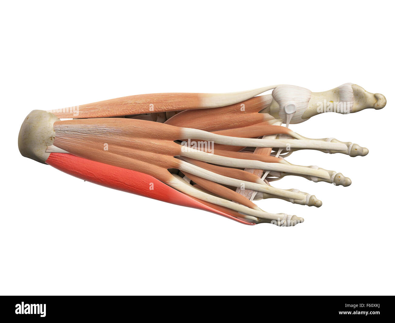 medically accurate illustration of the abductor digiti minimi Stock Photohttps://www.alamy.com/image-license-details/?v=1https://www.alamy.com/stock-photo-medically-accurate-illustration-of-the-abductor-digiti-minimi-89760710.html
RFF60XKJ–medically accurate illustration of the abductor digiti minimi
 medical accurate illustration of the abductor digiti minimi Stock Photohttps://www.alamy.com/image-license-details/?v=1https://www.alamy.com/stock-photo-medical-accurate-illustration-of-the-abductor-digiti-minimi-89735245.html
RFF5YP65–medical accurate illustration of the abductor digiti minimi
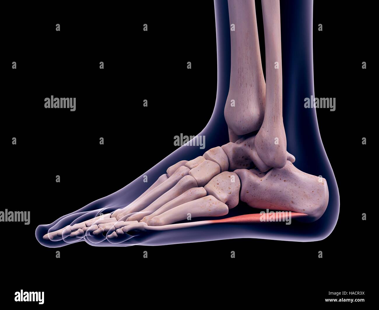 Illustration of the abductor digiti minimi muscle. Stock Photohttps://www.alamy.com/image-license-details/?v=1https://www.alamy.com/stock-photo-illustration-of-the-abductor-digiti-minimi-muscle-126900702.html
RFHACR3X–Illustration of the abductor digiti minimi muscle.
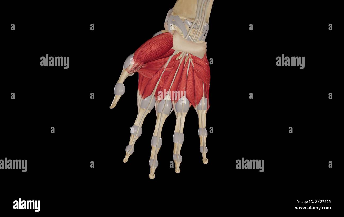 Muscles of Hand Palmar View Stock Photohttps://www.alamy.com/image-license-details/?v=1https://www.alamy.com/muscles-of-hand-palmar-view-image491880037.html
RF2KG7205–Muscles of Hand Palmar View
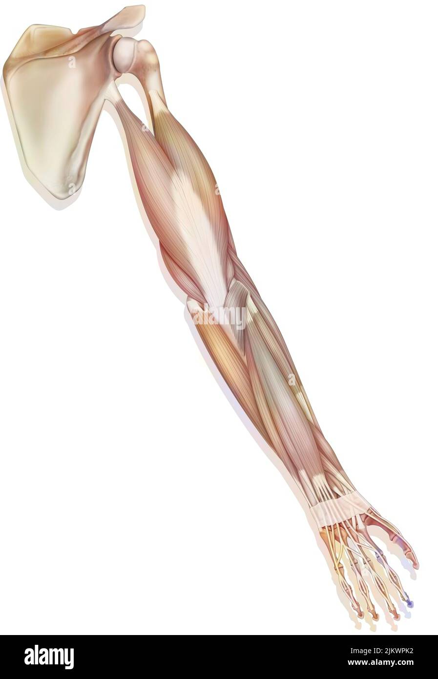 The muscles of the upper right limb in posterior view. Stock Photohttps://www.alamy.com/image-license-details/?v=1https://www.alamy.com/the-muscles-of-the-upper-right-limb-in-posterior-view-image476924982.html
RF2JKWPK2–The muscles of the upper right limb in posterior view.
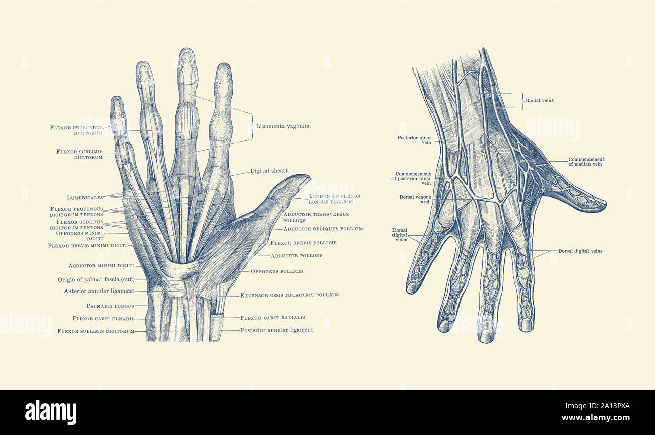 Dual-view diagram of the human hand, showcasing ligaments, muscles and veins. Stock Photohttps://www.alamy.com/image-license-details/?v=1https://www.alamy.com/dual-view-diagram-of-the-human-hand-showcasing-ligaments-muscles-and-veins-image327695490.html
RF2A13PXA–Dual-view diagram of the human hand, showcasing ligaments, muscles and veins.
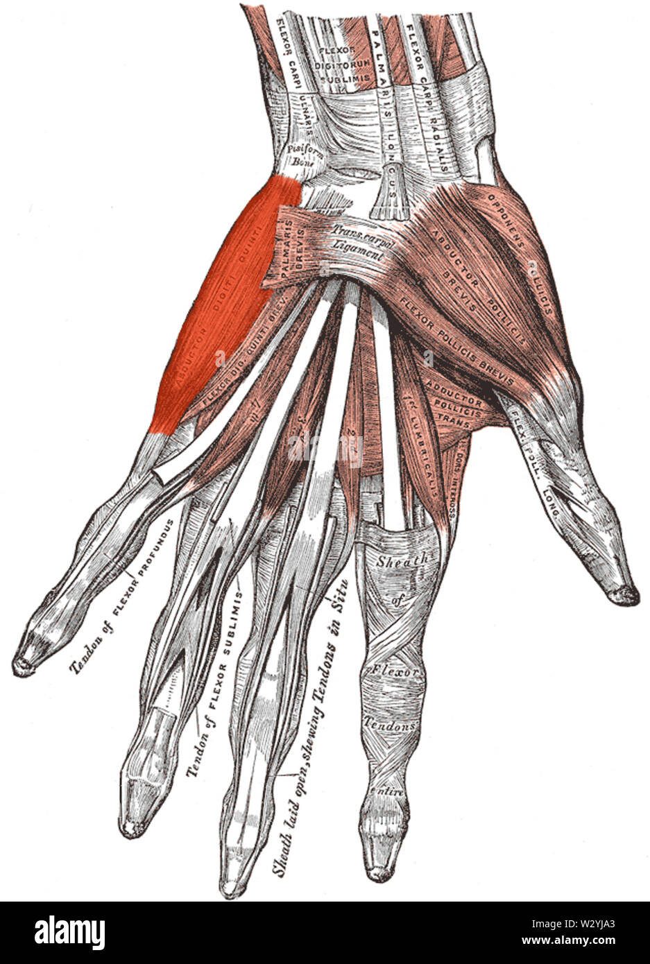 Musculus abductor digiti minimi (Hand) Stock Photohttps://www.alamy.com/image-license-details/?v=1https://www.alamy.com/musculus-abductor-digiti-minimi-hand-image259991931.html
RMW2YJA3–Musculus abductor digiti minimi (Hand)
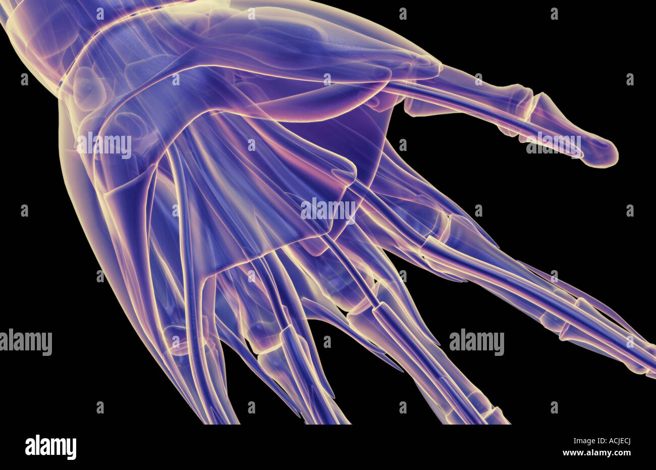 The muscles of the hand Stock Photohttps://www.alamy.com/image-license-details/?v=1https://www.alamy.com/stock-photo-the-muscles-of-the-hand-13169777.html
RFACJECJ–The muscles of the hand
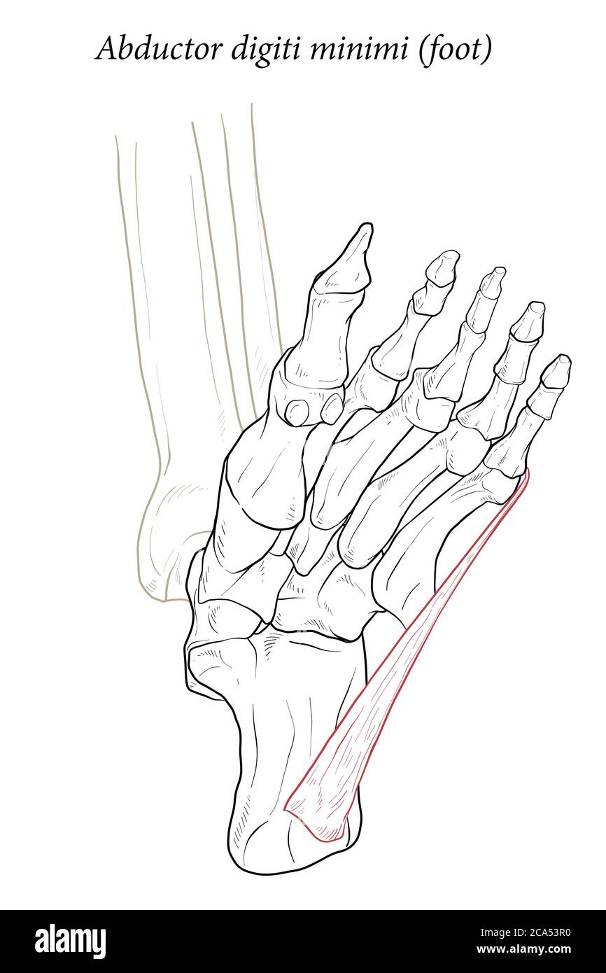 Abductor digiti minimi muscle of foot. Stock Vectorhttps://www.alamy.com/image-license-details/?v=1https://www.alamy.com/abductor-digiti-minimi-muscle-of-foot-image367677044.html
RF2CA53R0–Abductor digiti minimi muscle of foot.
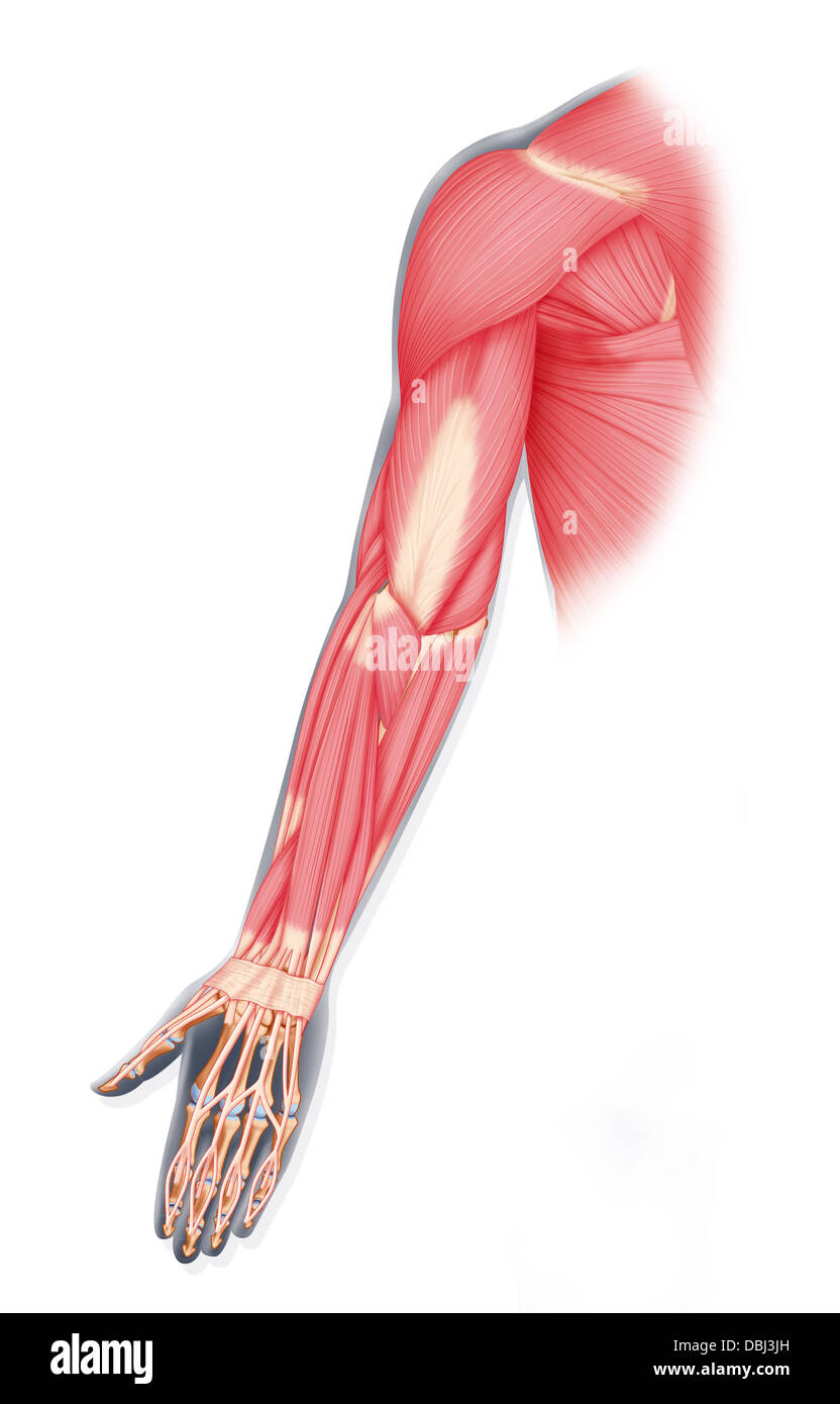 UPPER LIMB MUSCLE, DRAWING Stock Photohttps://www.alamy.com/image-license-details/?v=1https://www.alamy.com/stock-photo-upper-limb-muscle-drawing-58790329.html
RMDBJ3JH–UPPER LIMB MUSCLE, DRAWING
 . Practical electricity in medicine and surgery. r digitor. communislongus. M. peroneus brevis.? M. soleus. M. flexor hallucis longus. M. extensor digitor. communisbrevis. —.M. abductor digiti minimi ped. Fig. 162.—Motor Points of Outer Side op the Leg. rent and large electrode applied between the trochanter majorand the tuber ischii. Its stimulation produces contraction in theflexor muscles of the leg and foot, and pain in the area of distri-bution of the sensitive fibres. The heads of the biceps are each supplied with a branch MOTOR POINTS OF THE LOWER EXTREMITY. 185 of the sciatic nerve. Stock Photohttps://www.alamy.com/image-license-details/?v=1https://www.alamy.com/practical-electricity-in-medicine-and-surgery-r-digitor-communislongus-m-peroneus-brevis-m-soleus-m-flexor-hallucis-longus-m-extensor-digitor-communisbrevis-m-abductor-digiti-minimi-ped-fig-162motor-points-of-outer-side-op-the-leg-rent-and-large-electrode-applied-between-the-trochanter-majorand-the-tuber-ischii-its-stimulation-produces-contraction-in-theflexor-muscles-of-the-leg-and-foot-and-pain-in-the-area-of-distri-bution-of-the-sensitive-fibres-the-heads-of-the-biceps-are-each-supplied-with-a-branch-motor-points-of-the-lower-extremity-185-of-the-sciatic-nerve-image336686077.html
RM2AFNAEN–. Practical electricity in medicine and surgery. r digitor. communislongus. M. peroneus brevis.? M. soleus. M. flexor hallucis longus. M. extensor digitor. communisbrevis. —.M. abductor digiti minimi ped. Fig. 162.—Motor Points of Outer Side op the Leg. rent and large electrode applied between the trochanter majorand the tuber ischii. Its stimulation produces contraction in theflexor muscles of the leg and foot, and pain in the area of distri-bution of the sensitive fibres. The heads of the biceps are each supplied with a branch MOTOR POINTS OF THE LOWER EXTREMITY. 185 of the sciatic nerve.
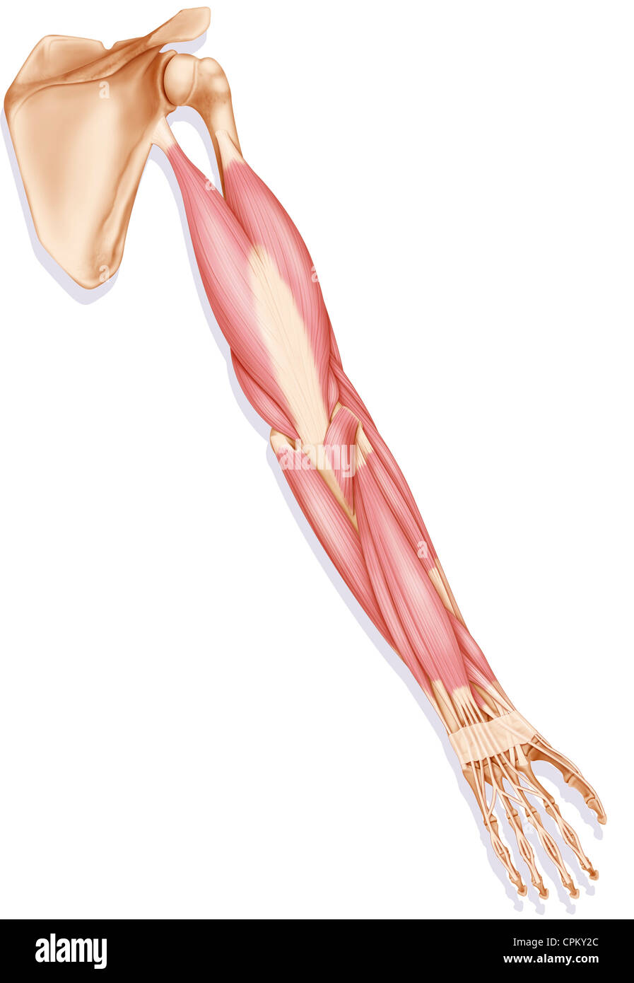 ARM MUSCLE, DRAWING Stock Photohttps://www.alamy.com/image-license-details/?v=1https://www.alamy.com/stock-photo-arm-muscle-drawing-48381492.html
RMCPKY2C–ARM MUSCLE, DRAWING
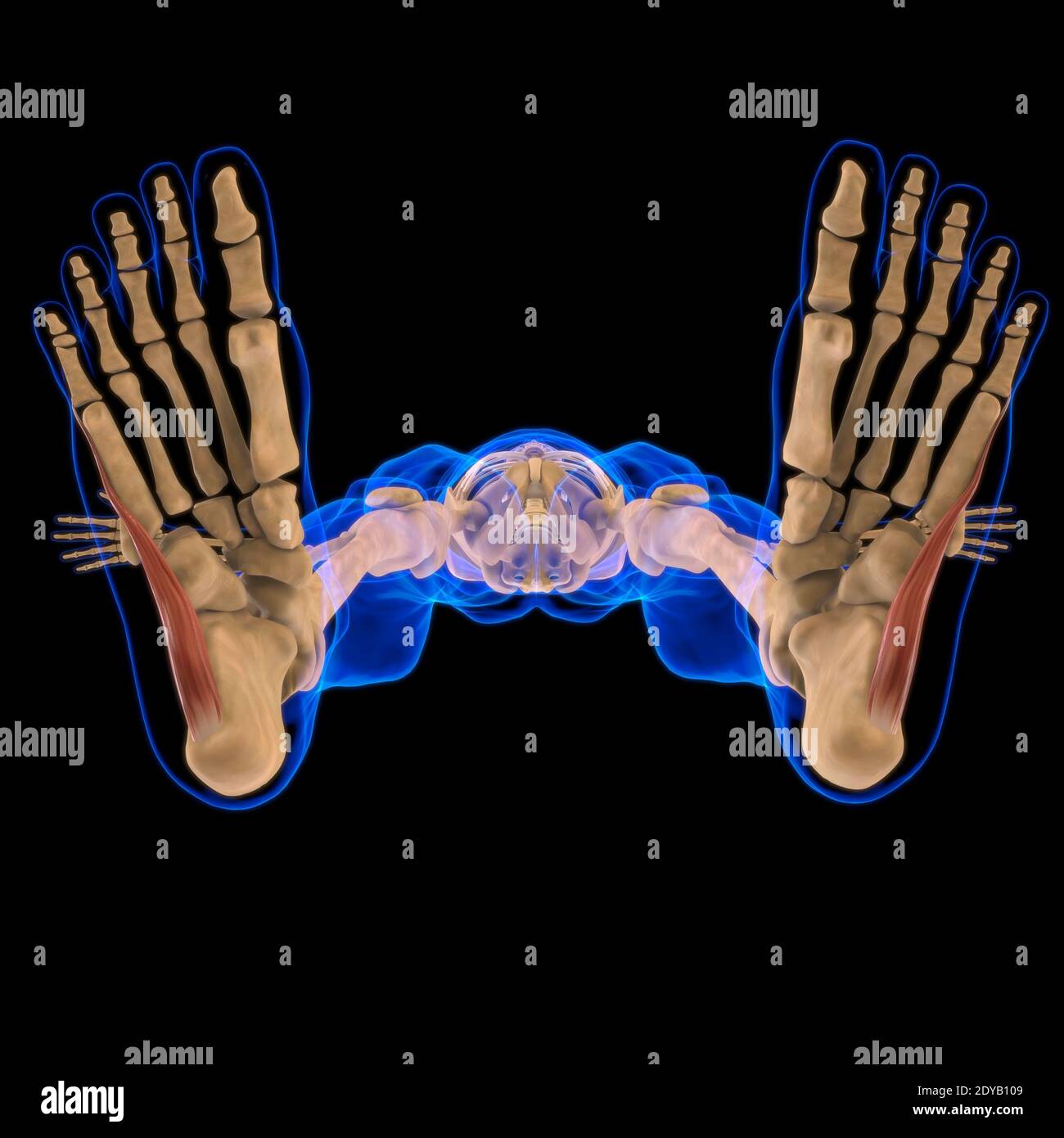 3D Illustration, Muscle is a soft tissue, Muscle cells contain proteins , producing a contraction that changes both the length and the shape of the ce Stock Photohttps://www.alamy.com/image-license-details/?v=1https://www.alamy.com/3d-illustration-muscle-is-a-soft-tissue-muscle-cells-contain-proteins-producing-a-contraction-that-changes-both-the-length-and-the-shape-of-the-ce-image395466073.html
RF2DYB109–3D Illustration, Muscle is a soft tissue, Muscle cells contain proteins , producing a contraction that changes both the length and the shape of the ce
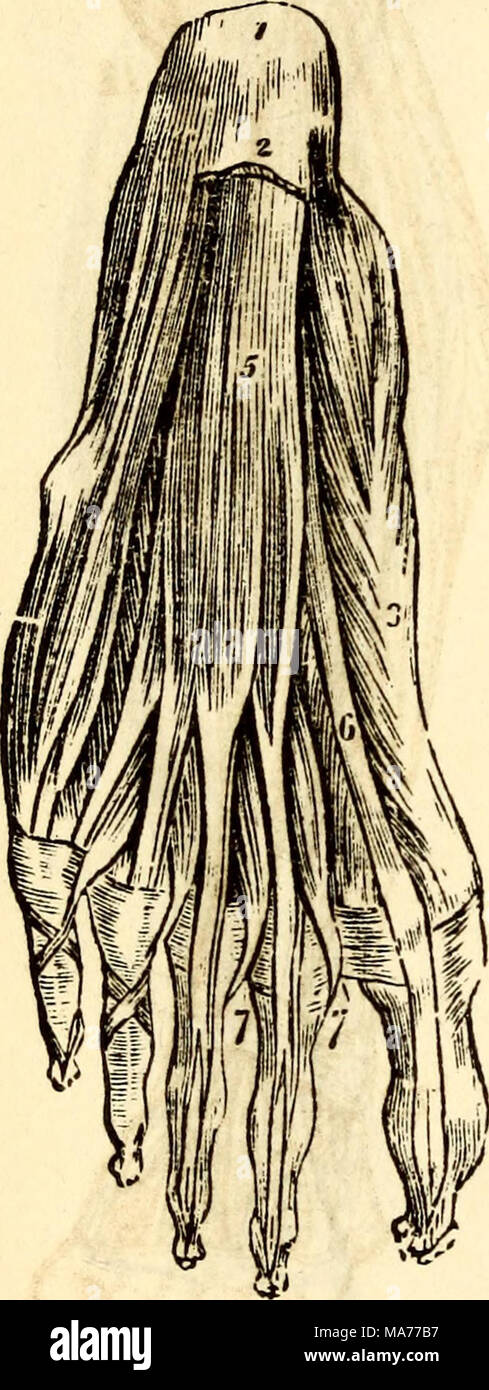 . Elementary anatomy and physiology : for colleges, academies, and other schools . A View of the Muscles on the Sole of the Foot immediately under the Plantar Fas- cia. 1, Os Calcis. 2, Section of the Fascia Plantaris. 3, Abductor Pollicis. 4, Ab- ductor Minimi Digiti. 5, Flexor Brevis Digitorum. 6, Tendon of the Flexor Longus Pollicis. 7, 7, Lumbricales. 261. What is the use of the Annular Ligaments ? What ornaments do they very closely /esemble ? Suppose these or their equivalent was wanting ? Stock Photohttps://www.alamy.com/image-license-details/?v=1https://www.alamy.com/elementary-anatomy-and-physiology-for-colleges-academies-and-other-schools-a-view-of-the-muscles-on-the-sole-of-the-foot-immediately-under-the-plantar-fas-cia-1-os-calcis-2-section-of-the-fascia-plantaris-3-abductor-pollicis-4-ab-ductor-minimi-digiti-5-flexor-brevis-digitorum-6-tendon-of-the-flexor-longus-pollicis-7-7-lumbricales-261-what-is-the-use-of-the-annular-ligaments-what-ornaments-do-they-very-closely-esemble-suppose-these-or-their-equivalent-was-wanting-image178409707.html
RMMA77B7–. Elementary anatomy and physiology : for colleges, academies, and other schools . A View of the Muscles on the Sole of the Foot immediately under the Plantar Fas- cia. 1, Os Calcis. 2, Section of the Fascia Plantaris. 3, Abductor Pollicis. 4, Ab- ductor Minimi Digiti. 5, Flexor Brevis Digitorum. 6, Tendon of the Flexor Longus Pollicis. 7, 7, Lumbricales. 261. What is the use of the Annular Ligaments ? What ornaments do they very closely /esemble ? Suppose these or their equivalent was wanting ?
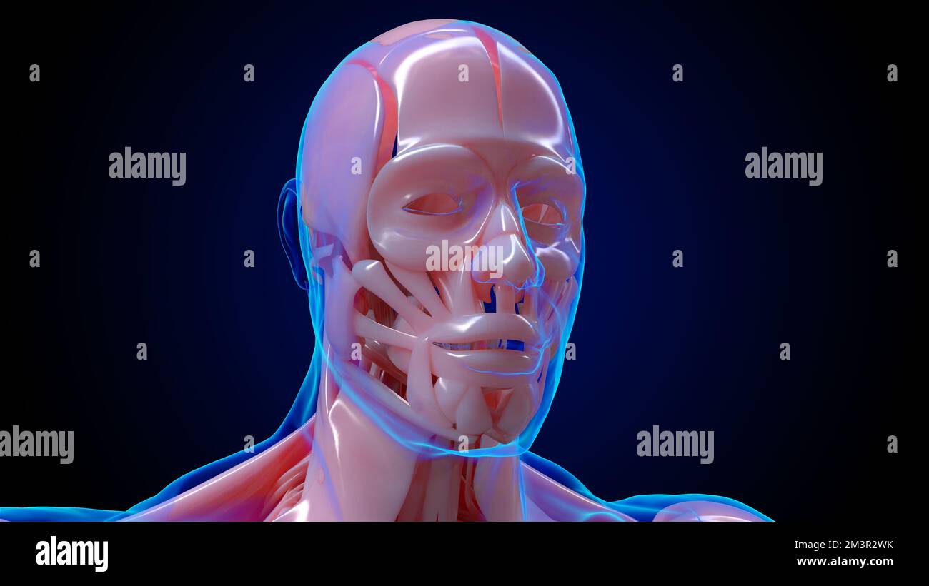 muscular system is an organ system responsible for providing strength 3D illustration Stock Photohttps://www.alamy.com/image-license-details/?v=1https://www.alamy.com/muscular-system-is-an-organ-system-responsible-for-providing-strength-3d-illustration-image501451823.html
RF2M3R2WK–muscular system is an organ system responsible for providing strength 3D illustration
 3D Illustration, Muscle is a soft tissue, Muscle cells contain proteins , producing a contraction that changes both the length and the shape of the ce Stock Photohttps://www.alamy.com/image-license-details/?v=1https://www.alamy.com/3d-illustration-muscle-is-a-soft-tissue-muscle-cells-contain-proteins-producing-a-contraction-that-changes-both-the-length-and-the-shape-of-the-ce-image395466112.html
RF2DYB11M–3D Illustration, Muscle is a soft tissue, Muscle cells contain proteins , producing a contraction that changes both the length and the shape of the ce
 medical accurate illustration of the abductor digiti minimi medial Stock Photohttps://www.alamy.com/image-license-details/?v=1https://www.alamy.com/stock-photo-medical-accurate-illustration-of-the-abductor-digiti-minimi-medial-89740146.html
RFF600D6–medical accurate illustration of the abductor digiti minimi medial
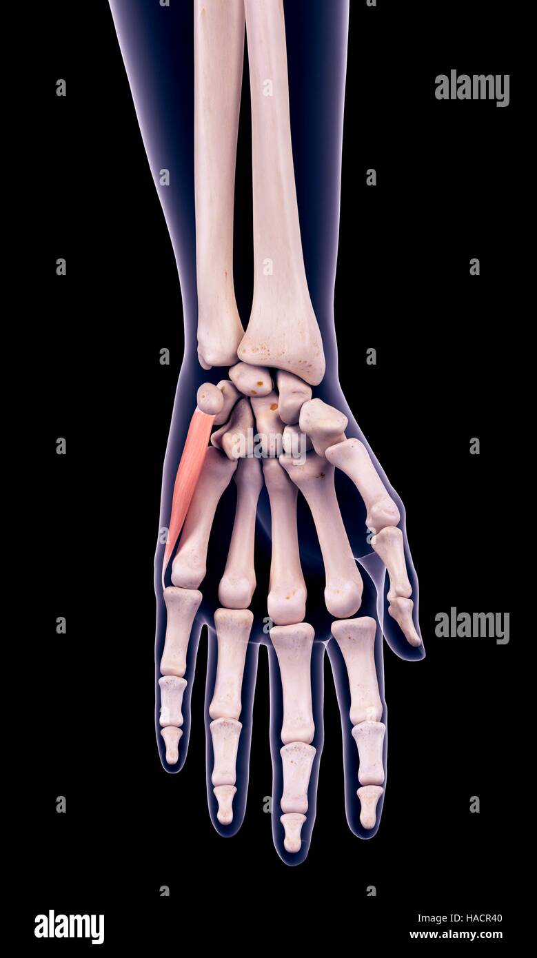 Illustration of the abductor digiti minimi muscle. Stock Photohttps://www.alamy.com/image-license-details/?v=1https://www.alamy.com/stock-photo-illustration-of-the-abductor-digiti-minimi-muscle-126900704.html
RFHACR40–Illustration of the abductor digiti minimi muscle.
 3d rendered medically accurate illustration of the abductor digiti minimi Stock Photohttps://www.alamy.com/image-license-details/?v=1https://www.alamy.com/3d-rendered-medically-accurate-illustration-of-the-abductor-digiti-minimi-image257792322.html
RFTYBCMJ–3d rendered medically accurate illustration of the abductor digiti minimi
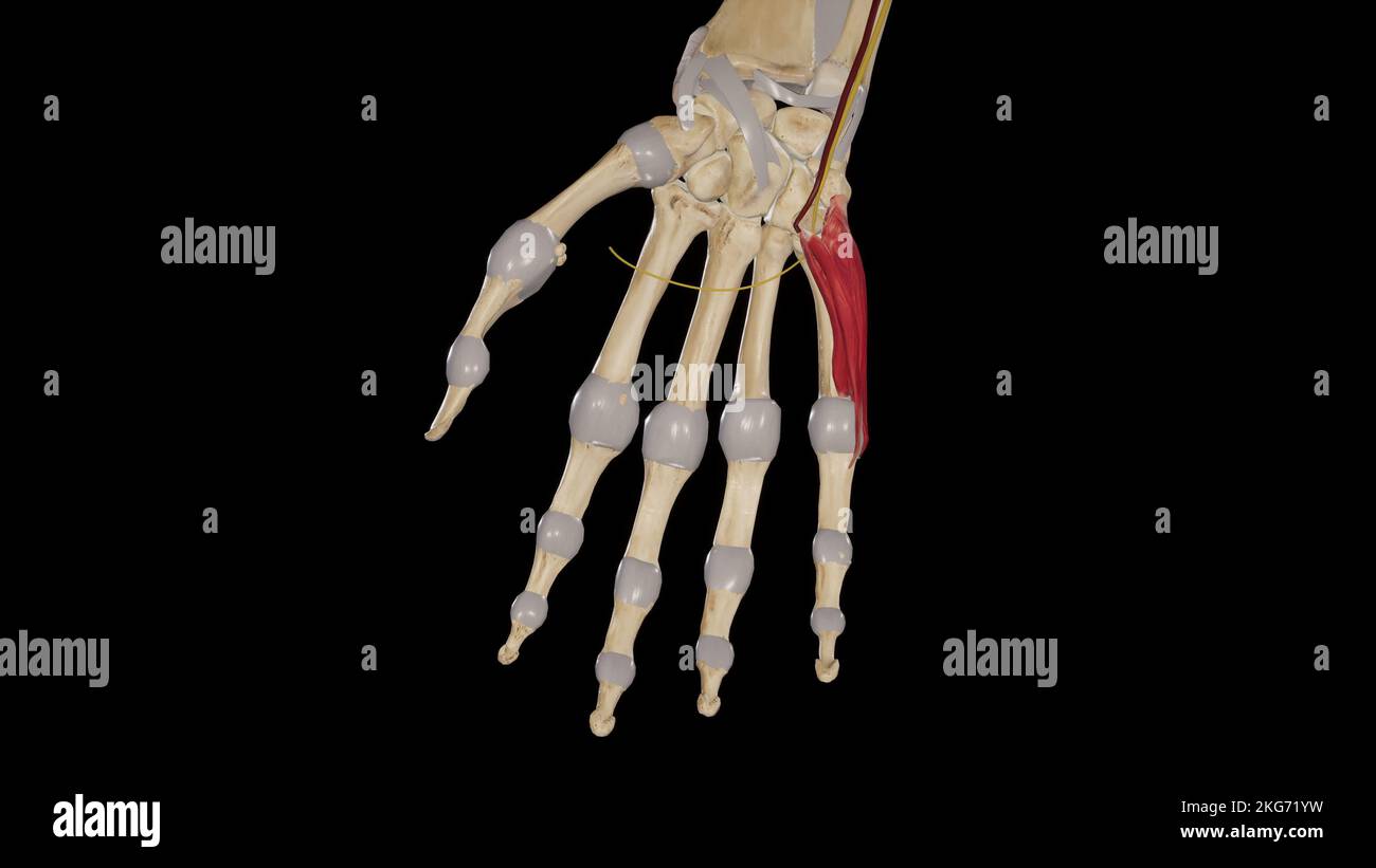 Hypothenar Muscles Stock Photohttps://www.alamy.com/image-license-details/?v=1https://www.alamy.com/hypothenar-muscles-image491880029.html
RF2KG71YW–Hypothenar Muscles
 3d rendered medically accurate illustration of the Abductor Digiti Minimi Stock Photohttps://www.alamy.com/image-license-details/?v=1https://www.alamy.com/3d-rendered-medically-accurate-illustration-of-the-abductor-digiti-minimi-image258154330.html
RFTYYXDE–3d rendered medically accurate illustration of the Abductor Digiti Minimi
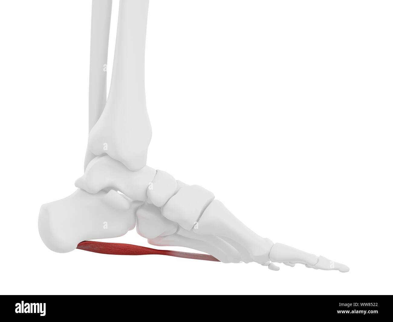 Abductor digiti minimi muscle, illustration Stock Photohttps://www.alamy.com/image-license-details/?v=1https://www.alamy.com/abductor-digiti-minimi-muscle-illustration-image273701514.html
RFWW8522–Abductor digiti minimi muscle, illustration
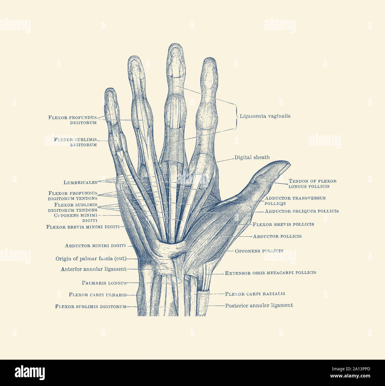 Diagram depicting the bones, ligaments and muscles throughout the hand and fingers. Stock Photohttps://www.alamy.com/image-license-details/?v=1https://www.alamy.com/diagram-depicting-the-bones-ligaments-and-muscles-throughout-the-hand-and-fingers-image327695381.html
RF2A13PPD–Diagram depicting the bones, ligaments and muscles throughout the hand and fingers.
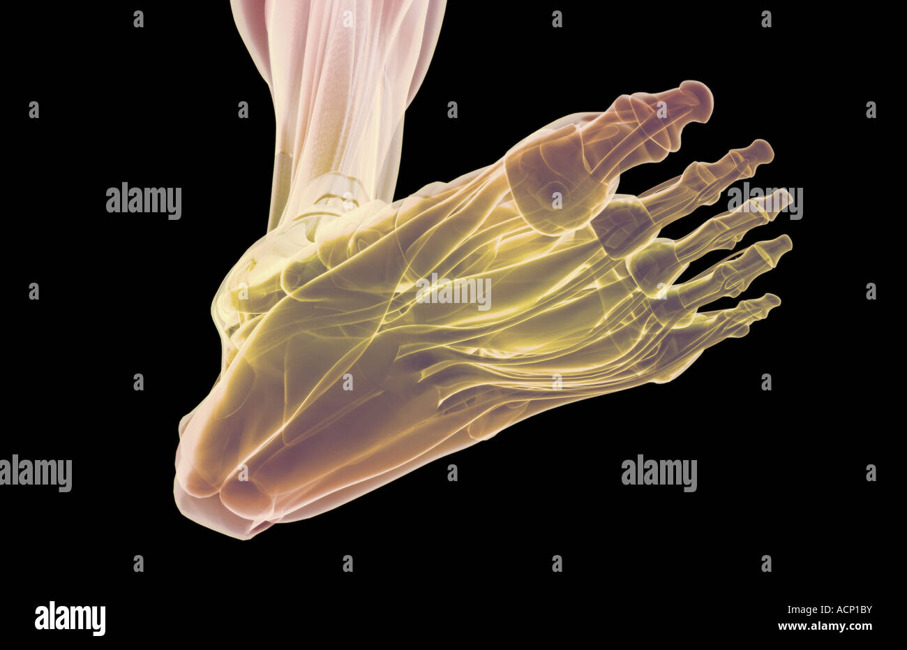 The muscles of the foot Stock Photohttps://www.alamy.com/image-license-details/?v=1https://www.alamy.com/stock-photo-the-muscles-of-the-foot-13203038.html
RFACP1BY–The muscles of the foot
 Abductor digiti minimi muscle of foot. Stock Vectorhttps://www.alamy.com/image-license-details/?v=1https://www.alamy.com/abductor-digiti-minimi-muscle-of-foot-image367673015.html
RF2CA4XK3–Abductor digiti minimi muscle of foot.
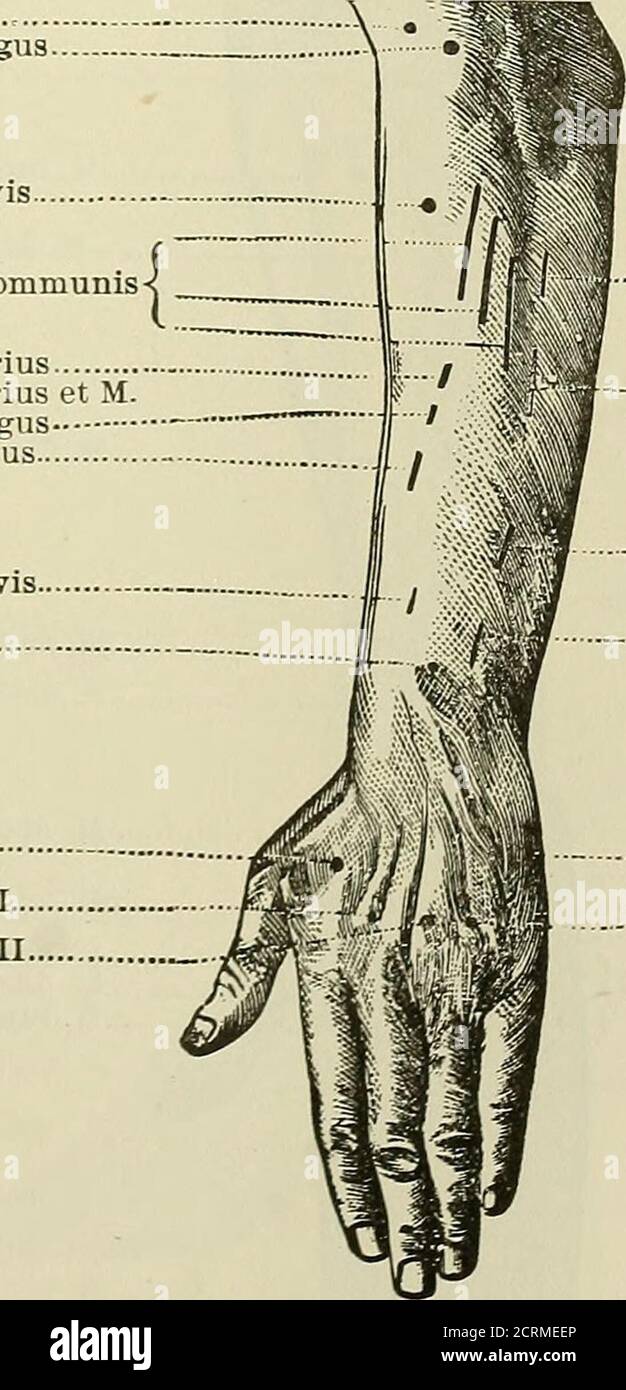 . Röntgen rays and electro-therapeutics : with chapters on radium and phototherapy . M. supinator longus M. radialis externus longus M. radialis externus brevis M. extensor digitorum communis-{ M. extensor indicis proprius M. extensor indicis proprius et M abductor pollicis longusM. abductor pollicis longus M. extensor pollicis brevisM. flexor pollicis longus M. interosseus dorsalis IM. interosseus dorsalis IIM. interosseus dorsalis III. .M. ulnaris e::^ternus. M. extensor digiti minimi pro-prius. M. extensor indicis proprius.M. extensor pollicis longus. M. abductor digiti minimi.M- interosseu Stock Photohttps://www.alamy.com/image-license-details/?v=1https://www.alamy.com/rntgen-rays-and-electro-therapeutics-with-chapters-on-radium-and-phototherapy-m-supinator-longus-m-radialis-externus-longus-m-radialis-externus-brevis-m-extensor-digitorum-communis-m-extensor-indicis-proprius-m-extensor-indicis-proprius-et-m-abductor-pollicis-longusm-abductor-pollicis-longus-m-extensor-pollicis-brevism-flexor-pollicis-longus-m-interosseus-dorsalis-im-interosseus-dorsalis-iim-interosseus-dorsalis-iii-m-ulnaris-eternus-m-extensor-digiti-minimi-pro-prius-m-extensor-indicis-propriusm-extensor-pollicis-longus-m-abductor-digiti-minimim-interosseu-image376005246.html
RM2CRMEEP–. Röntgen rays and electro-therapeutics : with chapters on radium and phototherapy . M. supinator longus M. radialis externus longus M. radialis externus brevis M. extensor digitorum communis-{ M. extensor indicis proprius M. extensor indicis proprius et M abductor pollicis longusM. abductor pollicis longus M. extensor pollicis brevisM. flexor pollicis longus M. interosseus dorsalis IM. interosseus dorsalis IIM. interosseus dorsalis III. .M. ulnaris e::^ternus. M. extensor digiti minimi pro-prius. M. extensor indicis proprius.M. extensor pollicis longus. M. abductor digiti minimi.M- interosseu
 . Elementary anatomy and physiology : for colleges, academies, and other schools . A View of the muscles on the Palm of the Hand. 1, Annular Ligament. 2, 2, Origin and Insertion of the Abductor Pol- licis. 3, Opponens Pollicis. 4, 5, Two Bellies of the Flexor Brevis Pollicis. 6, Adductor Pollicis. 7, 7, Lumbricales aris- ing from Tendons of the Flexor Profundus Dtgitorum. S, Shows how the Tendon of tho Flexor Profundus passes through the Flexor Sublimis. 9, Tendon of the Flexor Longus Pollicis. 10, Abductor Minimi Bigiti. 11, Flexor Parvus Minimi Digiti. 12, Pisiform Bone. 13, First Dorsal Int Stock Photohttps://www.alamy.com/image-license-details/?v=1https://www.alamy.com/elementary-anatomy-and-physiology-for-colleges-academies-and-other-schools-a-view-of-the-muscles-on-the-palm-of-the-hand-1-annular-ligament-2-2-origin-and-insertion-of-the-abductor-pol-licis-3-opponens-pollicis-4-5-two-bellies-of-the-flexor-brevis-pollicis-6-adductor-pollicis-7-7-lumbricales-aris-ing-from-tendons-of-the-flexor-profundus-dtgitorum-s-shows-how-the-tendon-of-tho-flexor-profundus-passes-through-the-flexor-sublimis-9-tendon-of-the-flexor-longus-pollicis-10-abductor-minimi-bigiti-11-flexor-parvus-minimi-digiti-12-pisiform-bone-13-first-dorsal-int-image178409721.html
RMMA77BN–. Elementary anatomy and physiology : for colleges, academies, and other schools . A View of the muscles on the Palm of the Hand. 1, Annular Ligament. 2, 2, Origin and Insertion of the Abductor Pol- licis. 3, Opponens Pollicis. 4, 5, Two Bellies of the Flexor Brevis Pollicis. 6, Adductor Pollicis. 7, 7, Lumbricales aris- ing from Tendons of the Flexor Profundus Dtgitorum. S, Shows how the Tendon of tho Flexor Profundus passes through the Flexor Sublimis. 9, Tendon of the Flexor Longus Pollicis. 10, Abductor Minimi Bigiti. 11, Flexor Parvus Minimi Digiti. 12, Pisiform Bone. 13, First Dorsal Int
 3D Illustration, Muscle is a soft tissue, Muscle cells contain proteins , producing a contraction that changes both the length and the shape Stock Photohttps://www.alamy.com/image-license-details/?v=1https://www.alamy.com/3d-illustration-muscle-is-a-soft-tissue-muscle-cells-contain-proteins-producing-a-contraction-that-changes-both-the-length-and-the-shape-image395479783.html
RF2DYBJDY–3D Illustration, Muscle is a soft tissue, Muscle cells contain proteins , producing a contraction that changes both the length and the shape
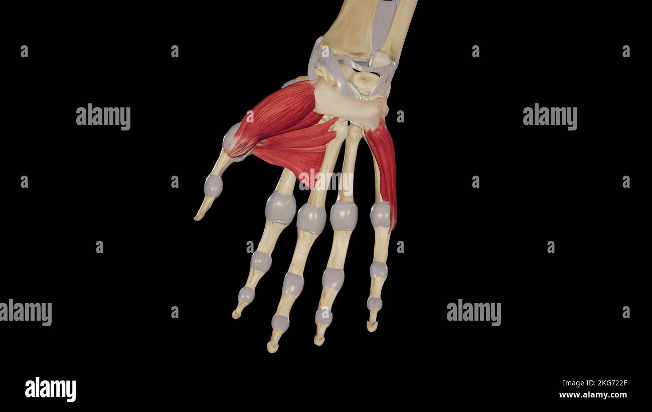 Thenar and Hypothenar Muscles Stock Photohttps://www.alamy.com/image-license-details/?v=1https://www.alamy.com/thenar-and-hypothenar-muscles-image491880103.html
RF2KG722F–Thenar and Hypothenar Muscles
 3d rendered medically accurate illustration of the Abductor Digiti Minimi Stock Photohttps://www.alamy.com/image-license-details/?v=1https://www.alamy.com/3d-rendered-medically-accurate-illustration-of-the-abductor-digiti-minimi-image257880591.html
RFTYFD93–3d rendered medically accurate illustration of the Abductor Digiti Minimi
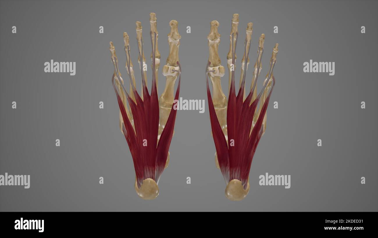 Muscles of Sole of Foot First Layer Stock Photohttps://www.alamy.com/image-license-details/?v=1https://www.alamy.com/muscles-of-sole-of-foot-first-layer-image490198437.html
RF2KDED31–Muscles of Sole of Foot First Layer
 Abductor digiti minimi muscle, illustration Stock Photohttps://www.alamy.com/image-license-details/?v=1https://www.alamy.com/abductor-digiti-minimi-muscle-illustration-image273701515.html
RFWW8523–Abductor digiti minimi muscle, illustration
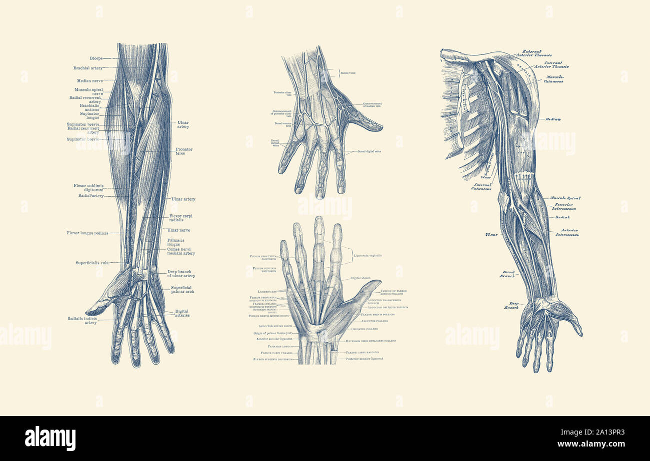 Multi-view diagram showcasing ligaments, muscles and veins throughout hand, arm and fingers. Stock Photohttps://www.alamy.com/image-license-details/?v=1https://www.alamy.com/multi-view-diagram-showcasing-ligaments-muscles-and-veins-throughout-hand-arm-and-fingers-image327695399.html
RF2A13PR3–Multi-view diagram showcasing ligaments, muscles and veins throughout hand, arm and fingers.
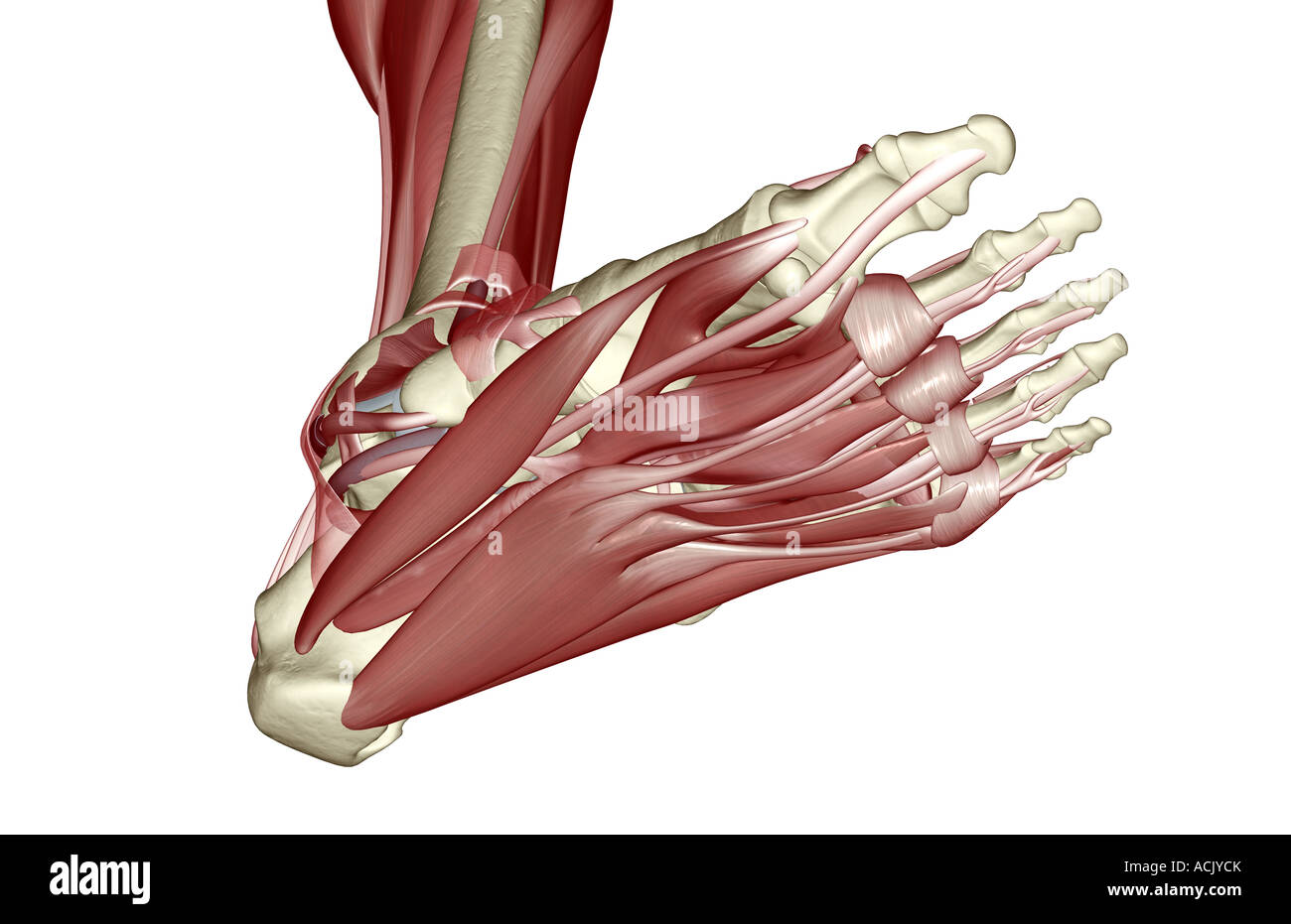 The muscles of the foot Stock Photohttps://www.alamy.com/image-license-details/?v=1https://www.alamy.com/stock-photo-the-muscles-of-the-foot-13174146.html
RFACJYCK–The muscles of the foot
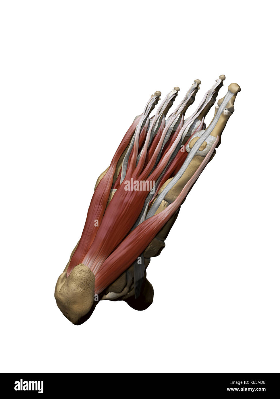 3D model of the foot depicting the plantar superficial muscles and bone structures. Stock Photohttps://www.alamy.com/image-license-details/?v=1https://www.alamy.com/stock-image-3d-model-of-the-foot-depicting-the-plantar-superficial-muscles-and-163616471.html
RFKE5ADB–3D model of the foot depicting the plantar superficial muscles and bone structures.
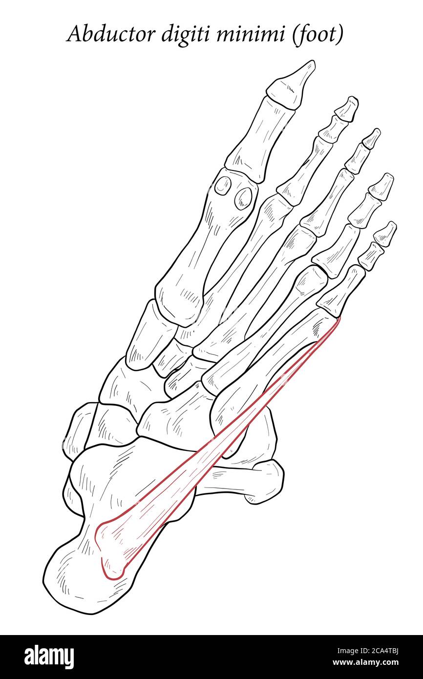 Abductor digiti minimi muscle of foot. Stock Vectorhttps://www.alamy.com/image-license-details/?v=1https://www.alamy.com/abductor-digiti-minimi-muscle-of-foot-image367671238.html
RF2CA4TBJ–Abductor digiti minimi muscle of foot.
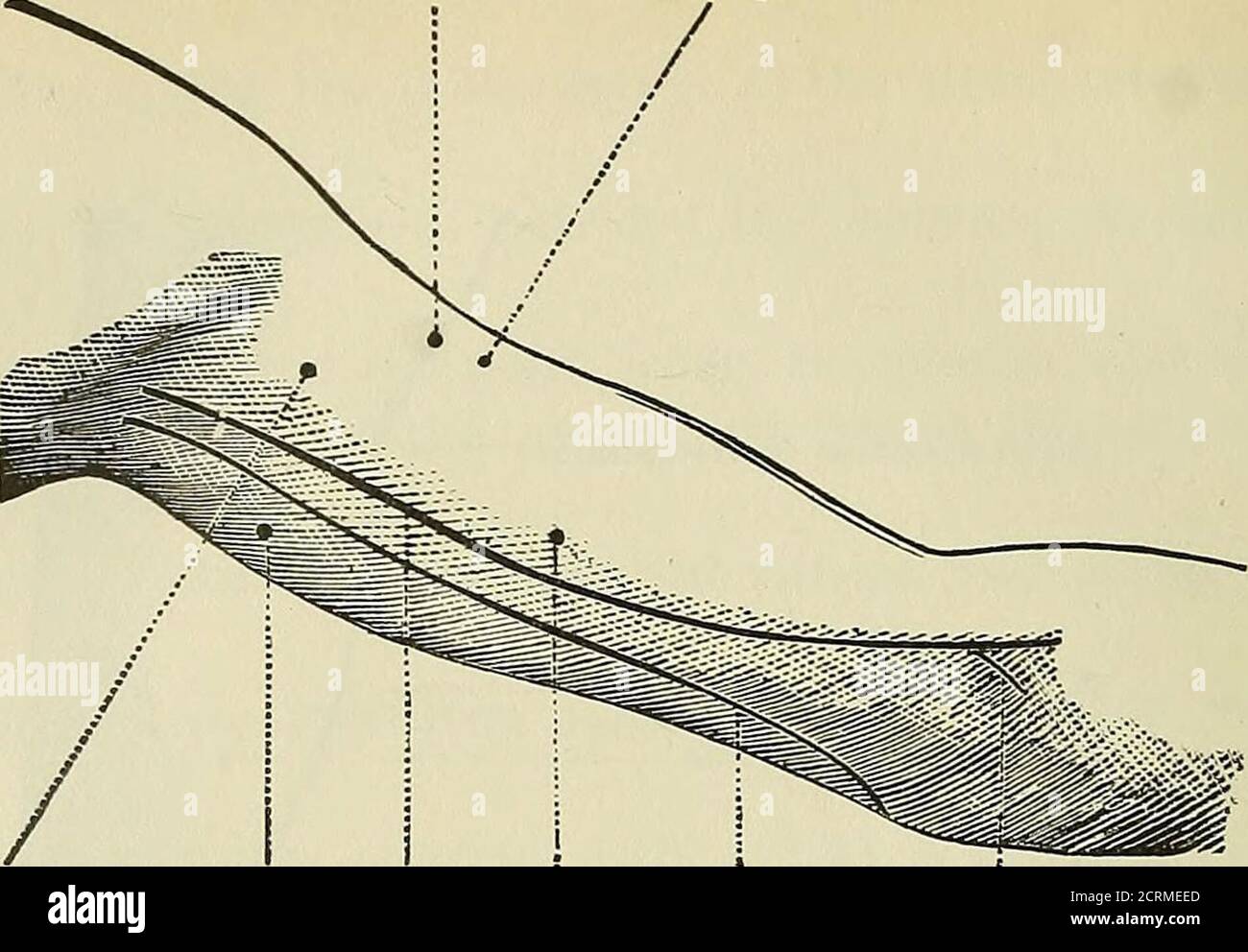 . Röntgen rays and electro-therapeutics : with chapters on radium and phototherapy . .M. ulnaris e::^ternus. M. extensor digiti minimi pro-prius. M. extensor indicis proprius.M. extensor pollicis longus. M. abductor digiti minimi.M- interosseus dorsalis IV. Fig. 37.—Motor points of the forearm and hand. 60 ELECTRO-THERAPEUTICS. N. Musculo-cuianeus. M. biceps.. N.musculo-Caput in- N. Media- ; N. ulnaris. Rami N. medianicutaneus. ternus M. nus. M. brachialis pro M. pronatore tricipitis. ■internus. radii terete. Fig. 38.—Motor points of the arm (front view). Rami Nervi mediani pro M. pronatore ra Stock Photohttps://www.alamy.com/image-license-details/?v=1https://www.alamy.com/rntgen-rays-and-electro-therapeutics-with-chapters-on-radium-and-phototherapy-m-ulnaris-eternus-m-extensor-digiti-minimi-pro-prius-m-extensor-indicis-propriusm-extensor-pollicis-longus-m-abductor-digiti-minimim-interosseus-dorsalis-iv-fig-37motor-points-of-the-forearm-and-hand-60-electro-therapeutics-n-musculo-cuianeus-m-biceps-nmusculo-caput-in-n-media-n-ulnaris-rami-n-medianicutaneus-ternus-m-nus-m-brachialis-pro-m-pronatore-tricipitis-internus-radii-terete-fig-38motor-points-of-the-arm-front-view-rami-nervi-mediani-pro-m-pronatore-ra-image376005237.html
RM2CRMEED–. Röntgen rays and electro-therapeutics : with chapters on radium and phototherapy . .M. ulnaris e::^ternus. M. extensor digiti minimi pro-prius. M. extensor indicis proprius.M. extensor pollicis longus. M. abductor digiti minimi.M- interosseus dorsalis IV. Fig. 37.—Motor points of the forearm and hand. 60 ELECTRO-THERAPEUTICS. N. Musculo-cuianeus. M. biceps.. N.musculo-Caput in- N. Media- ; N. ulnaris. Rami N. medianicutaneus. ternus M. nus. M. brachialis pro M. pronatore tricipitis. ■internus. radii terete. Fig. 38.—Motor points of the arm (front view). Rami Nervi mediani pro M. pronatore ra
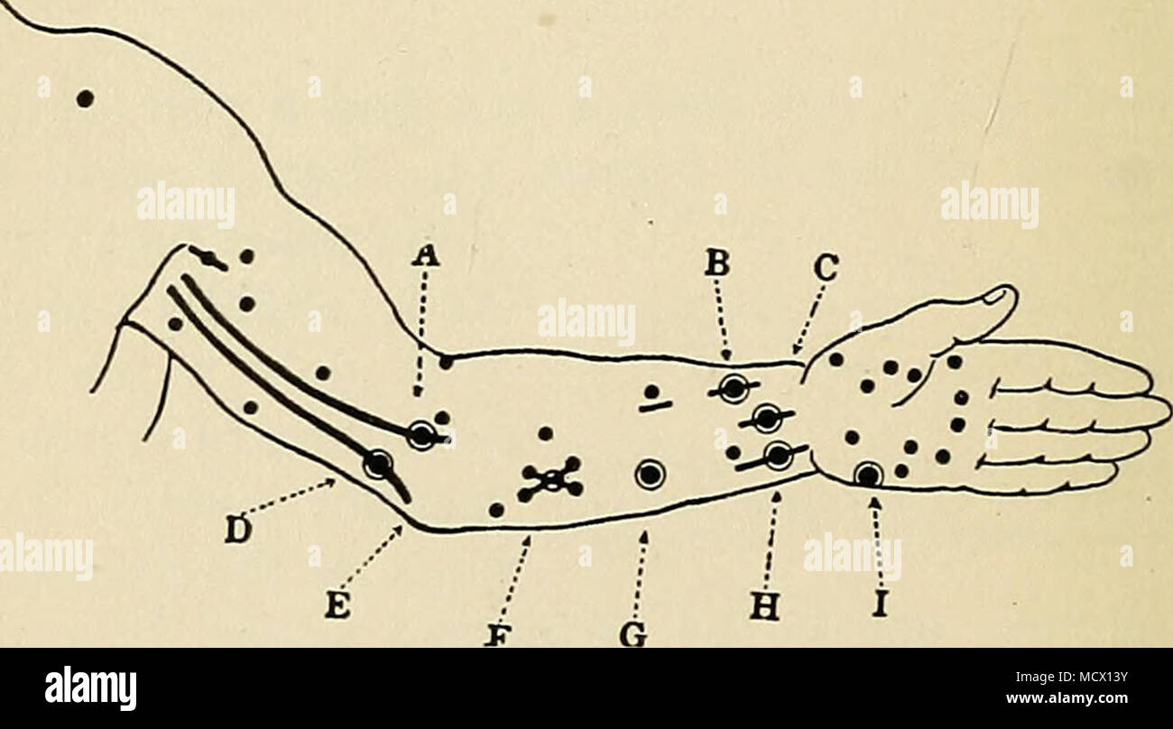 . Fig. 15. Diagram of location of motor points on flexor side of arm. (After Erb.) A, median nerve in upper arm; B, flexor longus policis; C, median nerve at wrist; D, ulnar nerve in upper arm; E, ulnar nerve in groove between the internal condyle of the humerus and the olecranon process; F, flexor profundus digitorum; G, flexor sublimis digitorum; H, ulnar nerve at wrist; I, abductor minimi digiti. more efficient pole of the secondary circuit is the one that is the kathode when the primary circuit is broken. Make a note of this point, stating whether the more efficient pole is the one to whic Stock Photohttps://www.alamy.com/image-license-details/?v=1https://www.alamy.com/fig-15-diagram-of-location-of-motor-points-on-flexor-side-of-arm-after-erb-a-median-nerve-in-upper-arm-b-flexor-longus-policis-c-median-nerve-at-wrist-d-ulnar-nerve-in-upper-arm-e-ulnar-nerve-in-groove-between-the-internal-condyle-of-the-humerus-and-the-olecranon-process-f-flexor-profundus-digitorum-g-flexor-sublimis-digitorum-h-ulnar-nerve-at-wrist-i-abductor-minimi-digiti-more-efficient-pole-of-the-secondary-circuit-is-the-one-that-is-the-kathode-when-the-primary-circuit-is-broken-make-a-note-of-this-point-stating-whether-the-more-efficient-pole-is-the-one-to-whic-image180051199.html
RMMCX13Y–. Fig. 15. Diagram of location of motor points on flexor side of arm. (After Erb.) A, median nerve in upper arm; B, flexor longus policis; C, median nerve at wrist; D, ulnar nerve in upper arm; E, ulnar nerve in groove between the internal condyle of the humerus and the olecranon process; F, flexor profundus digitorum; G, flexor sublimis digitorum; H, ulnar nerve at wrist; I, abductor minimi digiti. more efficient pole of the secondary circuit is the one that is the kathode when the primary circuit is broken. Make a note of this point, stating whether the more efficient pole is the one to whic
 3D Illustration, Muscle is a soft tissue, Muscle cells contain proteins , producing a contraction that changes both the length and the shape Stock Photohttps://www.alamy.com/image-license-details/?v=1https://www.alamy.com/3d-illustration-muscle-is-a-soft-tissue-muscle-cells-contain-proteins-producing-a-contraction-that-changes-both-the-length-and-the-shape-image395479782.html
RF2DYBJDX–3D Illustration, Muscle is a soft tissue, Muscle cells contain proteins , producing a contraction that changes both the length and the shape
 3d rendered medically accurate illustration of the Abductor Digiti Minimi Stock Photohttps://www.alamy.com/image-license-details/?v=1https://www.alamy.com/3d-rendered-medically-accurate-illustration-of-the-abductor-digiti-minimi-image257880624.html
RFTYFDA8–3d rendered medically accurate illustration of the Abductor Digiti Minimi
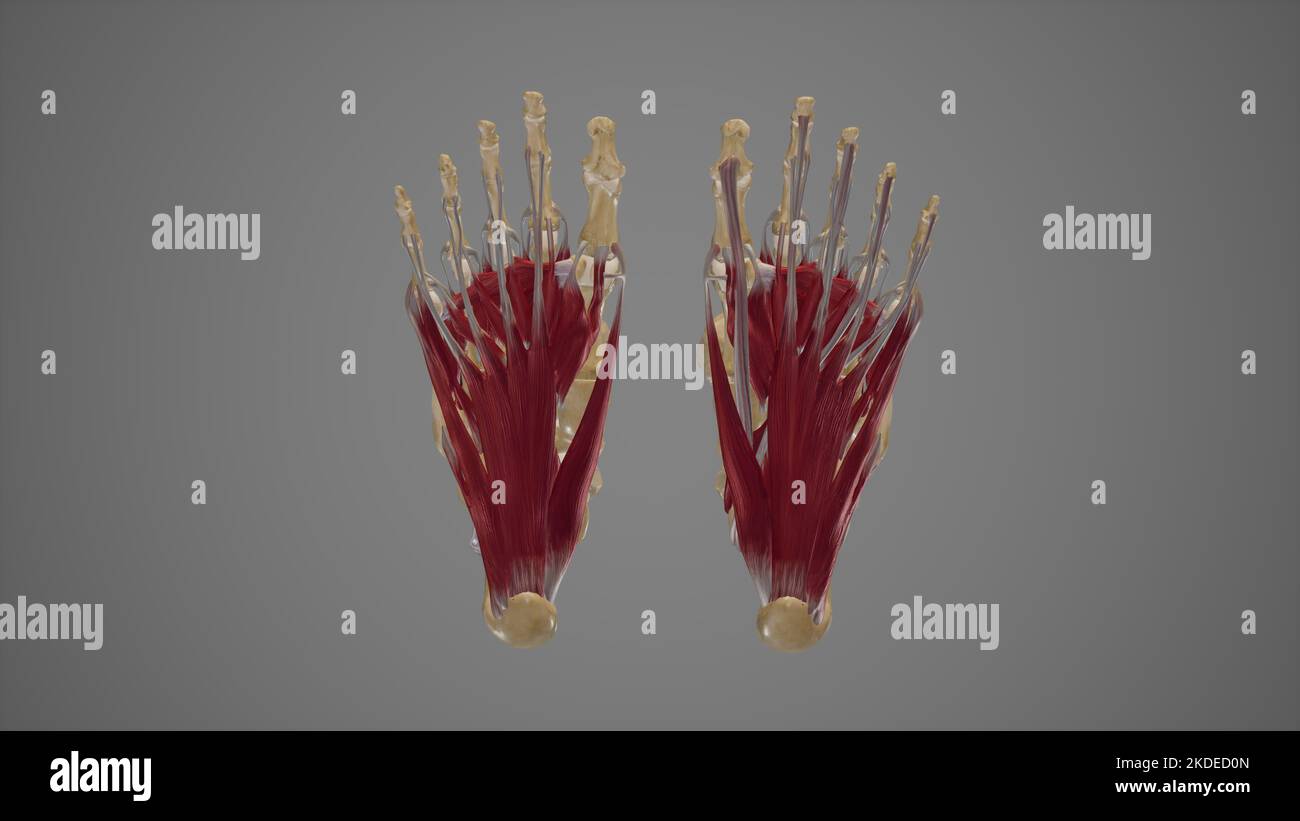 Intrinsic Muscles of Foot Stock Photohttps://www.alamy.com/image-license-details/?v=1https://www.alamy.com/intrinsic-muscles-of-foot-image490198373.html
RF2KDED0N–Intrinsic Muscles of Foot
 Abductor digiti minimi muscle, illustration Stock Photohttps://www.alamy.com/image-license-details/?v=1https://www.alamy.com/abductor-digiti-minimi-muscle-illustration-image273701535.html
RFWW852R–Abductor digiti minimi muscle, illustration
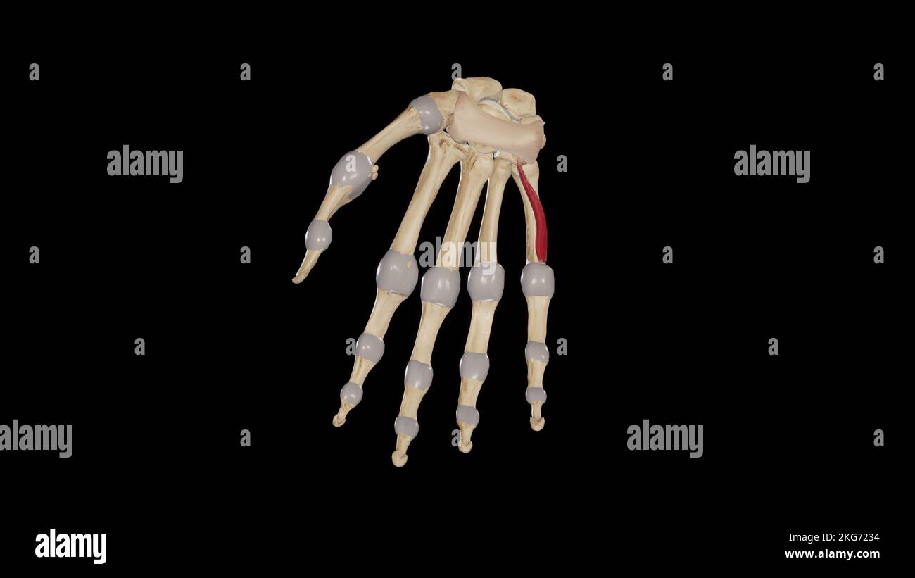 Opponens Digiti Minimi Stock Photohttps://www.alamy.com/image-license-details/?v=1https://www.alamy.com/opponens-digiti-minimi-image491880120.html
RF2KG7234–Opponens Digiti Minimi
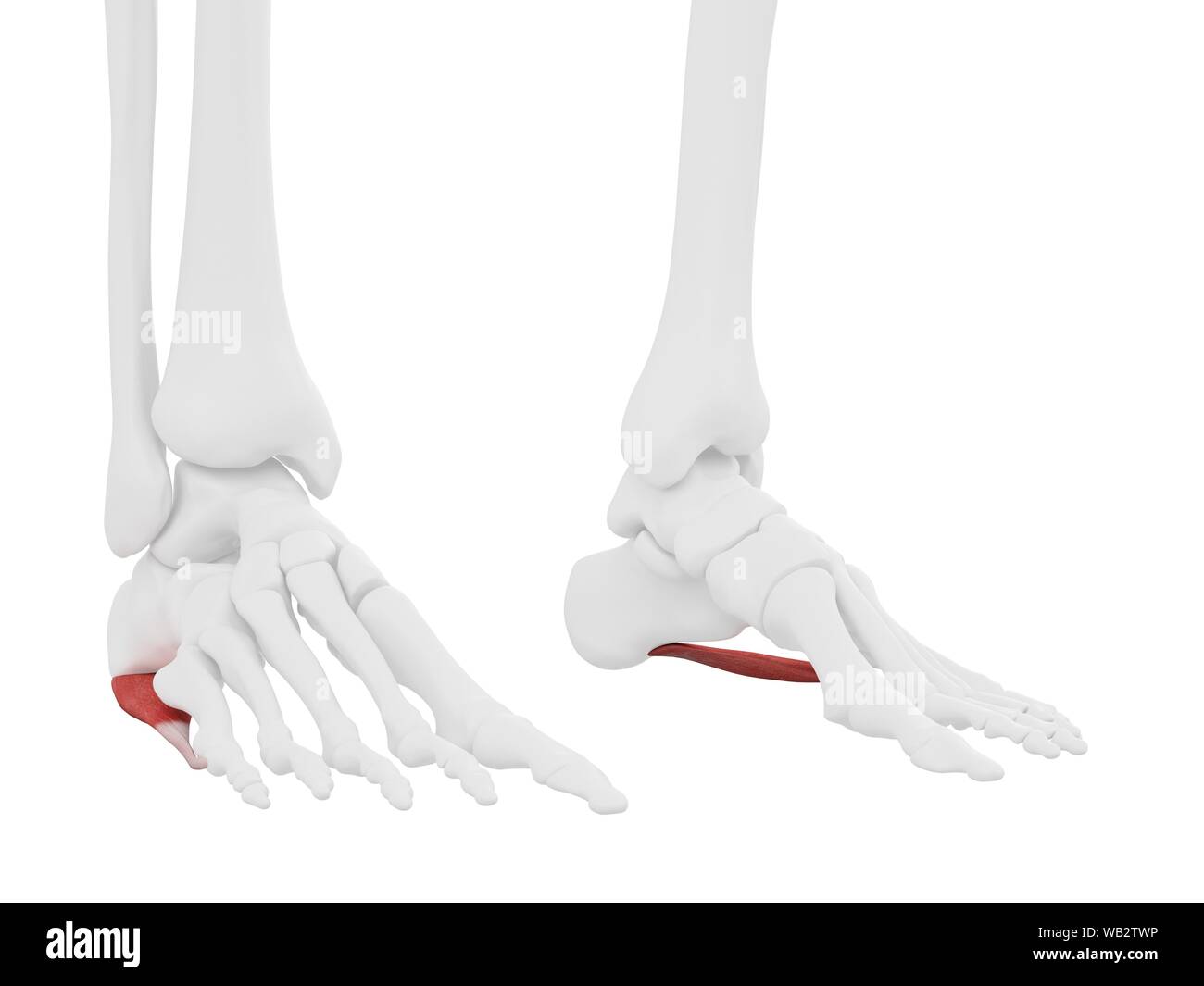 Abductor digiti minimi muscle, computer illustration. Stock Photohttps://www.alamy.com/image-license-details/?v=1https://www.alamy.com/abductor-digiti-minimi-muscle-computer-illustration-image264980178.html
RFWB2TWP–Abductor digiti minimi muscle, computer illustration.
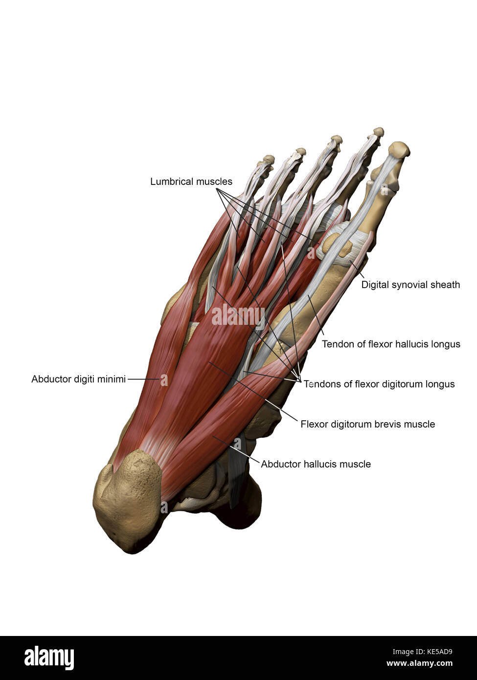 3D model of the foot depicting the plantar superficial muscles and bone structures. Stock Photohttps://www.alamy.com/image-license-details/?v=1https://www.alamy.com/stock-image-3d-model-of-the-foot-depicting-the-plantar-superficial-muscles-and-163616469.html
RFKE5AD9–3D model of the foot depicting the plantar superficial muscles and bone structures.
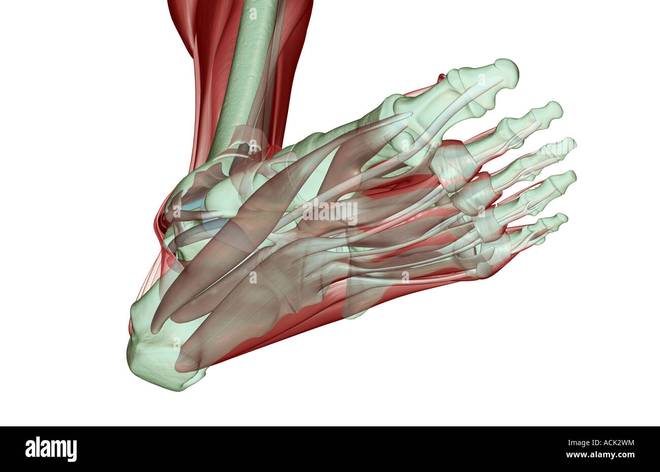 The musculoskeleton of the foot Stock Photohttps://www.alamy.com/image-license-details/?v=1https://www.alamy.com/stock-photo-the-musculoskeleton-of-the-foot-13175311.html
RFACK2WM–The musculoskeleton of the foot
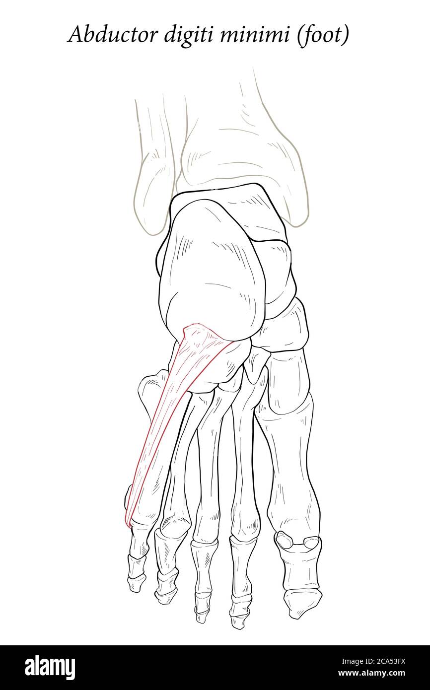 Abductor digiti minimi muscle of foot. Stock Vectorhttps://www.alamy.com/image-license-details/?v=1https://www.alamy.com/abductor-digiti-minimi-muscle-of-foot-image367676846.html
RF2CA53FX–Abductor digiti minimi muscle of foot.
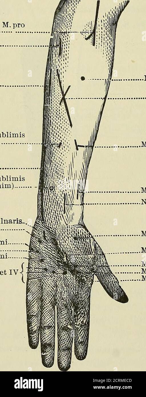 . Röntgen rays and electro-therapeutics : with chapters on radium and phototherapy . N.musculo-Caput in- N. Media- ; N. ulnaris. Rami N. medianicutaneus. ternus M. nus. M. brachialis pro M. pronatore tricipitis. ■internus. radii terete. Fig. 38.—Motor points of the arm (front view). Rami Nervi mediani pro M. pronatore radii terete^ M. palmaris longus.. Jll. ulnaris internus. M. flexor digitorum sublimis(digitt. II et III.) N. ulnaris M. flexor digitorum sublimis(digitt. indicis et minim) Rami volar, prof. Nervi ulnaris.M. palmaris brevis M. abductor digiti minimi M. flexor digiti minimi M. opp Stock Photohttps://www.alamy.com/image-license-details/?v=1https://www.alamy.com/rntgen-rays-and-electro-therapeutics-with-chapters-on-radium-and-phototherapy-nmusculo-caput-in-n-media-n-ulnaris-rami-n-medianicutaneus-ternus-m-nus-m-brachialis-pro-m-pronatore-tricipitis-internus-radii-terete-fig-38motor-points-of-the-arm-front-view-rami-nervi-mediani-pro-m-pronatore-radii-terete-m-palmaris-longus-jll-ulnaris-internus-m-flexor-digitorum-sublimisdigitt-ii-et-iii-n-ulnaris-m-flexor-digitorum-sublimisdigitt-indicis-et-minim-rami-volar-prof-nervi-ulnarism-palmaris-brevis-m-abductor-digiti-minimi-m-flexor-digiti-minimi-m-opp-image376005181.html
RM2CRMECD–. Röntgen rays and electro-therapeutics : with chapters on radium and phototherapy . N.musculo-Caput in- N. Media- ; N. ulnaris. Rami N. medianicutaneus. ternus M. nus. M. brachialis pro M. pronatore tricipitis. ■internus. radii terete. Fig. 38.—Motor points of the arm (front view). Rami Nervi mediani pro M. pronatore radii terete^ M. palmaris longus.. Jll. ulnaris internus. M. flexor digitorum sublimis(digitt. II et III.) N. ulnaris M. flexor digitorum sublimis(digitt. indicis et minim) Rami volar, prof. Nervi ulnaris.M. palmaris brevis M. abductor digiti minimi M. flexor digiti minimi M. opp
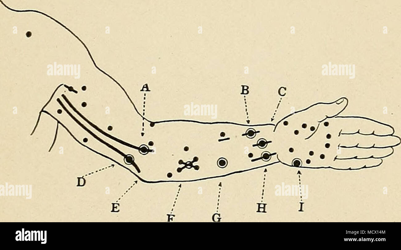 . Fig. 17. Diagram of location of motor points on flexor side of arm. (After Frb.) A, median nerve in upper arm; B, flexor longus policis; C, median nerve at wrist; D, ulnar nerve in upper arm ; E, tilnar nerve in groove between the internal condyle of the humerus and the olecranon process; F, flexor profundus digitorum; G, fl.exor sublimis digitorum; H, tilnar nerve at wrist; I, abductor minimi digiti. gauze pad between to prevent the metal from touching the skin. Let your companion press the active electrode firmly upon the skin at the point to be stimulated, and make and break the circuit, Stock Photohttps://www.alamy.com/image-license-details/?v=1https://www.alamy.com/fig-17-diagram-of-location-of-motor-points-on-flexor-side-of-arm-after-frb-a-median-nerve-in-upper-arm-b-flexor-longus-policis-c-median-nerve-at-wrist-d-ulnar-nerve-in-upper-arm-e-tilnar-nerve-in-groove-between-the-internal-condyle-of-the-humerus-and-the-olecranon-process-f-flexor-profundus-digitorum-g-flexor-sublimis-digitorum-h-tilnar-nerve-at-wrist-i-abductor-minimi-digiti-gauze-pad-between-to-prevent-the-metal-from-touching-the-skin-let-your-companion-press-the-active-electrode-firmly-upon-the-skin-at-the-point-to-be-stimulated-and-make-and-break-the-circuit-image180051220.html
RMMCX14M–. Fig. 17. Diagram of location of motor points on flexor side of arm. (After Frb.) A, median nerve in upper arm; B, flexor longus policis; C, median nerve at wrist; D, ulnar nerve in upper arm ; E, tilnar nerve in groove between the internal condyle of the humerus and the olecranon process; F, flexor profundus digitorum; G, fl.exor sublimis digitorum; H, tilnar nerve at wrist; I, abductor minimi digiti. gauze pad between to prevent the metal from touching the skin. Let your companion press the active electrode firmly upon the skin at the point to be stimulated, and make and break the circuit,
 3D Illustration, Muscle is a soft tissue, Muscle cells contain proteins , producing a contraction that changes both the length and the shape of the ce Stock Photohttps://www.alamy.com/image-license-details/?v=1https://www.alamy.com/3d-illustration-muscle-is-a-soft-tissue-muscle-cells-contain-proteins-producing-a-contraction-that-changes-both-the-length-and-the-shape-of-the-ce-image395466418.html
RF2DYB1CJ–3D Illustration, Muscle is a soft tissue, Muscle cells contain proteins , producing a contraction that changes both the length and the shape of the ce
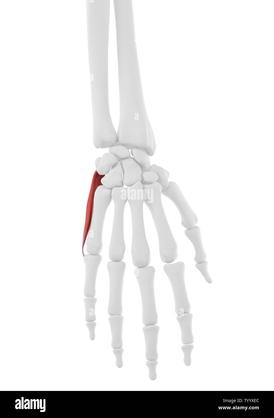 3d rendered medically accurate illustration of the Abductor Digiti Minimi Stock Photohttps://www.alamy.com/image-license-details/?v=1https://www.alamy.com/3d-rendered-medically-accurate-illustration-of-the-abductor-digiti-minimi-image258154356.html
RFTYYXEC–3d rendered medically accurate illustration of the Abductor Digiti Minimi
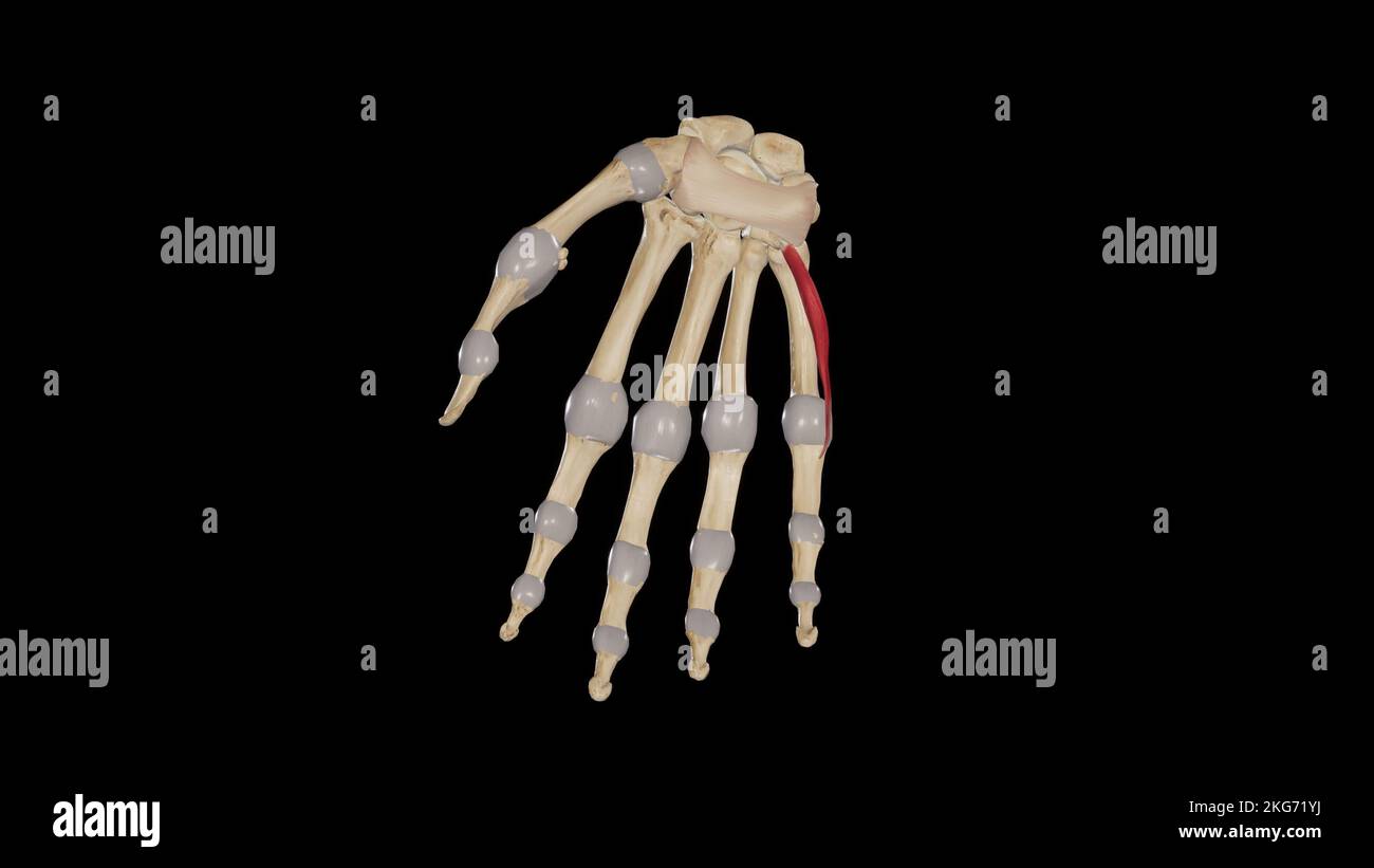 Flexor Digiti Minimi Brevis Stock Photohttps://www.alamy.com/image-license-details/?v=1https://www.alamy.com/flexor-digiti-minimi-brevis-image491880022.html
RF2KG71YJ–Flexor Digiti Minimi Brevis
 Abductor digiti minimi muscle, computer illustration. Stock Photohttps://www.alamy.com/image-license-details/?v=1https://www.alamy.com/abductor-digiti-minimi-muscle-computer-illustration-image264980198.html
RFWB2TXE–Abductor digiti minimi muscle, computer illustration.
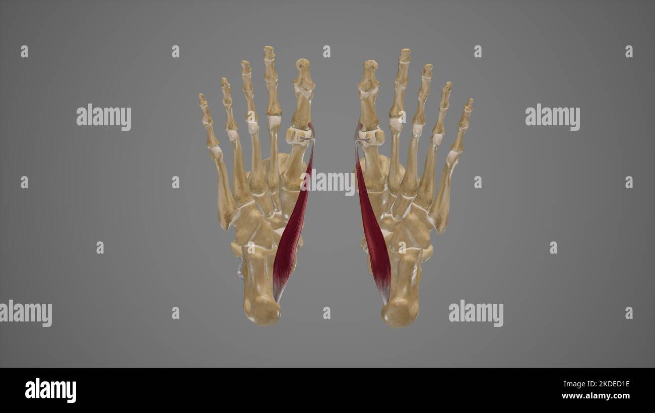 Medical Illustration of Abductor Hallucis Stock Photohttps://www.alamy.com/image-license-details/?v=1https://www.alamy.com/medical-illustration-of-abductor-hallucis-image490198394.html
RF2KDED1E–Medical Illustration of Abductor Hallucis
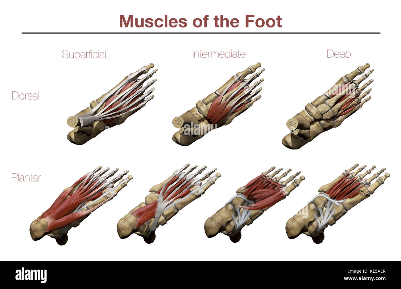 Muscles of the Foot Stock Photohttps://www.alamy.com/image-license-details/?v=1https://www.alamy.com/stock-image-muscles-of-the-foot-163616511.html
RFKE5AER–Muscles of the Foot
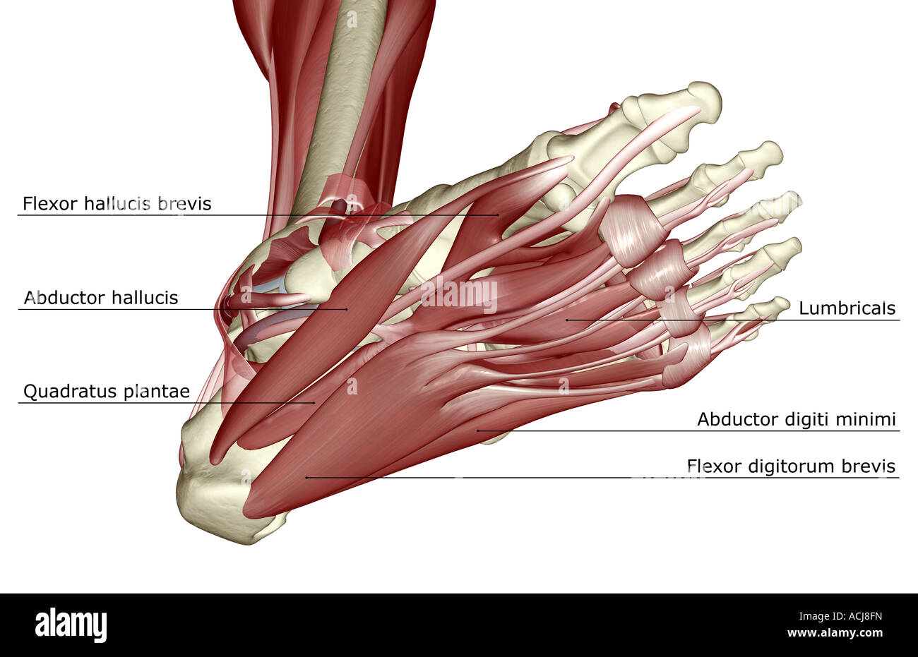 The muscles of the foot Stock Photohttps://www.alamy.com/image-license-details/?v=1https://www.alamy.com/stock-photo-the-muscles-of-the-foot-13167800.html
RFACJ8FN–The muscles of the foot
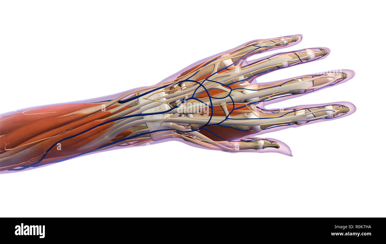 Anatomy of human hand, dorsal view. Stock Photohttps://www.alamy.com/image-license-details/?v=1https://www.alamy.com/anatomy-of-human-hand-dorsal-view-image224157846.html
RFR0K7HA–Anatomy of human hand, dorsal view.
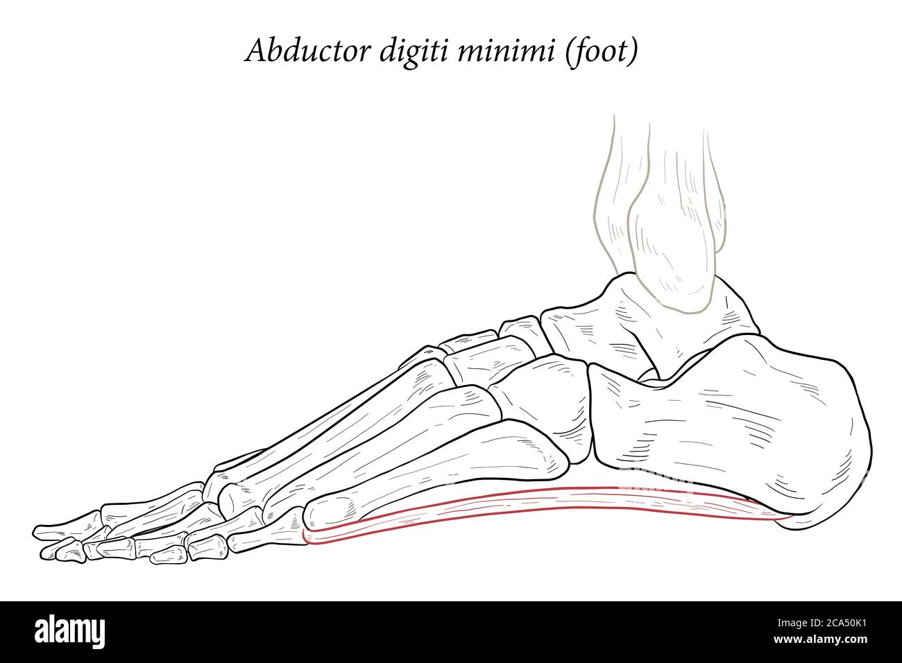 Abductor digiti minimi muscle of foot. Stock Vectorhttps://www.alamy.com/image-license-details/?v=1https://www.alamy.com/abductor-digiti-minimi-muscle-of-foot-image367674581.html
RF2CA50K1–Abductor digiti minimi muscle of foot.
 . The anatomy of the human body. Human anatomy; Anatomy. THE ABDUCTOR DIGITI MINIMI, ETC. 289 External Plantar Region. The Abductor Digiti Minimi. Dissection.—This is common to the abductor and the flexor brevis. The first is ex- posed by simply removing the external plantar fascia, and the second by removing or re- flecting down the first. ,. The abductor digiti mi)umi {v,fig. 13l) is of the same form, the same structure, and al- most the same size as the abductor pollicis, and extends from the os calcis to the first phalanx of the little toe. It arises by tendinous and fleshy fibres from the Stock Photohttps://www.alamy.com/image-license-details/?v=1https://www.alamy.com/the-anatomy-of-the-human-body-human-anatomy-anatomy-the-abductor-digiti-minimi-etc-289-external-plantar-region-the-abductor-digiti-minimi-dissectionthis-is-common-to-the-abductor-and-the-flexor-brevis-the-first-is-ex-posed-by-simply-removing-the-external-plantar-fascia-and-the-second-by-removing-or-re-flecting-down-the-first-the-abductor-digiti-miumi-vfig-13l-is-of-the-same-form-the-same-structure-and-al-most-the-same-size-as-the-abductor-pollicis-and-extends-from-the-os-calcis-to-the-first-phalanx-of-the-little-toe-it-arises-by-tendinous-and-fleshy-fibres-from-the-image236797482.html
RMRN71GX–. The anatomy of the human body. Human anatomy; Anatomy. THE ABDUCTOR DIGITI MINIMI, ETC. 289 External Plantar Region. The Abductor Digiti Minimi. Dissection.—This is common to the abductor and the flexor brevis. The first is ex- posed by simply removing the external plantar fascia, and the second by removing or re- flecting down the first. ,. The abductor digiti mi)umi {v,fig. 13l) is of the same form, the same structure, and al- most the same size as the abductor pollicis, and extends from the os calcis to the first phalanx of the little toe. It arises by tendinous and fleshy fibres from the
 3D Illustration, Muscle is a soft tissue, Muscle cells contain proteins , producing a contraction that changes both the length and the shape Stock Photohttps://www.alamy.com/image-license-details/?v=1https://www.alamy.com/3d-illustration-muscle-is-a-soft-tissue-muscle-cells-contain-proteins-producing-a-contraction-that-changes-both-the-length-and-the-shape-image395479774.html
RF2DYBJDJ–3D Illustration, Muscle is a soft tissue, Muscle cells contain proteins , producing a contraction that changes both the length and the shape
 3d rendered medically accurate illustration of the Abductor Digiti Minimi Stock Photohttps://www.alamy.com/image-license-details/?v=1https://www.alamy.com/3d-rendered-medically-accurate-illustration-of-the-abductor-digiti-minimi-image257880522.html
RFTYFD6J–3d rendered medically accurate illustration of the Abductor Digiti Minimi
 Abductor digiti minimi muscle, computer illustration. Stock Photohttps://www.alamy.com/image-license-details/?v=1https://www.alamy.com/abductor-digiti-minimi-muscle-computer-illustration-image264980186.html
RFWB2TX2–Abductor digiti minimi muscle, computer illustration.
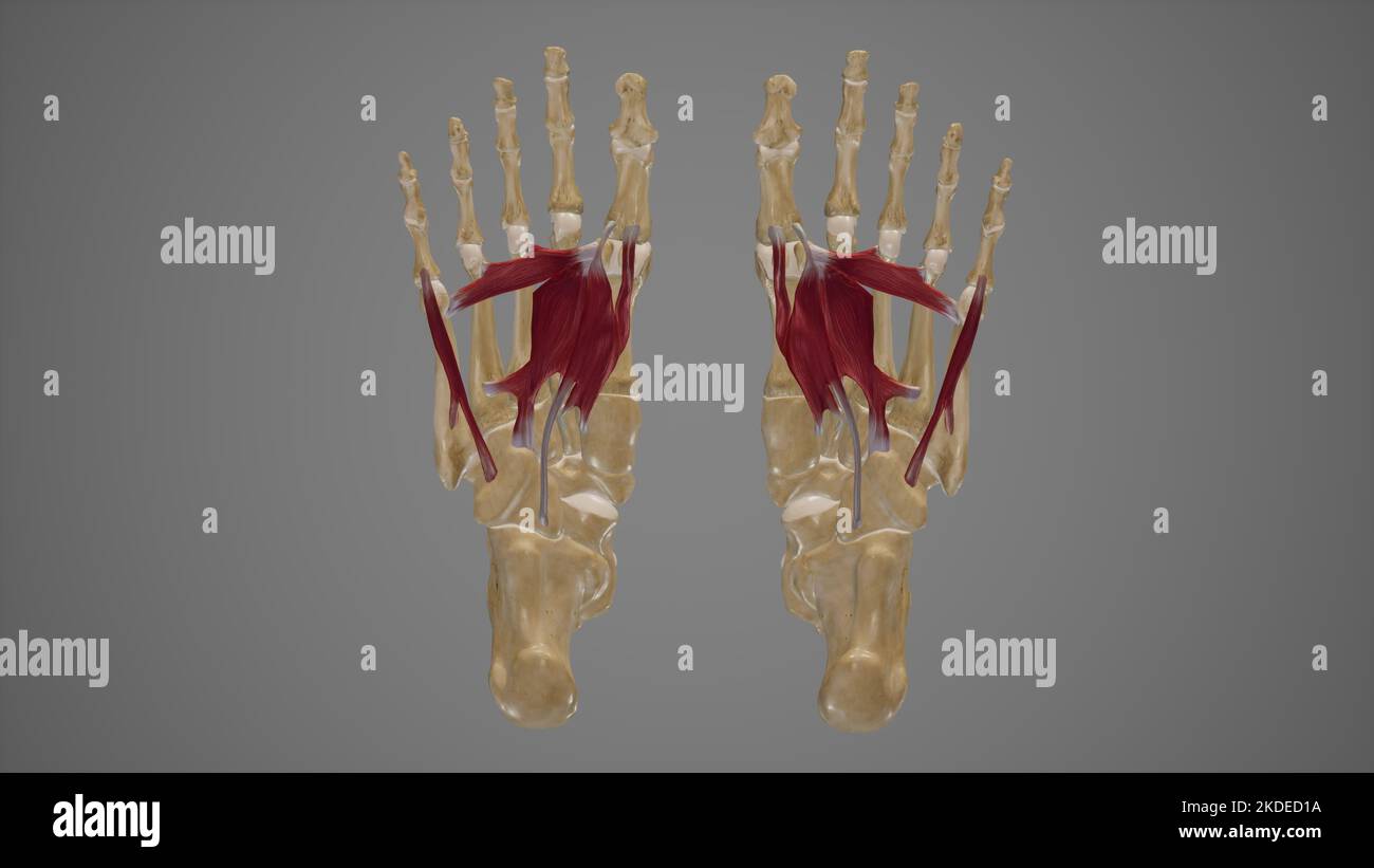 Muscles of Sole of Foot Third Layer Stock Photohttps://www.alamy.com/image-license-details/?v=1https://www.alamy.com/muscles-of-sole-of-foot-third-layer-image490198390.html
RF2KDED1A–Muscles of Sole of Foot Third Layer
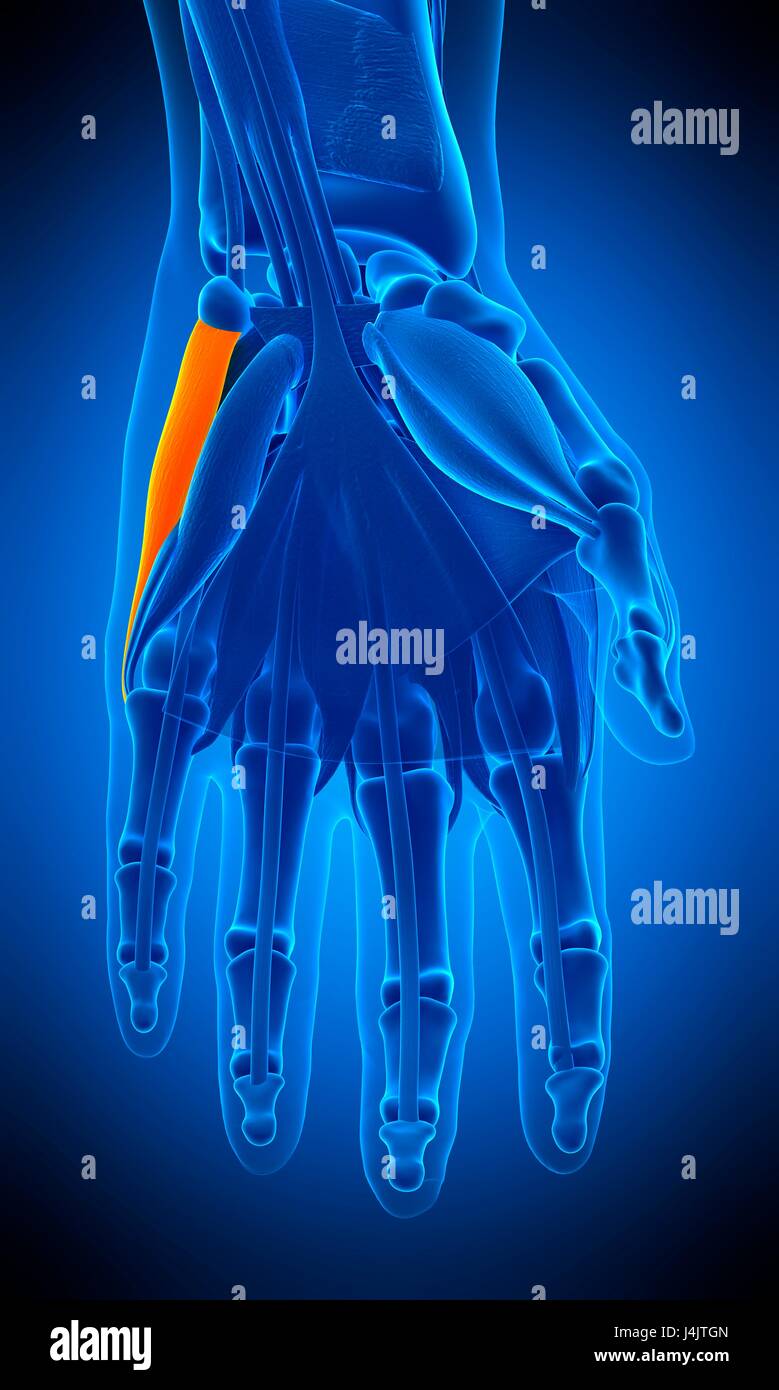 Illustration of the abductor digiti minimi muscle. Stock Photohttps://www.alamy.com/image-license-details/?v=1https://www.alamy.com/stock-photo-illustration-of-the-abductor-digiti-minimi-muscle-140555989.html
RFJ4JTGN–Illustration of the abductor digiti minimi muscle.
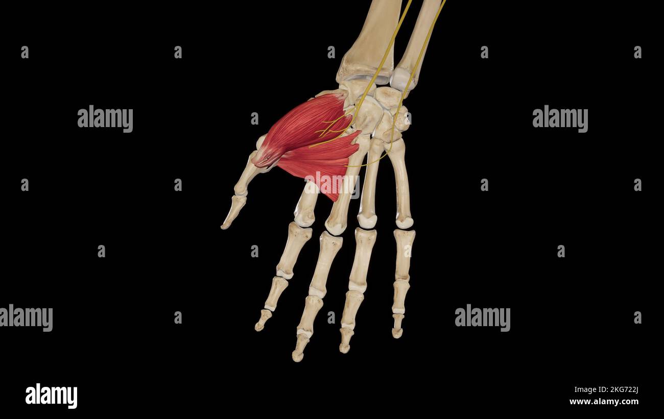 Thenar Muscles Stock Photohttps://www.alamy.com/image-license-details/?v=1https://www.alamy.com/thenar-muscles-image491880106.html
RF2KG722J–Thenar Muscles
 Human foot musculature, artwork Stock Photohttps://www.alamy.com/image-license-details/?v=1https://www.alamy.com/human-foot-musculature-artwork-image65249613.html
RFDP4AF9–Human foot musculature, artwork
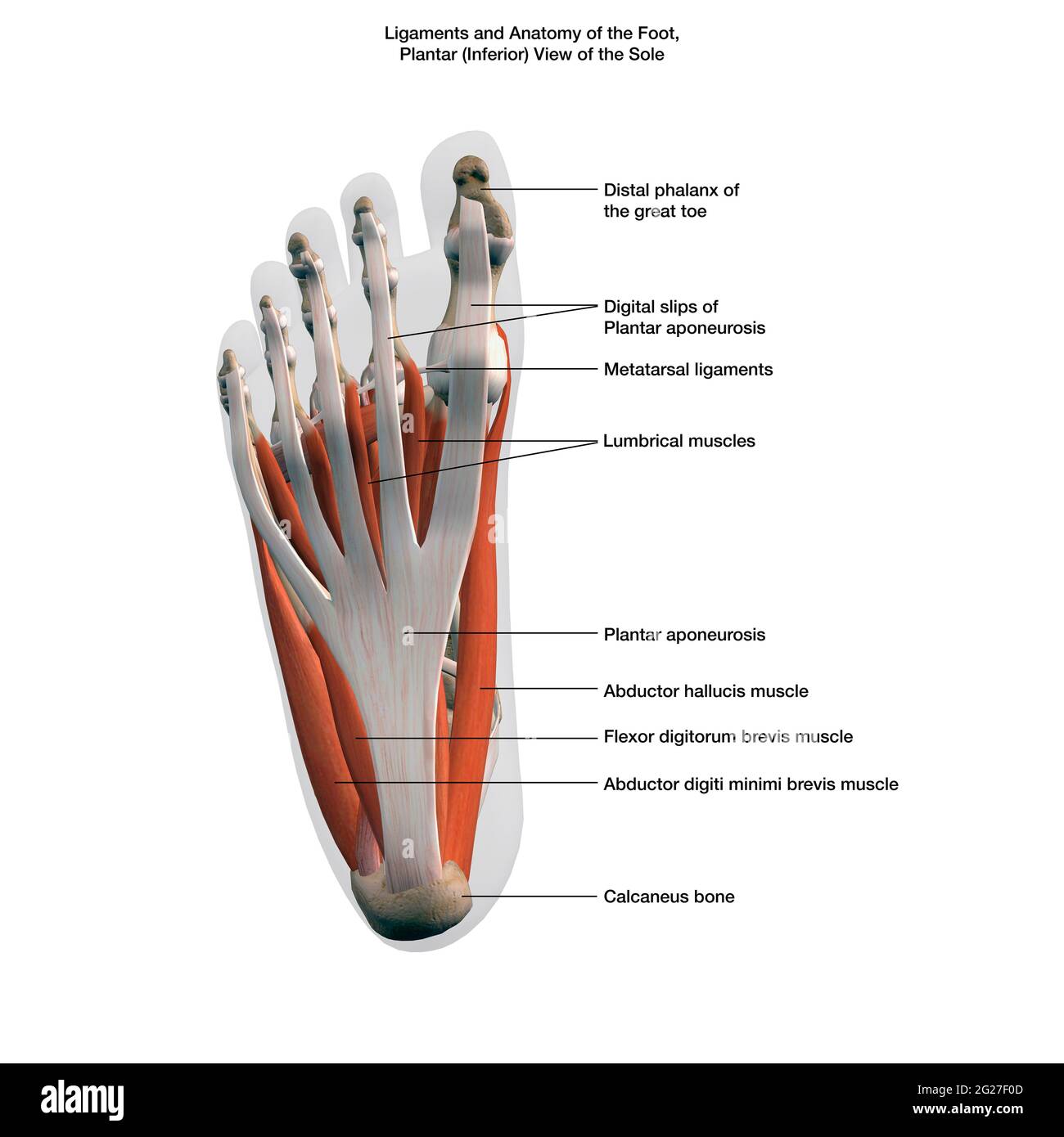 Ligaments and muscles of the human foot, planar view of the sole with labels. Stock Photohttps://www.alamy.com/image-license-details/?v=1https://www.alamy.com/ligaments-and-muscles-of-the-human-foot-planar-view-of-the-sole-with-labels-image431653949.html
RF2G27F0D–Ligaments and muscles of the human foot, planar view of the sole with labels.
 Human leg musculature, artwork Stock Photohttps://www.alamy.com/image-license-details/?v=1https://www.alamy.com/human-leg-musculature-artwork-image65248367.html
RFDP48XR–Human leg musculature, artwork
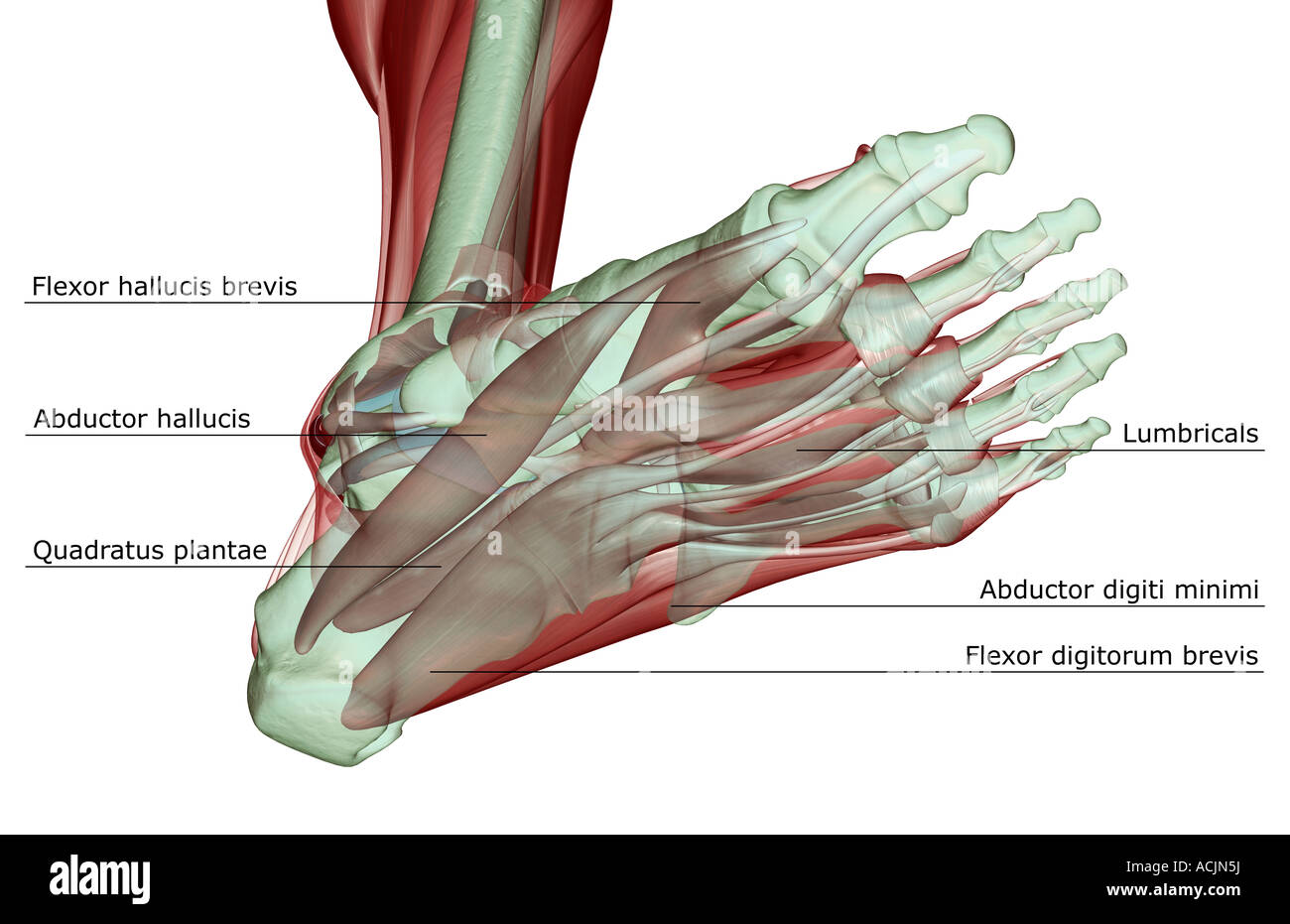 The musculoskeleton of the foot Stock Photohttps://www.alamy.com/image-license-details/?v=1https://www.alamy.com/stock-photo-the-musculoskeleton-of-the-foot-13172045.html
RFACJN5J–The musculoskeleton of the foot
 Human leg musculature, artwork Stock Photohttps://www.alamy.com/image-license-details/?v=1https://www.alamy.com/human-leg-musculature-artwork-image65238329.html
RFDP3T49–Human leg musculature, artwork
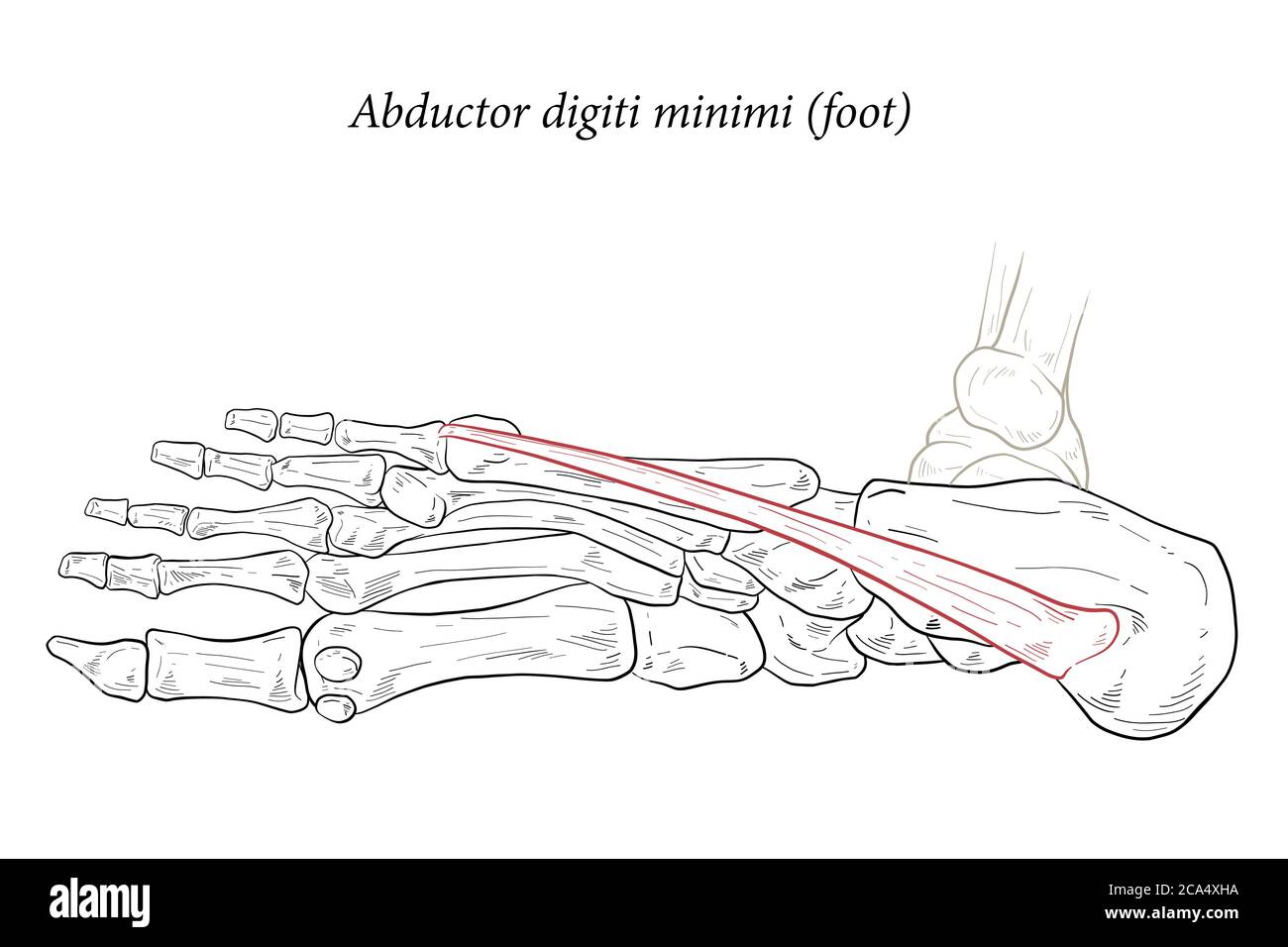 Abductor digiti minimi muscle of foot. Stock Vectorhttps://www.alamy.com/image-license-details/?v=1https://www.alamy.com/abductor-digiti-minimi-muscle-of-foot-image367672966.html
RF2CA4XHA–Abductor digiti minimi muscle of foot.
 Human foot musculature, artwork Stock Photohttps://www.alamy.com/image-license-details/?v=1https://www.alamy.com/human-foot-musculature-artwork-image65254827.html
RFDP4H5F–Human foot musculature, artwork
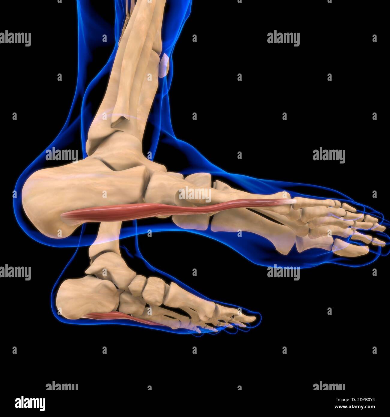 3D Illustration, Muscle is a soft tissue, Muscle cells contain proteins , producing a contraction that changes both the length and the shape of the ce Stock Photohttps://www.alamy.com/image-license-details/?v=1https://www.alamy.com/3d-illustration-muscle-is-a-soft-tissue-muscle-cells-contain-proteins-producing-a-contraction-that-changes-both-the-length-and-the-shape-of-the-ce-image395466040.html
RF2DYB0Y4–3D Illustration, Muscle is a soft tissue, Muscle cells contain proteins , producing a contraction that changes both the length and the shape of the ce
 . Bulletin - United States National Museum. Science. Figure 33.—Musculature of the hand: a, cross section (1, m. opponens digiti qumti (=opponensdigiti minimi of N.A.);2, m. abductor digiti quinti (= abductor digiti minimi of N.A.); 3, m. flexor digiti quinti (= flexor digiti minimi of N.A.); 4, tendon of m. flexor profundus to digit I; 5, m. flexor poUicis brevis, caput radiale; 6, m. abductor pollicis brevis; 7, m. opponens pollicis; 8, m. flexor pollicis brevis, caput profundum; 9 m. flexor pollicis brevis, caput ulnare; 10, m. adductor pollicis (m. contrahens I); 11, m. contrahens II; 12, Stock Photohttps://www.alamy.com/image-license-details/?v=1https://www.alamy.com/bulletin-united-states-national-museum-science-figure-33musculature-of-the-hand-a-cross-section-1-m-opponens-digiti-qumti-=opponensdigiti-minimi-of-na2-m-abductor-digiti-quinti-=-abductor-digiti-minimi-of-na-3-m-flexor-digiti-quinti-=-flexor-digiti-minimi-of-na-4-tendon-of-m-flexor-profundus-to-digit-i-5-m-flexor-pouicis-brevis-caput-radiale-6-m-abductor-pollicis-brevis-7-m-opponens-pollicis-8-m-flexor-pollicis-brevis-caput-profundum-9-m-flexor-pollicis-brevis-caput-ulnare-10-m-adductor-pollicis-m-contrahens-i-11-m-contrahens-ii-12-image233731761.html
RMRG7B6W–. Bulletin - United States National Museum. Science. Figure 33.—Musculature of the hand: a, cross section (1, m. opponens digiti qumti (=opponensdigiti minimi of N.A.);2, m. abductor digiti quinti (= abductor digiti minimi of N.A.); 3, m. flexor digiti quinti (= flexor digiti minimi of N.A.); 4, tendon of m. flexor profundus to digit I; 5, m. flexor poUicis brevis, caput radiale; 6, m. abductor pollicis brevis; 7, m. opponens pollicis; 8, m. flexor pollicis brevis, caput profundum; 9 m. flexor pollicis brevis, caput ulnare; 10, m. adductor pollicis (m. contrahens I); 11, m. contrahens II; 12,
 3d rendered medically accurate illustration of the Abductor Digiti Minimi Stock Photohttps://www.alamy.com/image-license-details/?v=1https://www.alamy.com/3d-rendered-medically-accurate-illustration-of-the-abductor-digiti-minimi-image258154332.html
RFTYYXDG–3d rendered medically accurate illustration of the Abductor Digiti Minimi
