Quick filters:
Acoustic meatus Stock Photos and Images
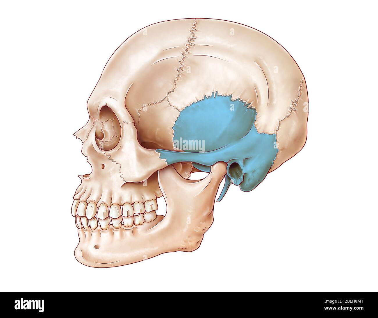 Human Skull, Temporal Bone Stock Photohttps://www.alamy.com/image-license-details/?v=1https://www.alamy.com/human-skull-temporal-bone-image353192584.html
RM2BEH8MT–Human Skull, Temporal Bone
 Doctors surgery. Examination of the ear, acoustic meatus, ear canal of a patient, with a loupe Stock Photohttps://www.alamy.com/image-license-details/?v=1https://www.alamy.com/stock-photo-doctors-surgery-examination-of-the-ear-acoustic-meatus-ear-canal-of-31849175.html
RMBRPRXF–Doctors surgery. Examination of the ear, acoustic meatus, ear canal of a patient, with a loupe
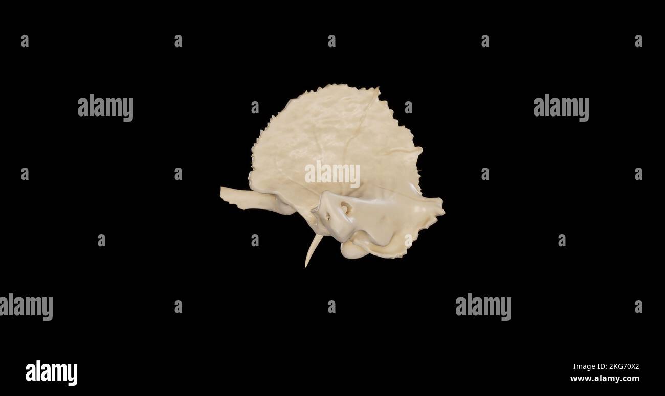 Left view of Right Temporal Bone Stock Photohttps://www.alamy.com/image-license-details/?v=1https://www.alamy.com/left-view-of-right-temporal-bone-image491879194.html
RF2KG70X2–Left view of Right Temporal Bone
 Ear anatomy hand drawn vector illustration. Ear canal and skull cross section vintage engraving style drawing. Human acoustic meatus internal parts. Stock Vectorhttps://www.alamy.com/image-license-details/?v=1https://www.alamy.com/ear-anatomy-hand-drawn-vector-illustration-ear-canal-and-skull-cross-section-vintage-engraving-style-drawing-human-acoustic-meatus-internal-parts-image434341309.html
RF2G6HXNH–Ear anatomy hand drawn vector illustration. Ear canal and skull cross section vintage engraving style drawing. Human acoustic meatus internal parts.
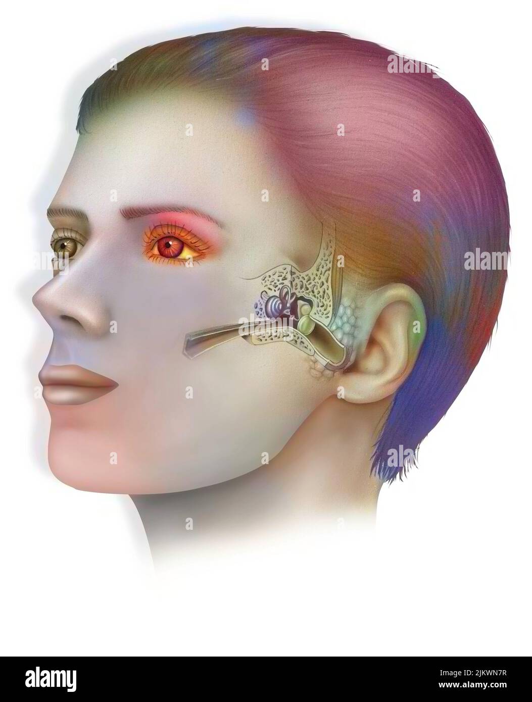 Anatomy of the inner ear showing the eardrum, the cochlea. Stock Photohttps://www.alamy.com/image-license-details/?v=1https://www.alamy.com/anatomy-of-the-inner-ear-showing-the-eardrum-the-cochlea-image476923883.html
RF2JKWN7R–Anatomy of the inner ear showing the eardrum, the cochlea.
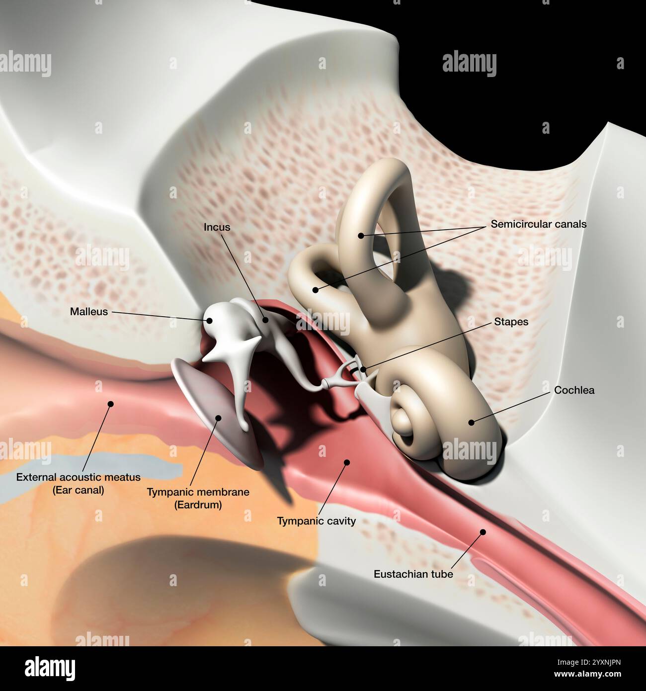 Cross section of the human ear, with labels. Stock Photohttps://www.alamy.com/image-license-details/?v=1https://www.alamy.com/cross-section-of-the-human-ear-with-labels-image636030045.html
RF2YXNJPN–Cross section of the human ear, with labels.
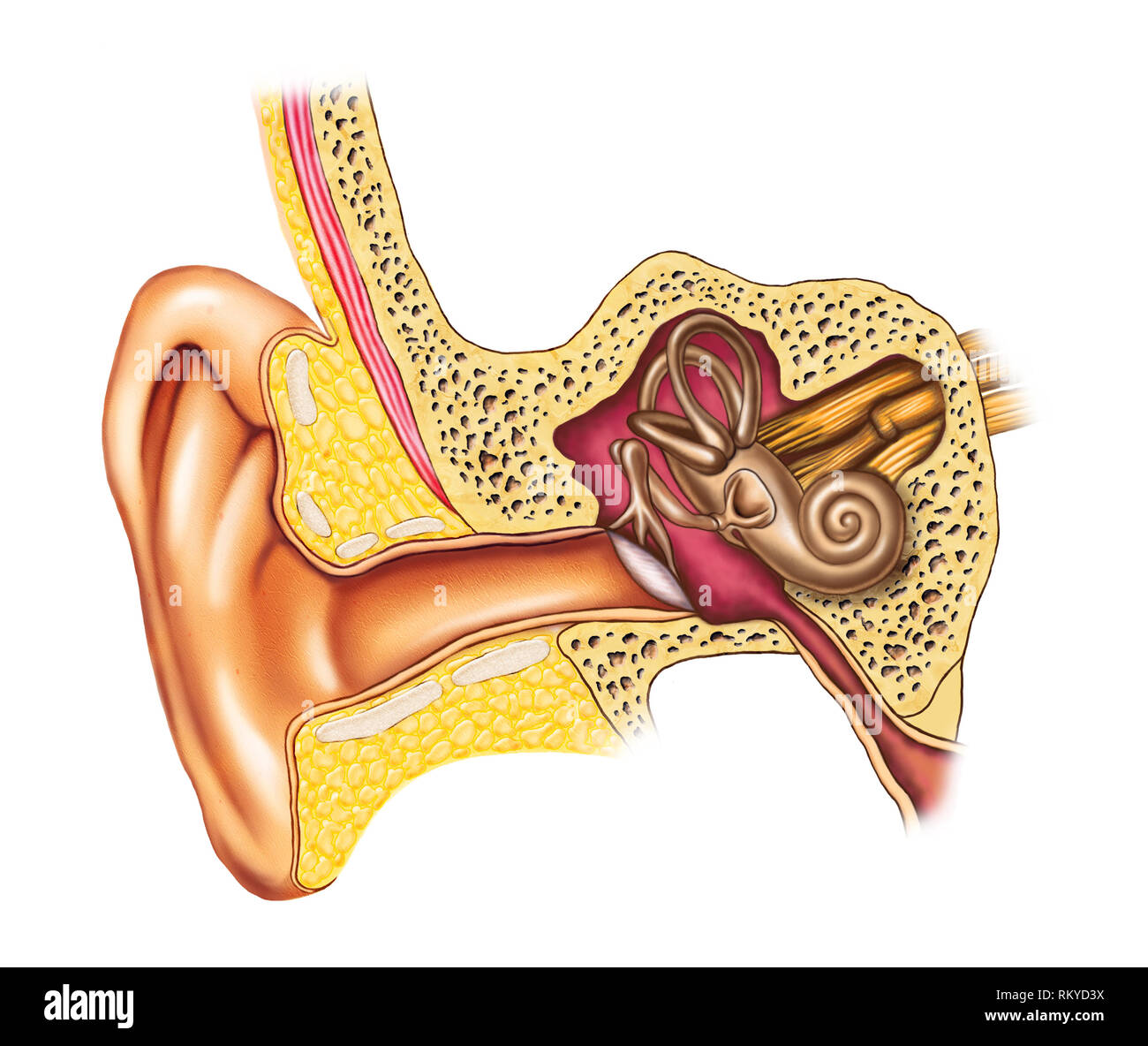 Illustration showing the interiors of an human ear. Digital illustration. Stock Photohttps://www.alamy.com/image-license-details/?v=1https://www.alamy.com/illustration-showing-the-interiors-of-an-human-ear-digital-illustration-image236016254.html
RFRKYD3X–Illustration showing the interiors of an human ear. Digital illustration.
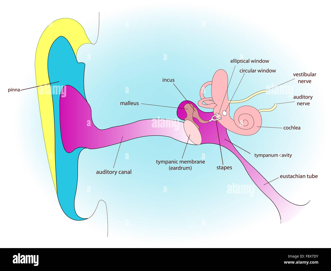 ear anatomy Stock Photohttps://www.alamy.com/image-license-details/?v=1https://www.alamy.com/stock-photo-ear-anatomy-91545719.html
RFF8X7DY–ear anatomy
 Human ear cross-section model isolated on white background Stock Photohttps://www.alamy.com/image-license-details/?v=1https://www.alamy.com/stock-image-human-ear-cross-section-model-isolated-on-white-background-164248974.html
RFKF656P–Human ear cross-section model isolated on white background
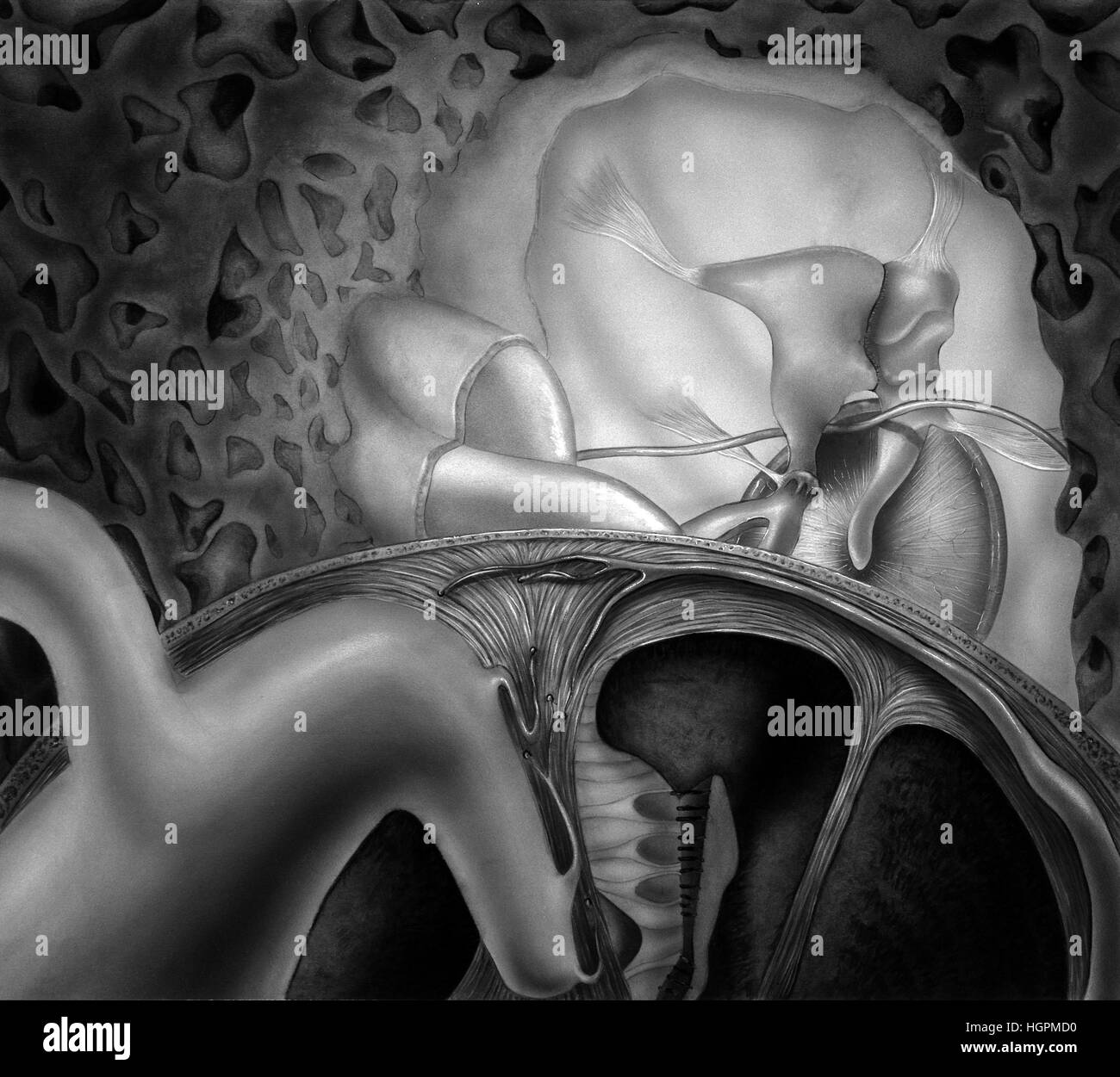 Inner Ear Structures. Shown are the vestibular labyrinth, cochlear labyrinth, cochlea, tensor tympani muscle, auditory tube,tympanic cavity, Tympanic Stock Photohttps://www.alamy.com/image-license-details/?v=1https://www.alamy.com/stock-photo-inner-ear-structures-shown-are-the-vestibular-labyrinth-cochlear-labyrinth-130806060.html
RFHGPMD0–Inner Ear Structures. Shown are the vestibular labyrinth, cochlear labyrinth, cochlea, tensor tympani muscle, auditory tube,tympanic cavity, Tympanic
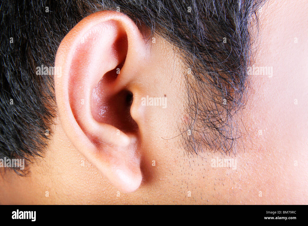 close up of male external ear Stock Photohttps://www.alamy.com/image-license-details/?v=1https://www.alamy.com/stock-photo-close-up-of-male-external-ear-29664864.html
RFBM79RC–close up of male external ear
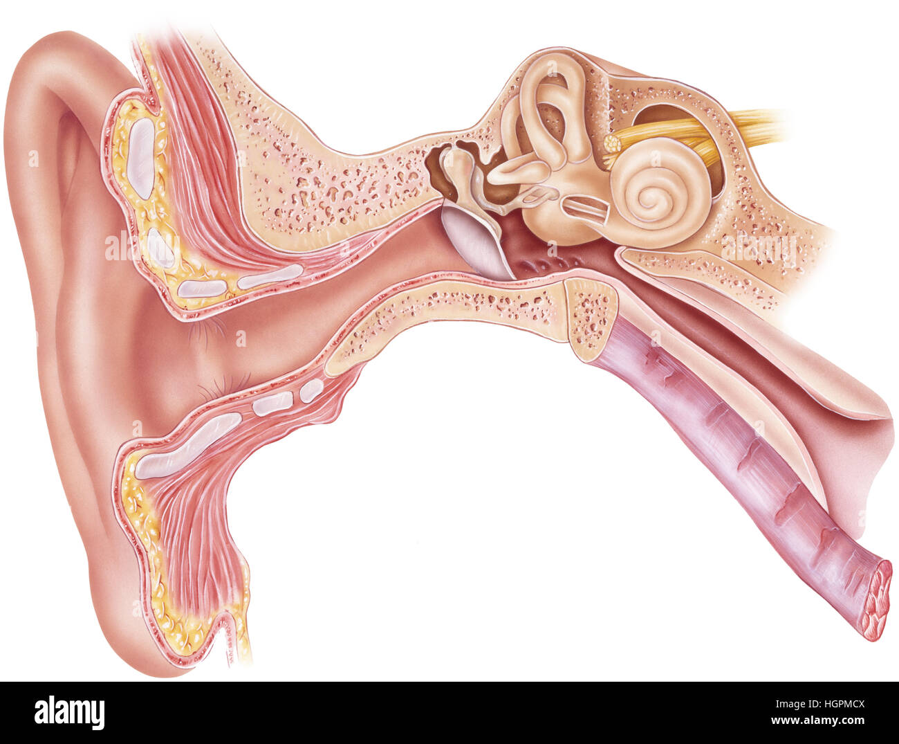 Frontal section through the right external, middle, and internal ear. Shown are the vestibular labyrinth, cochlear labyrinth, cochlea, tensor tympani Stock Photohttps://www.alamy.com/image-license-details/?v=1https://www.alamy.com/stock-photo-frontal-section-through-the-right-external-middle-and-internal-ear-130806058.html
RFHGPMCX–Frontal section through the right external, middle, and internal ear. Shown are the vestibular labyrinth, cochlear labyrinth, cochlea, tensor tympani
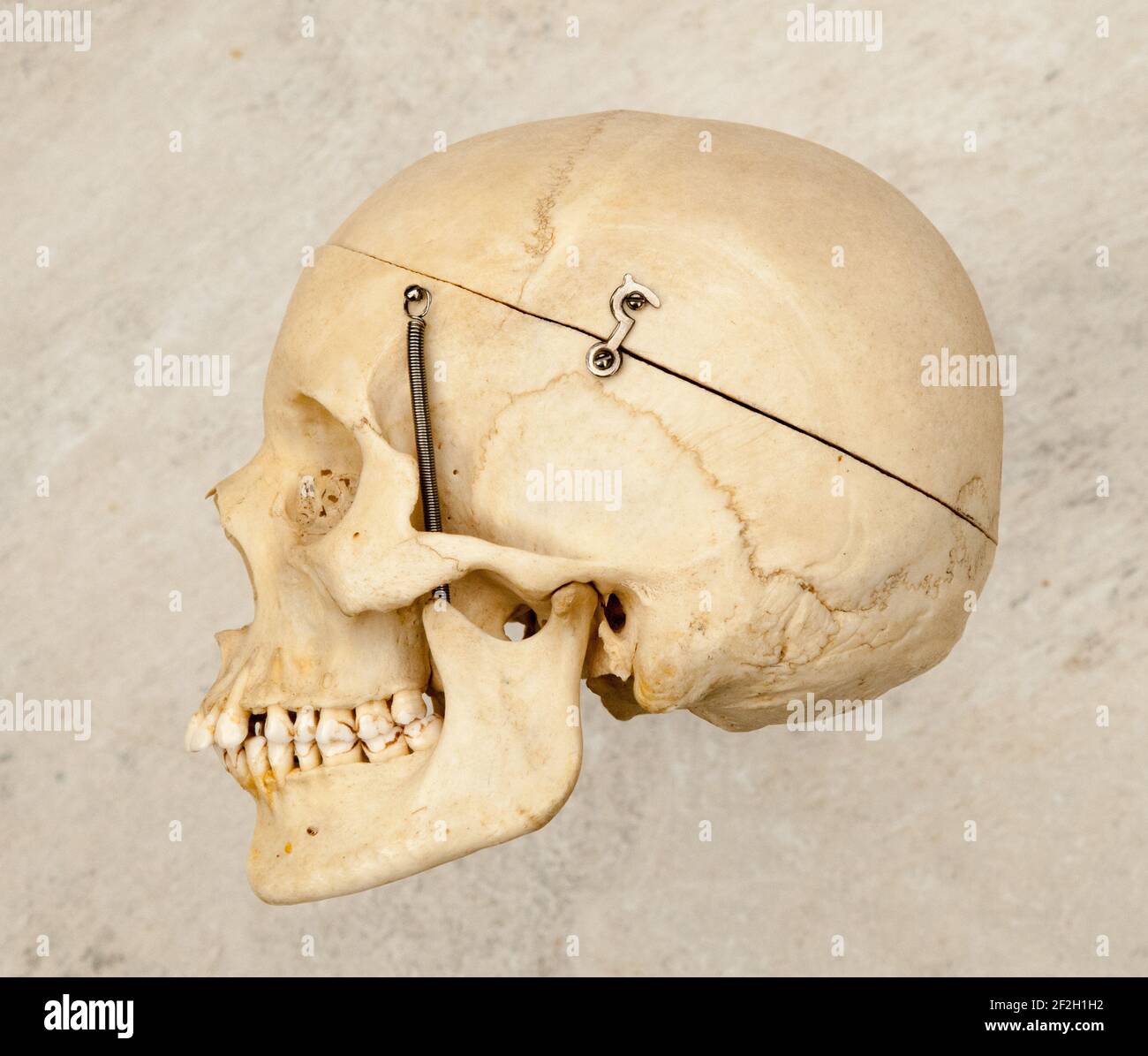 Sideways or profile view of a human skull which has been prepared for medical studies. Stock Photohttps://www.alamy.com/image-license-details/?v=1https://www.alamy.com/sideways-or-profile-view-of-a-human-skull-which-has-been-prepared-for-medical-studies-image414652590.html
RF2F2H1H2–Sideways or profile view of a human skull which has been prepared for medical studies.
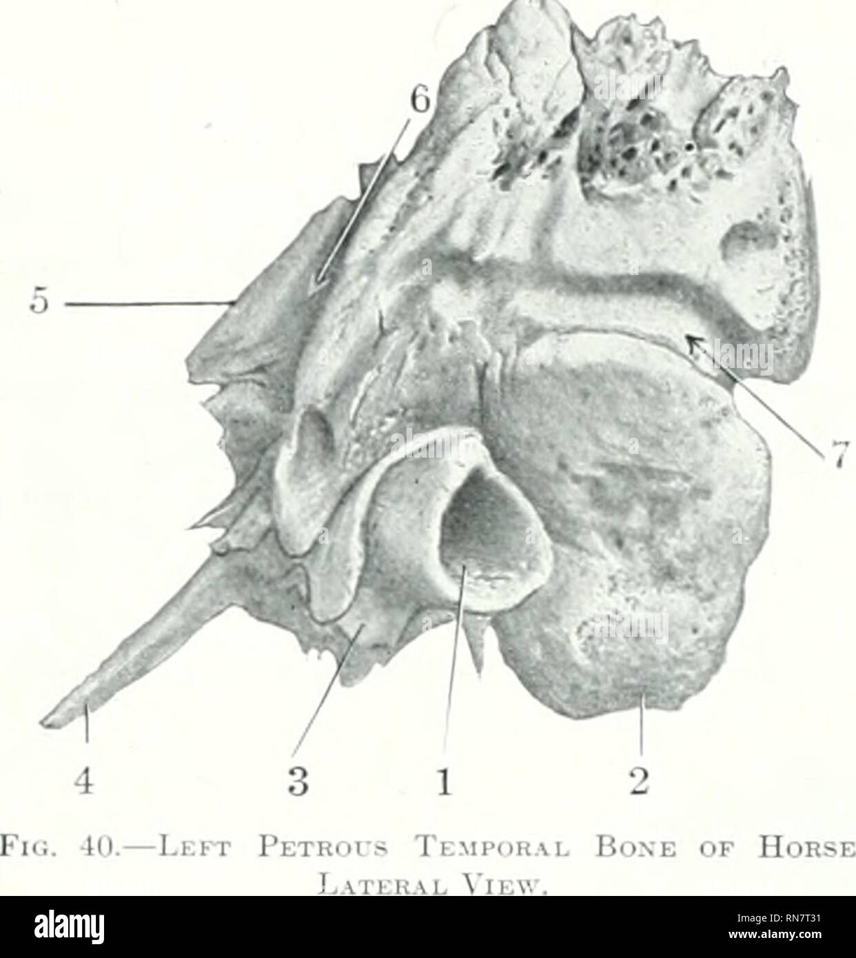 . The anatomy of the domestic animals. Veterinary anatomy. 1, External acoustic meatus; 2, mastoid process: 3, hyoid process; 4, muscular process; 5, petrosal crest; 6, groove which concurs in formation of temporal canal; 7. groove for posterior meningeal arterj'.. Please note that these images are extracted from scanned page images that may have been digitally enhanced for readability - coloration and appearance of these illustrations may not perfectly resemble the original work.. Sisson, Septimus, 1865-1924. Philadelphia Saunders Stock Photohttps://www.alamy.com/image-license-details/?v=1https://www.alamy.com/the-anatomy-of-the-domestic-animals-veterinary-anatomy-1-external-acoustic-meatus-2-mastoid-process-3-hyoid-process-4-muscular-process-5-petrosal-crest-6-groove-which-concurs-in-formation-of-temporal-canal-7-groove-for-posterior-meningeal-arterj-please-note-that-these-images-are-extracted-from-scanned-page-images-that-may-have-been-digitally-enhanced-for-readability-coloration-and-appearance-of-these-illustrations-may-not-perfectly-resemble-the-original-work-sisson-septimus-1865-1924-philadelphia-saunders-image236815125.html
RMRN7T31–. The anatomy of the domestic animals. Veterinary anatomy. 1, External acoustic meatus; 2, mastoid process: 3, hyoid process; 4, muscular process; 5, petrosal crest; 6, groove which concurs in formation of temporal canal; 7. groove for posterior meningeal arterj'.. Please note that these images are extracted from scanned page images that may have been digitally enhanced for readability - coloration and appearance of these illustrations may not perfectly resemble the original work.. Sisson, Septimus, 1865-1924. Philadelphia Saunders
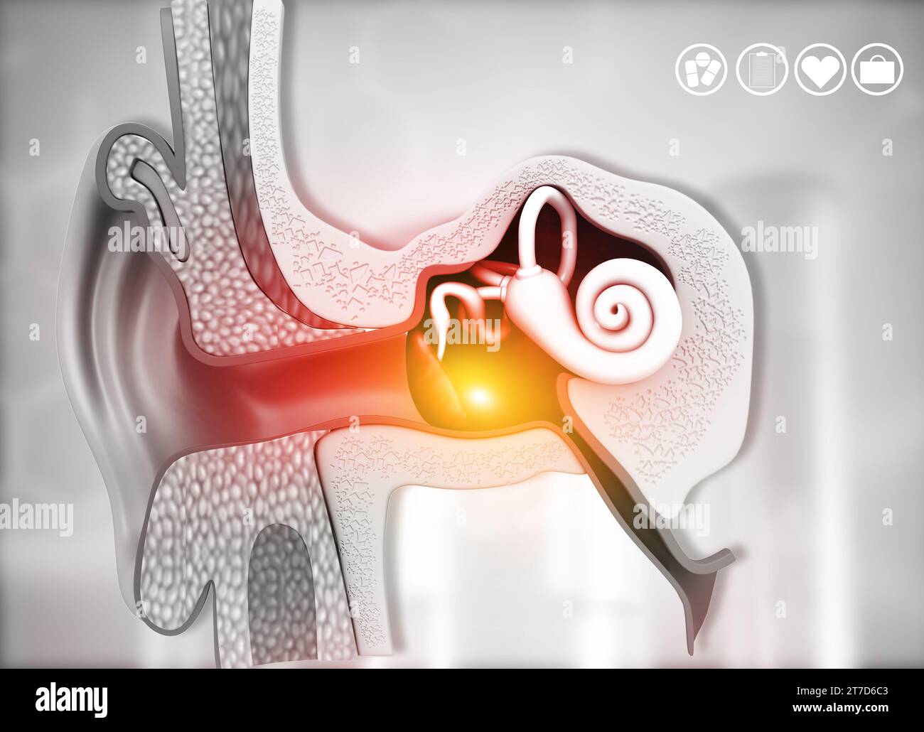 Cross-section of the human ear. 3d illustration Stock Photohttps://www.alamy.com/image-license-details/?v=1https://www.alamy.com/cross-section-of-the-human-ear-3d-illustration-image572535155.html
RF2T7D6C3–Cross-section of the human ear. 3d illustration
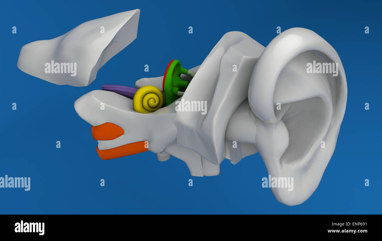 Human ear anatomy on blue background Stock Photohttps://www.alamy.com/image-license-details/?v=1https://www.alamy.com/stock-photo-human-ear-anatomy-on-blue-background-82237149.html
RMENP691–Human ear anatomy on blue background
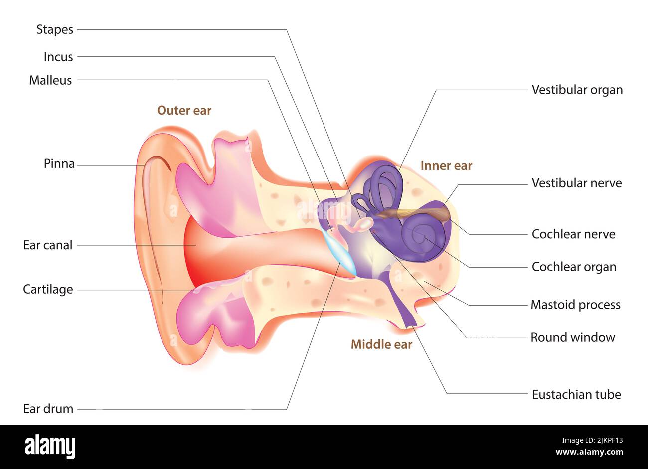 Ear structure Stock Photohttps://www.alamy.com/image-license-details/?v=1https://www.alamy.com/ear-structure-image476853135.html
RF2JKPF13–Ear structure
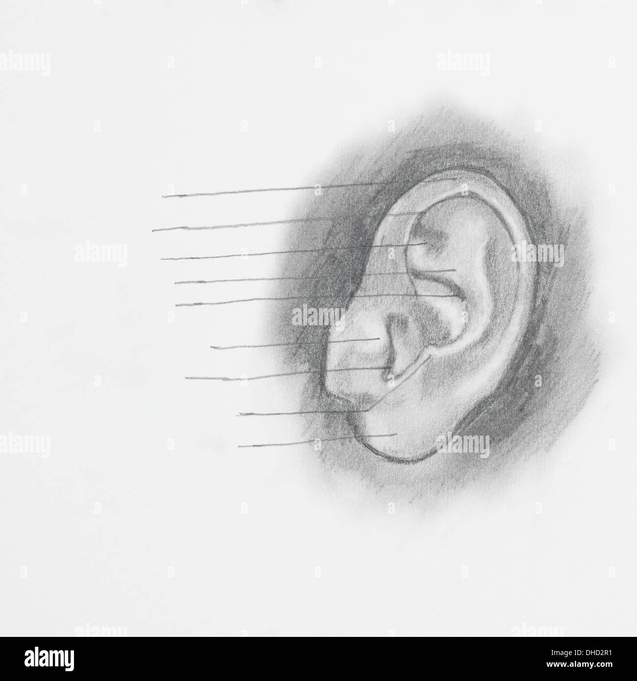 Detail of ear pencil drawing on white paper Stock Photohttps://www.alamy.com/image-license-details/?v=1https://www.alamy.com/detail-of-ear-pencil-drawing-on-white-paper-image62367845.html
RFDHD2R1–Detail of ear pencil drawing on white paper
 illustration balance Stock Photohttps://www.alamy.com/image-license-details/?v=1https://www.alamy.com/stock-photo-illustration-balance-131589980.html
RFHJ2CA4–illustration balance
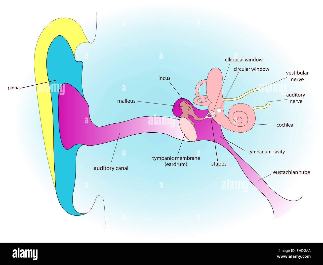 ear anatomy Stock Photohttps://www.alamy.com/image-license-details/?v=1https://www.alamy.com/stock-photo-ear-anatomy-79588834.html
RFEHDGAA–ear anatomy
 Doctors surgery. Examination of the ear, acoustic meatus, ear canal of a patient, with a loupe Stock Photohttps://www.alamy.com/image-license-details/?v=1https://www.alamy.com/stock-photo-doctors-surgery-examination-of-the-ear-acoustic-meatus-ear-canal-of-31849273.html
RMBRPT21–Doctors surgery. Examination of the ear, acoustic meatus, ear canal of a patient, with a loupe
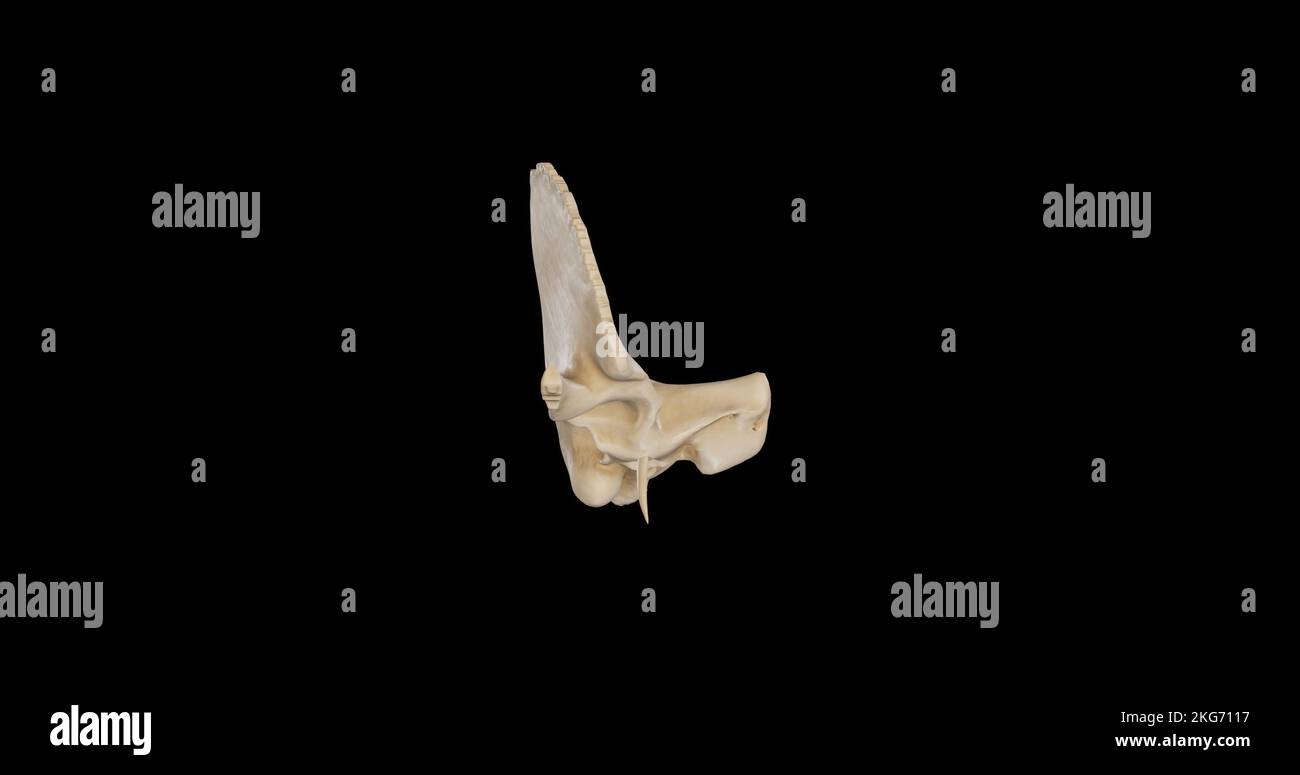 Anterior view of Right Temporal Bone Stock Photohttps://www.alamy.com/image-license-details/?v=1https://www.alamy.com/anterior-view-of-right-temporal-bone-image491879283.html
RF2KG7117–Anterior view of Right Temporal Bone
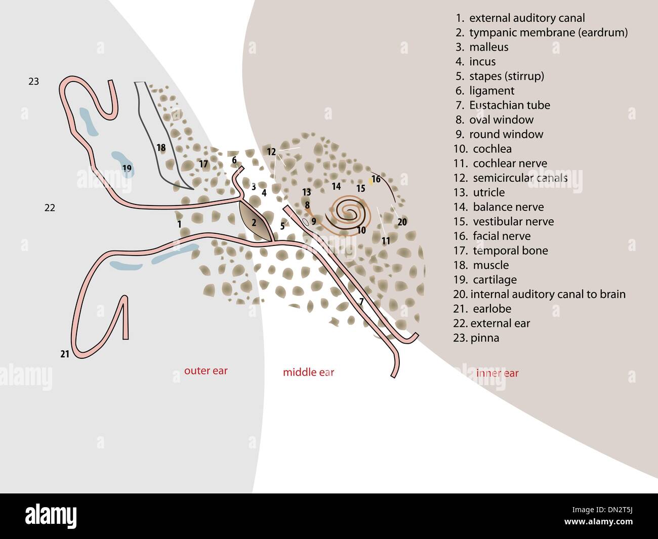 ear anatomy Stock Vectorhttps://www.alamy.com/image-license-details/?v=1https://www.alamy.com/ear-anatomy-image64601758.html
RFDN2T5J–ear anatomy
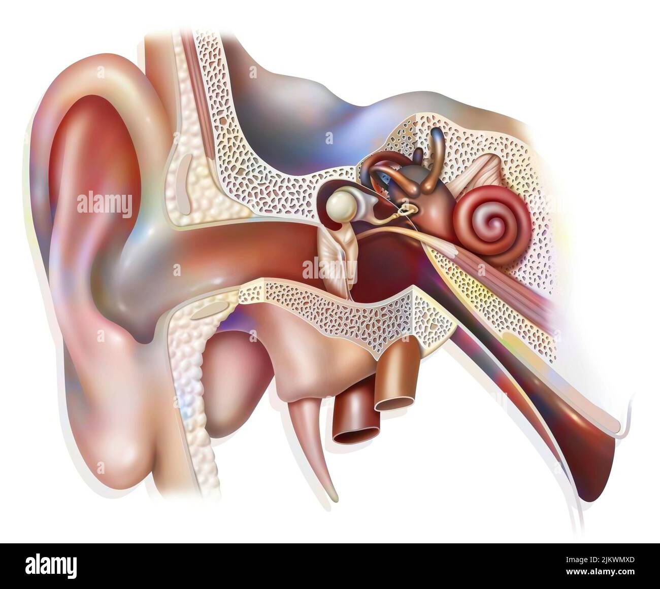 Anatomy of the inner ear showing the eardrum, the cochlea. Stock Photohttps://www.alamy.com/image-license-details/?v=1https://www.alamy.com/anatomy-of-the-inner-ear-showing-the-eardrum-the-cochlea-image476923621.html
RF2JKWMXD–Anatomy of the inner ear showing the eardrum, the cochlea.
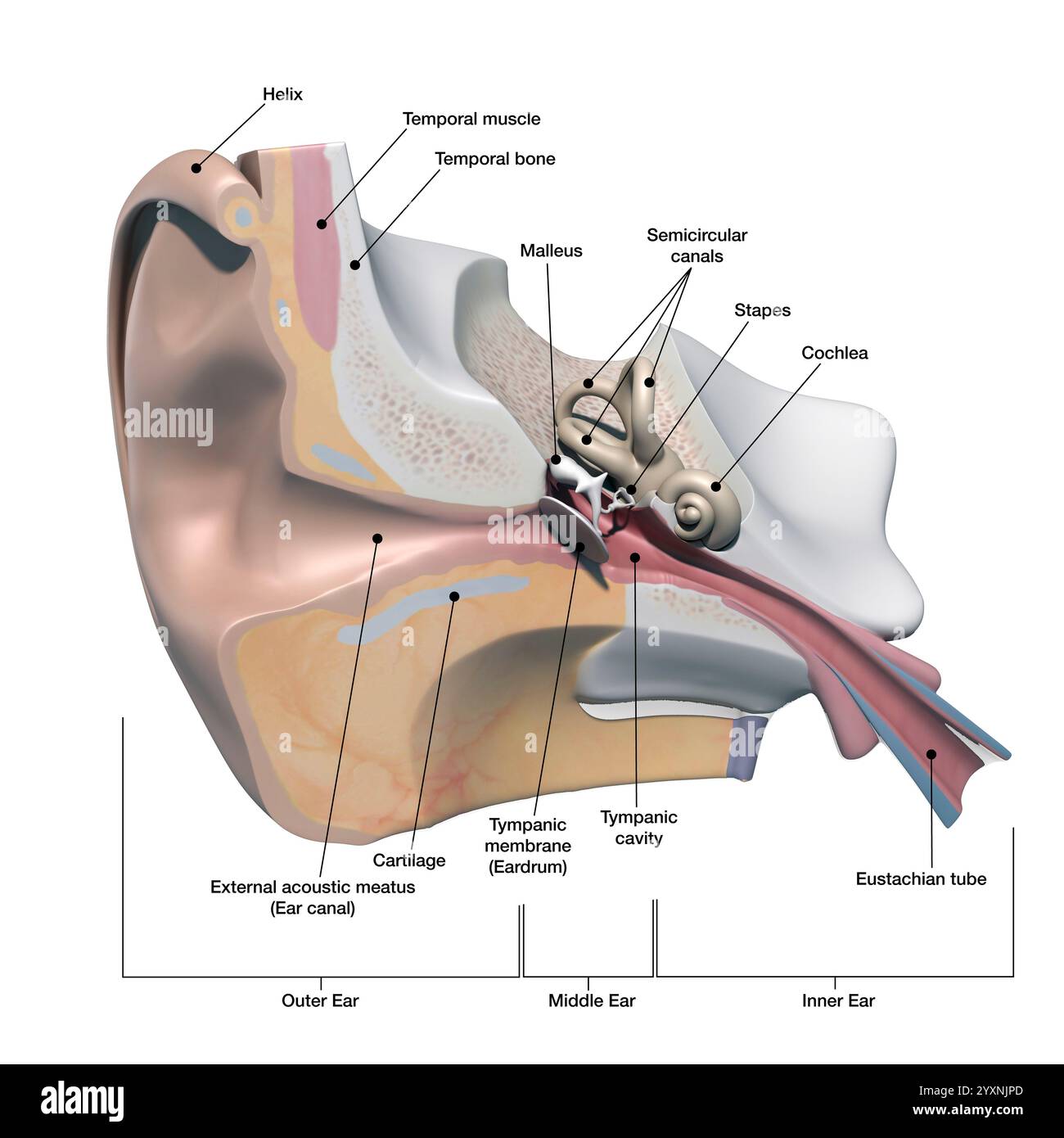 Cross section of the human ear, with labels. Stock Photohttps://www.alamy.com/image-license-details/?v=1https://www.alamy.com/cross-section-of-the-human-ear-with-labels-image636030037.html
RF2YXNJPD–Cross section of the human ear, with labels.
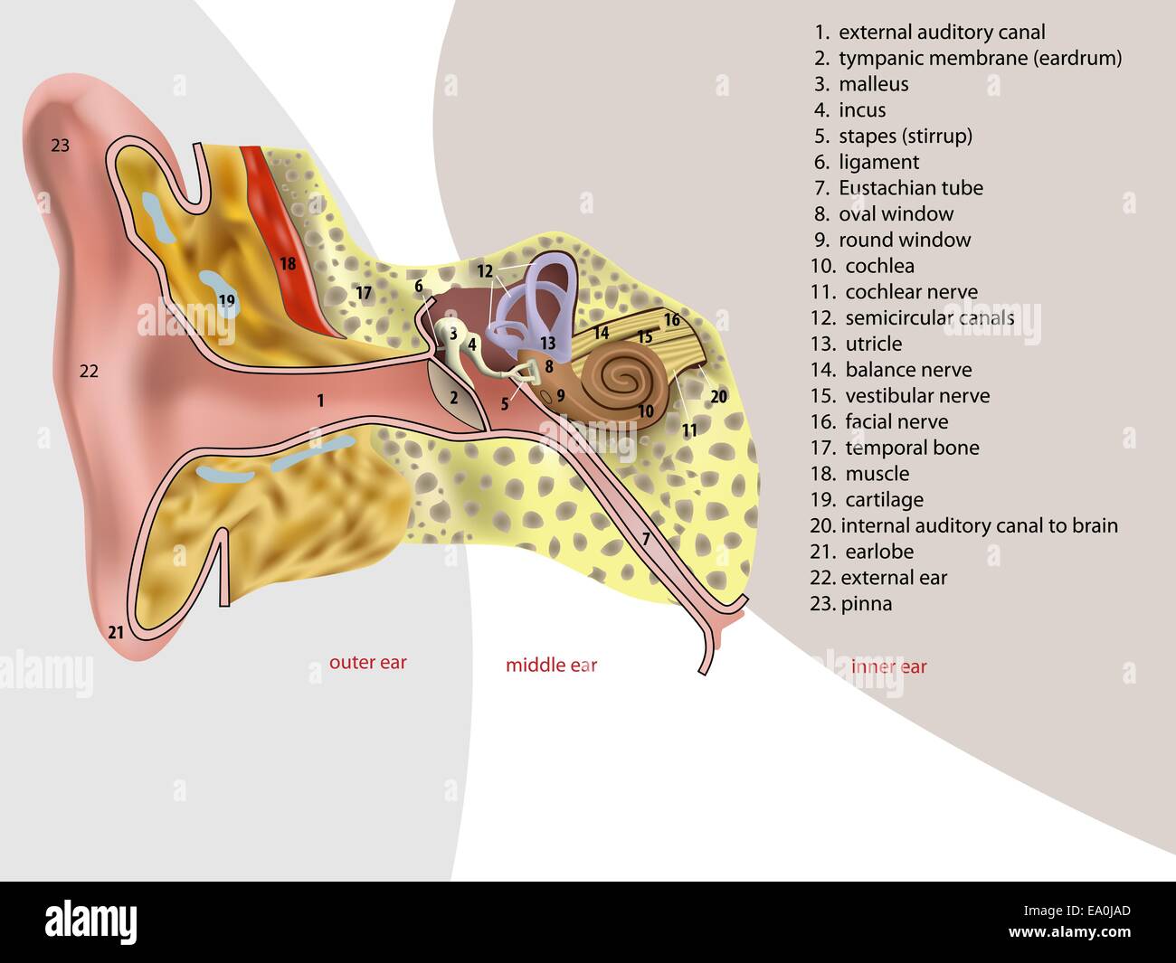 vector illustration of ear anatomy Stock Vectorhttps://www.alamy.com/image-license-details/?v=1https://www.alamy.com/stock-photo-vector-illustration-of-ear-anatomy-75002437.html
RFEA0JAD–vector illustration of ear anatomy
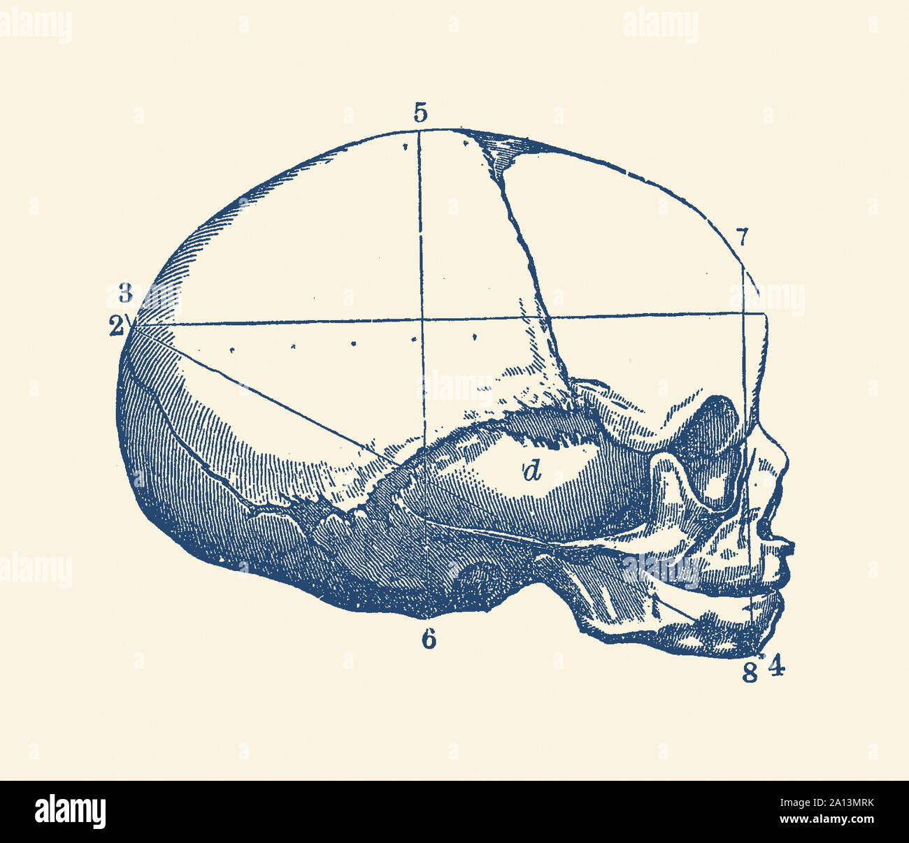 Vintage anatomy print showing a side view of the human skull. Stock Photohttps://www.alamy.com/image-license-details/?v=1https://www.alamy.com/vintage-anatomy-print-showing-a-side-view-of-the-human-skull-image327693847.html
RF2A13MRK–Vintage anatomy print showing a side view of the human skull.
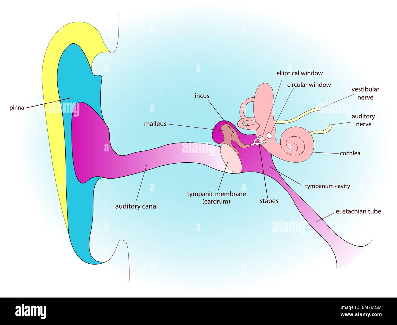 ear anatomy Stock Vectorhttps://www.alamy.com/image-license-details/?v=1https://www.alamy.com/stock-photo-ear-anatomy-81304404.html
RFEM7MGM–ear anatomy
 Cotton swabs / Q-tips Stock Photohttps://www.alamy.com/image-license-details/?v=1https://www.alamy.com/stock-photo-cotton-swabs-q-tips-76346541.html
RMEC5TP5–Cotton swabs / Q-tips
 Archive image from page 225 of Cunningham's Text-book of anatomy (1914). Cunningham's Text-book of anatomy cunninghamstextb00cunn Year: 1914 ( 102 OSTEOLOGY. roofed in by the thin tegmen tympani, which separates it from the middle cranial fossa. The obliquity of the medial end of the external acoustic meatus, together with the groove for the attachment of the tympanic membrane is well seen, and the thickness of the upper wall of that passage is also noteworthy. The floor of the meatus, formed by the tympanic plate, which separ- ates it from the mandibular fossa, is much thinner, but in the re Stock Photohttps://www.alamy.com/image-license-details/?v=1https://www.alamy.com/archive-image-from-page-225-of-cunninghams-text-book-of-anatomy-1914-cunninghams-text-book-of-anatomy-cunninghamstextb00cunn-year-1914-102-osteology-roofed-in-by-the-thin-tegmen-tympani-which-separates-it-from-the-middle-cranial-fossa-the-obliquity-of-the-medial-end-of-the-external-acoustic-meatus-together-with-the-groove-for-the-attachment-of-the-tympanic-membrane-is-well-seen-and-the-thickness-of-the-upper-wall-of-that-passage-is-also-noteworthy-the-floor-of-the-meatus-formed-by-the-tympanic-plate-which-separ-ates-it-from-the-mandibular-fossa-is-much-thinner-but-in-the-re-image264051642.html
RMW9GGFP–Archive image from page 225 of Cunningham's Text-book of anatomy (1914). Cunningham's Text-book of anatomy cunninghamstextb00cunn Year: 1914 ( 102 OSTEOLOGY. roofed in by the thin tegmen tympani, which separates it from the middle cranial fossa. The obliquity of the medial end of the external acoustic meatus, together with the groove for the attachment of the tympanic membrane is well seen, and the thickness of the upper wall of that passage is also noteworthy. The floor of the meatus, formed by the tympanic plate, which separ- ates it from the mandibular fossa, is much thinner, but in the re
 Zygoma word cloud concept Stock Photohttps://www.alamy.com/image-license-details/?v=1https://www.alamy.com/zygoma-word-cloud-concept-image262839896.html
RFW7HAY4–Zygoma word cloud concept
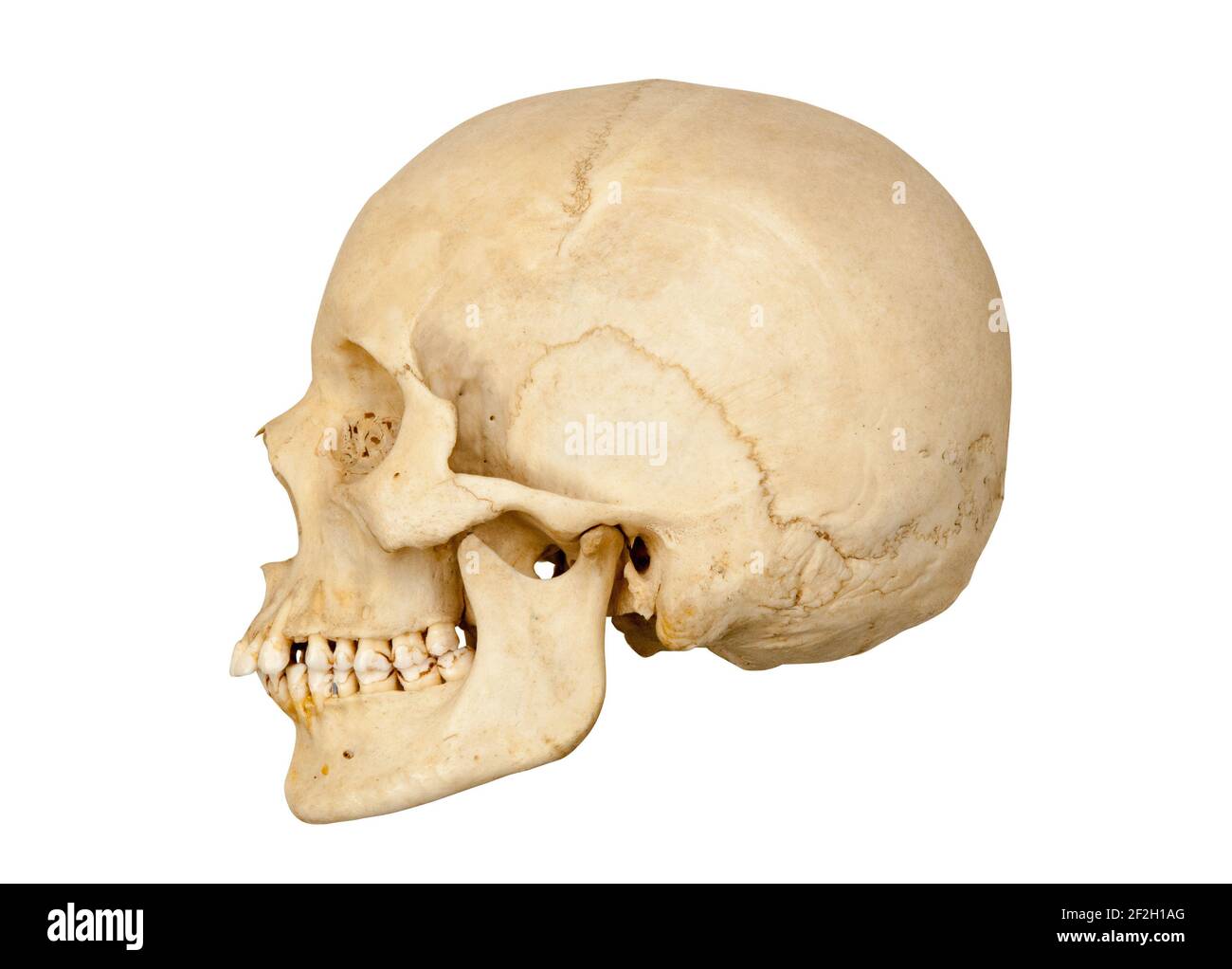 Sideways or profile view of left side of a human skull cut out on a white background. Stock Photohttps://www.alamy.com/image-license-details/?v=1https://www.alamy.com/sideways-or-profile-view-of-left-side-of-a-human-skull-cut-out-on-a-white-background-image414652408.html
RF2F2H1AG–Sideways or profile view of left side of a human skull cut out on a white background.
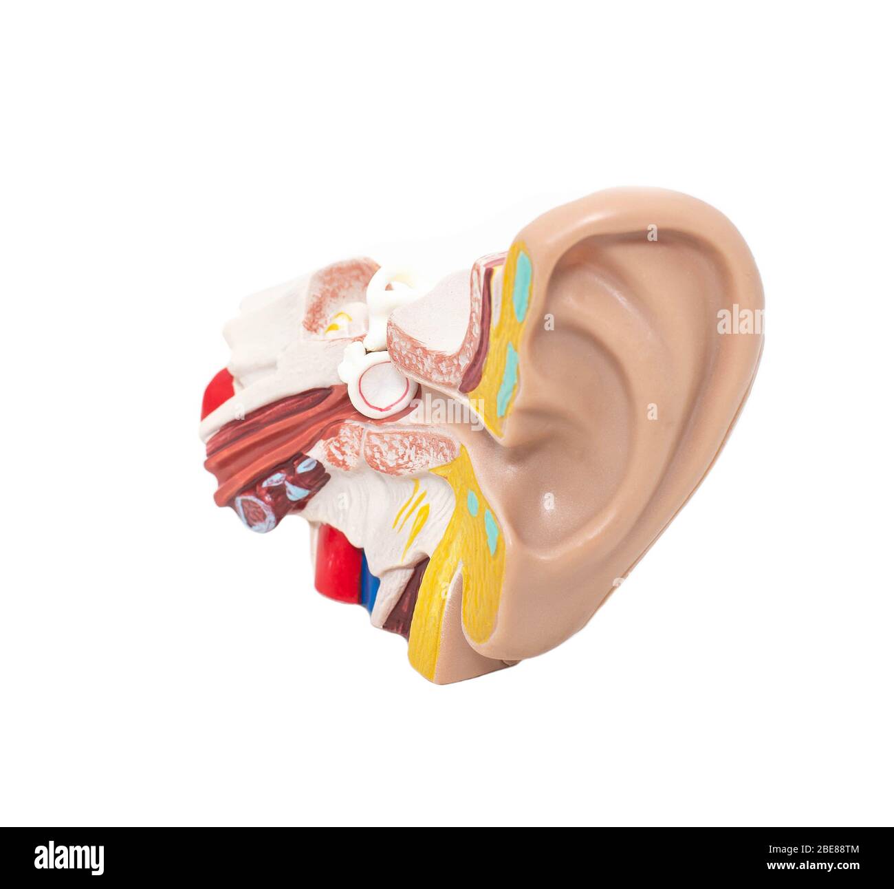 Mock ear with eardrum and auditory tube on a white background, isolate. The concept of diseases in otolaryngology Stock Photohttps://www.alamy.com/image-license-details/?v=1https://www.alamy.com/mock-ear-with-eardrum-and-auditory-tube-on-a-white-background-isolate-the-concept-of-diseases-in-otolaryngology-image352995124.html
RF2BE88TM–Mock ear with eardrum and auditory tube on a white background, isolate. The concept of diseases in otolaryngology
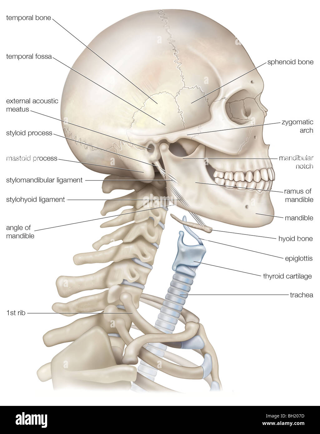 Human head and neck Stock Photohttps://www.alamy.com/image-license-details/?v=1https://www.alamy.com/stock-photo-human-head-and-neck-27703633.html
RMBH207D–Human head and neck
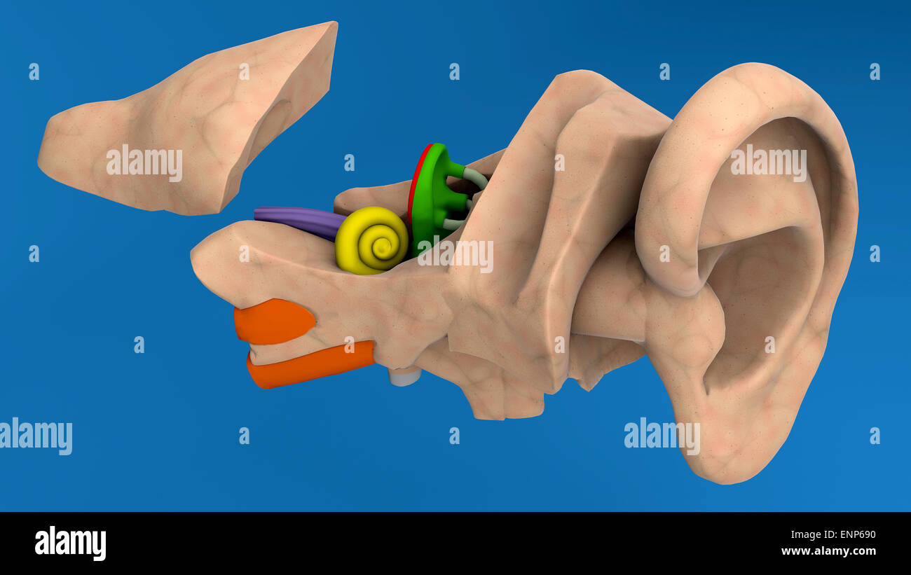 Human ear anatomy on blue background Stock Photohttps://www.alamy.com/image-license-details/?v=1https://www.alamy.com/stock-photo-human-ear-anatomy-on-blue-background-82237148.html
RMENP690–Human ear anatomy on blue background
![The sense of hearing and hearing mechanics are explained. The infographic includes three parts of the hearing system (external ear, middle and inner), the processing of sound and the elements involved, and a comparator frequency table. It also illustrates the sense of equlibrium. [6259x4015]. Stock Photo The sense of hearing and hearing mechanics are explained. The infographic includes three parts of the hearing system (external ear, middle and inner), the processing of sound and the elements involved, and a comparator frequency table. It also illustrates the sense of equlibrium. [6259x4015]. Stock Photo](https://c8.alamy.com/comp/2NEC6BY/the-sense-of-hearing-and-hearing-mechanics-are-explained-the-infographic-includes-three-parts-of-the-hearing-system-external-ear-middle-and-inner-the-processing-of-sound-and-the-elements-involved-and-a-comparator-frequency-table-it-also-illustrates-the-sense-of-equlibrium-6259x4015-2NEC6BY.jpg) The sense of hearing and hearing mechanics are explained. The infographic includes three parts of the hearing system (external ear, middle and inner), the processing of sound and the elements involved, and a comparator frequency table. It also illustrates the sense of equlibrium. [6259x4015]. Stock Photohttps://www.alamy.com/image-license-details/?v=1https://www.alamy.com/the-sense-of-hearing-and-hearing-mechanics-are-explained-the-infographic-includes-three-parts-of-the-hearing-system-external-ear-middle-and-inner-the-processing-of-sound-and-the-elements-involved-and-a-comparator-frequency-table-it-also-illustrates-the-sense-of-equlibrium-6259x4015-image525184687.html
The sense of hearing and hearing mechanics are explained. The infographic includes three parts of the hearing system (external ear, middle and inner), the processing of sound and the elements involved, and a comparator frequency table. It also illustrates the sense of equlibrium. [6259x4015]. Stock Photohttps://www.alamy.com/image-license-details/?v=1https://www.alamy.com/the-sense-of-hearing-and-hearing-mechanics-are-explained-the-infographic-includes-three-parts-of-the-hearing-system-external-ear-middle-and-inner-the-processing-of-sound-and-the-elements-involved-and-a-comparator-frequency-table-it-also-illustrates-the-sense-of-equlibrium-6259x4015-image525184687.htmlRM2NEC6BY–The sense of hearing and hearing mechanics are explained. The infographic includes three parts of the hearing system (external ear, middle and inner), the processing of sound and the elements involved, and a comparator frequency table. It also illustrates the sense of equlibrium. [6259x4015].
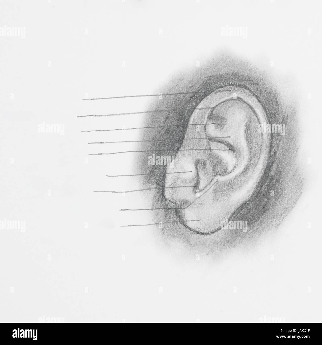 Detail of ear pencil drawing on white paper Stock Photohttps://www.alamy.com/image-license-details/?v=1https://www.alamy.com/stock-photo-detail-of-ear-pencil-drawing-on-white-paper-144267019.html
RFJAKX1F–Detail of ear pencil drawing on white paper
 Model from auditory canal - isolated on white backgriound Stock Photohttps://www.alamy.com/image-license-details/?v=1https://www.alamy.com/stock-photo-model-from-auditory-canal-isolated-on-white-backgriound-134333266.html
RFHPFBCJ–Model from auditory canal - isolated on white backgriound
![. The anatomy of the domestic animals. Veterinary anatomy. 898 THE SENSE ORGANS AND COMMON INTEGUMENT OF THE OX ?wider part is pale, and arises beneath the cervico-auricularis siiperfieiaHs indirectly from the scutifonn cartilage. The two unite and are inserteil into the lower aspect of the base of the conchal cartilage. (9) The scutulo-auricularis ]3rofundus niinor arises from the temporal crest above the external acoustic meatus and is inserted into the anterior part of the deep face of the sc-utiform cartilage. The cavum tympani is small; it communicates ventrally with the air-cells of the Stock Photo . The anatomy of the domestic animals. Veterinary anatomy. 898 THE SENSE ORGANS AND COMMON INTEGUMENT OF THE OX ?wider part is pale, and arises beneath the cervico-auricularis siiperfieiaHs indirectly from the scutifonn cartilage. The two unite and are inserteil into the lower aspect of the base of the conchal cartilage. (9) The scutulo-auricularis ]3rofundus niinor arises from the temporal crest above the external acoustic meatus and is inserted into the anterior part of the deep face of the sc-utiform cartilage. The cavum tympani is small; it communicates ventrally with the air-cells of the Stock Photo](https://c8.alamy.com/comp/RN5X2N/the-anatomy-of-the-domestic-animals-veterinary-anatomy-898-the-sense-organs-and-common-integument-of-the-ox-wider-part-is-pale-and-arises-beneath-the-cervico-auricularis-siiperfieiahs-indirectly-from-the-scutifonn-cartilage-the-two-unite-and-are-inserteil-into-the-lower-aspect-of-the-base-of-the-conchal-cartilage-9-the-scutulo-auricularis-3rofundus-niinor-arises-from-the-temporal-crest-above-the-external-acoustic-meatus-and-is-inserted-into-the-anterior-part-of-the-deep-face-of-the-sc-utiform-cartilage-the-cavum-tympani-is-small-it-communicates-ventrally-with-the-air-cells-of-the-RN5X2N.jpg) . The anatomy of the domestic animals. Veterinary anatomy. 898 THE SENSE ORGANS AND COMMON INTEGUMENT OF THE OX ?wider part is pale, and arises beneath the cervico-auricularis siiperfieiaHs indirectly from the scutifonn cartilage. The two unite and are inserteil into the lower aspect of the base of the conchal cartilage. (9) The scutulo-auricularis ]3rofundus niinor arises from the temporal crest above the external acoustic meatus and is inserted into the anterior part of the deep face of the sc-utiform cartilage. The cavum tympani is small; it communicates ventrally with the air-cells of the Stock Photohttps://www.alamy.com/image-license-details/?v=1https://www.alamy.com/the-anatomy-of-the-domestic-animals-veterinary-anatomy-898-the-sense-organs-and-common-integument-of-the-ox-wider-part-is-pale-and-arises-beneath-the-cervico-auricularis-siiperfieiahs-indirectly-from-the-scutifonn-cartilage-the-two-unite-and-are-inserteil-into-the-lower-aspect-of-the-base-of-the-conchal-cartilage-9-the-scutulo-auricularis-3rofundus-niinor-arises-from-the-temporal-crest-above-the-external-acoustic-meatus-and-is-inserted-into-the-anterior-part-of-the-deep-face-of-the-sc-utiform-cartilage-the-cavum-tympani-is-small-it-communicates-ventrally-with-the-air-cells-of-the-image236772781.html
. The anatomy of the domestic animals. Veterinary anatomy. 898 THE SENSE ORGANS AND COMMON INTEGUMENT OF THE OX ?wider part is pale, and arises beneath the cervico-auricularis siiperfieiaHs indirectly from the scutifonn cartilage. The two unite and are inserteil into the lower aspect of the base of the conchal cartilage. (9) The scutulo-auricularis ]3rofundus niinor arises from the temporal crest above the external acoustic meatus and is inserted into the anterior part of the deep face of the sc-utiform cartilage. The cavum tympani is small; it communicates ventrally with the air-cells of the Stock Photohttps://www.alamy.com/image-license-details/?v=1https://www.alamy.com/the-anatomy-of-the-domestic-animals-veterinary-anatomy-898-the-sense-organs-and-common-integument-of-the-ox-wider-part-is-pale-and-arises-beneath-the-cervico-auricularis-siiperfieiahs-indirectly-from-the-scutifonn-cartilage-the-two-unite-and-are-inserteil-into-the-lower-aspect-of-the-base-of-the-conchal-cartilage-9-the-scutulo-auricularis-3rofundus-niinor-arises-from-the-temporal-crest-above-the-external-acoustic-meatus-and-is-inserted-into-the-anterior-part-of-the-deep-face-of-the-sc-utiform-cartilage-the-cavum-tympani-is-small-it-communicates-ventrally-with-the-air-cells-of-the-image236772781.htmlRMRN5X2N–. The anatomy of the domestic animals. Veterinary anatomy. 898 THE SENSE ORGANS AND COMMON INTEGUMENT OF THE OX ?wider part is pale, and arises beneath the cervico-auricularis siiperfieiaHs indirectly from the scutifonn cartilage. The two unite and are inserteil into the lower aspect of the base of the conchal cartilage. (9) The scutulo-auricularis ]3rofundus niinor arises from the temporal crest above the external acoustic meatus and is inserted into the anterior part of the deep face of the sc-utiform cartilage. The cavum tympani is small; it communicates ventrally with the air-cells of the
 Medical practice, Doctors surgery. Examination of the ear, acoustic meatus, ear canal of a patient, with a loupe. Stock Photohttps://www.alamy.com/image-license-details/?v=1https://www.alamy.com/stock-photo-medical-practice-doctors-surgery-examination-of-the-ear-acoustic-meatus-31853364.html
RMBRR184–Medical practice, Doctors surgery. Examination of the ear, acoustic meatus, ear canal of a patient, with a loupe.
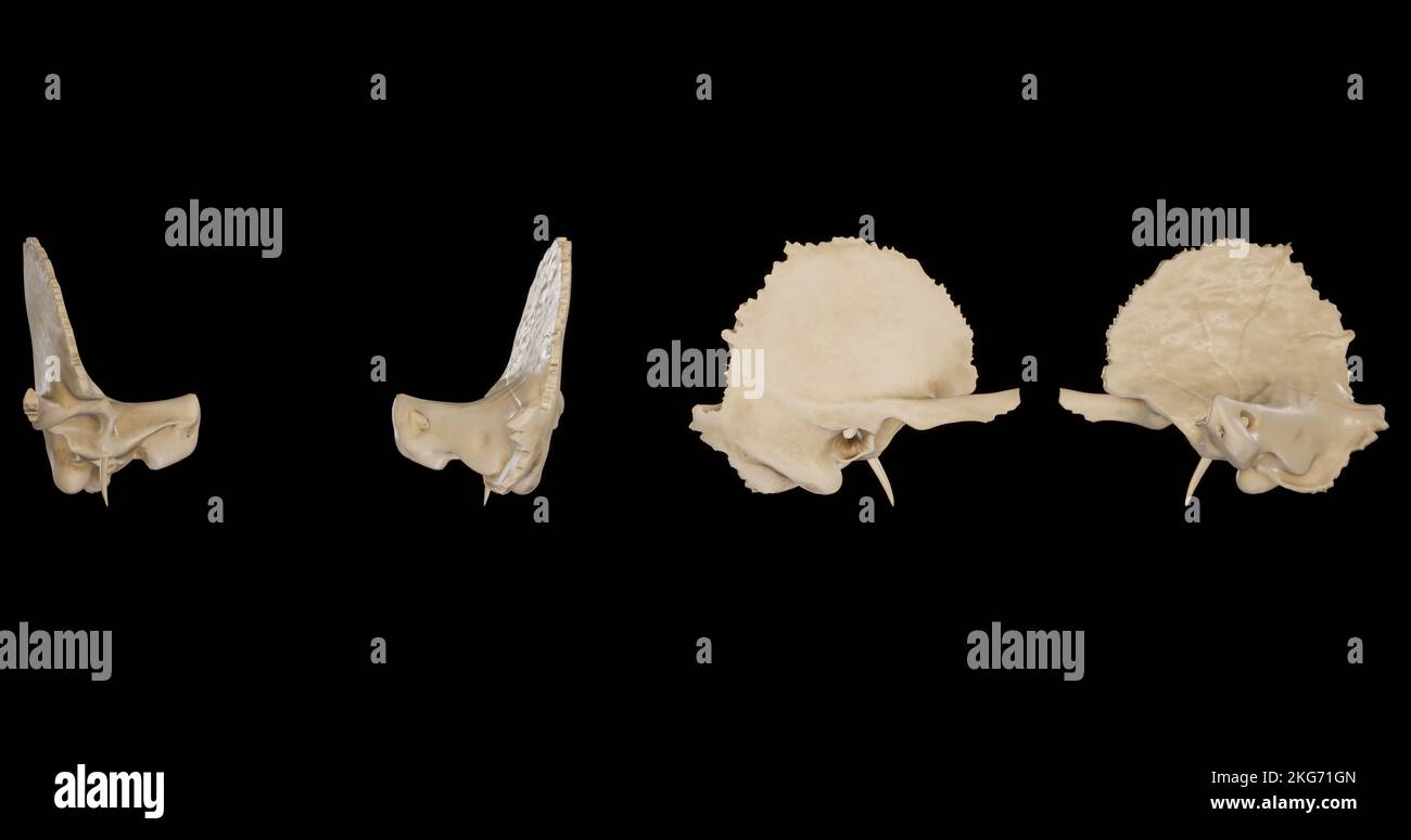 Right Temporal Bone-Multiple Views Stock Photohttps://www.alamy.com/image-license-details/?v=1https://www.alamy.com/right-temporal-bone-multiple-views-image491879717.html
RF2KG71GN–Right Temporal Bone-Multiple Views
 . Cunningham's Text-book of anatomy. Anatomy. THE LYMPH GLANDS OF THE HEAD. 999 from the external acoustic meatus, the tympanum, the soft palate, the posterior part of the nose, and the deeper portions of the cheek. Their efferents open into the upper deep cervical glands. The Superficial Facial Lymph Glands.—Several lymph glands, or groups of lymph glands, have been found in the region of the face but, apparently, they are irregular, both in occurrence and in position. Those which appear to be most frequently found are : Infra-orbital, which lie along the angle between the nose and the cheek, Stock Photohttps://www.alamy.com/image-license-details/?v=1https://www.alamy.com/cunninghams-text-book-of-anatomy-anatomy-the-lymph-glands-of-the-head-999-from-the-external-acoustic-meatus-the-tympanum-the-soft-palate-the-posterior-part-of-the-nose-and-the-deeper-portions-of-the-cheek-their-efferents-open-into-the-upper-deep-cervical-glands-the-superficial-facial-lymph-glandsseveral-lymph-glands-or-groups-of-lymph-glands-have-been-found-in-the-region-of-the-face-but-apparently-they-are-irregular-both-in-occurrence-and-in-position-those-which-appear-to-be-most-frequently-found-are-infra-orbital-which-lie-along-the-angle-between-the-nose-and-the-cheek-image216334302.html
RMPFXTH2–. Cunningham's Text-book of anatomy. Anatomy. THE LYMPH GLANDS OF THE HEAD. 999 from the external acoustic meatus, the tympanum, the soft palate, the posterior part of the nose, and the deeper portions of the cheek. Their efferents open into the upper deep cervical glands. The Superficial Facial Lymph Glands.—Several lymph glands, or groups of lymph glands, have been found in the region of the face but, apparently, they are irregular, both in occurrence and in position. Those which appear to be most frequently found are : Infra-orbital, which lie along the angle between the nose and the cheek,
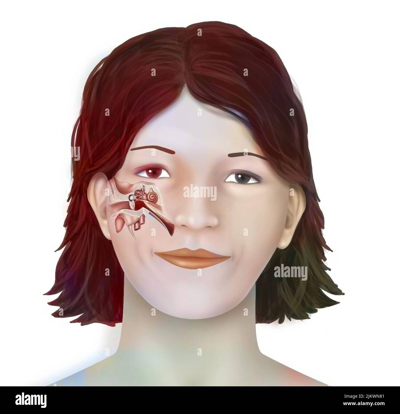 Anatomy of the inner ear showing the eardrum, the cochlea. Stock Photohttps://www.alamy.com/image-license-details/?v=1https://www.alamy.com/anatomy-of-the-inner-ear-showing-the-eardrum-the-cochlea-image476923889.html
RF2JKWN81–Anatomy of the inner ear showing the eardrum, the cochlea.
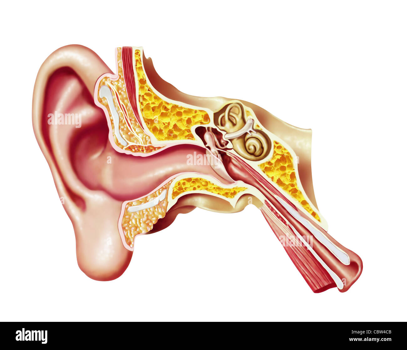 Human ear cutaway diagram. Anatomy illustration. Stock Photohttps://www.alamy.com/image-license-details/?v=1https://www.alamy.com/stock-photo-human-ear-cutaway-diagram-anatomy-illustration-41734235.html
RFCBW4CB–Human ear cutaway diagram. Anatomy illustration.
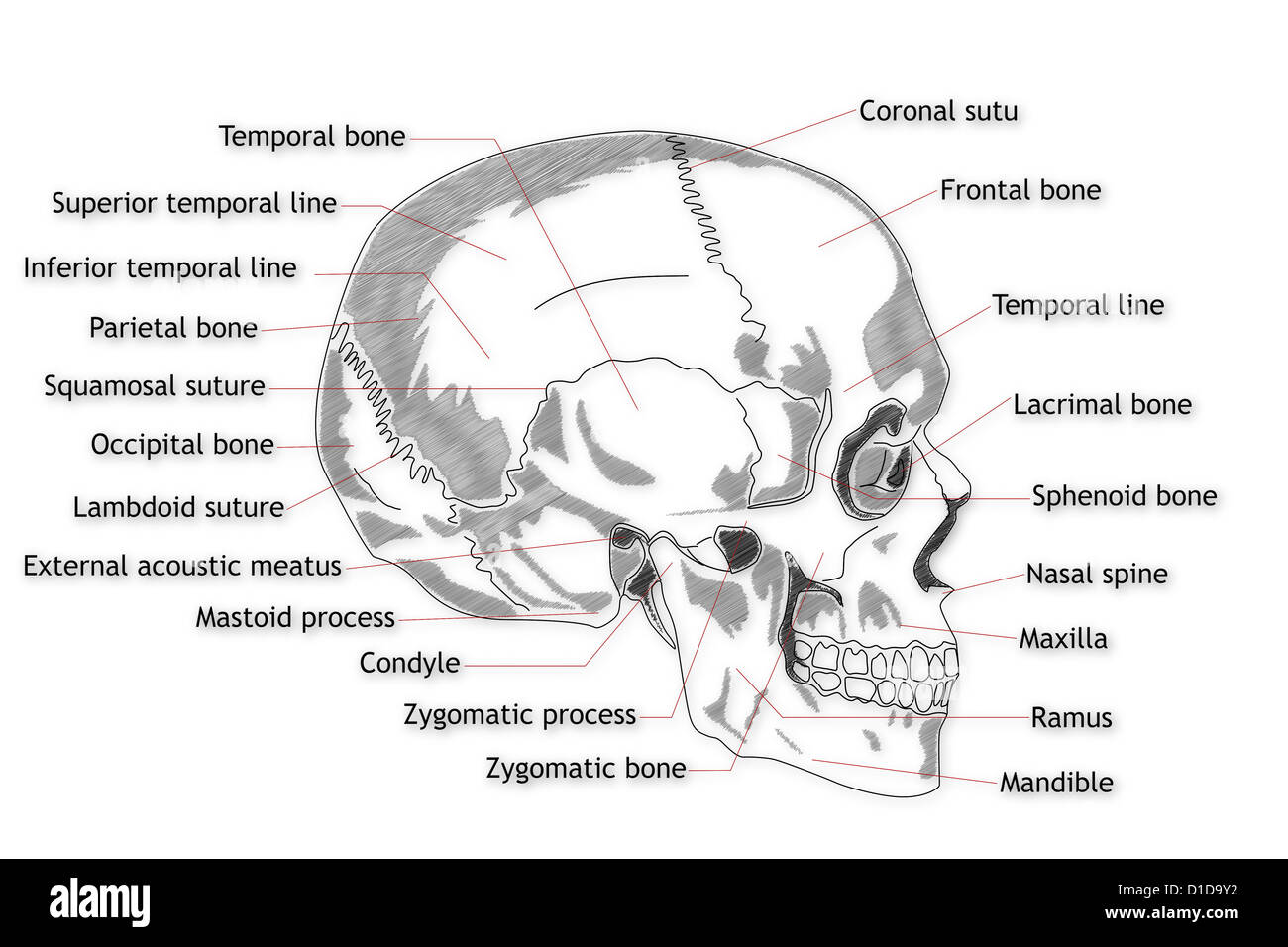 Human Skull structure Stock Photohttps://www.alamy.com/image-license-details/?v=1https://www.alamy.com/stock-photo-human-skull-structure-52538950.html
RFD1D9Y2–Human Skull structure
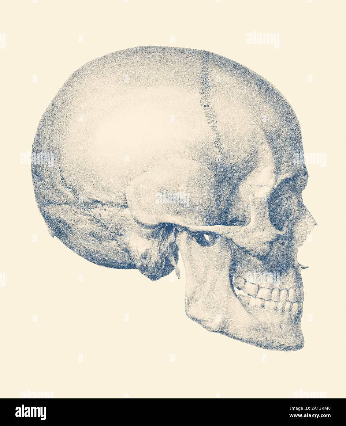 Vintage anatomy print features a side view of the human skull. Stock Photohttps://www.alamy.com/image-license-details/?v=1https://www.alamy.com/vintage-anatomy-print-features-a-side-view-of-the-human-skull-image327696096.html
RF2A13RM0–Vintage anatomy print features a side view of the human skull.
 Illustration of music entering ear, listening to music Stock Photohttps://www.alamy.com/image-license-details/?v=1https://www.alamy.com/stock-photo-illustration-of-music-entering-ear-listening-to-music-54671594.html
RFD4XE4X–Illustration of music entering ear, listening to music
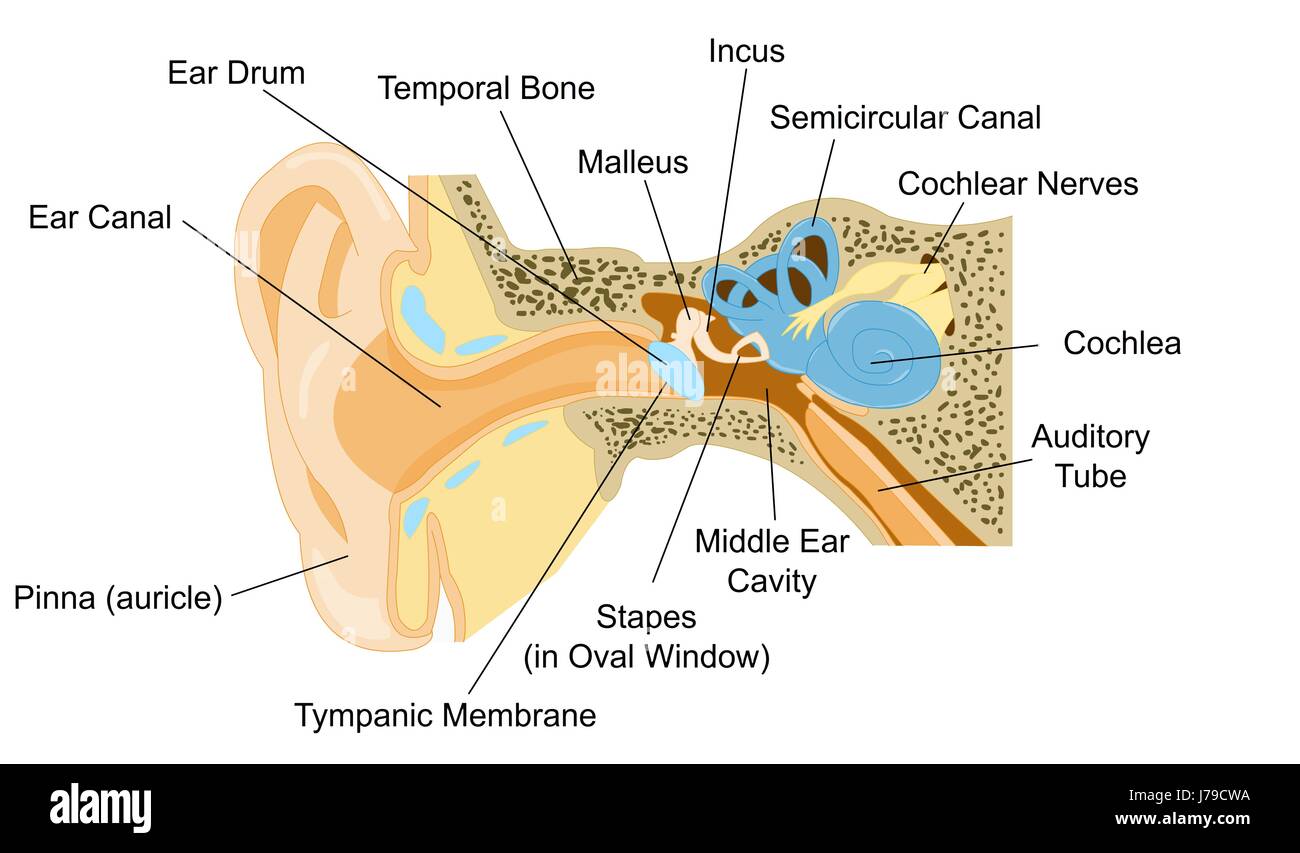 Ear Anatomy Stock Photohttps://www.alamy.com/image-license-details/?v=1https://www.alamy.com/stock-photo-ear-anatomy-142193222.html
RFJ79CWA–Ear Anatomy
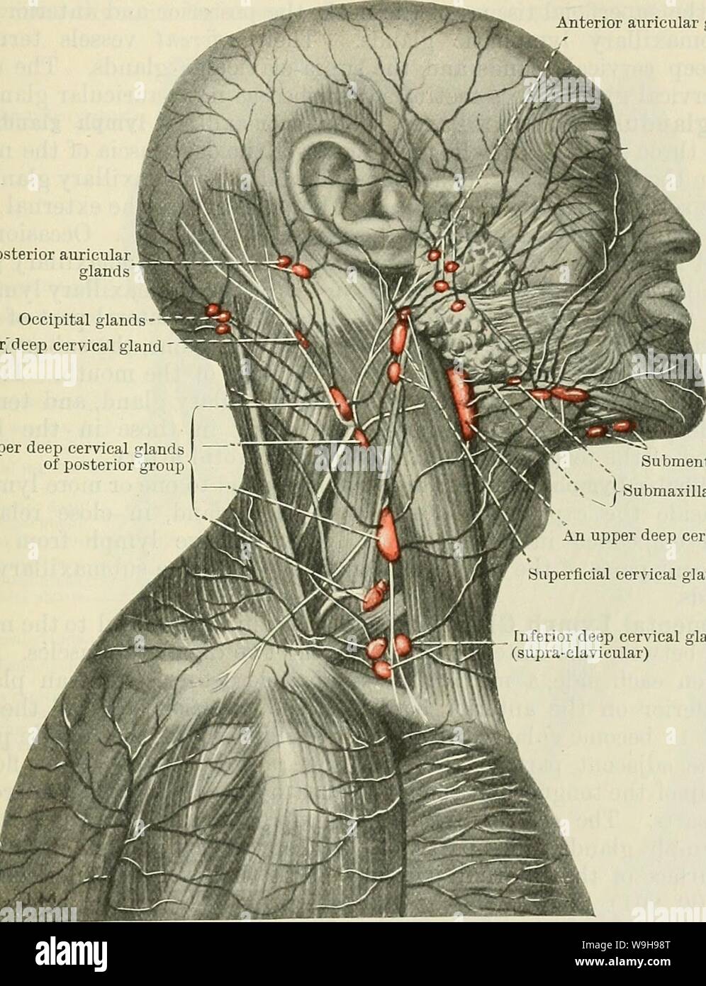 Archive image from page 1032 of Cunningham's Text-book of anatomy (1914). Cunningham's Text-book of anatomy cunninghamstextb00cunn Year: 1914 ( THE LYMPH GLANDS OF THE HEAD. 999 from the external acoustic meatus, the tympanum, the soft palate, the posterior part of the nose, and the deeper portions of the cheek. Their efferents open into the upper deep cervical glands. The Superficial Facial Lymph Glands.—Several lymph glands, or groups of lymph glands, have been found in the region of the face but, apparently, they are irregular, both in occurrence and in position. Those which appear to be m Stock Photohttps://www.alamy.com/image-license-details/?v=1https://www.alamy.com/archive-image-from-page-1032-of-cunninghams-text-book-of-anatomy-1914-cunninghams-text-book-of-anatomy-cunninghamstextb00cunn-year-1914-the-lymph-glands-of-the-head-999-from-the-external-acoustic-meatus-the-tympanum-the-soft-palate-the-posterior-part-of-the-nose-and-the-deeper-portions-of-the-cheek-their-efferents-open-into-the-upper-deep-cervical-glands-the-superficial-facial-lymph-glandsseveral-lymph-glands-or-groups-of-lymph-glands-have-been-found-in-the-region-of-the-face-but-apparently-they-are-irregular-both-in-occurrence-and-in-position-those-which-appear-to-be-m-image264067912.html
RMW9H98T–Archive image from page 1032 of Cunningham's Text-book of anatomy (1914). Cunningham's Text-book of anatomy cunninghamstextb00cunn Year: 1914 ( THE LYMPH GLANDS OF THE HEAD. 999 from the external acoustic meatus, the tympanum, the soft palate, the posterior part of the nose, and the deeper portions of the cheek. Their efferents open into the upper deep cervical glands. The Superficial Facial Lymph Glands.—Several lymph glands, or groups of lymph glands, have been found in the region of the face but, apparently, they are irregular, both in occurrence and in position. Those which appear to be m
 sound, medicinally, medical, science, cross, human, human being, sense, Stock Photohttps://www.alamy.com/image-license-details/?v=1https://www.alamy.com/stock-photo-sound-medicinally-medical-science-cross-human-human-being-sense-131574647.html
RFHJ1MPF–sound, medicinally, medical, science, cross, human, human being, sense,
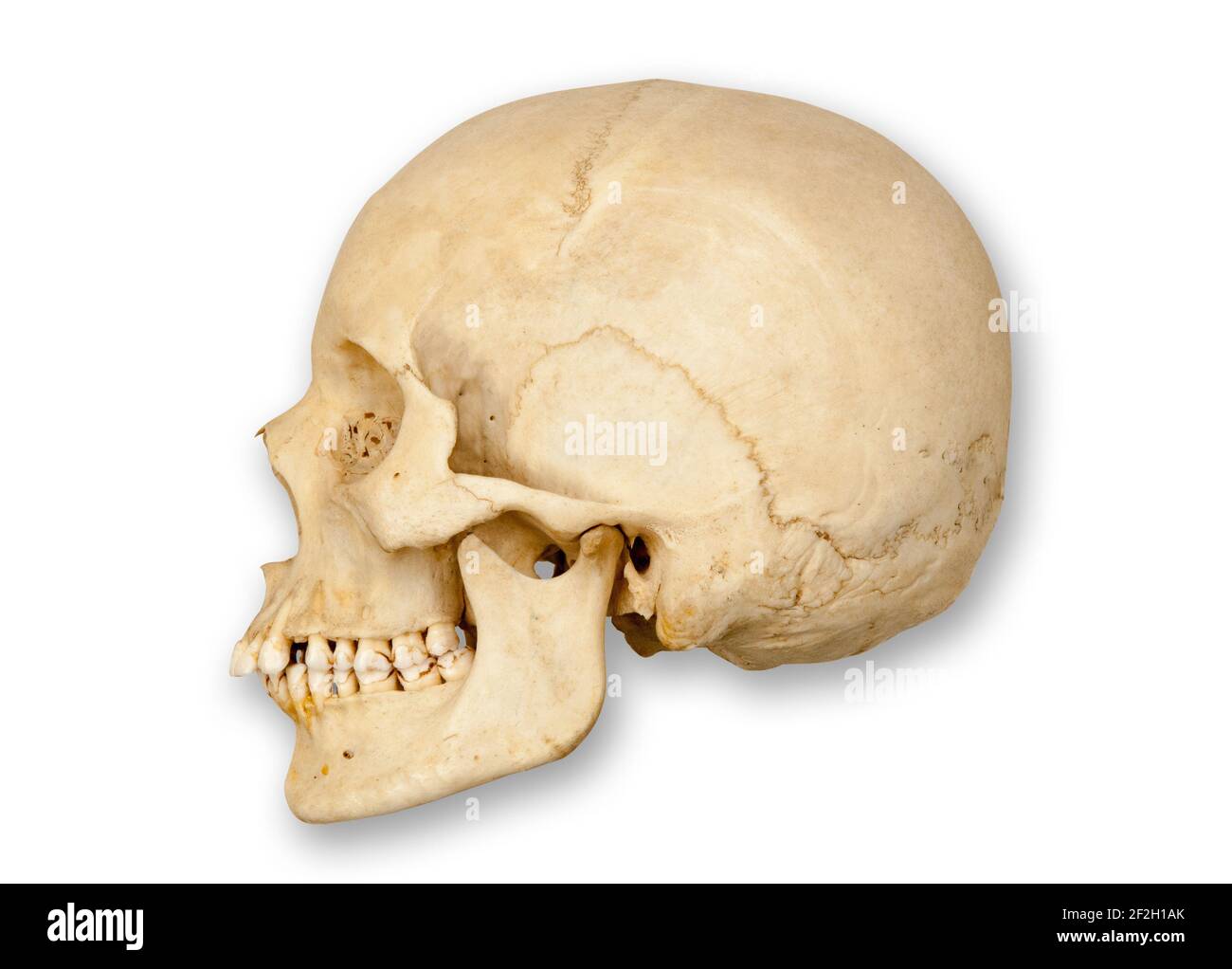 Sideways or profile view of left side of a human skull cut out with soft drop shadow on a white background. Stock Photohttps://www.alamy.com/image-license-details/?v=1https://www.alamy.com/sideways-or-profile-view-of-left-side-of-a-human-skull-cut-out-with-soft-drop-shadow-on-a-white-background-image414652411.html
RF2F2H1AK–Sideways or profile view of left side of a human skull cut out with soft drop shadow on a white background.
 Zygoma word cloud concept Stock Photohttps://www.alamy.com/image-license-details/?v=1https://www.alamy.com/zygoma-word-cloud-concept-image268036388.html
RFWG2344–Zygoma word cloud concept
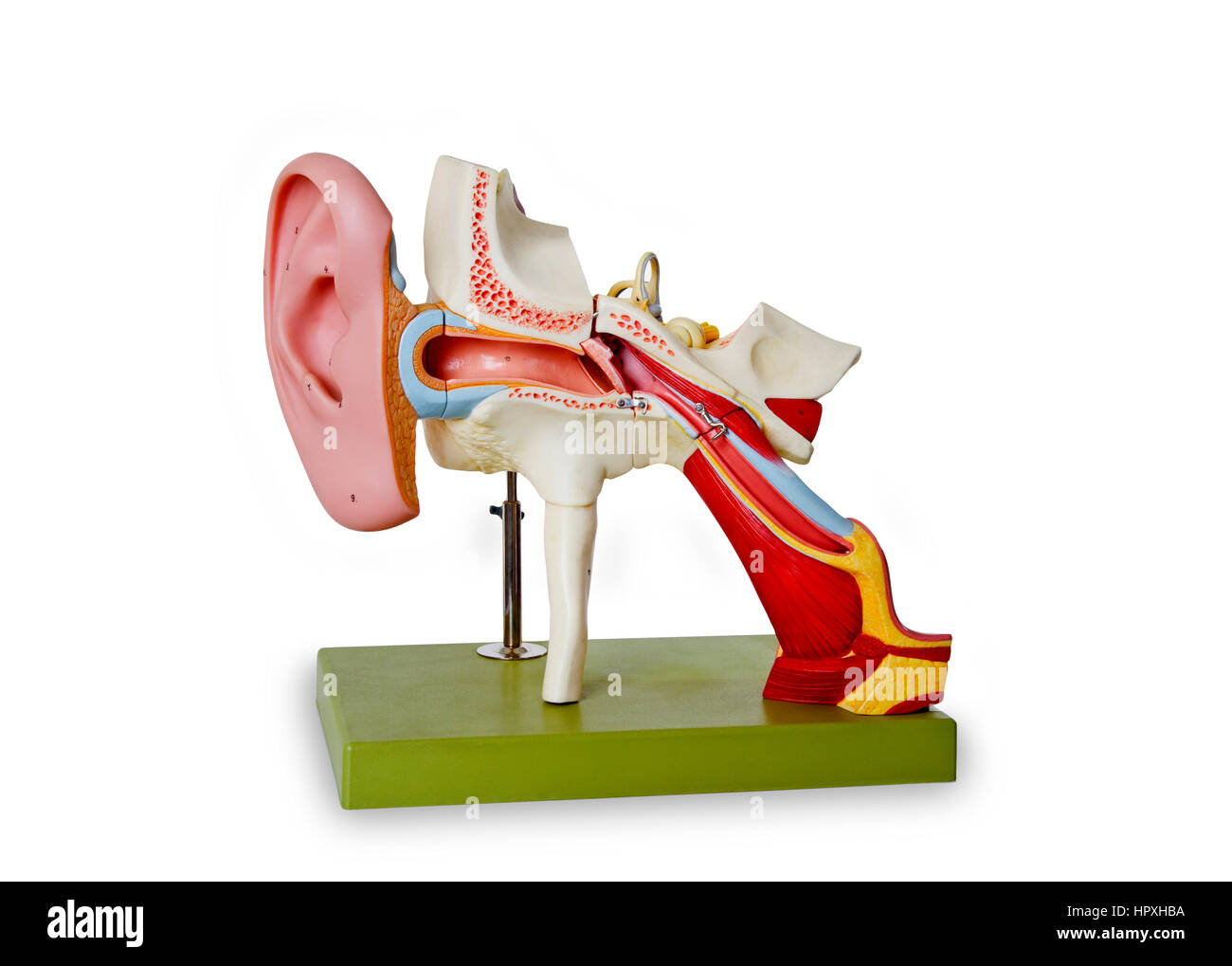 Model from auditory canal - isolated on white backgriound Stock Photohttps://www.alamy.com/image-license-details/?v=1https://www.alamy.com/stock-photo-model-from-auditory-canal-isolated-on-white-backgriound-134579406.html
RFHPXHBA–Model from auditory canal - isolated on white backgriound
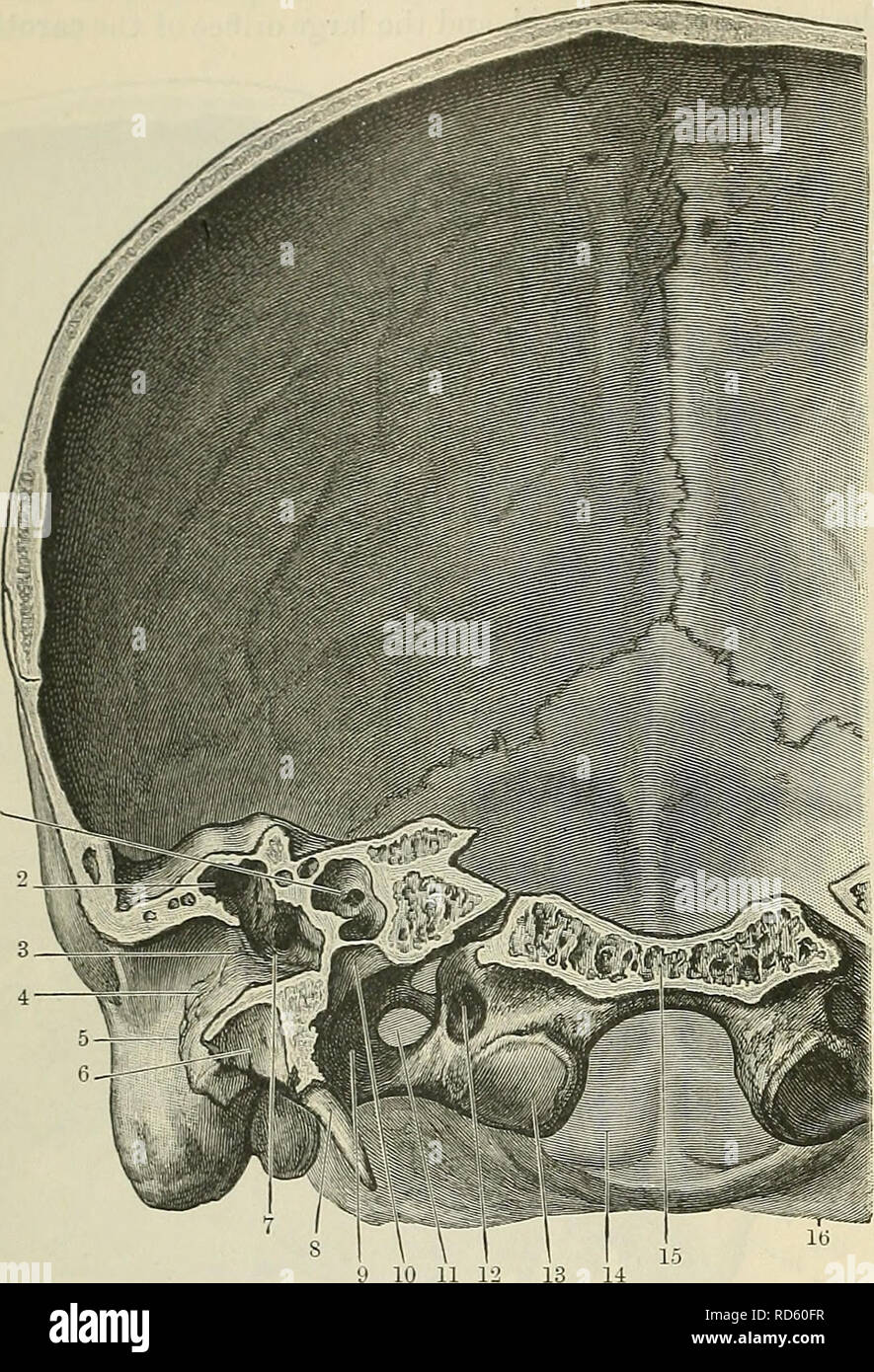 . Cunningham's Text-book of anatomy. Anatomy. 102 OSTEOLOGY. roofed in by the thin tegmen tympani, which separates it from the middle cranial fossa. The obliquity of the medial end of the external acoustic meatus, together with the groove for the attachment of the tympanic membrane is well seen, and the thickness of the upper wall of that passage is also noteworthy. The floor of the meatus, formed by the tympanic plate, which separ- ates it from the mandibular fossa, is much thinner, but in the region of the root of the styloid process there is a mass- ing together of dense bone. HORIZONTAL SE Stock Photohttps://www.alamy.com/image-license-details/?v=1https://www.alamy.com/cunninghams-text-book-of-anatomy-anatomy-102-osteology-roofed-in-by-the-thin-tegmen-tympani-which-separates-it-from-the-middle-cranial-fossa-the-obliquity-of-the-medial-end-of-the-external-acoustic-meatus-together-with-the-groove-for-the-attachment-of-the-tympanic-membrane-is-well-seen-and-the-thickness-of-the-upper-wall-of-that-passage-is-also-noteworthy-the-floor-of-the-meatus-formed-by-the-tympanic-plate-which-separ-ates-it-from-the-mandibular-fossa-is-much-thinner-but-in-the-region-of-the-root-of-the-styloid-process-there-is-a-mass-ing-together-of-dense-bone-horizontal-se-image231857467.html
RMRD60FR–. Cunningham's Text-book of anatomy. Anatomy. 102 OSTEOLOGY. roofed in by the thin tegmen tympani, which separates it from the middle cranial fossa. The obliquity of the medial end of the external acoustic meatus, together with the groove for the attachment of the tympanic membrane is well seen, and the thickness of the upper wall of that passage is also noteworthy. The floor of the meatus, formed by the tympanic plate, which separ- ates it from the mandibular fossa, is much thinner, but in the region of the root of the styloid process there is a mass- ing together of dense bone. HORIZONTAL SE
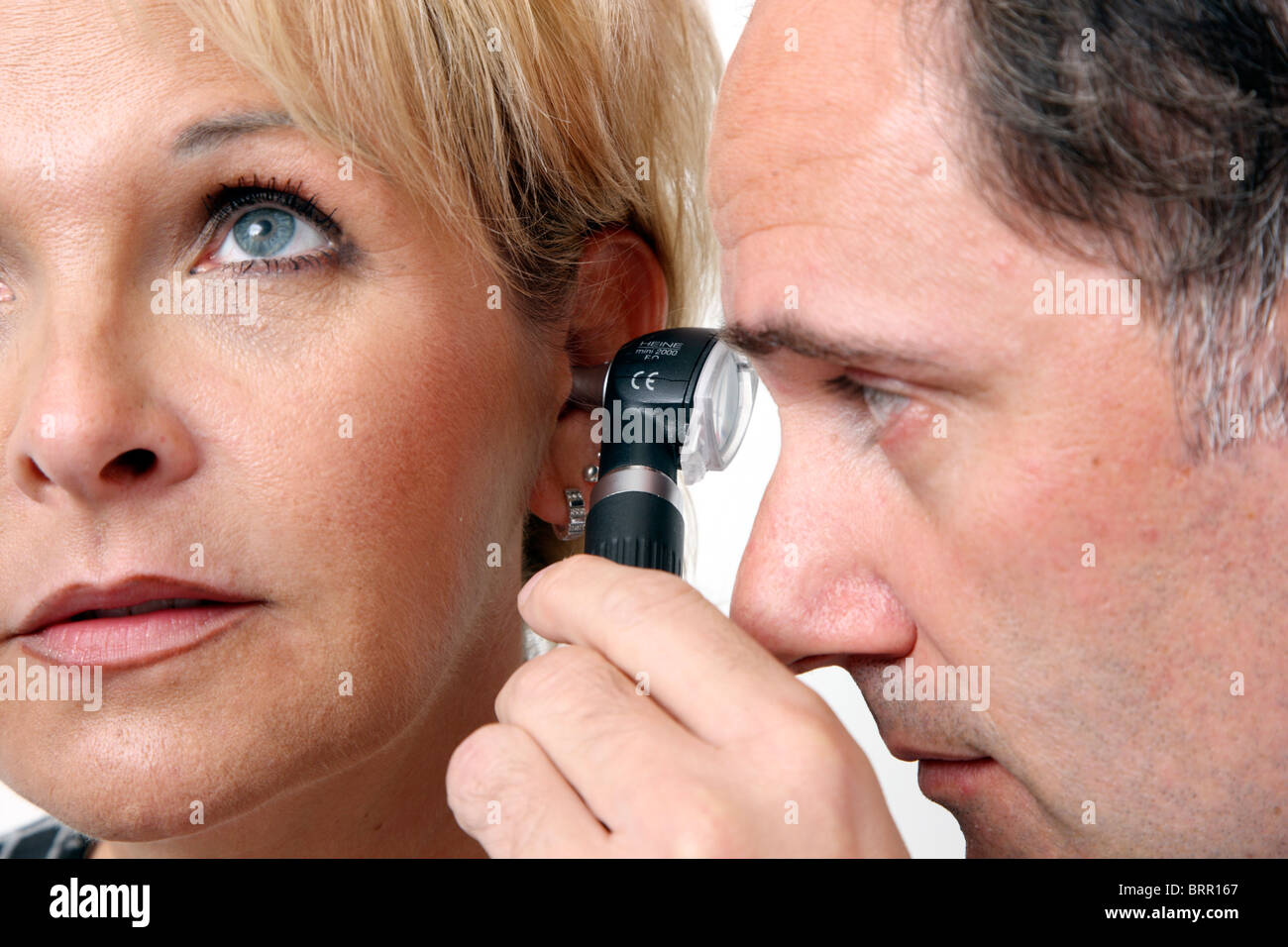 Medical practice, Doctors surgery. Examination of the ear, acoustic meatus, ear canal of a patient, with a loupe. Stock Photohttps://www.alamy.com/image-license-details/?v=1https://www.alamy.com/stock-photo-medical-practice-doctors-surgery-examination-of-the-ear-acoustic-meatus-31853311.html
RMBRR167–Medical practice, Doctors surgery. Examination of the ear, acoustic meatus, ear canal of a patient, with a loupe.
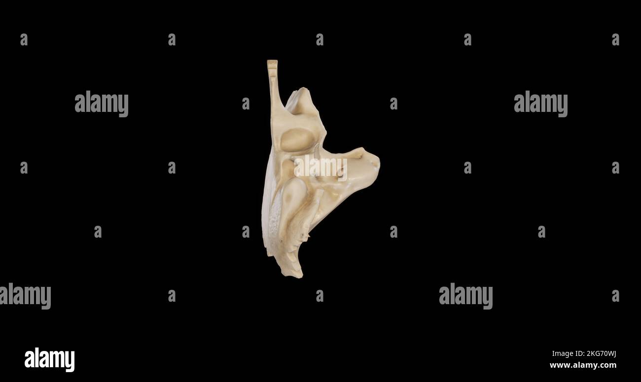 Inferior view of Right Temporal Bone Stock Photohttps://www.alamy.com/image-license-details/?v=1https://www.alamy.com/inferior-view-of-right-temporal-bone-image491879182.html
RF2KG70WJ–Inferior view of Right Temporal Bone
 Model from auditory canal - isolated on white backgriound Stock Photohttps://www.alamy.com/image-license-details/?v=1https://www.alamy.com/stock-photo-model-from-auditory-canal-isolated-on-white-backgriound-136754022.html
RFHXDK46–Model from auditory canal - isolated on white backgriound
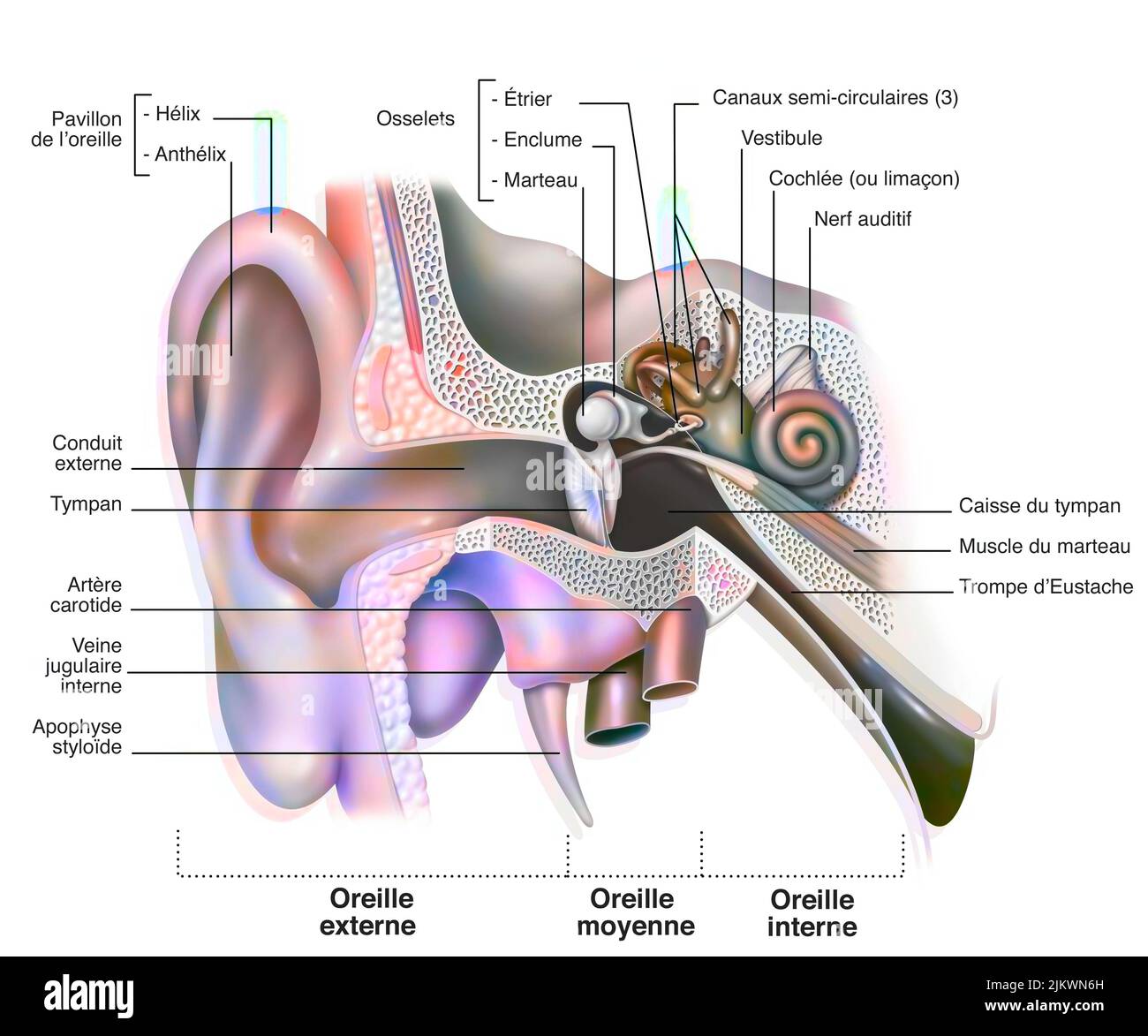 Anatomy of the inner ear showing the eardrum, the cochlea. Stock Photohttps://www.alamy.com/image-license-details/?v=1https://www.alamy.com/anatomy-of-the-inner-ear-showing-the-eardrum-the-cochlea-image476923849.html
RF2JKWN6H–Anatomy of the inner ear showing the eardrum, the cochlea.
 . Cunningham's Text-book of anatomy. Anatomy. 102 OSTEOLOGY. roofed in by the thin tegmen tympani, which separates it from the middle cranial fossa. The obliquity of the medial end of the external acoustic meatus, together with the groove for the attachment of the tympanic membrane is well seen, and the thickness of the upper wall of that passage is also noteworthy. The floor of the meatus, formed by the tympanic plate, which separ- ates it from the mandibular fossa, is much thinner, but in the region of the root of the styloid process there is a mass- ing together of dense bone. HORIZONTAL SE Stock Photohttps://www.alamy.com/image-license-details/?v=1https://www.alamy.com/cunninghams-text-book-of-anatomy-anatomy-102-osteology-roofed-in-by-the-thin-tegmen-tympani-which-separates-it-from-the-middle-cranial-fossa-the-obliquity-of-the-medial-end-of-the-external-acoustic-meatus-together-with-the-groove-for-the-attachment-of-the-tympanic-membrane-is-well-seen-and-the-thickness-of-the-upper-wall-of-that-passage-is-also-noteworthy-the-floor-of-the-meatus-formed-by-the-tympanic-plate-which-separ-ates-it-from-the-mandibular-fossa-is-much-thinner-but-in-the-region-of-the-root-of-the-styloid-process-there-is-a-mass-ing-together-of-dense-bone-horizontal-se-image216346663.html
RMPFYCAF–. Cunningham's Text-book of anatomy. Anatomy. 102 OSTEOLOGY. roofed in by the thin tegmen tympani, which separates it from the middle cranial fossa. The obliquity of the medial end of the external acoustic meatus, together with the groove for the attachment of the tympanic membrane is well seen, and the thickness of the upper wall of that passage is also noteworthy. The floor of the meatus, formed by the tympanic plate, which separ- ates it from the mandibular fossa, is much thinner, but in the region of the root of the styloid process there is a mass- ing together of dense bone. HORIZONTAL SE
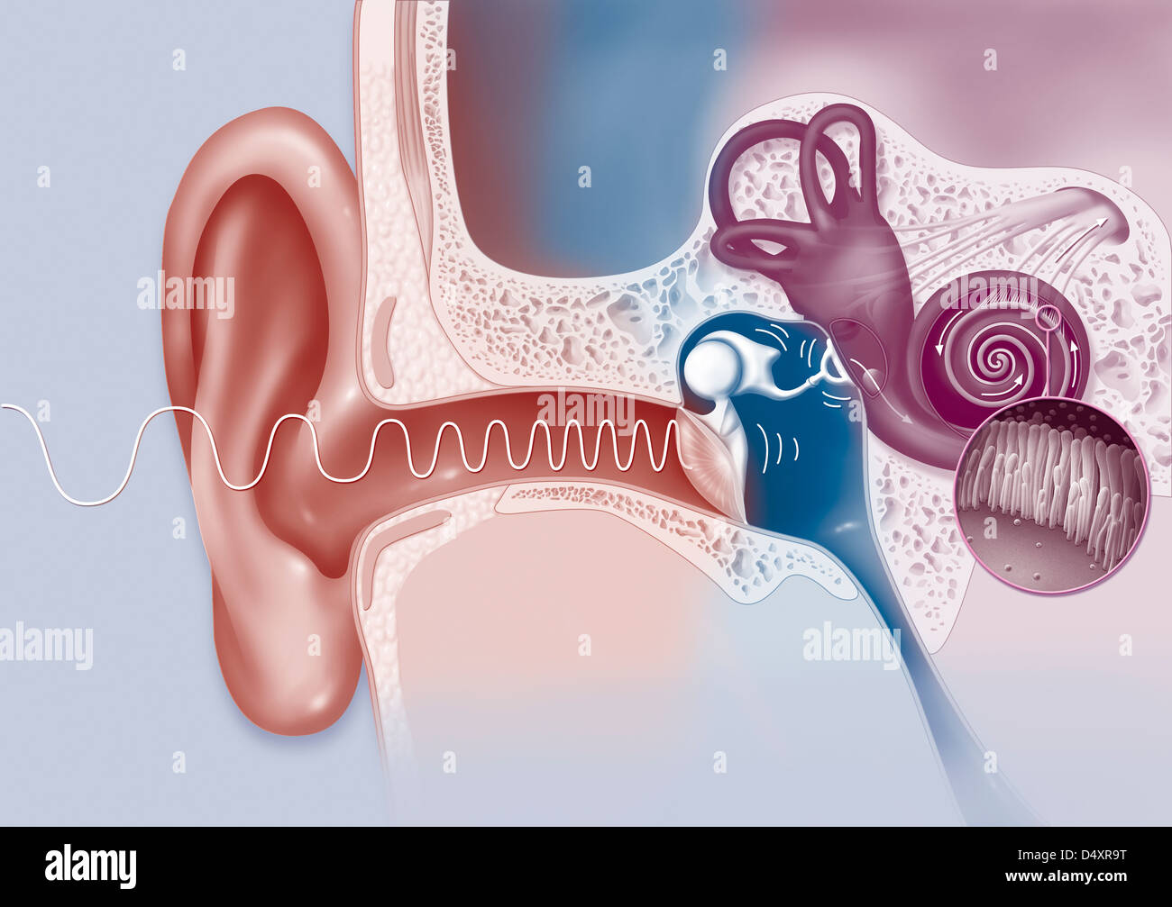 EAR, DRAWING Stock Photohttps://www.alamy.com/image-license-details/?v=1https://www.alamy.com/stock-photo-ear-drawing-54678788.html
RMD4XR9T–EAR, DRAWING
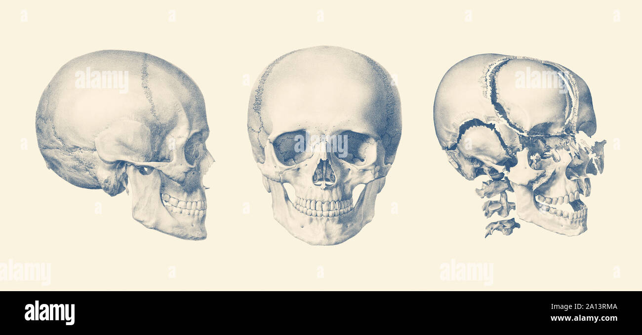 A multi view of the human skull, showing the bone breakdown. Stock Photohttps://www.alamy.com/image-license-details/?v=1https://www.alamy.com/a-multi-view-of-the-human-skull-showing-the-bone-breakdown-image327696106.html
RF2A13RMA–A multi view of the human skull, showing the bone breakdown.
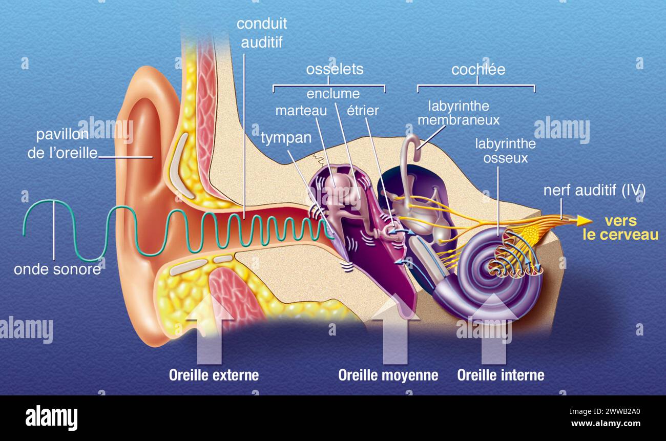 Representation of the anatomy of the ear, from left to right: - external: pavilion, auditory canal, eardrum - middle: ossicles. Stock Photohttps://www.alamy.com/image-license-details/?v=1https://www.alamy.com/representation-of-the-anatomy-of-the-ear-from-left-to-right-external-pavilion-auditory-canal-eardrum-middle-ossicles-image600762232.html
RM2WWB2A0–Representation of the anatomy of the ear, from left to right: - external: pavilion, auditory canal, eardrum - middle: ossicles.
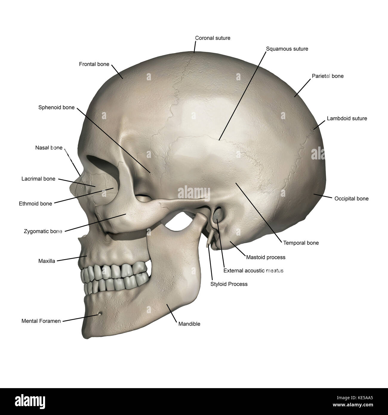 Lateral view of human skull anatomy with annotations. Stock Photohttps://www.alamy.com/image-license-details/?v=1https://www.alamy.com/stock-image-lateral-view-of-human-skull-anatomy-with-annotations-163616381.html
RFKE5AA5–Lateral view of human skull anatomy with annotations.
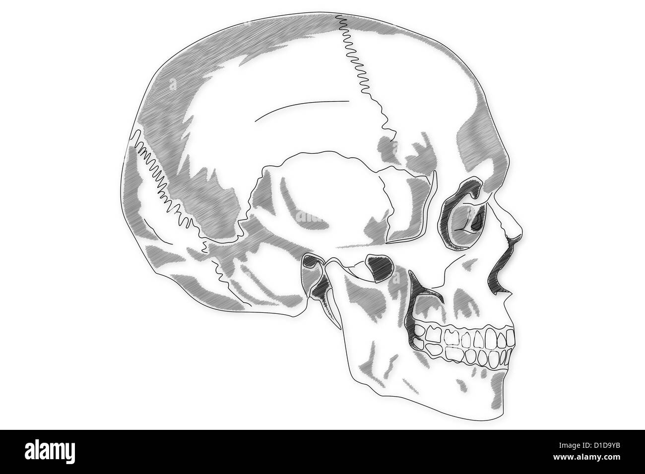 Human Skull structure Stock Photohttps://www.alamy.com/image-license-details/?v=1https://www.alamy.com/stock-photo-human-skull-structure-52538959.html
RFD1D9YB–Human Skull structure
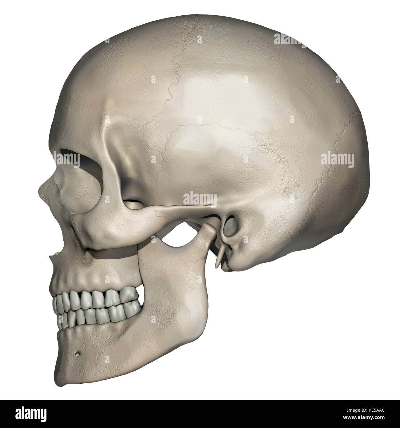 Lateral view of human skull anatomy. Stock Photohttps://www.alamy.com/image-license-details/?v=1https://www.alamy.com/stock-image-lateral-view-of-human-skull-anatomy-163616388.html
RFKE5AAC–Lateral view of human skull anatomy.
 Illustration of music entering ear, listening to music Stock Photohttps://www.alamy.com/image-license-details/?v=1https://www.alamy.com/stock-photo-illustration-of-music-entering-ear-listening-to-music-54671602.html
RFD4XE56–Illustration of music entering ear, listening to music
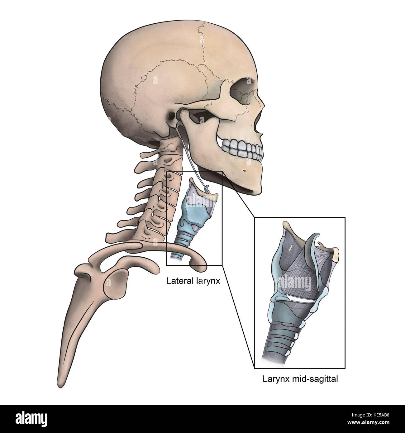 Lateral larynx and skeletal anatomy with mid-sagittal larynx view. Stock Photohttps://www.alamy.com/image-license-details/?v=1https://www.alamy.com/stock-image-lateral-larynx-and-skeletal-anatomy-with-mid-sagittal-larynx-view-163616412.html
RFKE5AB8–Lateral larynx and skeletal anatomy with mid-sagittal larynx view.
 sound, medicinally, medical, science, cross, human, human being, sense, Stock Photohttps://www.alamy.com/image-license-details/?v=1https://www.alamy.com/stock-photo-sound-medicinally-medical-science-cross-human-human-being-sense-131487393.html
RFHHWNE9–sound, medicinally, medical, science, cross, human, human being, sense,
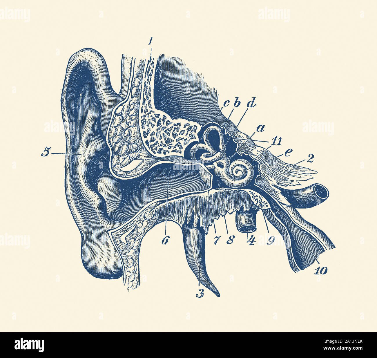 Vintage anatomy print showing a diagram of the inner ear. Stock Photohttps://www.alamy.com/image-license-details/?v=1https://www.alamy.com/vintage-anatomy-print-showing-a-diagram-of-the-inner-ear-image327694379.html
RF2A13NEK–Vintage anatomy print showing a diagram of the inner ear.
 . The anatomy of the domestic animals. Veterinary anatomy. 190 SKELETON OF THE DOG extensively with the corresponding process of tlie malar. The articular surface for the condyle of the mandible consists of a transverse groove which is continued upon the front of the large postglenoid process. Behind the latter is the lower opening of the temporal canal. There is no condyle. The mastoid part is small, but bears a distinct mastoid process. The external acoustic meatus is wide and very short, so that one can see into the tympanum in the dry skull. The bulla ossea is very large and is rounded and Stock Photohttps://www.alamy.com/image-license-details/?v=1https://www.alamy.com/the-anatomy-of-the-domestic-animals-veterinary-anatomy-190-skeleton-of-the-dog-extensively-with-the-corresponding-process-of-tlie-malar-the-articular-surface-for-the-condyle-of-the-mandible-consists-of-a-transverse-groove-which-is-continued-upon-the-front-of-the-large-postglenoid-process-behind-the-latter-is-the-lower-opening-of-the-temporal-canal-there-is-no-condyle-the-mastoid-part-is-small-but-bears-a-distinct-mastoid-process-the-external-acoustic-meatus-is-wide-and-very-short-so-that-one-can-see-into-the-tympanum-in-the-dry-skull-the-bulla-ossea-is-very-large-and-is-rounded-and-image236802729.html
RMRN7889–. The anatomy of the domestic animals. Veterinary anatomy. 190 SKELETON OF THE DOG extensively with the corresponding process of tlie malar. The articular surface for the condyle of the mandible consists of a transverse groove which is continued upon the front of the large postglenoid process. Behind the latter is the lower opening of the temporal canal. There is no condyle. The mastoid part is small, but bears a distinct mastoid process. The external acoustic meatus is wide and very short, so that one can see into the tympanum in the dry skull. The bulla ossea is very large and is rounded and
 Medical practice, Doctors surgery. Examination of the ear, acoustic meatus, ear canal of a patient, with a loupe. Stock Photohttps://www.alamy.com/image-license-details/?v=1https://www.alamy.com/stock-photo-medical-practice-doctors-surgery-examination-of-the-ear-acoustic-meatus-31850662.html
RMBRPWRJ–Medical practice, Doctors surgery. Examination of the ear, acoustic meatus, ear canal of a patient, with a loupe.
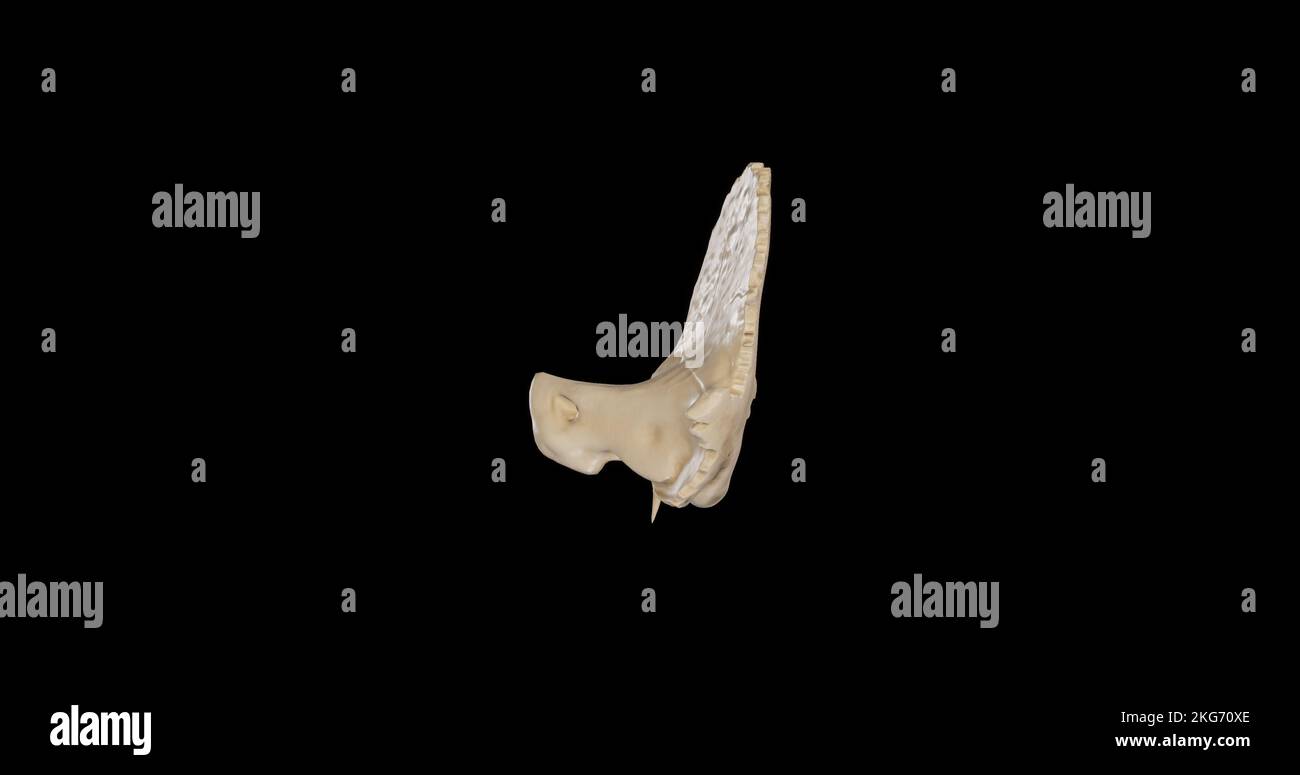 Posterior view of Right Temporal Bone Stock Photohttps://www.alamy.com/image-license-details/?v=1https://www.alamy.com/posterior-view-of-right-temporal-bone-image491879206.html
RF2KG70XE–Posterior view of Right Temporal Bone
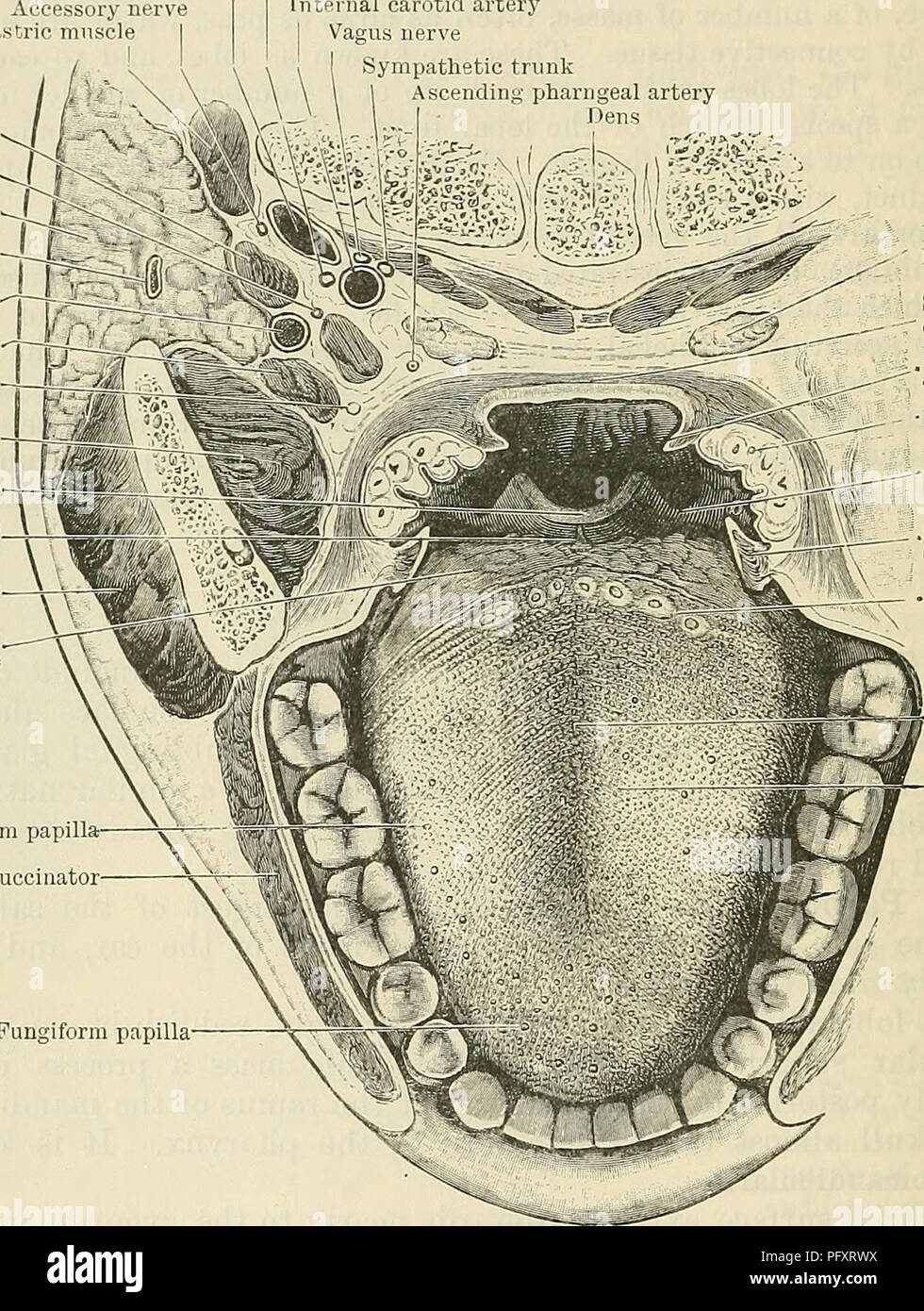 . Cunningham's Text-book of anatomy. Anatomy. 1134 THE DIGESTIVE SYSTEM. and lined on the other, by fascia. The covering layer is specially known as the fascia parotideomasseterica (O.T. parotid fascia) and both it and the lining layer are derived from the deep cervical fascia, which divides to enclose the gland. The parotideo-masseteric fascia is connected, on the surface, above to the zygoma ; posteriorly, to the acoustic meatus and anterior border of the sterno-mastoid; below, it is continuous with the deep cervical fascia, and anteriorly it passes over the masseter, and blends with the fas Stock Photohttps://www.alamy.com/image-license-details/?v=1https://www.alamy.com/cunninghams-text-book-of-anatomy-anatomy-1134-the-digestive-system-and-lined-on-the-other-by-fascia-the-covering-layer-is-specially-known-as-the-fascia-parotideomasseterica-ot-parotid-fascia-and-both-it-and-the-lining-layer-are-derived-from-the-deep-cervical-fascia-which-divides-to-enclose-the-gland-the-parotideo-masseteric-fascia-is-connected-on-the-surface-above-to-the-zygoma-posteriorly-to-the-acoustic-meatus-and-anterior-border-of-the-sterno-mastoid-below-it-is-continuous-with-the-deep-cervical-fascia-and-anteriorly-it-passes-over-the-masseter-and-blends-with-the-fas-image216333766.html
RMPFXRWX–. Cunningham's Text-book of anatomy. Anatomy. 1134 THE DIGESTIVE SYSTEM. and lined on the other, by fascia. The covering layer is specially known as the fascia parotideomasseterica (O.T. parotid fascia) and both it and the lining layer are derived from the deep cervical fascia, which divides to enclose the gland. The parotideo-masseteric fascia is connected, on the surface, above to the zygoma ; posteriorly, to the acoustic meatus and anterior border of the sterno-mastoid; below, it is continuous with the deep cervical fascia, and anteriorly it passes over the masseter, and blends with the fas
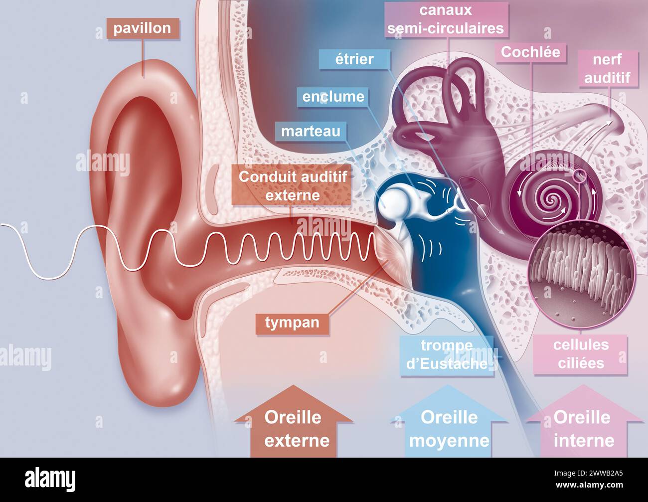 The path of sound in the ear. Representation of the path of sound in the ear. Stock Photohttps://www.alamy.com/image-license-details/?v=1https://www.alamy.com/the-path-of-sound-in-the-ear-representation-of-the-path-of-sound-in-the-ear-image600762237.html
RM2WWB2A5–The path of sound in the ear. Representation of the path of sound in the ear.
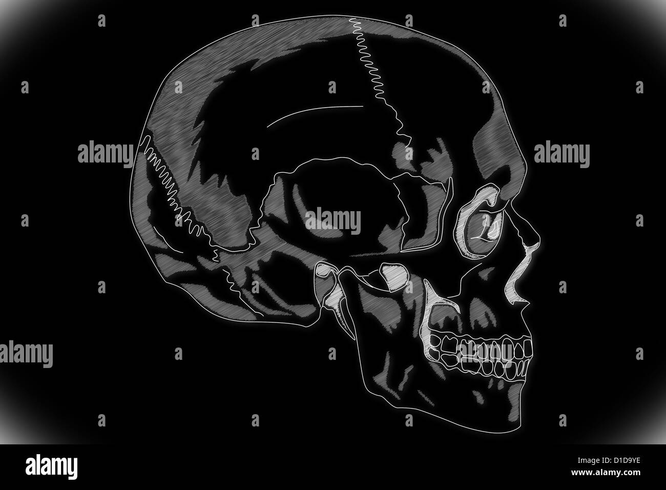 Human Skull structure Stock Photohttps://www.alamy.com/image-license-details/?v=1https://www.alamy.com/stock-photo-human-skull-structure-52538962.html
RFD1D9YE–Human Skull structure
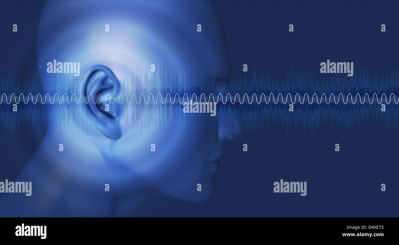 Sound waves, waveforms passing through an ear Stock Photohttps://www.alamy.com/image-license-details/?v=1https://www.alamy.com/stock-photo-sound-waves-waveforms-passing-through-an-ear-54671655.html
RFD4XE73–Sound waves, waveforms passing through an ear
 sound, medicinally, medical, science, cross, human, human being, sense, Stock Photohttps://www.alamy.com/image-license-details/?v=1https://www.alamy.com/stock-photo-sound-medicinally-medical-science-cross-human-human-being-sense-131574646.html
RFHJ1MPE–sound, medicinally, medical, science, cross, human, human being, sense,
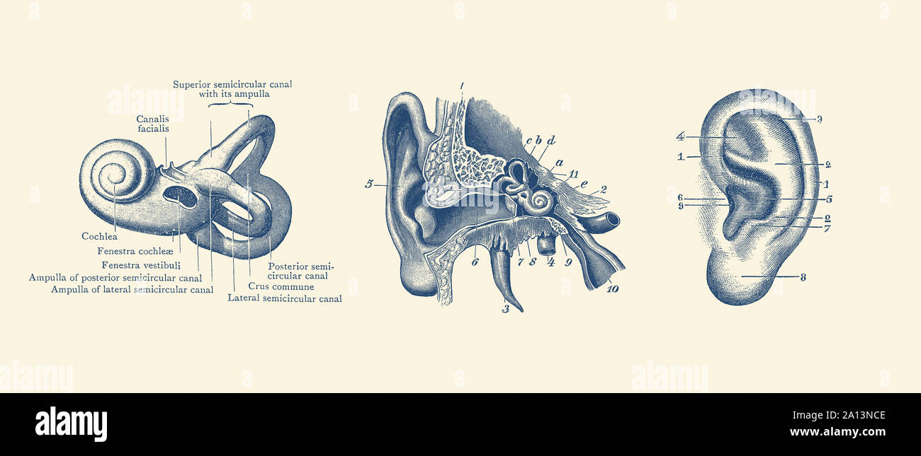 Vintage anatomy print showing three views of the human ear. Stock Photohttps://www.alamy.com/image-license-details/?v=1https://www.alamy.com/vintage-anatomy-print-showing-three-views-of-the-human-ear-image327694318.html
RF2A13NCE–Vintage anatomy print showing three views of the human ear.
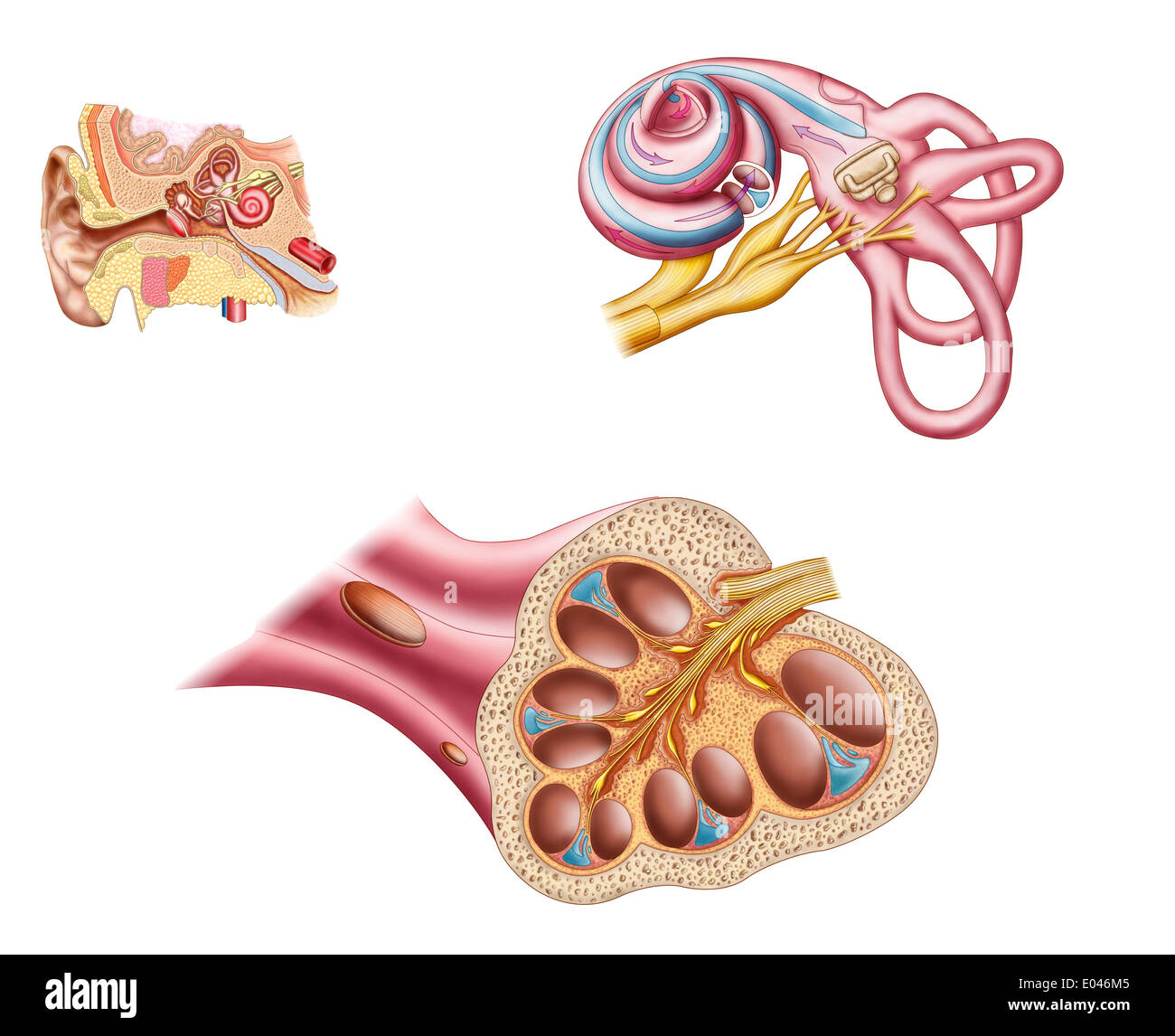 Anatomy of the cochlear duct in the human ear. Stock Photohttps://www.alamy.com/image-license-details/?v=1https://www.alamy.com/anatomy-of-the-cochlear-duct-in-the-human-ear-image68934549.html
RFE046M5–Anatomy of the cochlear duct in the human ear.
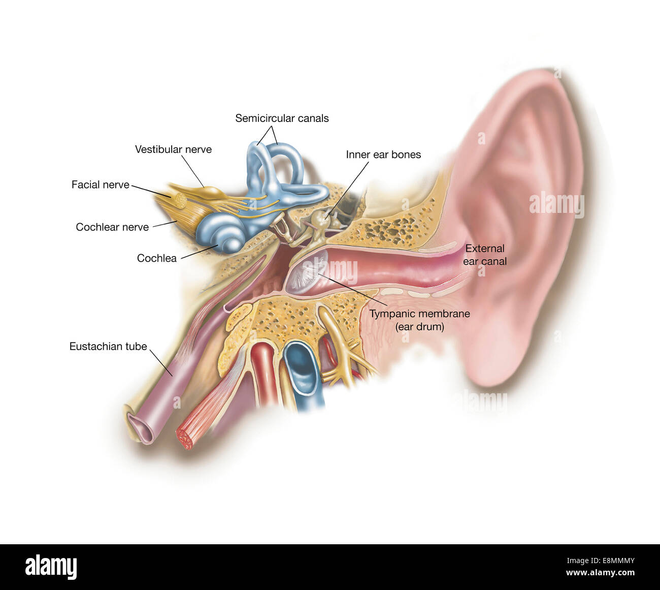 Anatomy of human ear. Stock Photohttps://www.alamy.com/image-license-details/?v=1https://www.alamy.com/stock-photo-anatomy-of-human-ear-74214027.html
RME8MMMY–Anatomy of human ear.
 Medical practice, Doctors surgery. Examination of the ear, acoustic meatus, ear canal of a patient, with a loupe. Stock Photohttps://www.alamy.com/image-license-details/?v=1https://www.alamy.com/stock-photo-medical-practice-doctors-surgery-examination-of-the-ear-acoustic-meatus-31849199.html
RMBRPRYB–Medical practice, Doctors surgery. Examination of the ear, acoustic meatus, ear canal of a patient, with a loupe.
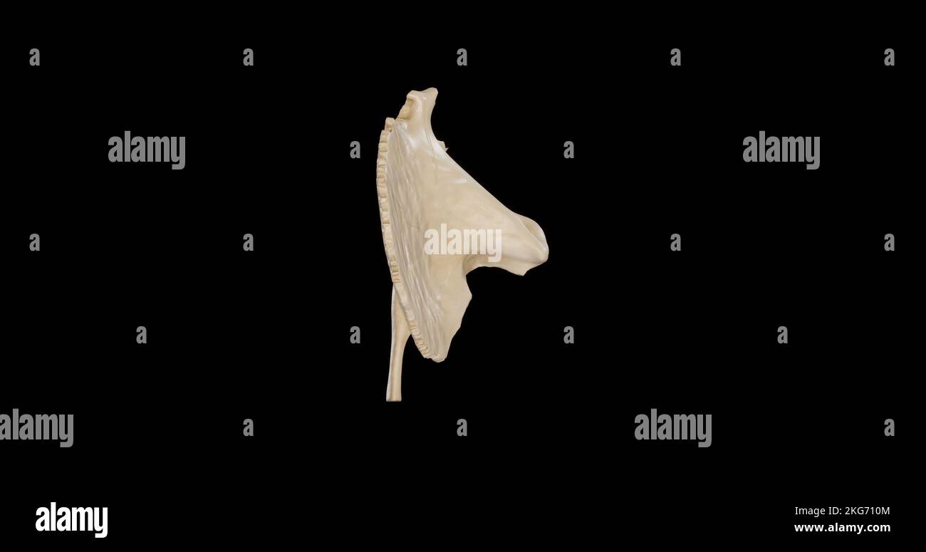 Superior view of Right Temporal Bone Stock Photohttps://www.alamy.com/image-license-details/?v=1https://www.alamy.com/superior-view-of-right-temporal-bone-image491879268.html
RF2KG710M–Superior view of Right Temporal Bone
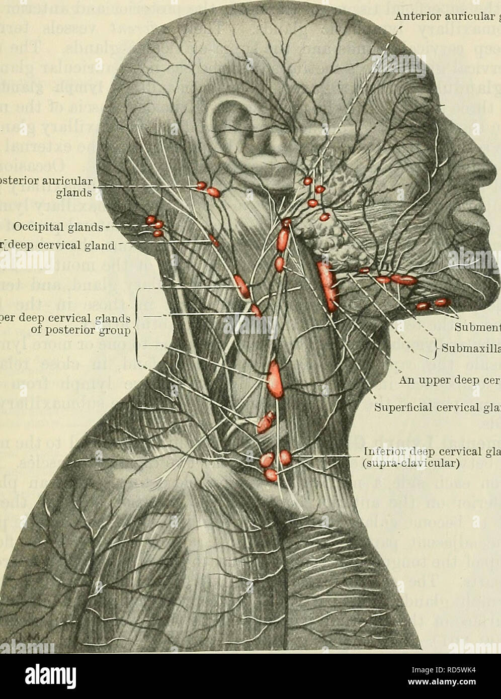 . Cunningham's Text-book of anatomy. Anatomy. THE LYMPH GLANDS OF THE HEAD. 999 from the external acoustic meatus, the tympanum, the soft palate, the posterior part of the nose, and the deeper portions of the cheek. Their efferents open into the upper deep cervical glands. The Superficial Facial Lymph Glands.—Several lymph glands, or groups of lymph glands, have been found in the region of the face but, apparently, they are irregular, both in occurrence and in position. Those which appear to be most frequently found are : Infra-orbital, which lie along the angle between the nose and the cheek, Stock Photohttps://www.alamy.com/image-license-details/?v=1https://www.alamy.com/cunninghams-text-book-of-anatomy-anatomy-the-lymph-glands-of-the-head-999-from-the-external-acoustic-meatus-the-tympanum-the-soft-palate-the-posterior-part-of-the-nose-and-the-deeper-portions-of-the-cheek-their-efferents-open-into-the-upper-deep-cervical-glands-the-superficial-facial-lymph-glandsseveral-lymph-glands-or-groups-of-lymph-glands-have-been-found-in-the-region-of-the-face-but-apparently-they-are-irregular-both-in-occurrence-and-in-position-those-which-appear-to-be-most-frequently-found-are-infra-orbital-which-lie-along-the-angle-between-the-nose-and-the-cheek-image231855208.html
RMRD5WK4–. Cunningham's Text-book of anatomy. Anatomy. THE LYMPH GLANDS OF THE HEAD. 999 from the external acoustic meatus, the tympanum, the soft palate, the posterior part of the nose, and the deeper portions of the cheek. Their efferents open into the upper deep cervical glands. The Superficial Facial Lymph Glands.—Several lymph glands, or groups of lymph glands, have been found in the region of the face but, apparently, they are irregular, both in occurrence and in position. Those which appear to be most frequently found are : Infra-orbital, which lie along the angle between the nose and the cheek,
 . Cunningham's Text-book of anatomy. Anatomy. 782 THE NERVOUS SYSTEM. Branches and Communications.—(i.) In the internal acoustic meatus the nervus intermedins lying between the facial and acoustic, sends communicating branches to both nerves. The branch to the acoustic nerve probably separates from it again to ioin the genicular ganglion of the facial nerve. , (ii) In the canalis facialis the ganglion genicnli is formed at the point where the facial nerve bends backwards. It is an oval swelling on the nerve, and is ioined by a branch from the upper (vestibular) trunk of the acoustic nerve by w Stock Photohttps://www.alamy.com/image-license-details/?v=1https://www.alamy.com/cunninghams-text-book-of-anatomy-anatomy-782-the-nervous-system-branches-and-communicationsi-in-the-internal-acoustic-meatus-the-nervus-intermedins-lying-between-the-facial-and-acoustic-sends-communicating-branches-to-both-nerves-the-branch-to-the-acoustic-nerve-probably-separates-from-it-again-to-ioin-the-genicular-ganglion-of-the-facial-nerve-ii-in-the-canalis-facialis-the-ganglion-genicnli-is-formed-at-the-point-where-the-facial-nerve-bends-backwards-it-is-an-oval-swelling-on-the-nerve-and-is-ioined-by-a-branch-from-the-upper-vestibular-trunk-of-the-acoustic-nerve-by-w-image216345133.html
RMPFYABW–. Cunningham's Text-book of anatomy. Anatomy. 782 THE NERVOUS SYSTEM. Branches and Communications.—(i.) In the internal acoustic meatus the nervus intermedins lying between the facial and acoustic, sends communicating branches to both nerves. The branch to the acoustic nerve probably separates from it again to ioin the genicular ganglion of the facial nerve. , (ii) In the canalis facialis the ganglion genicnli is formed at the point where the facial nerve bends backwards. It is an oval swelling on the nerve, and is ioined by a branch from the upper (vestibular) trunk of the acoustic nerve by w
 The path of sound in the ear. Representation of the path of sound in the ear. Stock Photohttps://www.alamy.com/image-license-details/?v=1https://www.alamy.com/the-path-of-sound-in-the-ear-representation-of-the-path-of-sound-in-the-ear-image600762225.html
RM2WWB29N–The path of sound in the ear. Representation of the path of sound in the ear.
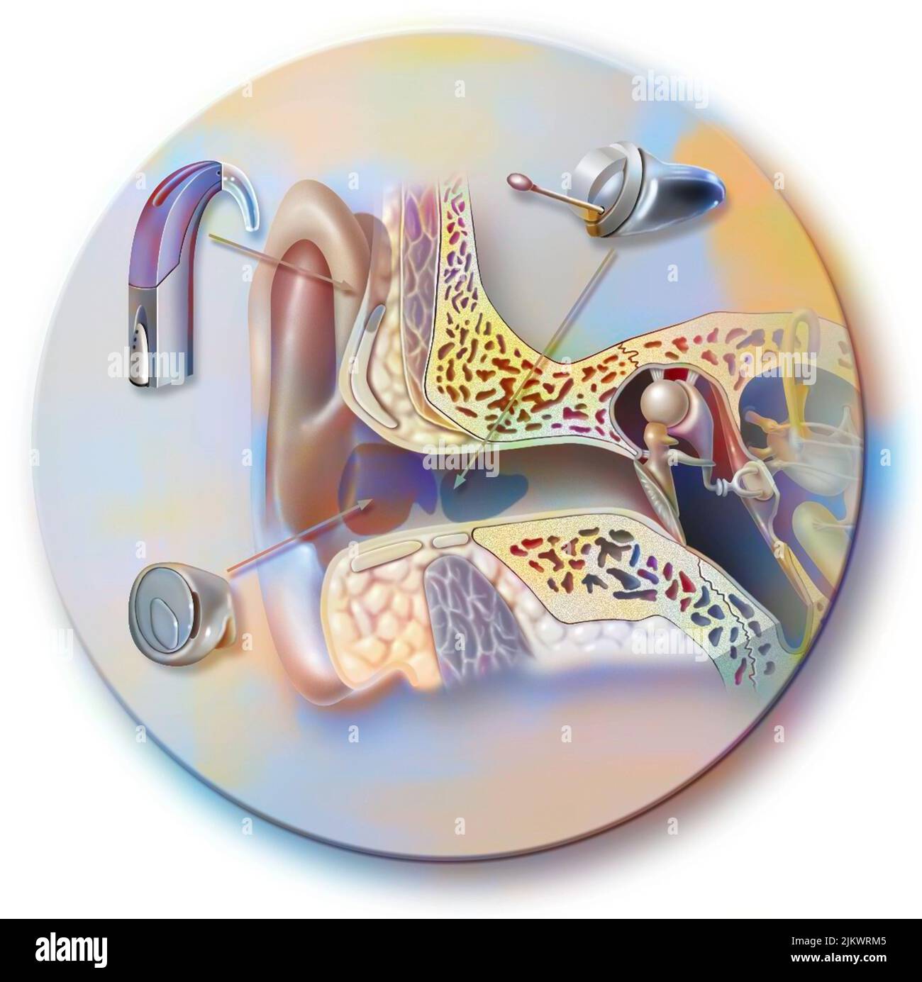 Hearing aids and their location in the outer ear. Stock Photohttps://www.alamy.com/image-license-details/?v=1https://www.alamy.com/hearing-aids-and-their-location-in-the-outer-ear-image476925797.html
RF2JKWRM5–Hearing aids and their location in the outer ear.
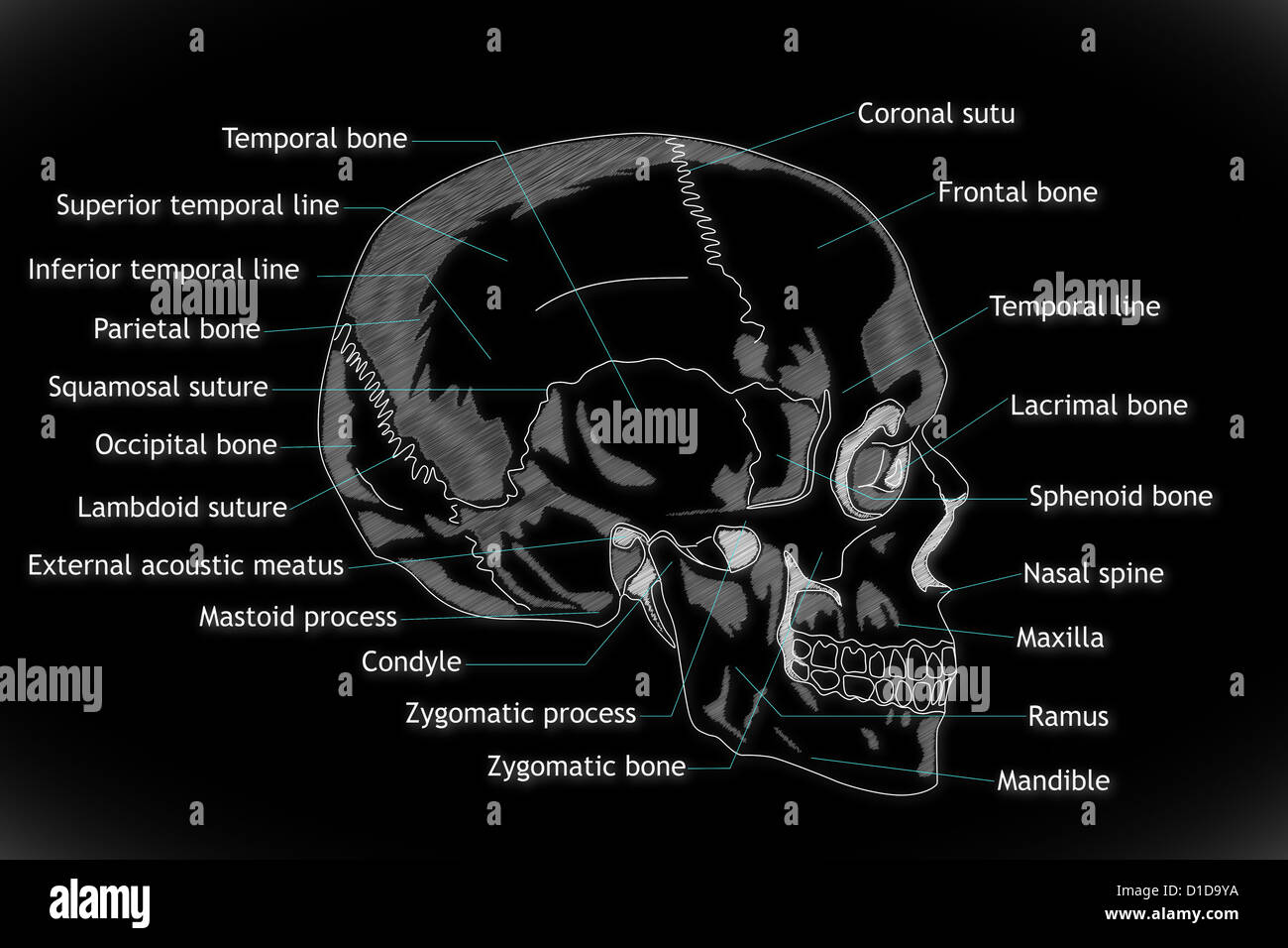 Human Skull structure Stock Photohttps://www.alamy.com/image-license-details/?v=1https://www.alamy.com/stock-photo-human-skull-structure-52538958.html
RFD1D9YA–Human Skull structure
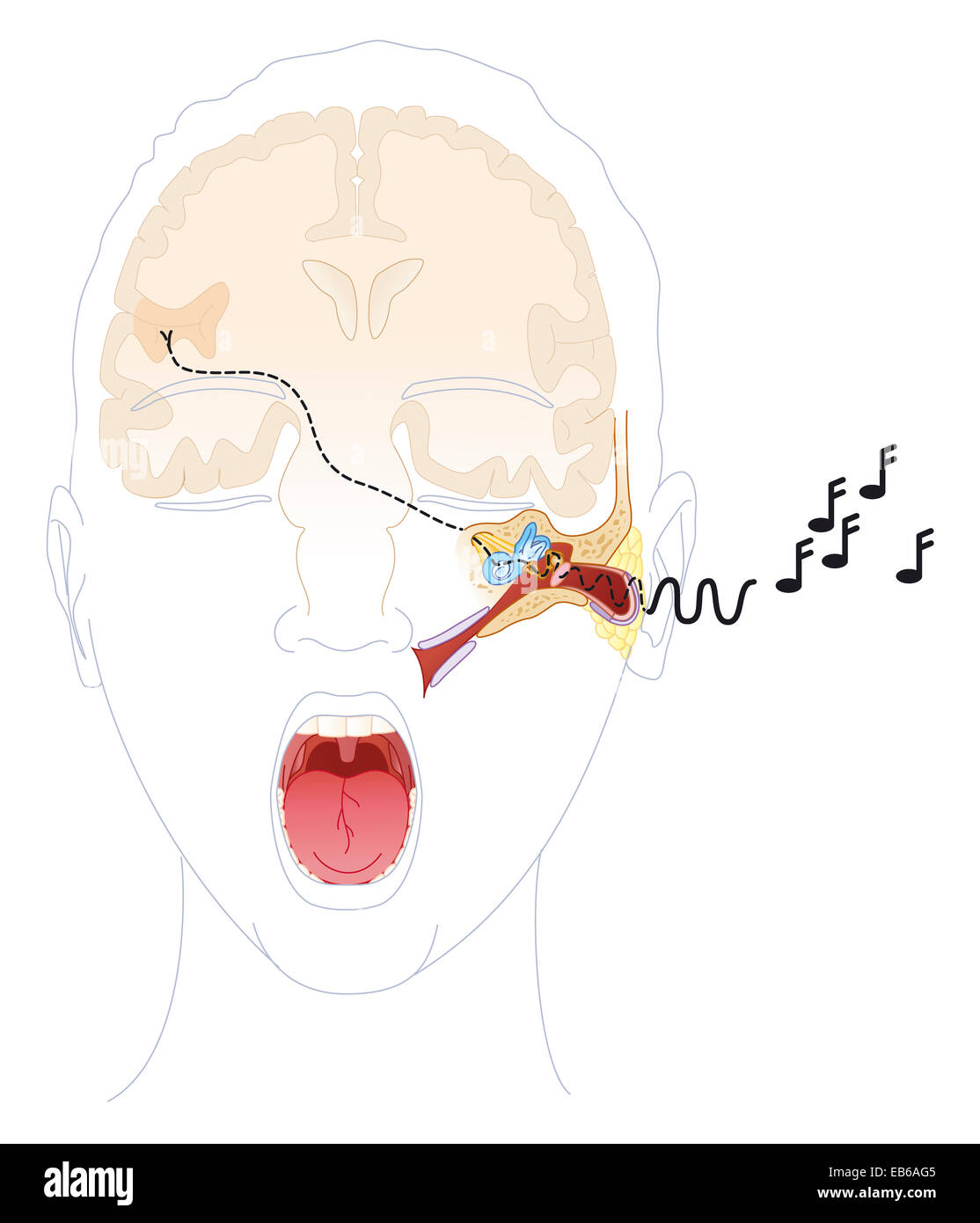 HEARING, DRAWING Stock Photohttps://www.alamy.com/image-license-details/?v=1https://www.alamy.com/stock-photo-hearing-drawing-75742693.html
RMEB6AG5–HEARING, DRAWING
 sound, medicinally, medical, science, cross, human, human being, sense, Stock Photohttps://www.alamy.com/image-license-details/?v=1https://www.alamy.com/stock-photo-sound-medicinally-medical-science-cross-human-human-being-sense-131566817.html
RFHJ1APW–sound, medicinally, medical, science, cross, human, human being, sense,
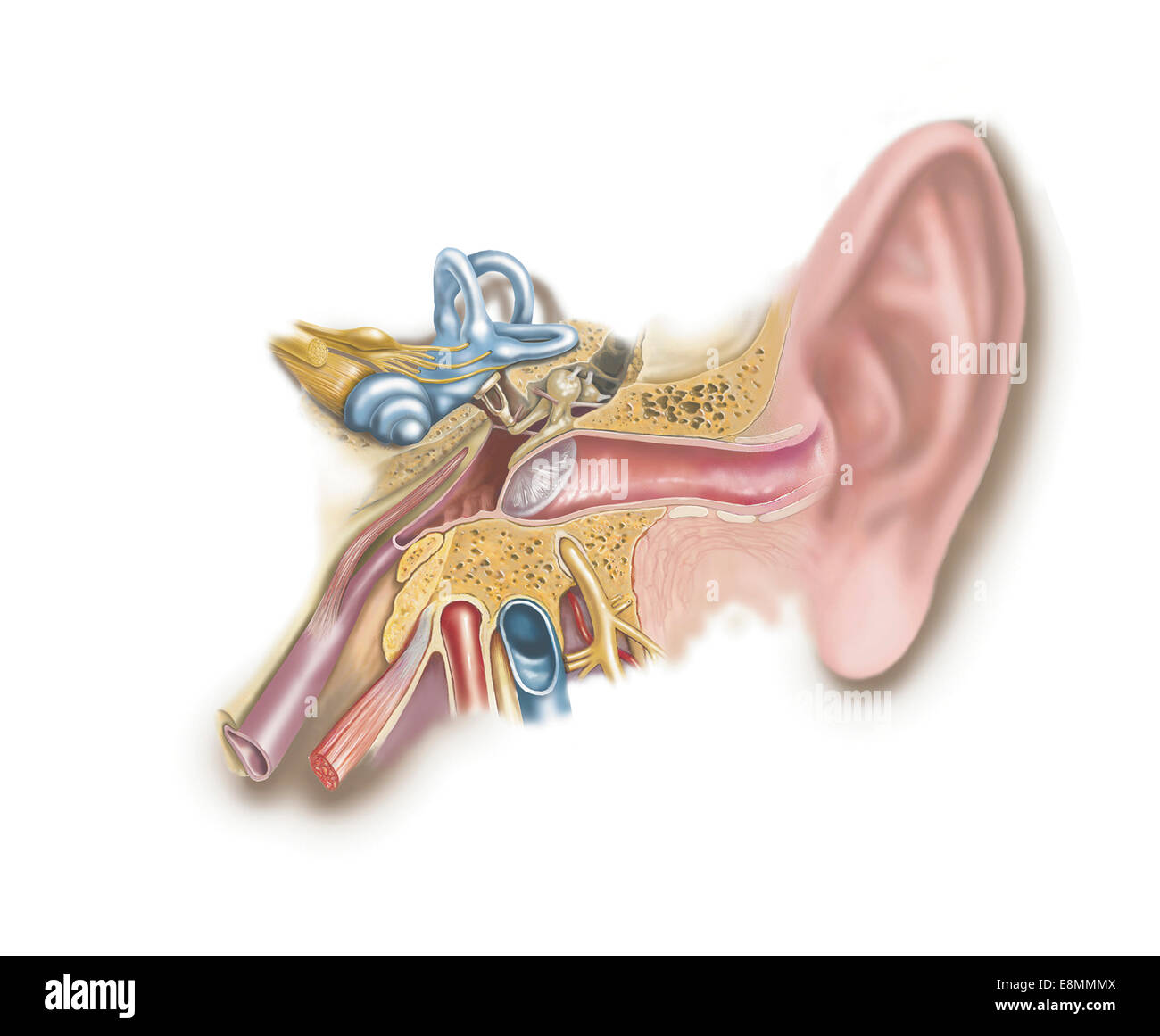 Anatomy of human ear. Stock Photohttps://www.alamy.com/image-license-details/?v=1https://www.alamy.com/stock-photo-anatomy-of-human-ear-74214026.html
RME8MMMX–Anatomy of human ear.
 Medical practice, Doctors surgery. Examination of the ear, acoustic meatus, ear canal of a patient, with a loupe. Stock Photohttps://www.alamy.com/image-license-details/?v=1https://www.alamy.com/stock-photo-medical-practice-doctors-surgery-examination-of-the-ear-acoustic-meatus-31853370.html
RMBRR18A–Medical practice, Doctors surgery. Examination of the ear, acoustic meatus, ear canal of a patient, with a loupe.
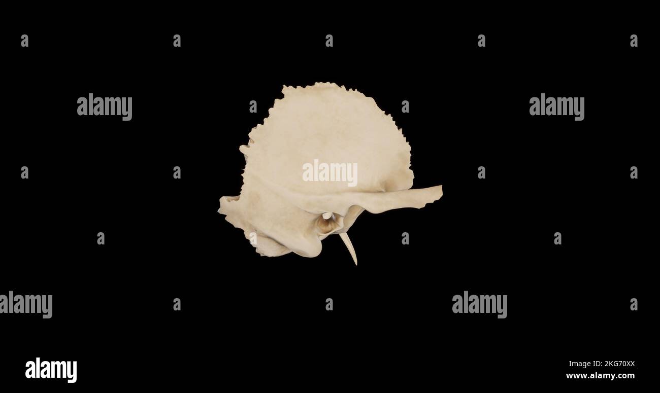 Right view of Right Temporal Bone Stock Photohttps://www.alamy.com/image-license-details/?v=1https://www.alamy.com/right-view-of-right-temporal-bone-image491879218.html
RF2KG70XX–Right view of Right Temporal Bone
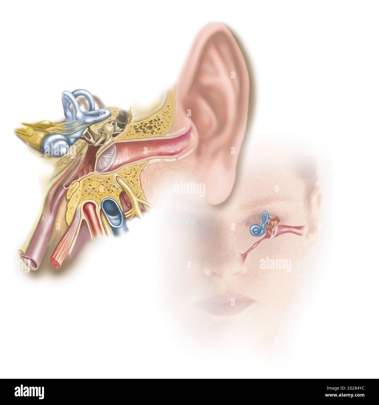 Position of inner ear in relationship to head and face. Stock Photohttps://www.alamy.com/image-license-details/?v=1https://www.alamy.com/position-of-inner-ear-in-relationship-to-head-and-face-image431668032.html
RM2G284YC–Position of inner ear in relationship to head and face.
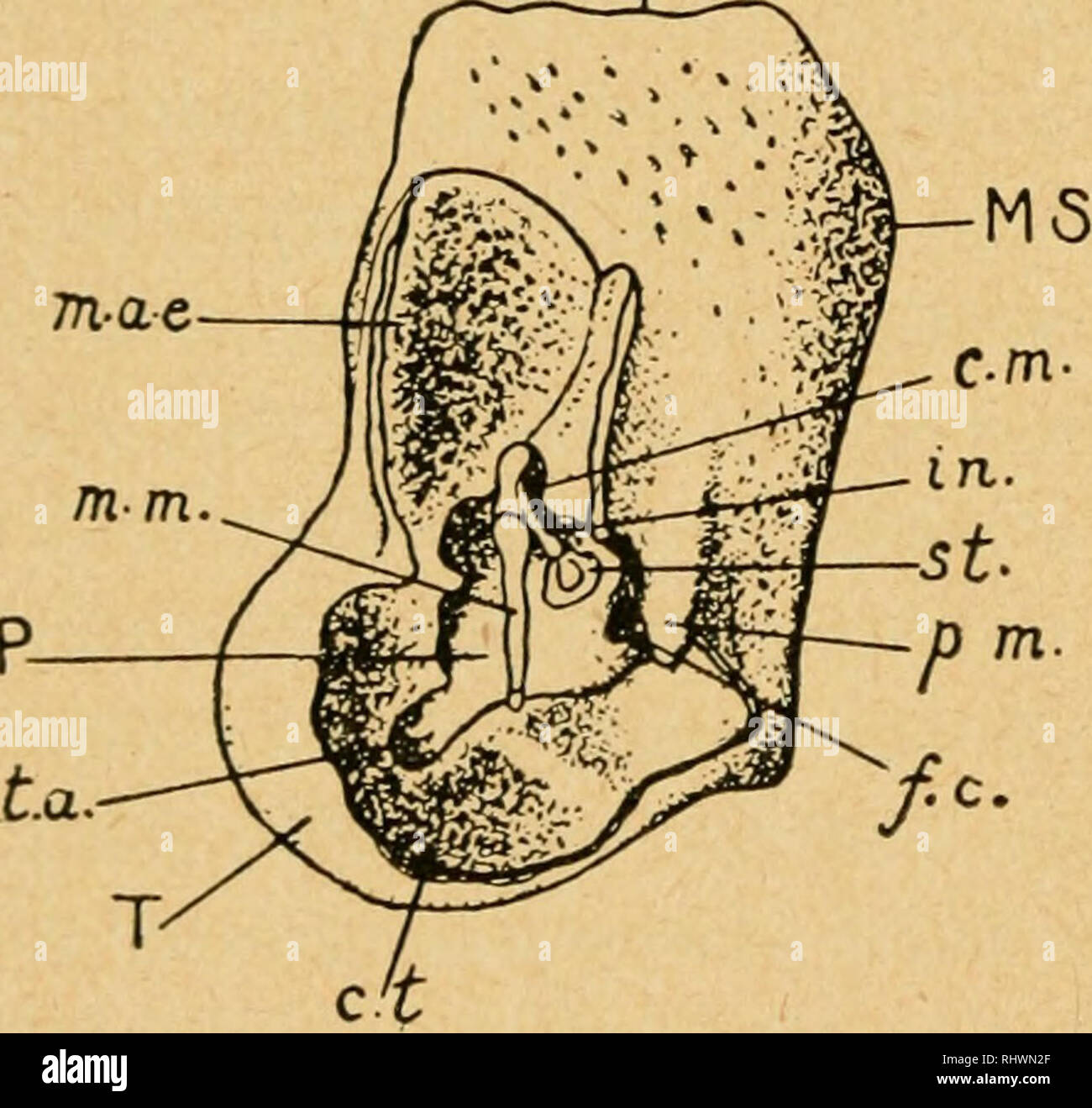 . Bensley's Practical anatomy of the rabbit : an elementary laboratory text-book in mammalian anatomy. Rabbits -- Anatomy. 18G ANATOMY OF THE RABBIT m-so. stylomastoid foramen lies between the latter and the external acoustic meatus. The petrous portion, as viewed from its medial surface, is roughly oblong; it is placed obliquely with reference to the basi- occipital and basisphenoid. The parafloccular fossa occupies its posterodorsal portion, and extends into the substance of the bone, forming a much larger depression than is indicated by the diameter of its rim. The related dorsal margin of Stock Photohttps://www.alamy.com/image-license-details/?v=1https://www.alamy.com/bensleys-practical-anatomy-of-the-rabbit-an-elementary-laboratory-text-book-in-mammalian-anatomy-rabbits-anatomy-18g-anatomy-of-the-rabbit-m-so-stylomastoid-foramen-lies-between-the-latter-and-the-external-acoustic-meatus-the-petrous-portion-as-viewed-from-its-medial-surface-is-roughly-oblong-it-is-placed-obliquely-with-reference-to-the-basi-occipital-and-basisphenoid-the-parafloccular-fossa-occupies-its-posterodorsal-portion-and-extends-into-the-substance-of-the-bone-forming-a-much-larger-depression-than-is-indicated-by-the-diameter-of-its-rim-the-related-dorsal-margin-of-image234749271.html
RMRHWN2F–. Bensley's Practical anatomy of the rabbit : an elementary laboratory text-book in mammalian anatomy. Rabbits -- Anatomy. 18G ANATOMY OF THE RABBIT m-so. stylomastoid foramen lies between the latter and the external acoustic meatus. The petrous portion, as viewed from its medial surface, is roughly oblong; it is placed obliquely with reference to the basi- occipital and basisphenoid. The parafloccular fossa occupies its posterodorsal portion, and extends into the substance of the bone, forming a much larger depression than is indicated by the diameter of its rim. The related dorsal margin of
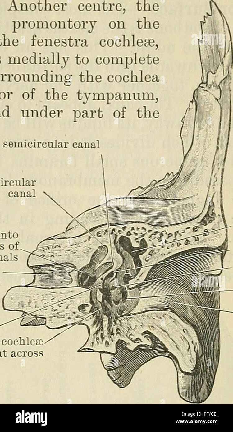 . Cunningham's Text-book of anatomy. Anatomy. 132 OSTEOLOGY. facial canal. Reaching forwards, it extends to the apex of the petrous part; whilst laterally it forms part of the medial wall'of the tympanum, surrounds the fenestra vesti- bulij and encloses within its substance portions of the cochlea, vestibule, and superior semicircular canal. Another centre, the Opisthotic, appears in the vicinity of the promontory on the medial wall of the tympanum, surrounds the fenestra cochleae, forms the floor of the vestibule, and extends medially to complete the floor of the internal acoustic meatus. Sur Stock Photohttps://www.alamy.com/image-license-details/?v=1https://www.alamy.com/cunninghams-text-book-of-anatomy-anatomy-132-osteology-facial-canal-reaching-forwards-it-extends-to-the-apex-of-the-petrous-part-whilst-laterally-it-forms-part-of-the-medial-wallof-the-tympanum-surrounds-the-fenestra-vesti-bulij-and-encloses-within-its-substance-portions-of-the-cochlea-vestibule-and-superior-semicircular-canal-another-centre-the-opisthotic-appears-in-the-vicinity-of-the-promontory-on-the-medial-wall-of-the-tympanum-surrounds-the-fenestra-cochleae-forms-the-floor-of-the-vestibule-and-extends-medially-to-complete-the-floor-of-the-internal-acoustic-meatus-sur-image216346778.html
RMPFYCEJ–. Cunningham's Text-book of anatomy. Anatomy. 132 OSTEOLOGY. facial canal. Reaching forwards, it extends to the apex of the petrous part; whilst laterally it forms part of the medial wall'of the tympanum, surrounds the fenestra vesti- bulij and encloses within its substance portions of the cochlea, vestibule, and superior semicircular canal. Another centre, the Opisthotic, appears in the vicinity of the promontory on the medial wall of the tympanum, surrounds the fenestra cochleae, forms the floor of the vestibule, and extends medially to complete the floor of the internal acoustic meatus. Sur
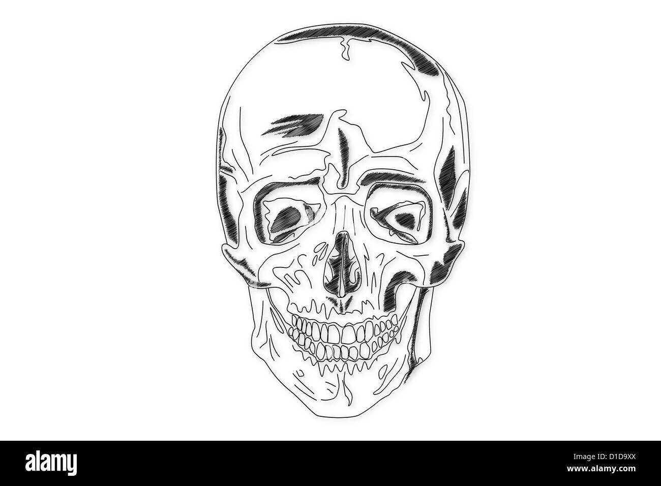 Human Skull structure Stock Photohttps://www.alamy.com/image-license-details/?v=1https://www.alamy.com/stock-photo-human-skull-structure-52538946.html
RFD1D9XX–Human Skull structure
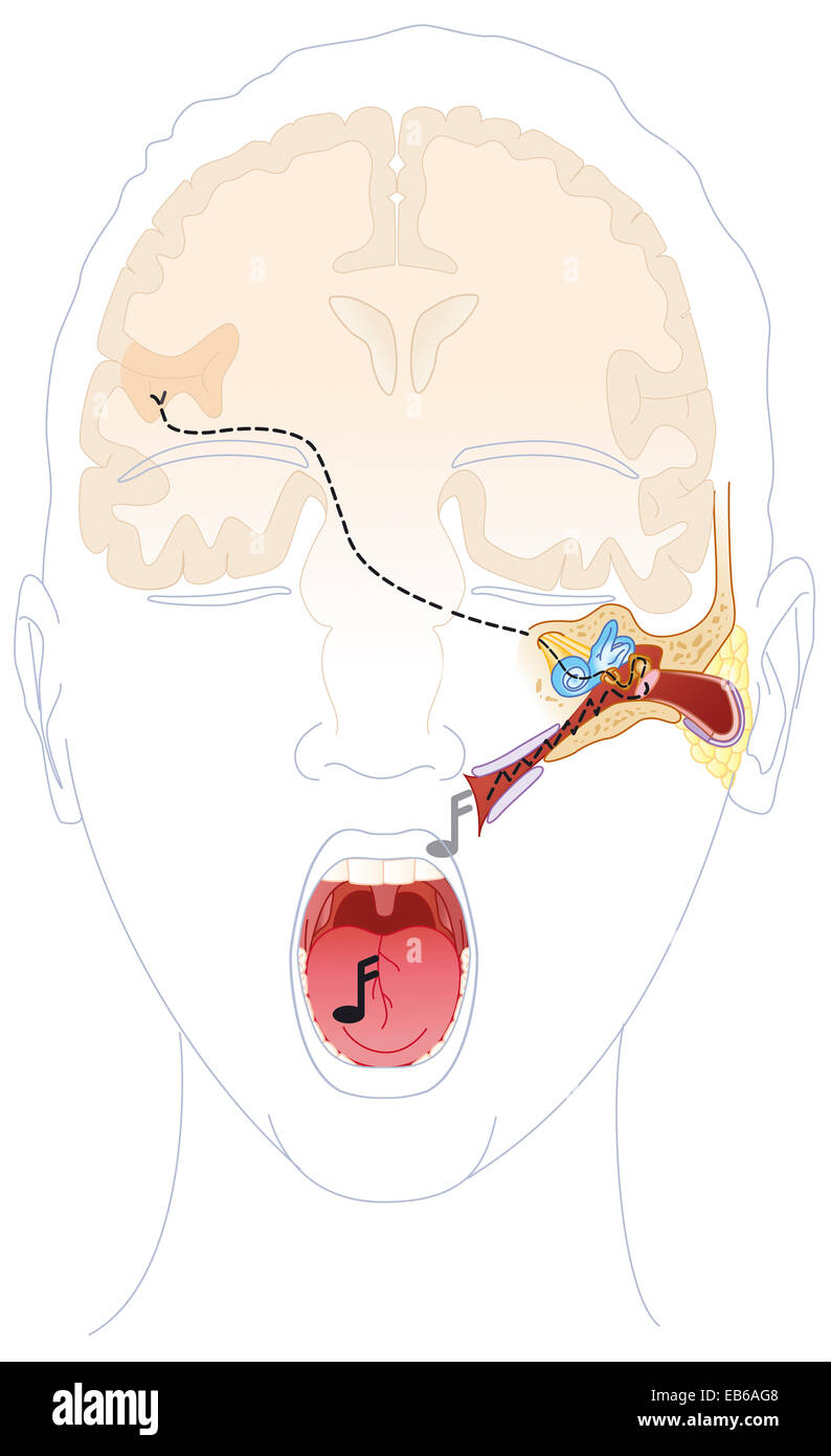 HEARING, DRAWING Stock Photohttps://www.alamy.com/image-license-details/?v=1https://www.alamy.com/stock-photo-hearing-drawing-75742696.html
RMEB6AG8–HEARING, DRAWING
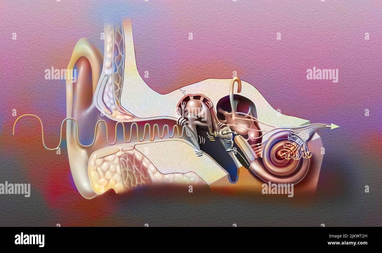 Anatomy of the ear showing the eardrum, ossicles, hammer, anvil. Stock Photohttps://www.alamy.com/image-license-details/?v=1https://www.alamy.com/anatomy-of-the-ear-showing-the-eardrum-ossicles-hammer-anvil-image476926089.html
RF2JKWT2H–Anatomy of the ear showing the eardrum, ossicles, hammer, anvil.
 sound, medicinally, medical, science, cross, human, human being, sense, Stock Photohttps://www.alamy.com/image-license-details/?v=1https://www.alamy.com/stock-photo-sound-medicinally-medical-science-cross-human-human-being-sense-131566818.html
RFHJ1APX–sound, medicinally, medical, science, cross, human, human being, sense,
