Anniella pulchra Stock Photos and Images
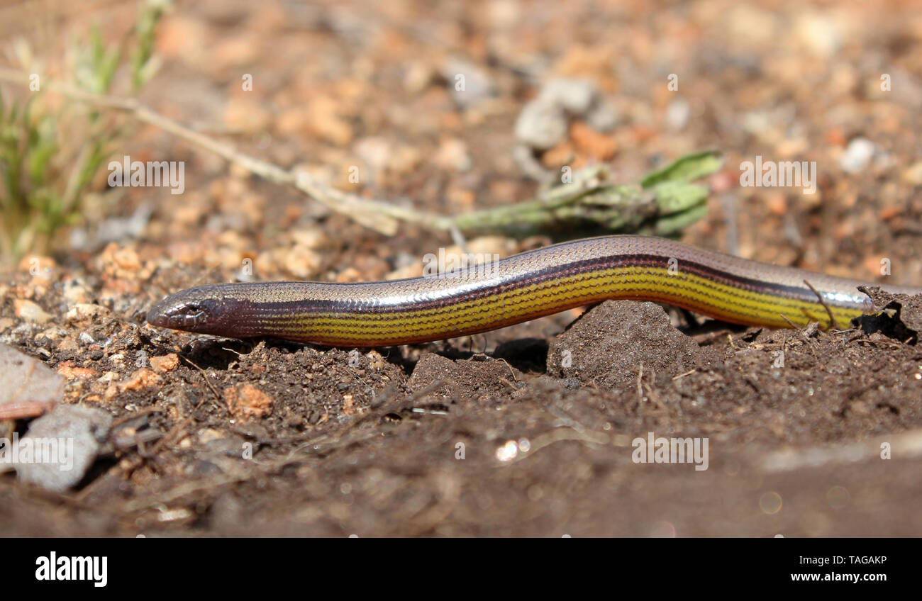 California Legless Lizard (Anniella pulchra) Stock Photohttps://www.alamy.com/image-license-details/?v=1https://www.alamy.com/california-legless-lizard-anniella-pulchra-image247451338.html
RFTAGAKP–California Legless Lizard (Anniella pulchra)
 Northern Legless Lizard (Anniella pulchra) Reptilia Stock Photohttps://www.alamy.com/image-license-details/?v=1https://www.alamy.com/northern-legless-lizard-anniella-pulchra-reptilia-image620364746.html
RM2Y181FP–Northern Legless Lizard (Anniella pulchra) Reptilia
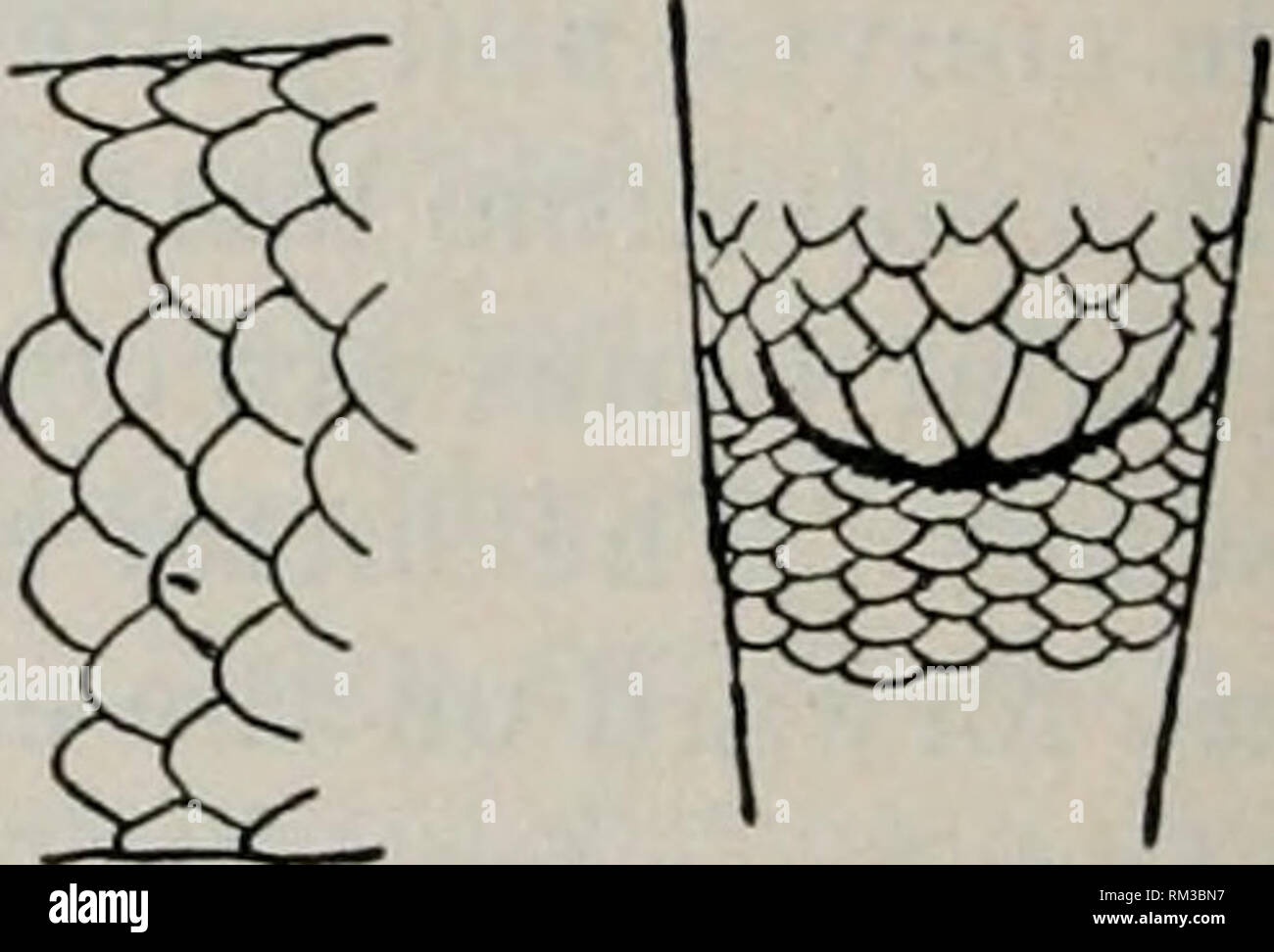 . Annual report of the Board of Regents of the Smithsonian Institution. Smithsonian Institution; Smithsonian Institution. Archives; Discoveries in science. Fig. 138. ANNIELLA PULCHRA GRAY. X3. Cat. No. 16022, U.S.N.M. Body depressed cylindric >tail obtuse, about one-half as long as body, but varying somewhat in length. Scales smooth, everywhere equal, in generally thirty rows, but sometimes in twenty-eight and even twenty- six. Head but little wider than body posteriorly, contracting radially to an obtuse, moderately depressed muzzle, which projects beyond the lower jaw. Preanal scales gene Stock Photohttps://www.alamy.com/image-license-details/?v=1https://www.alamy.com/annual-report-of-the-board-of-regents-of-the-smithsonian-institution-smithsonian-institution-smithsonian-institution-archives-discoveries-in-science-fig-138-anniella-pulchra-gray-x3-cat-no-16022-usnm-body-depressed-cylindric-gttail-obtuse-about-one-half-as-long-as-body-but-varying-somewhat-in-length-scales-smooth-everywhere-equal-in-generally-thirty-rows-but-sometimes-in-twenty-eight-and-even-twenty-six-head-but-little-wider-than-body-posteriorly-contracting-radially-to-an-obtuse-moderately-depressed-muzzle-which-projects-beyond-the-lower-jaw-preanal-scales-gene-image236102979.html
RMRM3BN7–. Annual report of the Board of Regents of the Smithsonian Institution. Smithsonian Institution; Smithsonian Institution. Archives; Discoveries in science. Fig. 138. ANNIELLA PULCHRA GRAY. X3. Cat. No. 16022, U.S.N.M. Body depressed cylindric >tail obtuse, about one-half as long as body, but varying somewhat in length. Scales smooth, everywhere equal, in generally thirty rows, but sometimes in twenty-eight and even twenty- six. Head but little wider than body posteriorly, contracting radially to an obtuse, moderately depressed muzzle, which projects beyond the lower jaw. Preanal scales gene
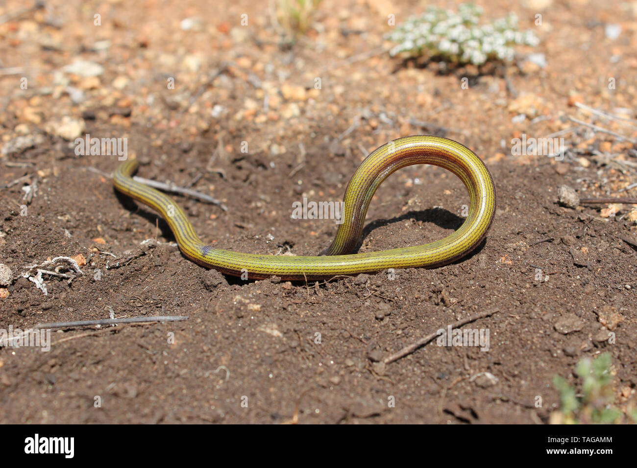 California Legless Lizard (Anniella pulchra) Stock Photohttps://www.alamy.com/image-license-details/?v=1https://www.alamy.com/california-legless-lizard-anniella-pulchra-image247451364.html
RFTAGAMM–California Legless Lizard (Anniella pulchra)
 Northern Legless Lizard (Anniella pulchra) Reptilia Stock Photohttps://www.alamy.com/image-license-details/?v=1https://www.alamy.com/northern-legless-lizard-anniella-pulchra-reptilia-image620364360.html
RM2Y18120–Northern Legless Lizard (Anniella pulchra) Reptilia
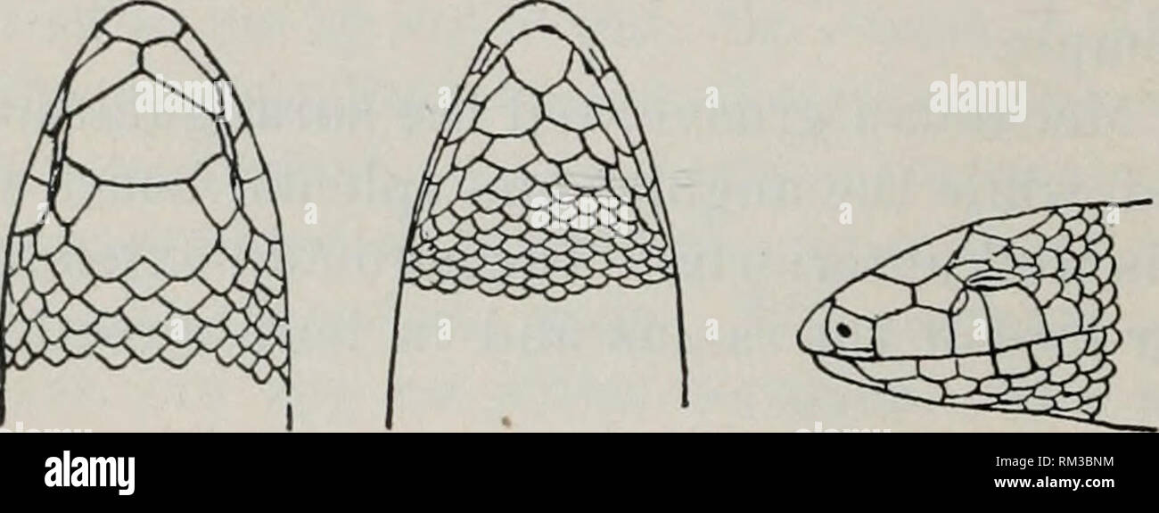 . Annual report of the Board of Regents of the Smithsonian Institution. Smithsonian Institution; Smithsonian Institution. Archives; Discoveries in science. 674 REPORT OF NATIONAL MUSEUM, 1898. Three supposed species have been described, but I believe that two of them are referable to a siugle, rather variable form. The range of the genus is confined, with present knowledge, to the southern part of the Pacific district. ANNIELLA PULCHRA Gray. Anniella pulehra Gray, Ann. Mag. Nat. Hist. (2), X, 1852, p. 440; Zool. Her- ald, p. 154, pi. XXVIII.—Cope, Proc. Acad. Nat. Sci. PLila., 1864, p. 230.— B Stock Photohttps://www.alamy.com/image-license-details/?v=1https://www.alamy.com/annual-report-of-the-board-of-regents-of-the-smithsonian-institution-smithsonian-institution-smithsonian-institution-archives-discoveries-in-science-674-report-of-national-museum-1898-three-supposed-species-have-been-described-but-i-believe-that-two-of-them-are-referable-to-a-siugle-rather-variable-form-the-range-of-the-genus-is-confined-with-present-knowledge-to-the-southern-part-of-the-pacific-district-anniella-pulchra-gray-anniella-pulehra-gray-ann-mag-nat-hist-2-x-1852-p-440-zool-her-ald-p-154-pi-xxviiicope-proc-acad-nat-sci-plila-1864-p-230-b-image236102992.html
RMRM3BNM–. Annual report of the Board of Regents of the Smithsonian Institution. Smithsonian Institution; Smithsonian Institution. Archives; Discoveries in science. 674 REPORT OF NATIONAL MUSEUM, 1898. Three supposed species have been described, but I believe that two of them are referable to a siugle, rather variable form. The range of the genus is confined, with present knowledge, to the southern part of the Pacific district. ANNIELLA PULCHRA Gray. Anniella pulehra Gray, Ann. Mag. Nat. Hist. (2), X, 1852, p. 440; Zool. Her- ald, p. 154, pi. XXVIII.—Cope, Proc. Acad. Nat. Sci. PLila., 1864, p. 230.— B
 Northern Legless Lizard (Anniella pulchra) Reptilia Stock Photohttps://www.alamy.com/image-license-details/?v=1https://www.alamy.com/northern-legless-lizard-anniella-pulchra-reptilia-image620363358.html
RM2Y17YP6–Northern Legless Lizard (Anniella pulchra) Reptilia
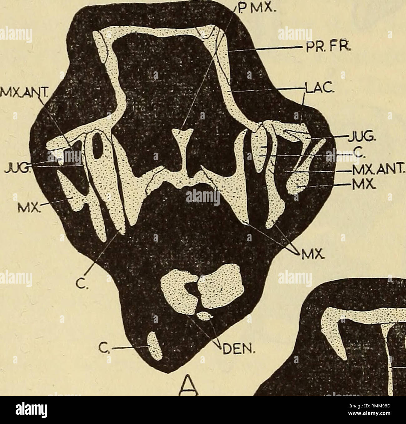 . Annals of the South African Museum = Annale van die Suid-Afrikaanse Museum. Natural history. 140 ANNALS OF THE SOUTH AFRICAN MUSEUM nerves into the nostril, as it still does in the lizards Cordylns polyzonus (Van Pletzen, 1946) and Monopeltis capensis (Kritzinger, 1946). In these two species the ramus medialis nasi V, accompanied by a small artery, passes through a foramen in the anterior tip of the septomaxillary. In Anniella pulchra, however, the blood-vessel and nerve are accommodated in a deep groove on the dorsal surface of the bone (Toerien, 1950). Williston (1925) claims that the prim Stock Photohttps://www.alamy.com/image-license-details/?v=1https://www.alamy.com/annals-of-the-south-african-museum-=-annale-van-die-suid-afrikaanse-museum-natural-history-140-annals-of-the-south-african-museum-nerves-into-the-nostril-as-it-still-does-in-the-lizards-cordylns-polyzonus-van-pletzen-1946-and-monopeltis-capensis-kritzinger-1946-in-these-two-species-the-ramus-medialis-nasi-v-accompanied-by-a-small-artery-passes-through-a-foramen-in-the-anterior-tip-of-the-septomaxillary-in-anniella-pulchra-however-the-blood-vessel-and-nerve-are-accommodated-in-a-deep-groove-on-the-dorsal-surface-of-the-bone-toerien-1950-williston-1925-claims-that-the-prim-image236474237.html
RMRMM98D–. Annals of the South African Museum = Annale van die Suid-Afrikaanse Museum. Natural history. 140 ANNALS OF THE SOUTH AFRICAN MUSEUM nerves into the nostril, as it still does in the lizards Cordylns polyzonus (Van Pletzen, 1946) and Monopeltis capensis (Kritzinger, 1946). In these two species the ramus medialis nasi V, accompanied by a small artery, passes through a foramen in the anterior tip of the septomaxillary. In Anniella pulchra, however, the blood-vessel and nerve are accommodated in a deep groove on the dorsal surface of the bone (Toerien, 1950). Williston (1925) claims that the prim
 . Anatomischer Anzeiger. Anatomy, Comparative; Anatomy, Comparative. 6B6. Fig. 6. Brain of Anniella pulchra. Sagittal section, X 28. pc. paracoele. Epen. Cerebellum. ^y no Fig. 7. Fore part of the cranium of Anniella pulchra. Transverse section, X ^^^ b bone. I lens, r retina, n nerve. Fig. 8. Transverse section of the olfactory bulbs near the fore-brain, X ^8. (An- niella pulchra.) s v y r> T 1 Fig. 9. Transverse section of the fore-brain (Anniella pulchra), X 2». dc diacoele. c chiasma.. Please note that these images are extracted from scanned page images that may have been digitally enha Stock Photohttps://www.alamy.com/image-license-details/?v=1https://www.alamy.com/anatomischer-anzeiger-anatomy-comparative-anatomy-comparative-6b6-fig-6-brain-of-anniella-pulchra-sagittal-section-x-28-pc-paracoele-epen-cerebellum-y-no-fig-7-fore-part-of-the-cranium-of-anniella-pulchra-transverse-section-x-b-bone-i-lens-r-retina-n-nerve-fig-8-transverse-section-of-the-olfactory-bulbs-near-the-fore-brain-x-8-an-niella-pulchra-s-v-y-rgt-t-1-fig-9-transverse-section-of-the-fore-brain-anniella-pulchra-x-2-dc-diacoele-c-chiasma-please-note-that-these-images-are-extracted-from-scanned-page-images-that-may-have-been-digitally-enha-image236807048.html
RMRN7DPG–. Anatomischer Anzeiger. Anatomy, Comparative; Anatomy, Comparative. 6B6. Fig. 6. Brain of Anniella pulchra. Sagittal section, X 28. pc. paracoele. Epen. Cerebellum. ^y no Fig. 7. Fore part of the cranium of Anniella pulchra. Transverse section, X ^^^ b bone. I lens, r retina, n nerve. Fig. 8. Transverse section of the olfactory bulbs near the fore-brain, X ^8. (An- niella pulchra.) s v y r> T 1 Fig. 9. Transverse section of the fore-brain (Anniella pulchra), X 2». dc diacoele. c chiasma.. Please note that these images are extracted from scanned page images that may have been digitally enha
 . Anatomischer Anzeiger. Anatomy, Comparative; Anatomy, Comparative. Rhinen Mv. Mesen. Fig. nerves. 5 Fig. Fig. 1. Brain of a California horned toad. Dorsal view, X 3. Rhinen. Rhinencephal. olf. olfactory tract. Prosen. Prosencephal. Meten. Metencephal. ep. Epi- physis. Jlesen. Mesencephal. Epen. Epencephal. Fig. 2. Brain of a snake. Dorsal view, X 3. Fig. 3. Brain of Anniella pulchra. Dorsal view, X 10. tr. trigeminous lobe. Jly. Myelcncephal. 4. Brain of Anniella pulchra. Ventral view, X 10. 2 second pair of cranial fifth pair of cranial nerves. 5. Brain of Anniella pulchra. Lateral view, X Stock Photohttps://www.alamy.com/image-license-details/?v=1https://www.alamy.com/anatomischer-anzeiger-anatomy-comparative-anatomy-comparative-rhinen-mv-mesen-fig-nerves-5-fig-fig-1-brain-of-a-california-horned-toad-dorsal-view-x-3-rhinen-rhinencephal-olf-olfactory-tract-prosen-prosencephal-meten-metencephal-ep-epi-physis-jlesen-mesencephal-epen-epencephal-fig-2-brain-of-a-snake-dorsal-view-x-3-fig-3-brain-of-anniella-pulchra-dorsal-view-x-10-tr-trigeminous-lobe-jly-myelcncephal-4-brain-of-anniella-pulchra-ventral-view-x-10-2-second-pair-of-cranial-fifth-pair-of-cranial-nerves-5-brain-of-anniella-pulchra-lateral-view-x-image236807056.html
RMRN7DPT–. Anatomischer Anzeiger. Anatomy, Comparative; Anatomy, Comparative. Rhinen Mv. Mesen. Fig. nerves. 5 Fig. Fig. 1. Brain of a California horned toad. Dorsal view, X 3. Rhinen. Rhinencephal. olf. olfactory tract. Prosen. Prosencephal. Meten. Metencephal. ep. Epi- physis. Jlesen. Mesencephal. Epen. Epencephal. Fig. 2. Brain of a snake. Dorsal view, X 3. Fig. 3. Brain of Anniella pulchra. Dorsal view, X 10. tr. trigeminous lobe. Jly. Myelcncephal. 4. Brain of Anniella pulchra. Ventral view, X 10. 2 second pair of cranial fifth pair of cranial nerves. 5. Brain of Anniella pulchra. Lateral view, X