Anterior superior iliac spine Stock Photos and Images
(74)See anterior superior iliac spine stock video clipsQuick filters:
Anterior superior iliac spine Stock Photos and Images
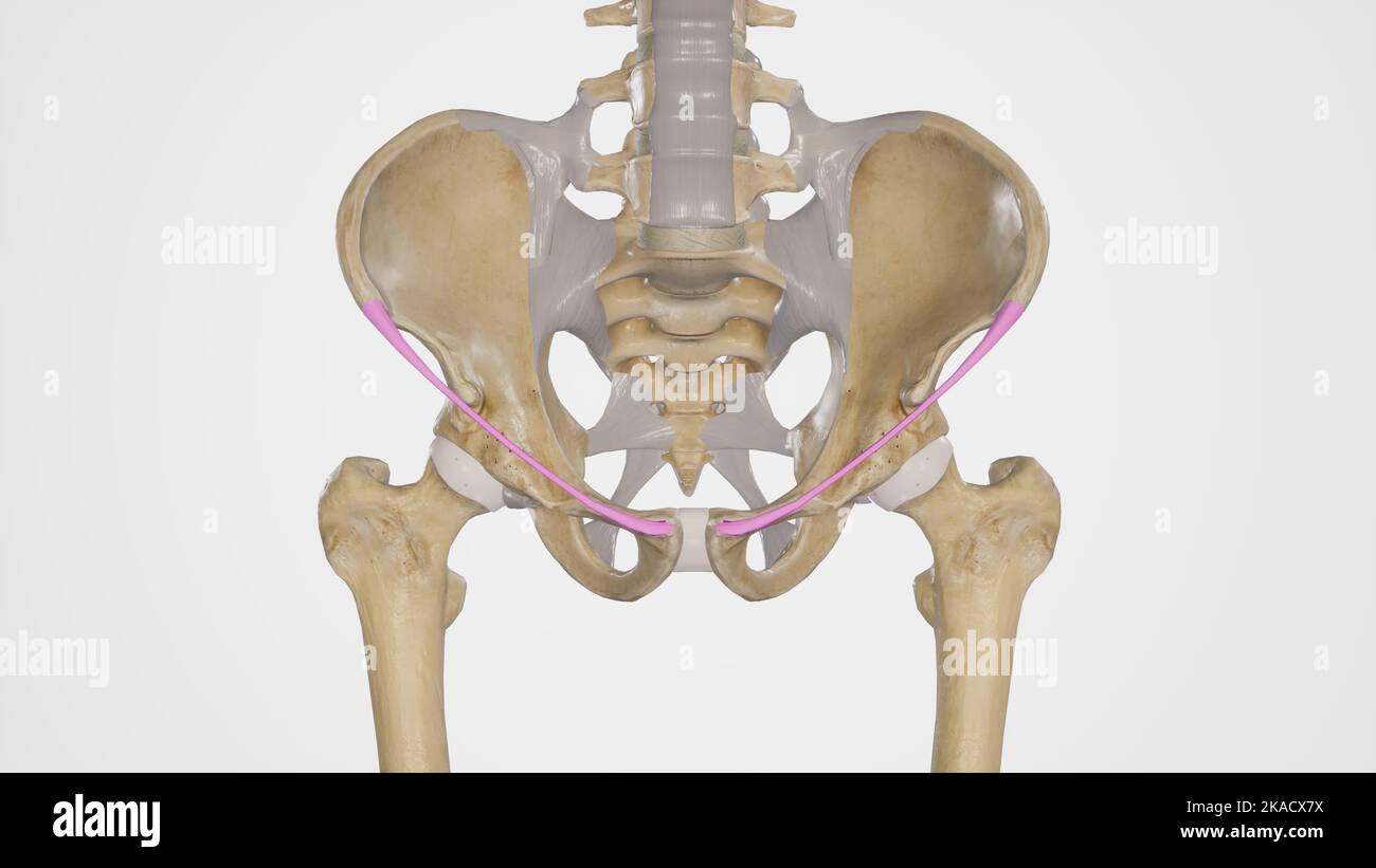 medical accurate illustration of the inguinal ligament Stock Photohttps://www.alamy.com/image-license-details/?v=1https://www.alamy.com/medical-accurate-illustration-of-the-inguinal-ligament-image488320894.html
RF2KACX7X–medical accurate illustration of the inguinal ligament
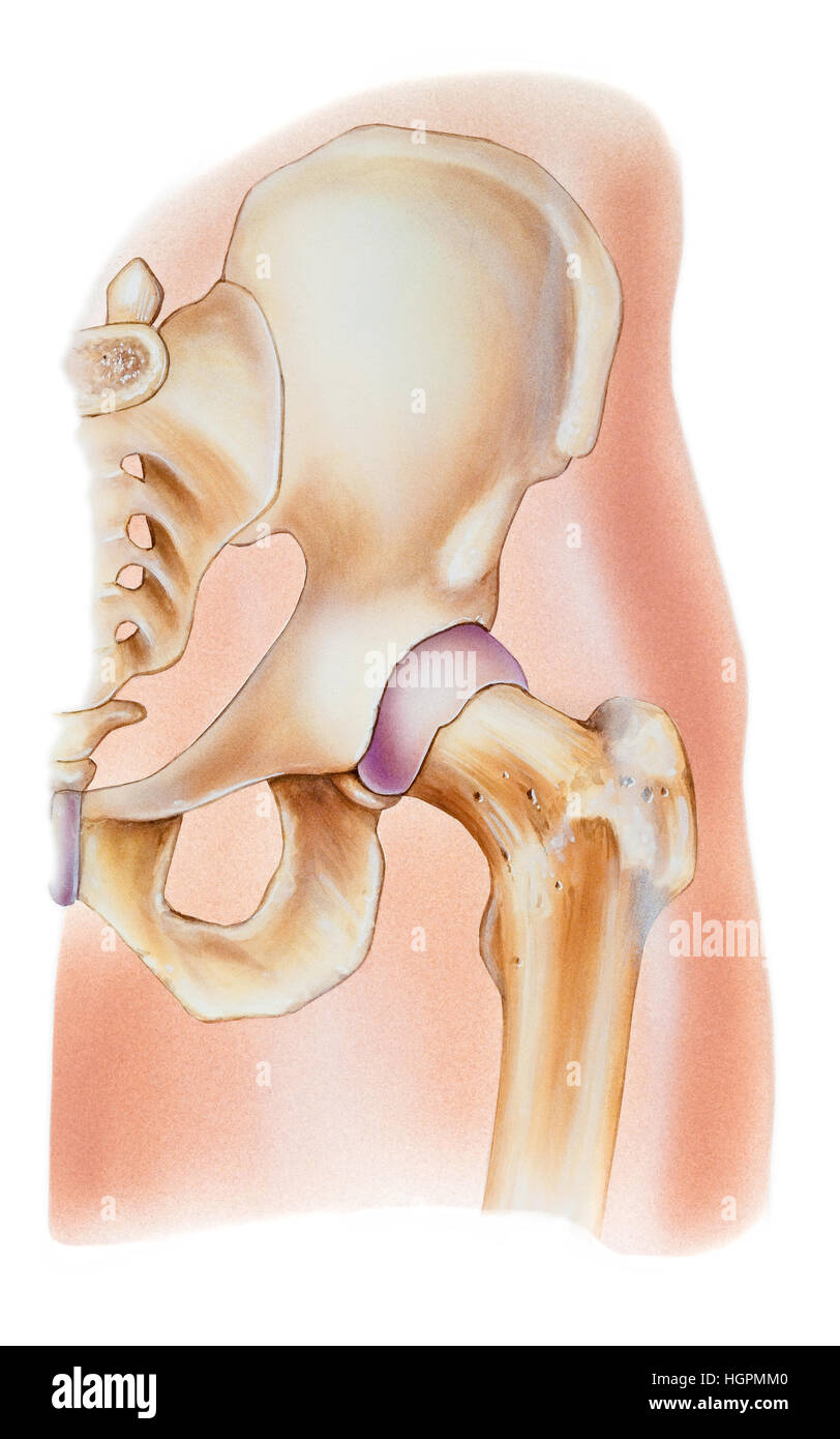 Normal human anatomy of a hip joint. Shown are the ilium, pelvis, anterior superior iliac spine, iliac crest, anterior inferior iliac spine, smooth ar Stock Photohttps://www.alamy.com/image-license-details/?v=1https://www.alamy.com/stock-photo-normal-human-anatomy-of-a-hip-joint-shown-are-the-ilium-pelvis-anterior-130806256.html
RFHGPMM0–Normal human anatomy of a hip joint. Shown are the ilium, pelvis, anterior superior iliac spine, iliac crest, anterior inferior iliac spine, smooth ar
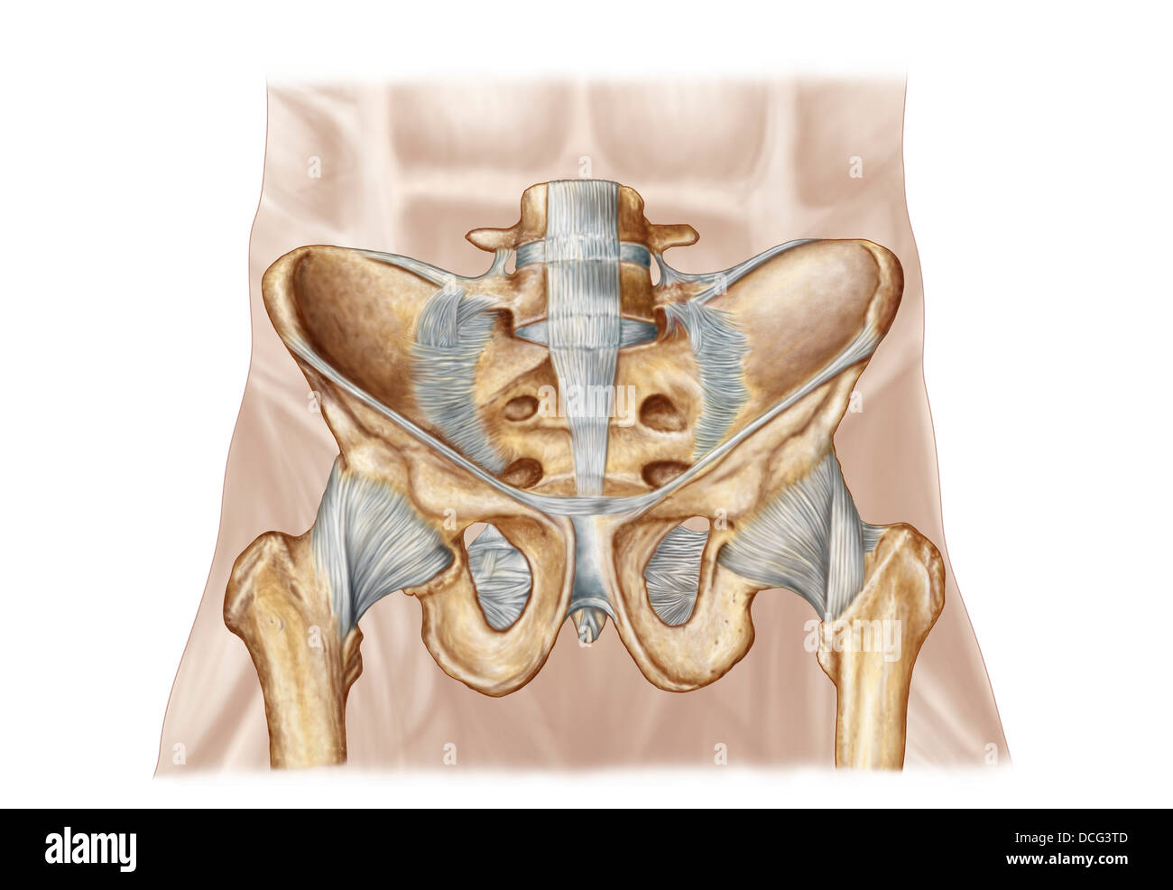 Anatomy of human pelvic bone and ligaments. Stock Photohttps://www.alamy.com/image-license-details/?v=1https://www.alamy.com/stock-photo-anatomy-of-human-pelvic-bone-and-ligaments-59361245.html
RFDCG3TD–Anatomy of human pelvic bone and ligaments.
 Abdomen. Tummy tuck. It is retensioning skin and abdominal muscles, by removing redundant skin and musculature plication. Before surgery should be taken anatomical references, such as the midline of the abdomen and the height of the anterior superior iliac spine. Plastic surgery. Doctor attending to patient medical consultation. Stock Photohttps://www.alamy.com/image-license-details/?v=1https://www.alamy.com/abdomen-tummy-tuck-it-is-retensioning-skin-and-abdominal-muscles-by-removing-redundant-skin-and-musculature-plication-before-surgery-should-be-taken-anatomical-references-such-as-the-midline-of-the-abdomen-and-the-height-of-the-anterior-superior-iliac-spine-plastic-surgery-doctor-attending-to-patient-medical-consultation-image602048387.html
RM2WYDJT3–Abdomen. Tummy tuck. It is retensioning skin and abdominal muscles, by removing redundant skin and musculature plication. Before surgery should be taken anatomical references, such as the midline of the abdomen and the height of the anterior superior iliac spine. Plastic surgery. Doctor attending to patient medical consultation.
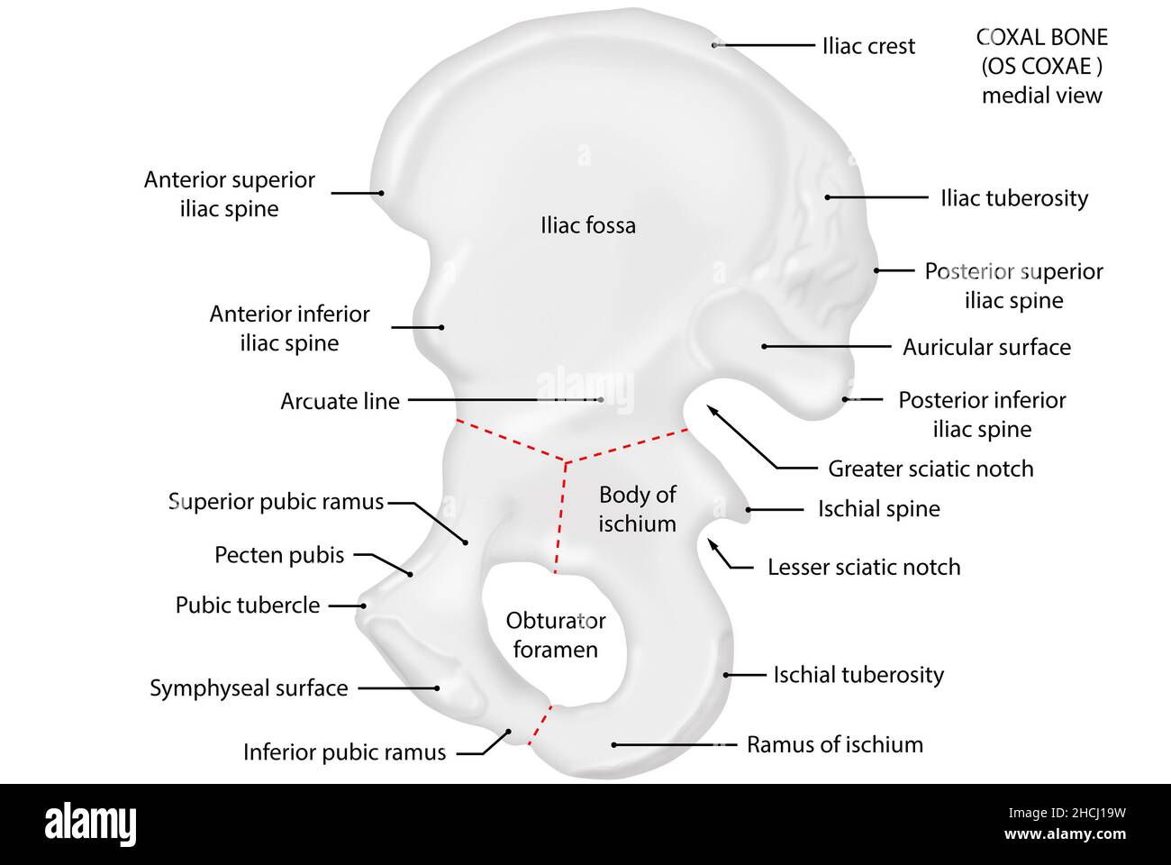 Os coxae, coxal bone, medial view, anatomy Stock Photohttps://www.alamy.com/image-license-details/?v=1https://www.alamy.com/os-coxae-coxal-bone-medial-view-anatomy-image455241637.html
RF2HCJ19W–Os coxae, coxal bone, medial view, anatomy
![. A manual of dissections of the human body [electronic resource] : for the use of students and more particularly for those preparing for the higher examinations in anatomy . anterior-superior Iliac spine to the ante- rior-inferior angle of the great Trochanter. 2. From the upper end of No. 1, along the anterior two thirds of the Iliac crest, and then downwardsand backwards to a point three inches below theposterior-superior Iliac spine. 174 A MANUAL OF DISSECTIONS. Reflect the flap downwards, and expose the loose fasciaof the buttock, containing— 1. The posterior branches of the External Cuta Stock Photo . A manual of dissections of the human body [electronic resource] : for the use of students and more particularly for those preparing for the higher examinations in anatomy . anterior-superior Iliac spine to the ante- rior-inferior angle of the great Trochanter. 2. From the upper end of No. 1, along the anterior two thirds of the Iliac crest, and then downwardsand backwards to a point three inches below theposterior-superior Iliac spine. 174 A MANUAL OF DISSECTIONS. Reflect the flap downwards, and expose the loose fasciaof the buttock, containing— 1. The posterior branches of the External Cuta Stock Photo](https://c8.alamy.com/comp/2CE400D/a-manual-of-dissections-of-the-human-body-electronic-resource-for-the-use-of-students-and-more-particularly-for-those-preparing-for-the-higher-examinations-in-anatomy-anterior-superior-iliac-spine-to-the-ante-rior-inferior-angle-of-the-great-trochanter-2-from-the-upper-end-of-no-1-along-the-anterior-two-thirds-of-the-iliac-crest-and-then-downwardsand-backwards-to-a-point-three-inches-below-theposterior-superior-iliac-spine-174-a-manual-of-dissections-reflect-the-flap-downwards-and-expose-the-loose-fasciaof-the-buttock-containing-1-the-posterior-branches-of-the-external-cuta-2CE400D.jpg) . A manual of dissections of the human body [electronic resource] : for the use of students and more particularly for those preparing for the higher examinations in anatomy . anterior-superior Iliac spine to the ante- rior-inferior angle of the great Trochanter. 2. From the upper end of No. 1, along the anterior two thirds of the Iliac crest, and then downwardsand backwards to a point three inches below theposterior-superior Iliac spine. 174 A MANUAL OF DISSECTIONS. Reflect the flap downwards, and expose the loose fasciaof the buttock, containing— 1. The posterior branches of the External Cuta Stock Photohttps://www.alamy.com/image-license-details/?v=1https://www.alamy.com/a-manual-of-dissections-of-the-human-body-electronic-resource-for-the-use-of-students-and-more-particularly-for-those-preparing-for-the-higher-examinations-in-anatomy-anterior-superior-iliac-spine-to-the-ante-rior-inferior-angle-of-the-great-trochanter-2-from-the-upper-end-of-no-1-along-the-anterior-two-thirds-of-the-iliac-crest-and-then-downwardsand-backwards-to-a-point-three-inches-below-theposterior-superior-iliac-spine-174-a-manual-of-dissections-reflect-the-flap-downwards-and-expose-the-loose-fasciaof-the-buttock-containing-1-the-posterior-branches-of-the-external-cuta-image370110733.html
. A manual of dissections of the human body [electronic resource] : for the use of students and more particularly for those preparing for the higher examinations in anatomy . anterior-superior Iliac spine to the ante- rior-inferior angle of the great Trochanter. 2. From the upper end of No. 1, along the anterior two thirds of the Iliac crest, and then downwardsand backwards to a point three inches below theposterior-superior Iliac spine. 174 A MANUAL OF DISSECTIONS. Reflect the flap downwards, and expose the loose fasciaof the buttock, containing— 1. The posterior branches of the External Cuta Stock Photohttps://www.alamy.com/image-license-details/?v=1https://www.alamy.com/a-manual-of-dissections-of-the-human-body-electronic-resource-for-the-use-of-students-and-more-particularly-for-those-preparing-for-the-higher-examinations-in-anatomy-anterior-superior-iliac-spine-to-the-ante-rior-inferior-angle-of-the-great-trochanter-2-from-the-upper-end-of-no-1-along-the-anterior-two-thirds-of-the-iliac-crest-and-then-downwardsand-backwards-to-a-point-three-inches-below-theposterior-superior-iliac-spine-174-a-manual-of-dissections-reflect-the-flap-downwards-and-expose-the-loose-fasciaof-the-buttock-containing-1-the-posterior-branches-of-the-external-cuta-image370110733.htmlRM2CE400D–. A manual of dissections of the human body [electronic resource] : for the use of students and more particularly for those preparing for the higher examinations in anatomy . anterior-superior Iliac spine to the ante- rior-inferior angle of the great Trochanter. 2. From the upper end of No. 1, along the anterior two thirds of the Iliac crest, and then downwardsand backwards to a point three inches below theposterior-superior Iliac spine. 174 A MANUAL OF DISSECTIONS. Reflect the flap downwards, and expose the loose fasciaof the buttock, containing— 1. The posterior branches of the External Cuta
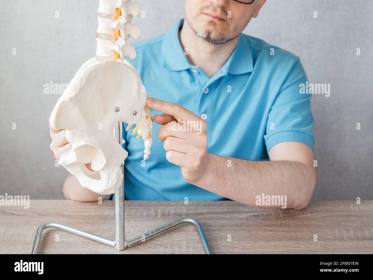 Close up of male doctor's hand pointing at anterior superior iliac spine ASIS on a skeleton spine model Stock Photohttps://www.alamy.com/image-license-details/?v=1https://www.alamy.com/close-up-of-male-doctors-hand-pointing-at-anterior-superior-iliac-spine-asis-on-a-skeleton-spine-model-image425347857.html
RF2FM07EW–Close up of male doctor's hand pointing at anterior superior iliac spine ASIS on a skeleton spine model
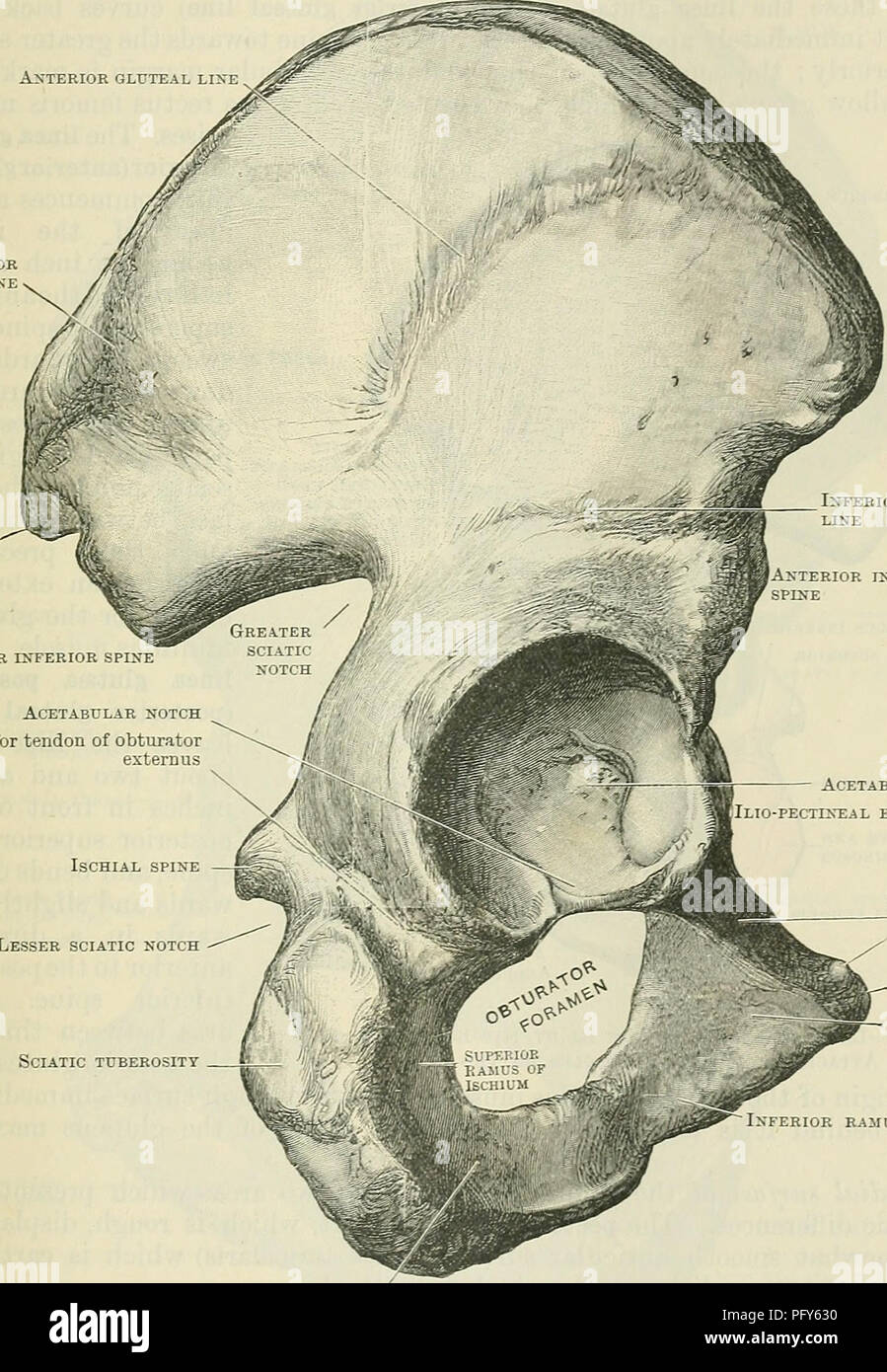 . Cunningham's Text-book of anatomy. Anatomy. THE HIP BONE. 229 anterior end of the external lip of the iliac crest the tensor fasciae latse muscle takes origin. The anterior border of the ilium stretches from the anterior superior iliac spine to the margin of the acetabulum below. Above, it is thin; but below, it forms a thick blunt process, the spina iliaca anterior inferior (anterior inferior iliac spine). From this the rectus femoris muscle arises, whilst the stout fibres of the Crest of the ilium ANTERIOR GLUTEAL LINE Posterior gluteal line Anterior inferior spine. Posterior superior spin Stock Photohttps://www.alamy.com/image-license-details/?v=1https://www.alamy.com/cunninghams-text-book-of-anatomy-anatomy-the-hip-bone-229-anterior-end-of-the-external-lip-of-the-iliac-crest-the-tensor-fasciae-latse-muscle-takes-origin-the-anterior-border-of-the-ilium-stretches-from-the-anterior-superior-iliac-spine-to-the-margin-of-the-acetabulum-below-above-it-is-thin-but-below-it-forms-a-thick-blunt-process-the-spina-iliaca-anterior-inferior-anterior-inferior-iliac-spine-from-this-the-rectus-femoris-muscle-arises-whilst-the-stout-fibres-of-the-crest-of-the-ilium-anterior-gluteal-line-posterior-gluteal-line-anterior-inferior-spine-posterior-superior-spin-image216341748.html
RMPFY630–. Cunningham's Text-book of anatomy. Anatomy. THE HIP BONE. 229 anterior end of the external lip of the iliac crest the tensor fasciae latse muscle takes origin. The anterior border of the ilium stretches from the anterior superior iliac spine to the margin of the acetabulum below. Above, it is thin; but below, it forms a thick blunt process, the spina iliaca anterior inferior (anterior inferior iliac spine). From this the rectus femoris muscle arises, whilst the stout fibres of the Crest of the ilium ANTERIOR GLUTEAL LINE Posterior gluteal line Anterior inferior spine. Posterior superior spin
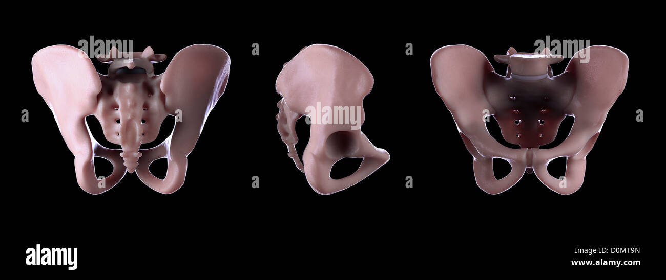 Three different view of the pelvis. Stock Photohttps://www.alamy.com/image-license-details/?v=1https://www.alamy.com/stock-photo-three-different-view-of-the-pelvis-52089233.html
RMD0MT9N–Three different view of the pelvis.
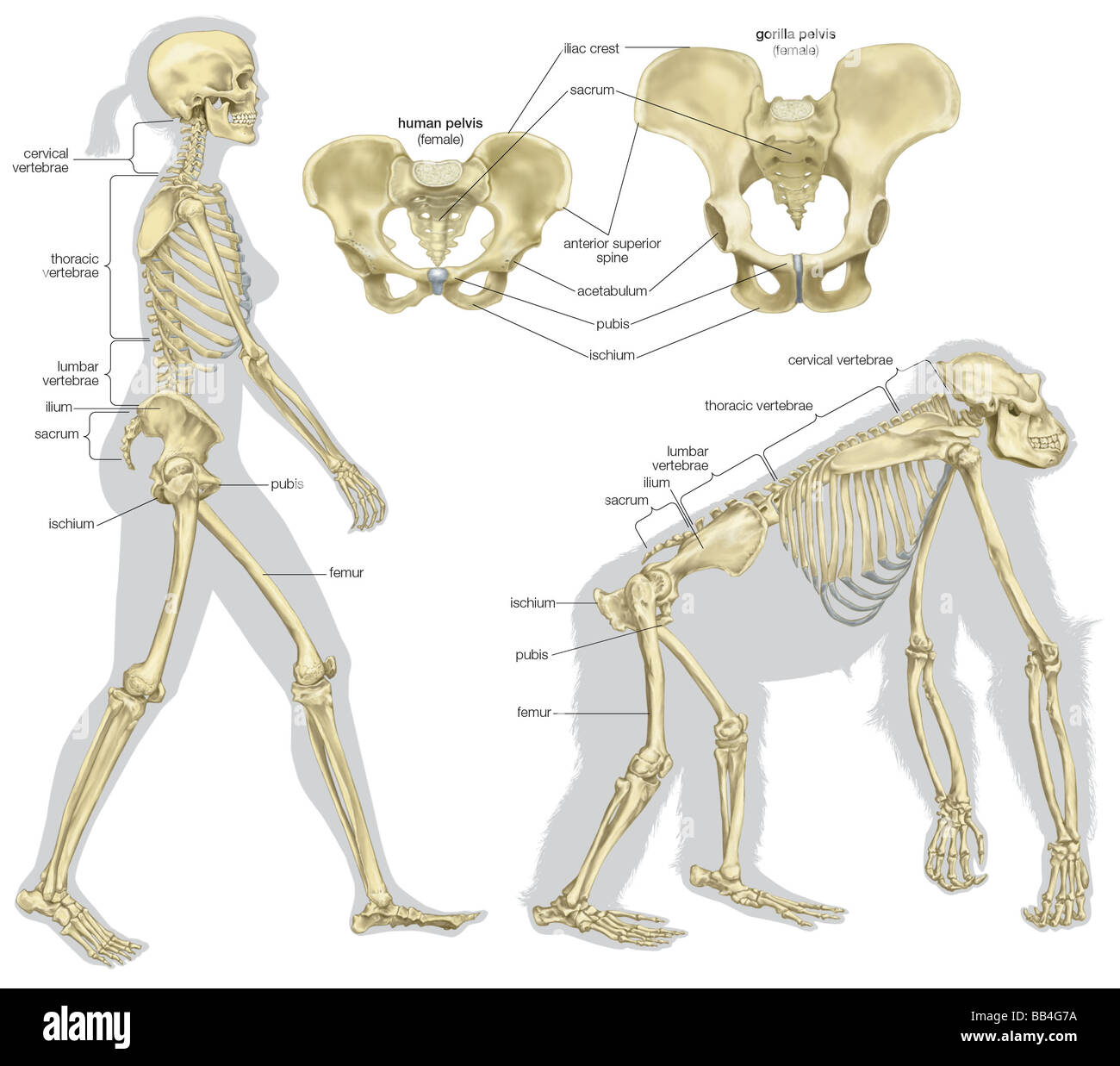 The skeletal structures of a human being and of a gorilla. Stock Photohttps://www.alamy.com/image-license-details/?v=1https://www.alamy.com/stock-photo-the-skeletal-structures-of-a-human-being-and-of-a-gorilla-24072142.html
RMBB4G7A–The skeletal structures of a human being and of a gorilla.
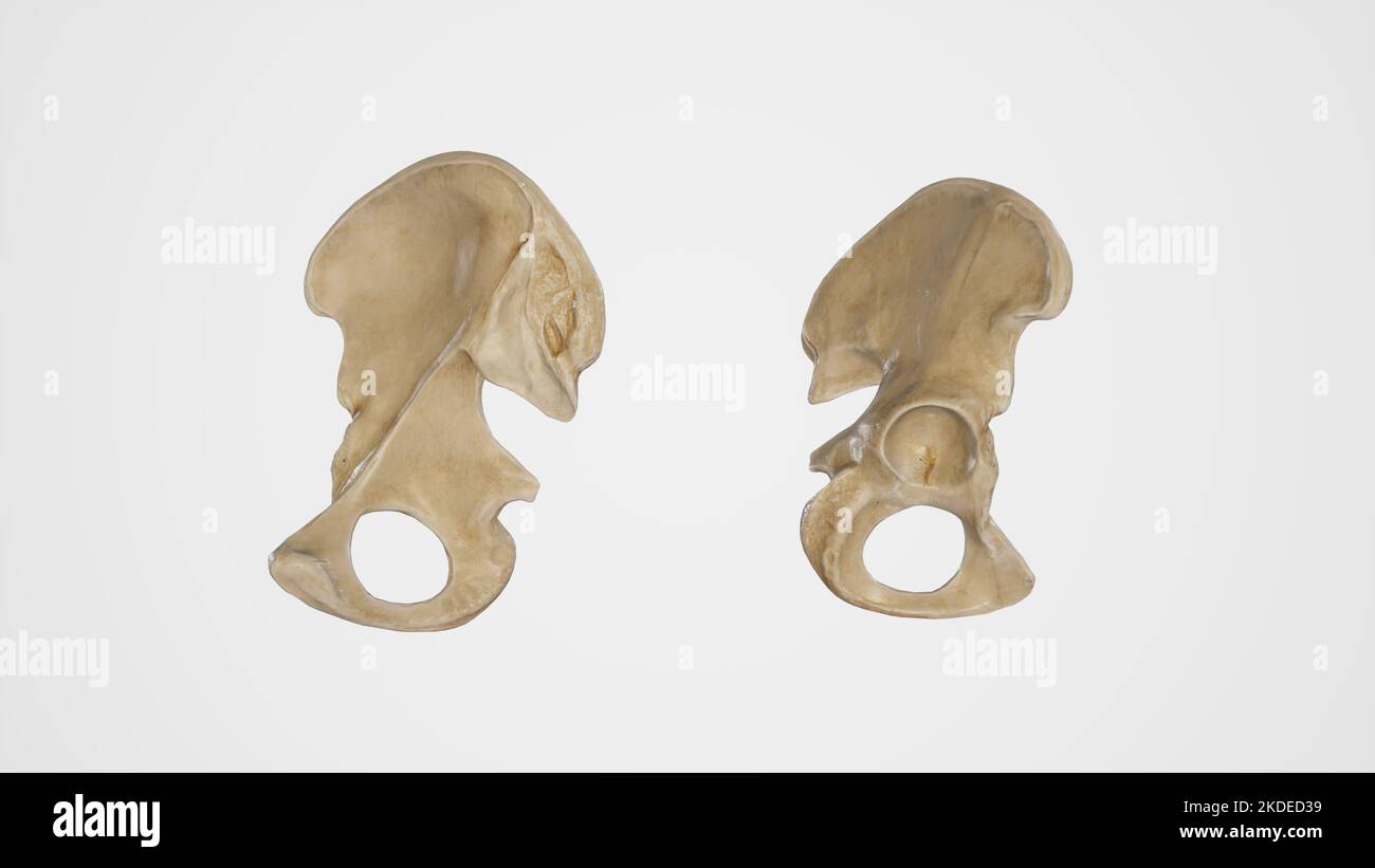 Medial and Lateral View of Hip Bone Stock Photohttps://www.alamy.com/image-license-details/?v=1https://www.alamy.com/medial-and-lateral-view-of-hip-bone-image490198445.html
RF2KDED39–Medial and Lateral View of Hip Bone
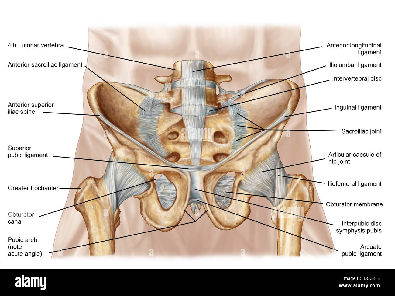 Anatomy of human pelvic bone and ligaments. Stock Photohttps://www.alamy.com/image-license-details/?v=1https://www.alamy.com/stock-photo-anatomy-of-human-pelvic-bone-and-ligaments-59361246.html
RFDCG3TE–Anatomy of human pelvic bone and ligaments.
 Abdomen. Tummy tuck. It is retensioning skin and abdominal muscles, by removing redundant skin and musculature plication. Before surgery should be taken anatomical references, such as the midline of the abdomen and the height of the anterior superior iliac spine. Plastic surgery. Doctor attending to patient medical consultation. Stock Photohttps://www.alamy.com/image-license-details/?v=1https://www.alamy.com/abdomen-tummy-tuck-it-is-retensioning-skin-and-abdominal-muscles-by-removing-redundant-skin-and-musculature-plication-before-surgery-should-be-taken-anatomical-references-such-as-the-midline-of-the-abdomen-and-the-height-of-the-anterior-superior-iliac-spine-plastic-surgery-doctor-attending-to-patient-medical-consultation-image602215497.html
RM2WYN809–Abdomen. Tummy tuck. It is retensioning skin and abdominal muscles, by removing redundant skin and musculature plication. Before surgery should be taken anatomical references, such as the midline of the abdomen and the height of the anterior superior iliac spine. Plastic surgery. Doctor attending to patient medical consultation.
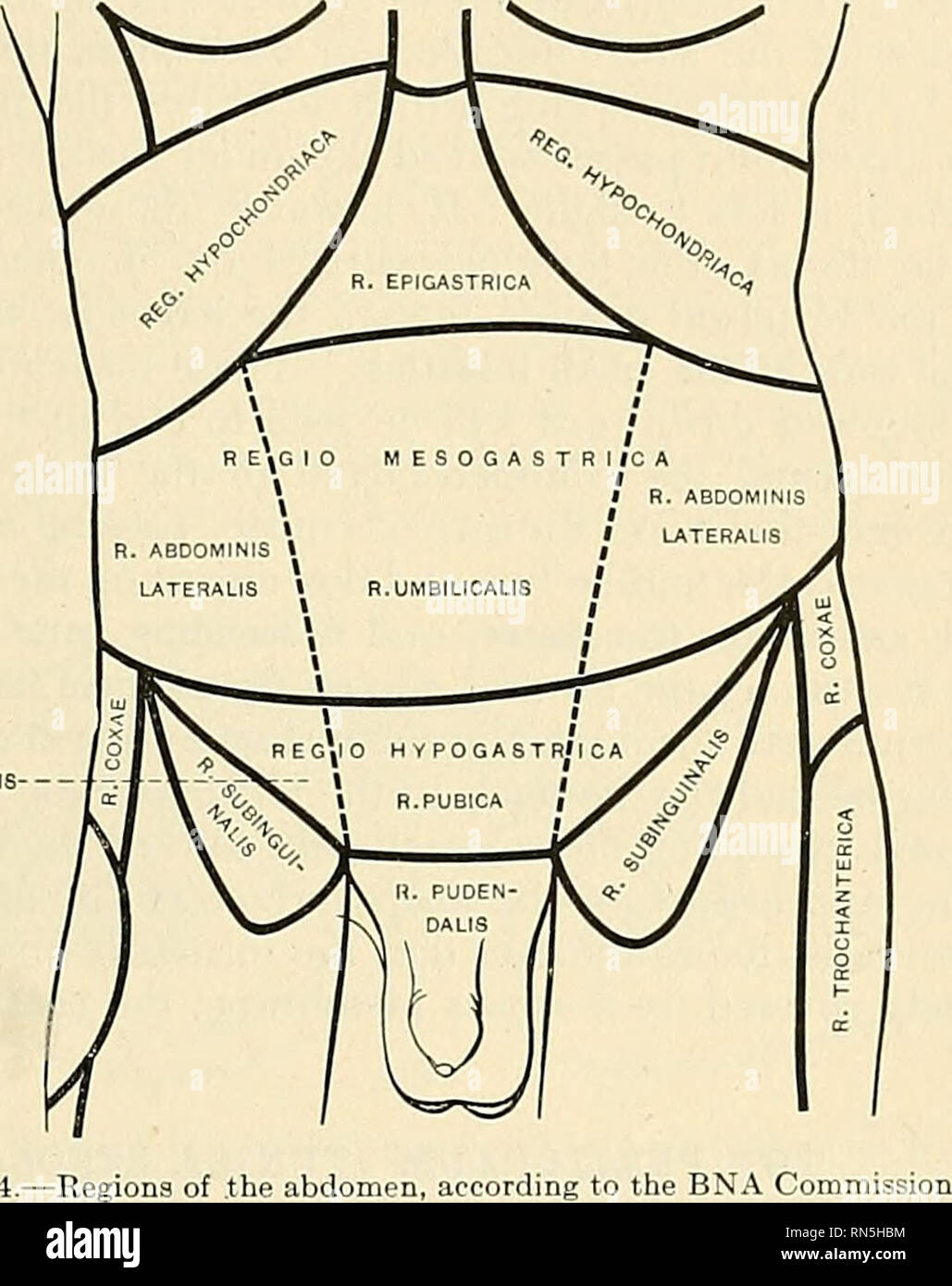 . Anatomy, descriptive and applied. Anatomy. THE ABDOMEN 1243 the anterior superior iliac spine. By means of these imaginary planes the abdomen is divided into three zones, which are named, from above downward, ?mbcostal, umbilical, and hypogastric zones. Each of these is furtlier sul)divided by two sagittal planes, which are indicated on the surface by lines drawn vertically through points half way between the anterior superior iliac spines and tlie symphysis puns. The regions as outlined by the BNA Commission are shown in Fig. 974.' The middle region of the upper zone is called the epigastr Stock Photohttps://www.alamy.com/image-license-details/?v=1https://www.alamy.com/anatomy-descriptive-and-applied-anatomy-the-abdomen-1243-the-anterior-superior-iliac-spine-by-means-of-these-imaginary-planes-the-abdomen-is-divided-into-three-zones-which-are-named-from-above-downward-mbcostal-umbilical-and-hypogastric-zones-each-of-these-is-furtlier-suldivided-by-two-sagittal-planes-which-are-indicated-on-the-surface-by-lines-drawn-vertically-through-points-half-way-between-the-anterior-superior-iliac-spines-and-tlie-symphysis-puns-the-regions-as-outlined-by-the-bna-commission-are-shown-in-fig-974-the-middle-region-of-the-upper-zone-is-called-the-epigastr-image236765976.html
RMRN5HBM–. Anatomy, descriptive and applied. Anatomy. THE ABDOMEN 1243 the anterior superior iliac spine. By means of these imaginary planes the abdomen is divided into three zones, which are named, from above downward, ?mbcostal, umbilical, and hypogastric zones. Each of these is furtlier sul)divided by two sagittal planes, which are indicated on the surface by lines drawn vertically through points half way between the anterior superior iliac spines and tlie symphysis puns. The regions as outlined by the BNA Commission are shown in Fig. 974.' The middle region of the upper zone is called the epigastr
 . Cunningham's Text-book of anatomy. Anatomy. 478 THE MUSCULAE SYSTEM. spermatic funiculus or round ligament in their further passage beyond the abdominal wall. Lig. Inguinale Reflexum Collesi.—The reflexed inguinal ligament of Colles (O.T. triangular fascia), lastly, is a triangular band of fibres placed behind the medial superior crus of the subcutaneous inguinal ring. It consists of fibres from the. t"f«" Pectoralis major Serratus anterior Latissimus dorsi BLIQUUS EXTERNVS ABDOMINIS if JM Sheath of rectus f£j,t W abdominis --Iliac crest .Anterior superior iliac spine ...... The in Stock Photohttps://www.alamy.com/image-license-details/?v=1https://www.alamy.com/cunninghams-text-book-of-anatomy-anatomy-478-the-musculae-system-spermatic-funiculus-or-round-ligament-in-their-further-passage-beyond-the-abdominal-wall-lig-inguinale-reflexum-collesithe-reflexed-inguinal-ligament-of-colles-ot-triangular-fascia-lastly-is-a-triangular-band-of-fibres-placed-behind-the-medial-superior-crus-of-the-subcutaneous-inguinal-ring-it-consists-of-fibres-from-the-tquotfquot-pectoralis-major-serratus-anterior-latissimus-dorsi-bliquus-externvs-abdominis-if-jm-sheath-of-rectus-fjt-w-abdominis-iliac-crest-anterior-superior-iliac-spine-the-in-image216346272.html
RMPFYBTG–. Cunningham's Text-book of anatomy. Anatomy. 478 THE MUSCULAE SYSTEM. spermatic funiculus or round ligament in their further passage beyond the abdominal wall. Lig. Inguinale Reflexum Collesi.—The reflexed inguinal ligament of Colles (O.T. triangular fascia), lastly, is a triangular band of fibres placed behind the medial superior crus of the subcutaneous inguinal ring. It consists of fibres from the. t"f«" Pectoralis major Serratus anterior Latissimus dorsi BLIQUUS EXTERNVS ABDOMINIS if JM Sheath of rectus f£j,t W abdominis --Iliac crest .Anterior superior iliac spine ...... The in
 Lateral Femoral Cutaneous Nerve Stock Photohttps://www.alamy.com/image-license-details/?v=1https://www.alamy.com/lateral-femoral-cutaneous-nerve-image490198315.html
RF2KDECXK–Lateral Femoral Cutaneous Nerve
 Abdomen. Tummy tuck. It is retensioning skin and abdominal muscles, by removing redundant skin and musculature plication. Before surgery should be taken anatomical references, such as the midline of the abdomen and the height of the anterior superior iliac spine. Plastic surgery. Doctor attending to patient medical consultation. Stock Photohttps://www.alamy.com/image-license-details/?v=1https://www.alamy.com/abdomen-tummy-tuck-it-is-retensioning-skin-and-abdominal-muscles-by-removing-redundant-skin-and-musculature-plication-before-surgery-should-be-taken-anatomical-references-such-as-the-midline-of-the-abdomen-and-the-height-of-the-anterior-superior-iliac-spine-plastic-surgery-doctor-attending-to-patient-medical-consultation-image601986043.html
RM2WYAR9F–Abdomen. Tummy tuck. It is retensioning skin and abdominal muscles, by removing redundant skin and musculature plication. Before surgery should be taken anatomical references, such as the midline of the abdomen and the height of the anterior superior iliac spine. Plastic surgery. Doctor attending to patient medical consultation.
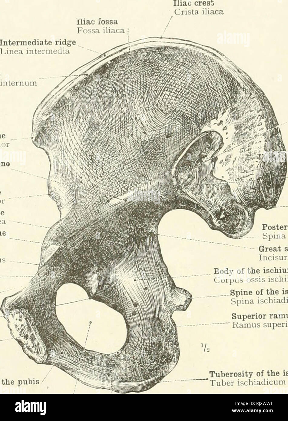 . An atlas of human anatomy for students and physicians. Anatomy. 128 THE SKELETON OF THE LOWER EXTREMITY Intermediate ridge Finea intermedia Inner lip Labium internum Anterior superior iliac spine Spina iliaca anterior superior Iliac portion of the iliopectineal line Linea arcuata Anterior inferior iliac spine Spina iliaca anterior inferior Iliopectineal eminence Eminentia iliopectinea Pubic portion of the iliopectineal line Pecten ossis pubis Obturator groove Sulcus obturatorius *Anterior obturator tubercle •Tuberculum obturatorium anterius Superior or ascending ramus of the pubis Ramus supe Stock Photohttps://www.alamy.com/image-license-details/?v=1https://www.alamy.com/an-atlas-of-human-anatomy-for-students-and-physicians-anatomy-128-the-skeleton-of-the-lower-extremity-intermediate-ridge-finea-intermedia-inner-lip-labium-internum-anterior-superior-iliac-spine-spina-iliaca-anterior-superior-iliac-portion-of-the-iliopectineal-line-linea-arcuata-anterior-inferior-iliac-spine-spina-iliaca-anterior-inferior-iliopectineal-eminence-eminentia-iliopectinea-pubic-portion-of-the-iliopectineal-line-pecten-ossis-pubis-obturator-groove-sulcus-obturatorius-anterior-obturator-tubercle-tuberculum-obturatorium-anterius-superior-or-ascending-ramus-of-the-pubis-ramus-supe-image235389668.html
RMRJXWWT–. An atlas of human anatomy for students and physicians. Anatomy. 128 THE SKELETON OF THE LOWER EXTREMITY Intermediate ridge Finea intermedia Inner lip Labium internum Anterior superior iliac spine Spina iliaca anterior superior Iliac portion of the iliopectineal line Linea arcuata Anterior inferior iliac spine Spina iliaca anterior inferior Iliopectineal eminence Eminentia iliopectinea Pubic portion of the iliopectineal line Pecten ossis pubis Obturator groove Sulcus obturatorius *Anterior obturator tubercle •Tuberculum obturatorium anterius Superior or ascending ramus of the pubis Ramus supe
 . Cunningham's Text-book of anatomy. Anatomy. 1460 SURFACE AND SUKGICAL ANATOMY. hip-joint. One of the commonest situations to meet with an abscess in hip-joint disease is in the cellular tissue and fat under the tensor fasciae lata?; or the pus may pass below and to the medial side of the neck of the femur, and thence along the course of the medial circumflex artery of the thigh to the back of the thigh. To tap or explore the hip-joint, the puncture should be made in the interval between the sartorius and the tensor fascia? lata}, 2 to 3 in. distal to the superior anterior iliac spine ; if th Stock Photohttps://www.alamy.com/image-license-details/?v=1https://www.alamy.com/cunninghams-text-book-of-anatomy-anatomy-1460-surface-and-sukgical-anatomy-hip-joint-one-of-the-commonest-situations-to-meet-with-an-abscess-in-hip-joint-disease-is-in-the-cellular-tissue-and-fat-under-the-tensor-fasciae-lata-or-the-pus-may-pass-below-and-to-the-medial-side-of-the-neck-of-the-femur-and-thence-along-the-course-of-the-medial-circumflex-artery-of-the-thigh-to-the-back-of-the-thigh-to-tap-or-explore-the-hip-joint-the-puncture-should-be-made-in-the-interval-between-the-sartorius-and-the-tensor-fascia-lata-2-to-3-in-distal-to-the-superior-anterior-iliac-spine-if-th-image216339195.html
RMPFY2RR–. Cunningham's Text-book of anatomy. Anatomy. 1460 SURFACE AND SUKGICAL ANATOMY. hip-joint. One of the commonest situations to meet with an abscess in hip-joint disease is in the cellular tissue and fat under the tensor fasciae lata?; or the pus may pass below and to the medial side of the neck of the femur, and thence along the course of the medial circumflex artery of the thigh to the back of the thigh. To tap or explore the hip-joint, the puncture should be made in the interval between the sartorius and the tensor fascia? lata}, 2 to 3 in. distal to the superior anterior iliac spine ; if th
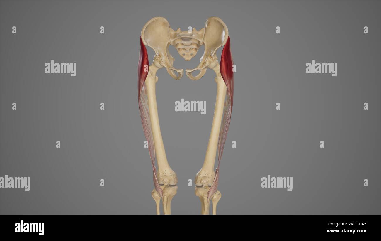 Medical Illustration of Tensor Fascia Lata Muscle Stock Photohttps://www.alamy.com/image-license-details/?v=1https://www.alamy.com/medical-illustration-of-tensor-fascia-lata-muscle-image490198491.html
RF2KDED4Y–Medical Illustration of Tensor Fascia Lata Muscle
 Abdomen. Tummy tuck. It is retensioning skin and abdominal muscles, by removing redundant skin and musculature plication. Before surgery should be taken anatomical references, such as the midline of the abdomen and the height of the anterior superior iliac spine. Plastic surgery. Doctor attending to patient medical consultation. Stock Photohttps://www.alamy.com/image-license-details/?v=1https://www.alamy.com/abdomen-tummy-tuck-it-is-retensioning-skin-and-abdominal-muscles-by-removing-redundant-skin-and-musculature-plication-before-surgery-should-be-taken-anatomical-references-such-as-the-midline-of-the-abdomen-and-the-height-of-the-anterior-superior-iliac-spine-plastic-surgery-doctor-attending-to-patient-medical-consultation-image602125245.html
RM2WYH4W1–Abdomen. Tummy tuck. It is retensioning skin and abdominal muscles, by removing redundant skin and musculature plication. Before surgery should be taken anatomical references, such as the midline of the abdomen and the height of the anterior superior iliac spine. Plastic surgery. Doctor attending to patient medical consultation.
 . Modern surgery, general and operative. employed in youths and middle-aged individuals if the general conditionof the patient or some particular dis-eased state does not forbid, and if pain issevere and disability is pronounced. Leonard Freeman advises an anteriorincision beginning below and external tothe anterior superior iliac spine and ex-tending downward, external to thesartorious, for 3 or 4 inches (Annals ofSurg., Oct., 1904). When the frag-ments are exposed, the connective tissuebetween them is cut away by means ofscissors, the surfaces of the fragmentsare freshened by a chisel or a c Stock Photohttps://www.alamy.com/image-license-details/?v=1https://www.alamy.com/modern-surgery-general-and-operative-employed-in-youths-and-middle-aged-individuals-if-the-general-conditionof-the-patient-or-some-particular-dis-eased-state-does-not-forbid-and-if-pain-issevere-and-disability-is-pronounced-leonard-freeman-advises-an-anteriorincision-beginning-below-and-external-tothe-anterior-superior-iliac-spine-and-ex-tending-downward-external-to-thesartorious-for-3-or-4-inches-annals-ofsurg-oct-1904-when-the-frag-ments-are-exposed-the-connective-tissuebetween-them-is-cut-away-by-means-ofscissors-the-surfaces-of-the-fragmentsare-freshened-by-a-chisel-or-a-c-image337078838.html
RM2AGB7DX–. Modern surgery, general and operative. employed in youths and middle-aged individuals if the general conditionof the patient or some particular dis-eased state does not forbid, and if pain issevere and disability is pronounced. Leonard Freeman advises an anteriorincision beginning below and external tothe anterior superior iliac spine and ex-tending downward, external to thesartorious, for 3 or 4 inches (Annals ofSurg., Oct., 1904). When the frag-ments are exposed, the connective tissuebetween them is cut away by means ofscissors, the surfaces of the fragmentsare freshened by a chisel or a c
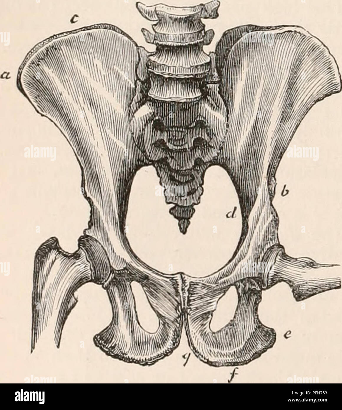 . The cyclopædia of anatomy and physiology. Anatomy; Physiology; Zoology. 152 PELVIS. wings, the anterior part seems deficient, the anterior superior spine'(a) being placed directly over the cotyloid cavity ; and the crest (c) being, consequently, very short, terminating abruptly at the vertical rib mentioned in the description of the human ilium. The ales are more expanded in the Uran than the Chimpanzee. The crest does not present the lateral /-like curvature, and is less arched than in man. The anterior iliac spines are more widely separated, the inferior (6) being scarcely discernible, and Stock Photohttps://www.alamy.com/image-license-details/?v=1https://www.alamy.com/the-cyclopdia-of-anatomy-and-physiology-anatomy-physiology-zoology-152-pelvis-wings-the-anterior-part-seems-deficient-the-anterior-superior-spinea-being-placed-directly-over-the-cotyloid-cavity-and-the-crest-c-being-consequently-very-short-terminating-abruptly-at-the-vertical-rib-mentioned-in-the-description-of-the-human-ilium-the-ales-are-more-expanded-in-the-uran-than-the-chimpanzee-the-crest-does-not-present-the-lateral-like-curvature-and-is-less-arched-than-in-man-the-anterior-iliac-spines-are-more-widely-separated-the-inferior-6-being-scarcely-discernible-and-image216210879.html
RMPFN753–. The cyclopædia of anatomy and physiology. Anatomy; Physiology; Zoology. 152 PELVIS. wings, the anterior part seems deficient, the anterior superior spine'(a) being placed directly over the cotyloid cavity ; and the crest (c) being, consequently, very short, terminating abruptly at the vertical rib mentioned in the description of the human ilium. The ales are more expanded in the Uran than the Chimpanzee. The crest does not present the lateral /-like curvature, and is less arched than in man. The anterior iliac spines are more widely separated, the inferior (6) being scarcely discernible, and
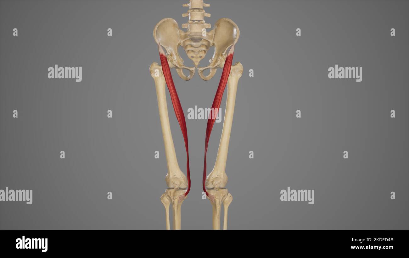 Medical Illustration of Sartorius Muscle Stock Photohttps://www.alamy.com/image-license-details/?v=1https://www.alamy.com/medical-illustration-of-sartorius-muscle-image490198475.html
RF2KDED4B–Medical Illustration of Sartorius Muscle
 Anterior view of Right Hip Bone Stock Photohttps://www.alamy.com/image-license-details/?v=1https://www.alamy.com/anterior-view-of-right-hip-bone-image491878999.html
RF2KG70K3–Anterior view of Right Hip Bone
 Abdomen. Tummy tuck. It is retensioning skin and abdominal muscles, by removing redundant skin and musculature plication. Before surgery should be taken anatomical references, such as the midline of the abdomen and the height of the anterior superior iliac spine. Plastic surgery. Doctor attending to patient medical consultation. Stock Photohttps://www.alamy.com/image-license-details/?v=1https://www.alamy.com/abdomen-tummy-tuck-it-is-retensioning-skin-and-abdominal-muscles-by-removing-redundant-skin-and-musculature-plication-before-surgery-should-be-taken-anatomical-references-such-as-the-midline-of-the-abdomen-and-the-height-of-the-anterior-superior-iliac-spine-plastic-surgery-doctor-attending-to-patient-medical-consultation-image602263583.html
RM2WYRD9K–Abdomen. Tummy tuck. It is retensioning skin and abdominal muscles, by removing redundant skin and musculature plication. Before surgery should be taken anatomical references, such as the midline of the abdomen and the height of the anterior superior iliac spine. Plastic surgery. Doctor attending to patient medical consultation.
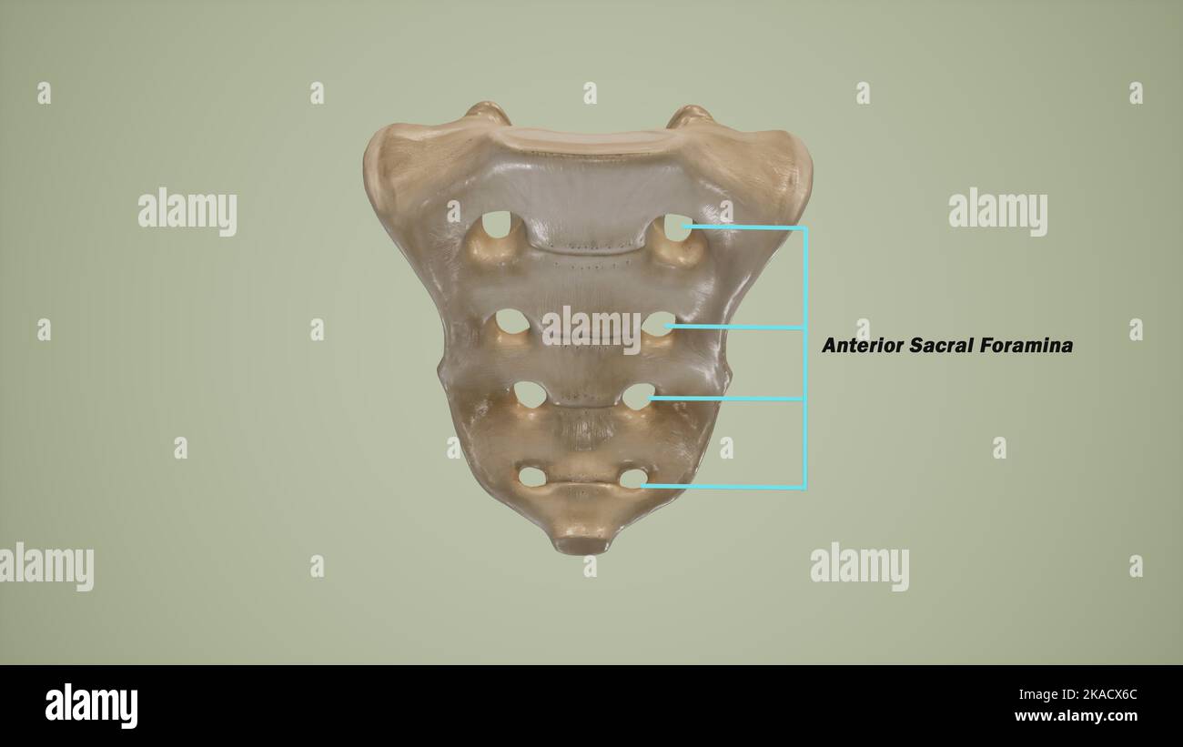 Anterior view of human sacrum showing the anterior sacral foramina-Labeled Stock Photohttps://www.alamy.com/image-license-details/?v=1https://www.alamy.com/anterior-view-of-human-sacrum-showing-the-anterior-sacral-foramina-labeled-image488320852.html
RF2KACX6C–Anterior view of human sacrum showing the anterior sacral foramina-Labeled
 . The diagnosis and treatment of diseases of women. e with the ordinary tape-line. When measuring apatient, take enough measurements to make an accurate record. Measurementsalong the lines shown in Fig. 40 will show variations with a large growth in any part of the peritoneal cavity. They are as follows: 1. From umbilicus to sternal notch. 2. From umbilicus to pubes. 3. From umbilicus to right anterior superior iliac spine. 4. From umbihcus to left anterior superior iliac spine. 5. Circumference of body at level of umbilicus. 6. Circumference of body 3 inches above umbilicus. 7. Circumference Stock Photohttps://www.alamy.com/image-license-details/?v=1https://www.alamy.com/the-diagnosis-and-treatment-of-diseases-of-women-e-with-the-ordinary-tape-line-when-measuring-apatient-take-enough-measurements-to-make-an-accurate-record-measurementsalong-the-lines-shown-in-fig-40-will-show-variations-with-a-large-growth-in-any-part-of-the-peritoneal-cavity-they-are-as-follows-1-from-umbilicus-to-sternal-notch-2-from-umbilicus-to-pubes-3-from-umbilicus-to-right-anterior-superior-iliac-spine-4-from-umbihcus-to-left-anterior-superior-iliac-spine-5-circumference-of-body-at-level-of-umbilicus-6-circumference-of-body-3-inches-above-umbilicus-7-circumference-image336936075.html
RM2AG4NB7–. The diagnosis and treatment of diseases of women. e with the ordinary tape-line. When measuring apatient, take enough measurements to make an accurate record. Measurementsalong the lines shown in Fig. 40 will show variations with a large growth in any part of the peritoneal cavity. They are as follows: 1. From umbilicus to sternal notch. 2. From umbilicus to pubes. 3. From umbilicus to right anterior superior iliac spine. 4. From umbihcus to left anterior superior iliac spine. 5. Circumference of body at level of umbilicus. 6. Circumference of body 3 inches above umbilicus. 7. Circumference
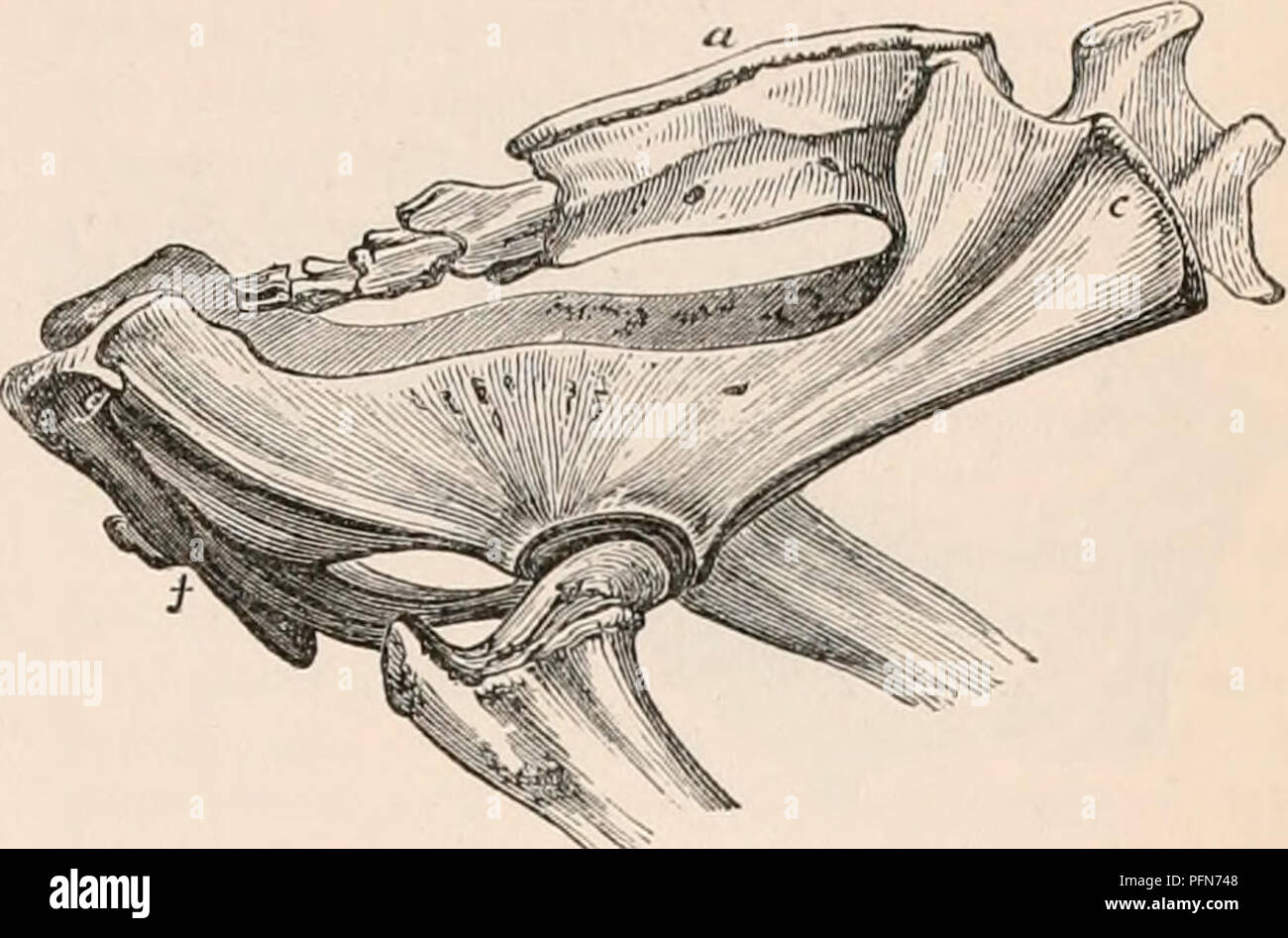 . The cyclopædia of anatomy and physiology. Anatomy; Physiology; Zoology. PELVIS. 157 are not united in a crest. The ilia approach in shape to those of the Tapir, being in a less marked degree T-shaped ; the posterior limb of the iliac wings projecting inwards as far as the sacral spines; the anterior superior spine often presenting an epiphysis, and the shaft being long and blade-like. The ischia are comparatively long, and much more slender than in the Ruminants, being placed nearly parallel with the coccygeal vertebra, and with prolonged tuberosities. The jnibcs are small and short, and dir Stock Photohttps://www.alamy.com/image-license-details/?v=1https://www.alamy.com/the-cyclopdia-of-anatomy-and-physiology-anatomy-physiology-zoology-pelvis-157-are-not-united-in-a-crest-the-ilia-approach-in-shape-to-those-of-the-tapir-being-in-a-less-marked-degree-t-shaped-the-posterior-limb-of-the-iliac-wings-projecting-inwards-as-far-as-the-sacral-spines-the-anterior-superior-spine-often-presenting-an-epiphysis-and-the-shaft-being-long-and-blade-like-the-ischia-are-comparatively-long-and-much-more-slender-than-in-the-ruminants-being-placed-nearly-parallel-with-the-coccygeal-vertebra-and-with-prolonged-tuberosities-the-jnibcs-are-small-and-short-and-dir-image216210856.html
RMPFN748–. The cyclopædia of anatomy and physiology. Anatomy; Physiology; Zoology. PELVIS. 157 are not united in a crest. The ilia approach in shape to those of the Tapir, being in a less marked degree T-shaped ; the posterior limb of the iliac wings projecting inwards as far as the sacral spines; the anterior superior spine often presenting an epiphysis, and the shaft being long and blade-like. The ischia are comparatively long, and much more slender than in the Ruminants, being placed nearly parallel with the coccygeal vertebra, and with prolonged tuberosities. The jnibcs are small and short, and dir
 Abdomen. Tummy tuck. It is retensioning skin and abdominal muscles, by removing redundant skin and musculature plication. Before surgery should be taken anatomical references, such as the midline of the abdomen and the height of the anterior superior iliac spine. Plastic surgery. Doctor attending to patient medical consultation. Stock Photohttps://www.alamy.com/image-license-details/?v=1https://www.alamy.com/abdomen-tummy-tuck-it-is-retensioning-skin-and-abdominal-muscles-by-removing-redundant-skin-and-musculature-plication-before-surgery-should-be-taken-anatomical-references-such-as-the-midline-of-the-abdomen-and-the-height-of-the-anterior-superior-iliac-spine-plastic-surgery-doctor-attending-to-patient-medical-consultation-image602036531.html
RM2WYD3MK–Abdomen. Tummy tuck. It is retensioning skin and abdominal muscles, by removing redundant skin and musculature plication. Before surgery should be taken anatomical references, such as the midline of the abdomen and the height of the anterior superior iliac spine. Plastic surgery. Doctor attending to patient medical consultation.
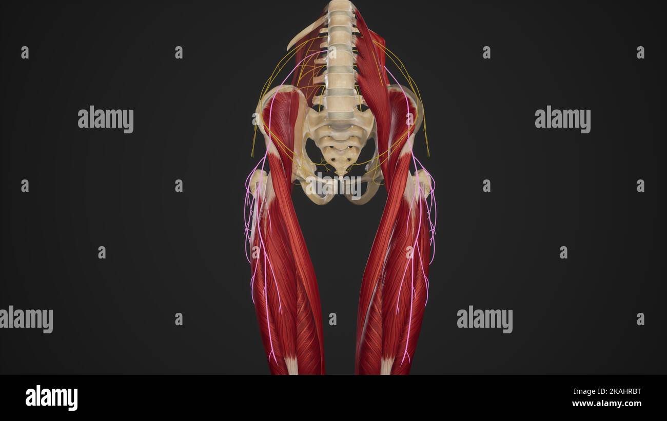 Course of Lateral Femoral Cutaneous Nerve Stock Photohttps://www.alamy.com/image-license-details/?v=1https://www.alamy.com/course-of-lateral-femoral-cutaneous-nerve-image488428412.html
RF2KAHRBT–Course of Lateral Femoral Cutaneous Nerve
 . The diagnosis and treatment of diseases of women. / ^> -^^ L ^:^ .. Fig. 4u. Showing the lines for Men.suiation. The measurements are made with the ordinary tape-line. When measuring apatient, take enough measurements to make an accurate record. Measurementsalong the lines shown in Fig. 40 will show variations with a large growth in any part of the peritoneal cavity. They are as follows: 1. From umbilicus to sternal notch. 2. From umbilicus to pubes. 3. From umbilicus to right anterior superior iliac spine. 4. From umbihcus to left anterior superior iliac spine. 5. Circumference of bod Stock Photohttps://www.alamy.com/image-license-details/?v=1https://www.alamy.com/the-diagnosis-and-treatment-of-diseases-of-women-gt-l-fig-4u-showing-the-lines-for-mensuiation-the-measurements-are-made-with-the-ordinary-tape-line-when-measuring-apatient-take-enough-measurements-to-make-an-accurate-record-measurementsalong-the-lines-shown-in-fig-40-will-show-variations-with-a-large-growth-in-any-part-of-the-peritoneal-cavity-they-are-as-follows-1-from-umbilicus-to-sternal-notch-2-from-umbilicus-to-pubes-3-from-umbilicus-to-right-anterior-superior-iliac-spine-4-from-umbihcus-to-left-anterior-superior-iliac-spine-5-circumference-of-bod-image336936201.html
RM2AG4NFN–. The diagnosis and treatment of diseases of women. / ^> -^^ L ^:^ .. Fig. 4u. Showing the lines for Men.suiation. The measurements are made with the ordinary tape-line. When measuring apatient, take enough measurements to make an accurate record. Measurementsalong the lines shown in Fig. 40 will show variations with a large growth in any part of the peritoneal cavity. They are as follows: 1. From umbilicus to sternal notch. 2. From umbilicus to pubes. 3. From umbilicus to right anterior superior iliac spine. 4. From umbihcus to left anterior superior iliac spine. 5. Circumference of bod
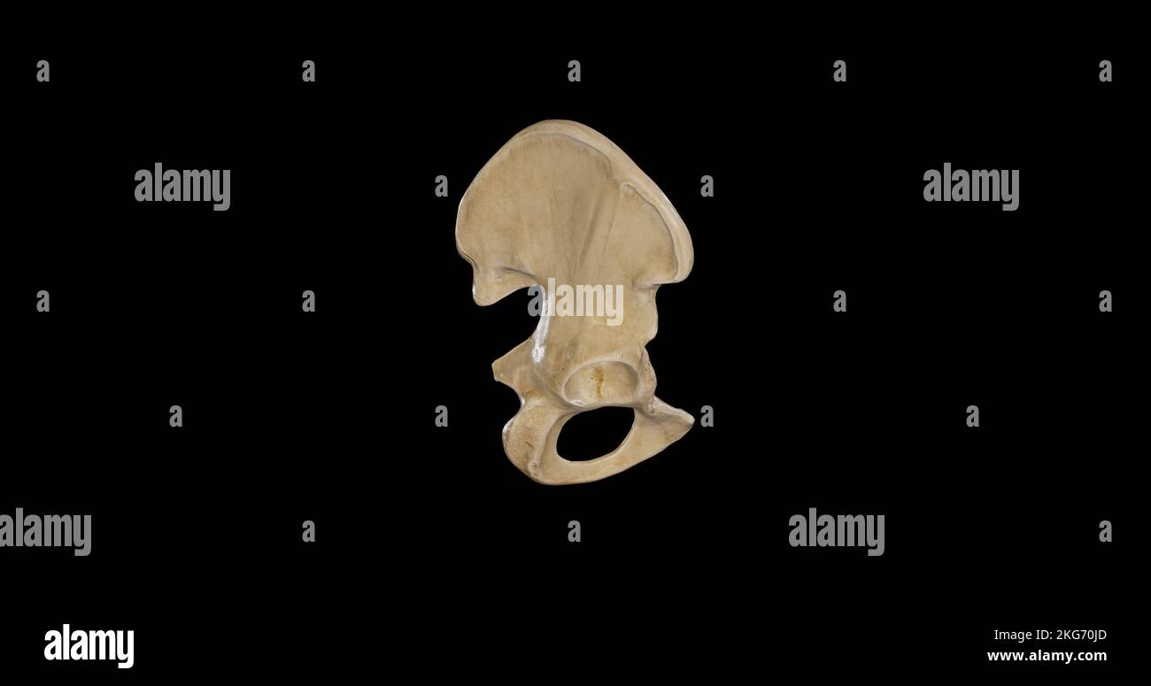 Lateral view of Right Hip Bone Stock Photohttps://www.alamy.com/image-license-details/?v=1https://www.alamy.com/lateral-view-of-right-hip-bone-image491878981.html
RF2KG70JD–Lateral view of Right Hip Bone
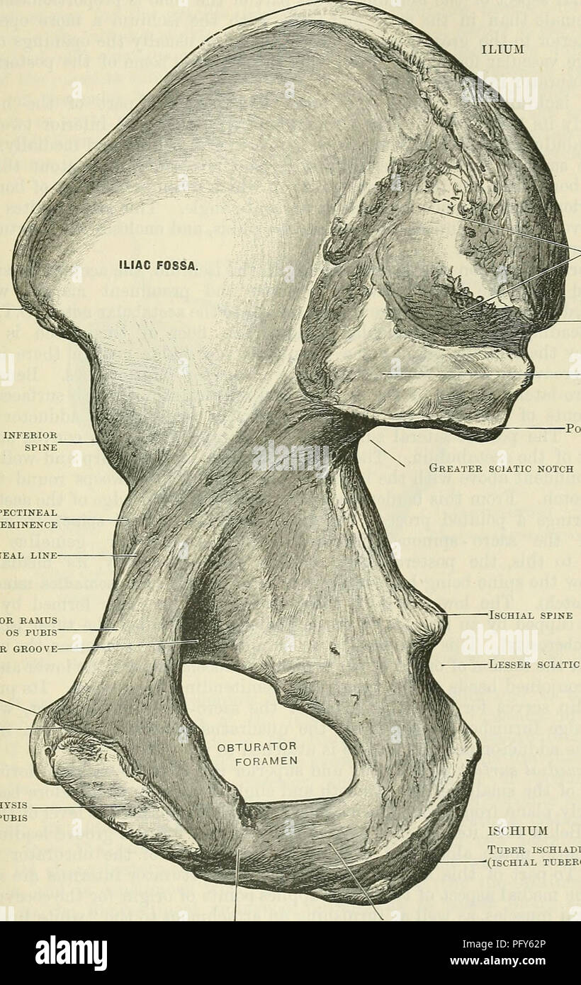 . Cunningham's Text-book of anatomy. Anatomy. THE HIP BONE. 231 is subdivided by an oblique ridge, called the ilio-pectineal line (linea arcuata), which passes forwards and distally, from the most prominent point of the auricular surface towards the medial side of the ilio-pectineal eminence, which is placed just above and in front of the acetabulum and marks the fusion of the Crest of the ilium ILIUM Anterior superior spine Anterior inferior spine'. Tuberosity for posterior sacro-iliac ligament Posterior superior SPINE Articular surface Post. inf. spine Ilio-pectineal eminence Ilio-pectineal Stock Photohttps://www.alamy.com/image-license-details/?v=1https://www.alamy.com/cunninghams-text-book-of-anatomy-anatomy-the-hip-bone-231-is-subdivided-by-an-oblique-ridge-called-the-ilio-pectineal-line-linea-arcuata-which-passes-forwards-and-distally-from-the-most-prominent-point-of-the-auricular-surface-towards-the-medial-side-of-the-ilio-pectineal-eminence-which-is-placed-just-above-and-in-front-of-the-acetabulum-and-marks-the-fusion-of-the-crest-of-the-ilium-ilium-anterior-superior-spine-anterior-inferior-spine-tuberosity-for-posterior-sacro-iliac-ligament-posterior-superior-spine-articular-surface-post-inf-spine-ilio-pectineal-eminence-ilio-pectineal-image216341742.html
RMPFY62P–. Cunningham's Text-book of anatomy. Anatomy. THE HIP BONE. 231 is subdivided by an oblique ridge, called the ilio-pectineal line (linea arcuata), which passes forwards and distally, from the most prominent point of the auricular surface towards the medial side of the ilio-pectineal eminence, which is placed just above and in front of the acetabulum and marks the fusion of the Crest of the ilium ILIUM Anterior superior spine Anterior inferior spine'. Tuberosity for posterior sacro-iliac ligament Posterior superior SPINE Articular surface Post. inf. spine Ilio-pectineal eminence Ilio-pectineal
 Abdomen. Tummy tuck. It is retensioning skin and abdominal muscles, by removing redundant skin and musculature plication. Before surgery should be taken anatomical references, such as the midline of the abdomen and the height of the anterior superior iliac spine. Plastic surgery. Doctor attending to patient medical consultation. Stock Photohttps://www.alamy.com/image-license-details/?v=1https://www.alamy.com/abdomen-tummy-tuck-it-is-retensioning-skin-and-abdominal-muscles-by-removing-redundant-skin-and-musculature-plication-before-surgery-should-be-taken-anatomical-references-such-as-the-midline-of-the-abdomen-and-the-height-of-the-anterior-superior-iliac-spine-plastic-surgery-doctor-attending-to-patient-medical-consultation-image602371458.html
RM2X00AXA–Abdomen. Tummy tuck. It is retensioning skin and abdominal muscles, by removing redundant skin and musculature plication. Before surgery should be taken anatomical references, such as the midline of the abdomen and the height of the anterior superior iliac spine. Plastic surgery. Doctor attending to patient medical consultation.
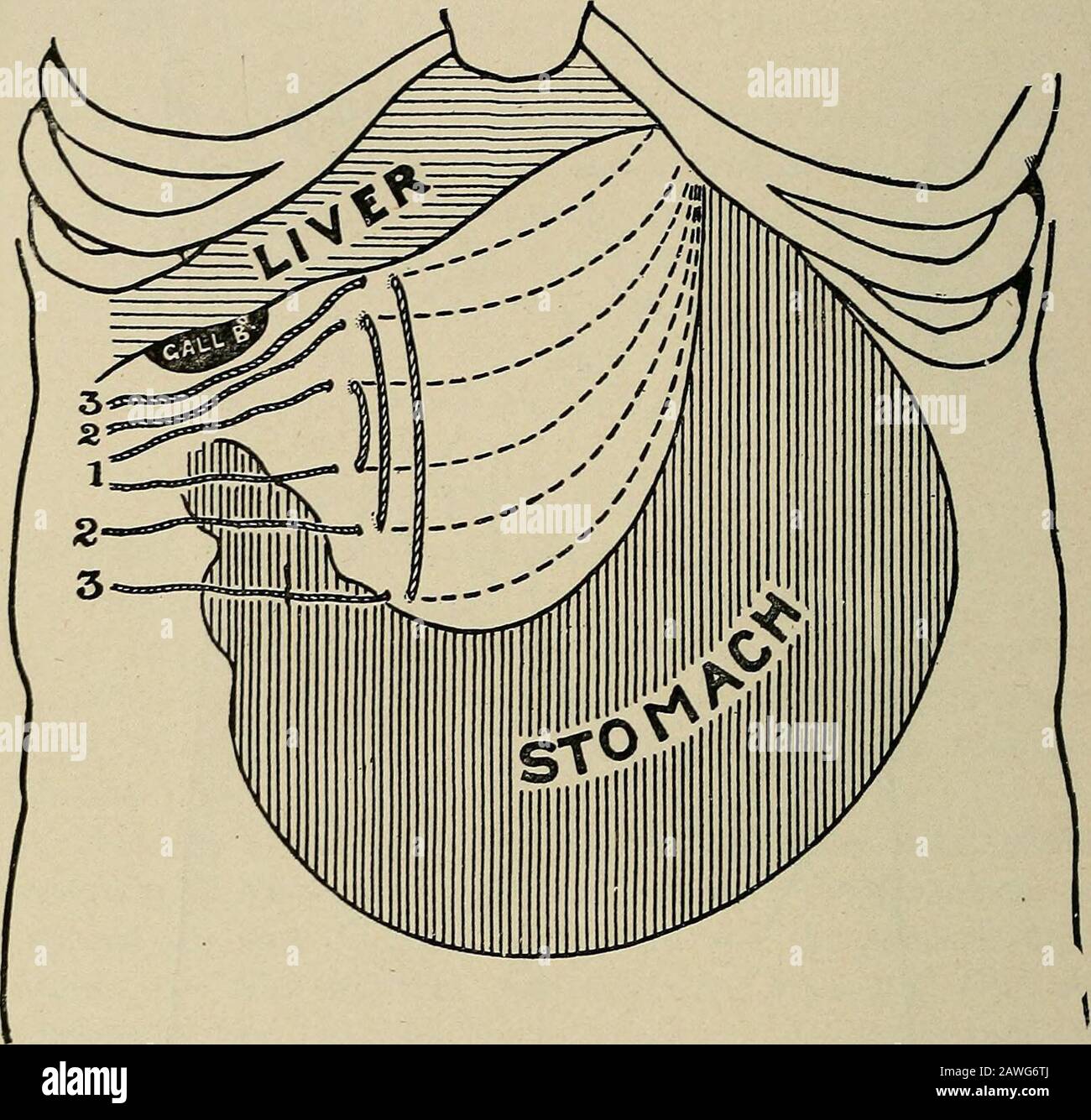 Operative surgery, for students and practitioners . Fig. 161.—Various Abdominal Incisions. B, mid-rectus incision; C, incisionfor left Inguinal colostomy: F, Fenger incision for stomach; G, vertical andoblique incisions for gall-bladder, etc.; H, von Hackers incision for gastrostomy;M, McBurney Incision for appendicectomy; S, incision for suprapubic cystotomy.In middle line above umbilicus is linea alba incision for operations upon stomach.X indicates location of anterior superior iliac spine. Dotted line drawn fromspine to the umbilicus. 363 ABDOMEN AND BACK. the sutures are all tied^ first t Stock Photohttps://www.alamy.com/image-license-details/?v=1https://www.alamy.com/operative-surgery-for-students-and-practitioners-fig-161various-abdominal-incisions-b-mid-rectus-incision-c-incisionfor-left-inguinal-colostomy-f-fenger-incision-for-stomach-g-vertical-andoblique-incisions-for-gall-bladder-etc-h-von-hackers-incision-for-gastrostomym-mcburney-incision-for-appendicectomy-s-incision-for-suprapubic-cystotomyin-middle-line-above-umbilicus-is-linea-alba-incision-for-operations-upon-stomachx-indicates-location-of-anterior-superior-iliac-spine-dotted-line-drawn-fromspine-to-the-umbilicus-363-abdomen-and-back-the-sutures-are-all-tied-first-t-image342720018.html
RM2AWG6TJ–Operative surgery, for students and practitioners . Fig. 161.—Various Abdominal Incisions. B, mid-rectus incision; C, incisionfor left Inguinal colostomy: F, Fenger incision for stomach; G, vertical andoblique incisions for gall-bladder, etc.; H, von Hackers incision for gastrostomy;M, McBurney Incision for appendicectomy; S, incision for suprapubic cystotomy.In middle line above umbilicus is linea alba incision for operations upon stomach.X indicates location of anterior superior iliac spine. Dotted line drawn fromspine to the umbilicus. 363 ABDOMEN AND BACK. the sutures are all tied^ first t
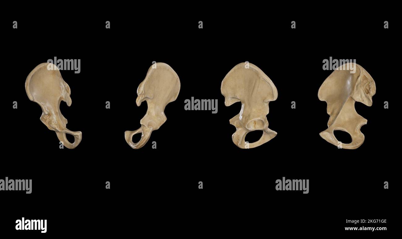 Right Hip Bone from multiple sides Stock Photohttps://www.alamy.com/image-license-details/?v=1https://www.alamy.com/right-hip-bone-from-multiple-sides-image491879710.html
RF2KG71GE–Right Hip Bone from multiple sides
 . A practical treatise on fractures and dislocations. Oblique fracture of shaft of femur; rearview. Oblique fracture of the shaft: front view.(U. S. Army Med. Museum.) the abdomen from one anterior superior iliac spine to the other, anda second one at right angles to the first from its centre downward,and then to place the ankles at equal distances from the second line.The shortening may vary from a small fraction of an inch toseveral inches. Abnormal mobility may be recognized by placing the hand underthe thigh at the suspected seat of fracture and gently litting it, orby holding the upper po Stock Photohttps://www.alamy.com/image-license-details/?v=1https://www.alamy.com/a-practical-treatise-on-fractures-and-dislocations-oblique-fracture-of-shaft-of-femur-rearview-oblique-fracture-of-the-shaft-front-viewu-s-army-med-museum-the-abdomen-from-one-anterior-superior-iliac-spine-to-the-other-anda-second-one-at-right-angles-to-the-first-from-its-centre-downwardand-then-to-place-the-ankles-at-equal-distances-from-the-second-linethe-shortening-may-vary-from-a-small-fraction-of-an-inch-toseveral-inches-abnormal-mobility-may-be-recognized-by-placing-the-hand-underthe-thigh-at-the-suspected-seat-of-fracture-and-gently-litting-it-orby-holding-the-upper-po-image336720334.html
RM2AFPX66–. A practical treatise on fractures and dislocations. Oblique fracture of shaft of femur; rearview. Oblique fracture of the shaft: front view.(U. S. Army Med. Museum.) the abdomen from one anterior superior iliac spine to the other, anda second one at right angles to the first from its centre downward,and then to place the ankles at equal distances from the second line.The shortening may vary from a small fraction of an inch toseveral inches. Abnormal mobility may be recognized by placing the hand underthe thigh at the suspected seat of fracture and gently litting it, orby holding the upper po
 Posterior view of Right Hip Bone Stock Photohttps://www.alamy.com/image-license-details/?v=1https://www.alamy.com/posterior-view-of-right-hip-bone-image491878990.html
RF2KG70JP–Posterior view of Right Hip Bone
 A manual of operative surgery . The patientis rolled nearly over on tothe face ; the limb is al-lowed to hang over theedge of the table ; thethigh is rotated in. The fig. 347.—incisions for the gluteal, sciatic, i ,, AND PUDIC ARTEKIES. surgeon stands upon the n. ? ..... ? i j -i t , A» Posterior superior iliac spine ; B, Great trochanter; side to be dealt With. c, Tuber ischii; D, Anterior superior iliac spine ; An incision five inches %%£$?* ^ : AC SdatiC aUd ^ (MaC-in length is made along the line just given. The centre of the incision should correspond to thepoint of exit of the artery (F Stock Photohttps://www.alamy.com/image-license-details/?v=1https://www.alamy.com/a-manual-of-operative-surgery-the-patientis-rolled-nearly-over-on-tothe-face-the-limb-is-al-lowed-to-hang-over-theedge-of-the-table-thethigh-is-rotated-in-the-fig-347incisions-for-the-gluteal-sciatic-i-and-pudic-artekies-surgeon-stands-upon-the-n-i-j-i-t-a-posterior-superior-iliac-spine-b-great-trochanter-side-to-be-dealt-with-c-tuber-ischii-d-anterior-superior-iliac-spine-an-incision-five-inches-ac-sdatic-aud-mac-in-length-is-made-along-the-line-just-given-the-centre-of-the-incision-should-correspond-to-thepoint-of-exit-of-the-artery-f-image343322304.html
RM2AXFK2T–A manual of operative surgery . The patientis rolled nearly over on tothe face ; the limb is al-lowed to hang over theedge of the table ; thethigh is rotated in. The fig. 347.—incisions for the gluteal, sciatic, i ,, AND PUDIC ARTEKIES. surgeon stands upon the n. ? ..... ? i j -i t , A» Posterior superior iliac spine ; B, Great trochanter; side to be dealt With. c, Tuber ischii; D, Anterior superior iliac spine ; An incision five inches %%£$?* ^ : AC SdatiC aUd ^ (MaC-in length is made along the line just given. The centre of the incision should correspond to thepoint of exit of the artery (F
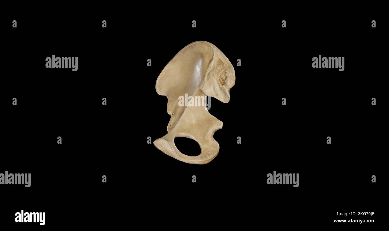 Medial view of Right Hip Bone Stock Photohttps://www.alamy.com/image-license-details/?v=1https://www.alamy.com/medial-view-of-right-hip-bone-image491878983.html
RF2KG70JF–Medial view of Right Hip Bone
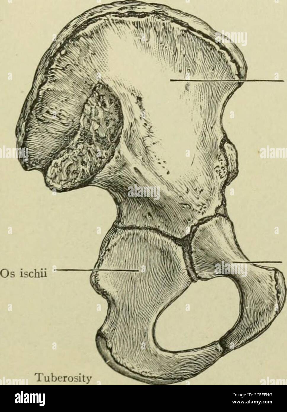 . Text-book of anatomy and physiology for nurses. Fig. 42.—Hip-bone, Exterior.—(Morris.) The OS ilium is the highest part of the hip-bone and has abroad expanded portion called the wing (or ala). The medialsurface of the wing is the iliac fossa, which is filled with the iliacmuscle; the lateral surface is crossed by three curved lines (calledthe posterior gluteal, the anterior gluteal, and the inferior ghiteallines). BONES OF THE PELVIS. 45 The superior border is called the crest. It can be easily felt, andthe anterior extremity is known as the anterior superior iliac spine,more often called t Stock Photohttps://www.alamy.com/image-license-details/?v=1https://www.alamy.com/text-book-of-anatomy-and-physiology-for-nurses-fig-42hip-bone-exteriormorris-the-os-ilium-is-the-highest-part-of-the-hip-bone-and-has-abroad-expanded-portion-called-the-wing-or-ala-the-medialsurface-of-the-wing-is-the-iliac-fossa-which-is-filled-with-the-iliacmuscle-the-lateral-surface-is-crossed-by-three-curved-lines-calledthe-posterior-gluteal-the-anterior-gluteal-and-the-inferior-ghiteallines-bones-of-the-pelvis-45-the-superior-border-is-called-the-crest-it-can-be-easily-felt-andthe-anterior-extremity-is-known-as-the-anterior-superior-iliac-spinemore-often-called-t-image370342604.html
RM2CEEFNG–. Text-book of anatomy and physiology for nurses. Fig. 42.—Hip-bone, Exterior.—(Morris.) The OS ilium is the highest part of the hip-bone and has abroad expanded portion called the wing (or ala). The medialsurface of the wing is the iliac fossa, which is filled with the iliacmuscle; the lateral surface is crossed by three curved lines (calledthe posterior gluteal, the anterior gluteal, and the inferior ghiteallines). BONES OF THE PELVIS. 45 The superior border is called the crest. It can be easily felt, andthe anterior extremity is known as the anterior superior iliac spine,more often called t
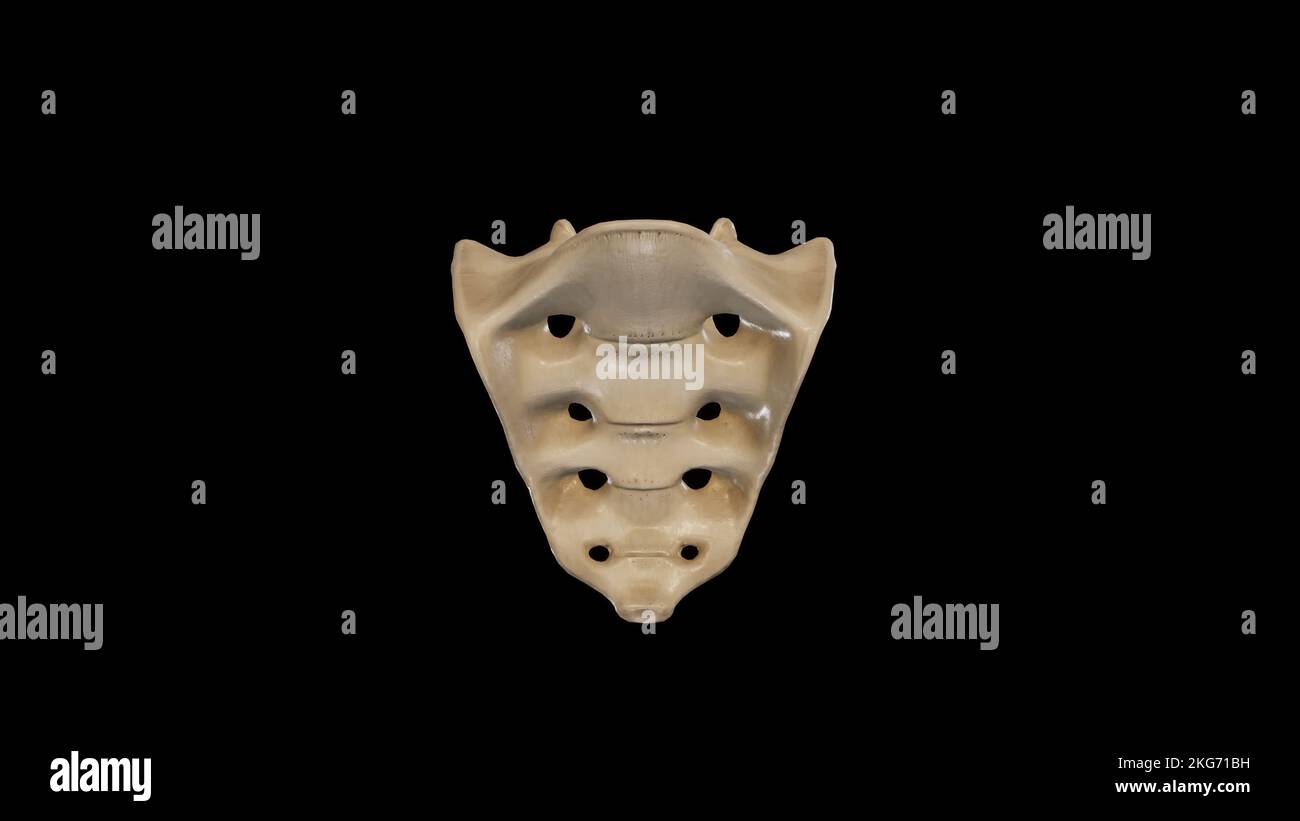 Anterior view of Sacrum Stock Photohttps://www.alamy.com/image-license-details/?v=1https://www.alamy.com/anterior-view-of-sacrum-image491879573.html
RF2KG71BH–Anterior view of Sacrum
 . Regional anesthesia : its technic and clinical application . run almost parallel with the twelfth Ant. Sup. iliac spine External cutaneous n. Anterior cnaral n:. Genito crural n Obturator n. Obturator externus mAdductor magnus m Femoral A andV 1 Adductor brevis m ^ Adductor longus m. ^, 4?J^ Fig. 186.—The external cutaneous, anterior crural, and obturator nerves at their exitfrom the pelvis. thoracic nerve and reach the anterior superior iliac spine side by side,between the transversalis and internal oblique muscles. These nervescan, therefore, be blocked by injecting their common trunk, the Stock Photohttps://www.alamy.com/image-license-details/?v=1https://www.alamy.com/regional-anesthesia-its-technic-and-clinical-application-run-almost-parallel-with-the-twelfth-ant-sup-iliac-spine-external-cutaneous-n-anterior-cnaral-n-genito-crural-n-obturator-n-obturator-externus-madductor-magnus-m-femoral-a-andv-1-adductor-brevis-m-adductor-longus-m-4j-fig-186the-external-cutaneous-anterior-crural-and-obturator-nerves-at-their-exitfrom-the-pelvis-thoracic-nerve-and-reach-the-anterior-superior-iliac-spine-side-by-sidebetween-the-transversalis-and-internal-oblique-muscles-these-nervescan-therefore-be-blocked-by-injecting-their-common-trunk-the-image370061495.html
RM2CE1N5Y–. Regional anesthesia : its technic and clinical application . run almost parallel with the twelfth Ant. Sup. iliac spine External cutaneous n. Anterior cnaral n:. Genito crural n Obturator n. Obturator externus mAdductor magnus m Femoral A andV 1 Adductor brevis m ^ Adductor longus m. ^, 4?J^ Fig. 186.—The external cutaneous, anterior crural, and obturator nerves at their exitfrom the pelvis. thoracic nerve and reach the anterior superior iliac spine side by side,between the transversalis and internal oblique muscles. These nervescan, therefore, be blocked by injecting their common trunk, the
 . Virginia medical semi-monthly. n fromthe anterior superior iliac spine to the tuber-osity of the ischium : this line crosses the centreof the acetabulum just above the greater tro-chanter. Hip joint dislocations are therefore of twochief varieties: 1. Backward Dislocations, those behindNelatoirs line, on to the back of the ilium orinto the great sacro-sciatic notch. 2. Forward Dislocations, those in front ofNelatons line, on to the front of the pubes orinto the obturator foramen. On September 20, 1916, dames Edwards, 22years old, a colored laborer on the constructionwork of the Southern Ry. Stock Photohttps://www.alamy.com/image-license-details/?v=1https://www.alamy.com/virginia-medical-semi-monthly-n-fromthe-anterior-superior-iliac-spine-to-the-tuber-osity-of-the-ischium-this-line-crosses-the-centreof-the-acetabulum-just-above-the-greater-tro-chanter-hip-joint-dislocations-are-therefore-of-twochief-varieties-1-backward-dislocations-those-behindnelatoirs-line-on-to-the-back-of-the-ilium-orinto-the-great-sacro-sciatic-notch-2-forward-dislocations-those-in-front-ofnelatons-line-on-to-the-front-of-the-pubes-orinto-the-obturator-foramen-on-september-20-1916-dames-edwards-22years-old-a-colored-laborer-on-the-constructionwork-of-the-southern-ry-image370515266.html
RM2CEPC02–. Virginia medical semi-monthly. n fromthe anterior superior iliac spine to the tuber-osity of the ischium : this line crosses the centreof the acetabulum just above the greater tro-chanter. Hip joint dislocations are therefore of twochief varieties: 1. Backward Dislocations, those behindNelatoirs line, on to the back of the ilium orinto the great sacro-sciatic notch. 2. Forward Dislocations, those in front ofNelatons line, on to the front of the pubes orinto the obturator foramen. On September 20, 1916, dames Edwards, 22years old, a colored laborer on the constructionwork of the Southern Ry.
 . Regional anesthesia : its technic and clinical application . e needle with the left forefinger placed belowPouparts ligament, at the level of the femoral ring, and in contact with tx ^ ? ^ Fig, 278.—Field-block for unilat.Tal reducible herni,the technic and on the other the nerve supply: 1, Para-iliaisubinguinal wheal; X, anterior superior iliac spine. , illustrating on one sidewheal; 2, pubic wheal; 3, the horizontal ramus of the pubis. One fingerbreadth to the inner sideof the femoral artery is a limit beyond which the needle must nottrespass. Through the same site of puncture subcutane Stock Photohttps://www.alamy.com/image-license-details/?v=1https://www.alamy.com/regional-anesthesia-its-technic-and-clinical-application-e-needle-with-the-left-forefinger-placed-belowpouparts-ligament-at-the-level-of-the-femoral-ring-and-in-contact-with-tx-fig-278field-block-for-unilattal-reducible-hernithe-technic-and-on-the-other-the-nerve-supply-1-para-iliaisubinguinal-wheal-x-anterior-superior-iliac-spine-illustrating-on-one-sidewheal-2-pubic-wheal-3-the-horizontal-ramus-of-the-pubis-one-fingerbreadth-to-the-inner-sideof-the-femoral-artery-is-a-limit-beyond-which-the-needle-must-nottrespass-through-the-same-site-of-puncture-subcutane-image370058523.html
RM2CE1HBR–. Regional anesthesia : its technic and clinical application . e needle with the left forefinger placed belowPouparts ligament, at the level of the femoral ring, and in contact with tx ^ ? ^ Fig, 278.—Field-block for unilat.Tal reducible herni,the technic and on the other the nerve supply: 1, Para-iliaisubinguinal wheal; X, anterior superior iliac spine. , illustrating on one sidewheal; 2, pubic wheal; 3, the horizontal ramus of the pubis. One fingerbreadth to the inner sideof the femoral artery is a limit beyond which the needle must nottrespass. Through the same site of puncture subcutane
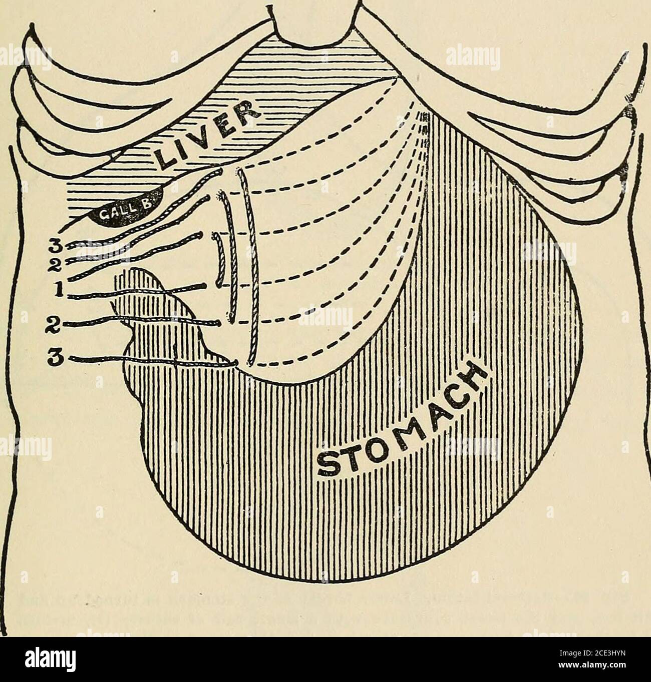 . Operative surgery, for students and practitioners . Fig. 94.—Various Abdominal Incisions. B, Battle incision; C, incision forleft inguinal colostomy; F, Fenger incision for stomach; G, Vertical andoblique incisions for gall-bladder, etc.; H, von Hackers incision for gastros-tomy; M, McBurney incision for appendicectomy; S, incision for suprapubiccystotomy. In middle line above umbilicus is linea alba incision for opera-tions upon stomach. X indicates location of anterior superior iliac spine.Dotted line drawn from spine to the umbilicus. OPERATIONS UPON THE STOMACH. 243 second row, and last Stock Photohttps://www.alamy.com/image-license-details/?v=1https://www.alamy.com/operative-surgery-for-students-and-practitioners-fig-94various-abdominal-incisions-b-battle-incision-c-incision-forleft-inguinal-colostomy-f-fenger-incision-for-stomach-g-vertical-andoblique-incisions-for-gall-bladder-etc-h-von-hackers-incision-for-gastros-tomy-m-mcburney-incision-for-appendicectomy-s-incision-for-suprapubiccystotomy-in-middle-line-above-umbilicus-is-linea-alba-incision-for-opera-tions-upon-stomach-x-indicates-location-of-anterior-superior-iliac-spinedotted-line-drawn-from-spine-to-the-umbilicus-operations-upon-the-stomach-243-second-row-and-last-image370102873.html
RM2CE3HYN–. Operative surgery, for students and practitioners . Fig. 94.—Various Abdominal Incisions. B, Battle incision; C, incision forleft inguinal colostomy; F, Fenger incision for stomach; G, Vertical andoblique incisions for gall-bladder, etc.; H, von Hackers incision for gastros-tomy; M, McBurney incision for appendicectomy; S, incision for suprapubiccystotomy. In middle line above umbilicus is linea alba incision for opera-tions upon stomach. X indicates location of anterior superior iliac spine.Dotted line drawn from spine to the umbilicus. OPERATIONS UPON THE STOMACH. 243 second row, and last
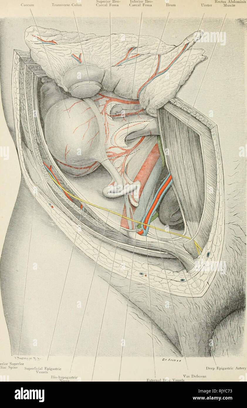 . Atlas of applied (topographical) human anatomy for students and practitioners. Anatomy. Inferior Ileo- Caeca I Fossa Ileum Rectus Abdominis Ureter Muscle. Anterior Superior Iliac Spine Superficial Epigastric / Vessels / Deep Epigastric Artery Ilio-hypogastric Nerve Spermatic Vessels Plica Epigastrica Vas Deferens External Iliac Vessels Fig. 137. Vermiform Appendix and Caecum. Nat. Size.. Please note that these images are extracted from scanned page images that may have been digitally enhanced for readability - coloration and appearance of these illustrations may not perfectly resemble the or Stock Photohttps://www.alamy.com/image-license-details/?v=1https://www.alamy.com/atlas-of-applied-topographical-human-anatomy-for-students-and-practitioners-anatomy-inferior-ileo-caeca-i-fossa-ileum-rectus-abdominis-ureter-muscle-anterior-superior-iliac-spine-superficial-epigastric-vessels-deep-epigastric-artery-ilio-hypogastric-nerve-spermatic-vessels-plica-epigastrica-vas-deferens-external-iliac-vessels-fig-137-vermiform-appendix-and-caecum-nat-size-please-note-that-these-images-are-extracted-from-scanned-page-images-that-may-have-been-digitally-enhanced-for-readability-coloration-and-appearance-of-these-illustrations-may-not-perfectly-resemble-the-or-image235400903.html
RMRJYC73–. Atlas of applied (topographical) human anatomy for students and practitioners. Anatomy. Inferior Ileo- Caeca I Fossa Ileum Rectus Abdominis Ureter Muscle. Anterior Superior Iliac Spine Superficial Epigastric / Vessels / Deep Epigastric Artery Ilio-hypogastric Nerve Spermatic Vessels Plica Epigastrica Vas Deferens External Iliac Vessels Fig. 137. Vermiform Appendix and Caecum. Nat. Size.. Please note that these images are extracted from scanned page images that may have been digitally enhanced for readability - coloration and appearance of these illustrations may not perfectly resemble the or
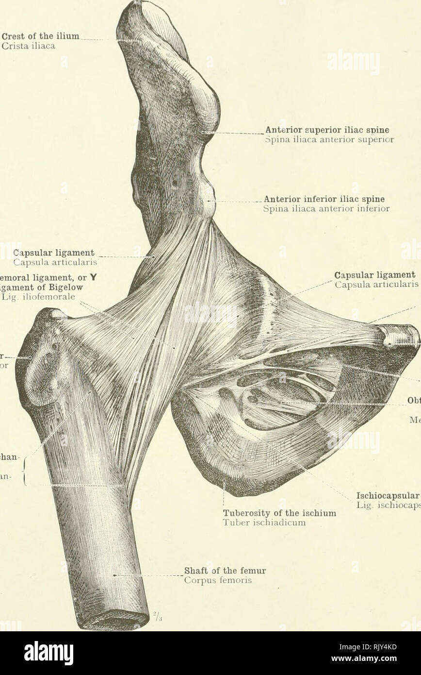 . An atlas of human anatomy for students and physicians. Anatomy. 222 THE ARTICULATIONS OF THE LOW!,:-: LIMB Crest of the ilium Crista iliaca Anterior superior iliac spine Spina iliaca anterior superior . Anterior inferior iliac spine Spina iliaca anterior inferior Capsular ligament Capsula articularis Iliofemoral ligament, or Y ligament of Bigelow Lie. iliofem. Capsular ligament Capsula articularis Great trochanter Trochanter maj Anterior intertrochan teric line Linea intertrochan- t-rica. Pubofemoral ligament Lig pubocapsulare Spine of the pubis Tuberculum pubicum Obturator canal Canalis obt Stock Photohttps://www.alamy.com/image-license-details/?v=1https://www.alamy.com/an-atlas-of-human-anatomy-for-students-and-physicians-anatomy-222-the-articulations-of-the-low!-limb-crest-of-the-ilium-crista-iliaca-anterior-superior-iliac-spine-spina-iliaca-anterior-superior-anterior-inferior-iliac-spine-spina-iliaca-anterior-inferior-capsular-ligament-capsula-articularis-iliofemoral-ligament-or-y-ligament-of-bigelow-lie-iliofem-capsular-ligament-capsula-articularis-great-trochanter-trochanter-maj-anterior-intertrochan-teric-line-linea-intertrochan-t-rica-pubofemoral-ligament-lig-pubocapsulare-spine-of-the-pubis-tuberculum-pubicum-obturator-canal-canalis-obt-image235394977.html
RMRJY4KD–. An atlas of human anatomy for students and physicians. Anatomy. 222 THE ARTICULATIONS OF THE LOW!,:-: LIMB Crest of the ilium Crista iliaca Anterior superior iliac spine Spina iliaca anterior superior . Anterior inferior iliac spine Spina iliaca anterior inferior Capsular ligament Capsula articularis Iliofemoral ligament, or Y ligament of Bigelow Lie. iliofem. Capsular ligament Capsula articularis Great trochanter Trochanter maj Anterior intertrochan teric line Linea intertrochan- t-rica. Pubofemoral ligament Lig pubocapsulare Spine of the pubis Tuberculum pubicum Obturator canal Canalis obt
 . Cunningham's Text-book of anatomy. Anatomy. THE HIP BONE. 229 anterior end of the external lip of the iliac crest the tensor fasciae latse muscle takes origin. The anterior border of the ilium stretches from the anterior superior iliac spine to the margin of the acetabulum below. Above, it is thin; but below, it forms a thick blunt process, the spina iliaca anterior inferior (anterior inferior iliac spine). From this the rectus femoris muscle arises, whilst the stout fibres of the Crest of the ilium ANTERIOR GLUTEAL LINE Posterior gluteal line Anterior inferior spine. Posterior superior spin Stock Photohttps://www.alamy.com/image-license-details/?v=1https://www.alamy.com/cunninghams-text-book-of-anatomy-anatomy-the-hip-bone-229-anterior-end-of-the-external-lip-of-the-iliac-crest-the-tensor-fasciae-latse-muscle-takes-origin-the-anterior-border-of-the-ilium-stretches-from-the-anterior-superior-iliac-spine-to-the-margin-of-the-acetabulum-below-above-it-is-thin-but-below-it-forms-a-thick-blunt-process-the-spina-iliaca-anterior-inferior-anterior-inferior-iliac-spine-from-this-the-rectus-femoris-muscle-arises-whilst-the-stout-fibres-of-the-crest-of-the-ilium-anterior-gluteal-line-posterior-gluteal-line-anterior-inferior-spine-posterior-superior-spin-image231857205.html
RMRD606D–. Cunningham's Text-book of anatomy. Anatomy. THE HIP BONE. 229 anterior end of the external lip of the iliac crest the tensor fasciae latse muscle takes origin. The anterior border of the ilium stretches from the anterior superior iliac spine to the margin of the acetabulum below. Above, it is thin; but below, it forms a thick blunt process, the spina iliaca anterior inferior (anterior inferior iliac spine). From this the rectus femoris muscle arises, whilst the stout fibres of the Crest of the ilium ANTERIOR GLUTEAL LINE Posterior gluteal line Anterior inferior spine. Posterior superior spin
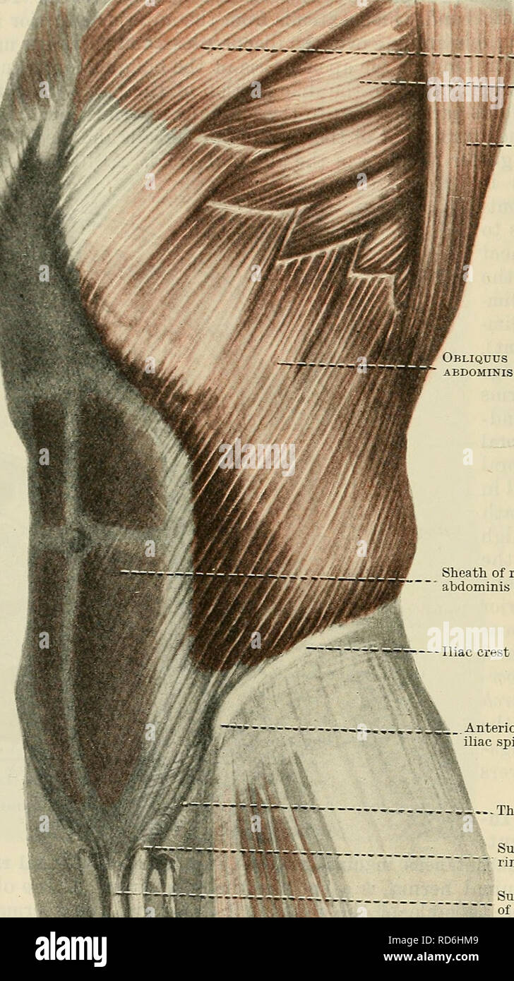 . Cunningham's Text-book of anatomy. Anatomy. 478 THE MUSCULAE SYSTEM. spermatic funiculus or round ligament in their further passage beyond the abdominal wall. Lig. Inguinale Reflexum Collesi.—The reflexed inguinal ligament of Colles (O.T. triangular fascia), lastly, is a triangular band of fibres placed behind the medial superior crus of the subcutaneous inguinal ring. It consists of fibres from the. t"f«" Pectoralis major Serratus anterior Latissimus dorsi BLIQUUS EXTERNVS ABDOMINIS if JM Sheath of rectus f£j,t W abdominis --Iliac crest .Anterior superior iliac spine ...... The in Stock Photohttps://www.alamy.com/image-license-details/?v=1https://www.alamy.com/cunninghams-text-book-of-anatomy-anatomy-478-the-musculae-system-spermatic-funiculus-or-round-ligament-in-their-further-passage-beyond-the-abdominal-wall-lig-inguinale-reflexum-collesithe-reflexed-inguinal-ligament-of-colles-ot-triangular-fascia-lastly-is-a-triangular-band-of-fibres-placed-behind-the-medial-superior-crus-of-the-subcutaneous-inguinal-ring-it-consists-of-fibres-from-the-tquotfquot-pectoralis-major-serratus-anterior-latissimus-dorsi-bliquus-externvs-abdominis-if-jm-sheath-of-rectus-fjt-w-abdominis-iliac-crest-anterior-superior-iliac-spine-the-in-image231870921.html
RMRD6HM9–. Cunningham's Text-book of anatomy. Anatomy. 478 THE MUSCULAE SYSTEM. spermatic funiculus or round ligament in their further passage beyond the abdominal wall. Lig. Inguinale Reflexum Collesi.—The reflexed inguinal ligament of Colles (O.T. triangular fascia), lastly, is a triangular band of fibres placed behind the medial superior crus of the subcutaneous inguinal ring. It consists of fibres from the. t"f«" Pectoralis major Serratus anterior Latissimus dorsi BLIQUUS EXTERNVS ABDOMINIS if JM Sheath of rectus f£j,t W abdominis --Iliac crest .Anterior superior iliac spine ...... The in
 . The anatomical record. Anatomy; Anatomy. DUPLICATION OF INFERIOR VENA CAVA 477 left, inferior vena cava; 5 cm. inferior to the entrance of the large anterior renal vein (fig. 1). In the fall of 1911 a condition similar to the first was found in another subject (no. 401, Cornell series), a white male, aged fifty, who died of cirrhosis of the liver. On the right the external iliac vein is joined at the level of the anterior superior iliac spine by the internal iliac vein to form the AZYGOS ANASTOMOSING BRANCH RIGHT SUPRARENAL RENALS RIGHT SPERMATIC RIGHT INFERIOR VENA CAVA RIGHT COMMON ILIAC R Stock Photohttps://www.alamy.com/image-license-details/?v=1https://www.alamy.com/the-anatomical-record-anatomy-anatomy-duplication-of-inferior-vena-cava-477-left-inferior-vena-cava-5-cm-inferior-to-the-entrance-of-the-large-anterior-renal-vein-fig-1-in-the-fall-of-1911-a-condition-similar-to-the-first-was-found-in-another-subject-no-401-cornell-series-a-white-male-aged-fifty-who-died-of-cirrhosis-of-the-liver-on-the-right-the-external-iliac-vein-is-joined-at-the-level-of-the-anterior-superior-iliac-spine-by-the-internal-iliac-vein-to-form-the-azygos-anastomosing-branch-right-suprarenal-renals-right-spermatic-right-inferior-vena-cava-right-common-iliac-r-image236863276.html
RMRNA1EM–. The anatomical record. Anatomy; Anatomy. DUPLICATION OF INFERIOR VENA CAVA 477 left, inferior vena cava; 5 cm. inferior to the entrance of the large anterior renal vein (fig. 1). In the fall of 1911 a condition similar to the first was found in another subject (no. 401, Cornell series), a white male, aged fifty, who died of cirrhosis of the liver. On the right the external iliac vein is joined at the level of the anterior superior iliac spine by the internal iliac vein to form the AZYGOS ANASTOMOSING BRANCH RIGHT SUPRARENAL RENALS RIGHT SPERMATIC RIGHT INFERIOR VENA CAVA RIGHT COMMON ILIAC R
 Gynecology . Fig. 398.—Nephrectomy. The Incision. rassing difficulties and dangerous accidents. For this purpose a long incisionis necessary. As in making the incision for suspension, one must first determinethe location of the twelfth rib, the outer border of the sacrospinalis muscles, andthe curve of the iliac crest as far as the anterior superior spine. Within the 692 GYNECOLOGY angle made by the twelfth rib and the muscle border is an area which is softerto the feel than the surrounding parts, and it is in this area that the incisionstarts. It is then carried in a curving sweep toward the Stock Photohttps://www.alamy.com/image-license-details/?v=1https://www.alamy.com/gynecology-fig-398nephrectomy-the-incision-rassing-difficulties-and-dangerous-accidents-for-this-purpose-a-long-incisionis-necessary-as-in-making-the-incision-for-suspension-one-must-first-determinethe-location-of-the-twelfth-rib-the-outer-border-of-the-sacrospinalis-muscles-andthe-curve-of-the-iliac-crest-as-far-as-the-anterior-superior-spine-within-the-692-gynecology-angle-made-by-the-twelfth-rib-and-the-muscle-border-is-an-area-which-is-softerto-the-feel-than-the-surrounding-parts-and-it-is-in-this-area-that-the-incisionstarts-it-is-then-carried-in-a-curving-sweep-toward-the-image340254866.html
RM2ANFXFE–Gynecology . Fig. 398.—Nephrectomy. The Incision. rassing difficulties and dangerous accidents. For this purpose a long incisionis necessary. As in making the incision for suspension, one must first determinethe location of the twelfth rib, the outer border of the sacrospinalis muscles, andthe curve of the iliac crest as far as the anterior superior spine. Within the 692 GYNECOLOGY angle made by the twelfth rib and the muscle border is an area which is softerto the feel than the surrounding parts, and it is in this area that the incisionstarts. It is then carried in a curving sweep toward the
![Practical human anatomy [electronic resource] : a working-guide for students of medicine and a ready-reference for surgeons and physicians . ilium, the anterior sacro-iliac ligament;from the spine of the ischium to the sacrum and coccyx, thesmall sacro-sciatic ligament; from the sacrum to the body ofthe ischium, the great sacro-sciatic ligament; from the anteriorsurface of the inferior segment of the sacrum to the anterior ofthe coccyx, the anterior sacro-coccygeal ligament; at the sym-physis pubis the posterior, superior and inferior pubic liga-ments. Dissection.—If the dissector has the lowe Stock Photo Practical human anatomy [electronic resource] : a working-guide for students of medicine and a ready-reference for surgeons and physicians . ilium, the anterior sacro-iliac ligament;from the spine of the ischium to the sacrum and coccyx, thesmall sacro-sciatic ligament; from the sacrum to the body ofthe ischium, the great sacro-sciatic ligament; from the anteriorsurface of the inferior segment of the sacrum to the anterior ofthe coccyx, the anterior sacro-coccygeal ligament; at the sym-physis pubis the posterior, superior and inferior pubic liga-ments. Dissection.—If the dissector has the lowe Stock Photo](https://c8.alamy.com/comp/2AX7AXJ/practical-human-anatomy-electronic-resource-a-working-guide-for-students-of-medicine-and-a-ready-reference-for-surgeons-and-physicians-ilium-the-anterior-sacro-iliac-ligamentfrom-the-spine-of-the-ischium-to-the-sacrum-and-coccyx-thesmall-sacro-sciatic-ligament-from-the-sacrum-to-the-body-ofthe-ischium-the-great-sacro-sciatic-ligament-from-the-anteriorsurface-of-the-inferior-segment-of-the-sacrum-to-the-anterior-ofthe-coccyx-the-anterior-sacro-coccygeal-ligament-at-the-sym-physis-pubis-the-posterior-superior-and-inferior-pubic-liga-ments-dissectionif-the-dissector-has-the-lowe-2AX7AXJ.jpg) Practical human anatomy [electronic resource] : a working-guide for students of medicine and a ready-reference for surgeons and physicians . ilium, the anterior sacro-iliac ligament;from the spine of the ischium to the sacrum and coccyx, thesmall sacro-sciatic ligament; from the sacrum to the body ofthe ischium, the great sacro-sciatic ligament; from the anteriorsurface of the inferior segment of the sacrum to the anterior ofthe coccyx, the anterior sacro-coccygeal ligament; at the sym-physis pubis the posterior, superior and inferior pubic liga-ments. Dissection.—If the dissector has the lowe Stock Photohttps://www.alamy.com/image-license-details/?v=1https://www.alamy.com/practical-human-anatomy-electronic-resource-a-working-guide-for-students-of-medicine-and-a-ready-reference-for-surgeons-and-physicians-ilium-the-anterior-sacro-iliac-ligamentfrom-the-spine-of-the-ischium-to-the-sacrum-and-coccyx-thesmall-sacro-sciatic-ligament-from-the-sacrum-to-the-body-ofthe-ischium-the-great-sacro-sciatic-ligament-from-the-anteriorsurface-of-the-inferior-segment-of-the-sacrum-to-the-anterior-ofthe-coccyx-the-anterior-sacro-coccygeal-ligament-at-the-sym-physis-pubis-the-posterior-superior-and-inferior-pubic-liga-ments-dissectionif-the-dissector-has-the-lowe-image343140298.html
Practical human anatomy [electronic resource] : a working-guide for students of medicine and a ready-reference for surgeons and physicians . ilium, the anterior sacro-iliac ligament;from the spine of the ischium to the sacrum and coccyx, thesmall sacro-sciatic ligament; from the sacrum to the body ofthe ischium, the great sacro-sciatic ligament; from the anteriorsurface of the inferior segment of the sacrum to the anterior ofthe coccyx, the anterior sacro-coccygeal ligament; at the sym-physis pubis the posterior, superior and inferior pubic liga-ments. Dissection.—If the dissector has the lowe Stock Photohttps://www.alamy.com/image-license-details/?v=1https://www.alamy.com/practical-human-anatomy-electronic-resource-a-working-guide-for-students-of-medicine-and-a-ready-reference-for-surgeons-and-physicians-ilium-the-anterior-sacro-iliac-ligamentfrom-the-spine-of-the-ischium-to-the-sacrum-and-coccyx-thesmall-sacro-sciatic-ligament-from-the-sacrum-to-the-body-ofthe-ischium-the-great-sacro-sciatic-ligament-from-the-anteriorsurface-of-the-inferior-segment-of-the-sacrum-to-the-anterior-ofthe-coccyx-the-anterior-sacro-coccygeal-ligament-at-the-sym-physis-pubis-the-posterior-superior-and-inferior-pubic-liga-ments-dissectionif-the-dissector-has-the-lowe-image343140298.htmlRM2AX7AXJ–Practical human anatomy [electronic resource] : a working-guide for students of medicine and a ready-reference for surgeons and physicians . ilium, the anterior sacro-iliac ligament;from the spine of the ischium to the sacrum and coccyx, thesmall sacro-sciatic ligament; from the sacrum to the body ofthe ischium, the great sacro-sciatic ligament; from the anteriorsurface of the inferior segment of the sacrum to the anterior ofthe coccyx, the anterior sacro-coccygeal ligament; at the sym-physis pubis the posterior, superior and inferior pubic liga-ments. Dissection.—If the dissector has the lowe
 The surgeon's handbook on the treatment of wounded in war : a prize essay . to the outer side the anterior cruralnerve covered by the iliac fascia; the genital branch of the genito-crural nerve crosses the artery obliquely. 56 Plate XV. Ligature of the femoral artery (common femoral) below Poupartsligament (right). 1. The cutaneous incision commences at a point midway betweenthe anterior superior spine of the ilium and the symphysis pubis,2mm above Pouparts ligament, and is carried downwards for 5cm. 2. The superficial fascia is divided. 3. The subcutaneous tissue is divided; the lymphatic gl Stock Photohttps://www.alamy.com/image-license-details/?v=1https://www.alamy.com/the-surgeons-handbook-on-the-treatment-of-wounded-in-war-a-prize-essay-to-the-outer-side-the-anterior-cruralnerve-covered-by-the-iliac-fascia-the-genital-branch-of-the-genito-crural-nerve-crosses-the-artery-obliquely-56-plate-xv-ligature-of-the-femoral-artery-common-femoral-below-poupartsligament-right-1-the-cutaneous-incision-commences-at-a-point-midway-betweenthe-anterior-superior-spine-of-the-ilium-and-the-symphysis-pubis2mm-above-pouparts-ligament-and-is-carried-downwards-for-5cm-2-the-superficial-fascia-is-divided-3-the-subcutaneous-tissue-is-divided-the-lymphatic-gl-image343314348.html
RM2AXF8XM–The surgeon's handbook on the treatment of wounded in war : a prize essay . to the outer side the anterior cruralnerve covered by the iliac fascia; the genital branch of the genito-crural nerve crosses the artery obliquely. 56 Plate XV. Ligature of the femoral artery (common femoral) below Poupartsligament (right). 1. The cutaneous incision commences at a point midway betweenthe anterior superior spine of the ilium and the symphysis pubis,2mm above Pouparts ligament, and is carried downwards for 5cm. 2. The superficial fascia is divided. 3. The subcutaneous tissue is divided; the lymphatic gl
 A system of human anatomy, general and special . artery. 5. The circumflexa ilii.6. The internal iliac artery. 7. Its anterior trunk. 8. Its posterior trunk. 9. The umbi-lical artery giving off (10) the superior vesical artery. After the origin of this branch,the umbilical artery becomes converted into a fibrous cord—the umbilical ligament.11. The internal pudic artery passing behind the spine of the ischium (12) and lessersacro-ischiatic ligament. 13. The middle hemorrhoidal artery. 14. The ischiatic artery,also passing behind the anterior sacro-ischiatic ligament to escape from the pelvis. 1 Stock Photohttps://www.alamy.com/image-license-details/?v=1https://www.alamy.com/a-system-of-human-anatomy-general-and-special-artery-5-the-circumflexa-ilii6-the-internal-iliac-artery-7-its-anterior-trunk-8-its-posterior-trunk-9-the-umbi-lical-artery-giving-off-10-the-superior-vesical-artery-after-the-origin-of-this-branchthe-umbilical-artery-becomes-converted-into-a-fibrous-cordthe-umbilical-ligament11-the-internal-pudic-artery-passing-behind-the-spine-of-the-ischium-12-and-lessersacro-ischiatic-ligament-13-the-middle-hemorrhoidal-artery-14-the-ischiatic-arteryalso-passing-behind-the-anterior-sacro-ischiatic-ligament-to-escape-from-the-pelvis-1-image342721920.html
RM2AWG98G–A system of human anatomy, general and special . artery. 5. The circumflexa ilii.6. The internal iliac artery. 7. Its anterior trunk. 8. Its posterior trunk. 9. The umbi-lical artery giving off (10) the superior vesical artery. After the origin of this branch,the umbilical artery becomes converted into a fibrous cord—the umbilical ligament.11. The internal pudic artery passing behind the spine of the ischium (12) and lessersacro-ischiatic ligament. 13. The middle hemorrhoidal artery. 14. The ischiatic artery,also passing behind the anterior sacro-ischiatic ligament to escape from the pelvis. 1
 Surgery; its theory and practice . opposite foot. The head of the bone, at least in thin subjects,can be felt in its abnormal situation on rotating the limb. Thegreat trochanter is above a line drawn from the anterior superioriliac spine to the most prominent part of the tuberosity of theischium {Nelatons line) (Fig- i??) J s^id the distance from thetop of the great trochanter to a line drawn horizontally round thepelvis on a level with the anterior superior iliac spines {Bryantsline) is less on the injured than on the sound side. 2. Dislocation into the sciatic notch {the dorsalbeloiu the ten Stock Photohttps://www.alamy.com/image-license-details/?v=1https://www.alamy.com/surgery-its-theory-and-practice-opposite-foot-the-head-of-the-bone-at-least-in-thin-subjectscan-be-felt-in-its-abnormal-situation-on-rotating-the-limb-thegreat-trochanter-is-above-a-line-drawn-from-the-anterior-superioriliac-spine-to-the-most-prominent-part-of-the-tuberosity-of-theischium-nelatons-line-fig-i-j-sid-the-distance-from-thetop-of-the-great-trochanter-to-a-line-drawn-horizontally-round-thepelvis-on-a-level-with-the-anterior-superior-iliac-spines-bryantsline-is-less-on-the-injured-than-on-the-sound-side-2-dislocation-into-the-sciatic-notch-the-dorsalbeloiu-the-ten-image343086384.html
RM2AX4X54–Surgery; its theory and practice . opposite foot. The head of the bone, at least in thin subjects,can be felt in its abnormal situation on rotating the limb. Thegreat trochanter is above a line drawn from the anterior superioriliac spine to the most prominent part of the tuberosity of theischium {Nelatons line) (Fig- i??) J s^id the distance from thetop of the great trochanter to a line drawn horizontally round thepelvis on a level with the anterior superior iliac spines {Bryantsline) is less on the injured than on the sound side. 2. Dislocation into the sciatic notch {the dorsalbeloiu the ten
 An atlas of human anatomy for students and physicians . unis Internal iliac arteryA. hypogastuca Sacral promontory Promontorium Anterior superior spine of the iliumSpina iliaca anterior superior iPouparts ligament (superficial femoral arch)I Lig- inguinale (Iouparti) I Internal or deep abdominal ring- fi)1 Deep or inferior epigastric arteryi and vein ? A et Vv. epi;-;astric;i- inferiores Vestige of the obliterated hypogastric artery, orexternal umbilical ligament (•;!,- Obturator artery and vein ;L V. ol.turatoria ^Rectal fascia^ Iri^ ia diaphragmatis pelvis superior ^ Pubic symphysis vmphysis Stock Photohttps://www.alamy.com/image-license-details/?v=1https://www.alamy.com/an-atlas-of-human-anatomy-for-students-and-physicians-unis-internal-iliac-arterya-hypogastuca-sacral-promontory-promontorium-anterior-superior-spine-of-the-iliumspina-iliaca-anterior-superior-ipouparts-ligament-superficial-femoral-archi-lig-inguinale-iouparti-i-internal-or-deep-abdominal-ring-fi1-deep-or-inferior-epigastric-arteryi-and-vein-a-et-vv-epi-astrici-inferiores-vestige-of-the-obliterated-hypogastric-artery-orexternal-umbilical-ligament-!-obturator-artery-and-vein-l-v-olturatoria-rectal-fascia-iri-ia-diaphragmatis-pelvis-superior-pubic-symphysis-vmphysis-image338272564.html
RM2AJ9J30–An atlas of human anatomy for students and physicians . unis Internal iliac arteryA. hypogastuca Sacral promontory Promontorium Anterior superior spine of the iliumSpina iliaca anterior superior iPouparts ligament (superficial femoral arch)I Lig- inguinale (Iouparti) I Internal or deep abdominal ring- fi)1 Deep or inferior epigastric arteryi and vein ? A et Vv. epi;-;astric;i- inferiores Vestige of the obliterated hypogastric artery, orexternal umbilical ligament (•;!,- Obturator artery and vein ;L V. ol.turatoria ^Rectal fascia^ Iri^ ia diaphragmatis pelvis superior ^ Pubic symphysis vmphysis
 A manual of operative surgery . d upon thesame plane as the artery, andentirely to the inner side (Fig. 343). The internal abdominalring is situated about half aninch above Pouparts liga-ment, opposite a point mid-way between the anterior-superior spine of the ilium andthe symphysis pubis. Line of the Artery.—A linedrawn on the surface of theabdomen from a spot abouta fingers breadth to the leftof and below the navel, to apoint midway between theanterior superior iliac spineand the symphysis pubis. Theupper third of this line repre-sents the common iliac, thelower two-thirds the externaliliac Stock Photohttps://www.alamy.com/image-license-details/?v=1https://www.alamy.com/a-manual-of-operative-surgery-d-upon-thesame-plane-as-the-artery-andentirely-to-the-inner-side-fig-343-the-internal-abdominalring-is-situated-about-half-aninch-above-pouparts-liga-ment-opposite-a-point-mid-way-between-the-anterior-superior-spine-of-the-ilium-andthe-symphysis-pubis-line-of-the-arterya-linedrawn-on-the-surface-of-theabdomen-from-a-spot-abouta-fingers-breadth-to-the-leftof-and-below-the-navel-to-apoint-midway-between-theanterior-superior-iliac-spineand-the-symphysis-pubis-theupper-third-of-this-line-repre-sents-the-common-iliac-thelower-two-thirds-the-externaliliac-image343323131.html
RM2AXFM4B–A manual of operative surgery . d upon thesame plane as the artery, andentirely to the inner side (Fig. 343). The internal abdominalring is situated about half aninch above Pouparts liga-ment, opposite a point mid-way between the anterior-superior spine of the ilium andthe symphysis pubis. Line of the Artery.—A linedrawn on the surface of theabdomen from a spot abouta fingers breadth to the leftof and below the navel, to apoint midway between theanterior superior iliac spineand the symphysis pubis. Theupper third of this line repre-sents the common iliac, thelower two-thirds the externaliliac
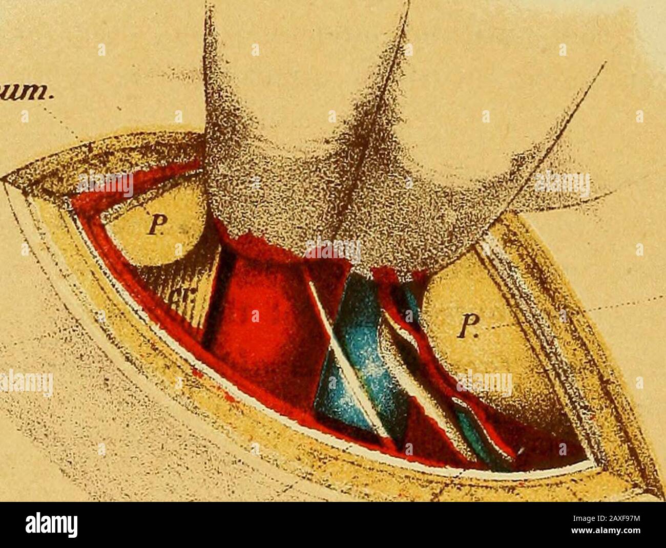 The surgeon's handbook on the treatment of wounded in war : a prize essay . m. iliopsoas,ii. cruralis. ureter. Spffttt a/it. sup. iiern. sperm at. ex>$§ernu.s. Tab. IE. Peritoneum nero. cruralis. Perilojifum. nerv. sjiermatbcus eccternus / funiculus sp£-rmaticae. m Plate XIV.Ligature of the external iliac artery (right). 1. The cutaneous incision, which is lcm above and parallel toPouparts ligament, 8—10cm in length, and slightly convex, begins ocmto the inner side of the anterior superior spine, and ends opposite tothe internal inguinal ring (without exposing the ring or the sper-matic cor Stock Photohttps://www.alamy.com/image-license-details/?v=1https://www.alamy.com/the-surgeons-handbook-on-the-treatment-of-wounded-in-war-a-prize-essay-m-iliopsoasii-cruralis-ureter-spffttt-ait-sup-iiern-sperm-at-exgternus-tab-ie-peritoneum-nero-cruralis-perilojifum-nerv-sjiermatbcus-eccternus-funiculus-sp-rmaticae-m-plate-xivligature-of-the-external-iliac-artery-right-1-the-cutaneous-incision-which-is-lcm-above-and-parallel-topouparts-ligament-810cm-in-length-and-slightly-convex-begins-ocmto-the-inner-side-of-the-anterior-superior-spine-and-ends-opposite-tothe-internal-inguinal-ring-without-exposing-the-ring-or-the-sper-matic-cor-image343314600.html
RM2AXF97M–The surgeon's handbook on the treatment of wounded in war : a prize essay . m. iliopsoas,ii. cruralis. ureter. Spffttt a/it. sup. iiern. sperm at. ex>$§ernu.s. Tab. IE. Peritoneum nero. cruralis. Perilojifum. nerv. sjiermatbcus eccternus / funiculus sp£-rmaticae. m Plate XIV.Ligature of the external iliac artery (right). 1. The cutaneous incision, which is lcm above and parallel toPouparts ligament, 8—10cm in length, and slightly convex, begins ocmto the inner side of the anterior superior spine, and ends opposite tothe internal inguinal ring (without exposing the ring or the sper-matic cor
 Operative surgery . Fig. 1116.—Direct inguinal her-nia, a. Integument and fascia.b. Aponeurosis of external ob-lique muscle, d. Spermaticcord. e. Epigastric vessels. /.Sac. g. Hernial contents. 910 OPERATIVE SURGERY.. Fig. 1117.—Transverse section below Pouparts ligament.a. Anterior superior spine of the ilium, h. Iliac fascia,c. Anterior crural nerve, d. Femoral artery, e. Femoralvein. /. Septum crurale. g. Gimbernats ligament, h.Spine of pubes. i. Pectineal fascia, j. Ilio-pectinealeminence, k. Iliac bursa. /. Rectus femoris muscle.m. Sartorius muscle, n. Transversalis fascia. of the femoral Stock Photohttps://www.alamy.com/image-license-details/?v=1https://www.alamy.com/operative-surgery-fig-1116direct-inguinal-her-nia-a-integument-and-fasciab-aponeurosis-of-external-ob-lique-muscle-d-spermaticcord-e-epigastric-vessels-sac-g-hernial-contents-910-operative-surgery-fig-1117transverse-section-below-pouparts-ligamenta-anterior-superior-spine-of-the-ilium-h-iliac-fasciac-anterior-crural-nerve-d-femoral-artery-e-femoralvein-septum-crurale-g-gimbernats-ligament-hspine-of-pubes-i-pectineal-fascia-j-ilio-pectinealeminence-k-iliac-bursa-rectus-femoris-musclem-sartorius-muscle-n-transversalis-fascia-of-the-femoral-image342668615.html
RM2AWDW8R–Operative surgery . Fig. 1116.—Direct inguinal her-nia, a. Integument and fascia.b. Aponeurosis of external ob-lique muscle, d. Spermaticcord. e. Epigastric vessels. /.Sac. g. Hernial contents. 910 OPERATIVE SURGERY.. Fig. 1117.—Transverse section below Pouparts ligament.a. Anterior superior spine of the ilium, h. Iliac fascia,c. Anterior crural nerve, d. Femoral artery, e. Femoralvein. /. Septum crurale. g. Gimbernats ligament, h.Spine of pubes. i. Pectineal fascia, j. Ilio-pectinealeminence, k. Iliac bursa. /. Rectus femoris muscle.m. Sartorius muscle, n. Transversalis fascia. of the femoral
 A text-book of clinical anatomy : for students and practitioners . Fig. 51.—Surface markings of thoracic and abdominal viscera viewed from the leftside; also view of Roser-Nelaton line. C.C., Costo-clavicular line. Ax, Mid-axillary line.U.L., Upper lobe of lung. L.L., Lower lobe. 2, Lower margin of lung. 3, Lower marginof pleura. 4, Anterior superior spine of ilium. 5, Costal arch. S, Spleen. T.C., Trans-verse colon. D.C., Descending colon. S.F., Sigmoid flexure (iliac colon). R.N., Roser-Nelaton line passing from 4 across top of trochanter major (T) to tuberosity of ischium(Is.). 163. Fig. 52 Stock Photohttps://www.alamy.com/image-license-details/?v=1https://www.alamy.com/a-text-book-of-clinical-anatomy-for-students-and-practitioners-fig-51surface-markings-of-thoracic-and-abdominal-viscera-viewed-from-the-leftside-also-view-of-roser-nelaton-line-cc-costo-clavicular-line-ax-mid-axillary-lineul-upper-lobe-of-lung-ll-lower-lobe-2-lower-margin-of-lung-3-lower-marginof-pleura-4-anterior-superior-spine-of-ilium-5-costal-arch-s-spleen-tc-trans-verse-colon-dc-descending-colon-sf-sigmoid-flexure-iliac-colon-rn-roser-nelaton-line-passing-from-4-across-top-of-trochanter-major-t-to-tuberosity-of-ischiumis-163-fig-52-image340225391.html
RM2ANEGXR–A text-book of clinical anatomy : for students and practitioners . Fig. 51.—Surface markings of thoracic and abdominal viscera viewed from the leftside; also view of Roser-Nelaton line. C.C., Costo-clavicular line. Ax, Mid-axillary line.U.L., Upper lobe of lung. L.L., Lower lobe. 2, Lower margin of lung. 3, Lower marginof pleura. 4, Anterior superior spine of ilium. 5, Costal arch. S, Spleen. T.C., Trans-verse colon. D.C., Descending colon. S.F., Sigmoid flexure (iliac colon). R.N., Roser-Nelaton line passing from 4 across top of trochanter major (T) to tuberosity of ischium(Is.). 163. Fig. 52
 . Gynecology : . Fig. 464.—Nephrectomy. The Incision. rassing difficulties and dangerous accidents. For this purpose a long incisionis necessary. As in making the incision for suspension, one must first determinethe location of the twelfth rib, the outer border of the sacrospinalis muscles, andthe curve of the iliac crest as far as the anterior superior spine. Within the s:i GYNECOLOGY angle niade by the twelfth rib and the muscle border is an area which is softerto the feel than the surrounding parts, and it is in this area that the incisionstarts. It is then carried in a curving sweep toward Stock Photohttps://www.alamy.com/image-license-details/?v=1https://www.alamy.com/gynecology-fig-464nephrectomy-the-incision-rassing-difficulties-and-dangerous-accidents-for-this-purpose-a-long-incisionis-necessary-as-in-making-the-incision-for-suspension-one-must-first-determinethe-location-of-the-twelfth-rib-the-outer-border-of-the-sacrospinalis-muscles-andthe-curve-of-the-iliac-crest-as-far-as-the-anterior-superior-spine-within-the-si-gynecology-angle-niade-by-the-twelfth-rib-and-the-muscle-border-is-an-area-which-is-softerto-the-feel-than-the-surrounding-parts-and-it-is-in-this-area-that-the-incisionstarts-it-is-then-carried-in-a-curving-sweep-toward-image369767672.html
RM2CDGAC8–. Gynecology : . Fig. 464.—Nephrectomy. The Incision. rassing difficulties and dangerous accidents. For this purpose a long incisionis necessary. As in making the incision for suspension, one must first determinethe location of the twelfth rib, the outer border of the sacrospinalis muscles, andthe curve of the iliac crest as far as the anterior superior spine. Within the s:i GYNECOLOGY angle niade by the twelfth rib and the muscle border is an area which is softerto the feel than the surrounding parts, and it is in this area that the incisionstarts. It is then carried in a curving sweep toward
![. Hernia, strangulated and reducible. With cure by subcutaneous injections, together with sugcested [!] and improved methods for kelotomy. Also an appendix giving a short account of various new surgical instruments. l-developed persons, will be of especial value.Although the distances will be somewhat different accordingas the person be large or small, the relative proportions willbe the same. From S3mphysis pubis to anterior superior spine of ilium .to tuberosity of pubesto inner margin of the lower open-ing of the abdominal canalto inner edge of the upper openingto middle of iliac artery to Stock Photo . Hernia, strangulated and reducible. With cure by subcutaneous injections, together with sugcested [!] and improved methods for kelotomy. Also an appendix giving a short account of various new surgical instruments. l-developed persons, will be of especial value.Although the distances will be somewhat different accordingas the person be large or small, the relative proportions willbe the same. From S3mphysis pubis to anterior superior spine of ilium .to tuberosity of pubesto inner margin of the lower open-ing of the abdominal canalto inner edge of the upper openingto middle of iliac artery to Stock Photo](https://c8.alamy.com/comp/2CEDEHA/hernia-strangulated-and-reducible-with-cure-by-subcutaneous-injections-together-with-sugcested-!-and-improved-methods-for-kelotomy-also-an-appendix-giving-a-short-account-of-various-new-surgical-instruments-l-developed-persons-will-be-of-especial-valuealthough-the-distances-will-be-somewhat-different-accordingas-the-person-be-large-or-small-the-relative-proportions-willbe-the-same-from-s3mphysis-pubis-to-anterior-superior-spine-of-ilium-to-tuberosity-of-pubesto-inner-margin-of-the-lower-open-ing-of-the-abdominal-canalto-inner-edge-of-the-upper-openingto-middle-of-iliac-artery-to-2CEDEHA.jpg) . Hernia, strangulated and reducible. With cure by subcutaneous injections, together with sugcested [!] and improved methods for kelotomy. Also an appendix giving a short account of various new surgical instruments. l-developed persons, will be of especial value.Although the distances will be somewhat different accordingas the person be large or small, the relative proportions willbe the same. From S3mphysis pubis to anterior superior spine of ilium .to tuberosity of pubesto inner margin of the lower open-ing of the abdominal canalto inner edge of the upper openingto middle of iliac artery to Stock Photohttps://www.alamy.com/image-license-details/?v=1https://www.alamy.com/hernia-strangulated-and-reducible-with-cure-by-subcutaneous-injections-together-with-sugcested-!-and-improved-methods-for-kelotomy-also-an-appendix-giving-a-short-account-of-various-new-surgical-instruments-l-developed-persons-will-be-of-especial-valuealthough-the-distances-will-be-somewhat-different-accordingas-the-person-be-large-or-small-the-relative-proportions-willbe-the-same-from-s3mphysis-pubis-to-anterior-superior-spine-of-ilium-to-tuberosity-of-pubesto-inner-margin-of-the-lower-open-ing-of-the-abdominal-canalto-inner-edge-of-the-upper-openingto-middle-of-iliac-artery-to-image370319750.html
. Hernia, strangulated and reducible. With cure by subcutaneous injections, together with sugcested [!] and improved methods for kelotomy. Also an appendix giving a short account of various new surgical instruments. l-developed persons, will be of especial value.Although the distances will be somewhat different accordingas the person be large or small, the relative proportions willbe the same. From S3mphysis pubis to anterior superior spine of ilium .to tuberosity of pubesto inner margin of the lower open-ing of the abdominal canalto inner edge of the upper openingto middle of iliac artery to Stock Photohttps://www.alamy.com/image-license-details/?v=1https://www.alamy.com/hernia-strangulated-and-reducible-with-cure-by-subcutaneous-injections-together-with-sugcested-!-and-improved-methods-for-kelotomy-also-an-appendix-giving-a-short-account-of-various-new-surgical-instruments-l-developed-persons-will-be-of-especial-valuealthough-the-distances-will-be-somewhat-different-accordingas-the-person-be-large-or-small-the-relative-proportions-willbe-the-same-from-s3mphysis-pubis-to-anterior-superior-spine-of-ilium-to-tuberosity-of-pubesto-inner-margin-of-the-lower-open-ing-of-the-abdominal-canalto-inner-edge-of-the-upper-openingto-middle-of-iliac-artery-to-image370319750.htmlRM2CEDEHA–. Hernia, strangulated and reducible. With cure by subcutaneous injections, together with sugcested [!] and improved methods for kelotomy. Also an appendix giving a short account of various new surgical instruments. l-developed persons, will be of especial value.Although the distances will be somewhat different accordingas the person be large or small, the relative proportions willbe the same. From S3mphysis pubis to anterior superior spine of ilium .to tuberosity of pubesto inner margin of the lower open-ing of the abdominal canalto inner edge of the upper openingto middle of iliac artery to
 . Regional anesthesia : its technic and clinical application . viz.,the field-block and the paravertebral block. Field-block is the pro- ?cedure usually employed, no matter what the size and consistency ofthe hernia, whether it be reducible or irreducible, strangulated or not. 1. Field-Uock.—Four wheals are raised as shown in Fig. 286.Wheal 1 is the para-iliac wheal of the procedure used for inguinal herni-otomy. It lies 2.5 cm. medial to and above the anterior superior spineof the iliimi. Wheal 2 is the pubic wheal raised over the pubic spine.Wheal 3 occupies the lateral margin of the hernial Stock Photohttps://www.alamy.com/image-license-details/?v=1https://www.alamy.com/regional-anesthesia-its-technic-and-clinical-application-vizthe-field-block-and-the-paravertebral-block-field-block-is-the-pro-cedure-usually-employed-no-matter-what-the-size-and-consistency-ofthe-hernia-whether-it-be-reducible-or-irreducible-strangulated-or-not-1-field-uockfour-wheals-are-raised-as-shown-in-fig-286wheal-1-is-the-para-iliac-wheal-of-the-procedure-used-for-inguinal-herni-otomy-it-lies-25-cm-medial-to-and-above-the-anterior-superior-spineof-the-iliimi-wheal-2-is-the-pubic-wheal-raised-over-the-pubic-spinewheal-3-occupies-the-lateral-margin-of-the-hernial-image370058240.html
RM2CE1H1M–. Regional anesthesia : its technic and clinical application . viz.,the field-block and the paravertebral block. Field-block is the pro- ?cedure usually employed, no matter what the size and consistency ofthe hernia, whether it be reducible or irreducible, strangulated or not. 1. Field-Uock.—Four wheals are raised as shown in Fig. 286.Wheal 1 is the para-iliac wheal of the procedure used for inguinal herni-otomy. It lies 2.5 cm. medial to and above the anterior superior spineof the iliimi. Wheal 2 is the pubic wheal raised over the pubic spine.Wheal 3 occupies the lateral margin of the hernial
 . A manual of operative surgery . ne, asshown in Fig. 89, runs vertically down from the tip of the last ribto a point on the iliac crest half an inch behind the middle of thelatter, measuring from the anterior superior spine backwards. The anatomy of the operation is illustrated in Fig. 89. After the skin and superficial structures have been divided theexternal oblique and latissimus dorsi muscles will be exposed. Thefibres of those muscles are in this situation vertical. They should bedivided by a single clean cut through the whole length of the incision. The layer of the internal oblique wil Stock Photohttps://www.alamy.com/image-license-details/?v=1https://www.alamy.com/a-manual-of-operative-surgery-ne-asshown-in-fig-89-runs-vertically-down-from-the-tip-of-the-last-ribto-a-point-on-the-iliac-crest-half-an-inch-behind-the-middle-of-thelatter-measuring-from-the-anterior-superior-spine-backwards-the-anatomy-of-the-operation-is-illustrated-in-fig-89-after-the-skin-and-superficial-structures-have-been-divided-theexternal-oblique-and-latissimus-dorsi-muscles-will-be-exposed-thefibres-of-those-muscles-are-in-this-situation-vertical-they-should-bedivided-by-a-single-clean-cut-through-the-whole-length-of-the-incision-the-layer-of-the-internal-oblique-wil-image372562479.html
RM2CJ3K6R–. A manual of operative surgery . ne, asshown in Fig. 89, runs vertically down from the tip of the last ribto a point on the iliac crest half an inch behind the middle of thelatter, measuring from the anterior superior spine backwards. The anatomy of the operation is illustrated in Fig. 89. After the skin and superficial structures have been divided theexternal oblique and latissimus dorsi muscles will be exposed. Thefibres of those muscles are in this situation vertical. They should bedivided by a single clean cut through the whole length of the incision. The layer of the internal oblique wil
 . Cunningham's Text-book of anatomy. Anatomy. THE HIP BONE. 231 is subdivided by an oblique ridge, called the ilio-pectineal line (linea arcuata), which passes forwards and distally, from the most prominent point of the auricular surface towards the medial side of the ilio-pectineal eminence, which is placed just above and in front of the acetabulum and marks the fusion of the Crest of the ilium ILIUM Anterior superior spine Anterior inferior spine'. Tuberosity for posterior sacro-iliac ligament Posterior superior SPINE Articular surface Post. inf. spine Ilio-pectineal eminence Ilio-pectineal Stock Photohttps://www.alamy.com/image-license-details/?v=1https://www.alamy.com/cunninghams-text-book-of-anatomy-anatomy-the-hip-bone-231-is-subdivided-by-an-oblique-ridge-called-the-ilio-pectineal-line-linea-arcuata-which-passes-forwards-and-distally-from-the-most-prominent-point-of-the-auricular-surface-towards-the-medial-side-of-the-ilio-pectineal-eminence-which-is-placed-just-above-and-in-front-of-the-acetabulum-and-marks-the-fusion-of-the-crest-of-the-ilium-ilium-anterior-superior-spine-anterior-inferior-spine-tuberosity-for-posterior-sacro-iliac-ligament-posterior-superior-spine-articular-surface-post-inf-spine-ilio-pectineal-eminence-ilio-pectineal-image231857195.html
RMRD6063–. Cunningham's Text-book of anatomy. Anatomy. THE HIP BONE. 231 is subdivided by an oblique ridge, called the ilio-pectineal line (linea arcuata), which passes forwards and distally, from the most prominent point of the auricular surface towards the medial side of the ilio-pectineal eminence, which is placed just above and in front of the acetabulum and marks the fusion of the Crest of the ilium ILIUM Anterior superior spine Anterior inferior spine'. Tuberosity for posterior sacro-iliac ligament Posterior superior SPINE Articular surface Post. inf. spine Ilio-pectineal eminence Ilio-pectineal
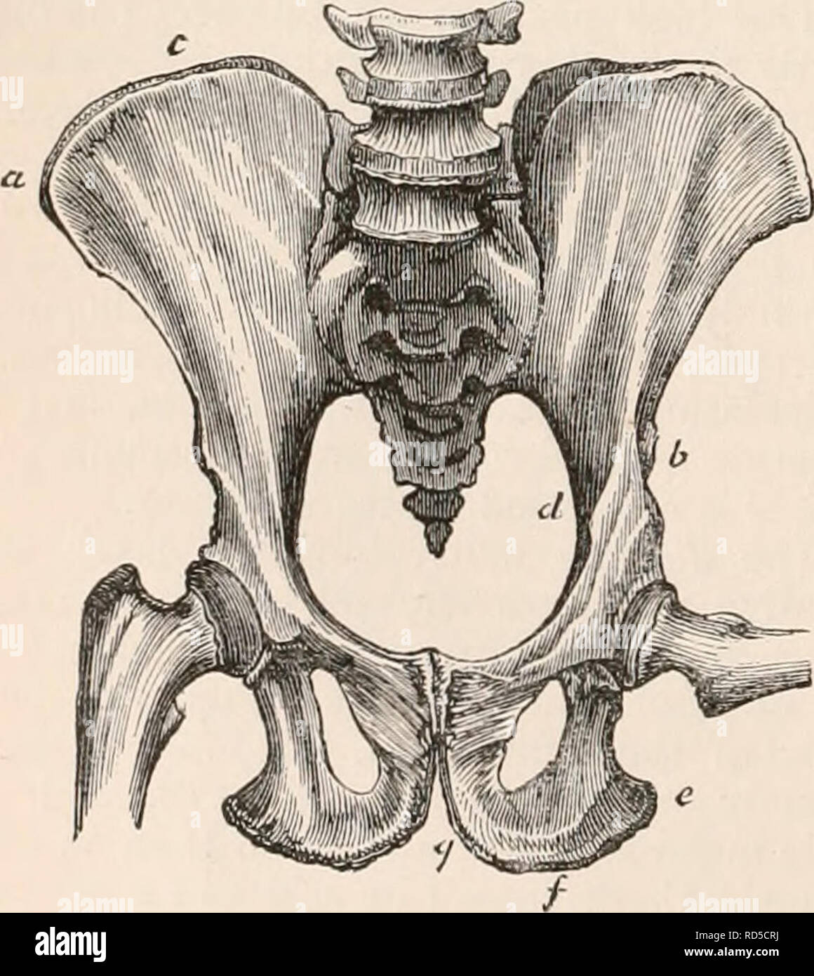 . The cyclopædia of anatomy and physiology. Anatomy; Physiology; Zoology. 152 PELVIS. wings, the anterior part seems deficient, the anterior superior spine'(a) being placed directly over the cotyloid cavity ; and the crest (c) being, consequently, very short, terminating abruptly at the vertical rib mentioned in the description of the human ilium. The ales are more expanded in the Uran than the Chimpanzee. The crest does not present the lateral /-like curvature, and is less arched than in man. The anterior iliac spines are more widely separated, the inferior (6) being scarcely discernible, and Stock Photohttps://www.alamy.com/image-license-details/?v=1https://www.alamy.com/the-cyclopdia-of-anatomy-and-physiology-anatomy-physiology-zoology-152-pelvis-wings-the-anterior-part-seems-deficient-the-anterior-superior-spinea-being-placed-directly-over-the-cotyloid-cavity-and-the-crest-c-being-consequently-very-short-terminating-abruptly-at-the-vertical-rib-mentioned-in-the-description-of-the-human-ilium-the-ales-are-more-expanded-in-the-uran-than-the-chimpanzee-the-crest-does-not-present-the-lateral-like-curvature-and-is-less-arched-than-in-man-the-anterior-iliac-spines-are-more-widely-separated-the-inferior-6-being-scarcely-discernible-and-image231845142.html
RMRD5CRJ–. The cyclopædia of anatomy and physiology. Anatomy; Physiology; Zoology. 152 PELVIS. wings, the anterior part seems deficient, the anterior superior spine'(a) being placed directly over the cotyloid cavity ; and the crest (c) being, consequently, very short, terminating abruptly at the vertical rib mentioned in the description of the human ilium. The ales are more expanded in the Uran than the Chimpanzee. The crest does not present the lateral /-like curvature, and is less arched than in man. The anterior iliac spines are more widely separated, the inferior (6) being scarcely discernible, and
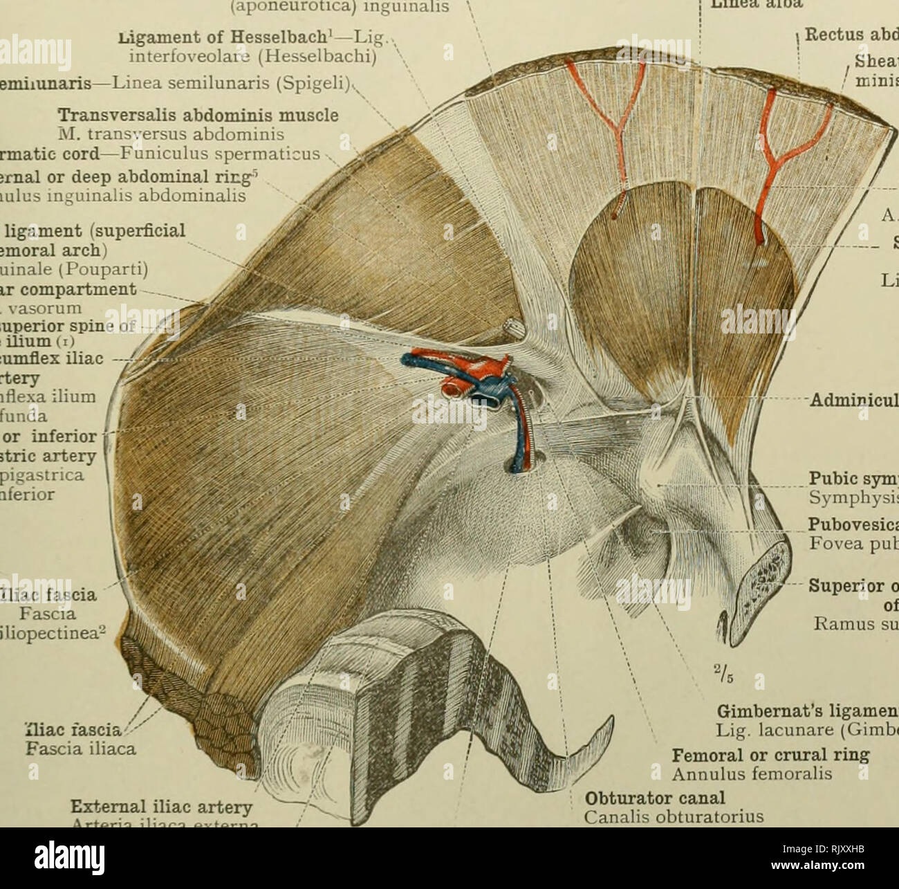 . An atlas of human anatomy for students and physicians. Anatomy. INGUINAL CANAL AND FEMORAL CANAL 389 Ligament of Henle'—Falx (aponeurotica) inguinalis Llnea semilunaris- Ligament of Hesselbach'—Lig interfoveolare (Hesselbachi) -Linea semilunaris (Spigel:. Transversalis abdominis muscle M. transversus abdominis Spermatic- cord—Funiculus sperraaticus Internal or deep abdominal nng^ Annulus inguinalis abdommalis Poupart's ligament (superficial femoral arch Lig. inguinale (I'ouparti) Vascular compartment Lacuna vasorum Anterior superior spine of the ilium (i) Deep circumflex iliac - artery A. c Stock Photohttps://www.alamy.com/image-license-details/?v=1https://www.alamy.com/an-atlas-of-human-anatomy-for-students-and-physicians-anatomy-inguinal-canal-and-femoral-canal-389-ligament-of-henlefalx-aponeurotica-inguinalis-llnea-semilunaris-ligament-of-hesselbachlig-interfoveolare-hesselbachi-linea-semilunaris-spigel-transversalis-abdominis-muscle-m-transversus-abdominis-spermatic-cordfuniculus-sperraaticus-internal-or-deep-abdominal-nng-annulus-inguinalis-abdommalis-pouparts-ligament-superficial-femoral-arch-lig-inguinale-iouparti-vascular-compartment-lacuna-vasorum-anterior-superior-spine-of-the-ilium-i-deep-circumflex-iliac-artery-a-c-image235390215.html
RMRJXXHB–. An atlas of human anatomy for students and physicians. Anatomy. INGUINAL CANAL AND FEMORAL CANAL 389 Ligament of Henle'—Falx (aponeurotica) inguinalis Llnea semilunaris- Ligament of Hesselbach'—Lig interfoveolare (Hesselbachi) -Linea semilunaris (Spigel:. Transversalis abdominis muscle M. transversus abdominis Spermatic- cord—Funiculus sperraaticus Internal or deep abdominal nng^ Annulus inguinalis abdommalis Poupart's ligament (superficial femoral arch Lig. inguinale (I'ouparti) Vascular compartment Lacuna vasorum Anterior superior spine of the ilium (i) Deep circumflex iliac - artery A. c
 . The cyclopædia of anatomy and physiology. Anatomy; Physiology; Zoology. PELVIS. 157 are not united in a crest. The ilia approach in shape to those of the Tapir, being in a less marked degree T-shaped ; the posterior limb of the iliac wings projecting inwards as far as the sacral spines; the anterior superior spine often presenting an epiphysis, and the shaft being long and blade-like. The ischia are comparatively long, and much more slender than in the Ruminants, being placed nearly parallel with the coccygeal vertebra, and with prolonged tuberosities. The jnibcs are small and short, and dir Stock Photohttps://www.alamy.com/image-license-details/?v=1https://www.alamy.com/the-cyclopdia-of-anatomy-and-physiology-anatomy-physiology-zoology-pelvis-157-are-not-united-in-a-crest-the-ilia-approach-in-shape-to-those-of-the-tapir-being-in-a-less-marked-degree-t-shaped-the-posterior-limb-of-the-iliac-wings-projecting-inwards-as-far-as-the-sacral-spines-the-anterior-superior-spine-often-presenting-an-epiphysis-and-the-shaft-being-long-and-blade-like-the-ischia-are-comparatively-long-and-much-more-slender-than-in-the-ruminants-being-placed-nearly-parallel-with-the-coccygeal-vertebra-and-with-prolonged-tuberosities-the-jnibcs-are-small-and-short-and-dir-image231845125.html
RMRD5CR1–. The cyclopædia of anatomy and physiology. Anatomy; Physiology; Zoology. PELVIS. 157 are not united in a crest. The ilia approach in shape to those of the Tapir, being in a less marked degree T-shaped ; the posterior limb of the iliac wings projecting inwards as far as the sacral spines; the anterior superior spine often presenting an epiphysis, and the shaft being long and blade-like. The ischia are comparatively long, and much more slender than in the Ruminants, being placed nearly parallel with the coccygeal vertebra, and with prolonged tuberosities. The jnibcs are small and short, and dir
 . Cunningham's Text-book of anatomy. Anatomy. 1460 SURFACE AND SUKGICAL ANATOMY. hip-joint. One of the commonest situations to meet with an abscess in hip-joint disease is in the cellular tissue and fat under the tensor fasciae lata?; or the pus may pass below and to the medial side of the neck of the femur, and thence along the course of the medial circumflex artery of the thigh to the back of the thigh. To tap or explore the hip-joint, the puncture should be made in the interval between the sartorius and the tensor fascia? lata}, 2 to 3 in. distal to the superior anterior iliac spine ; if th Stock Photohttps://www.alamy.com/image-license-details/?v=1https://www.alamy.com/cunninghams-text-book-of-anatomy-anatomy-1460-surface-and-sukgical-anatomy-hip-joint-one-of-the-commonest-situations-to-meet-with-an-abscess-in-hip-joint-disease-is-in-the-cellular-tissue-and-fat-under-the-tensor-fasciae-lata-or-the-pus-may-pass-below-and-to-the-medial-side-of-the-neck-of-the-femur-and-thence-along-the-course-of-the-medial-circumflex-artery-of-the-thigh-to-the-back-of-the-thigh-to-tap-or-explore-the-hip-joint-the-puncture-should-be-made-in-the-interval-between-the-sartorius-and-the-tensor-fascia-lata-2-to-3-in-distal-to-the-superior-anterior-iliac-spine-if-th-image231866876.html
RMRD6CFT–. Cunningham's Text-book of anatomy. Anatomy. 1460 SURFACE AND SUKGICAL ANATOMY. hip-joint. One of the commonest situations to meet with an abscess in hip-joint disease is in the cellular tissue and fat under the tensor fasciae lata?; or the pus may pass below and to the medial side of the neck of the femur, and thence along the course of the medial circumflex artery of the thigh to the back of the thigh. To tap or explore the hip-joint, the puncture should be made in the interval between the sartorius and the tensor fascia? lata}, 2 to 3 in. distal to the superior anterior iliac spine ; if th
 . An atlas of human anatomy for students and physicians. Anatomy. 390 INGUINAL CANAL AND FEMORAL CANAL Anterior superior spine of the ilium Spina iliaca anterior superior /⢠Iliac compartment Z Lacuna musculorum. Fig. 639- Anterior inferior spine of the ilium Spina ihaca anterior inferior Iliac fascia Fascia iliopectinea^ Poupart s ligament (superficial femoral arch) Lig inguinale (Pouparti) Vascular com.partment Lacuna vasorum Iliopectineal eminence Eminentia iliopectinea Pectineal fascia ' Fascia pectinea y Gimbemat's ligament / Lig lacunare (Gimbernati) Triangular fascia' / Lig. inguinale Stock Photohttps://www.alamy.com/image-license-details/?v=1https://www.alamy.com/an-atlas-of-human-anatomy-for-students-and-physicians-anatomy-390-inguinal-canal-and-femoral-canal-anterior-superior-spine-of-the-ilium-spina-iliaca-anterior-superior-iliac-compartment-z-lacuna-musculorum-fig-639-anterior-inferior-spine-of-the-ilium-spina-ihaca-anterior-inferior-iliac-fascia-fascia-iliopectinea-poupart-s-ligament-superficial-femoral-arch-lig-inguinale-pouparti-vascular-compartment-lacuna-vasorum-iliopectineal-eminence-eminentia-iliopectinea-pectineal-fascia-fascia-pectinea-y-gimbemats-ligament-lig-lacunare-gimbernati-triangular-fascia-lig-inguinale-image235390191.html
RMRJXXGF–. An atlas of human anatomy for students and physicians. Anatomy. 390 INGUINAL CANAL AND FEMORAL CANAL Anterior superior spine of the ilium Spina iliaca anterior superior /⢠Iliac compartment Z Lacuna musculorum. Fig. 639- Anterior inferior spine of the ilium Spina ihaca anterior inferior Iliac fascia Fascia iliopectinea^ Poupart s ligament (superficial femoral arch) Lig inguinale (Pouparti) Vascular com.partment Lacuna vasorum Iliopectineal eminence Eminentia iliopectinea Pectineal fascia ' Fascia pectinea y Gimbemat's ligament / Lig lacunare (Gimbernati) Triangular fascia' / Lig. inguinale
![. An atlas of human anatomy for students and physicians. Anatomy. MUSCLES OF THE PERINEUM 533 Vas deferens and artery of the vas deferens or deferential artery Ductus deferens et A. deferentiali-- Iliac fascia Fascia iliaca Spermatic or pampiniform^ plexus I'iexus pampiniformis ^ Obturator nerve ^, N. obturatorius. External iliac artery^. A. iliaca externa External iliac vein V. iliaca externa Ante] lor superior spine of the ilium ""Spina iliaca anterior superior ,Poupart's ligament (superficial femoral arch) ILg inguinile ( r lupirti) 1 Internal or deep abdominal ring- (i) I Deep o Stock Photo . An atlas of human anatomy for students and physicians. Anatomy. MUSCLES OF THE PERINEUM 533 Vas deferens and artery of the vas deferens or deferential artery Ductus deferens et A. deferentiali-- Iliac fascia Fascia iliaca Spermatic or pampiniform^ plexus I'iexus pampiniformis ^ Obturator nerve ^, N. obturatorius. External iliac artery^. A. iliaca externa External iliac vein V. iliaca externa Ante] lor superior spine of the ilium ""Spina iliaca anterior superior ,Poupart's ligament (superficial femoral arch) ILg inguinile ( r lupirti) 1 Internal or deep abdominal ring- (i) I Deep o Stock Photo](https://c8.alamy.com/comp/RJY2WN/an-atlas-of-human-anatomy-for-students-and-physicians-anatomy-muscles-of-the-perineum-533-vas-deferens-and-artery-of-the-vas-deferens-or-deferential-artery-ductus-deferens-et-a-deferentiali-iliac-fascia-fascia-iliaca-spermatic-or-pampiniform-plexus-iiexus-pampiniformis-obturator-nerve-n-obturatorius-external-iliac-artery-a-iliaca-externa-external-iliac-vein-v-iliaca-externa-ante-lor-superior-spine-of-the-ilium-quotquotspina-iliaca-anterior-superior-pouparts-ligament-superficial-femoral-arch-ilg-inguinile-r-lupirti-1-internal-or-deep-abdominal-ring-i-i-deep-o-RJY2WN.jpg) . An atlas of human anatomy for students and physicians. Anatomy. MUSCLES OF THE PERINEUM 533 Vas deferens and artery of the vas deferens or deferential artery Ductus deferens et A. deferentiali-- Iliac fascia Fascia iliaca Spermatic or pampiniform^ plexus I'iexus pampiniformis ^ Obturator nerve ^, N. obturatorius. External iliac artery^. A. iliaca externa External iliac vein V. iliaca externa Ante] lor superior spine of the ilium ""Spina iliaca anterior superior ,Poupart's ligament (superficial femoral arch) ILg inguinile ( r lupirti) 1 Internal or deep abdominal ring- (i) I Deep o Stock Photohttps://www.alamy.com/image-license-details/?v=1https://www.alamy.com/an-atlas-of-human-anatomy-for-students-and-physicians-anatomy-muscles-of-the-perineum-533-vas-deferens-and-artery-of-the-vas-deferens-or-deferential-artery-ductus-deferens-et-a-deferentiali-iliac-fascia-fascia-iliaca-spermatic-or-pampiniform-plexus-iiexus-pampiniformis-obturator-nerve-n-obturatorius-external-iliac-artery-a-iliaca-externa-external-iliac-vein-v-iliaca-externa-ante-lor-superior-spine-of-the-ilium-quotquotspina-iliaca-anterior-superior-pouparts-ligament-superficial-femoral-arch-ilg-inguinile-r-lupirti-1-internal-or-deep-abdominal-ring-i-i-deep-o-image235393585.html
. An atlas of human anatomy for students and physicians. Anatomy. MUSCLES OF THE PERINEUM 533 Vas deferens and artery of the vas deferens or deferential artery Ductus deferens et A. deferentiali-- Iliac fascia Fascia iliaca Spermatic or pampiniform^ plexus I'iexus pampiniformis ^ Obturator nerve ^, N. obturatorius. External iliac artery^. A. iliaca externa External iliac vein V. iliaca externa Ante] lor superior spine of the ilium ""Spina iliaca anterior superior ,Poupart's ligament (superficial femoral arch) ILg inguinile ( r lupirti) 1 Internal or deep abdominal ring- (i) I Deep o Stock Photohttps://www.alamy.com/image-license-details/?v=1https://www.alamy.com/an-atlas-of-human-anatomy-for-students-and-physicians-anatomy-muscles-of-the-perineum-533-vas-deferens-and-artery-of-the-vas-deferens-or-deferential-artery-ductus-deferens-et-a-deferentiali-iliac-fascia-fascia-iliaca-spermatic-or-pampiniform-plexus-iiexus-pampiniformis-obturator-nerve-n-obturatorius-external-iliac-artery-a-iliaca-externa-external-iliac-vein-v-iliaca-externa-ante-lor-superior-spine-of-the-ilium-quotquotspina-iliaca-anterior-superior-pouparts-ligament-superficial-femoral-arch-ilg-inguinile-r-lupirti-1-internal-or-deep-abdominal-ring-i-i-deep-o-image235393585.htmlRMRJY2WN–. An atlas of human anatomy for students and physicians. Anatomy. MUSCLES OF THE PERINEUM 533 Vas deferens and artery of the vas deferens or deferential artery Ductus deferens et A. deferentiali-- Iliac fascia Fascia iliaca Spermatic or pampiniform^ plexus I'iexus pampiniformis ^ Obturator nerve ^, N. obturatorius. External iliac artery^. A. iliaca externa External iliac vein V. iliaca externa Ante] lor superior spine of the ilium ""Spina iliaca anterior superior ,Poupart's ligament (superficial femoral arch) ILg inguinile ( r lupirti) 1 Internal or deep abdominal ring- (i) I Deep o
![. An atlas of human anatomy for students and physicians. Anatomy. External iliac artery^. A. iliaca externa External iliac vein V. iliaca externa Ante] lor superior spine of the ilium ""Spina iliaca anterior superior ,Poupart's ligament (superficial femoral arch) ILg inguinile ( r lupirti) 1 Internal or deep abdominal ring- (i) I Deep or inferior epigastric artery —k 1 and vein ^ ' ^ et V epi-,astric I inferiores Vestige of the obliterated hypogastric artery, or 'external umbilical ligament" (•;) Obturator artery and vein I 1 1 jturia ^Rectal fascia' L liaphragmatis pelvis Stock Photo . An atlas of human anatomy for students and physicians. Anatomy. External iliac artery^. A. iliaca externa External iliac vein V. iliaca externa Ante] lor superior spine of the ilium ""Spina iliaca anterior superior ,Poupart's ligament (superficial femoral arch) ILg inguinile ( r lupirti) 1 Internal or deep abdominal ring- (i) I Deep or inferior epigastric artery —k 1 and vein ^ ' ^ et V epi-,astric I inferiores Vestige of the obliterated hypogastric artery, or 'external umbilical ligament" (•;) Obturator artery and vein I 1 1 jturia ^Rectal fascia' L liaphragmatis pelvis Stock Photo](https://c8.alamy.com/comp/RJY2W0/an-atlas-of-human-anatomy-for-students-and-physicians-anatomy-external-iliac-artery-a-iliaca-externa-external-iliac-vein-v-iliaca-externa-ante-lor-superior-spine-of-the-ilium-quotquotspina-iliaca-anterior-superior-pouparts-ligament-superficial-femoral-arch-ilg-inguinile-r-lupirti-1-internal-or-deep-abdominal-ring-i-i-deep-or-inferior-epigastric-artery-k-1-and-vein-et-v-epi-astric-i-inferiores-vestige-of-the-obliterated-hypogastric-artery-or-external-umbilical-ligamentquot-obturator-artery-and-vein-i-1-1-jturia-rectal-fascia-l-liaphragmatis-pelvis-RJY2W0.jpg) . An atlas of human anatomy for students and physicians. Anatomy. External iliac artery^. A. iliaca externa External iliac vein V. iliaca externa Ante] lor superior spine of the ilium ""Spina iliaca anterior superior ,Poupart's ligament (superficial femoral arch) ILg inguinile ( r lupirti) 1 Internal or deep abdominal ring- (i) I Deep or inferior epigastric artery —k 1 and vein ^ ' ^ et V epi-,astric I inferiores Vestige of the obliterated hypogastric artery, or 'external umbilical ligament" (•;) Obturator artery and vein I 1 1 jturia ^Rectal fascia' L liaphragmatis pelvis Stock Photohttps://www.alamy.com/image-license-details/?v=1https://www.alamy.com/an-atlas-of-human-anatomy-for-students-and-physicians-anatomy-external-iliac-artery-a-iliaca-externa-external-iliac-vein-v-iliaca-externa-ante-lor-superior-spine-of-the-ilium-quotquotspina-iliaca-anterior-superior-pouparts-ligament-superficial-femoral-arch-ilg-inguinile-r-lupirti-1-internal-or-deep-abdominal-ring-i-i-deep-or-inferior-epigastric-artery-k-1-and-vein-et-v-epi-astric-i-inferiores-vestige-of-the-obliterated-hypogastric-artery-or-external-umbilical-ligamentquot-obturator-artery-and-vein-i-1-1-jturia-rectal-fascia-l-liaphragmatis-pelvis-image235393564.html
. An atlas of human anatomy for students and physicians. Anatomy. External iliac artery^. A. iliaca externa External iliac vein V. iliaca externa Ante] lor superior spine of the ilium ""Spina iliaca anterior superior ,Poupart's ligament (superficial femoral arch) ILg inguinile ( r lupirti) 1 Internal or deep abdominal ring- (i) I Deep or inferior epigastric artery —k 1 and vein ^ ' ^ et V epi-,astric I inferiores Vestige of the obliterated hypogastric artery, or 'external umbilical ligament" (•;) Obturator artery and vein I 1 1 jturia ^Rectal fascia' L liaphragmatis pelvis Stock Photohttps://www.alamy.com/image-license-details/?v=1https://www.alamy.com/an-atlas-of-human-anatomy-for-students-and-physicians-anatomy-external-iliac-artery-a-iliaca-externa-external-iliac-vein-v-iliaca-externa-ante-lor-superior-spine-of-the-ilium-quotquotspina-iliaca-anterior-superior-pouparts-ligament-superficial-femoral-arch-ilg-inguinile-r-lupirti-1-internal-or-deep-abdominal-ring-i-i-deep-or-inferior-epigastric-artery-k-1-and-vein-et-v-epi-astric-i-inferiores-vestige-of-the-obliterated-hypogastric-artery-or-external-umbilical-ligamentquot-obturator-artery-and-vein-i-1-1-jturia-rectal-fascia-l-liaphragmatis-pelvis-image235393564.htmlRMRJY2W0–. An atlas of human anatomy for students and physicians. Anatomy. External iliac artery^. A. iliaca externa External iliac vein V. iliaca externa Ante] lor superior spine of the ilium ""Spina iliaca anterior superior ,Poupart's ligament (superficial femoral arch) ILg inguinile ( r lupirti) 1 Internal or deep abdominal ring- (i) I Deep or inferior epigastric artery —k 1 and vein ^ ' ^ et V epi-,astric I inferiores Vestige of the obliterated hypogastric artery, or 'external umbilical ligament" (•;) Obturator artery and vein I 1 1 jturia ^Rectal fascia' L liaphragmatis pelvis