Quick filters:
Apex of lung Stock Photos and Images
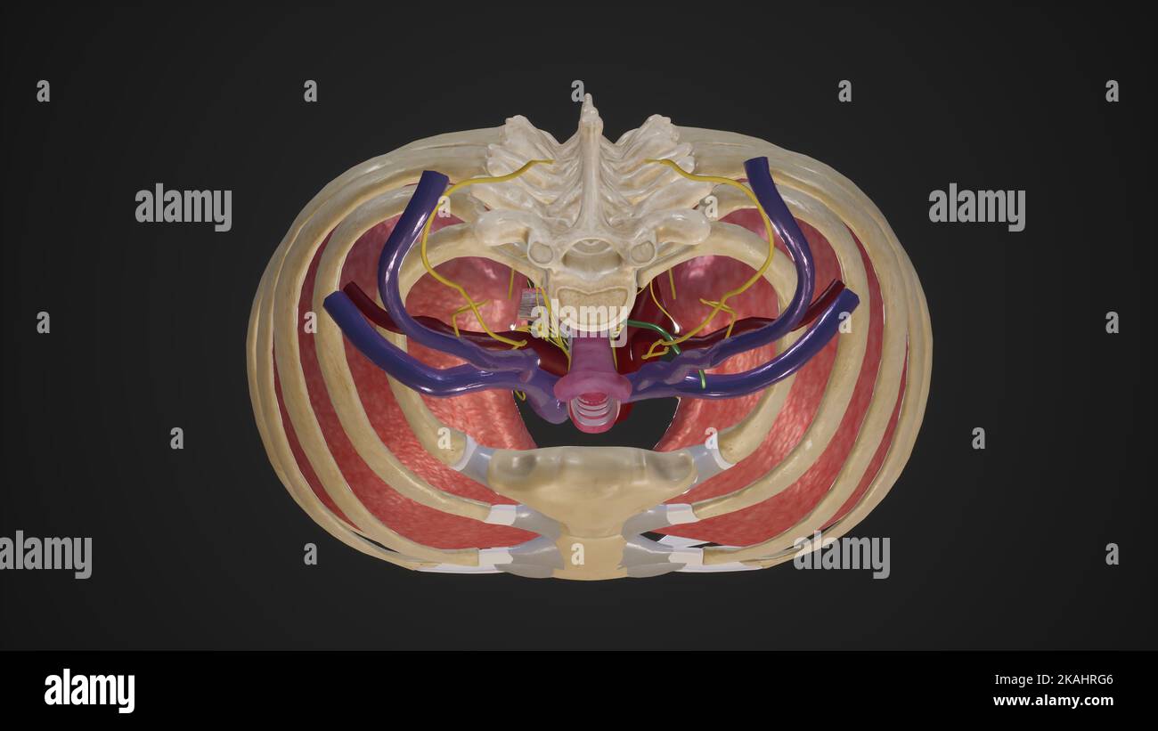 Superior Thoracic Aperture Stock Photohttps://www.alamy.com/image-license-details/?v=1https://www.alamy.com/superior-thoracic-aperture-image488428534.html
RF2KAHRG6–Superior Thoracic Aperture
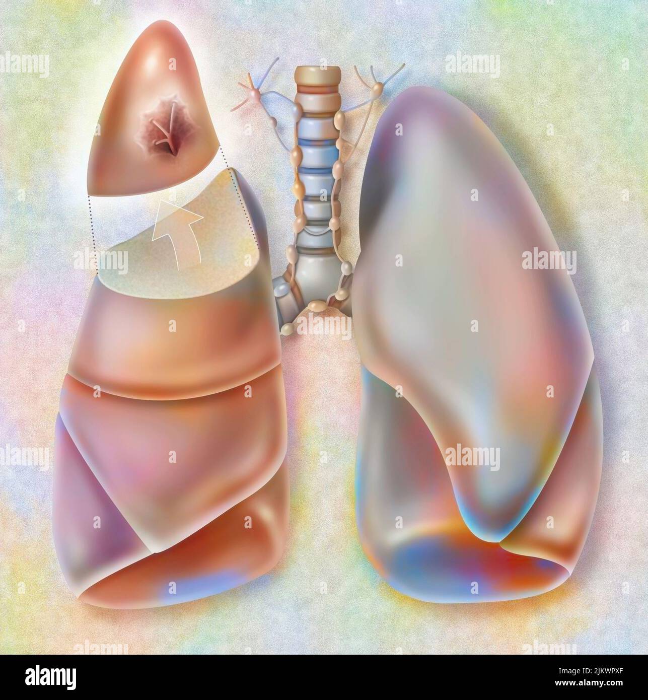 Removal of the apical segment of the right lung affected by a cancerous tumor. Stock Photohttps://www.alamy.com/image-license-details/?v=1https://www.alamy.com/removal-of-the-apical-segment-of-the-right-lung-affected-by-a-cancerous-tumor-image476925191.html
RF2JKWPXF–Removal of the apical segment of the right lung affected by a cancerous tumor.
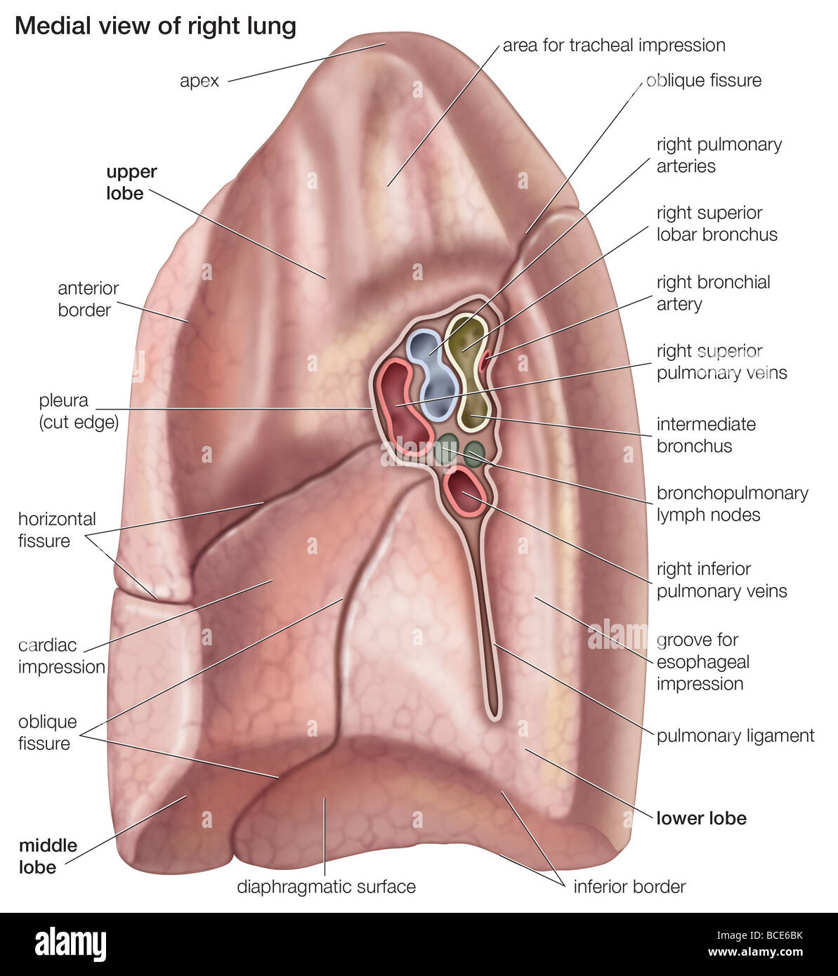 Medial view of the right lung. Stock Photohttps://www.alamy.com/image-license-details/?v=1https://www.alamy.com/stock-photo-medial-view-of-the-right-lung-24898599.html
RMBCE6BK–Medial view of the right lung.
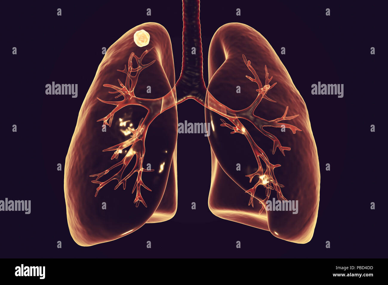 Secondary tuberculosis infection. Computer illustration showing small-sized solid nodular mass located in the upper lobe of right lung near lung apex. Stock Photohttps://www.alamy.com/image-license-details/?v=1https://www.alamy.com/secondary-tuberculosis-infection-computer-illustration-showing-small-sized-solid-nodular-mass-located-in-the-upper-lobe-of-right-lung-near-lung-apex-image213574521.html
RFPBD4DD–Secondary tuberculosis infection. Computer illustration showing small-sized solid nodular mass located in the upper lobe of right lung near lung apex.
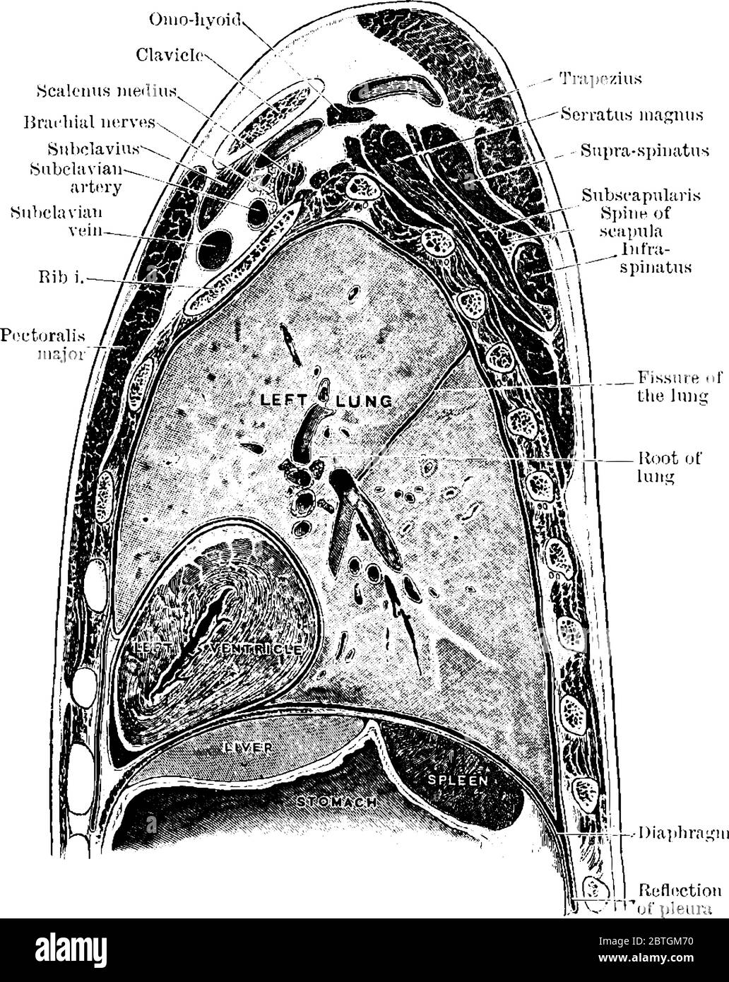 The sagittal section through the left shoulder, lung, and apex of the heart, vintage line drawing or engraving illustration. Stock Vectorhttps://www.alamy.com/image-license-details/?v=1https://www.alamy.com/the-sagittal-section-through-the-left-shoulder-lung-and-apex-of-the-heart-vintage-line-drawing-or-engraving-illustration-image359326212.html
RF2BTGM70–The sagittal section through the left shoulder, lung, and apex of the heart, vintage line drawing or engraving illustration.
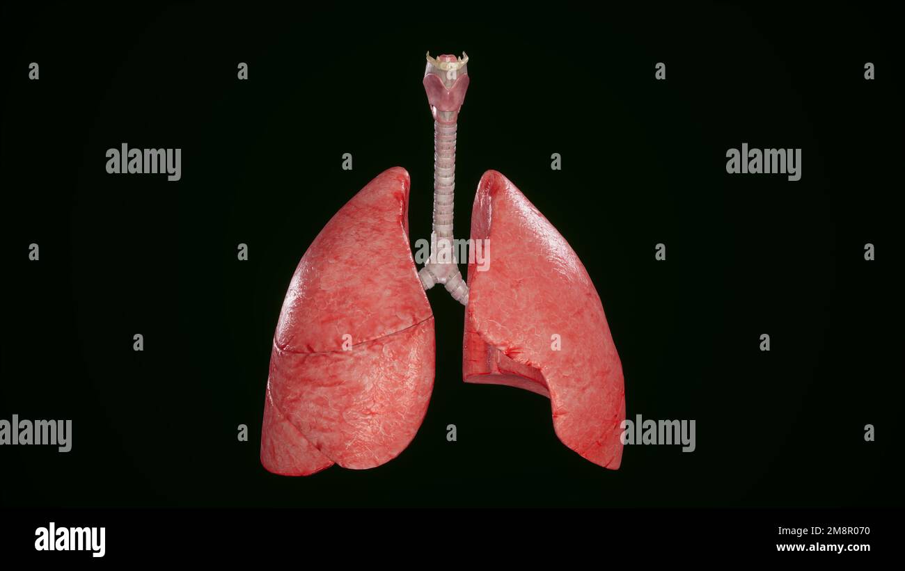 Human lungs and trachea isolated,3D rendering Stock Photohttps://www.alamy.com/image-license-details/?v=1https://www.alamy.com/human-lungs-and-trachea-isolated3d-rendering-image504523012.html
RF2M8R070–Human lungs and trachea isolated,3D rendering
 A proposal of marriage in Cape Town's Kirstensbosch gardens and the apex of a network of conservation gardens in South Africa Stock Photohttps://www.alamy.com/image-license-details/?v=1https://www.alamy.com/a-proposal-of-marriage-in-cape-towns-kirstensbosch-gardens-and-the-apex-of-a-network-of-conservation-gardens-in-south-africa-image455618546.html
RM2HD762X–A proposal of marriage in Cape Town's Kirstensbosch gardens and the apex of a network of conservation gardens in South Africa
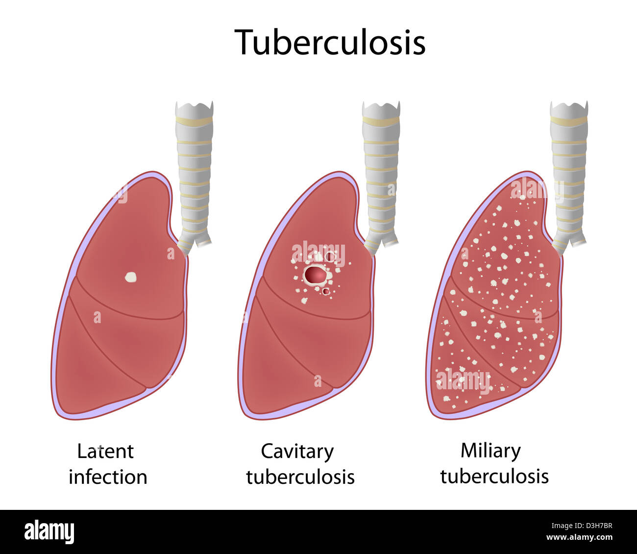 Pulmonary tuberculosis, latent infection, cavitary and miliary TB. Stock Photohttps://www.alamy.com/image-license-details/?v=1https://www.alamy.com/stock-photo-pulmonary-tuberculosis-latent-infection-cavitary-and-miliary-tb-53854075.html
RFD3H7BR–Pulmonary tuberculosis, latent infection, cavitary and miliary TB.
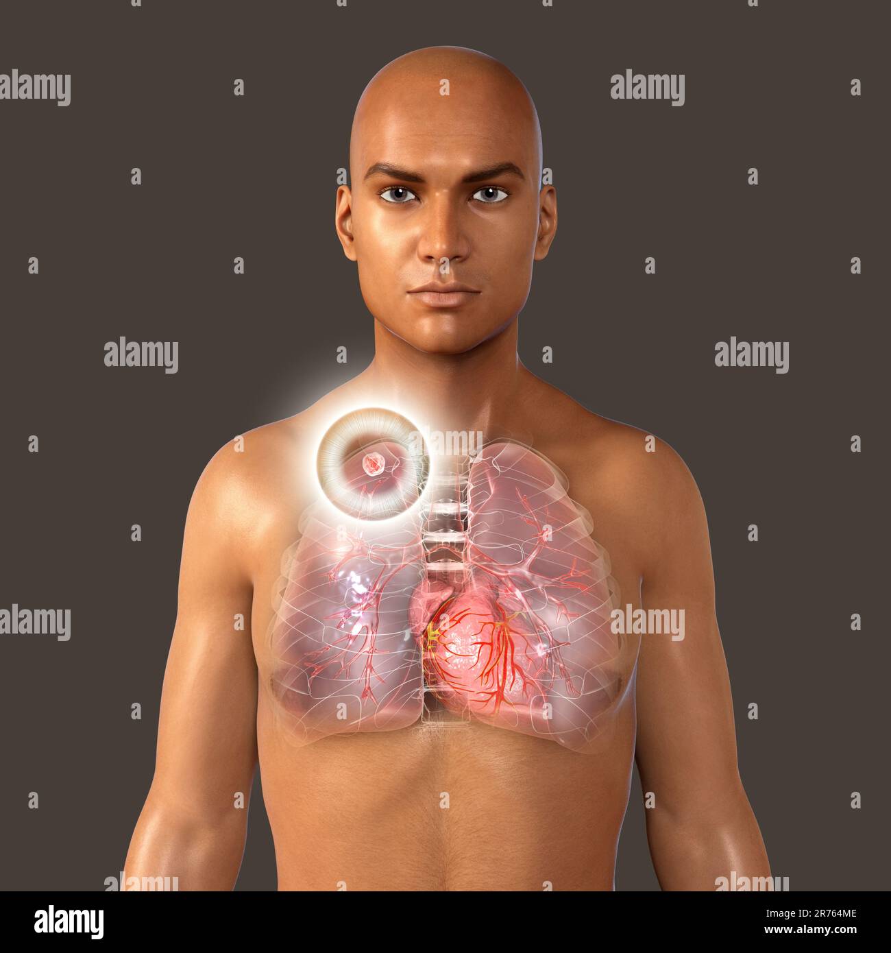 Secondary tuberculosis infection. Computer illustration showing small-sized solid nodular mass located in the upper lobe of right lung near lung apex. Stock Photohttps://www.alamy.com/image-license-details/?v=1https://www.alamy.com/secondary-tuberculosis-infection-computer-illustration-showing-small-sized-solid-nodular-mass-located-in-the-upper-lobe-of-right-lung-near-lung-apex-image555169790.html
RF2R764ME–Secondary tuberculosis infection. Computer illustration showing small-sized solid nodular mass located in the upper lobe of right lung near lung apex.
 The Practitioner . Fig- 5-X-ray plioto^raph of lumour at apex of left luni^ i<T. 4. Paralysis of L. cervical sympatheticfrom tumour at apex of lung. Pl-ATK XVII.. lie Stock Photohttps://www.alamy.com/image-license-details/?v=1https://www.alamy.com/the-practitioner-fig-5-x-ray-pliotoraph-of-lumour-at-apex-of-left-luni-iltt-4-paralysis-of-l-cervical-sympatheticfrom-tumour-at-apex-of-lung-pl-atk-xvii-lie-image342733147.html
RM2AWGRHF–The Practitioner . Fig- 5-X-ray plioto^raph of lumour at apex of left luni^ i<T. 4. Paralysis of L. cervical sympatheticfrom tumour at apex of lung. Pl-ATK XVII.. lie
 Anatomically accurate realistic 3d illustration of human internal organ - lungs isolated on white background. Lungs structure with properly placed text captions Stock Photohttps://www.alamy.com/image-license-details/?v=1https://www.alamy.com/anatomically-accurate-realistic-3d-illustration-of-human-internal-organ-lungs-isolated-on-white-background-lungs-structure-with-properly-placed-text-captions-image565981186.html
RF2RTPJNP–Anatomically accurate realistic 3d illustration of human internal organ - lungs isolated on white background. Lungs structure with properly placed text captions
 The great cardiac vein (left coronary vein) is a vein of the heart3d illustration It begins at the apex of the heart and ascends along the anterior in Stock Photohttps://www.alamy.com/image-license-details/?v=1https://www.alamy.com/the-great-cardiac-vein-left-coronary-vein-is-a-vein-of-the-heart3d-illustration-it-begins-at-the-apex-of-the-heart-and-ascends-along-the-anterior-in-image596575897.html
RF2WJGAJ1–The great cardiac vein (left coronary vein) is a vein of the heart3d illustration It begins at the apex of the heart and ascends along the anterior in
 Archive image from page 252 of The cyclopædia of anatomy and. The cyclopædia of anatomy and physiology cyclopdiaofana0402todd Year: 1849 THORAX. 1037 same characters in both sexes. There is little difference in the height of the two apices. The elevation of the liver on the right side does not necessarily cause the right apex to be the higher. The right lung is more shallow than the left; but this is not because it is ' pushed up,' but because, in order to ac- commodate the liver, there is less lung- substance on the right side. If the mean of a series of observations represents the right si Stock Photohttps://www.alamy.com/image-license-details/?v=1https://www.alamy.com/archive-image-from-page-252-of-the-cyclopdia-of-anatomy-and-the-cyclopdia-of-anatomy-and-physiology-cyclopdiaofana0402todd-year-1849-thorax-1037-same-characters-in-both-sexes-there-is-little-difference-in-the-height-of-the-two-apices-the-elevation-of-the-liver-on-the-right-side-does-not-necessarily-cause-the-right-apex-to-be-the-higher-the-right-lung-is-more-shallow-than-the-left-but-this-is-not-because-it-is-pushed-up-but-because-in-order-to-ac-commodate-the-liver-there-is-less-lung-substance-on-the-right-side-if-the-mean-of-a-series-of-observations-represents-the-right-si-image259545471.html
RMW278W3–Archive image from page 252 of The cyclopædia of anatomy and. The cyclopædia of anatomy and physiology cyclopdiaofana0402todd Year: 1849 THORAX. 1037 same characters in both sexes. There is little difference in the height of the two apices. The elevation of the liver on the right side does not necessarily cause the right apex to be the higher. The right lung is more shallow than the left; but this is not because it is ' pushed up,' but because, in order to ac- commodate the liver, there is less lung- substance on the right side. If the mean of a series of observations represents the right si
 Cartoon lungs human body organ character, vector respiratory system health care and anatomy medicine. Happy lungs personage with funny faces, healthy pink lobes, pulmonary alveoli and trachea Stock Vectorhttps://www.alamy.com/image-license-details/?v=1https://www.alamy.com/cartoon-lungs-human-body-organ-character-vector-respiratory-system-health-care-and-anatomy-medicine-happy-lungs-personage-with-funny-faces-healthy-pink-lobes-pulmonary-alveoli-and-trachea-image517677932.html
RF2N267DG–Cartoon lungs human body organ character, vector respiratory system health care and anatomy medicine. Happy lungs personage with funny faces, healthy pink lobes, pulmonary alveoli and trachea
 Tuberculosis Stock Photohttps://www.alamy.com/image-license-details/?v=1https://www.alamy.com/stock-photo-tuberculosis-51650860.html
RFD00W5G–Tuberculosis
 . The cyclopædia of anatomy and physiology. Anatomy; Physiology; Zoology. THORAX. 1037 same characters in both sexes. There is little difference in the height of the two apices. The elevation of the liver on the right side does not necessarily cause the right apex to be the higher. The right lung is more shallow than the left; but this is not because it is " pushed up," but because, in order to ac- commodate the liver, there is less lung- substance on the right side. If the mean of a series of observations represents the right side of the thoracic cavity as equal to 151, the left may Stock Photohttps://www.alamy.com/image-license-details/?v=1https://www.alamy.com/the-cyclopdia-of-anatomy-and-physiology-anatomy-physiology-zoology-thorax-1037-same-characters-in-both-sexes-there-is-little-difference-in-the-height-of-the-two-apices-the-elevation-of-the-liver-on-the-right-side-does-not-necessarily-cause-the-right-apex-to-be-the-higher-the-right-lung-is-more-shallow-than-the-left-but-this-is-not-because-it-is-quot-pushed-upquot-but-because-in-order-to-ac-commodate-the-liver-there-is-less-lung-substance-on-the-right-side-if-the-mean-of-a-series-of-observations-represents-the-right-side-of-the-thoracic-cavity-as-equal-to-151-the-left-may-image216189997.html
RMPFM8F9–. The cyclopædia of anatomy and physiology. Anatomy; Physiology; Zoology. THORAX. 1037 same characters in both sexes. There is little difference in the height of the two apices. The elevation of the liver on the right side does not necessarily cause the right apex to be the higher. The right lung is more shallow than the left; but this is not because it is " pushed up," but because, in order to ac- commodate the liver, there is less lung- substance on the right side. If the mean of a series of observations represents the right side of the thoracic cavity as equal to 151, the left may
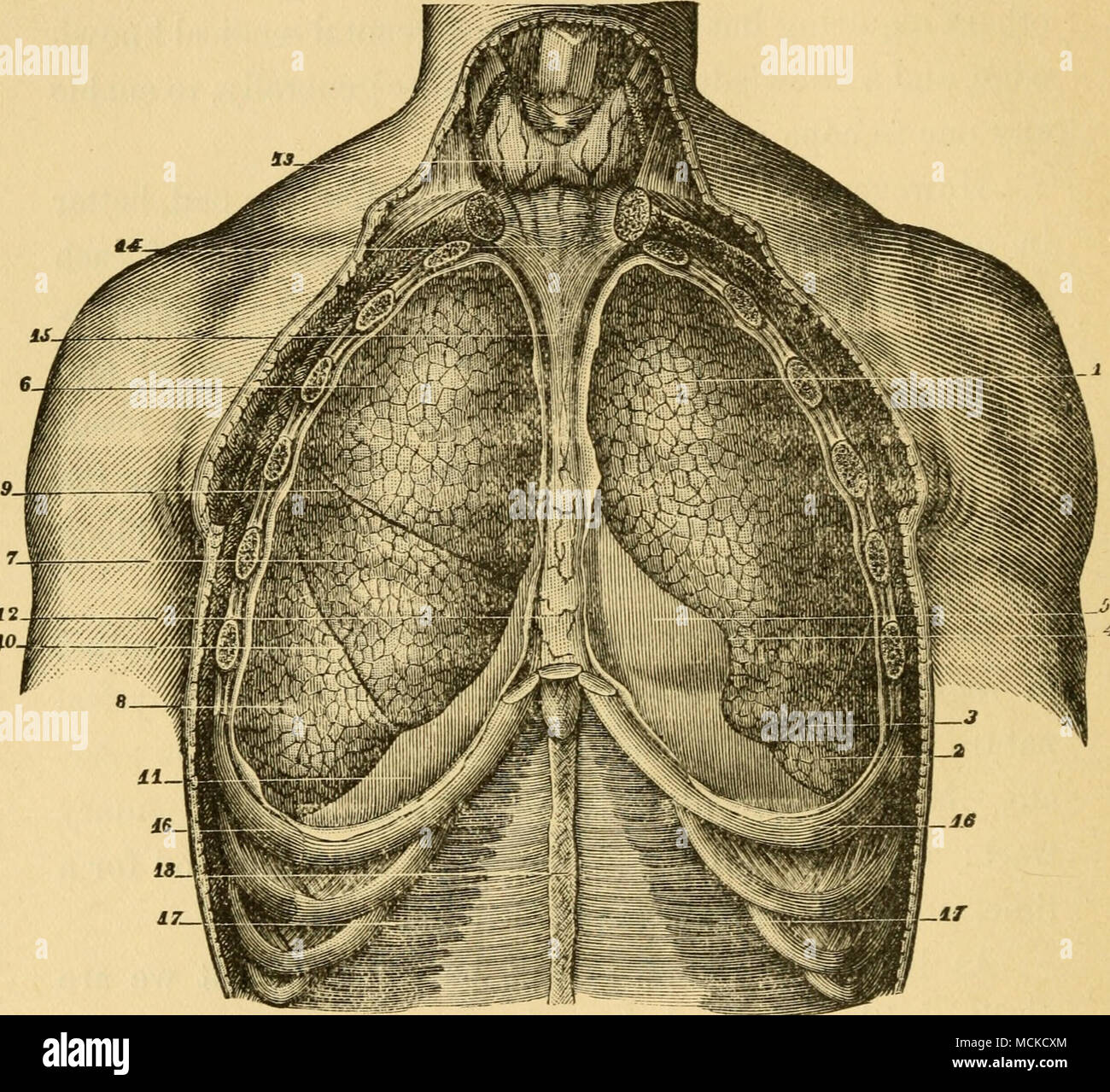 . FiG. 12.—Lungs, Anterior View (Sappey). 1, upper lobe of left lung ; 2, lower lobe ; 3, fissure ; 4, notch corresponding to apex of heart; 5, pericardium ; 6, upper lobe of right lung : 7. middle lobe ; 8, lower lobe ; 9, fissure ; 10, fissm'e ; 11, diaphragm ; 12, anterior mediasti- num ; 13. thyroid gland ; 14, middle cervical aponeurosis; 15, process of at- tachment of mediastinum to pericardium ; 16, 16, seventh ribs ; 17, 17, trans- versales muscles ; 18, linea alba. Though this cut refers to the human sub- ject, the relations of parts are substantially the same in the dog. ing the nose Stock Photohttps://www.alamy.com/image-license-details/?v=1https://www.alamy.com/fig-12lungs-anterior-view-sappey-1-upper-lobe-of-left-lung-2-lower-lobe-3-fissure-4-notch-corresponding-to-apex-of-heart-5-pericardium-6-upper-lobe-of-right-lung-7-middle-lobe-8-lower-lobe-9-fissure-10-fissme-11-diaphragm-12-anterior-mediasti-num-13-thyroid-gland-14-middle-cervical-aponeurosis-15-process-of-at-tachment-of-mediastinum-to-pericardium-16-16-seventh-ribs-17-17-trans-versales-muscles-18-linea-alba-though-this-cut-refers-to-the-human-sub-ject-the-relations-of-parts-are-substantially-the-same-in-the-dog-ing-the-nose-image179906796.html
RMMCKCXM–. FiG. 12.—Lungs, Anterior View (Sappey). 1, upper lobe of left lung ; 2, lower lobe ; 3, fissure ; 4, notch corresponding to apex of heart; 5, pericardium ; 6, upper lobe of right lung : 7. middle lobe ; 8, lower lobe ; 9, fissure ; 10, fissm'e ; 11, diaphragm ; 12, anterior mediasti- num ; 13. thyroid gland ; 14, middle cervical aponeurosis; 15, process of at- tachment of mediastinum to pericardium ; 16, 16, seventh ribs ; 17, 17, trans- versales muscles ; 18, linea alba. Though this cut refers to the human sub- ject, the relations of parts are substantially the same in the dog. ing the nose
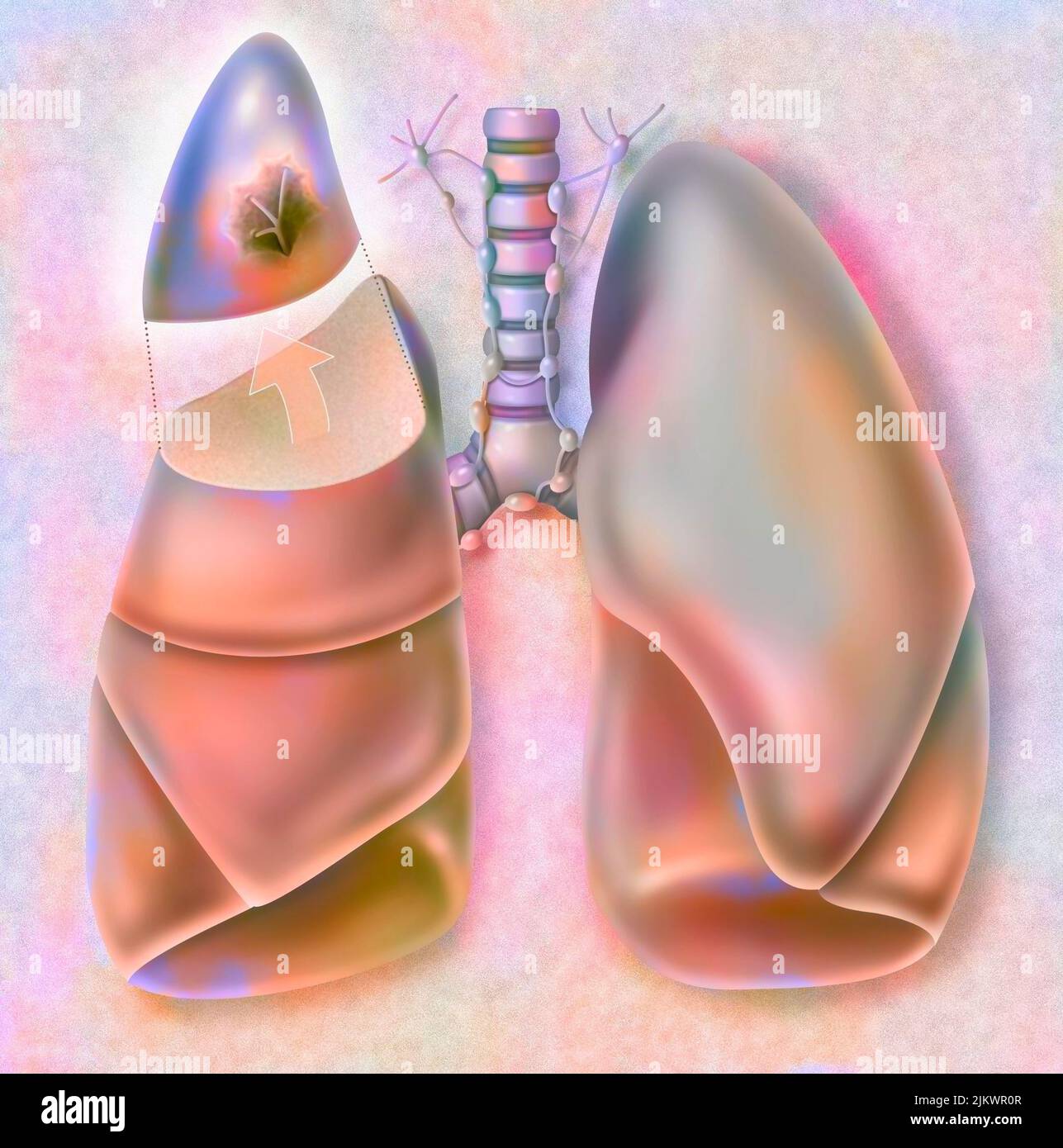 Removal of the apical segment of the right lung affected by a cancerous tumor. Stock Photohttps://www.alamy.com/image-license-details/?v=1https://www.alamy.com/removal-of-the-apical-segment-of-the-right-lung-affected-by-a-cancerous-tumor-image476925255.html
RF2JKWR0R–Removal of the apical segment of the right lung affected by a cancerous tumor.
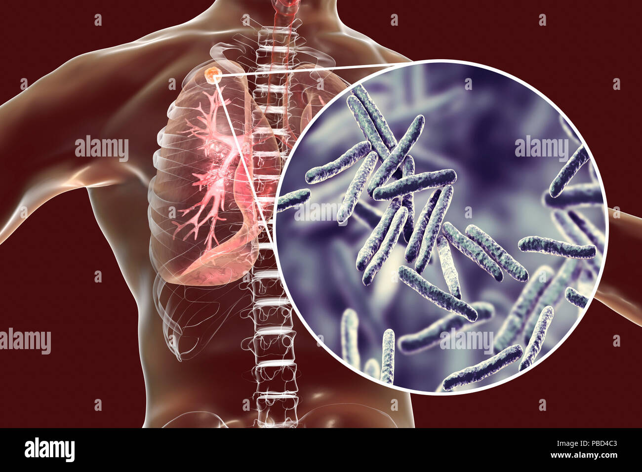 Secondary tuberculosis infection and close-up view of Mycobacterium tuberculosis bacteria, the causative agent of tuberculosis. Computer illustration showing small-sized solid nodular mass located in the upper lobe of right lung near lung apex. Stock Photohttps://www.alamy.com/image-license-details/?v=1https://www.alamy.com/secondary-tuberculosis-infection-and-close-up-view-of-mycobacterium-tuberculosis-bacteria-the-causative-agent-of-tuberculosis-computer-illustration-showing-small-sized-solid-nodular-mass-located-in-the-upper-lobe-of-right-lung-near-lung-apex-image213574483.html
RFPBD4C3–Secondary tuberculosis infection and close-up view of Mycobacterium tuberculosis bacteria, the causative agent of tuberculosis. Computer illustration showing small-sized solid nodular mass located in the upper lobe of right lung near lung apex.
 CANCER SURGERY Stock Photohttps://www.alamy.com/image-license-details/?v=1https://www.alamy.com/stock-photo-cancer-surgery-49280614.html
RMCT4WWX–CANCER SURGERY
 Cape Town's Kirstensbosch gardens is the apex of a network of conservation gardens in South Africa protecting indigenous and other plant species Stock Photohttps://www.alamy.com/image-license-details/?v=1https://www.alamy.com/cape-towns-kirstensbosch-gardens-is-the-apex-of-a-network-of-conservation-gardens-in-south-africa-protecting-indigenous-and-other-plant-species-image455618667.html
RF2HD7677–Cape Town's Kirstensbosch gardens is the apex of a network of conservation gardens in South Africa protecting indigenous and other plant species
 Secondary tuberculosis infection. Computer illustration showing small-sized solid nodular mass located in the upper lobe of right lung near lung apex. Stock Photohttps://www.alamy.com/image-license-details/?v=1https://www.alamy.com/secondary-tuberculosis-infection-computer-illustration-showing-small-sized-solid-nodular-mass-located-in-the-upper-lobe-of-right-lung-near-lung-apex-image555169787.html
RF2R764MB–Secondary tuberculosis infection. Computer illustration showing small-sized solid nodular mass located in the upper lobe of right lung near lung apex.
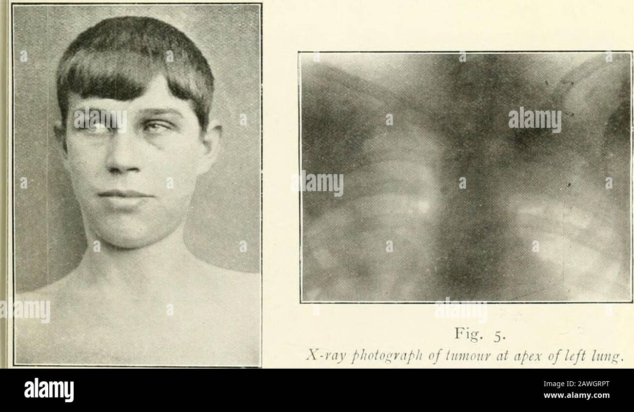 The Practitioner . Fig. 2. Paralysis of R. cervicalsynipati die from opcrationtnOiiiid. Fig. 3. Paralysis of R. cervical5; mpathetic from rupture of brachial plexus.. Fig- 5-X-ray plioto^raph of lumour at apex of left luni^ i<T. 4. Paralysis of L. cervical sympatheticfrom tumour at apex of lung. Pl-ATK XVII. Stock Photohttps://www.alamy.com/image-license-details/?v=1https://www.alamy.com/the-practitioner-fig-2-paralysis-of-r-cervicalsynipati-die-from-opcrationtnoiiiid-fig-3-paralysis-of-r-cervical5-mpathetic-from-rupture-of-brachial-plexus-fig-5-x-ray-pliotoraph-of-lumour-at-apex-of-left-luni-iltt-4-paralysis-of-l-cervical-sympatheticfrom-tumour-at-apex-of-lung-pl-atk-xvii-image342733296.html
RM2AWGRPT–The Practitioner . Fig. 2. Paralysis of R. cervicalsynipati die from opcrationtnOiiiid. Fig. 3. Paralysis of R. cervical5; mpathetic from rupture of brachial plexus.. Fig- 5-X-ray plioto^raph of lumour at apex of left luni^ i<T. 4. Paralysis of L. cervical sympatheticfrom tumour at apex of lung. Pl-ATK XVII.
 The great cardiac vein (left coronary vein) is a vein of the heart3d illustration It begins at the apex of the heart and ascends along the anterior in Stock Photohttps://www.alamy.com/image-license-details/?v=1https://www.alamy.com/the-great-cardiac-vein-left-coronary-vein-is-a-vein-of-the-heart3d-illustration-it-begins-at-the-apex-of-the-heart-and-ascends-along-the-anterior-in-image596584115.html
RF2WJGN3F–The great cardiac vein (left coronary vein) is a vein of the heart3d illustration It begins at the apex of the heart and ascends along the anterior in
 Archive image from page 1126 of Cunningham's Text-book of anatomy (1914). Cunningham's Text-book of anatomy cunninghamstextb00cunn Year: 1914 ( Lower lobe Cardiac notch Fig. 869.- -The Trachea, Bronchi, and Lungs of hardened by formalin injection. Child, Groove for left . /innominate vein neck, short mit, and a groove, the sulcus subclavius, corresponding to the vessel, is apparent upon it. At a lower level on the apex pulmonis a shallower and wider groove upon its medial and ventral aspects marks the position of the innominate vein. Although these vessels impress the lung they are separated Stock Photohttps://www.alamy.com/image-license-details/?v=1https://www.alamy.com/archive-image-from-page-1126-of-cunninghams-text-book-of-anatomy-1914-cunninghams-text-book-of-anatomy-cunninghamstextb00cunn-year-1914-lower-lobe-cardiac-notch-fig-869-the-trachea-bronchi-and-lungs-of-hardened-by-formalin-injection-child-groove-for-left-innominate-vein-neck-short-mit-and-a-groove-the-sulcus-subclavius-corresponding-to-the-vessel-is-apparent-upon-it-at-a-lower-level-on-the-apex-pulmonis-a-shallower-and-wider-groove-upon-its-medial-and-ventral-aspects-marks-the-position-of-the-innominate-vein-although-these-vessels-impress-the-lung-they-are-separated-image264068134.html
RMW9H9GP–Archive image from page 1126 of Cunningham's Text-book of anatomy (1914). Cunningham's Text-book of anatomy cunninghamstextb00cunn Year: 1914 ( Lower lobe Cardiac notch Fig. 869.- -The Trachea, Bronchi, and Lungs of hardened by formalin injection. Child, Groove for left . /innominate vein neck, short mit, and a groove, the sulcus subclavius, corresponding to the vessel, is apparent upon it. At a lower level on the apex pulmonis a shallower and wider groove upon its medial and ventral aspects marks the position of the innominate vein. Although these vessels impress the lung they are separated
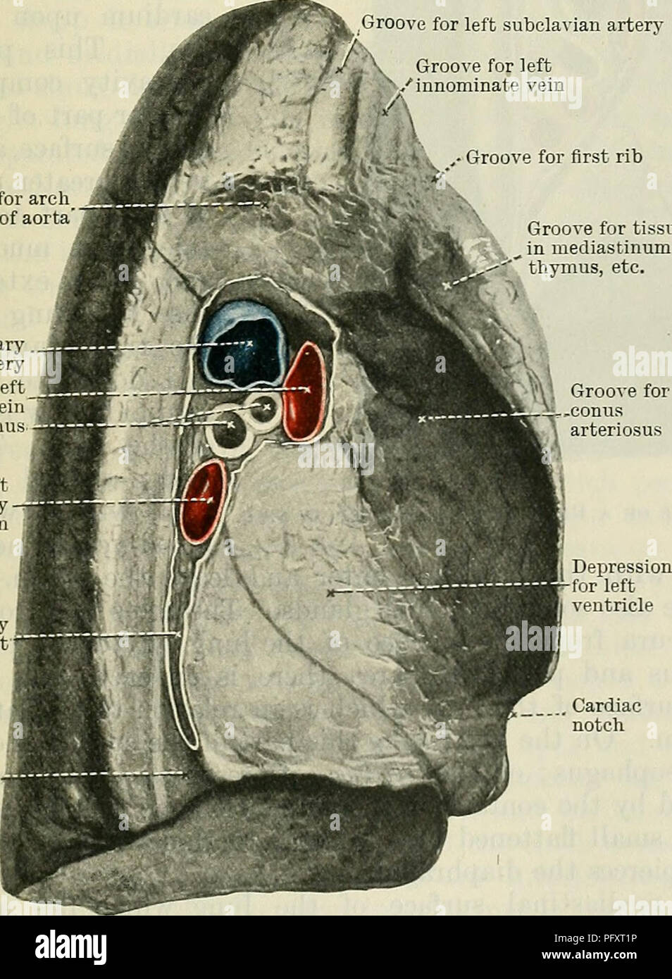 . Cunningham's Text-book of anatomy. Anatomy. Lower lobe Cardiac notch Fig. 869.- -The Trachea, Bronchi, and Lungs of hardened by formalin injection. Child, Groove for left . /innominate vein neck, short mit, and a groove, the sulcus subclavius, corresponding to the vessel, is apparent upon it. At a lower level on the apex pulmonis a shallower and wider groove upon its medial and ventral aspects marks the position of the innominate vein. Although these vessels impress the lung they are separated from it by the cupula pleurae. The diaphragmatic surface, or base of the lung, presents a semilunar Stock Photohttps://www.alamy.com/image-license-details/?v=1https://www.alamy.com/cunninghams-text-book-of-anatomy-anatomy-lower-lobe-cardiac-notch-fig-869-the-trachea-bronchi-and-lungs-of-hardened-by-formalin-injection-child-groove-for-left-innominate-vein-neck-short-mit-and-a-groove-the-sulcus-subclavius-corresponding-to-the-vessel-is-apparent-upon-it-at-a-lower-level-on-the-apex-pulmonis-a-shallower-and-wider-groove-upon-its-medial-and-ventral-aspects-marks-the-position-of-the-innominate-vein-although-these-vessels-impress-the-lung-they-are-separated-from-it-by-the-cupula-pleurae-the-diaphragmatic-surface-or-base-of-the-lung-presents-a-semilunar-image216333874.html
RMPFXT1P–. Cunningham's Text-book of anatomy. Anatomy. Lower lobe Cardiac notch Fig. 869.- -The Trachea, Bronchi, and Lungs of hardened by formalin injection. Child, Groove for left . /innominate vein neck, short mit, and a groove, the sulcus subclavius, corresponding to the vessel, is apparent upon it. At a lower level on the apex pulmonis a shallower and wider groove upon its medial and ventral aspects marks the position of the innominate vein. Although these vessels impress the lung they are separated from it by the cupula pleurae. The diaphragmatic surface, or base of the lung, presents a semilunar
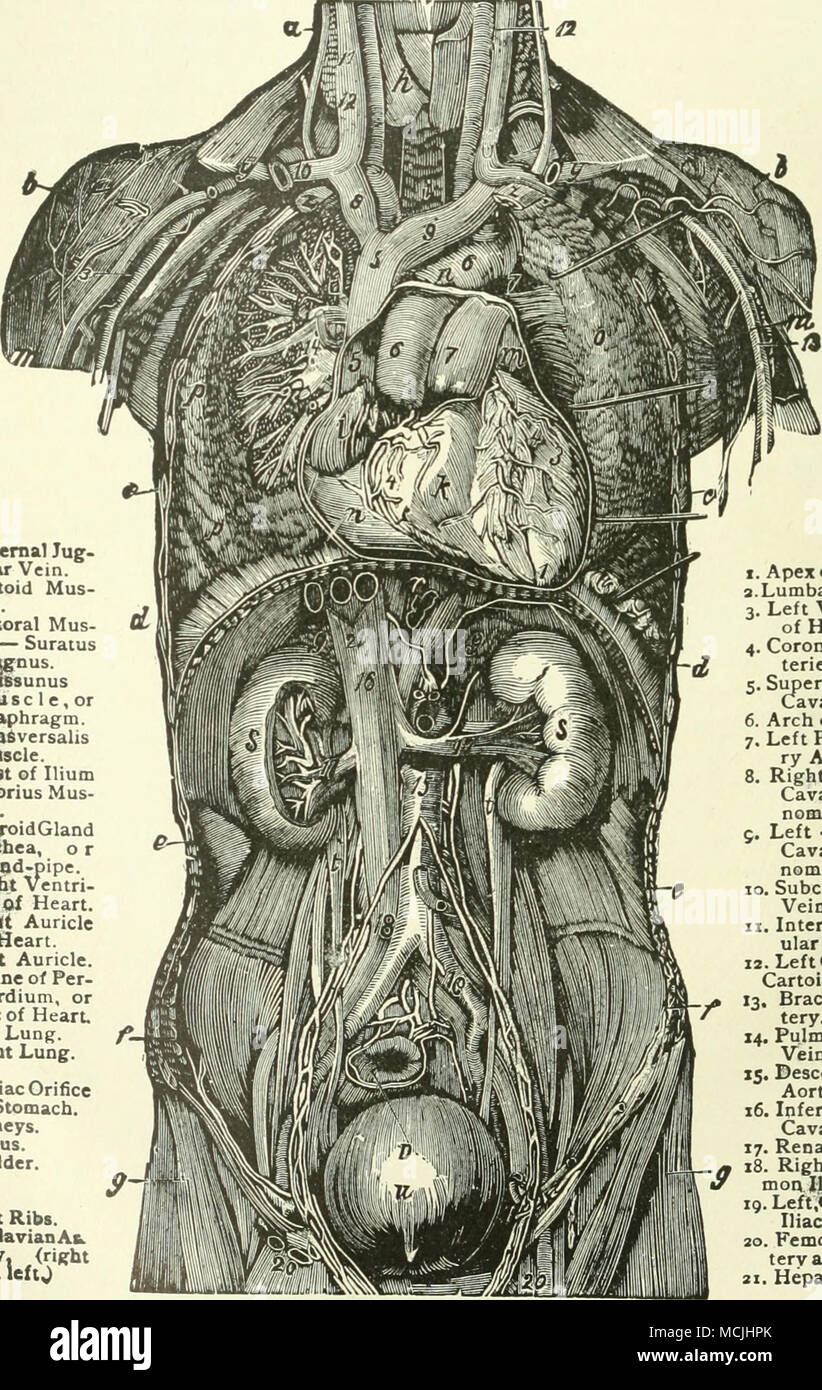 . «. Eaternal Tug- •Uu- Vein. *. DeHoid Mus- cle. #. Pmtoral Mus- cle— Suratus Magnus 4. L4tt68unus Mus Diaphragm 4. TV»a6versahs Mascle. /! jOrest of Ihum f. ^rtorius Mus- *. lliyroidGland •*. Trachea, o r Wiod-pipe. *. Rte^t Ventn- flft of Heart. /. Rinjt Auricle of Heart m. Vftx. Auricle. n.Ott^ne of Per- icardium, or Sac of Heart, #. Left Lung. /. Right Lung. 9- r. Cardiac Orifice of Stomach. *. Kidneys. t. Uretus. w. Bladder. f. Flnt Ribs. «> Soodavian At. t^. .(right I. Apex of Heart 3. Lu mbar Glanda 3. Left Ventrlel* of Heart. 4. Coronary Ar- teries, 5. Superior V««» Cava. (Vein.) 6 Stock Photohttps://www.alamy.com/image-license-details/?v=1https://www.alamy.com/eaternal-tug-uu-vein-dehoid-mus-cle-pmtoral-mus-cle-suratus-magnus-4-l4tt68unus-mus-diaphragm-4-tva6versahs-mascle-!-jorest-of-ihum-f-rtorius-mus-lliyroidgland-trachea-o-r-wiod-pipe-rtet-ventn-flft-of-heart-rinjt-auricle-of-heart-m-vftx-auricle-nottne-of-per-icardium-or-sac-of-heart-left-lung-right-lung-9-r-cardiac-orifice-of-stomach-kidneys-t-uretus-w-bladder-f-flnt-ribs-gt-soodavian-at-t-right-i-apex-of-heart-3-lu-mbar-glanda-3-left-ventrlel-of-heart-4-coronary-ar-teries-5-superior-v-cava-vein-6-image179888651.html
RMMCJHPK–. «. Eaternal Tug- •Uu- Vein. *. DeHoid Mus- cle. #. Pmtoral Mus- cle— Suratus Magnus 4. L4tt68unus Mus Diaphragm 4. TV»a6versahs Mascle. /! jOrest of Ihum f. ^rtorius Mus- *. lliyroidGland •*. Trachea, o r Wiod-pipe. *. Rte^t Ventn- flft of Heart. /. Rinjt Auricle of Heart m. Vftx. Auricle. n.Ott^ne of Per- icardium, or Sac of Heart, #. Left Lung. /. Right Lung. 9- r. Cardiac Orifice of Stomach. *. Kidneys. t. Uretus. w. Bladder. f. Flnt Ribs. «> Soodavian At. t^. .(right I. Apex of Heart 3. Lu mbar Glanda 3. Left Ventrlel* of Heart. 4. Coronary Ar- teries, 5. Superior V««» Cava. (Vein.) 6
 Secondary tuberculosis infection. Computer illustration showing small-sized solid nodular mass located in the upper lobe of right lung near lung apex. Image background represents illustration of Mycobacterium tuberculosis bacteria, the causative agent of tuberculosis. Stock Photohttps://www.alamy.com/image-license-details/?v=1https://www.alamy.com/secondary-tuberculosis-infection-computer-illustration-showing-small-sized-solid-nodular-mass-located-in-the-upper-lobe-of-right-lung-near-lung-apex-image-background-represents-illustration-of-mycobacterium-tuberculosis-bacteria-the-causative-agent-of-tuberculosis-image213574524.html
RFPBD4DG–Secondary tuberculosis infection. Computer illustration showing small-sized solid nodular mass located in the upper lobe of right lung near lung apex. Image background represents illustration of Mycobacterium tuberculosis bacteria, the causative agent of tuberculosis.
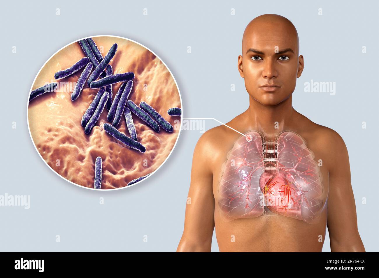 Secondary tuberculosis infection and close-up view of Mycobacterium tuberculosis bacteria, the causative agent of tuberculosis. Computer illustration Stock Photohttps://www.alamy.com/image-license-details/?v=1https://www.alamy.com/secondary-tuberculosis-infection-and-close-up-view-of-mycobacterium-tuberculosis-bacteria-the-causative-agent-of-tuberculosis-computer-illustration-image555169774.html
RF2R764KX–Secondary tuberculosis infection and close-up view of Mycobacterium tuberculosis bacteria, the causative agent of tuberculosis. Computer illustration
 Clinical tuberculosis . Effect of Spasm and Degeneration.—A con-crete example of the way this reflex acts is as follows. Suppose anindividual is suffering from an active tuberculous infiltration of theleft apex. The lung, being supplied by sympathetic fibers whichoriginate in the upper five or six thoracic segments of the cord, wheninflamed sends impulses back to the cord over the sensory sympa-thetics whose cell bodies lie in the posterior root ganglia correspond-ing to these segments. Here the impulses are transmitted to otherneurons which carry them upward in the cord to the cervical seg-me Stock Photohttps://www.alamy.com/image-license-details/?v=1https://www.alamy.com/clinical-tuberculosis-effect-of-spasm-and-degenerationa-con-crete-example-of-the-way-this-reflex-acts-is-as-follows-suppose-anindividual-is-suffering-from-an-active-tuberculous-infiltration-of-theleft-apex-the-lung-being-supplied-by-sympathetic-fibers-whichoriginate-in-the-upper-five-or-six-thoracic-segments-of-the-cord-wheninflamed-sends-impulses-back-to-the-cord-over-the-sensory-sympa-thetics-whose-cell-bodies-lie-in-the-posterior-root-ganglia-correspond-ing-to-these-segments-here-the-impulses-are-transmitted-to-otherneurons-which-carry-them-upward-in-the-cord-to-the-cervical-seg-me-image342967022.html
RM2AWYDX6–Clinical tuberculosis . Effect of Spasm and Degeneration.—A con-crete example of the way this reflex acts is as follows. Suppose anindividual is suffering from an active tuberculous infiltration of theleft apex. The lung, being supplied by sympathetic fibers whichoriginate in the upper five or six thoracic segments of the cord, wheninflamed sends impulses back to the cord over the sensory sympa-thetics whose cell bodies lie in the posterior root ganglia correspond-ing to these segments. Here the impulses are transmitted to otherneurons which carry them upward in the cord to the cervical seg-me
 The great cardiac vein (left coronary vein) is a vein of the heart3d illustration It begins at the apex of the heart and ascends along the anterior in Stock Photohttps://www.alamy.com/image-license-details/?v=1https://www.alamy.com/the-great-cardiac-vein-left-coronary-vein-is-a-vein-of-the-heart3d-illustration-it-begins-at-the-apex-of-the-heart-and-ascends-along-the-anterior-in-image596594793.html
RF2WJH6MW–The great cardiac vein (left coronary vein) is a vein of the heart3d illustration It begins at the apex of the heart and ascends along the anterior in
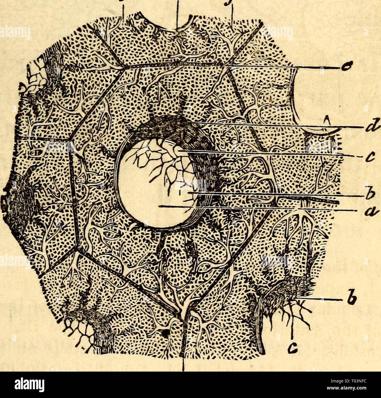 Elementary anatomy and physiology : for colleges, academies, and other schools . elementaryanato00hitc Year: 1869 AND PHYSIOLOGY. 247 are of a conical shape, the apex pointing upwards, the base resting on the Diaphragm, of a pinkish gray color, frequently clotted with black spots, and divided by a deep fissure into two lobes. The right lung is shorter in its Ions; diameter than the left, on account of the liver which raises the right side of the diaphragm higher than the left. The right lung is subdivided again, so that it is really made up of three lobes instead of two. It has also a larger Stock Photohttps://www.alamy.com/image-license-details/?v=1https://www.alamy.com/elementary-anatomy-and-physiology-for-colleges-academies-and-other-schools-elementaryanato00hitc-year-1869-and-physiology-247-are-of-a-conical-shape-the-apex-pointing-upwards-the-base-resting-on-the-diaphragm-of-a-pinkish-gray-color-frequently-clotted-with-black-spots-and-divided-by-a-deep-fissure-into-two-lobes-the-right-lung-is-shorter-in-its-ions-diameter-than-the-left-on-account-of-the-liver-which-raises-the-right-side-of-the-diaphragm-higher-than-the-left-the-right-lung-is-subdivided-again-so-that-it-is-really-made-up-of-three-lobes-instead-of-two-it-has-also-a-larger-image241027904.html
RMT03NFC–Elementary anatomy and physiology : for colleges, academies, and other schools . elementaryanato00hitc Year: 1869 AND PHYSIOLOGY. 247 are of a conical shape, the apex pointing upwards, the base resting on the Diaphragm, of a pinkish gray color, frequently clotted with black spots, and divided by a deep fissure into two lobes. The right lung is shorter in its Ions; diameter than the left, on account of the liver which raises the right side of the diaphragm higher than the left. The right lung is subdivided again, so that it is really made up of three lobes instead of two. It has also a larger
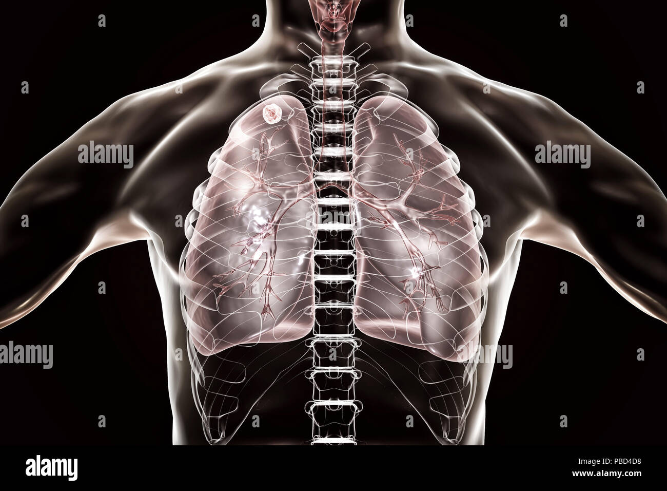 Secondary tuberculosis infection. Computer illustration showing small-sized solid nodular mass located in the upper lobe of right lung near lung apex. Stock Photohttps://www.alamy.com/image-license-details/?v=1https://www.alamy.com/secondary-tuberculosis-infection-computer-illustration-showing-small-sized-solid-nodular-mass-located-in-the-upper-lobe-of-right-lung-near-lung-apex-image213574516.html
RFPBD4D8–Secondary tuberculosis infection. Computer illustration showing small-sized solid nodular mass located in the upper lobe of right lung near lung apex.
 . Operative surgery, for students and practitioners . -Fig. S4.—Outline of Pleura, etc. Front view. ^4., apex of lung and domeof pleura; D, line of diaphragm; H, outline of heart; L, solid lines show theedges of the lungs; P, dotted lines correspond to the edges of the pleura.. — o o —> O «l ° 00 60 ™ ?a 3j£ to ft Stock Photohttps://www.alamy.com/image-license-details/?v=1https://www.alamy.com/operative-surgery-for-students-and-practitioners-fig-s4outline-of-pleura-etc-front-view-4-apex-of-lung-and-domeof-pleura-d-line-of-diaphragm-h-outline-of-heart-l-solid-lines-show-theedges-of-the-lungs-p-dotted-lines-correspond-to-the-edges-of-the-pleura-o-o-gt-o-l-00-60-a-3j-to-ft-image370103597.html
RM2CE3JWH–. Operative surgery, for students and practitioners . -Fig. S4.—Outline of Pleura, etc. Front view. ^4., apex of lung and domeof pleura; D, line of diaphragm; H, outline of heart; L, solid lines show theedges of the lungs; P, dotted lines correspond to the edges of the pleura.. — o o —> O «l ° 00 60 ™ ?a 3j£ to ft
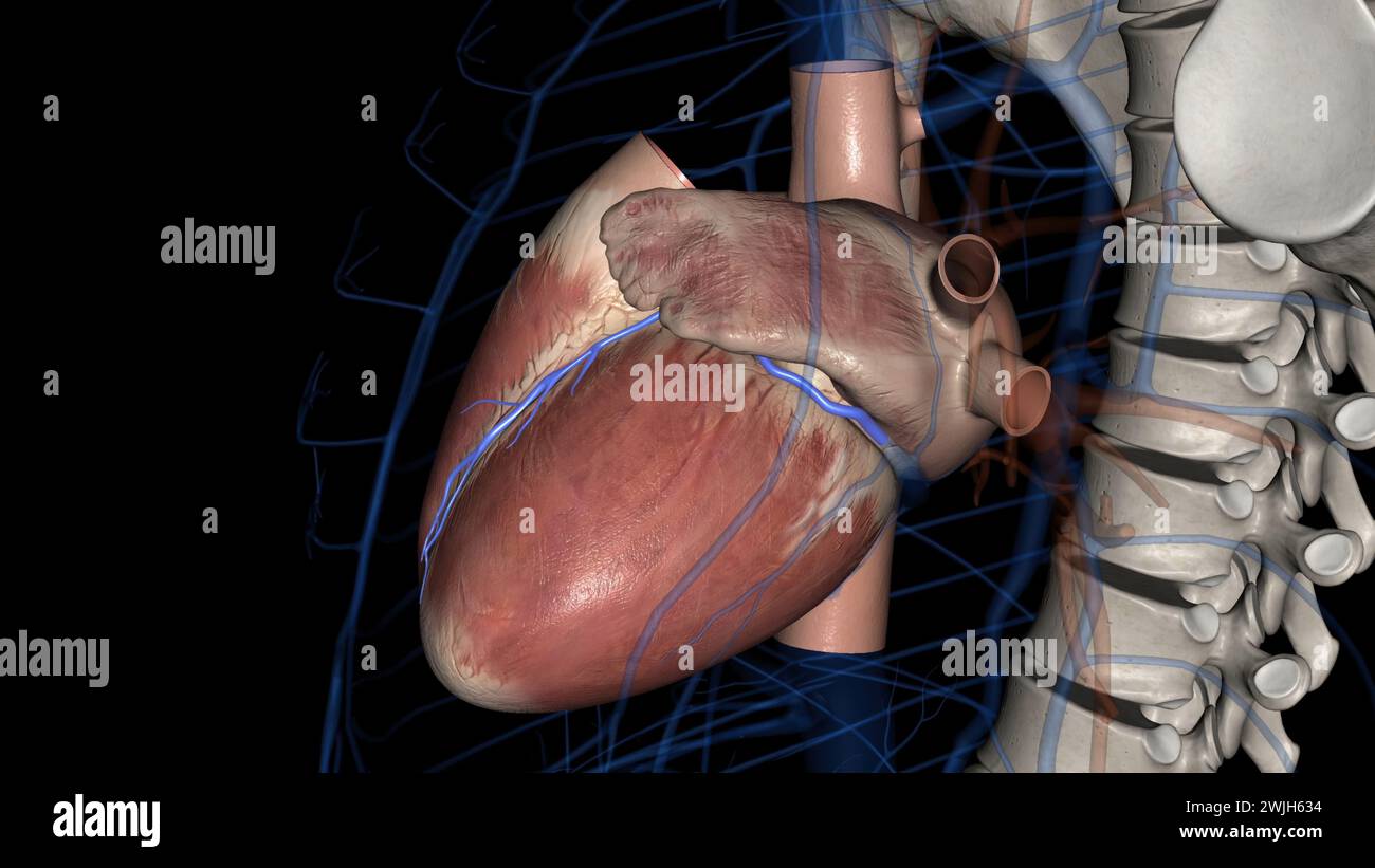 The great cardiac vein (left coronary vein) is a vein of the heart3d illustration It begins at the apex of the heart and ascends along the anterior in Stock Photohttps://www.alamy.com/image-license-details/?v=1https://www.alamy.com/the-great-cardiac-vein-left-coronary-vein-is-a-vein-of-the-heart3d-illustration-it-begins-at-the-apex-of-the-heart-and-ascends-along-the-anterior-in-image596594296.html
RF2WJH634–The great cardiac vein (left coronary vein) is a vein of the heart3d illustration It begins at the apex of the heart and ascends along the anterior in
 The dog in health and The dog in health and in disease : including his origin, history, varieties, breeding, education, and general management in health, and his treatment in disease doginhealthindis00mill Year: 1895 210 THE DOG IN DISEASE. ventilation of the blood pass, and extends from the nos- trils to the air-cells. Briefly, the tract consists of a mucous membrane cov- ered with epithelial cells, abounding in blood-vessels and lin- FiG. 12.—Lungs, Anterior View (Sappey). 1, upper lobe of left lung ; 2, lower lobe ; 3, fissure ; 4, notch corresponding to apex of heart; 5, pericardium ; Stock Photohttps://www.alamy.com/image-license-details/?v=1https://www.alamy.com/the-dog-in-health-and-the-dog-in-health-and-in-disease-including-his-origin-history-varieties-breeding-education-and-general-management-in-health-and-his-treatment-in-disease-doginhealthindis00mill-year-1895-210-the-dog-in-disease-ventilation-of-the-blood-pass-and-extends-from-the-nos-trils-to-the-air-cells-briefly-the-tract-consists-of-a-mucous-membrane-cov-ered-with-epithelial-cells-abounding-in-blood-vessels-and-lin-fig-12lungs-anterior-view-sappey-1-upper-lobe-of-left-lung-2-lower-lobe-3-fissure-4-notch-corresponding-to-apex-of-heart-5-pericardium-image241950022.html
RMT1HNM6–The dog in health and The dog in health and in disease : including his origin, history, varieties, breeding, education, and general management in health, and his treatment in disease doginhealthindis00mill Year: 1895 210 THE DOG IN DISEASE. ventilation of the blood pass, and extends from the nos- trils to the air-cells. Briefly, the tract consists of a mucous membrane cov- ered with epithelial cells, abounding in blood-vessels and lin- FiG. 12.—Lungs, Anterior View (Sappey). 1, upper lobe of left lung ; 2, lower lobe ; 3, fissure ; 4, notch corresponding to apex of heart; 5, pericardium ;
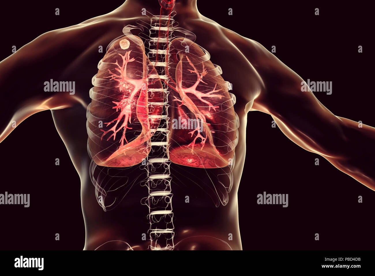 Secondary tuberculosis infection. Computer illustration showing small-sized solid nodular mass located in the upper lobe of right lung near lung apex. Stock Photohttps://www.alamy.com/image-license-details/?v=1https://www.alamy.com/secondary-tuberculosis-infection-computer-illustration-showing-small-sized-solid-nodular-mass-located-in-the-upper-lobe-of-right-lung-near-lung-apex-image213574519.html
RFPBD4DB–Secondary tuberculosis infection. Computer illustration showing small-sized solid nodular mass located in the upper lobe of right lung near lung apex.
 . Anatomy of the woodchuck (Marmota monax). Woodchuck; Mammals. 106 Anatomy of the Woodchuck, Marmota monax. Fig. 6-7. Thorax, left lateral view, intercostal muscles removed. 1 dorsal margin of left lung, 2 basal margin of left lung, 3 diaphragm, 4 undivided left lung, 5 ventral margin of left lung, 6 apex of lung.. Please note that these images are extracted from scanned page images that may have been digitally enhanced for readability - coloration and appearance of these illustrations may not perfectly resemble the original work.. Bezuidenhout, A. J. (Abraham Johannes), 1942-; Evans, Howard Stock Photohttps://www.alamy.com/image-license-details/?v=1https://www.alamy.com/anatomy-of-the-woodchuck-marmota-monax-woodchuck-mammals-106-anatomy-of-the-woodchuck-marmota-monax-fig-6-7-thorax-left-lateral-view-intercostal-muscles-removed-1-dorsal-margin-of-left-lung-2-basal-margin-of-left-lung-3-diaphragm-4-undivided-left-lung-5-ventral-margin-of-left-lung-6-apex-of-lung-please-note-that-these-images-are-extracted-from-scanned-page-images-that-may-have-been-digitally-enhanced-for-readability-coloration-and-appearance-of-these-illustrations-may-not-perfectly-resemble-the-original-work-bezuidenhout-a-j-abraham-johannes-1942-evans-howard-image236799807.html
RMRN74FY–. Anatomy of the woodchuck (Marmota monax). Woodchuck; Mammals. 106 Anatomy of the Woodchuck, Marmota monax. Fig. 6-7. Thorax, left lateral view, intercostal muscles removed. 1 dorsal margin of left lung, 2 basal margin of left lung, 3 diaphragm, 4 undivided left lung, 5 ventral margin of left lung, 6 apex of lung.. Please note that these images are extracted from scanned page images that may have been digitally enhanced for readability - coloration and appearance of these illustrations may not perfectly resemble the original work.. Bezuidenhout, A. J. (Abraham Johannes), 1942-; Evans, Howard
 The great cardiac vein (left coronary vein) is a vein of the heart3d illustration It begins at the apex of the heart and ascends along the anterior in Stock Photohttps://www.alamy.com/image-license-details/?v=1https://www.alamy.com/the-great-cardiac-vein-left-coronary-vein-is-a-vein-of-the-heart3d-illustration-it-begins-at-the-apex-of-the-heart-and-ascends-along-the-anterior-in-image596583235.html
RF2WJGM03–The great cardiac vein (left coronary vein) is a vein of the heart3d illustration It begins at the apex of the heart and ascends along the anterior in
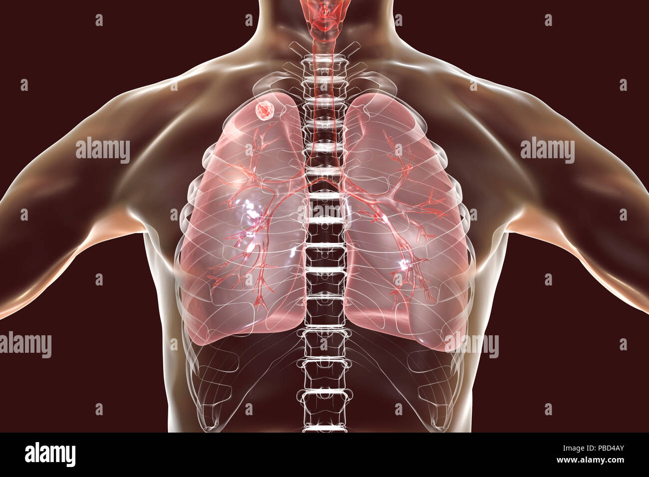 Secondary tuberculosis infection. Computer illustration showing small-sized solid nodular mass located in the upper lobe of right lung near lung apex. Stock Photohttps://www.alamy.com/image-license-details/?v=1https://www.alamy.com/secondary-tuberculosis-infection-computer-illustration-showing-small-sized-solid-nodular-mass-located-in-the-upper-lobe-of-right-lung-near-lung-apex-image213574451.html
RFPBD4AY–Secondary tuberculosis infection. Computer illustration showing small-sized solid nodular mass located in the upper lobe of right lung near lung apex.
 . Anatomy of the woodchuck (Marmota monax). Woodchuck; Mammals. Fig. 6-7. Thorax, left lateral view, intercostal muscles removed. 1 dorsal margin of left lung, 2 basal margin of left lung, 3 diaphragm, 4 undivided left lung, 5 ventral margin of left lung, 6 apex of lung.. 10^ fi Fig. 6-8. Thorax, ventral view, intercostal muscles removed. 1 manubrium, 2 apex of lung, 3 undivided left lung, 4 heart, 5 diaphragmatic surface on the base of lung, 6 diaphragm, 7 cranial epigastric a., 8 xiphoid cartilage, 9 costal arch, 10 caudal lobe of right lung, 1 1 middle lobe of right lung, 12 internal thorac Stock Photohttps://www.alamy.com/image-license-details/?v=1https://www.alamy.com/anatomy-of-the-woodchuck-marmota-monax-woodchuck-mammals-fig-6-7-thorax-left-lateral-view-intercostal-muscles-removed-1-dorsal-margin-of-left-lung-2-basal-margin-of-left-lung-3-diaphragm-4-undivided-left-lung-5-ventral-margin-of-left-lung-6-apex-of-lung-10-fi-fig-6-8-thorax-ventral-view-intercostal-muscles-removed-1-manubrium-2-apex-of-lung-3-undivided-left-lung-4-heart-5-diaphragmatic-surface-on-the-base-of-lung-6-diaphragm-7-cranial-epigastric-a-8-xiphoid-cartilage-9-costal-arch-10-caudal-lobe-of-right-lung-1-1-middle-lobe-of-right-lung-12-internal-thorac-image236799795.html
RMRN74FF–. Anatomy of the woodchuck (Marmota monax). Woodchuck; Mammals. Fig. 6-7. Thorax, left lateral view, intercostal muscles removed. 1 dorsal margin of left lung, 2 basal margin of left lung, 3 diaphragm, 4 undivided left lung, 5 ventral margin of left lung, 6 apex of lung.. 10^ fi Fig. 6-8. Thorax, ventral view, intercostal muscles removed. 1 manubrium, 2 apex of lung, 3 undivided left lung, 4 heart, 5 diaphragmatic surface on the base of lung, 6 diaphragm, 7 cranial epigastric a., 8 xiphoid cartilage, 9 costal arch, 10 caudal lobe of right lung, 1 1 middle lobe of right lung, 12 internal thorac
 The great cardiac vein (left coronary vein) is a vein of the heart3d illustration It begins at the apex of the heart and ascends along the anterior in Stock Photohttps://www.alamy.com/image-license-details/?v=1https://www.alamy.com/the-great-cardiac-vein-left-coronary-vein-is-a-vein-of-the-heart3d-illustration-it-begins-at-the-apex-of-the-heart-and-ascends-along-the-anterior-in-image596577062.html
RF2WJGC3J–The great cardiac vein (left coronary vein) is a vein of the heart3d illustration It begins at the apex of the heart and ascends along the anterior in
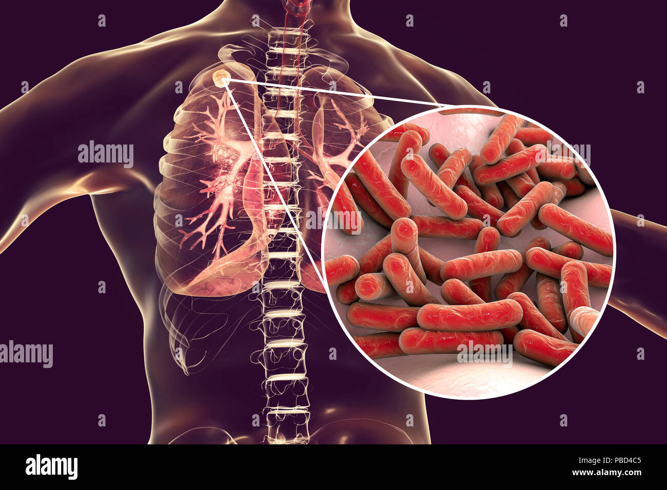 Secondary tuberculosis infection and close-up view of Mycobacterium tuberculosis bacteria, the causative agent of tuberculosis. Computer illustration showing small-sized solid nodular mass located in the upper lobe of right lung near lung apex. Stock Photohttps://www.alamy.com/image-license-details/?v=1https://www.alamy.com/secondary-tuberculosis-infection-and-close-up-view-of-mycobacterium-tuberculosis-bacteria-the-causative-agent-of-tuberculosis-computer-illustration-showing-small-sized-solid-nodular-mass-located-in-the-upper-lobe-of-right-lung-near-lung-apex-image213574485.html
RFPBD4C5–Secondary tuberculosis infection and close-up view of Mycobacterium tuberculosis bacteria, the causative agent of tuberculosis. Computer illustration showing small-sized solid nodular mass located in the upper lobe of right lung near lung apex.
 . Manual of antenatal pathology and hygiene : the foetus. Plate u upper part of Body of first dorsal vertebra. Spimtl cord. Apex o/ Right lung. Apex o/ Left lung.. Stock Photohttps://www.alamy.com/image-license-details/?v=1https://www.alamy.com/manual-of-antenatal-pathology-and-hygiene-the-foetus-plate-u-upper-part-of-body-of-first-dorsal-vertebra-spimtl-cord-apex-o-right-lung-apex-o-left-lung-image336749825.html
RM2AFT7RD–. Manual of antenatal pathology and hygiene : the foetus. Plate u upper part of Body of first dorsal vertebra. Spimtl cord. Apex o/ Right lung. Apex o/ Left lung..
 The great cardiac vein (left coronary vein) is a vein of the heart3d illustration It begins at the apex of the heart and ascends along the anterior in Stock Photohttps://www.alamy.com/image-license-details/?v=1https://www.alamy.com/the-great-cardiac-vein-left-coronary-vein-is-a-vein-of-the-heart3d-illustration-it-begins-at-the-apex-of-the-heart-and-ascends-along-the-anterior-in-image596594809.html
RF2WJH6ND–The great cardiac vein (left coronary vein) is a vein of the heart3d illustration It begins at the apex of the heart and ascends along the anterior in
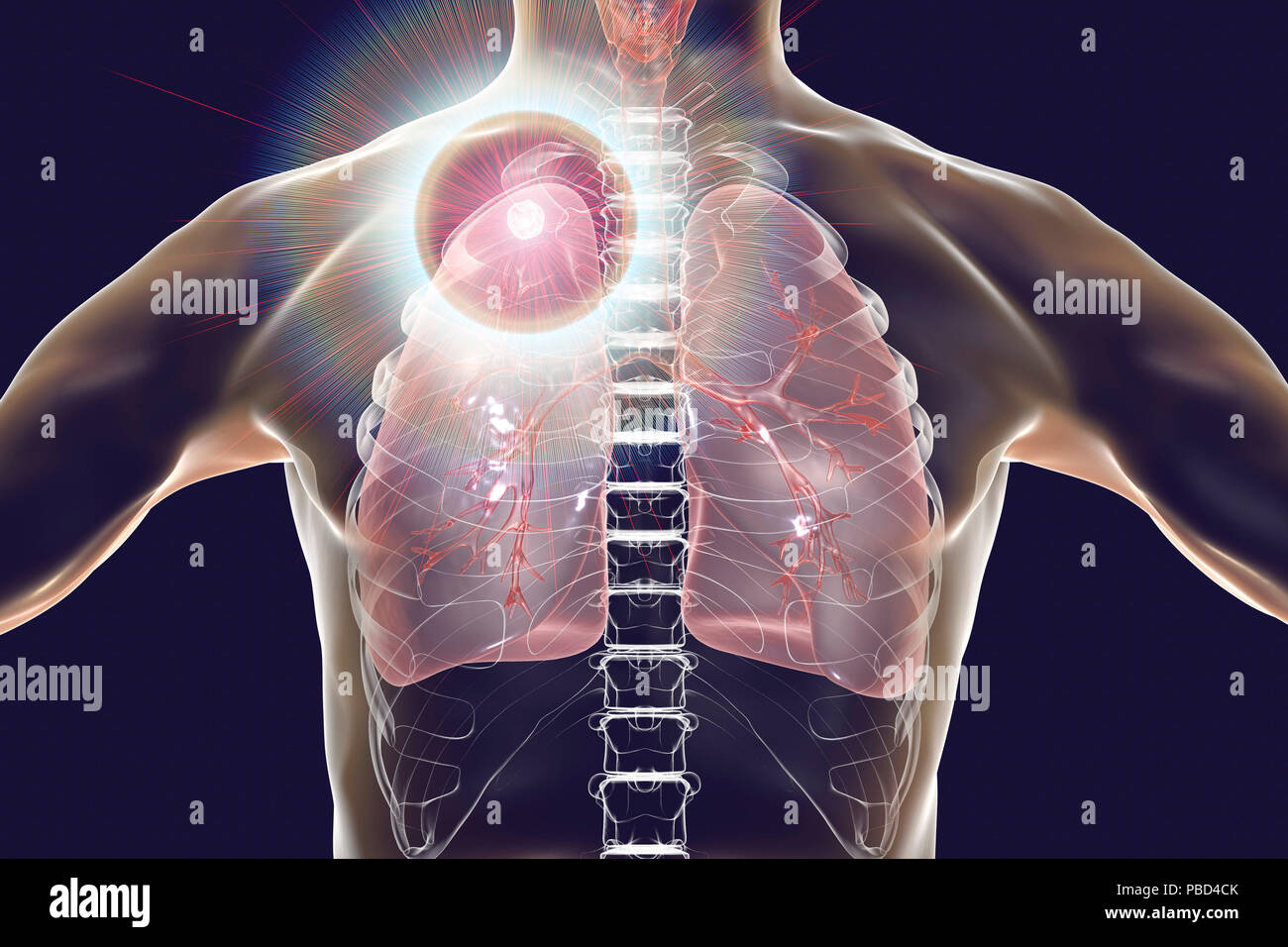 Secondary tuberculosis infection and close-up view of Mycobacterium tuberculosis bacteria, the causative agent of tuberculosis. Computer illustration showing small-sized solid nodular mass located in the upper lobe of right lung near lung apex. Stock Photohttps://www.alamy.com/image-license-details/?v=1https://www.alamy.com/secondary-tuberculosis-infection-and-close-up-view-of-mycobacterium-tuberculosis-bacteria-the-causative-agent-of-tuberculosis-computer-illustration-showing-small-sized-solid-nodular-mass-located-in-the-upper-lobe-of-right-lung-near-lung-apex-image213574499.html
RFPBD4CK–Secondary tuberculosis infection and close-up view of Mycobacterium tuberculosis bacteria, the causative agent of tuberculosis. Computer illustration showing small-sized solid nodular mass located in the upper lobe of right lung near lung apex.
 Atlas and text-book of topographic and applied anatomy . M. iliopsoas A. femoralis . anonyma sinistraA. sulclavia sin.Apex pulmonis Incisura cardiaca pulmonis sin.Incisura interlobaris Diaphragma Lig. teres Funiculus spermaticus Tuber ischiadicum VesicaForamen obturatum Left lung. TubentsiLy of ischium Stock Photohttps://www.alamy.com/image-license-details/?v=1https://www.alamy.com/atlas-and-text-book-of-topographic-and-applied-anatomy-m-iliopsoas-a-femoralis-anonyma-sinistraa-sulclavia-sinapex-pulmonis-incisura-cardiaca-pulmonis-sinincisura-interlobaris-diaphragma-lig-teres-funiculus-spermaticus-tuber-ischiadicum-vesicaforamen-obturatum-left-lung-tubentsily-of-ischium-image338250009.html
RM2AJ8H9D–Atlas and text-book of topographic and applied anatomy . M. iliopsoas A. femoralis . anonyma sinistraA. sulclavia sin.Apex pulmonis Incisura cardiaca pulmonis sin.Incisura interlobaris Diaphragma Lig. teres Funiculus spermaticus Tuber ischiadicum VesicaForamen obturatum Left lung. TubentsiLy of ischium
 The great cardiac vein (left coronary vein) is a vein of the heart3d illustration It begins at the apex of the heart and ascends along the anterior in Stock Photohttps://www.alamy.com/image-license-details/?v=1https://www.alamy.com/the-great-cardiac-vein-left-coronary-vein-is-a-vein-of-the-heart3d-illustration-it-begins-at-the-apex-of-the-heart-and-ascends-along-the-anterior-in-image596575612.html
RF2WJGA7T–The great cardiac vein (left coronary vein) is a vein of the heart3d illustration It begins at the apex of the heart and ascends along the anterior in
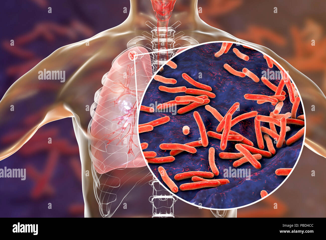 Secondary tuberculosis infection and close-up view of Mycobacterium tuberculosis bacteria, the causative agent of tuberculosis. Computer illustration showing small-sized solid nodular mass located in the upper lobe of right lung near lung apex. Stock Photohttps://www.alamy.com/image-license-details/?v=1https://www.alamy.com/secondary-tuberculosis-infection-and-close-up-view-of-mycobacterium-tuberculosis-bacteria-the-causative-agent-of-tuberculosis-computer-illustration-showing-small-sized-solid-nodular-mass-located-in-the-upper-lobe-of-right-lung-near-lung-apex-image213574492.html
RFPBD4CC–Secondary tuberculosis infection and close-up view of Mycobacterium tuberculosis bacteria, the causative agent of tuberculosis. Computer illustration showing small-sized solid nodular mass located in the upper lobe of right lung near lung apex.
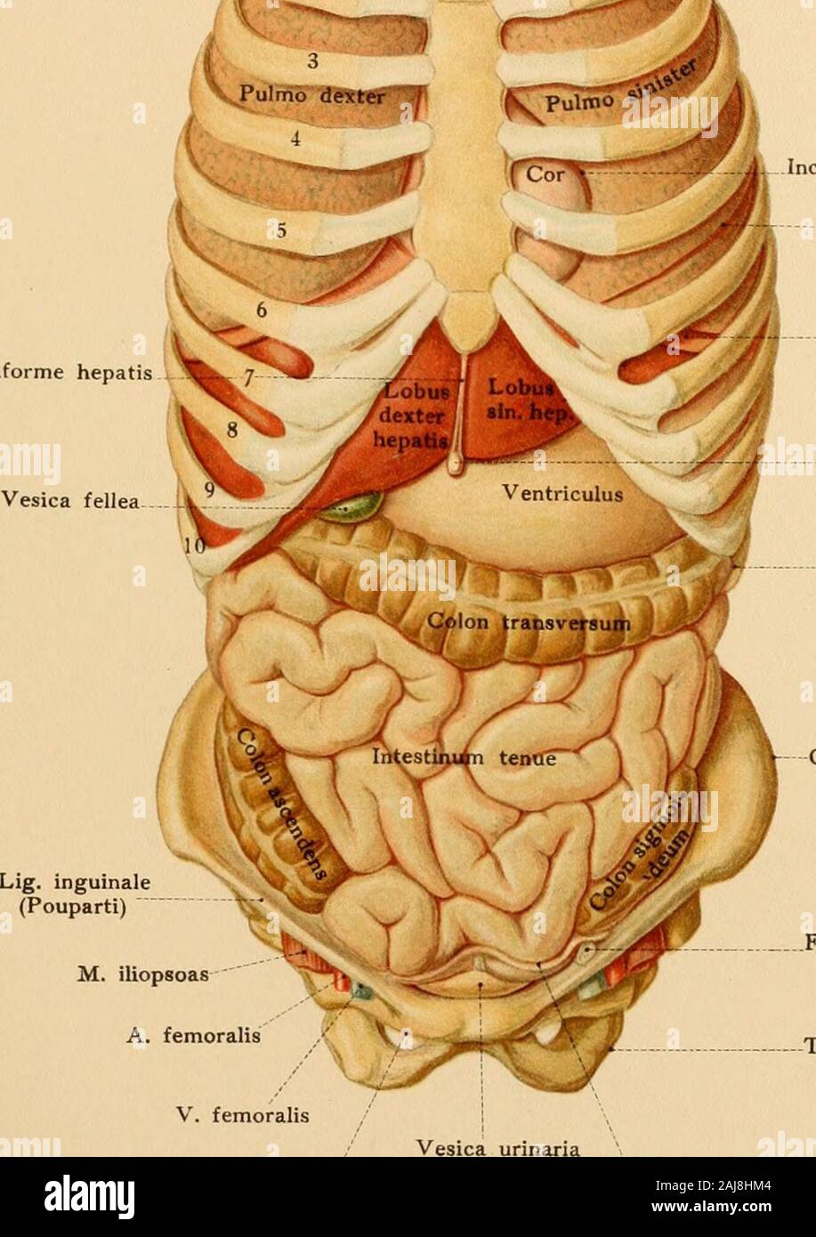 Atlas and text-book of topographic and applied anatomy . V. anonyma dextra : :- - Lig. falciforme hepat. M. iliopsoas A. femoralis . anonyma sinistraA. sulclavia sin.Apex pulmonis Incisura cardiaca pulmonis sin.Incisura interlobaris Diaphragma Lig. teres Funiculus spermaticus Tuber ischiadicum VesicaForamen obturatum Left lung Stock Photohttps://www.alamy.com/image-license-details/?v=1https://www.alamy.com/atlas-and-text-book-of-topographic-and-applied-anatomy-v-anonyma-dextra-lig-falciforme-hepat-m-iliopsoas-a-femoralis-anonyma-sinistraa-sulclavia-sinapex-pulmonis-incisura-cardiaca-pulmonis-sinincisura-interlobaris-diaphragma-lig-teres-funiculus-spermaticus-tuber-ischiadicum-vesicaforamen-obturatum-left-lung-image338250308.html
RM2AJ8HM4–Atlas and text-book of topographic and applied anatomy . V. anonyma dextra : :- - Lig. falciforme hepat. M. iliopsoas A. femoralis . anonyma sinistraA. sulclavia sin.Apex pulmonis Incisura cardiaca pulmonis sin.Incisura interlobaris Diaphragma Lig. teres Funiculus spermaticus Tuber ischiadicum VesicaForamen obturatum Left lung
 The great cardiac vein (left coronary vein) is a vein of the heart3d illustration It begins at the apex of the heart and ascends along the anterior in Stock Photohttps://www.alamy.com/image-license-details/?v=1https://www.alamy.com/the-great-cardiac-vein-left-coronary-vein-is-a-vein-of-the-heart3d-illustration-it-begins-at-the-apex-of-the-heart-and-ascends-along-the-anterior-in-image596595671.html
RF2WJH7T7–The great cardiac vein (left coronary vein) is a vein of the heart3d illustration It begins at the apex of the heart and ascends along the anterior in
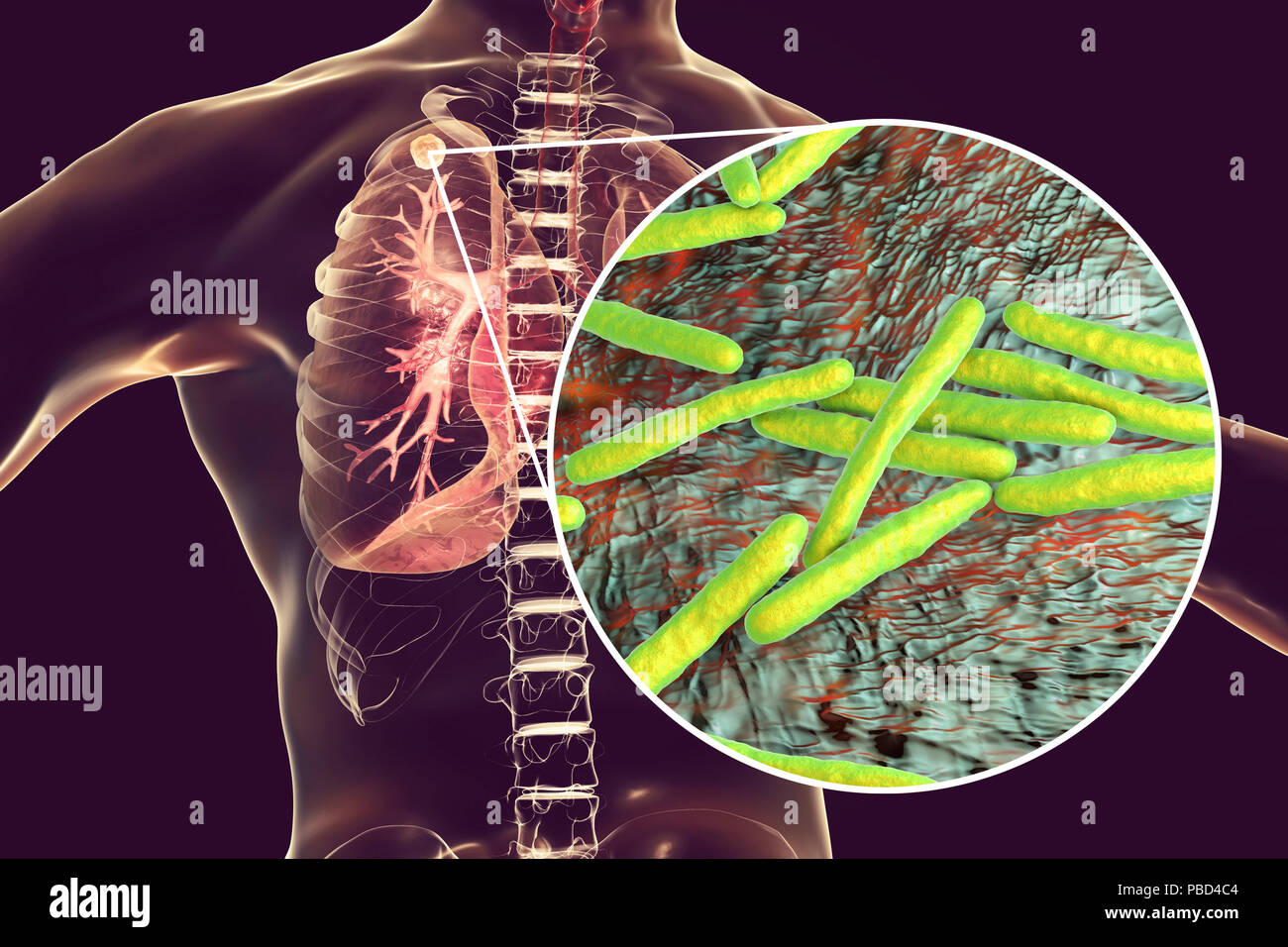 Secondary tuberculosis infection and close-up view of Mycobacterium tuberculosis bacteria, the causative agent of tuberculosis. Computer illustration showing small-sized solid nodular mass located in the upper lobe of right lung near lung apex. Stock Photohttps://www.alamy.com/image-license-details/?v=1https://www.alamy.com/secondary-tuberculosis-infection-and-close-up-view-of-mycobacterium-tuberculosis-bacteria-the-causative-agent-of-tuberculosis-computer-illustration-showing-small-sized-solid-nodular-mass-located-in-the-upper-lobe-of-right-lung-near-lung-apex-image213574484.html
RFPBD4C4–Secondary tuberculosis infection and close-up view of Mycobacterium tuberculosis bacteria, the causative agent of tuberculosis. Computer illustration showing small-sized solid nodular mass located in the upper lobe of right lung near lung apex.
 Atlas and text-book of topographic and applied anatomy . V. anonyma dextra : :- - Lig. falciforme hepat. M. iliopsoas A. femoralis . anonyma sinistraA. sulclavia sin.Apex pulmonis Incisura cardiaca pulmonis sin.Incisura interlobaris Diaphragma Lig. teres Funiculus spermaticus Tuber ischiadicum VesicaForamen obturatum Left lung Stock Photohttps://www.alamy.com/image-license-details/?v=1https://www.alamy.com/atlas-and-text-book-of-topographic-and-applied-anatomy-v-anonyma-dextra-lig-falciforme-hepat-m-iliopsoas-a-femoralis-anonyma-sinistraa-sulclavia-sinapex-pulmonis-incisura-cardiaca-pulmonis-sinincisura-interlobaris-diaphragma-lig-teres-funiculus-spermaticus-tuber-ischiadicum-vesicaforamen-obturatum-left-lung-image338259001.html
RM2AJ90PH–Atlas and text-book of topographic and applied anatomy . V. anonyma dextra : :- - Lig. falciforme hepat. M. iliopsoas A. femoralis . anonyma sinistraA. sulclavia sin.Apex pulmonis Incisura cardiaca pulmonis sin.Incisura interlobaris Diaphragma Lig. teres Funiculus spermaticus Tuber ischiadicum VesicaForamen obturatum Left lung
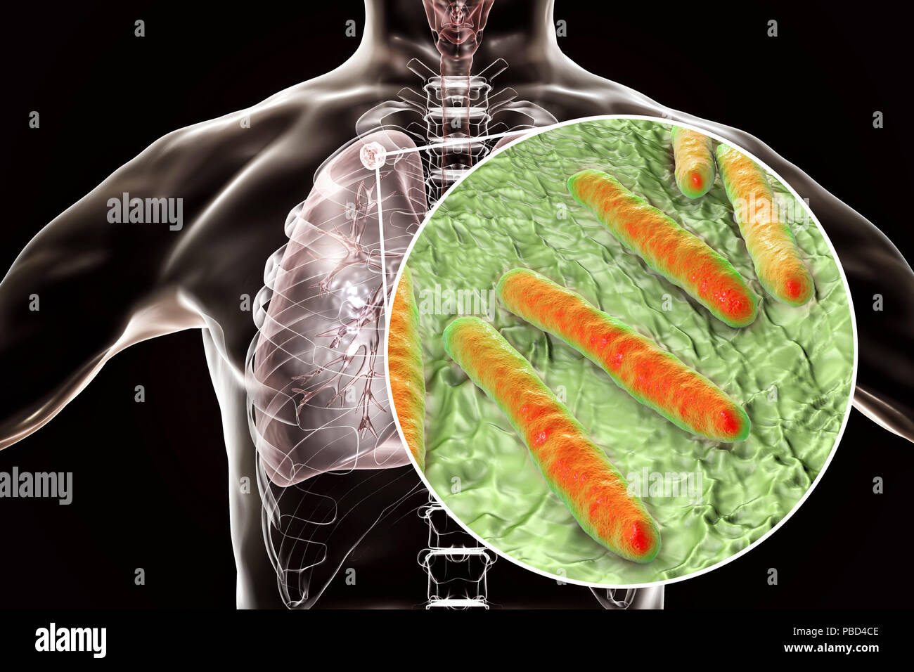 Secondary tuberculosis infection and close-up view of Mycobacterium tuberculosis bacteria, the causative agent of tuberculosis. Computer illustration showing small-sized solid nodular mass located in the upper lobe of right lung near lung apex. Stock Photohttps://www.alamy.com/image-license-details/?v=1https://www.alamy.com/secondary-tuberculosis-infection-and-close-up-view-of-mycobacterium-tuberculosis-bacteria-the-causative-agent-of-tuberculosis-computer-illustration-showing-small-sized-solid-nodular-mass-located-in-the-upper-lobe-of-right-lung-near-lung-apex-image213574494.html
RFPBD4CE–Secondary tuberculosis infection and close-up view of Mycobacterium tuberculosis bacteria, the causative agent of tuberculosis. Computer illustration showing small-sized solid nodular mass located in the upper lobe of right lung near lung apex.
 An atlas of human anatomy for students and physicians . Middle lobe Lobus medius Anterior border Maiso anterior Apex of the lung e pulmonis Upper lobe Lobus superior Outer or costalsurface Facies costalis Interlobar fissure Incisura interlobaris . Lower lobe Lobus inferior Lower lobe Lobus inferior. 1 Inferior border Mar£;o inferiorLower, phrenic, or diaphragmaticsurface—Facies diaphragmatica Stock Photohttps://www.alamy.com/image-license-details/?v=1https://www.alamy.com/an-atlas-of-human-anatomy-for-students-and-physicians-middle-lobe-lobus-medius-anterior-border-maiso-anterior-apex-of-the-lung-e-pulmonis-upper-lobe-lobus-superior-outer-or-costalsurface-facies-costalis-interlobar-fissure-incisura-interlobaris-lower-lobe-lobus-inferior-lower-lobe-lobus-inferior-1-inferior-border-maro-inferiorlower-phrenic-or-diaphragmaticsurfacefacies-diaphragmatica-image338317461.html
RM2AJBKAD–An atlas of human anatomy for students and physicians . Middle lobe Lobus medius Anterior border Maiso anterior Apex of the lung e pulmonis Upper lobe Lobus superior Outer or costalsurface Facies costalis Interlobar fissure Incisura interlobaris . Lower lobe Lobus inferior Lower lobe Lobus inferior. 1 Inferior border Mar£;o inferiorLower, phrenic, or diaphragmaticsurface—Facies diaphragmatica
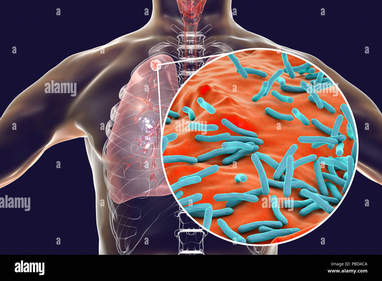 Secondary tuberculosis infection and close-up view of Mycobacterium tuberculosis bacteria, the causative agent of tuberculosis. Computer illustration showing small-sized solid nodular mass located in the upper lobe of right lung near lung apex. Stock Photohttps://www.alamy.com/image-license-details/?v=1https://www.alamy.com/secondary-tuberculosis-infection-and-close-up-view-of-mycobacterium-tuberculosis-bacteria-the-causative-agent-of-tuberculosis-computer-illustration-showing-small-sized-solid-nodular-mass-located-in-the-upper-lobe-of-right-lung-near-lung-apex-image213574490.html
RFPBD4CA–Secondary tuberculosis infection and close-up view of Mycobacterium tuberculosis bacteria, the causative agent of tuberculosis. Computer illustration showing small-sized solid nodular mass located in the upper lobe of right lung near lung apex.
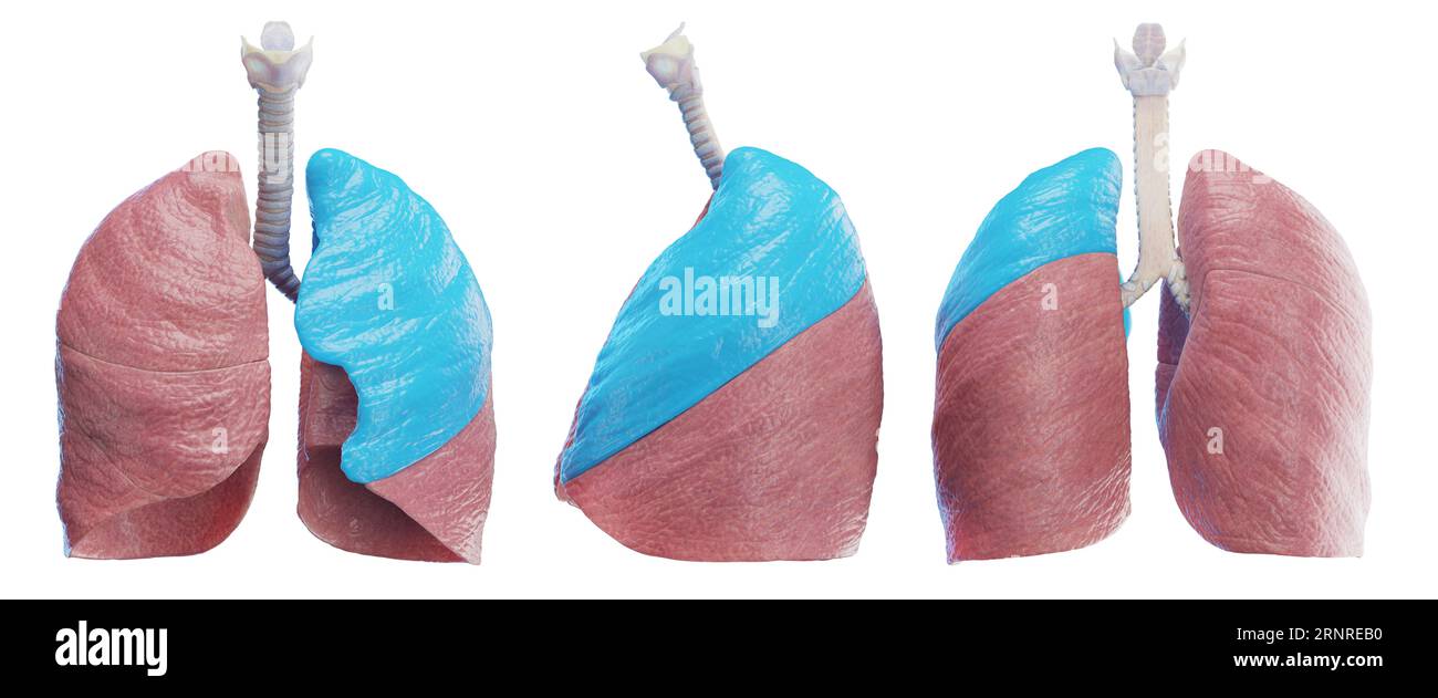 Left lung, illustration Stock Photohttps://www.alamy.com/image-license-details/?v=1https://www.alamy.com/left-lung-illustration-image564155732.html
RF2RNREB0–Left lung, illustration
 Clinical tuberculosis . Fig. 21.—Illustrating schciTiatically the movements of the various portions of the lung.The degree of motion is indicated according to the shading—the darker the portion, theless the motion. (Tendeloo.) time in the life of the child when it should show a natural re-sistance and is also the time when the child which has becomealready infected with the tubercle bacillus, is best protectedby the specific immunity which it has developed. Consideration of the predisposition of the apex of the adultlung to tuberculous infection has called forth a great deal ofdiscussion and m Stock Photohttps://www.alamy.com/image-license-details/?v=1https://www.alamy.com/clinical-tuberculosis-fig-21illustrating-schcitiatically-the-movements-of-the-various-portions-of-the-lungthe-degree-of-motion-is-indicated-according-to-the-shadingthe-darker-the-portion-theless-the-motion-tendeloo-time-in-the-life-of-the-child-when-it-should-show-a-natural-re-sistance-and-is-also-the-time-when-the-child-which-has-becomealready-infected-with-the-tubercle-bacillus-is-best-protectedby-the-specific-immunity-which-it-has-developed-consideration-of-the-predisposition-of-the-apex-of-the-adultlung-to-tuberculous-infection-has-called-forth-a-great-deal-ofdiscussion-and-m-image343004287.html
RM2AX15D3–Clinical tuberculosis . Fig. 21.—Illustrating schciTiatically the movements of the various portions of the lung.The degree of motion is indicated according to the shading—the darker the portion, theless the motion. (Tendeloo.) time in the life of the child when it should show a natural re-sistance and is also the time when the child which has becomealready infected with the tubercle bacillus, is best protectedby the specific immunity which it has developed. Consideration of the predisposition of the apex of the adultlung to tuberculous infection has called forth a great deal ofdiscussion and m
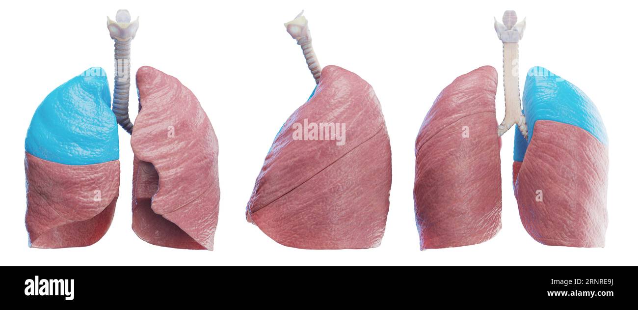 Right lung, illustration Stock Photohttps://www.alamy.com/image-license-details/?v=1https://www.alamy.com/right-lung-illustration-image564155694.html
RF2RNRE9J–Right lung, illustration
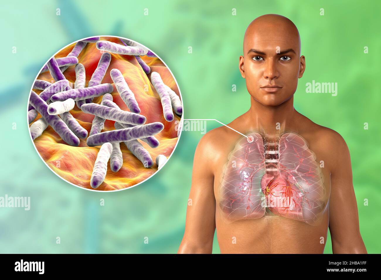 Illustration of a secondary tuberculosis infection and a close-up view of Mycobacterium tuberculosis bacteria, the causative agent of tuberculosis. There is a small-sized solid nodular mass located in the upper lobe of the right lung near its lung apex. Stock Photohttps://www.alamy.com/image-license-details/?v=1https://www.alamy.com/illustration-of-a-secondary-tuberculosis-infection-and-a-close-up-view-of-mycobacterium-tuberculosis-bacteria-the-causative-agent-of-tuberculosis-there-is-a-small-sized-solid-nodular-mass-located-in-the-upper-lobe-of-the-right-lung-near-its-lung-apex-image454451523.html
RF2HBA1FF–Illustration of a secondary tuberculosis infection and a close-up view of Mycobacterium tuberculosis bacteria, the causative agent of tuberculosis. There is a small-sized solid nodular mass located in the upper lobe of the right lung near its lung apex.
 Lungs, illustration Stock Photohttps://www.alamy.com/image-license-details/?v=1https://www.alamy.com/lungs-illustration-image560517068.html
RF2RFWN6M–Lungs, illustration
 Clinical tuberculosis . iscussion. Against these theories is the very importantphysiologic fact that one of the most important inspiratory actsis the contraction of the diaphragm which enlarges the lung notonly in the anteroposterior direction, but in the superoinferior di- 134 FACTORS WHICH PREDISPOSE TO TUBERCULOSIS Iection as svell. Keiih called attention to the accordion actionof the interlobular sulci of the lung and the general enlarge-ment which takes place with the contraction of the diaphragm. I regard the factors which make the apex of the lung especial-ly predisposed to tuberculous Stock Photohttps://www.alamy.com/image-license-details/?v=1https://www.alamy.com/clinical-tuberculosis-iscussion-against-these-theories-is-the-very-importantphysiologic-fact-that-one-of-the-most-important-inspiratory-actsis-the-contraction-of-the-diaphragm-which-enlarges-the-lung-notonly-in-the-anteroposterior-direction-but-in-the-superoinferior-di-134-factors-which-predispose-to-tuberculosis-iection-as-svell-keiih-called-attention-to-the-accordion-actionof-the-interlobular-sulci-of-the-lung-and-the-general-enlarge-ment-which-takes-place-with-the-contraction-of-the-diaphragm-i-regard-the-factors-which-make-the-apex-of-the-lung-especial-ly-predisposed-to-tuberculous-image343003501.html
RM2AX14D1–Clinical tuberculosis . iscussion. Against these theories is the very importantphysiologic fact that one of the most important inspiratory actsis the contraction of the diaphragm which enlarges the lung notonly in the anteroposterior direction, but in the superoinferior di- 134 FACTORS WHICH PREDISPOSE TO TUBERCULOSIS Iection as svell. Keiih called attention to the accordion actionof the interlobular sulci of the lung and the general enlarge-ment which takes place with the contraction of the diaphragm. I regard the factors which make the apex of the lung especial-ly predisposed to tuberculous
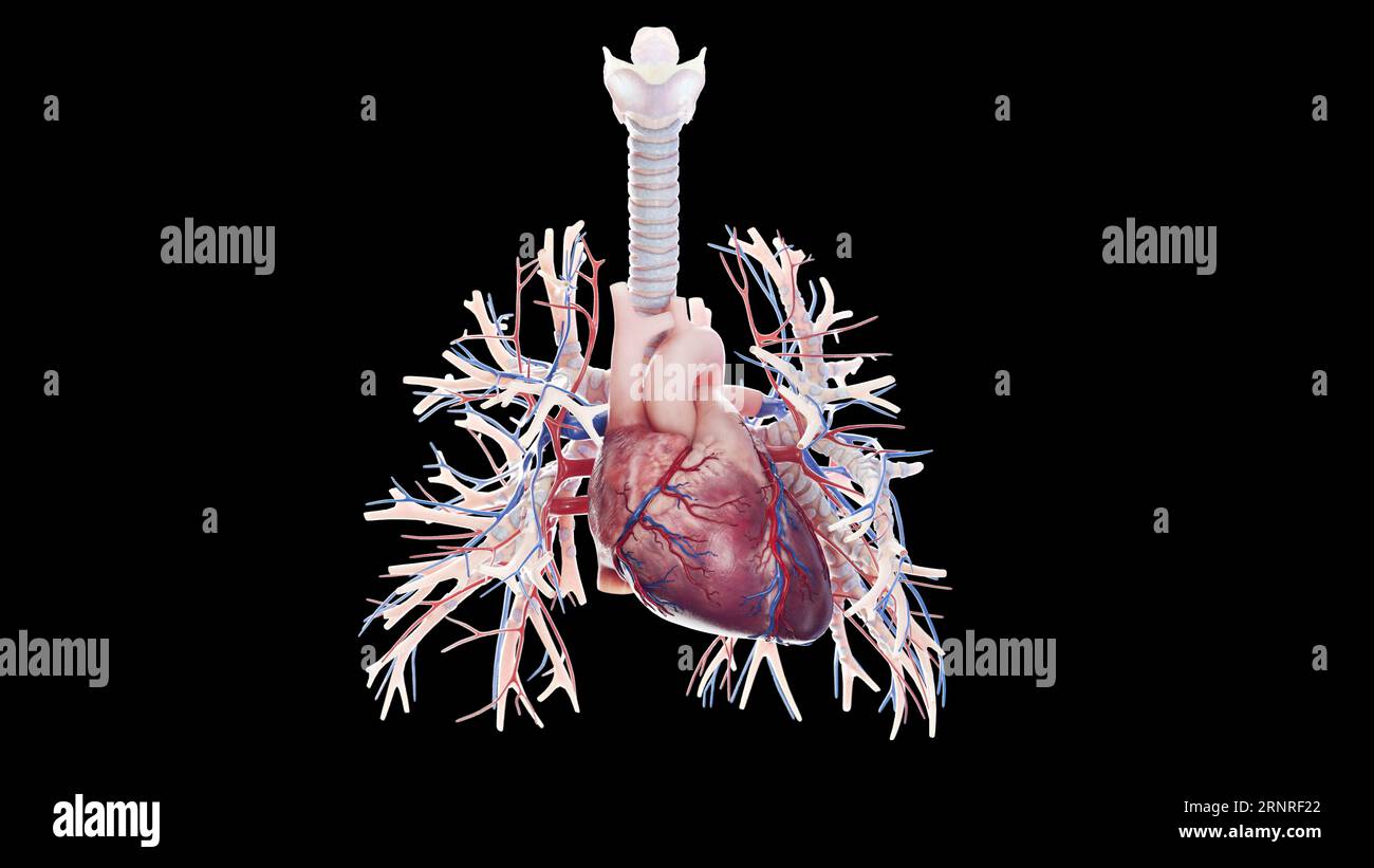 Cardiopulmonary system, illustration Stock Photohttps://www.alamy.com/image-license-details/?v=1https://www.alamy.com/cardiopulmonary-system-illustration-image564156266.html
RF2RNRF22–Cardiopulmonary system, illustration
 Lungs, illustration Stock Photohttps://www.alamy.com/image-license-details/?v=1https://www.alamy.com/lungs-illustration-image560517277.html
RF2RFWNE5–Lungs, illustration
 . Diseases of the heart and thoracic aorta. tion ofthe apex beat to the mid-sternum, and then to determinethe position of the vertical and oblique sides of the trianglerespectively, i.e. to define the anterior margins of the lungsby means of percussion. And it is important to rememberthat at those points at Avhich the absolute and relativeareas of cardiac dulness meet {i.e. where the margins ofthe lungs terminate), the percussion strokes must be lightlystruck, for otherwise the resonant note which is obtainedfrom the thin layer of lung will be, to some extent, obscuredby the dull, sound which Stock Photohttps://www.alamy.com/image-license-details/?v=1https://www.alamy.com/diseases-of-the-heart-and-thoracic-aorta-tion-ofthe-apex-beat-to-the-mid-sternum-and-then-to-determinethe-position-of-the-vertical-and-oblique-sides-of-the-trianglerespectively-ie-to-define-the-anterior-margins-of-the-lungsby-means-of-percussion-and-it-is-important-to-rememberthat-at-those-points-at-avhich-the-absolute-and-relativeareas-of-cardiac-dulness-meet-ie-where-the-margins-ofthe-lungs-terminate-the-percussion-strokes-must-be-lightlystruck-for-otherwise-the-resonant-note-which-is-obtainedfrom-the-thin-layer-of-lung-will-be-to-some-extent-obscuredby-the-dull-sound-which-image336749261.html
RM2AFT739–. Diseases of the heart and thoracic aorta. tion ofthe apex beat to the mid-sternum, and then to determinethe position of the vertical and oblique sides of the trianglerespectively, i.e. to define the anterior margins of the lungsby means of percussion. And it is important to rememberthat at those points at Avhich the absolute and relativeareas of cardiac dulness meet {i.e. where the margins ofthe lungs terminate), the percussion strokes must be lightlystruck, for otherwise the resonant note which is obtainedfrom the thin layer of lung will be, to some extent, obscuredby the dull, sound which
 Lungs, illustration Stock Photohttps://www.alamy.com/image-license-details/?v=1https://www.alamy.com/lungs-illustration-image560517219.html
RF2RFWNC3–Lungs, illustration
 Practical pathology; a manual for students and practitioners . milar changes or caseation may result throughout the wholeof the lung substance. Caseation and cavity formation are mostfrequent in the upper part of the lung, near the apex, where, too, theI)rocess is, as a rule, more chronic, but more advanced. Tubercle Bacilli 333. The association of the tubercle bacillus with tuberculousdisease cannot now be doubted; it is found in the lungs and sputumin various forms of tuberculosis and phthisis; it has also beendemonstrated in tuberculosis of the intestine; around the vessels intuberculous in Stock Photohttps://www.alamy.com/image-license-details/?v=1https://www.alamy.com/practical-pathology-a-manual-for-students-and-practitioners-milar-changes-or-caseation-may-result-throughout-the-wholeof-the-lung-substance-caseation-and-cavity-formation-are-mostfrequent-in-the-upper-part-of-the-lung-near-the-apex-where-too-theirocess-is-as-a-rule-more-chronic-but-more-advanced-tubercle-bacilli-333-the-association-of-the-tubercle-bacillus-with-tuberculousdisease-cannot-now-be-doubted-it-is-found-in-the-lungs-and-sputumin-various-forms-of-tuberculosis-and-phthisis-it-has-also-beendemonstrated-in-tuberculosis-of-the-intestine-around-the-vessels-intuberculous-in-image342980781.html
RM2AX03DH–Practical pathology; a manual for students and practitioners . milar changes or caseation may result throughout the wholeof the lung substance. Caseation and cavity formation are mostfrequent in the upper part of the lung, near the apex, where, too, theI)rocess is, as a rule, more chronic, but more advanced. Tubercle Bacilli 333. The association of the tubercle bacillus with tuberculousdisease cannot now be doubted; it is found in the lungs and sputumin various forms of tuberculosis and phthisis; it has also beendemonstrated in tuberculosis of the intestine; around the vessels intuberculous in
 Manual of pathology : including bacteriology, the technic of postmortems, and methods of pathologic research . ure the esophagus.Gentle protracted traction will strip the organs from the tissue behind,bringing forward with the pericardium the aorta, which must be severedat the diaphragm. If it is desired to leave the aorta, this can easily be doneby separating it from the pericardial attachments before delivering thelungs. When removed singly, the left lung is removed first; loosen alladhesions, pull the apex downward and the remainder forward, cuttingthe bronchi and vessels from behind; some Stock Photohttps://www.alamy.com/image-license-details/?v=1https://www.alamy.com/manual-of-pathology-including-bacteriology-the-technic-of-postmortems-and-methods-of-pathologic-research-ure-the-esophagusgentle-protracted-traction-will-strip-the-organs-from-the-tissue-behindbringing-forward-with-the-pericardium-the-aorta-which-must-be-severedat-the-diaphragm-if-it-is-desired-to-leave-the-aorta-this-can-easily-be-doneby-separating-it-from-the-pericardial-attachments-before-delivering-thelungs-when-removed-singly-the-left-lung-is-removed-first-loosen-alladhesions-pull-the-apex-downward-and-the-remainder-forward-cuttingthe-bronchi-and-vessels-from-behind-some-image338473990.html
RM2AJJR0P–Manual of pathology : including bacteriology, the technic of postmortems, and methods of pathologic research . ure the esophagus.Gentle protracted traction will strip the organs from the tissue behind,bringing forward with the pericardium the aorta, which must be severedat the diaphragm. If it is desired to leave the aorta, this can easily be doneby separating it from the pericardial attachments before delivering thelungs. When removed singly, the left lung is removed first; loosen alladhesions, pull the apex downward and the remainder forward, cuttingthe bronchi and vessels from behind; some
 Diseases of the chest and the principles of physical diagnosis . or this. Thepersistent absence of tubercle bacilli in the sputum, the marked dyspneaand the locaHzation of the physical signs in the anterior mediastinum, aboutthe roots or at the bases of the lungs should serve as warnings. Tubercu-losis is either locahzed at the summit of the lung or if the whole lung isdiseased, the most destructive lesions are found where the process first1 Boston Med. and Surg. Jour., October 28, 1915. 554 DISEASES OF THE BRONCHI, LUNGS, PLEURA, AND DIAPHRAGM started, namely, at the apex. Occasionally the ma Stock Photohttps://www.alamy.com/image-license-details/?v=1https://www.alamy.com/diseases-of-the-chest-and-the-principles-of-physical-diagnosis-or-this-thepersistent-absence-of-tubercle-bacilli-in-the-sputum-the-marked-dyspneaand-the-locahzation-of-the-physical-signs-in-the-anterior-mediastinum-aboutthe-roots-or-at-the-bases-of-the-lungs-should-serve-as-warnings-tubercu-losis-is-either-locahzed-at-the-summit-of-the-lung-or-if-the-whole-lung-isdiseased-the-most-destructive-lesions-are-found-where-the-process-first1-boston-med-and-surg-jour-october-28-1915-554-diseases-of-the-bronchi-lungs-pleura-and-diaphragm-started-namely-at-the-apex-occasionally-the-ma-image340224526.html
RM2ANEFRX–Diseases of the chest and the principles of physical diagnosis . or this. Thepersistent absence of tubercle bacilli in the sputum, the marked dyspneaand the locaHzation of the physical signs in the anterior mediastinum, aboutthe roots or at the bases of the lungs should serve as warnings. Tubercu-losis is either locahzed at the summit of the lung or if the whole lung isdiseased, the most destructive lesions are found where the process first1 Boston Med. and Surg. Jour., October 28, 1915. 554 DISEASES OF THE BRONCHI, LUNGS, PLEURA, AND DIAPHRAGM started, namely, at the apex. Occasionally the ma
 Diseases of the chest and the principles of physical diagnosis . g can be made to expand for an inch or morebelow the level of the tenth dorsal vertebra, providing it or the pleura DISEASES OF THE LUNGS 351 is free from disease. If, however, the lung is much diseased, or thepleural cavity is obliterated, or the diaphragm is immobile, the baseline on the affected side remains stationary. Having marked out the borders of the lung, the heart and viscerain relationship to the lungs should be outlined. Auscultation.—The fact that there normally exists a difference be-tween the right and left apex h Stock Photohttps://www.alamy.com/image-license-details/?v=1https://www.alamy.com/diseases-of-the-chest-and-the-principles-of-physical-diagnosis-g-can-be-made-to-expand-for-an-inch-or-morebelow-the-level-of-the-tenth-dorsal-vertebra-providing-it-or-the-pleura-diseases-of-the-lungs-351-is-free-from-disease-if-however-the-lung-is-much-diseased-or-thepleural-cavity-is-obliterated-or-the-diaphragm-is-immobile-the-baseline-on-the-affected-side-remains-stationary-having-marked-out-the-borders-of-the-lung-the-heart-and-viscerain-relationship-to-the-lungs-should-be-outlined-auscultationthe-fact-that-there-normally-exists-a-difference-be-tween-the-right-and-left-apex-h-image340242524.html
RM2ANFAPM–Diseases of the chest and the principles of physical diagnosis . g can be made to expand for an inch or morebelow the level of the tenth dorsal vertebra, providing it or the pleura DISEASES OF THE LUNGS 351 is free from disease. If, however, the lung is much diseased, or thepleural cavity is obliterated, or the diaphragm is immobile, the baseline on the affected side remains stationary. Having marked out the borders of the lung, the heart and viscerain relationship to the lungs should be outlined. Auscultation.—The fact that there normally exists a difference be-tween the right and left apex h
 Diseases of the chest and the principles of physical diagnosis . he disease first began, are characteristic of tuberculosis and serve asvaluable aids, inasmuch as the physical signs will be more marked atthe summit of the lung and while continuous, diminish as the base isapproached. This distribution of the disease also serves to distinguishtuberculosis from certain chronic inflammatory diseases of the lung, whichinvade the base rather than the apex. While these generalizations arenot absolute, the line of march in the great majority of cases un-doubtedly shows a great similarity. 318 DISEASES Stock Photohttps://www.alamy.com/image-license-details/?v=1https://www.alamy.com/diseases-of-the-chest-and-the-principles-of-physical-diagnosis-he-disease-first-began-are-characteristic-of-tuberculosis-and-serve-asvaluable-aids-inasmuch-as-the-physical-signs-will-be-more-marked-atthe-summit-of-the-lung-and-while-continuous-diminish-as-the-base-isapproached-this-distribution-of-the-disease-also-serves-to-distinguishtuberculosis-from-certain-chronic-inflammatory-diseases-of-the-lung-whichinvade-the-base-rather-than-the-apex-while-these-generalizations-arenot-absolute-the-line-of-march-in-the-great-majority-of-cases-un-doubtedly-shows-a-great-similarity-318-diseases-image340248804.html
RM2ANFJR0–Diseases of the chest and the principles of physical diagnosis . he disease first began, are characteristic of tuberculosis and serve asvaluable aids, inasmuch as the physical signs will be more marked atthe summit of the lung and while continuous, diminish as the base isapproached. This distribution of the disease also serves to distinguishtuberculosis from certain chronic inflammatory diseases of the lung, whichinvade the base rather than the apex. While these generalizations arenot absolute, the line of march in the great majority of cases un-doubtedly shows a great similarity. 318 DISEASES
 Diseases of the chest and the principles of physical diagnosis . f lung normal,{Jefferson Medical College Museum.) beyond their original boundaries. Thus at the apex where the originaldeposit occurred the infiltration becomes more and more dense andfurthermore the tubercles spread downward until the entire lung isinvolved although in the lower portions the infiltration is usually widelyscattered. This constitutes the second or moderately advanced stage. Fig.230 shows this very well. It will be noted that at the extreme apex theinfiltration, while very dense, has not replaced entirely all of th Stock Photohttps://www.alamy.com/image-license-details/?v=1https://www.alamy.com/diseases-of-the-chest-and-the-principles-of-physical-diagnosis-f-lung-normaljefferson-medical-college-museum-beyond-their-original-boundaries-thus-at-the-apex-where-the-originaldeposit-occurred-the-infiltration-becomes-more-and-more-dense-andfurthermore-the-tubercles-spread-downward-until-the-entire-lung-isinvolved-although-in-the-lower-portions-the-infiltration-is-usually-widelyscattered-this-constitutes-the-second-or-moderately-advanced-stage-fig230-shows-this-very-well-it-will-be-noted-that-at-the-extreme-apex-theinfiltration-while-very-dense-has-not-replaced-entirely-all-of-th-image340251797.html
RM2ANFPHW–Diseases of the chest and the principles of physical diagnosis . f lung normal,{Jefferson Medical College Museum.) beyond their original boundaries. Thus at the apex where the originaldeposit occurred the infiltration becomes more and more dense andfurthermore the tubercles spread downward until the entire lung isinvolved although in the lower portions the infiltration is usually widelyscattered. This constitutes the second or moderately advanced stage. Fig.230 shows this very well. It will be noted that at the extreme apex theinfiltration, while very dense, has not replaced entirely all of th
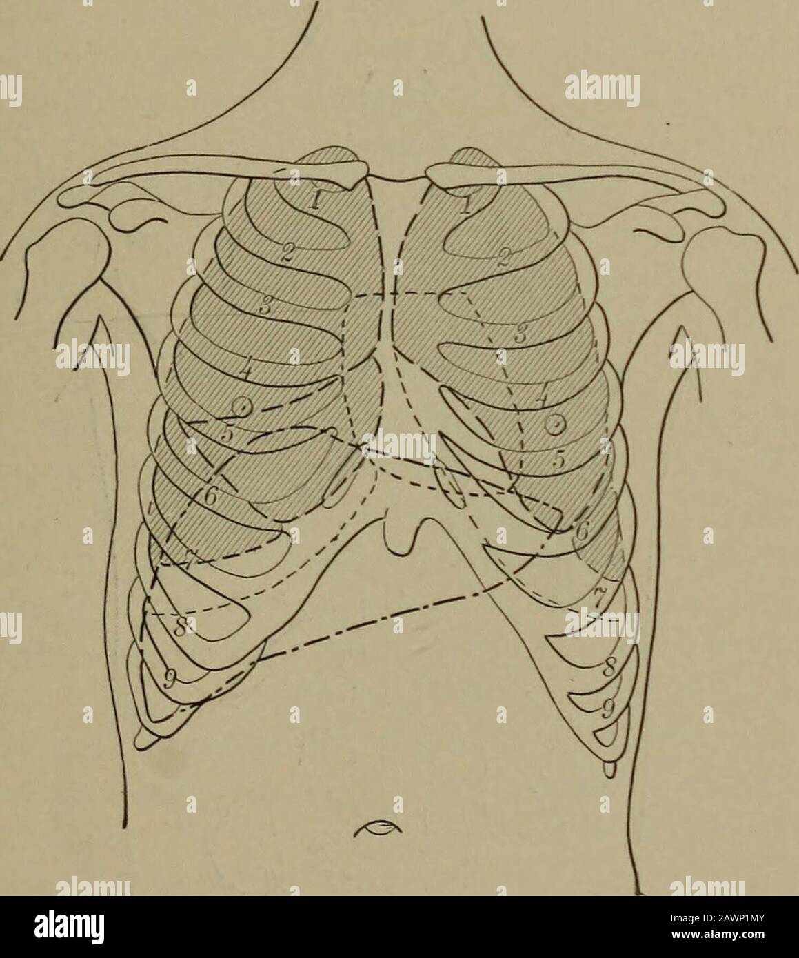 Physical diagnosis, including diseases of the thoracic and abdominal organs : a manual for students and physicians .. . present. These variations are noted inFig. 30. PERCUSSION OUTLINE OF LUNG. Percussion gives valuable information concerningthe size and mobility of the lung, and no examina-tion is complete unless the percussion outline of theborders of the lung during inspiration and expira-tion is determined/ The only boundaries or bordersthat can be definitely mapped out by percussion are thesuperior or apex of the lung, the inferior or bases, andthat portion of the anterior that is not co Stock Photohttps://www.alamy.com/image-license-details/?v=1https://www.alamy.com/physical-diagnosis-including-diseases-of-the-thoracic-and-abdominal-organs-a-manual-for-students-and-physicians-present-these-variations-are-noted-infig-30-percussion-outline-of-lung-percussion-gives-valuable-information-concerningthe-size-and-mobility-of-the-lung-and-no-examina-tion-is-complete-unless-the-percussion-outline-of-theborders-of-the-lung-during-inspiration-and-expira-tion-is-determined-the-only-boundaries-or-bordersthat-can-be-definitely-mapped-out-by-percussion-are-thesuperior-or-apex-of-the-lung-the-inferior-or-bases-andthat-portion-of-the-anterior-that-is-not-co-image342847707.html
RM2AWP1MY–Physical diagnosis, including diseases of the thoracic and abdominal organs : a manual for students and physicians .. . present. These variations are noted inFig. 30. PERCUSSION OUTLINE OF LUNG. Percussion gives valuable information concerningthe size and mobility of the lung, and no examina-tion is complete unless the percussion outline of theborders of the lung during inspiration and expira-tion is determined/ The only boundaries or bordersthat can be definitely mapped out by percussion are thesuperior or apex of the lung, the inferior or bases, andthat portion of the anterior that is not co
 Clinical tuberculosis . Fig. 49.—Showing the relationship of the anterior surface of the lung as conlined bythe bony thorax to the soft structures forming the anterior surface of the chest. they are on a level with the sixth rib in the mammary line, theeighth in the axillary line, the tenth in the scapular line, and atthe junction of the eleventh rib with the vertebra at the vertebralborder. (See pages 322, 323, and 324.) The apex is the nearest the surface Ijetween the two headsof the sternocleidomastoideus, as before mentioned. On ordi-nary quiet respiration the movement of the lower border Stock Photohttps://www.alamy.com/image-license-details/?v=1https://www.alamy.com/clinical-tuberculosis-fig-49showing-the-relationship-of-the-anterior-surface-of-the-lung-as-conlined-bythe-bony-thorax-to-the-soft-structures-forming-the-anterior-surface-of-the-chest-they-are-on-a-level-with-the-sixth-rib-in-the-mammary-line-theeighth-in-the-axillary-line-the-tenth-in-the-scapular-line-and-atthe-junction-of-the-eleventh-rib-with-the-vertebra-at-the-vertebralborder-see-pages-322-323-and-324-the-apex-is-the-nearest-the-surface-ijetween-the-two-headsof-the-sternocleidomastoideus-as-before-mentioned-on-ordi-nary-quiet-respiration-the-movement-of-the-lower-border-image342985722.html
RM2AX09P2–Clinical tuberculosis . Fig. 49.—Showing the relationship of the anterior surface of the lung as conlined bythe bony thorax to the soft structures forming the anterior surface of the chest. they are on a level with the sixth rib in the mammary line, theeighth in the axillary line, the tenth in the scapular line, and atthe junction of the eleventh rib with the vertebra at the vertebralborder. (See pages 322, 323, and 324.) The apex is the nearest the surface Ijetween the two headsof the sternocleidomastoideus, as before mentioned. On ordi-nary quiet respiration the movement of the lower border
 . Medical diagnosis for the student and practitioner. Fig. 126.—Unilateral Pulmonary Tuberculosis. Note marked infiltration of rightupper lung field. Pleuro-diaphragmatic adhesions. {Dr. Frank S. Bissell.) remain clear and uninvolved until late in the progress of other chronicinfections. Basal tuberculosis, while it has the same general characteristics, is muchmore difficult to differentiate from other chronic infections. Tuberculosis ROENTGENOGRAPHS EXAMINATION OF LUNGS AND PLEURAE 317 of the base, however, without concomitant involvement of the apex of one ormore lobes, is relatively rare. A Stock Photohttps://www.alamy.com/image-license-details/?v=1https://www.alamy.com/medical-diagnosis-for-the-student-and-practitioner-fig-126unilateral-pulmonary-tuberculosis-note-marked-infiltration-of-rightupper-lung-field-pleuro-diaphragmatic-adhesions-dr-frank-s-bissell-remain-clear-and-uninvolved-until-late-in-the-progress-of-other-chronicinfections-basal-tuberculosis-while-it-has-the-same-general-characteristics-is-muchmore-difficult-to-differentiate-from-other-chronic-infections-tuberculosis-roentgenographs-examination-of-lungs-and-pleurae-317-of-the-base-however-without-concomitant-involvement-of-the-apex-of-one-ormore-lobes-is-relatively-rare-a-image336897410.html
RM2AG302A–. Medical diagnosis for the student and practitioner. Fig. 126.—Unilateral Pulmonary Tuberculosis. Note marked infiltration of rightupper lung field. Pleuro-diaphragmatic adhesions. {Dr. Frank S. Bissell.) remain clear and uninvolved until late in the progress of other chronicinfections. Basal tuberculosis, while it has the same general characteristics, is muchmore difficult to differentiate from other chronic infections. Tuberculosis ROENTGENOGRAPHS EXAMINATION OF LUNGS AND PLEURAE 317 of the base, however, without concomitant involvement of the apex of one ormore lobes, is relatively rare. A
 Clinical tuberculosis . er lobe being involved, as a rule, more exten-sively than the lower; although large cavities and extensivefibrosis are often formed in the lower lobe also. The right lungis severely diseased less often; but when it is, the conditionsduplicate those in the left, the upper portion showing the moreextensive process. That the upper lobe should be the seat ofthe more extensive involvement is evident from the fact that theprimary pulmonary infection is usually near the apex whether itbe the lirst apex involved or the extension to the other lung; and,the disease spreads downwa Stock Photohttps://www.alamy.com/image-license-details/?v=1https://www.alamy.com/clinical-tuberculosis-er-lobe-being-involved-as-a-rule-more-exten-sively-than-the-lower-although-large-cavities-and-extensivefibrosis-are-often-formed-in-the-lower-lobe-also-the-right-lungis-severely-diseased-less-often-but-when-it-is-the-conditionsduplicate-those-in-the-left-the-upper-portion-showing-the-moreextensive-process-that-the-upper-lobe-should-be-the-seat-ofthe-more-extensive-involvement-is-evident-from-the-fact-that-theprimary-pulmonary-infection-is-usually-near-the-apex-whether-itbe-the-lirst-apex-involved-or-the-extension-to-the-other-lung-andthe-disease-spreads-downwa-image342992050.html
RM2AX0HT2–Clinical tuberculosis . er lobe being involved, as a rule, more exten-sively than the lower; although large cavities and extensivefibrosis are often formed in the lower lobe also. The right lungis severely diseased less often; but when it is, the conditionsduplicate those in the left, the upper portion showing the moreextensive process. That the upper lobe should be the seat ofthe more extensive involvement is evident from the fact that theprimary pulmonary infection is usually near the apex whether itbe the lirst apex involved or the extension to the other lung; and,the disease spreads downwa
 Clinical tuberculosis . a the accessory musclesof respiration, particularly the sternocleidomastoideus and scalenimay show increased tone because of the extra work thrown uponthem. Extremely rarely this may cause some confusion in thaithe increased tone is taken as indicating inflammation of the un-derlying apex. Careful analysis of all conditions present willusually make the diagnosis plain. Atrophy of the soft parts,must be looked upon as being expressive of a chronic, or it maybe healed, inflammatory process in the underlying lung or pleura.It may also lie due to occupational change in the Stock Photohttps://www.alamy.com/image-license-details/?v=1https://www.alamy.com/clinical-tuberculosis-a-the-accessory-musclesof-respiration-particularly-the-sternocleidomastoideus-and-scalenimay-show-increased-tone-because-of-the-extra-work-thrown-uponthem-extremely-rarely-this-may-cause-some-confusion-in-thaithe-increased-tone-is-taken-as-indicating-inflammation-of-the-un-derlying-apex-careful-analysis-of-all-conditions-present-willusually-make-the-diagnosis-plain-atrophy-of-the-soft-partsmust-be-looked-upon-as-being-expressive-of-a-chronic-or-it-maybe-healed-inflammatory-process-in-the-underlying-lung-or-pleurait-may-also-lie-due-to-occupational-change-in-the-image342958243.html
RM2AWY2MK–Clinical tuberculosis . a the accessory musclesof respiration, particularly the sternocleidomastoideus and scalenimay show increased tone because of the extra work thrown uponthem. Extremely rarely this may cause some confusion in thaithe increased tone is taken as indicating inflammation of the un-derlying apex. Careful analysis of all conditions present willusually make the diagnosis plain. Atrophy of the soft parts,must be looked upon as being expressive of a chronic, or it maybe healed, inflammatory process in the underlying lung or pleura.It may also lie due to occupational change in the
 Modern medicine : its theory and practice, in original contributions by American and foreign authors . as approximately circular,elevated, dark-red or blackish masses that in consistency are quite solid.On section they are more or less perfectly wedge-shaped, the typical infarctedarea forming a cone with the base toward the pleura. In some instances,owing to the abundant collateral circulation of the lung, the area is almostglobular. At or near the apex of the infarct may often be found the pluggedvessel responsible for the lesion. Occasionally, however, careful searchfails to reveal such obst Stock Photohttps://www.alamy.com/image-license-details/?v=1https://www.alamy.com/modern-medicine-its-theory-and-practice-in-original-contributions-by-american-and-foreign-authors-as-approximately-circularelevated-dark-red-or-blackish-masses-that-in-consistency-are-quite-solidon-section-they-are-more-or-less-perfectly-wedge-shaped-the-typical-infarctedarea-forming-a-cone-with-the-base-toward-the-pleura-in-some-instancesowing-to-the-abundant-collateral-circulation-of-the-lung-the-area-is-almostglobular-at-or-near-the-apex-of-the-infarct-may-often-be-found-the-pluggedvessel-responsible-for-the-lesion-occasionally-however-careful-searchfails-to-reveal-such-obst-image342762772.html
RM2AWJ5BG–Modern medicine : its theory and practice, in original contributions by American and foreign authors . as approximately circular,elevated, dark-red or blackish masses that in consistency are quite solid.On section they are more or less perfectly wedge-shaped, the typical infarctedarea forming a cone with the base toward the pleura. In some instances,owing to the abundant collateral circulation of the lung, the area is almostglobular. At or near the apex of the infarct may often be found the pluggedvessel responsible for the lesion. Occasionally, however, careful searchfails to reveal such obst
 Physical diagnosis, including diseases of the thoracic and abdominal organs : a manual for students and physicians .. . itedto the base. Fluoroscopic examinations have shown thatwhen there is a slight infiltration of the apex there is acorresponding loss of motion of the diaphragm on theaffected side. On the right side the relation of the lungto the liver renders mapping out of the lower border ofpulmonary resonance easy. On the left side, on accountof the relation of the lung to the resonant abdominalorgans, the lower level can only be determined over thatportion of the axillary line where th Stock Photohttps://www.alamy.com/image-license-details/?v=1https://www.alamy.com/physical-diagnosis-including-diseases-of-the-thoracic-and-abdominal-organs-a-manual-for-students-and-physicians-itedto-the-base-fluoroscopic-examinations-have-shown-thatwhen-there-is-a-slight-infiltration-of-the-apex-there-is-acorresponding-loss-of-motion-of-the-diaphragm-on-theaffected-side-on-the-right-side-the-relation-of-the-lungto-the-liver-renders-mapping-out-of-the-lower-border-ofpulmonary-resonance-easy-on-the-left-side-on-accountof-the-relation-of-the-lung-to-the-resonant-abdominalorgans-the-lower-level-can-only-be-determined-over-thatportion-of-the-axillary-line-where-th-image342847000.html
RM2AWP0RM–Physical diagnosis, including diseases of the thoracic and abdominal organs : a manual for students and physicians .. . itedto the base. Fluoroscopic examinations have shown thatwhen there is a slight infiltration of the apex there is acorresponding loss of motion of the diaphragm on theaffected side. On the right side the relation of the lungto the liver renders mapping out of the lower border ofpulmonary resonance easy. On the left side, on accountof the relation of the lung to the resonant abdominalorgans, the lower level can only be determined over thatportion of the axillary line where th
 . Internal medicine; a work for the practicing physician on diagnosis and treatment, with a complete Desk index. Fig. 348A.—Moderate pericardial effusion;quadrilateral flatness with border of relativedulness; effacement of cardiohepatic angle. Fig. 348B.—Massive pericardial effusion;pyramidal area of flatness with truncated apex;right border of relative dulness; downwarddisplacement of liver. toward the left and upward, later toward the right, displacing the bordersof the lung and forming at first a quadrilateral area of dulness with roundedcorners, which with larger effusions assumes a pear-s Stock Photohttps://www.alamy.com/image-license-details/?v=1https://www.alamy.com/internal-medicine-a-work-for-the-practicing-physician-on-diagnosis-and-treatment-with-a-complete-desk-index-fig-348amoderate-pericardial-effusionquadrilateral-flatness-with-border-of-relativedulness-effacement-of-cardiohepatic-angle-fig-348bmassive-pericardial-effusionpyramidal-area-of-flatness-with-truncated-apexright-border-of-relative-dulness-downwarddisplacement-of-liver-toward-the-left-and-upward-later-toward-the-right-displacing-the-bordersof-the-lung-and-forming-at-first-a-quadrilateral-area-of-dulness-with-roundedcorners-which-with-larger-effusions-assumes-a-pear-s-image336707533.html
RM2AFP9W1–. Internal medicine; a work for the practicing physician on diagnosis and treatment, with a complete Desk index. Fig. 348A.—Moderate pericardial effusion;quadrilateral flatness with border of relativedulness; effacement of cardiohepatic angle. Fig. 348B.—Massive pericardial effusion;pyramidal area of flatness with truncated apex;right border of relative dulness; downwarddisplacement of liver. toward the left and upward, later toward the right, displacing the bordersof the lung and forming at first a quadrilateral area of dulness with roundedcorners, which with larger effusions assumes a pear-s
 . The diseases of children : medical and surgical. or in front between tlie pericardium and anterior edge ofthe left lung, or between the lung and the diaphragm. We have known alocalised empyema situated at the posterior side of the apex of one lung ;there was fairly good resonance in front and behind, except over asmallareaat the back of the apex of the lung. These small empyemas are often asso-ciated with broncho-pneumonias and chronic tuberculosis of the lung. It isperfectly obvious that if these collections of fluid are not large and are sur-rounded by and backed up by crepitant lung, diag Stock Photohttps://www.alamy.com/image-license-details/?v=1https://www.alamy.com/the-diseases-of-children-medical-and-surgical-or-in-front-between-tlie-pericardium-and-anterior-edge-ofthe-left-lung-or-between-the-lung-and-the-diaphragm-we-have-known-alocalised-empyema-situated-at-the-posterior-side-of-the-apex-of-one-lung-there-was-fairly-good-resonance-in-front-and-behind-except-over-asmallareaat-the-back-of-the-apex-of-the-lung-these-small-empyemas-are-often-asso-ciated-with-broncho-pneumonias-and-chronic-tuberculosis-of-the-lung-it-isperfectly-obvious-that-if-these-collections-of-fluid-are-not-large-and-are-sur-rounded-by-and-backed-up-by-crepitant-lung-diag-image337032847.html
RM2AG94RB–. The diseases of children : medical and surgical. or in front between tlie pericardium and anterior edge ofthe left lung, or between the lung and the diaphragm. We have known alocalised empyema situated at the posterior side of the apex of one lung ;there was fairly good resonance in front and behind, except over asmallareaat the back of the apex of the lung. These small empyemas are often asso-ciated with broncho-pneumonias and chronic tuberculosis of the lung. It isperfectly obvious that if these collections of fluid are not large and are sur-rounded by and backed up by crepitant lung, diag
 . Diseases of the heart and thoracic aorta. Fig. 26. — Displacement of the heart to the right as the result of effusion into theleft pleural cavity. (Modified from Sibson,) these cases the apex beat not unfrequently corresponds tothe right nipple. Extreme displacement to the right mayalso be due to retraction of the right lung. Sibson quotesseveral cases of this description,^ and I have seen more thanone case in which the pulsation of the heart was situated justabove the right nipple. Russell Reynolds System of Medicine, vol. iv. p. 143. Displacements of the Heart. 109 Displaceinent of the hea Stock Photohttps://www.alamy.com/image-license-details/?v=1https://www.alamy.com/diseases-of-the-heart-and-thoracic-aorta-fig-26-displacement-of-the-heart-to-the-right-as-the-result-of-effusion-into-theleft-pleural-cavity-modified-from-sibson-these-cases-the-apex-beat-not-unfrequently-corresponds-tothe-right-nipple-extreme-displacement-to-the-right-mayalso-be-due-to-retraction-of-the-right-lung-sibson-quotesseveral-cases-of-this-description-and-i-have-seen-more-thanone-case-in-which-the-pulsation-of-the-heart-was-situated-justabove-the-right-nipple-russell-reynolds-system-of-medicine-vol-iv-p-143-displacements-of-the-heart-109-displaceinent-of-the-hea-image336751181.html
RM2AFT9FW–. Diseases of the heart and thoracic aorta. Fig. 26. — Displacement of the heart to the right as the result of effusion into theleft pleural cavity. (Modified from Sibson,) these cases the apex beat not unfrequently corresponds tothe right nipple. Extreme displacement to the right mayalso be due to retraction of the right lung. Sibson quotesseveral cases of this description,^ and I have seen more thanone case in which the pulsation of the heart was situated justabove the right nipple. Russell Reynolds System of Medicine, vol. iv. p. 143. Displacements of the Heart. 109 Displaceinent of the hea
 . Medical diagnosis for the student and practitioner. iver dulness begins atthe lower border of the lung and extends downward to the costal margin.Its lower border crosses the epigastrium from the tip of the tenth rightcartilage to the tip of the eighthleft and meets the left extrem-ity of the organ in the fifth leftintercostal space at a pointclosely approximating the loca-tion of the normal heart apex.Traubes Semilunar Space.—This is included between thelower border of the left lung,the spleen, the inferior costalmargin, and the left lobe of theliver, and is normally hyper-resonant because o Stock Photohttps://www.alamy.com/image-license-details/?v=1https://www.alamy.com/medical-diagnosis-for-the-student-and-practitioner-iver-dulness-begins-atthe-lower-border-of-the-lung-and-extends-downward-to-the-costal-marginits-lower-border-crosses-the-epigastrium-from-the-tip-of-the-tenth-rightcartilage-to-the-tip-of-the-eighthleft-and-meets-the-left-extrem-ity-of-the-organ-in-the-fifth-leftintercostal-space-at-a-pointclosely-approximating-the-loca-tion-of-the-normal-heart-apextraubes-semilunar-spacethis-is-included-between-thelower-border-of-the-left-lungthe-spleen-the-inferior-costalmargin-and-the-left-lobe-of-theliver-and-is-normally-hyper-resonant-because-o-image336905319.html
RM2AG3A4R–. Medical diagnosis for the student and practitioner. iver dulness begins atthe lower border of the lung and extends downward to the costal margin.Its lower border crosses the epigastrium from the tip of the tenth rightcartilage to the tip of the eighthleft and meets the left extrem-ity of the organ in the fifth leftintercostal space at a pointclosely approximating the loca-tion of the normal heart apex.Traubes Semilunar Space.—This is included between thelower border of the left lung,the spleen, the inferior costalmargin, and the left lobe of theliver, and is normally hyper-resonant because o
 . The diseases of children : medical and surgical. cardiac dulness extends from nipple to nipple,and the apex beat occupies perhaps the fifth, sixth, and seventh spaces outsidethe nipple fine, while the whole of the precordial region is bulged forward Chronic Heart Disease 411 by the hypertrophied heart. Often the left bronchus is pressed upon and thelower lobe of the lung becomes collapsed. During the last stages, whichmay be short or prolonged intermittently for many months or even years,the liver becomes congested and enlarged, there is albuminuria from con-gested kidneys, while the belly, Stock Photohttps://www.alamy.com/image-license-details/?v=1https://www.alamy.com/the-diseases-of-children-medical-and-surgical-cardiac-dulness-extends-from-nipple-to-nippleand-the-apex-beat-occupies-perhaps-the-fifth-sixth-and-seventh-spaces-outsidethe-nipple-fine-while-the-whole-of-the-precordial-region-is-bulged-forward-chronic-heart-disease-411-by-the-hypertrophied-heart-often-the-left-bronchus-is-pressed-upon-and-thelower-lobe-of-the-lung-becomes-collapsed-during-the-last-stages-whichmay-be-short-or-prolonged-intermittently-for-many-months-or-even-yearsthe-liver-becomes-congested-and-enlarged-there-is-albuminuria-from-con-gested-kidneys-while-the-belly-image337030855.html
RM2AG9287–. The diseases of children : medical and surgical. cardiac dulness extends from nipple to nipple,and the apex beat occupies perhaps the fifth, sixth, and seventh spaces outsidethe nipple fine, while the whole of the precordial region is bulged forward Chronic Heart Disease 411 by the hypertrophied heart. Often the left bronchus is pressed upon and thelower lobe of the lung becomes collapsed. During the last stages, whichmay be short or prolonged intermittently for many months or even years,the liver becomes congested and enlarged, there is albuminuria from con-gested kidneys, while the belly,
 The science and practice of medicine . half above the clavicle, the relative height being unequalbut variable. The inner margin of each lung passes downwardsand inwards from the apex, and meets with the inner margin ofthe other lung at the middle line, at a point between the lirst andsecond cartilao:es, or at the second. SITUATION OF THE OEGANS IN THE REGIONS OF THE THORAX. 547, The inner margin of the light lung continues vertically downwardsalong the centre of the sternum, or inclining a little to the left side,as far as the attachment of the xiphoid cartilage (Fig. 10). The inner margin of Stock Photohttps://www.alamy.com/image-license-details/?v=1https://www.alamy.com/the-science-and-practice-of-medicine-half-above-the-clavicle-the-relative-height-being-unequalbut-variable-the-inner-margin-of-each-lung-passes-downwardsand-inwards-from-the-apex-and-meets-with-the-inner-margin-ofthe-other-lung-at-the-middle-line-at-a-point-between-the-lirst-andsecond-cartilaoes-or-at-the-second-situation-of-the-oegans-in-the-regions-of-the-thorax-547-the-inner-margin-of-the-light-lung-continues-vertically-downwardsalong-the-centre-of-the-sternum-or-inclining-a-little-to-the-left-sideas-far-as-the-attachment-of-the-xiphoid-cartilage-fig-10-the-inner-margin-of-image339313876.html
RM2AM128M–The science and practice of medicine . half above the clavicle, the relative height being unequalbut variable. The inner margin of each lung passes downwardsand inwards from the apex, and meets with the inner margin ofthe other lung at the middle line, at a point between the lirst andsecond cartilao:es, or at the second. SITUATION OF THE OEGANS IN THE REGIONS OF THE THORAX. 547, The inner margin of the light lung continues vertically downwardsalong the centre of the sternum, or inclining a little to the left side,as far as the attachment of the xiphoid cartilage (Fig. 10). The inner margin of
 Manual of pathological anatomy . The apex of a lung affected with caseous pneumonia; the limitation of the disease was defined by a sharp line. The affected portion is sometimes separated by a sharp line fromthe healthy lung, sometimes a border of broncho-pneumonicgranulations is seen. There is not necessarily any miliary tubercle. EiCx. 114.. Cicatrix at the apex of a lung, resulting from the previous arrest oftubercular disease. So slow is the progress of disease that further changes take placebefore any large portion of the lung is affected. The centre of the 506 CHRONIC PHTHISIS (MIXED AND Stock Photohttps://www.alamy.com/image-license-details/?v=1https://www.alamy.com/manual-of-pathological-anatomy-the-apex-of-a-lung-affected-with-caseous-pneumonia-the-limitation-of-the-disease-was-defined-by-a-sharp-line-the-affected-portion-is-sometimes-separated-by-a-sharp-line-fromthe-healthy-lung-sometimes-a-border-of-broncho-pneumonicgranulations-is-seen-there-is-not-necessarily-any-miliary-tubercle-eicx-114-cicatrix-at-the-apex-of-a-lung-resulting-from-the-previous-arrest-oftubercular-disease-so-slow-is-the-progress-of-disease-that-further-changes-take-placebefore-any-large-portion-of-the-lung-is-affected-the-centre-of-the-506-chronic-phthisis-mixed-and-image339469430.html
RM2AM84M6–Manual of pathological anatomy . The apex of a lung affected with caseous pneumonia; the limitation of the disease was defined by a sharp line. The affected portion is sometimes separated by a sharp line fromthe healthy lung, sometimes a border of broncho-pneumonicgranulations is seen. There is not necessarily any miliary tubercle. EiCx. 114.. Cicatrix at the apex of a lung, resulting from the previous arrest oftubercular disease. So slow is the progress of disease that further changes take placebefore any large portion of the lung is affected. The centre of the 506 CHRONIC PHTHISIS (MIXED AND
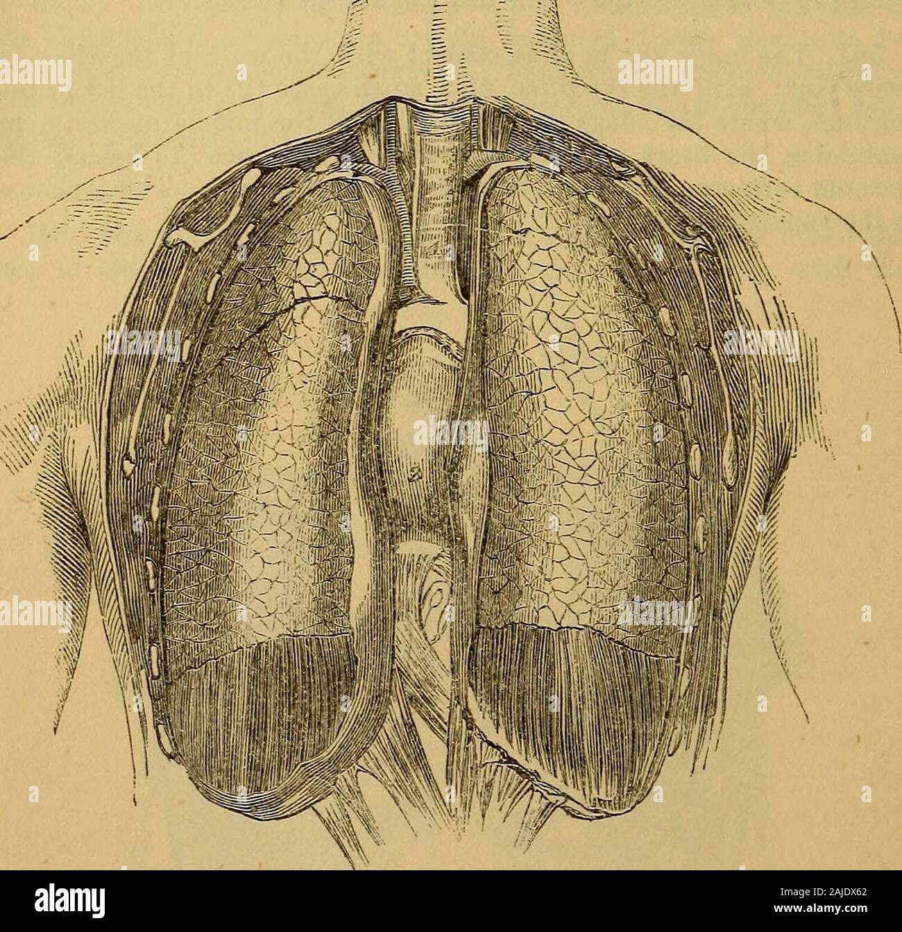 Hooper's physician's vade mecum, or, A manual of the principles and practice of physic . 142 SYMPTOMS AND SIGNS OF DISEASE. 562. Of the two lungs the right is the larger, but the left thelonger, its apex rising somewhat higher, and its base sinking lower.The right lung reaches to about the level of the sixth rib in front, ofthe eighth rib at the side, and still lower behind. The left lung extendsto the level of the seventh rib anteriorly, it reaches the eighth riblaterally, and descends still lower posteriorly. Both lungs applyingtheroselves closely to the diaphragm, descend much lower behind Stock Photohttps://www.alamy.com/image-license-details/?v=1https://www.alamy.com/hoopers-physicians-vade-mecum-or-a-manual-of-the-principles-and-practice-of-physic-142-symptoms-and-signs-of-disease-562-of-the-two-lungs-the-right-is-the-larger-but-the-left-thelonger-its-apex-rising-somewhat-higher-and-its-base-sinking-lowerthe-right-lung-reaches-to-about-the-level-of-the-sixth-rib-in-front-ofthe-eighth-rib-at-the-side-and-still-lower-behind-the-left-lung-extendsto-the-level-of-the-seventh-rib-anteriorly-it-reaches-the-eighth-riblaterally-and-descends-still-lower-posteriorly-both-lungs-applyingtheroselves-closely-to-the-diaphragm-descend-much-lower-behind-image338366730.html
RM2AJDX62–Hooper's physician's vade mecum, or, A manual of the principles and practice of physic . 142 SYMPTOMS AND SIGNS OF DISEASE. 562. Of the two lungs the right is the larger, but the left thelonger, its apex rising somewhat higher, and its base sinking lower.The right lung reaches to about the level of the sixth rib in front, ofthe eighth rib at the side, and still lower behind. The left lung extendsto the level of the seventh rib anteriorly, it reaches the eighth riblaterally, and descends still lower posteriorly. Both lungs applyingtheroselves closely to the diaphragm, descend much lower behind
 . Heart disease, with special reference to prognosis and treatment. FIG. 2.—THE HEART WITH LUNGS INSITU TO ILLUSTRATE AREA OF SUPER-FICIAL CARDIAC DULNESS (Sibsori).. FIG. 3.—THE HEART WITH OVERLYINGLUNG DISSECTED OFF TO ILLUSTRATEAREA OF DEEP DULNESS (Sibson). fifth cartilage, and curving, so as to form a hollow spacefor the lodgment of the apex, ends behind the sixthcartilage. The inner margin of the right lung continues its coursenearly straight downwards behind and a little to the leftof the centre of the sternum, to the level of the sixth RELATION OF HEART TO CHEST WALLS. 15 chondrosterna Stock Photohttps://www.alamy.com/image-license-details/?v=1https://www.alamy.com/heart-disease-with-special-reference-to-prognosis-and-treatment-fig-2the-heart-with-lungs-insitu-to-illustrate-area-of-super-ficial-cardiac-dulness-sibsori-fig-3the-heart-with-overlyinglung-dissected-off-to-illustratearea-of-deep-dulness-sibson-fifth-cartilage-and-curving-so-as-to-form-a-hollow-spacefor-the-lodgment-of-the-apex-ends-behind-the-sixthcartilage-the-inner-margin-of-the-right-lung-continues-its-coursenearly-straight-downwards-behind-and-a-little-to-the-leftof-the-centre-of-the-sternum-to-the-level-of-the-sixth-relation-of-heart-to-chest-walls-15-chondrosterna-image336853743.html
RM2AG10AR–. Heart disease, with special reference to prognosis and treatment. FIG. 2.—THE HEART WITH LUNGS INSITU TO ILLUSTRATE AREA OF SUPER-FICIAL CARDIAC DULNESS (Sibsori).. FIG. 3.—THE HEART WITH OVERLYINGLUNG DISSECTED OFF TO ILLUSTRATEAREA OF DEEP DULNESS (Sibson). fifth cartilage, and curving, so as to form a hollow spacefor the lodgment of the apex, ends behind the sixthcartilage. The inner margin of the right lung continues its coursenearly straight downwards behind and a little to the leftof the centre of the sternum, to the level of the sixth RELATION OF HEART TO CHEST WALLS. 15 chondrosterna
 Diseases of the chest and the principles of physical diagnosis . examination, is sufficiently frequent inthese cases to be worthv of note. 344 DISEASES OF THE BRONCHI, LUNGS, PLEURA, AND DIAPHRAGM Taking up the chest proper, it is usually best to note, when visible,the position of the apex beat of the heart, as in addition to disease of theheart itself, the apex may be displaced out of its normal position as theresult of disease of the lungs or pleura. An effusion displaces the hearttoward the opposite side while fibroid changes in the lung draw thelung toward the affected side. Coming now to Stock Photohttps://www.alamy.com/image-license-details/?v=1https://www.alamy.com/diseases-of-the-chest-and-the-principles-of-physical-diagnosis-examination-is-sufficiently-frequent-inthese-cases-to-be-worthv-of-note-344-diseases-of-the-bronchi-lungs-pleura-and-diaphragm-taking-up-the-chest-proper-it-is-usually-best-to-note-when-visiblethe-position-of-the-apex-beat-of-the-heart-as-in-addition-to-disease-of-theheart-itself-the-apex-may-be-displaced-out-of-its-normal-position-as-theresult-of-disease-of-the-lungs-or-pleura-an-effusion-displaces-the-hearttoward-the-opposite-side-while-fibroid-changes-in-the-lung-draw-thelung-toward-the-affected-side-coming-now-to-image340246778.html
RM2ANFG6J–Diseases of the chest and the principles of physical diagnosis . examination, is sufficiently frequent inthese cases to be worthv of note. 344 DISEASES OF THE BRONCHI, LUNGS, PLEURA, AND DIAPHRAGM Taking up the chest proper, it is usually best to note, when visible,the position of the apex beat of the heart, as in addition to disease of theheart itself, the apex may be displaced out of its normal position as theresult of disease of the lungs or pleura. An effusion displaces the hearttoward the opposite side while fibroid changes in the lung draw thelung toward the affected side. Coming now to
 American practitioner . tuberculosis, such restrictedmovement occurring before any definite shadow at the apex or otherportion of the lung tissue proper becomes evident. May it not bethat this restriction of movement may, in reality, be due to hilusdisease I glandular, or pulmonary, or both 1 restricting the air entry? A vertically placed tube like heart i> another confirm.rand is supposed to be characteristic of tuberculous soil and to be oneof the dystrophies which predisposes to tubercu When the heart occupies its normal oblique position the r< arc not of such importance. 290 The Amer Stock Photohttps://www.alamy.com/image-license-details/?v=1https://www.alamy.com/american-practitioner-tuberculosis-such-restrictedmovement-occurring-before-any-definite-shadow-at-the-apex-or-otherportion-of-the-lung-tissue-proper-becomes-evident-may-it-not-bethat-this-restriction-of-movement-may-in-reality-be-due-to-hilusdisease-i-glandular-or-pulmonary-or-both-1-restricting-the-air-entry-a-vertically-placed-tube-like-heart-igt-another-confirmrand-is-supposed-to-be-characteristic-of-tuberculous-soil-and-to-be-oneof-the-dystrophies-which-predisposes-to-tubercu-when-the-heart-occupies-its-normal-oblique-position-the-rlt-arc-not-of-such-importance-290-the-amer-image343396289.html
RM2AXK1D5–American practitioner . tuberculosis, such restrictedmovement occurring before any definite shadow at the apex or otherportion of the lung tissue proper becomes evident. May it not bethat this restriction of movement may, in reality, be due to hilusdisease I glandular, or pulmonary, or both 1 restricting the air entry? A vertically placed tube like heart i> another confirm.rand is supposed to be characteristic of tuberculous soil and to be oneof the dystrophies which predisposes to tubercu When the heart occupies its normal oblique position the r< arc not of such importance. 290 The Amer
 The practice of pediatrics . olidation isextensive, the resistance and dulness may, to some extent, simulate thatof effusion. When this is so, it is sometimes due to a layer of fluidoutside the solid lung, but not always. Around the consolidated areathe percussion note shades off gradually into that of healthy lung, LOBAR PNEUMONIA 635 though the note may be boxy over clear areas. Occasionally, thepercussion note may be truly tympanitic over the pneumonic lung, andthis more commonly, I think, when the process involves the apex(Fig. 138). Auscultation.—The breath sounds at the very commencement Stock Photohttps://www.alamy.com/image-license-details/?v=1https://www.alamy.com/the-practice-of-pediatrics-olidation-isextensive-the-resistance-and-dulness-may-to-some-extent-simulate-thatof-effusion-when-this-is-so-it-is-sometimes-due-to-a-layer-of-fluidoutside-the-solid-lung-but-not-always-around-the-consolidated-areathe-percussion-note-shades-off-gradually-into-that-of-healthy-lung-lobar-pneumonia-635-though-the-note-may-be-boxy-over-clear-areas-occasionally-thepercussion-note-may-be-truly-tympanitic-over-the-pneumonic-lung-andthis-more-commonly-i-think-when-the-process-involves-the-apexfig-138-auscultationthe-breath-sounds-at-the-very-commencement-image339039495.html
RM2AKGG9B–The practice of pediatrics . olidation isextensive, the resistance and dulness may, to some extent, simulate thatof effusion. When this is so, it is sometimes due to a layer of fluidoutside the solid lung, but not always. Around the consolidated areathe percussion note shades off gradually into that of healthy lung, LOBAR PNEUMONIA 635 though the note may be boxy over clear areas. Occasionally, thepercussion note may be truly tympanitic over the pneumonic lung, andthis more commonly, I think, when the process involves the apex(Fig. 138). Auscultation.—The breath sounds at the very commencement
 Interstate medical journal . the largest of these being 1.5 cm. in diameter. Thorax Cavity.—The pleural cavities are not remarkable in any way exceptthat one is obliterated by dense adhesions around the apex of the right lung.The lungs are normal in appearance. In the mediastinal space and surroundingthe larger vessels at the base of the heart, is a nodular mass, measuring5x6x8 cm. Upon section it appears to be a group of greatly enlarged lymphnodes which are bound together by adhesions. Many of the nodules are necroticand closely resemble tuberculous lesions. Pericardium and Heart.—The pariet Stock Photohttps://www.alamy.com/image-license-details/?v=1https://www.alamy.com/interstate-medical-journal-the-largest-of-these-being-15-cm-in-diameter-thorax-cavitythe-pleural-cavities-are-not-remarkable-in-any-way-exceptthat-one-is-obliterated-by-dense-adhesions-around-the-apex-of-the-right-lungthe-lungs-are-normal-in-appearance-in-the-mediastinal-space-and-surroundingthe-larger-vessels-at-the-base-of-the-heart-is-a-nodular-mass-measuring5x6x8-cm-upon-section-it-appears-to-be-a-group-of-greatly-enlarged-lymphnodes-which-are-bound-together-by-adhesions-many-of-the-nodules-are-necroticand-closely-resemble-tuberculous-lesions-pericardium-and-heartthe-pariet-image338143072.html
RM2AJ3MX8–Interstate medical journal . the largest of these being 1.5 cm. in diameter. Thorax Cavity.—The pleural cavities are not remarkable in any way exceptthat one is obliterated by dense adhesions around the apex of the right lung.The lungs are normal in appearance. In the mediastinal space and surroundingthe larger vessels at the base of the heart, is a nodular mass, measuring5x6x8 cm. Upon section it appears to be a group of greatly enlarged lymphnodes which are bound together by adhesions. Many of the nodules are necroticand closely resemble tuberculous lesions. Pericardium and Heart.—The pariet
 A manual of clinical medicine and physical diagnosis . ourth. The left auricleis covered by the right auricle and the left lung. Theleft ventricle lies mostly away from the surface, its upperedge is covered by the left lung, but its apex comesforward to the surface below, and beats between thefifth and sixth ribs, an inch or perhaps less to the inside,and one and a half inches below the left nipple. It willbe seen that there is a small space commencing abovewhere the left lung leaves its fellow, at the fourth cartilage,in which the heart is uncovered. The relative positionof 204 PHYSICAL DIAGN Stock Photohttps://www.alamy.com/image-license-details/?v=1https://www.alamy.com/a-manual-of-clinical-medicine-and-physical-diagnosis-ourth-the-left-auricleis-covered-by-the-right-auricle-and-the-left-lung-theleft-ventricle-lies-mostly-away-from-the-surface-its-upperedge-is-covered-by-the-left-lung-but-its-apex-comesforward-to-the-surface-below-and-beats-between-thefifth-and-sixth-ribs-an-inch-or-perhaps-less-to-the-insideand-one-and-a-half-inches-below-the-left-nipple-it-willbe-seen-that-there-is-a-small-space-commencing-abovewhere-the-left-lung-leaves-its-fellow-at-the-fourth-cartilagein-which-the-heart-is-uncovered-the-relative-positionof-204-physical-diagn-image340057383.html
RM2AN6XJF–A manual of clinical medicine and physical diagnosis . ourth. The left auricleis covered by the right auricle and the left lung. Theleft ventricle lies mostly away from the surface, its upperedge is covered by the left lung, but its apex comesforward to the surface below, and beats between thefifth and sixth ribs, an inch or perhaps less to the inside,and one and a half inches below the left nipple. It willbe seen that there is a small space commencing abovewhere the left lung leaves its fellow, at the fourth cartilage,in which the heart is uncovered. The relative positionof 204 PHYSICAL DIAGN
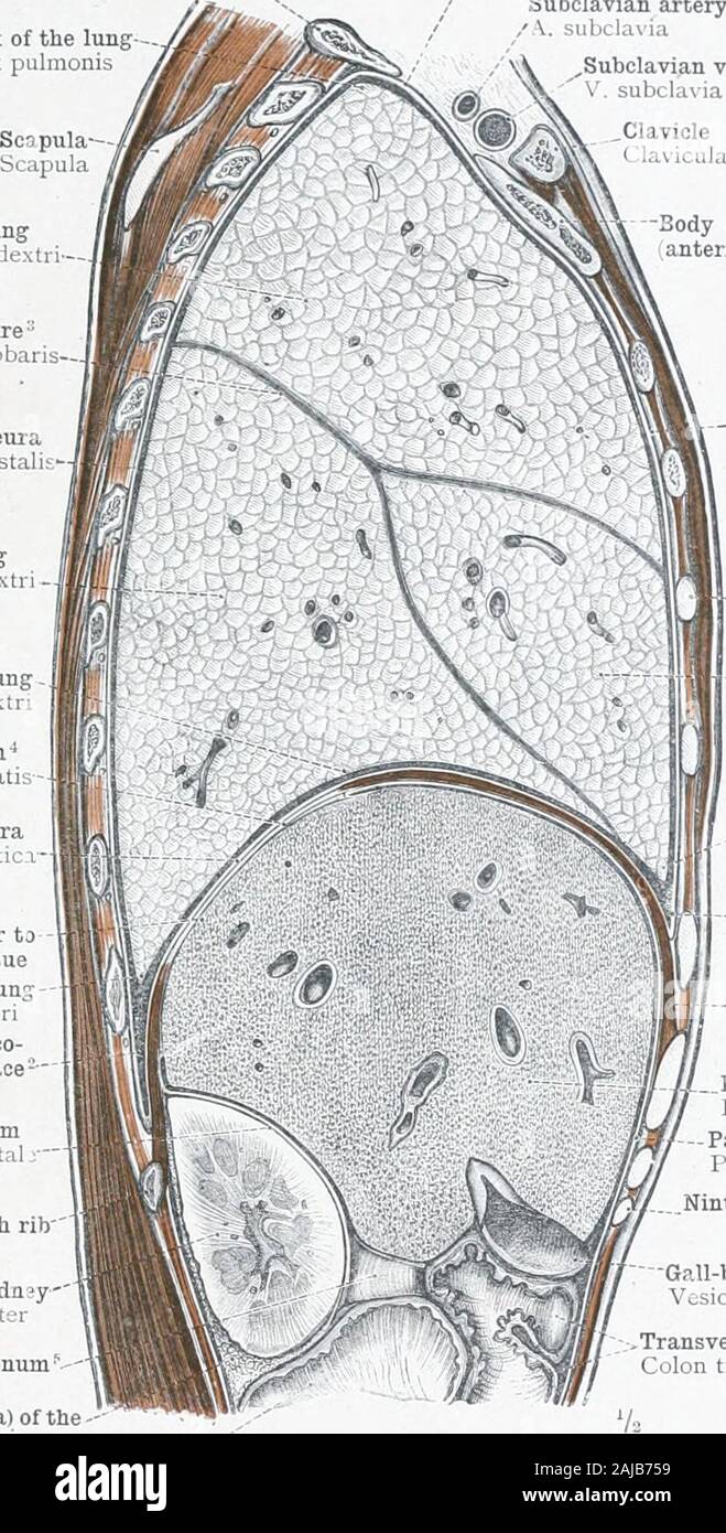 An atlas of human anatomy for students and physicians . mity). Corpus costx I, Apex of the lung-Apex pulmonis Cervical pleura yCupuIa pleura Subclavian artery / v. subclavia Subclavian vein subclaia Upper lobe of the right lungLobus superior pulmonis dextri-- Interlobar fissureIncisura interlobaris— Costal pleura Pleura costali Lower lobe of the right lung Lobus inferior pulmonis dextr -- Ease of the right lungBasis pulmonis dextri Central tendon of the diaphragm •* Centrum tendineum diaphragmatis Diaphragmatic pleura Pleura diaplira^matici Area of attachment of the liver to the diaphragm by Stock Photohttps://www.alamy.com/image-license-details/?v=1https://www.alamy.com/an-atlas-of-human-anatomy-for-students-and-physicians-mity-corpus-costx-i-apex-of-the-lung-apex-pulmonis-cervical-pleura-ycupuia-pleura-subclavian-artery-v-subclavia-subclavian-vein-subclaia-upper-lobe-of-the-right-lunglobus-superior-pulmonis-dextri-interlobar-fissureincisura-interlobaris-costal-pleura-pleura-costali-lower-lobe-of-the-right-lung-lobus-inferior-pulmonis-dextr-ease-of-the-right-lungbasis-pulmonis-dextri-central-tendon-of-the-diaphragm-centrum-tendineum-diaphragmatis-diaphragmatic-pleura-pleura-diapliramatici-area-of-attachment-of-the-liver-to-the-diaphragm-by-image338307909.html
RM2AJB759–An atlas of human anatomy for students and physicians . mity). Corpus costx I, Apex of the lung-Apex pulmonis Cervical pleura yCupuIa pleura Subclavian artery / v. subclavia Subclavian vein subclaia Upper lobe of the right lungLobus superior pulmonis dextri-- Interlobar fissureIncisura interlobaris— Costal pleura Pleura costali Lower lobe of the right lung Lobus inferior pulmonis dextr -- Ease of the right lungBasis pulmonis dextri Central tendon of the diaphragm •* Centrum tendineum diaphragmatis Diaphragmatic pleura Pleura diaplira^matici Area of attachment of the liver to the diaphragm by
 Pulmonary consumption, pneumonia, and allied diseases of the lungs : their etiology, pathology and treatment, with a chapter on physical diagnosis . Fig. 9.—Diagram showing the relation of the lobes of the lungs tothe front wall of the chest.—Fowler. It may be said that each lobe has a cone-like shape, withits apex directed either forward or backward. Thus theupper lobe of the left and the upper and middle lobes of theright lung have their apices behind and above, and theirbases in front; while the lower lobes of both lungs have theirapices in front and below, and their broad bases behind. 248 Stock Photohttps://www.alamy.com/image-license-details/?v=1https://www.alamy.com/pulmonary-consumption-pneumonia-and-allied-diseases-of-the-lungs-their-etiology-pathology-and-treatment-with-a-chapter-on-physical-diagnosis-fig-9diagram-showing-the-relation-of-the-lobes-of-the-lungs-tothe-front-wall-of-the-chestfowler-it-may-be-said-that-each-lobe-has-a-cone-like-shape-withits-apex-directed-either-forward-or-backward-thus-theupper-lobe-of-the-left-and-the-upper-and-middle-lobes-of-theright-lung-have-their-apices-behind-and-above-and-theirbases-in-front-while-the-lower-lobes-of-both-lungs-have-theirapices-in-front-and-below-and-their-broad-bases-behind-248-image339115143.html
RM2AKM0R3–Pulmonary consumption, pneumonia, and allied diseases of the lungs : their etiology, pathology and treatment, with a chapter on physical diagnosis . Fig. 9.—Diagram showing the relation of the lobes of the lungs tothe front wall of the chest.—Fowler. It may be said that each lobe has a cone-like shape, withits apex directed either forward or backward. Thus theupper lobe of the left and the upper and middle lobes of theright lung have their apices behind and above, and theirbases in front; while the lower lobes of both lungs have theirapices in front and below, and their broad bases behind. 248
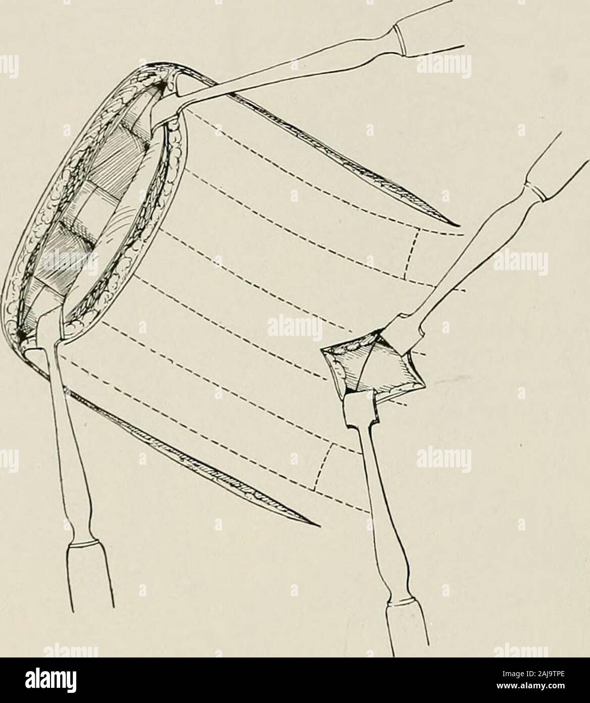 Surgical treatment; a practical treatise on the therapy of surgical diseases for the use of practitioners and students of surgery . this flap, the ribs may be broken. It is better to cut with forceps thetwo ribs adjacent to the incision and then divide the intervening rib or ribs through a smallincision at the base of the flap. 456 5 URGICAL TREA TMEN T For exposing the apex of the lung a U-shaped flap is made having itsconvexity at the border of the sternum, its base at the anterior axillary line,its upper arm at the first intercostal space, and its lower arm in the thirdintercostal space. If Stock Photohttps://www.alamy.com/image-license-details/?v=1https://www.alamy.com/surgical-treatment-a-practical-treatise-on-the-therapy-of-surgical-diseases-for-the-use-of-practitioners-and-students-of-surgery-this-flap-the-ribs-may-be-broken-it-is-better-to-cut-with-forceps-thetwo-ribs-adjacent-to-the-incision-and-then-divide-the-intervening-rib-or-ribs-through-a-smallincision-at-the-base-of-the-flap-456-5-urgical-trea-tmen-t-for-exposing-the-apex-of-the-lung-a-u-shaped-flap-is-made-having-itsconvexity-at-the-border-of-the-sternum-its-base-at-the-anterior-axillary-lineits-upper-arm-at-the-first-intercostal-space-and-its-lower-arm-in-the-thirdintercostal-space-if-image338277814.html
RM2AJ9TPE–Surgical treatment; a practical treatise on the therapy of surgical diseases for the use of practitioners and students of surgery . this flap, the ribs may be broken. It is better to cut with forceps thetwo ribs adjacent to the incision and then divide the intervening rib or ribs through a smallincision at the base of the flap. 456 5 URGICAL TREA TMEN T For exposing the apex of the lung a U-shaped flap is made having itsconvexity at the border of the sternum, its base at the anterior axillary line,its upper arm at the first intercostal space, and its lower arm in the thirdintercostal space. If
 Diseases of the chest and the principles of physical diagnosis . is present or not. If the apex beat is displaced to the left it may becaused by one of three things: (1) It may be due to hypertrophy or dila-tation of the heart itself. It is hardly likely that this would cause anyconfusion, because of other associated cardiac signs. (2) The heart maybe pulled to the left as the result of a fibrosis involving the left lung. Insuch a case, however, it will be noted that the left chest is retracted.(3) It may be due to a pleural effusion on the right side which has dis-placed the heart to the left Stock Photohttps://www.alamy.com/image-license-details/?v=1https://www.alamy.com/diseases-of-the-chest-and-the-principles-of-physical-diagnosis-is-present-or-not-if-the-apex-beat-is-displaced-to-the-left-it-may-becaused-by-one-of-three-things-1-it-may-be-due-to-hypertrophy-or-dila-tation-of-the-heart-itself-it-is-hardly-likely-that-this-would-cause-anyconfusion-because-of-other-associated-cardiac-signs-2-the-heart-maybe-pulled-to-the-left-as-the-result-of-a-fibrosis-involving-the-left-lung-insuch-a-case-however-it-will-be-noted-that-the-left-chest-is-retracted3-it-may-be-due-to-a-pleural-effusion-on-the-right-side-which-has-dis-placed-the-heart-to-the-left-image340220163.html
RM2ANEA83–Diseases of the chest and the principles of physical diagnosis . is present or not. If the apex beat is displaced to the left it may becaused by one of three things: (1) It may be due to hypertrophy or dila-tation of the heart itself. It is hardly likely that this would cause anyconfusion, because of other associated cardiac signs. (2) The heart maybe pulled to the left as the result of a fibrosis involving the left lung. Insuch a case, however, it will be noted that the left chest is retracted.(3) It may be due to a pleural effusion on the right side which has dis-placed the heart to the left
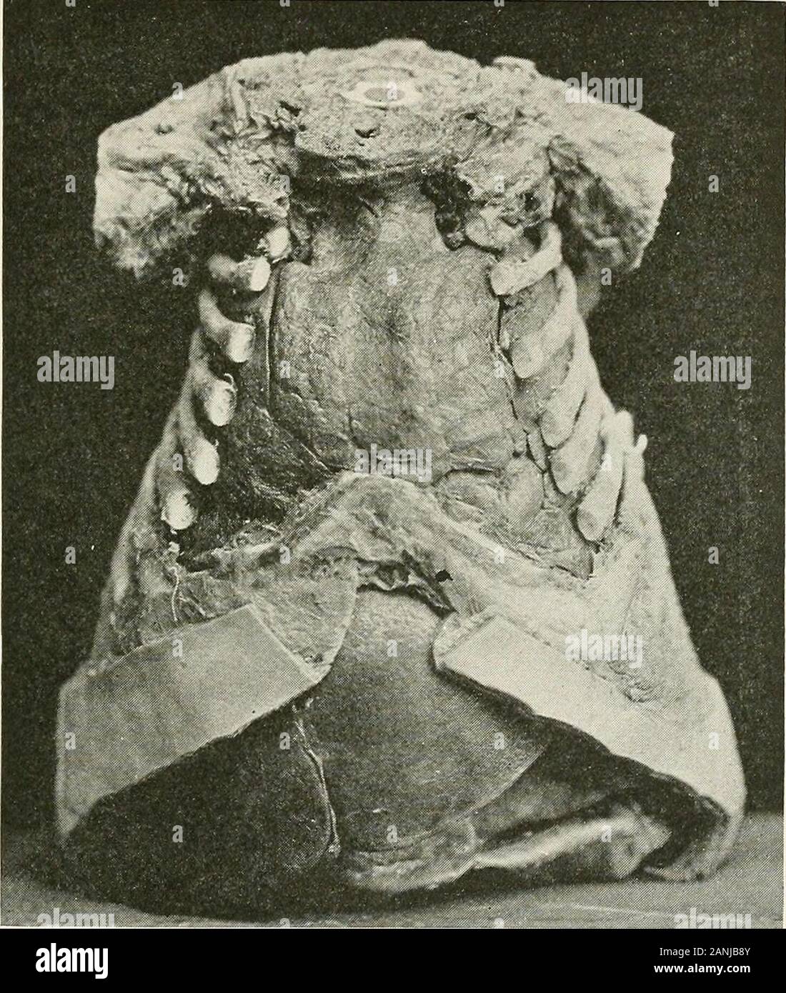 Diseases of the chest and the principles of physical diagnosis . nin adults (see vocal resonance). The apex heat is in the fourth interspace,just within or even to the left of the left mid-clavicular line. 1 For assistance in the preparation of the following paragraphs, we are indebted toDr. J. C. Gittings. 138 PHYSICAL FINDINGS IN INFANTS AND YOUNG CHILDREN 139 Percussion.—Percussion must be extremely light—finger percussion isoften necessary. If a forcible stroke be employed the whole lung as wellas the neighboring abdominal viscera will be thrown into vibration andtopographic percussion wil Stock Photohttps://www.alamy.com/image-license-details/?v=1https://www.alamy.com/diseases-of-the-chest-and-the-principles-of-physical-diagnosis-nin-adults-see-vocal-resonance-the-apex-heat-is-in-the-fourth-interspacejust-within-or-even-to-the-left-of-the-left-mid-clavicular-line-1-for-assistance-in-the-preparation-of-the-following-paragraphs-we-are-indebted-todr-j-c-gittings-138-physical-findings-in-infants-and-young-children-139-percussionpercussion-must-be-extremely-lightfinger-percussion-isoften-necessary-if-a-forcible-stroke-be-employed-the-whole-lung-as-wellas-the-neighboring-abdominal-viscera-will-be-thrown-into-vibration-andtopographic-percussion-wil-image340308779.html
RM2ANJB8Y–Diseases of the chest and the principles of physical diagnosis . nin adults (see vocal resonance). The apex heat is in the fourth interspace,just within or even to the left of the left mid-clavicular line. 1 For assistance in the preparation of the following paragraphs, we are indebted toDr. J. C. Gittings. 138 PHYSICAL FINDINGS IN INFANTS AND YOUNG CHILDREN 139 Percussion.—Percussion must be extremely light—finger percussion isoften necessary. If a forcible stroke be employed the whole lung as wellas the neighboring abdominal viscera will be thrown into vibration andtopographic percussion wil
 Manual of clinical medicine and physical diagnosis . the fourth. The left auricleis covered by the right auricle and the left lung. Theleft ventricle lies mostly away from the surface, its upperedge is covered by the left lung, but its apex comesforward to the surface below, and beats between thefifth and sixth ribs, an inch or perhaps less to the inside,and one and a half inches below the left nipple. It willbe seen that there is a small space commencing abovewhere the left lung leaves its fellow, at the fourth cartilage,in which the heart is uncovered. The relative position of 188 PHYSICAL D Stock Photohttps://www.alamy.com/image-license-details/?v=1https://www.alamy.com/manual-of-clinical-medicine-and-physical-diagnosis-the-fourth-the-left-auricleis-covered-by-the-right-auricle-and-the-left-lung-theleft-ventricle-lies-mostly-away-from-the-surface-its-upperedge-is-covered-by-the-left-lung-but-its-apex-comesforward-to-the-surface-below-and-beats-between-thefifth-and-sixth-ribs-an-inch-or-perhaps-less-to-the-insideand-one-and-a-half-inches-below-the-left-nipple-it-willbe-seen-that-there-is-a-small-space-commencing-abovewhere-the-left-lung-leaves-its-fellow-at-the-fourth-cartilagein-which-the-heart-is-uncovered-the-relative-position-of-188-physical-d-image339195894.html
RM2AKRKR2–Manual of clinical medicine and physical diagnosis . the fourth. The left auricleis covered by the right auricle and the left lung. Theleft ventricle lies mostly away from the surface, its upperedge is covered by the left lung, but its apex comesforward to the surface below, and beats between thefifth and sixth ribs, an inch or perhaps less to the inside,and one and a half inches below the left nipple. It willbe seen that there is a small space commencing abovewhere the left lung leaves its fellow, at the fourth cartilage,in which the heart is uncovered. The relative position of 188 PHYSICAL D
 . Medical diagnosis for the student and practitioner. Fig. no.—Anterior surface. Lungborders. Forced expiration. Fig. in.—Lateral surface. Lungborders. Forced inspiration.. Fig. ii2.—Posterior surface. Lungborders. Quiet breathing. Fig. 113.—Posterior surface. Lungborders. Forced inspiration. The Lung Borders.—The position and mobility of the lung borders areaffected in every serious chronic disease of the lung. In tuberculosis, they show a decided lack of mobility, both at apex andbase. * See Deutsche Klinik., Band ii, 1907. Importantsign. 296 MEDICAL DIAGNOSIS Compressedlung bases. Of limite Stock Photohttps://www.alamy.com/image-license-details/?v=1https://www.alamy.com/medical-diagnosis-for-the-student-and-practitioner-fig-noanterior-surface-lungborders-forced-expiration-fig-inlateral-surface-lungborders-forced-inspiration-fig-ii2posterior-surface-lungborders-quiet-breathing-fig-113posterior-surface-lungborders-forced-inspiration-the-lung-bordersthe-position-and-mobility-of-the-lung-borders-areaffected-in-every-serious-chronic-disease-of-the-lung-in-tuberculosis-they-show-a-decided-lack-of-mobility-both-at-apex-andbase-see-deutsche-klinik-band-ii-1907-importantsign-296-medical-diagnosis-compressedlung-bases-of-limite-image336900340.html
RM2AG33R0–. Medical diagnosis for the student and practitioner. Fig. no.—Anterior surface. Lungborders. Forced expiration. Fig. in.—Lateral surface. Lungborders. Forced inspiration.. Fig. ii2.—Posterior surface. Lungborders. Quiet breathing. Fig. 113.—Posterior surface. Lungborders. Forced inspiration. The Lung Borders.—The position and mobility of the lung borders areaffected in every serious chronic disease of the lung. In tuberculosis, they show a decided lack of mobility, both at apex andbase. * See Deutsche Klinik., Band ii, 1907. Importantsign. 296 MEDICAL DIAGNOSIS Compressedlung bases. Of limite