Atom electron rings Stock Photos and Images
 Glowing yellow orbits and a neutron forming atomic symbol on orange and black radial sunburst lines. Concept of quantum mechanics and computing Stock Photohttps://www.alamy.com/image-license-details/?v=1https://www.alamy.com/glowing-yellow-orbits-and-a-neutron-forming-atomic-symbol-on-orange-and-black-radial-sunburst-lines-concept-of-quantum-mechanics-and-computing-image600508216.html
RF2WTYEA0–Glowing yellow orbits and a neutron forming atomic symbol on orange and black radial sunburst lines. Concept of quantum mechanics and computing
 Floating blue sphere podium, platform or pedestal with rings and orbital spheres. 3d render, modern minimalistic futuristic science background. Stock Photohttps://www.alamy.com/image-license-details/?v=1https://www.alamy.com/floating-blue-sphere-podium-platform-or-pedestal-with-rings-and-orbital-spheres-3d-render-modern-minimalistic-futuristic-science-background-image561292233.html
RF2RH51Y5–Floating blue sphere podium, platform or pedestal with rings and orbital spheres. 3d render, modern minimalistic futuristic science background.
 Atom spheres with cyan organic background, 3d rendering. Computer digital drawing. Stock Photohttps://www.alamy.com/image-license-details/?v=1https://www.alamy.com/atom-spheres-with-cyan-organic-background-3d-rendering-computer-digital-drawing-image367490030.html
RF2C9TH7X–Atom spheres with cyan organic background, 3d rendering. Computer digital drawing.
 3D Model of an atom against a white background. Stock Photohttps://www.alamy.com/image-license-details/?v=1https://www.alamy.com/stock-photo-3d-model-of-an-atom-against-a-white-background-22671597.html
RFB8TNRW–3D Model of an atom against a white background.
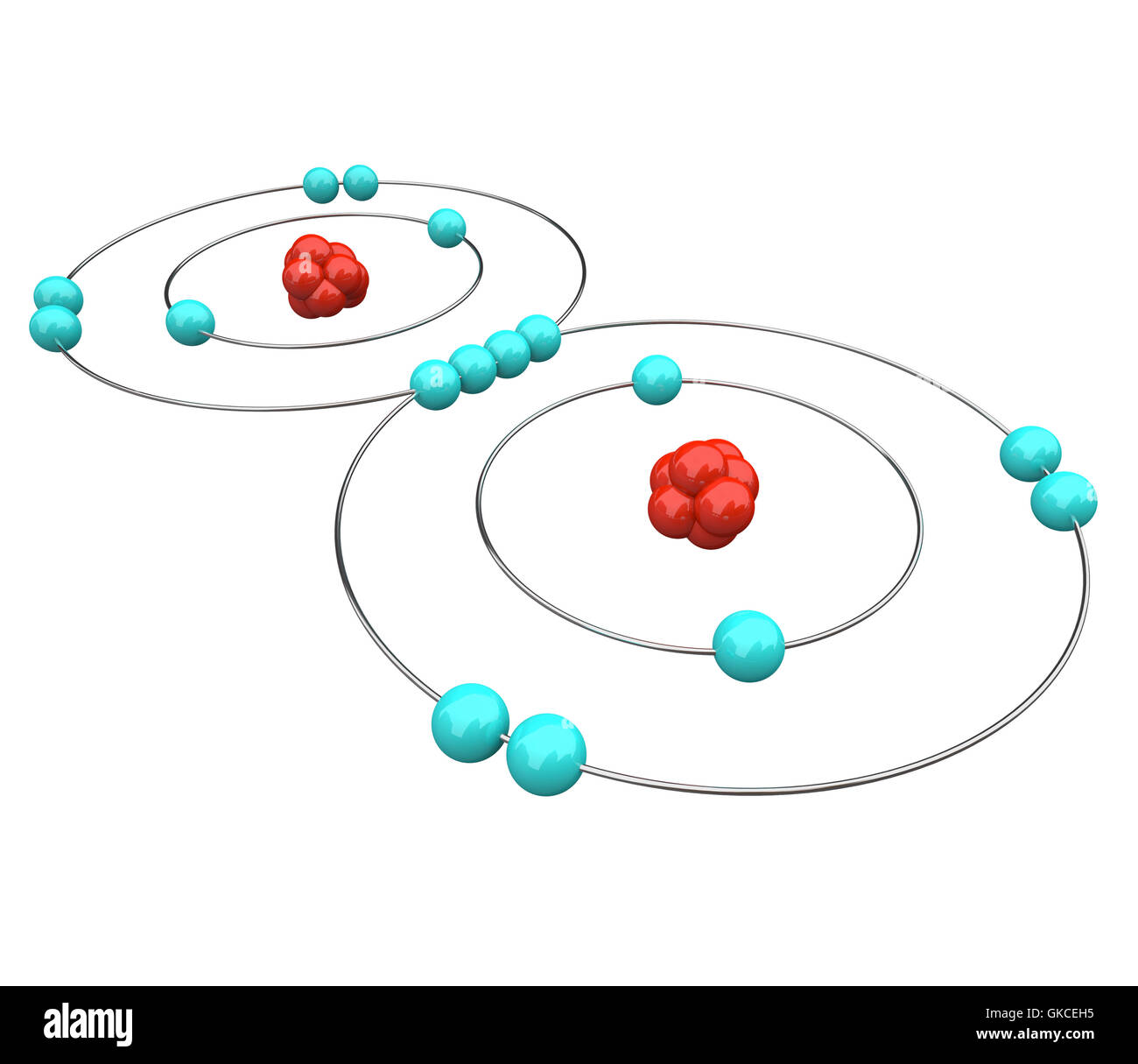 Oxygen - Atomic Diagram Stock Photohttps://www.alamy.com/image-license-details/?v=1https://www.alamy.com/stock-photo-oxygen-atomic-diagram-115215553.html
RFGKCEH5–Oxygen - Atomic Diagram
 Collection of global science future connection glowing abstract background vector illustration Stock Vectorhttps://www.alamy.com/image-license-details/?v=1https://www.alamy.com/collection-of-global-science-future-connection-glowing-abstract-background-vector-illustration-image472497043.html
RF2JCM2PB–Collection of global science future connection glowing abstract background vector illustration
 The structure of the graphene tube of nanotechnology 3d illustration Stock Photohttps://www.alamy.com/image-license-details/?v=1https://www.alamy.com/stock-photo-the-structure-of-the-graphene-tube-of-nanotechnology-3d-illustration-108018275.html
RMG7MJBF–The structure of the graphene tube of nanotechnology 3d illustration
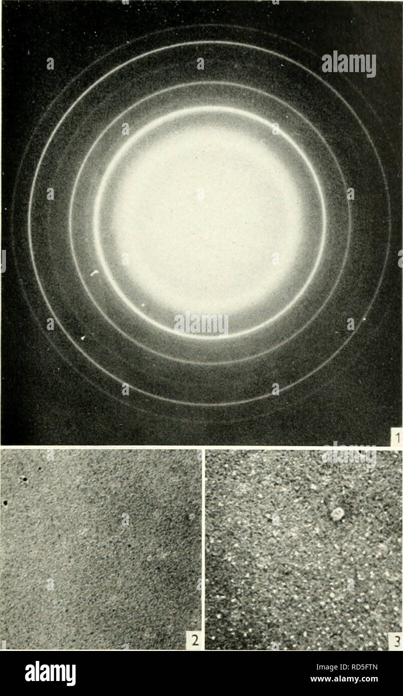 . Electron microscopy; proceedings of the Stockholm Conference, September, 1956. Electron microscopy. Identification of Minerals in Mine Dusts 353. Fig. 1. Diffraction pattern of PbTe layer. Fig. 2. PbTe layer exposed to intense electron beam during 10-20 seconds. Magnification 6000. Fig. 3. PbTe layer exposed to intense electron beam longer than 20 seconds. Magnification 6800. rings of the NaCl type structure, the intensity of which is determined by the difference of atom factors. Thus, a crystal lattice of the kind proposed shows an electron diiTraction pattern corresponding to simple cubic Stock Photohttps://www.alamy.com/image-license-details/?v=1https://www.alamy.com/electron-microscopy-proceedings-of-the-stockholm-conference-september-1956-electron-microscopy-identification-of-minerals-in-mine-dusts-353-fig-1-diffraction-pattern-of-pbte-layer-fig-2-pbte-layer-exposed-to-intense-electron-beam-during-10-20-seconds-magnification-6000-fig-3-pbte-layer-exposed-to-intense-electron-beam-longer-than-20-seconds-magnification-6800-rings-of-the-nacl-type-structure-the-intensity-of-which-is-determined-by-the-difference-of-atom-factors-thus-a-crystal-lattice-of-the-kind-proposed-shows-an-electron-diitraction-pattern-corresponding-to-simple-cubic-image231847525.html
RMRD5FTN–. Electron microscopy; proceedings of the Stockholm Conference, September, 1956. Electron microscopy. Identification of Minerals in Mine Dusts 353. Fig. 1. Diffraction pattern of PbTe layer. Fig. 2. PbTe layer exposed to intense electron beam during 10-20 seconds. Magnification 6000. Fig. 3. PbTe layer exposed to intense electron beam longer than 20 seconds. Magnification 6800. rings of the NaCl type structure, the intensity of which is determined by the difference of atom factors. Thus, a crystal lattice of the kind proposed shows an electron diiTraction pattern corresponding to simple cubic
 Abstract colorful explosion of neon nucleus isolated on black background. Amazing 3d impulse spreading into the sides surrounded by small particles. Stock Photohttps://www.alamy.com/image-license-details/?v=1https://www.alamy.com/abstract-colorful-explosion-of-neon-nucleus-isolated-on-black-background-amazing-3d-impulse-spreading-into-the-sides-surrounded-by-small-particles-image454868253.html
RF2HC112N–Abstract colorful explosion of neon nucleus isolated on black background. Amazing 3d impulse spreading into the sides surrounded by small particles.
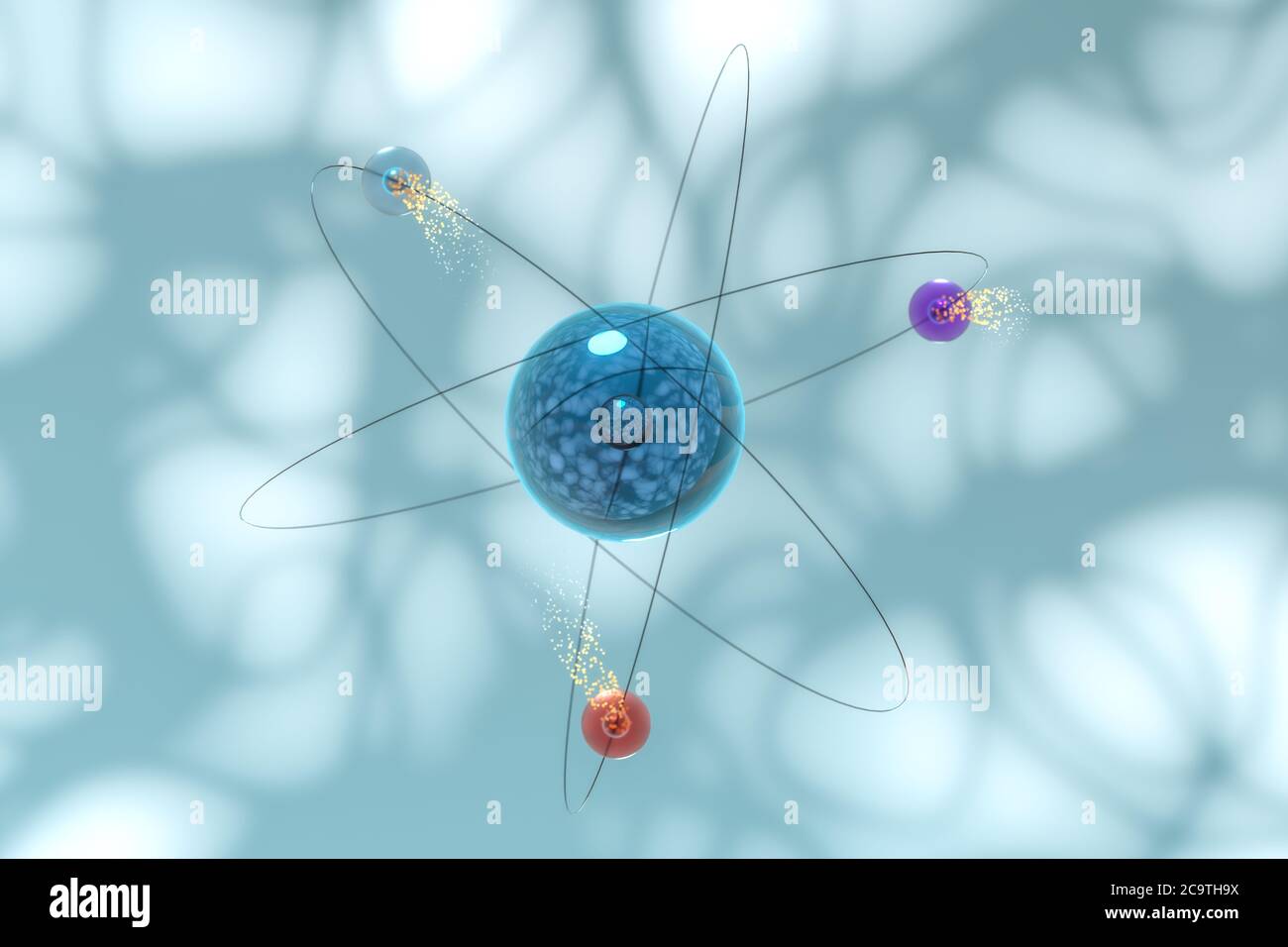 Atom spheres with blue organic background, 3d rendering. Computer digital drawing. Stock Photohttps://www.alamy.com/image-license-details/?v=1https://www.alamy.com/atom-spheres-with-blue-organic-background-3d-rendering-computer-digital-drawing-image367490086.html
RF2C9TH9X–Atom spheres with blue organic background, 3d rendering. Computer digital drawing.
 Heavy Uranium Atom Electrons Orbit Around Nucleus Science Concept - Abstract Background Texture Stock Photohttps://www.alamy.com/image-license-details/?v=1https://www.alamy.com/heavy-uranium-atom-electrons-orbit-around-nucleus-science-concept-abstract-background-texture-image363849876.html
RF2C3XP6C–Heavy Uranium Atom Electrons Orbit Around Nucleus Science Concept - Abstract Background Texture
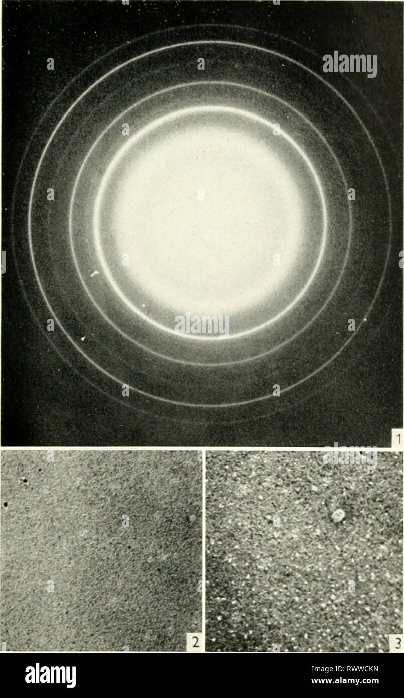 Electron microscopy; proceedings of the Electron microscopy; proceedings of the Stockholm Conference, September, 1956 electronmicrosco00euro Year: 1957 Identification of Minerals in Mine Dusts 353 Fig. 1. Diffraction pattern of PbTe layer. Fig. 2. PbTe layer exposed to intense electron beam during 10-20 seconds. Magnification 6000. Fig. 3. PbTe layer exposed to intense electron beam longer than 20 seconds. Magnification 6800. rings of the NaCl type structure, the intensity of which is determined by the difference of atom factors. Thus, a crystal lattice of the kind proposed shows an electr Stock Photohttps://www.alamy.com/image-license-details/?v=1https://www.alamy.com/electron-microscopy-proceedings-of-the-electron-microscopy-proceedings-of-the-stockholm-conference-september-1956-electronmicrosco00euro-year-1957-identification-of-minerals-in-mine-dusts-353-fig-1-diffraction-pattern-of-pbte-layer-fig-2-pbte-layer-exposed-to-intense-electron-beam-during-10-20-seconds-magnification-6000-fig-3-pbte-layer-exposed-to-intense-electron-beam-longer-than-20-seconds-magnification-6800-rings-of-the-nacl-type-structure-the-intensity-of-which-is-determined-by-the-difference-of-atom-factors-thus-a-crystal-lattice-of-the-kind-proposed-shows-an-electr-image239659945.html
RMRWWCKN–Electron microscopy; proceedings of the Electron microscopy; proceedings of the Stockholm Conference, September, 1956 electronmicrosco00euro Year: 1957 Identification of Minerals in Mine Dusts 353 Fig. 1. Diffraction pattern of PbTe layer. Fig. 2. PbTe layer exposed to intense electron beam during 10-20 seconds. Magnification 6000. Fig. 3. PbTe layer exposed to intense electron beam longer than 20 seconds. Magnification 6800. rings of the NaCl type structure, the intensity of which is determined by the difference of atom factors. Thus, a crystal lattice of the kind proposed shows an electr
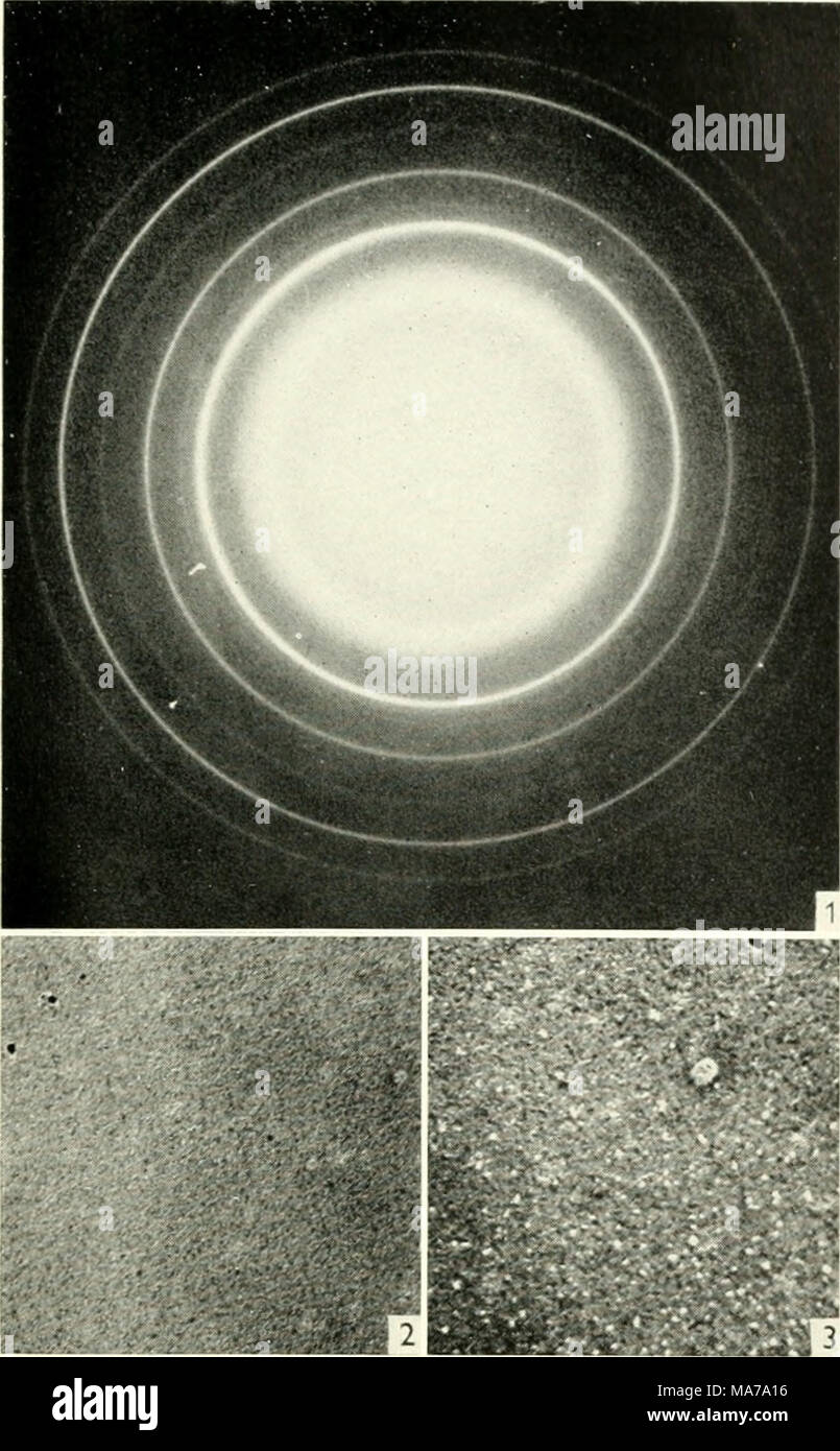 . Electron microscopy; proceedings of the Stockholm Conference, September, 1956 . Fig. 1. Diffraction pattern of PbTe layer. Fig. 2. PbTe layer exposed to intense electron beam during 10-20 seconds. Magnification 6000. Fig. 3. PbTe layer exposed to intense electron beam longer than 20 seconds. Magnification 6800. rings of the NaCl type structure, the intensity of which is determined by the difference of atom factors. Thus, a crystal lattice of the kind proposed shows an electron diiTraction pattern corresponding to simple cubic structure with the lattice constant halved. Of the three analogous Stock Photohttps://www.alamy.com/image-license-details/?v=1https://www.alamy.com/electron-microscopy-proceedings-of-the-stockholm-conference-september-1956-fig-1-diffraction-pattern-of-pbte-layer-fig-2-pbte-layer-exposed-to-intense-electron-beam-during-10-20-seconds-magnification-6000-fig-3-pbte-layer-exposed-to-intense-electron-beam-longer-than-20-seconds-magnification-6800-rings-of-the-nacl-type-structure-the-intensity-of-which-is-determined-by-the-difference-of-atom-factors-thus-a-crystal-lattice-of-the-kind-proposed-shows-an-electron-diitraction-pattern-corresponding-to-simple-cubic-structure-with-the-lattice-constant-halved-of-the-three-analogous-image178411778.html
RMMA7A16–. Electron microscopy; proceedings of the Stockholm Conference, September, 1956 . Fig. 1. Diffraction pattern of PbTe layer. Fig. 2. PbTe layer exposed to intense electron beam during 10-20 seconds. Magnification 6000. Fig. 3. PbTe layer exposed to intense electron beam longer than 20 seconds. Magnification 6800. rings of the NaCl type structure, the intensity of which is determined by the difference of atom factors. Thus, a crystal lattice of the kind proposed shows an electron diiTraction pattern corresponding to simple cubic structure with the lattice constant halved. Of the three analogous
 Floating yellow sphere podium, platform or pedestal with rings and orbital spheres. 3d render, modern minimalistic futuristic science background. Stock Photohttps://www.alamy.com/image-license-details/?v=1https://www.alamy.com/floating-yellow-sphere-podium-platform-or-pedestal-with-rings-and-orbital-spheres-3d-render-modern-minimalistic-futuristic-science-background-image563437936.html
RF2RMJPRC–Floating yellow sphere podium, platform or pedestal with rings and orbital spheres. 3d render, modern minimalistic futuristic science background.
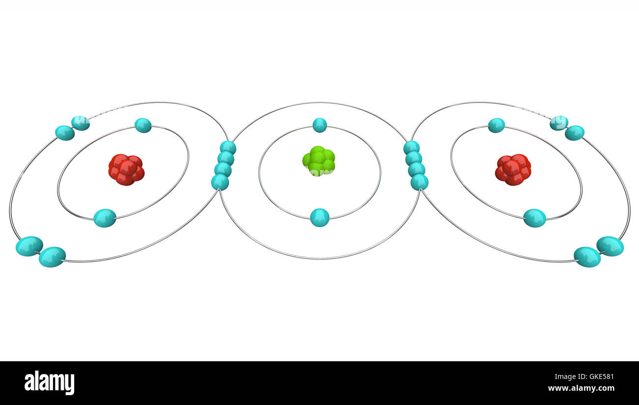 Carbon Dioxide CO2 - Atomic Diagram Stock Photohttps://www.alamy.com/image-license-details/?v=1https://www.alamy.com/stock-photo-carbon-dioxide-co2-atomic-diagram-115252145.html
RFGKE581–Carbon Dioxide CO2 - Atomic Diagram
RFWX53E9–Icons
 Abstract colorful explosion of neon nucleus isolated on black background. Amazing 3d impulse spreading into the sides surrounded by small particles. Stock Photohttps://www.alamy.com/image-license-details/?v=1https://www.alamy.com/abstract-colorful-explosion-of-neon-nucleus-isolated-on-black-background-amazing-3d-impulse-spreading-into-the-sides-surrounded-by-small-particles-image454970309.html
RF2HC5K7H–Abstract colorful explosion of neon nucleus isolated on black background. Amazing 3d impulse spreading into the sides surrounded by small particles.
 Atom spheres with blue organic background, 3d rendering. Computer digital drawing. Stock Photohttps://www.alamy.com/image-license-details/?v=1https://www.alamy.com/atom-spheres-with-blue-organic-background-3d-rendering-computer-digital-drawing-image367490089.html
RF2C9THA1–Atom spheres with blue organic background, 3d rendering. Computer digital drawing.
 Electron microscopy; proceedings of the Electron microscopy; proceedings of the Stockholm Conference, September, 1956 electronmicrosco00euro Year: 1957 Identification of Minerals in Mine Dusts 353 Fig. 1. Diffraction pattern of PbTe layer. Fig. 2. PbTe layer exposed to intense electron beam during 10-20 seconds. Magnification 6000. Fig. 3. PbTe layer exposed to intense electron beam longer than 20 seconds. Magnification 6800. rings of the NaCl type structure, the intensity of which is determined by the difference of atom factors. Thus, a crystal lattice of the kind proposed shows an electr Stock Photohttps://www.alamy.com/image-license-details/?v=1https://www.alamy.com/electron-microscopy-proceedings-of-the-electron-microscopy-proceedings-of-the-stockholm-conference-september-1956-electronmicrosco00euro-year-1957-identification-of-minerals-in-mine-dusts-353-fig-1-diffraction-pattern-of-pbte-layer-fig-2-pbte-layer-exposed-to-intense-electron-beam-during-10-20-seconds-magnification-6000-fig-3-pbte-layer-exposed-to-intense-electron-beam-longer-than-20-seconds-magnification-6800-rings-of-the-nacl-type-structure-the-intensity-of-which-is-determined-by-the-difference-of-atom-factors-thus-a-crystal-lattice-of-the-kind-proposed-shows-an-electr-image241231451.html
RMT0D14Y–Electron microscopy; proceedings of the Electron microscopy; proceedings of the Stockholm Conference, September, 1956 electronmicrosco00euro Year: 1957 Identification of Minerals in Mine Dusts 353 Fig. 1. Diffraction pattern of PbTe layer. Fig. 2. PbTe layer exposed to intense electron beam during 10-20 seconds. Magnification 6000. Fig. 3. PbTe layer exposed to intense electron beam longer than 20 seconds. Magnification 6800. rings of the NaCl type structure, the intensity of which is determined by the difference of atom factors. Thus, a crystal lattice of the kind proposed shows an electr
RFWX48PB–Icons
 Motion of atom spheres isolated on a black background, concept of chemistry and biology. Design. Small balls rotating around bigger black sphere Stock Photohttps://www.alamy.com/image-license-details/?v=1https://www.alamy.com/motion-of-atom-spheres-isolated-on-a-black-background-concept-of-chemistry-and-biology-design-small-balls-rotating-around-bigger-black-sphere-image463781764.html
RF2HXF2AC–Motion of atom spheres isolated on a black background, concept of chemistry and biology. Design. Small balls rotating around bigger black sphere
 Atom spheres with pink organic background, 3d rendering. Computer digital drawing. Stock Photohttps://www.alamy.com/image-license-details/?v=1https://www.alamy.com/atom-spheres-with-pink-organic-background-3d-rendering-computer-digital-drawing-image367490161.html
RF2C9THCH–Atom spheres with pink organic background, 3d rendering. Computer digital drawing.
 Abstract colorful explosion of neon nucleus isolated on black background. Animation. Amazing 3d impulse spreading into the sides surrounded by small Stock Photohttps://www.alamy.com/image-license-details/?v=1https://www.alamy.com/abstract-colorful-explosion-of-neon-nucleus-isolated-on-black-background-animation-amazing-3d-impulse-spreading-into-the-sides-surrounded-by-small-image463700217.html
RF2HXBAA1–Abstract colorful explosion of neon nucleus isolated on black background. Animation. Amazing 3d impulse spreading into the sides surrounded by small
 Atom spheres with pink organic background, 3d rendering. Computer digital drawing. Stock Photohttps://www.alamy.com/image-license-details/?v=1https://www.alamy.com/atom-spheres-with-pink-organic-background-3d-rendering-computer-digital-drawing-image367490085.html
RF2C9TH9W–Atom spheres with pink organic background, 3d rendering. Computer digital drawing.
 Atom spheres with pink organic background, 3d rendering. Computer digital drawing. Stock Photohttps://www.alamy.com/image-license-details/?v=1https://www.alamy.com/atom-spheres-with-pink-organic-background-3d-rendering-computer-digital-drawing-image367490025.html
RF2C9TH7N–Atom spheres with pink organic background, 3d rendering. Computer digital drawing.
 Atom spheres with pink organic background, 3d rendering. Computer digital drawing. Stock Photohttps://www.alamy.com/image-license-details/?v=1https://www.alamy.com/atom-spheres-with-pink-organic-background-3d-rendering-computer-digital-drawing-image367490088.html
RF2C9THA0–Atom spheres with pink organic background, 3d rendering. Computer digital drawing.
 Atom spheres with cyan organic background, 3d rendering. Computer digital drawing. Stock Photohttps://www.alamy.com/image-license-details/?v=1https://www.alamy.com/atom-spheres-with-cyan-organic-background-3d-rendering-computer-digital-drawing-image367490022.html
RF2C9TH7J–Atom spheres with cyan organic background, 3d rendering. Computer digital drawing.
 Atom spheres with pink organic background, 3d rendering. Computer digital drawing. Stock Photohttps://www.alamy.com/image-license-details/?v=1https://www.alamy.com/atom-spheres-with-pink-organic-background-3d-rendering-computer-digital-drawing-image367490087.html
RF2C9TH9Y–Atom spheres with pink organic background, 3d rendering. Computer digital drawing.
 Atom spheres with cyan organic background, 3d rendering. Computer digital drawing. Stock Photohttps://www.alamy.com/image-license-details/?v=1https://www.alamy.com/atom-spheres-with-cyan-organic-background-3d-rendering-computer-digital-drawing-image367490035.html
RF2C9TH83–Atom spheres with cyan organic background, 3d rendering. Computer digital drawing.