Quick filters:
Axis vertebra and human Stock Photos and Images
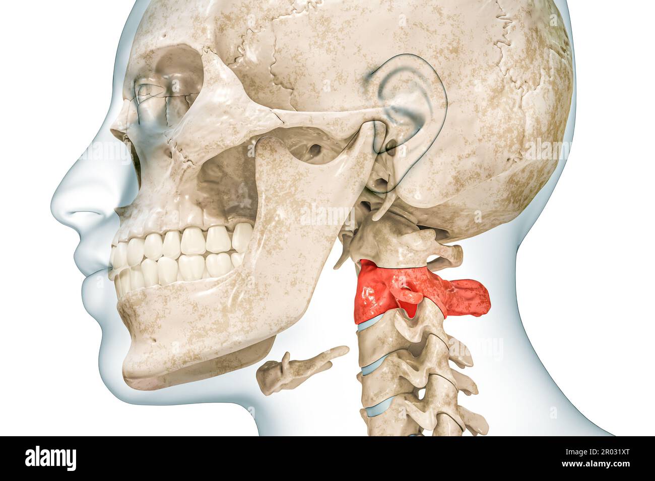 Axis second cervical vertebra in red color with body 3D rendering illustration isolated on white with copy space. Human skeleton ans spine anatomy, me Stock Photohttps://www.alamy.com/image-license-details/?v=1https://www.alamy.com/axis-second-cervical-vertebra-in-red-color-with-body-3d-rendering-illustration-isolated-on-white-with-copy-space-human-skeleton-ans-spine-anatomy-me-image550799168.html
RF2R031XT–Axis second cervical vertebra in red color with body 3D rendering illustration isolated on white with copy space. Human skeleton ans spine anatomy, me
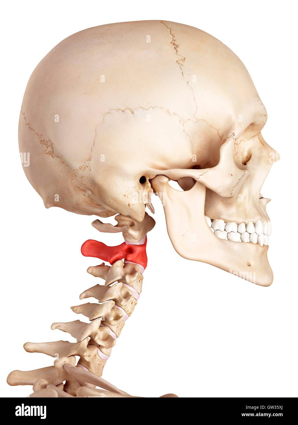 Human axis bone, illustration. Stock Photohttps://www.alamy.com/image-license-details/?v=1https://www.alamy.com/stock-photo-human-axis-bone-illustration-118699130.html
RFGW35XJ–Human axis bone, illustration.
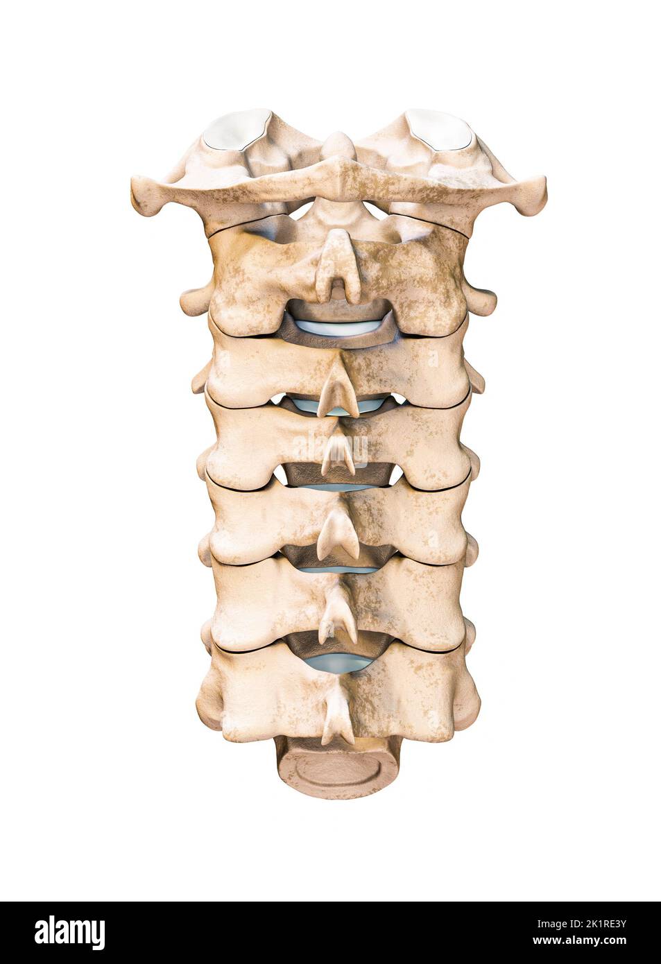 Posterior or rear or back view of the seven human cervical vertebrae isolated on white background 3D rendering illustration. Anatomy, osteology, blank Stock Photohttps://www.alamy.com/image-license-details/?v=1https://www.alamy.com/posterior-or-rear-or-back-view-of-the-seven-human-cervical-vertebrae-isolated-on-white-background-3d-rendering-illustration-anatomy-osteology-blank-image483020943.html
RF2K1RE3Y–Posterior or rear or back view of the seven human cervical vertebrae isolated on white background 3D rendering illustration. Anatomy, osteology, blank
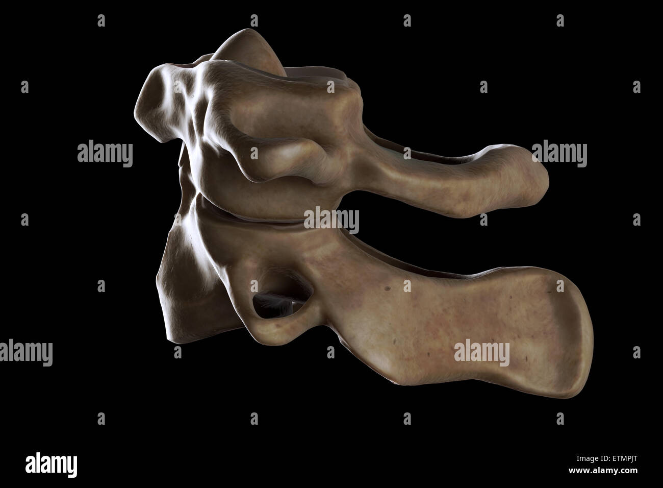 Illustration showing the atlas and axis vertebrae of the neck. Stock Photohttps://www.alamy.com/image-license-details/?v=1https://www.alamy.com/stock-photo-illustration-showing-the-atlas-and-axis-vertebrae-of-the-neck-84050032.html
RMETMPJT–Illustration showing the atlas and axis vertebrae of the neck.
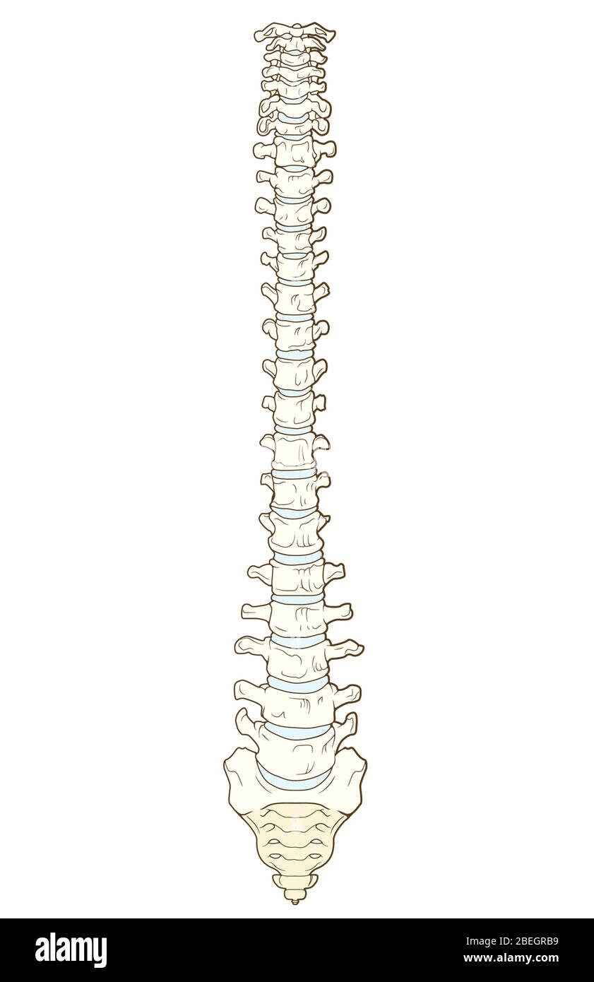 Vertebral Column Stock Photohttps://www.alamy.com/image-license-details/?v=1https://www.alamy.com/vertebral-column-image353182125.html
RM2BEGRB9–Vertebral Column
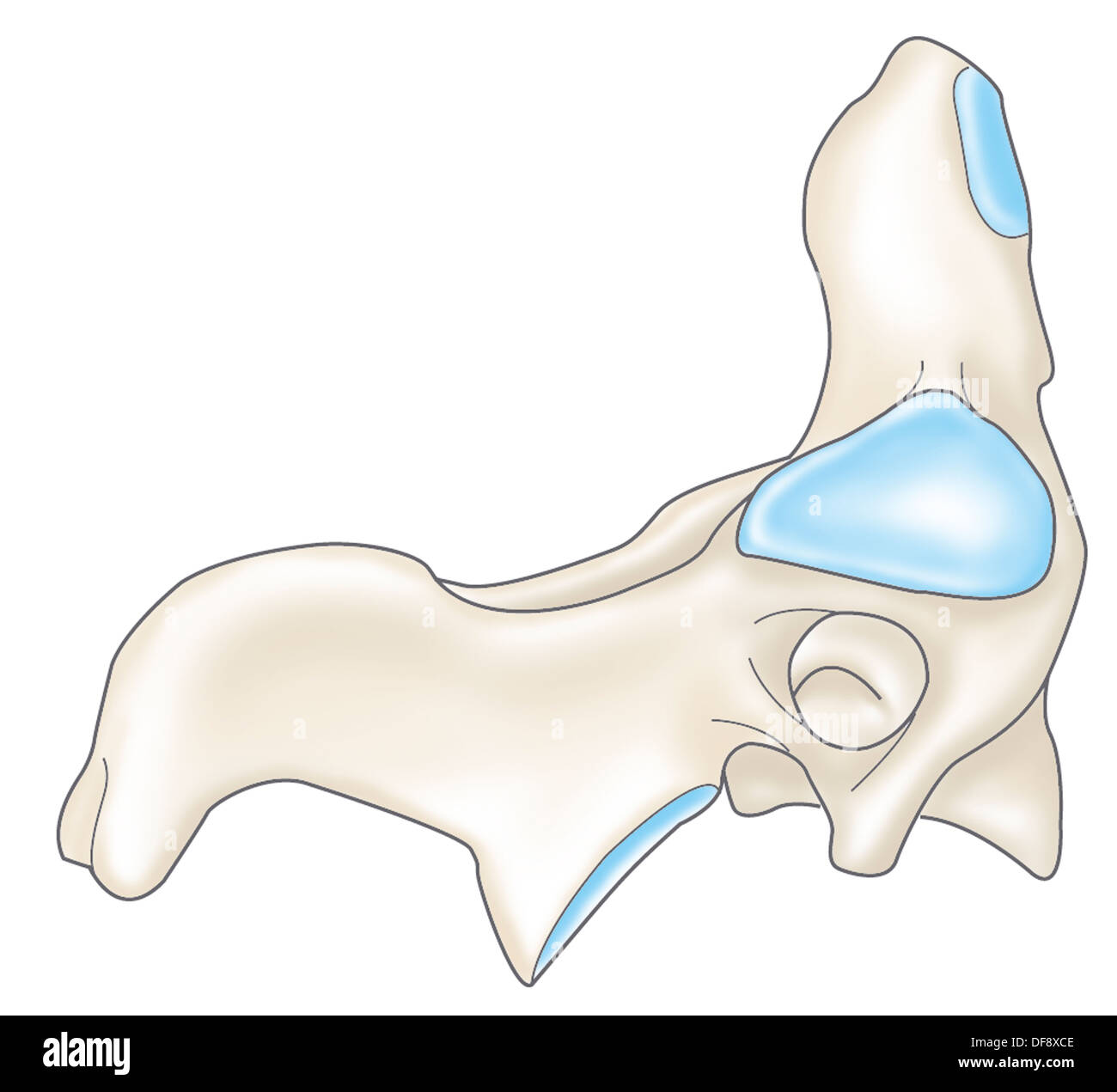 CERVICAL VERTEBRA, DRAWING Stock Photohttps://www.alamy.com/image-license-details/?v=1https://www.alamy.com/cervical-vertebra-drawing-image61047294.html
RMDF8XCE–CERVICAL VERTEBRA, DRAWING
 A normal human vertebral column (lateral view). Shown (from top to bottom) are the atlas (cervical vertebra 1: C1), axis (C2), cervical vertebrae 3-7 Stock Photohttps://www.alamy.com/image-license-details/?v=1https://www.alamy.com/stock-photo-a-normal-human-vertebral-column-lateral-view-shown-from-top-to-bottom-130806568.html
RFHGPN34–A normal human vertebral column (lateral view). Shown (from top to bottom) are the atlas (cervical vertebra 1: C1), axis (C2), cervical vertebrae 3-7
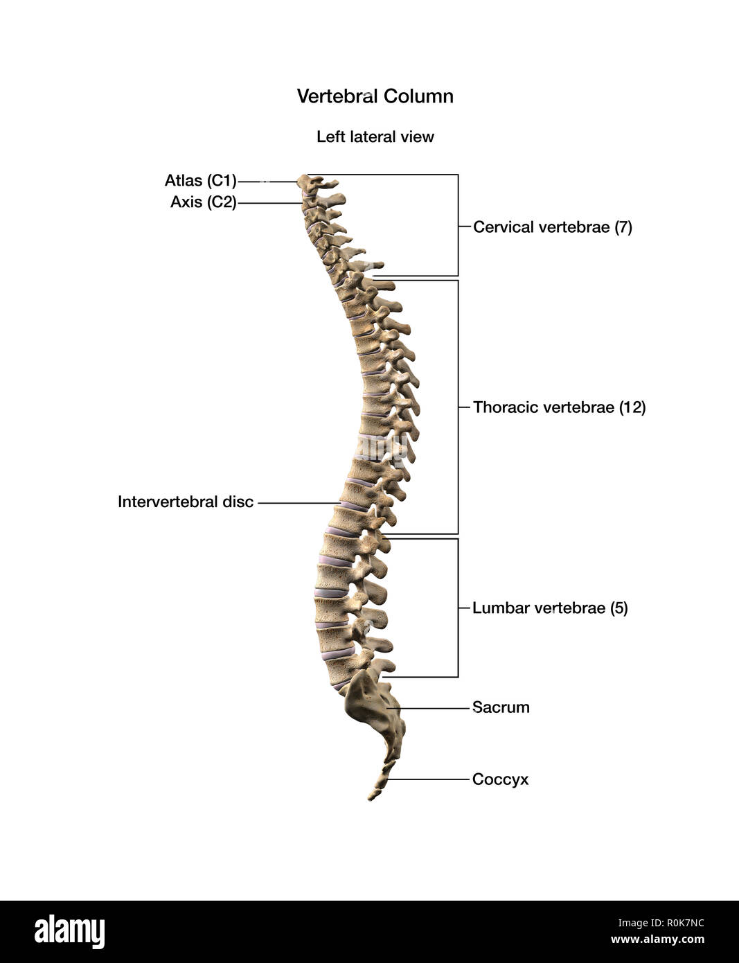 Human vertebral column with labels. Stock Photohttps://www.alamy.com/image-license-details/?v=1https://www.alamy.com/human-vertebral-column-with-labels-image224157960.html
RFR0K7NC–Human vertebral column with labels.
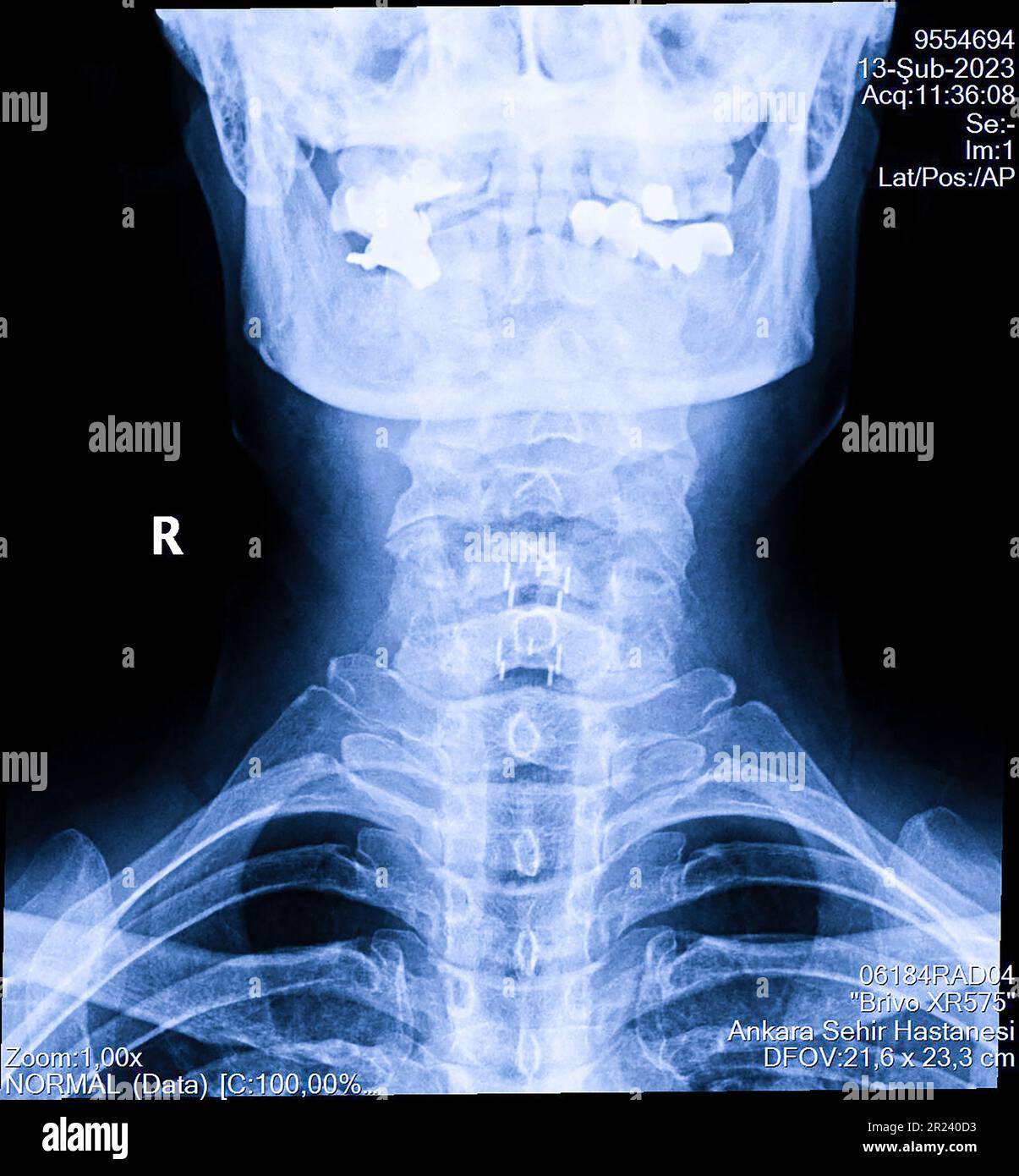 Human cervical spine x-ray, neck radiography Stock Photohttps://www.alamy.com/image-license-details/?v=1https://www.alamy.com/human-cervical-spine-x-ray-neck-radiography-image552049263.html
RF2R240D3–Human cervical spine x-ray, neck radiography
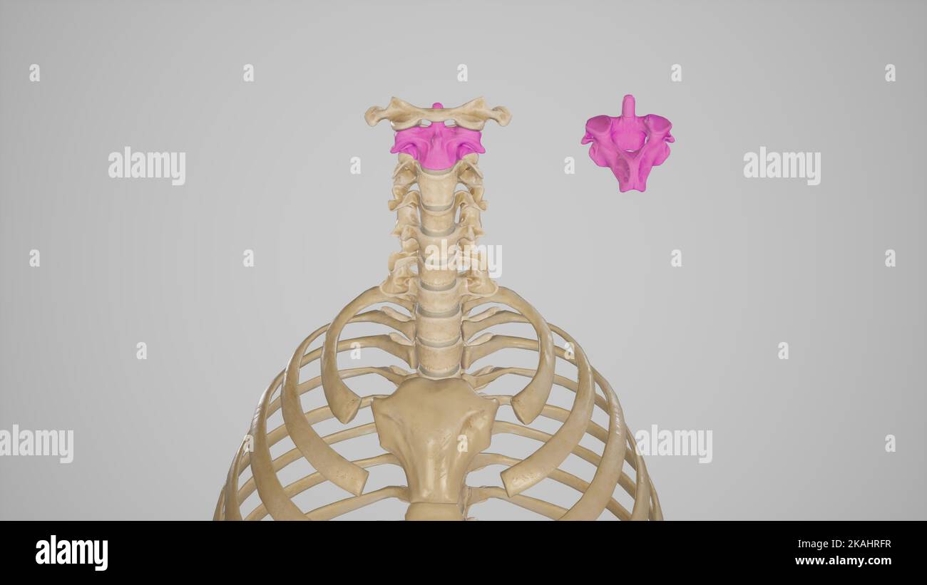 Anatomical Illustration of Second Cervical Vertebrae,Axis Stock Photohttps://www.alamy.com/image-license-details/?v=1https://www.alamy.com/anatomical-illustration-of-second-cervical-vertebraeaxis-image488428523.html
RF2KAHRFR–Anatomical Illustration of Second Cervical Vertebrae,Axis
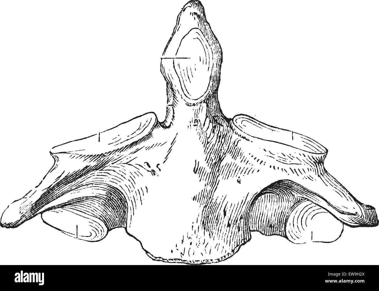 Fig. 136. Axis (second cervical vertebra), vintage engraved illustration. Magasin Pittoresque 1875. Stock Vectorhttps://www.alamy.com/image-license-details/?v=1https://www.alamy.com/stock-photo-fig-136-axis-second-cervical-vertebra-vintage-engraved-illustration-84418850.html
RFEW9H2X–Fig. 136. Axis (second cervical vertebra), vintage engraved illustration. Magasin Pittoresque 1875.
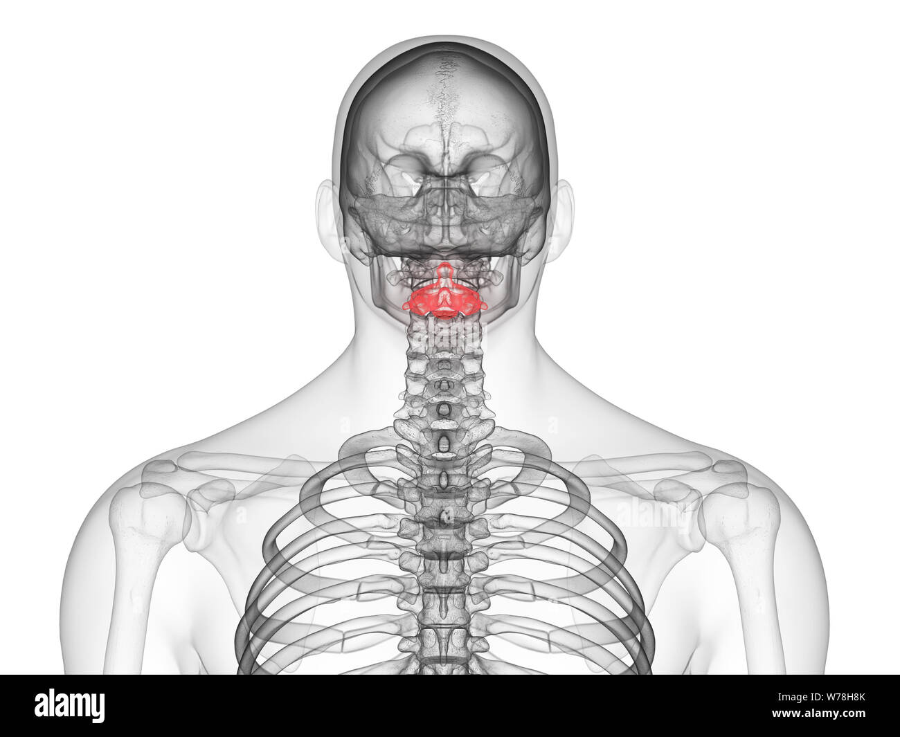 3d rendered medically accurate illustration of the axis vertebrae Stock Photohttps://www.alamy.com/image-license-details/?v=1https://www.alamy.com/3d-rendered-medically-accurate-illustration-of-the-axis-vertebrae-image262647299.html
RFW78H8K–3d rendered medically accurate illustration of the axis vertebrae
 X-ray C-spine or x-ray image of Cervical spine open mount view for fracture of cervical vertebra 2nd ( axis ). Stock Photohttps://www.alamy.com/image-license-details/?v=1https://www.alamy.com/x-ray-c-spine-or-x-ray-image-of-cervical-spine-open-mount-view-for-fracture-of-cervical-vertebra-2nd-axis-image546004992.html
RF2PM8JX8–X-ray C-spine or x-ray image of Cervical spine open mount view for fracture of cervical vertebra 2nd ( axis ).
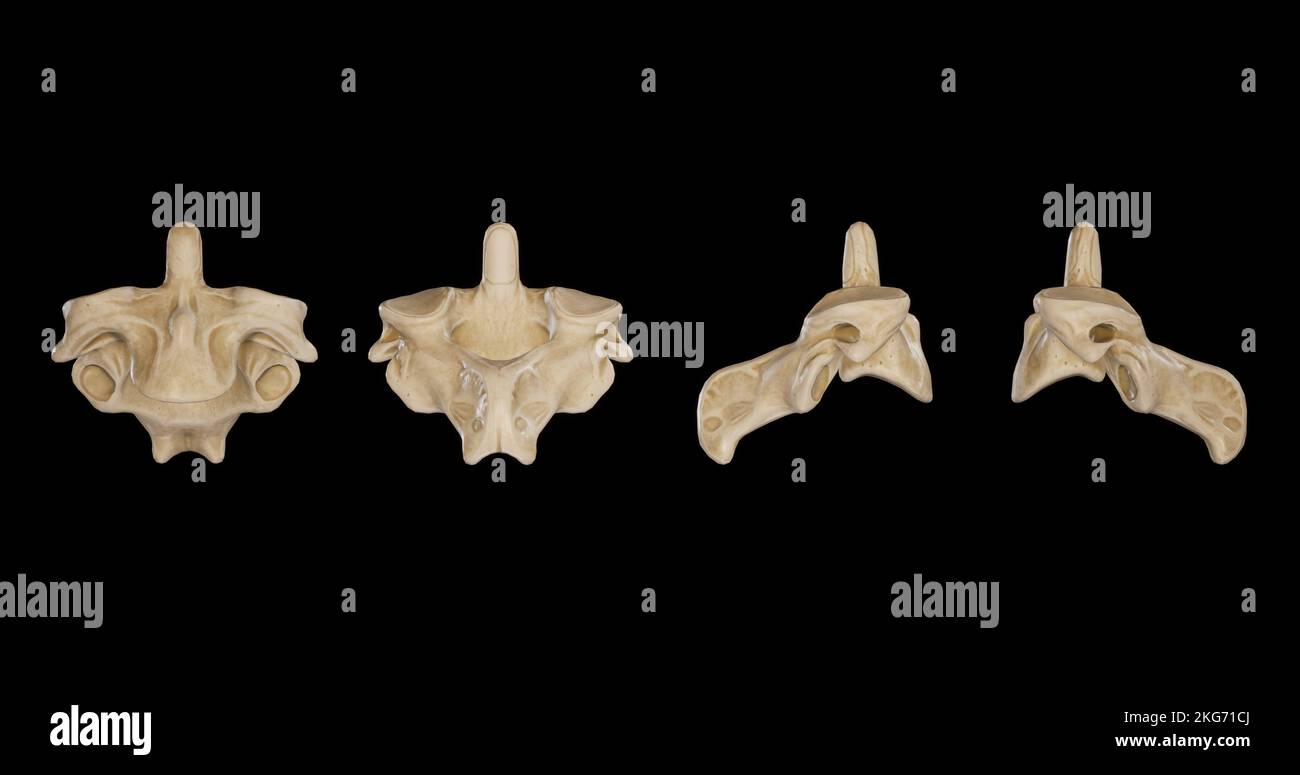 Second Cervical Vertebra (Axis) -Multiple Views.jpg Stock Photohttps://www.alamy.com/image-license-details/?v=1https://www.alamy.com/second-cervical-vertebra-axis-multiple-viewsjpg-image491879602.html
RF2KG71CJ–Second Cervical Vertebra (Axis) -Multiple Views.jpg
 The axis bone Stock Photohttps://www.alamy.com/image-license-details/?v=1https://www.alamy.com/stock-photo-the-axis-bone-13203092.html
RFACP1GN–The axis bone
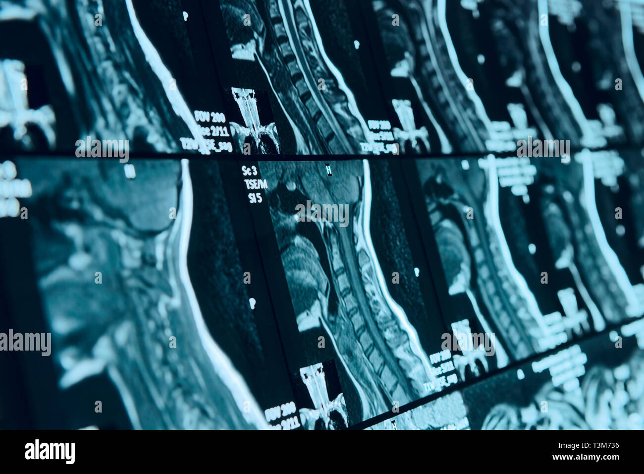 Head and neck MRI scan, anonymized, shallow focus depth Stock Photohttps://www.alamy.com/image-license-details/?v=1https://www.alamy.com/head-and-neck-mri-scan-anonymized-shallow-focus-depth-image243233738.html
RFT3M736–Head and neck MRI scan, anonymized, shallow focus depth
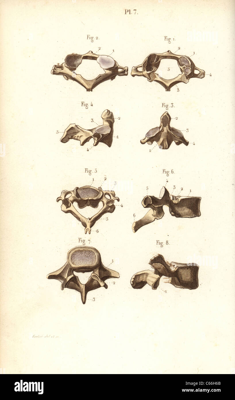 Axis and vertebrae. Stock Photohttps://www.alamy.com/image-license-details/?v=1https://www.alamy.com/stock-photo-axis-and-vertebrae-38253891.html
RMC66H6B–Axis and vertebrae.
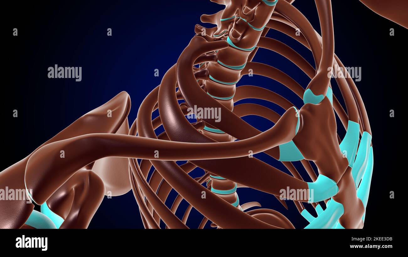 Costovertebral and Costotransverse Joints anatomy human rib cage for medical concept 3D Illustration Stock Photohttps://www.alamy.com/image-license-details/?v=1https://www.alamy.com/costovertebral-and-costotransverse-joints-anatomy-human-rib-cage-for-medical-concept-3d-illustration-image490805543.html
RF2KEE3DB–Costovertebral and Costotransverse Joints anatomy human rib cage for medical concept 3D Illustration
 Axis and vertebrae. Handcolored steel engraving by Corbie of a drawing by Corbie from Dr. Joseph Nicolas Masse's 'Petit Atlas complet d'Anatomie descriptive du Corps Humain,' Paris, 1864, published by Mequignon-Marvis. Masse's 'Pocket Anatomy of the Human Body' was first published in 1848 and went through many editions. Stock Photohttps://www.alamy.com/image-license-details/?v=1https://www.alamy.com/axis-and-vertebrae-handcolored-steel-engraving-by-corbie-of-a-drawing-by-corbie-from-dr-joseph-nicolas-masses-petit-atlas-complet-danatomie-descriptive-du-corps-humain-paris-1864-published-by-mequignon-marvis-masses-pocket-anatomy-of-the-human-body-was-first-published-in-1848-and-went-through-many-editions-image231204668.html
RMRC47WG–Axis and vertebrae. Handcolored steel engraving by Corbie of a drawing by Corbie from Dr. Joseph Nicolas Masse's 'Petit Atlas complet d'Anatomie descriptive du Corps Humain,' Paris, 1864, published by Mequignon-Marvis. Masse's 'Pocket Anatomy of the Human Body' was first published in 1848 and went through many editions.
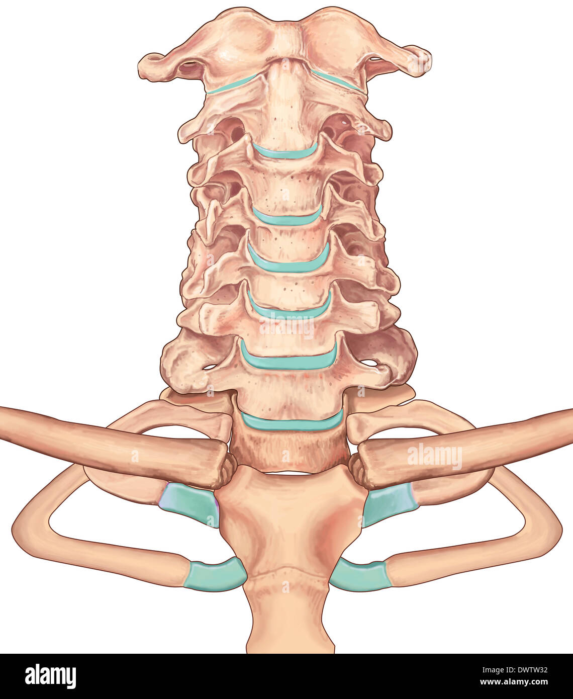 Cervical vertebra drawing Stock Photohttps://www.alamy.com/image-license-details/?v=1https://www.alamy.com/cervical-vertebra-drawing-image67544038.html
RMDWTW32–Cervical vertebra drawing
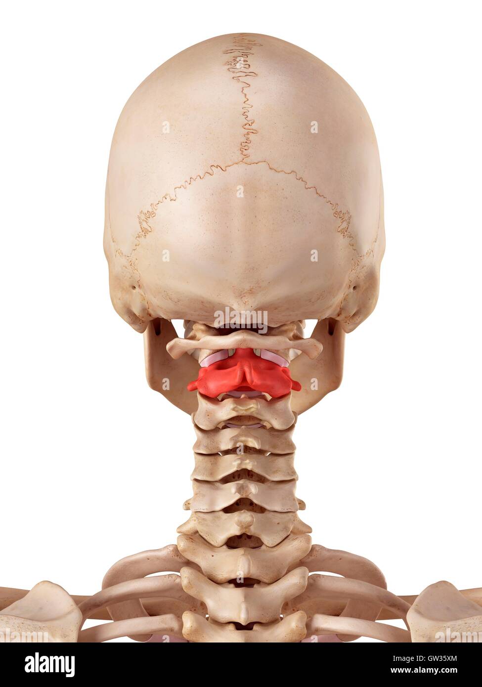 Human axis bone, illustration. Stock Photohttps://www.alamy.com/image-license-details/?v=1https://www.alamy.com/stock-photo-human-axis-bone-illustration-118699132.html
RFGW35XM–Human axis bone, illustration.
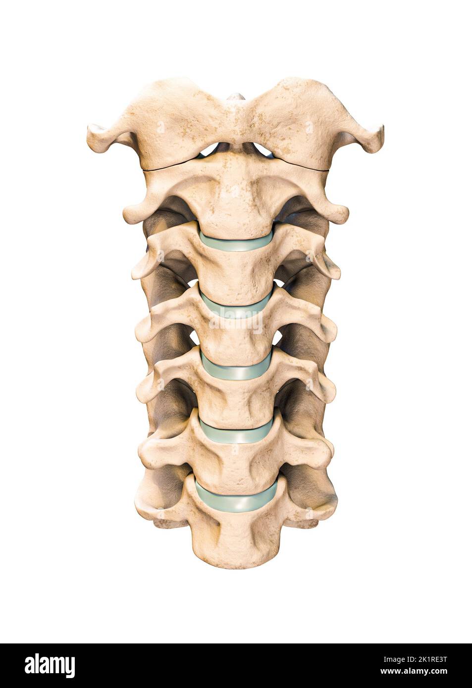 Anterior or front view of the seven human cervical vertebrae isolated on white background 3D rendering illustration. Anatomy, osteology, blank medical Stock Photohttps://www.alamy.com/image-license-details/?v=1https://www.alamy.com/anterior-or-front-view-of-the-seven-human-cervical-vertebrae-isolated-on-white-background-3d-rendering-illustration-anatomy-osteology-blank-medical-image483020940.html
RF2K1RE3T–Anterior or front view of the seven human cervical vertebrae isolated on white background 3D rendering illustration. Anatomy, osteology, blank medical
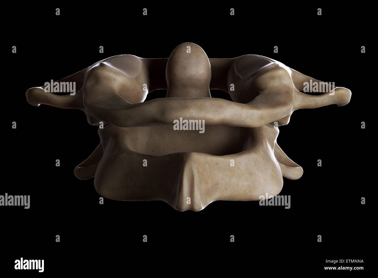 Illustration showing the atlas and axis vertebrae of the neck. Stock Photohttps://www.alamy.com/image-license-details/?v=1https://www.alamy.com/stock-photo-illustration-showing-the-atlas-and-axis-vertebrae-of-the-neck-84049318.html
RMETMNNA–Illustration showing the atlas and axis vertebrae of the neck.
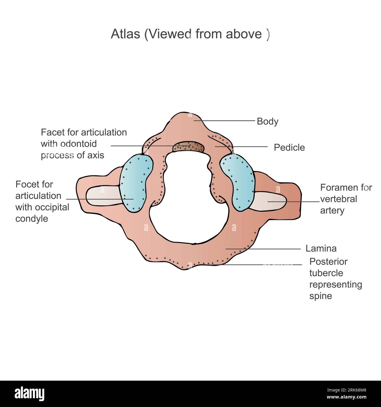 Illustration of an anterior view of the first cervical vertebra, also known as the Atlas C1 Stock Photohttps://www.alamy.com/image-license-details/?v=1https://www.alamy.com/illustration-of-an-anterior-view-of-the-first-cervical-vertebra-also-known-as-the-atlas-c1-image562548792.html
RF2RK68M8–Illustration of an anterior view of the first cervical vertebra, also known as the Atlas C1
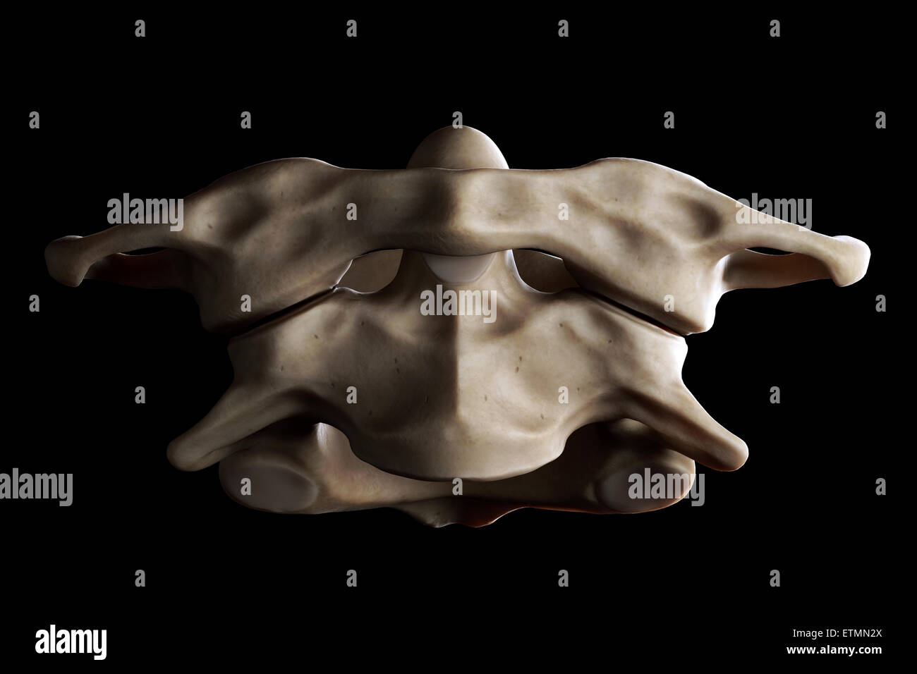 Illustration showing the atlas and axis vertebrae of the neck. Stock Photohttps://www.alamy.com/image-license-details/?v=1https://www.alamy.com/stock-photo-illustration-showing-the-atlas-and-axis-vertebrae-of-the-neck-84048802.html
RMETMN2X–Illustration showing the atlas and axis vertebrae of the neck.
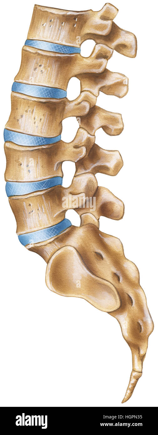 Spine - Lumbar Region - lateral view Stock Photohttps://www.alamy.com/image-license-details/?v=1https://www.alamy.com/stock-photo-spine-lumbar-region-lateral-view-130806569.html
RFHGPN35–Spine - Lumbar Region - lateral view
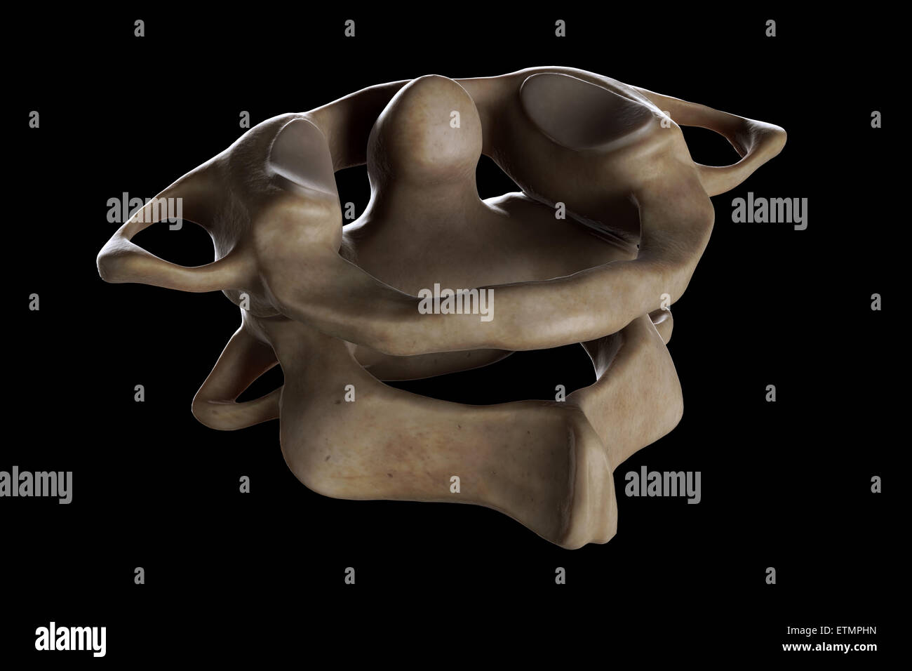 Illustration showing the atlas and axis vertebrae of the neck. Stock Photohttps://www.alamy.com/image-license-details/?v=1https://www.alamy.com/stock-photo-illustration-showing-the-atlas-and-axis-vertebrae-of-the-neck-84050001.html
RMETMPHN–Illustration showing the atlas and axis vertebrae of the neck.
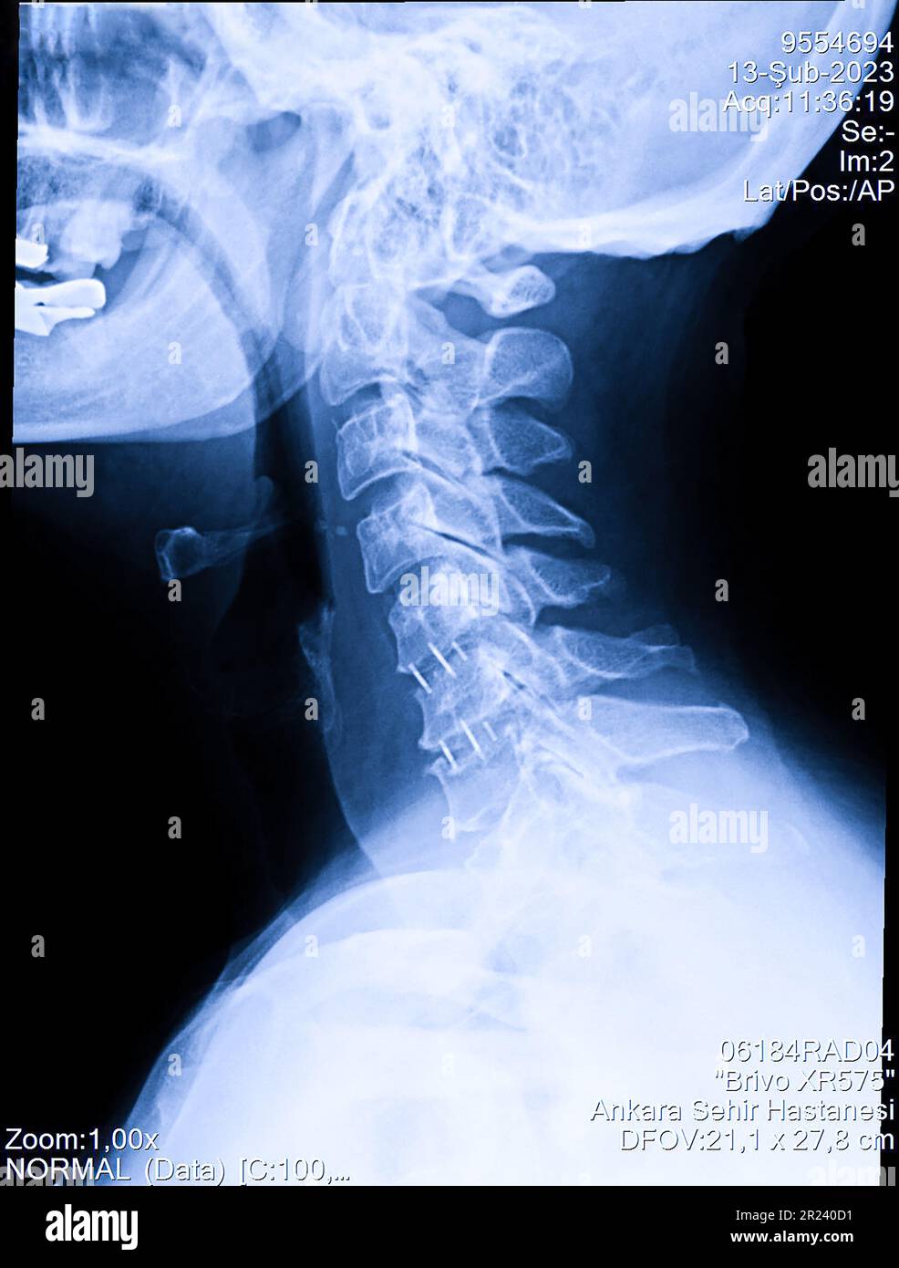 Woman's cervical spine x-ray, human neck radiography Stock Photohttps://www.alamy.com/image-license-details/?v=1https://www.alamy.com/womans-cervical-spine-x-ray-human-neck-radiography-image552049261.html
RF2R240D1–Woman's cervical spine x-ray, human neck radiography
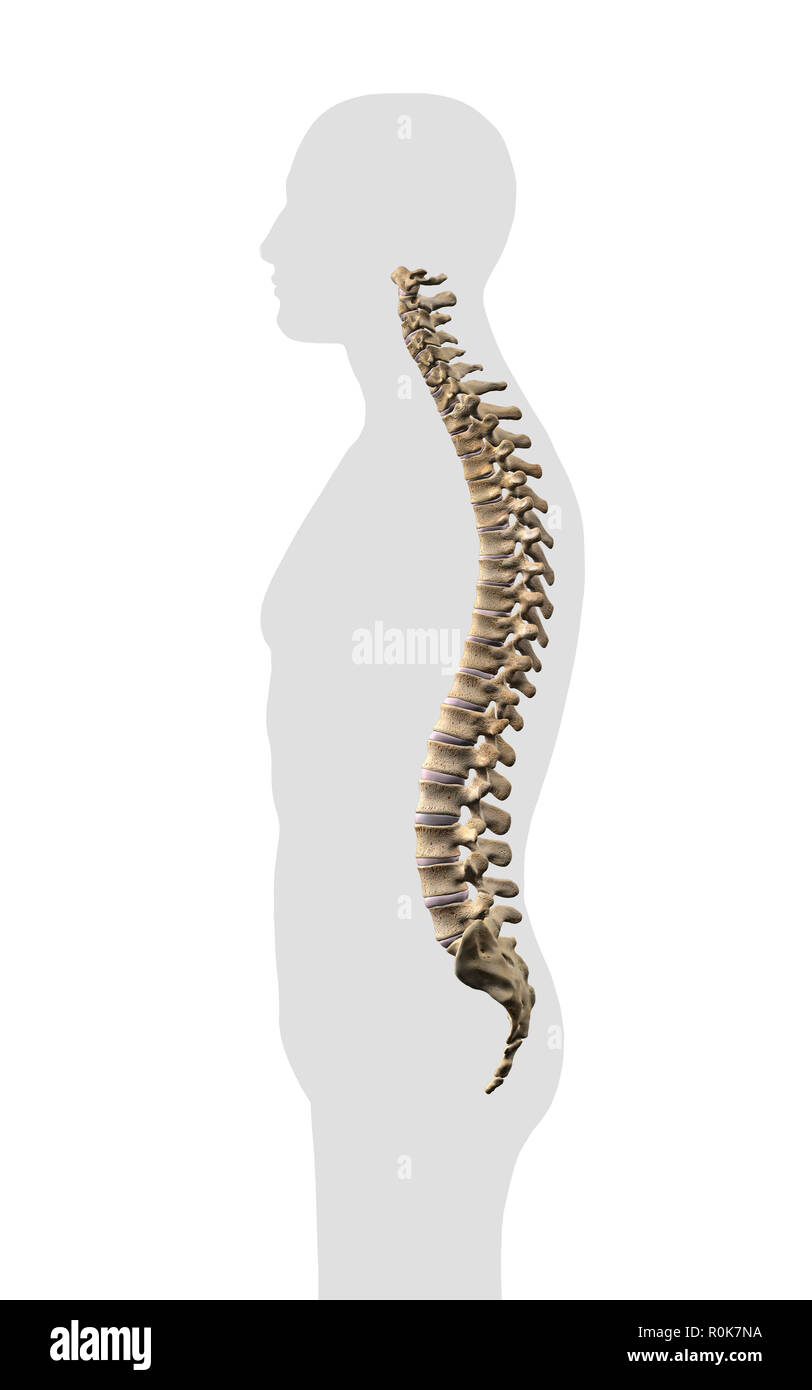 Human vertebral column, side view on white background. Stock Photohttps://www.alamy.com/image-license-details/?v=1https://www.alamy.com/human-vertebral-column-side-view-on-white-background-image224157958.html
RFR0K7NA–Human vertebral column, side view on white background.
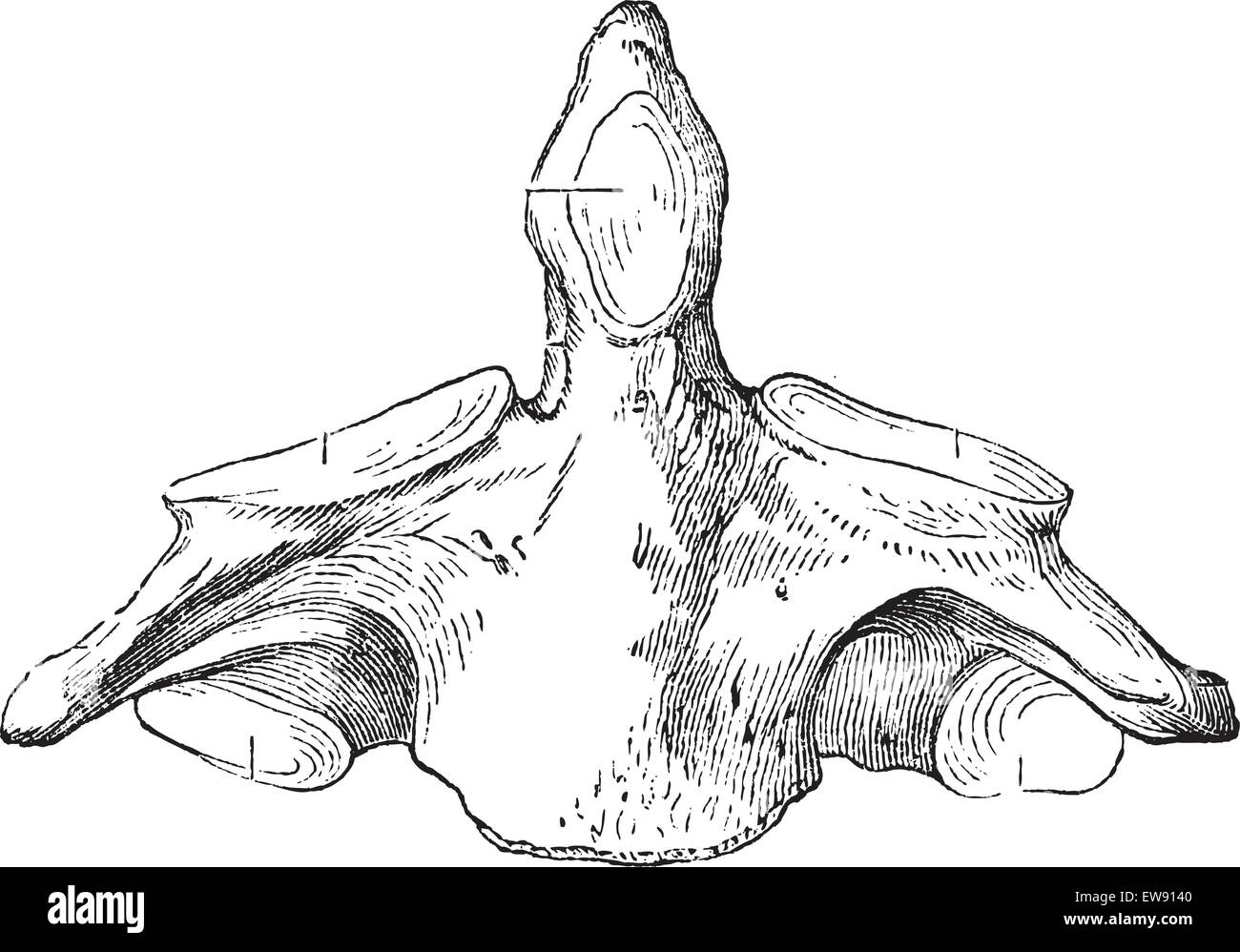 Fig. 136. Axis (second cervical vertebra), vintage engraved illustration. Magasin Pittoresque 1875. Stock Vectorhttps://www.alamy.com/image-license-details/?v=1https://www.alamy.com/stock-photo-fig-136-axis-second-cervical-vertebra-vintage-engraved-illustration-84406336.html
RFEW9140–Fig. 136. Axis (second cervical vertebra), vintage engraved illustration. Magasin Pittoresque 1875.
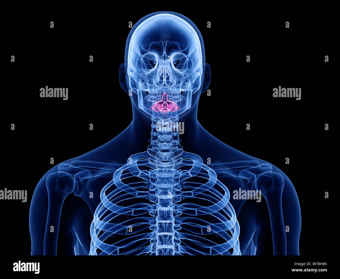 3d rendered medically accurate illustration of the axis vertebrae Stock Photohttps://www.alamy.com/image-license-details/?v=1https://www.alamy.com/3d-rendered-medically-accurate-illustration-of-the-axis-vertebrae-image262647286.html
RFW78H86–3d rendered medically accurate illustration of the axis vertebrae
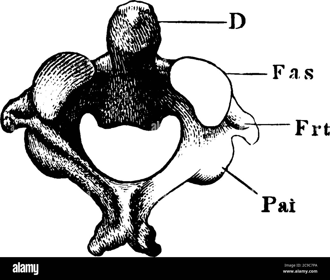 In anatomy, the second cervical vertebra of the spine is named the axis or epistropheus. Shown here is the anterior articular surface of axis vertebra Stock Vectorhttps://www.alamy.com/image-license-details/?v=1https://www.alamy.com/in-anatomy-the-second-cervical-vertebra-of-the-spine-is-named-the-axis-or-epistropheus-shown-here-is-the-anterior-articular-surface-of-axis-vertebra-image367219170.html
RF2C9C7PA–In anatomy, the second cervical vertebra of the spine is named the axis or epistropheus. Shown here is the anterior articular surface of axis vertebra
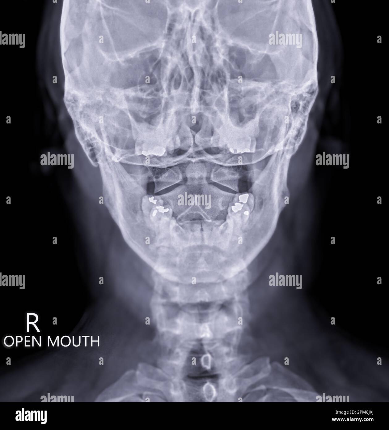 X-ray C-spine or x-ray image of Cervical spine open mount view for fracture of cervical vertebra 2nd ( axis ). Stock Photohttps://www.alamy.com/image-license-details/?v=1https://www.alamy.com/x-ray-c-spine-or-x-ray-image-of-cervical-spine-open-mount-view-for-fracture-of-cervical-vertebra-2nd-axis-image546005002.html
RF2PM8JXJ–X-ray C-spine or x-ray image of Cervical spine open mount view for fracture of cervical vertebra 2nd ( axis ).
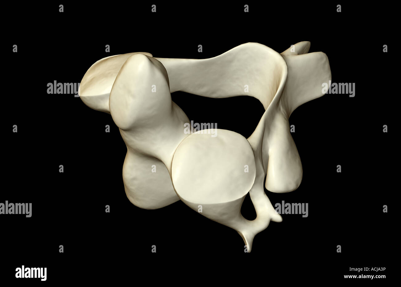 The axis bone Stock Photohttps://www.alamy.com/image-license-details/?v=1https://www.alamy.com/stock-photo-the-axis-bone-13168329.html
RFACJA3P–The axis bone
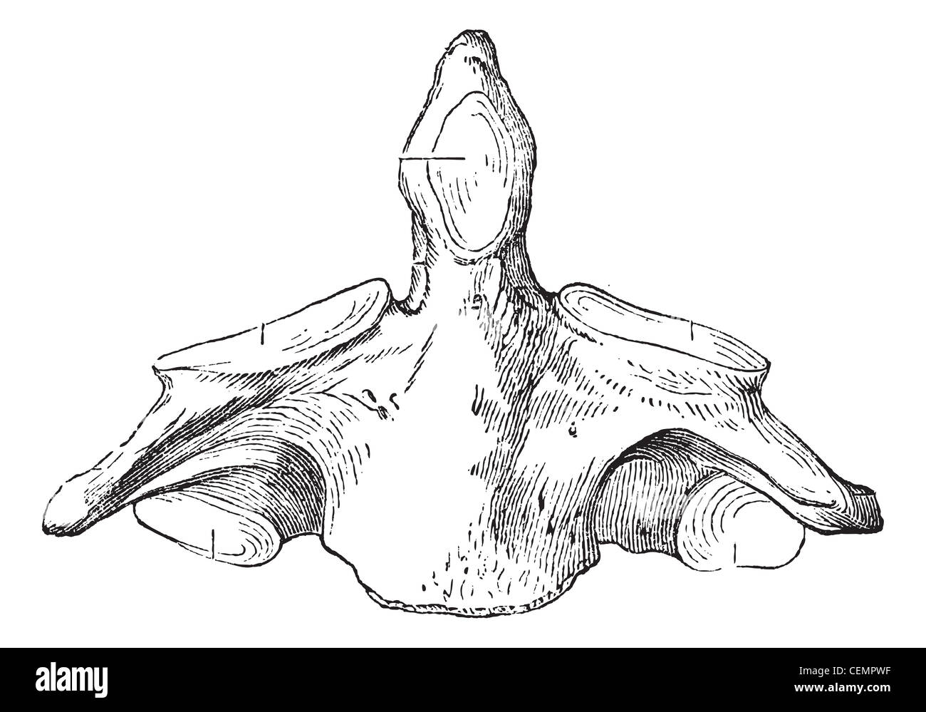 Fig. 136. Axis (second cervical vertebra), vintage engraved illustration. Magasin Pittoresque 1875. Stock Photohttps://www.alamy.com/image-license-details/?v=1https://www.alamy.com/stock-photo-fig-136-axis-second-cervical-vertebra-vintage-engraved-illustration-43482923.html
RFCEMPWF–Fig. 136. Axis (second cervical vertebra), vintage engraved illustration. Magasin Pittoresque 1875.
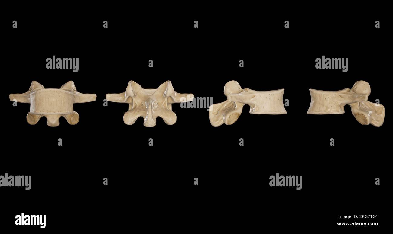 Lumbar Vertebra -Multiple Views Stock Photohttps://www.alamy.com/image-license-details/?v=1https://www.alamy.com/lumbar-vertebra-multiple-views-image491879700.html
RF2KG71G4–Lumbar Vertebra -Multiple Views
 Head and neck MRI scan, anonymized, shallow focus depth Stock Photohttps://www.alamy.com/image-license-details/?v=1https://www.alamy.com/head-and-neck-mri-scan-anonymized-shallow-focus-depth-image243233617.html
RFT3M6XW–Head and neck MRI scan, anonymized, shallow focus depth
 Costovertebral and Costotransverse Joints anatomy human rib cage for medical concept 3D Illustration Stock Photohttps://www.alamy.com/image-license-details/?v=1https://www.alamy.com/costovertebral-and-costotransverse-joints-anatomy-human-rib-cage-for-medical-concept-3d-illustration-image490805541.html
RF2KEE3D9–Costovertebral and Costotransverse Joints anatomy human rib cage for medical concept 3D Illustration
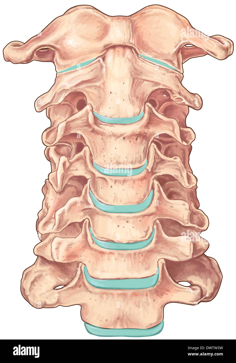 Cervical vertebra drawing Stock Photohttps://www.alamy.com/image-license-details/?v=1https://www.alamy.com/cervical-vertebra-drawing-image67544061.html
RMDWTW3W–Cervical vertebra drawing
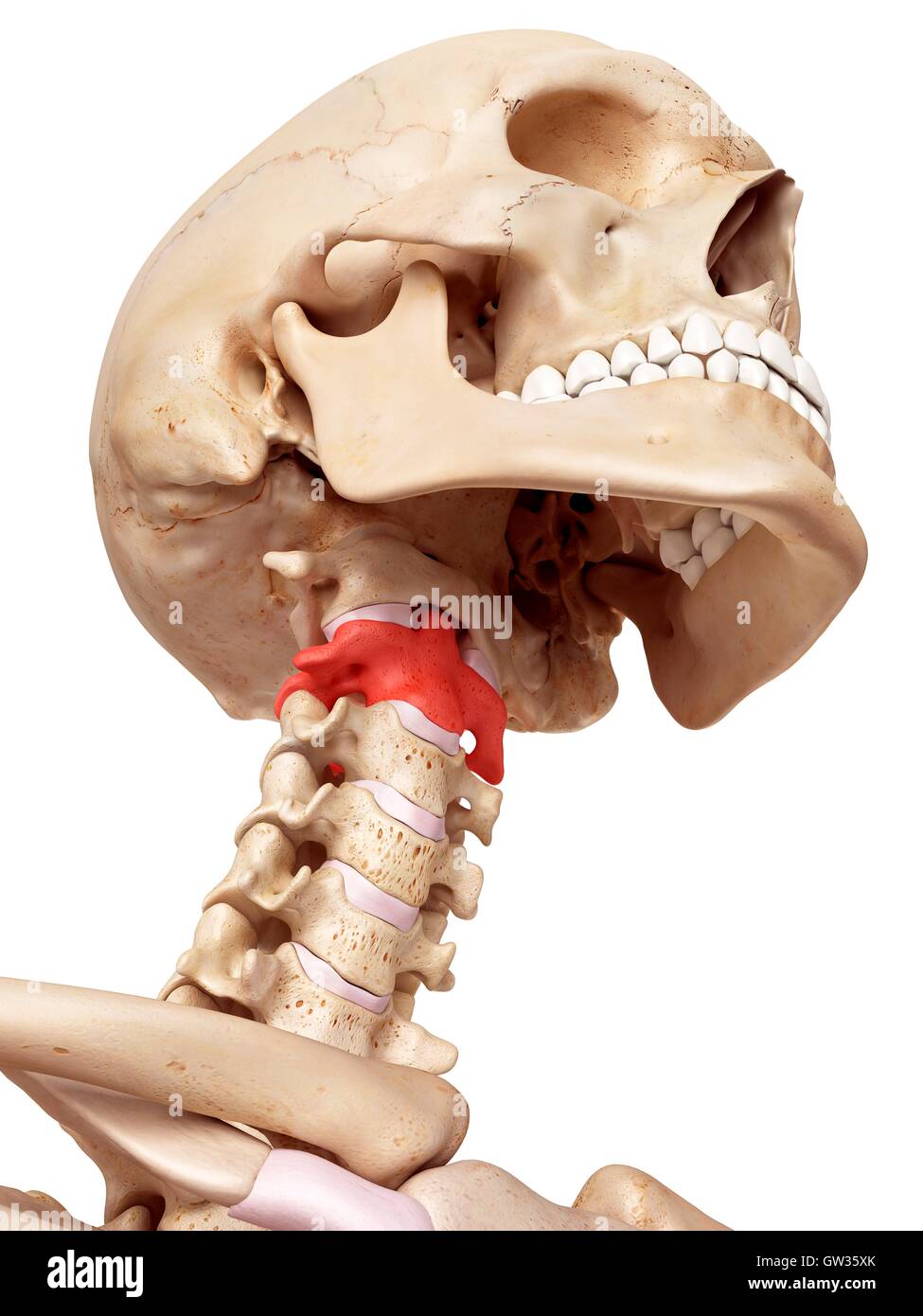 Human axis bone, illustration. Stock Photohttps://www.alamy.com/image-license-details/?v=1https://www.alamy.com/stock-photo-human-axis-bone-illustration-118699131.html
RFGW35XK–Human axis bone, illustration.
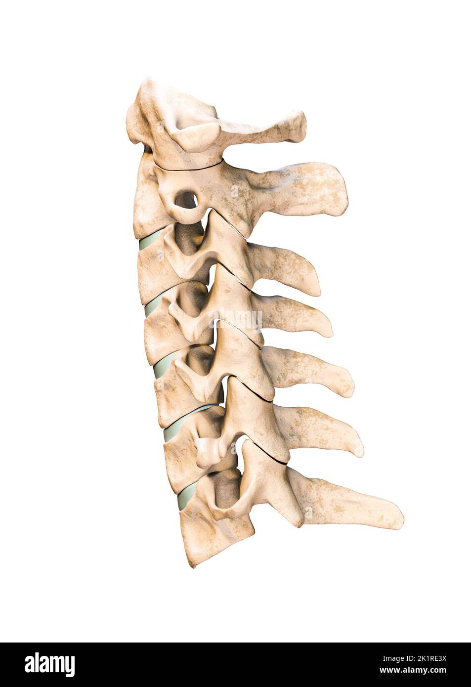 Lateral or profile view of the seven human cervical vertebrae isolated on white background 3D rendering illustration. Anatomy, osteology, blank medica Stock Photohttps://www.alamy.com/image-license-details/?v=1https://www.alamy.com/lateral-or-profile-view-of-the-seven-human-cervical-vertebrae-isolated-on-white-background-3d-rendering-illustration-anatomy-osteology-blank-medica-image483020942.html
RF2K1RE3X–Lateral or profile view of the seven human cervical vertebrae isolated on white background 3D rendering illustration. Anatomy, osteology, blank medica
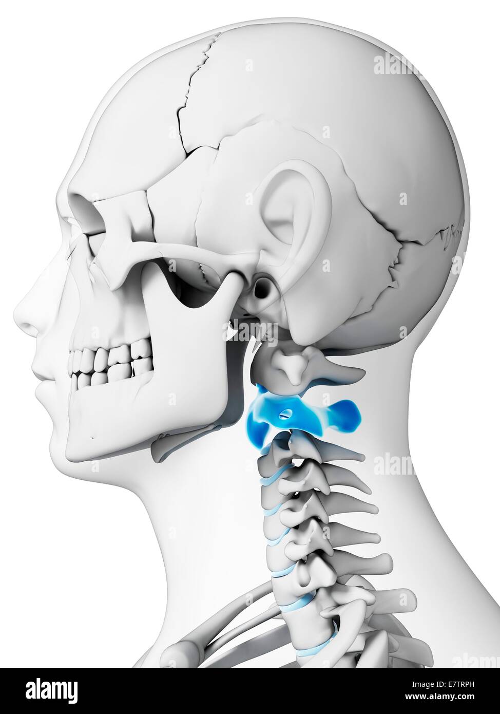 Human neck bones, computer artwork. Stock Photohttps://www.alamy.com/image-license-details/?v=1https://www.alamy.com/stock-photo-human-neck-bones-computer-artwork-73689577.html
RFE7TRPH–Human neck bones, computer artwork.
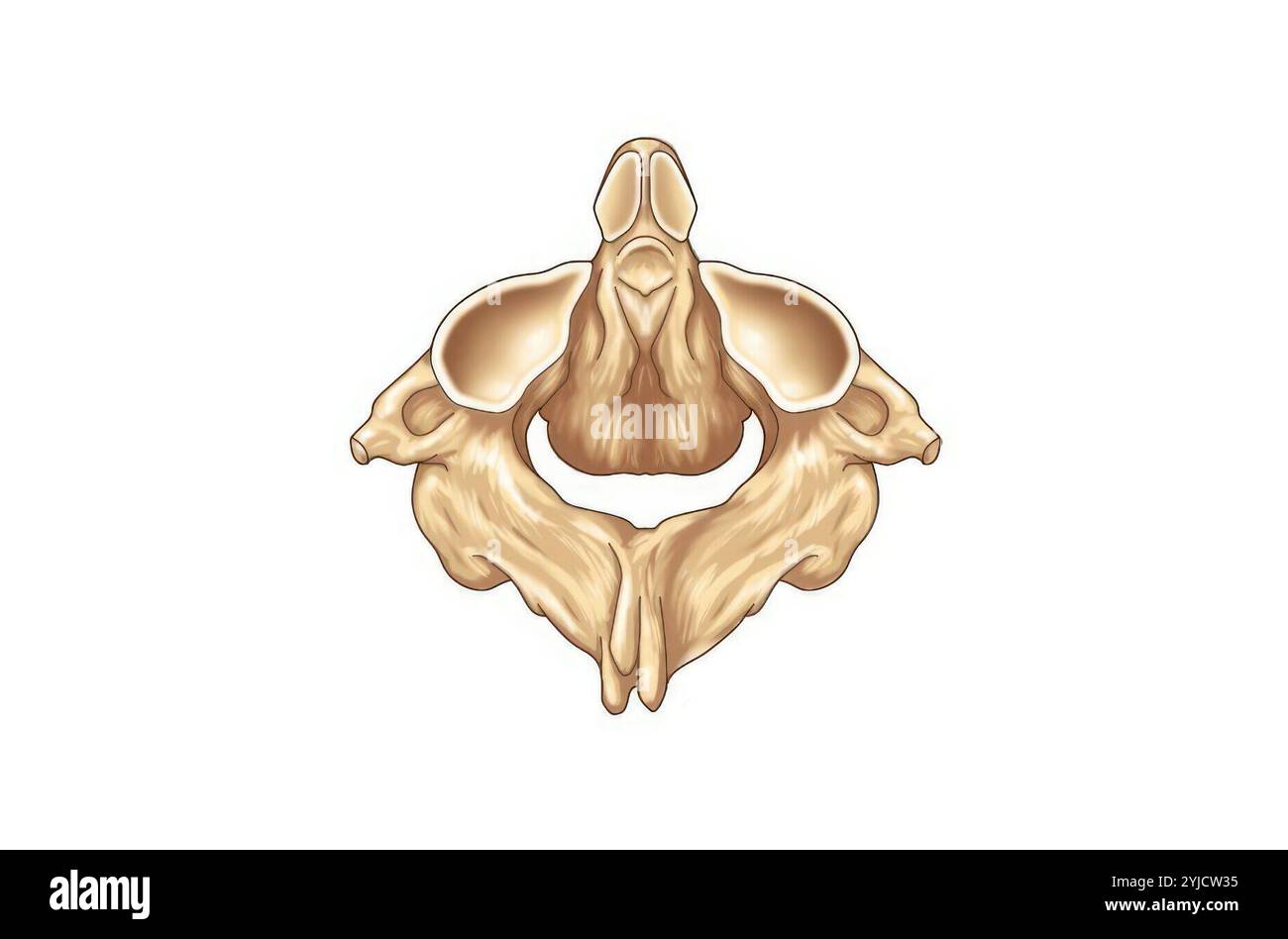 Axis -posterior view. Stock Photohttps://www.alamy.com/image-license-details/?v=1https://www.alamy.com/axis-posterior-view-image630920169.html
RM2YJCW35–Axis -posterior view.
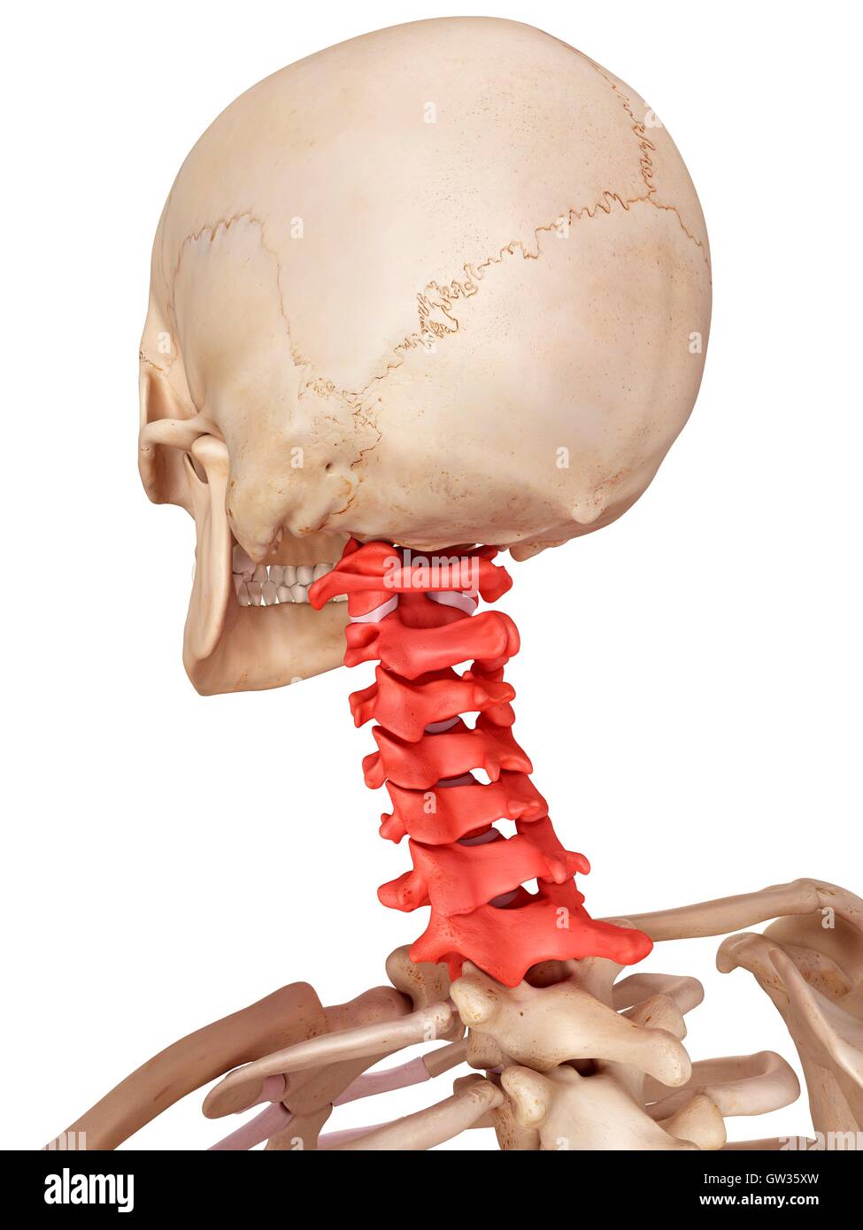 Human cervical spine, illustration. Stock Photohttps://www.alamy.com/image-license-details/?v=1https://www.alamy.com/stock-photo-human-cervical-spine-illustration-118699137.html
RFGW35XW–Human cervical spine, illustration.
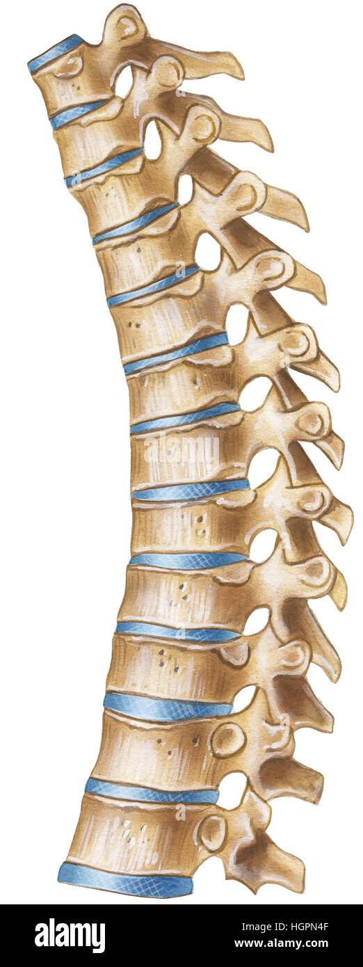 Spine - Thoracic Region - lateral Vvew Stock Photohttps://www.alamy.com/image-license-details/?v=1https://www.alamy.com/stock-photo-spine-thoracic-region-lateral-vvew-130806607.html
RFHGPN4F–Spine - Thoracic Region - lateral Vvew
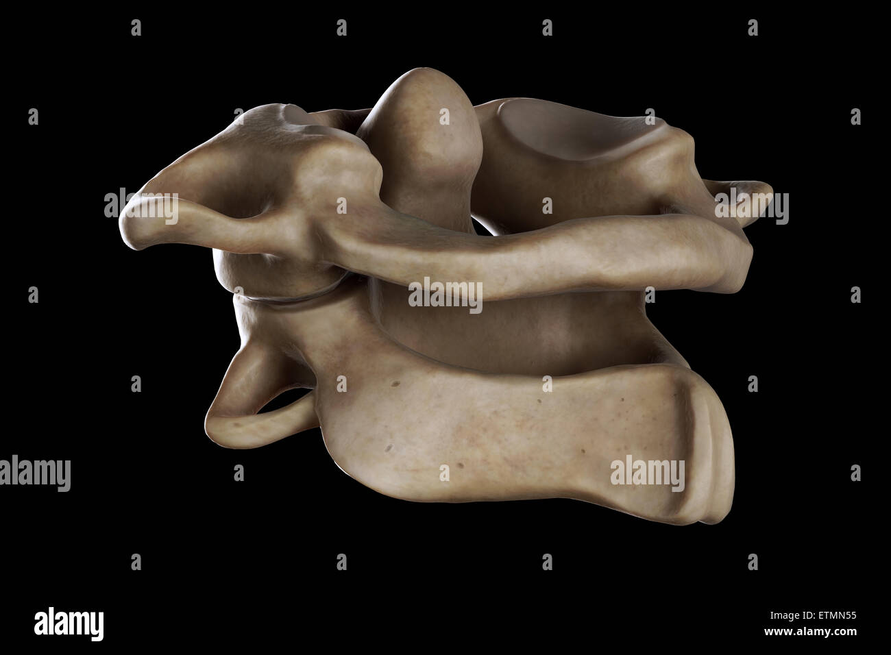 Illustration showing the atlas and axis vertebrae of the neck. Stock Photohttps://www.alamy.com/image-license-details/?v=1https://www.alamy.com/stock-photo-illustration-showing-the-atlas-and-axis-vertebrae-of-the-neck-84048865.html
RMETMN55–Illustration showing the atlas and axis vertebrae of the neck.
 X-ray of neck and cervical spine view. Image of radiography from patient who have neck pain, nerve root compression, numbness at arm hand wrist or fin Stock Photohttps://www.alamy.com/image-license-details/?v=1https://www.alamy.com/x-ray-of-neck-and-cervical-spine-view-image-of-radiography-from-patient-who-have-neck-pain-nerve-root-compression-numbness-at-arm-hand-wrist-or-fin-image552049260.html
RF2R240D0–X-ray of neck and cervical spine view. Image of radiography from patient who have neck pain, nerve root compression, numbness at arm hand wrist or fin
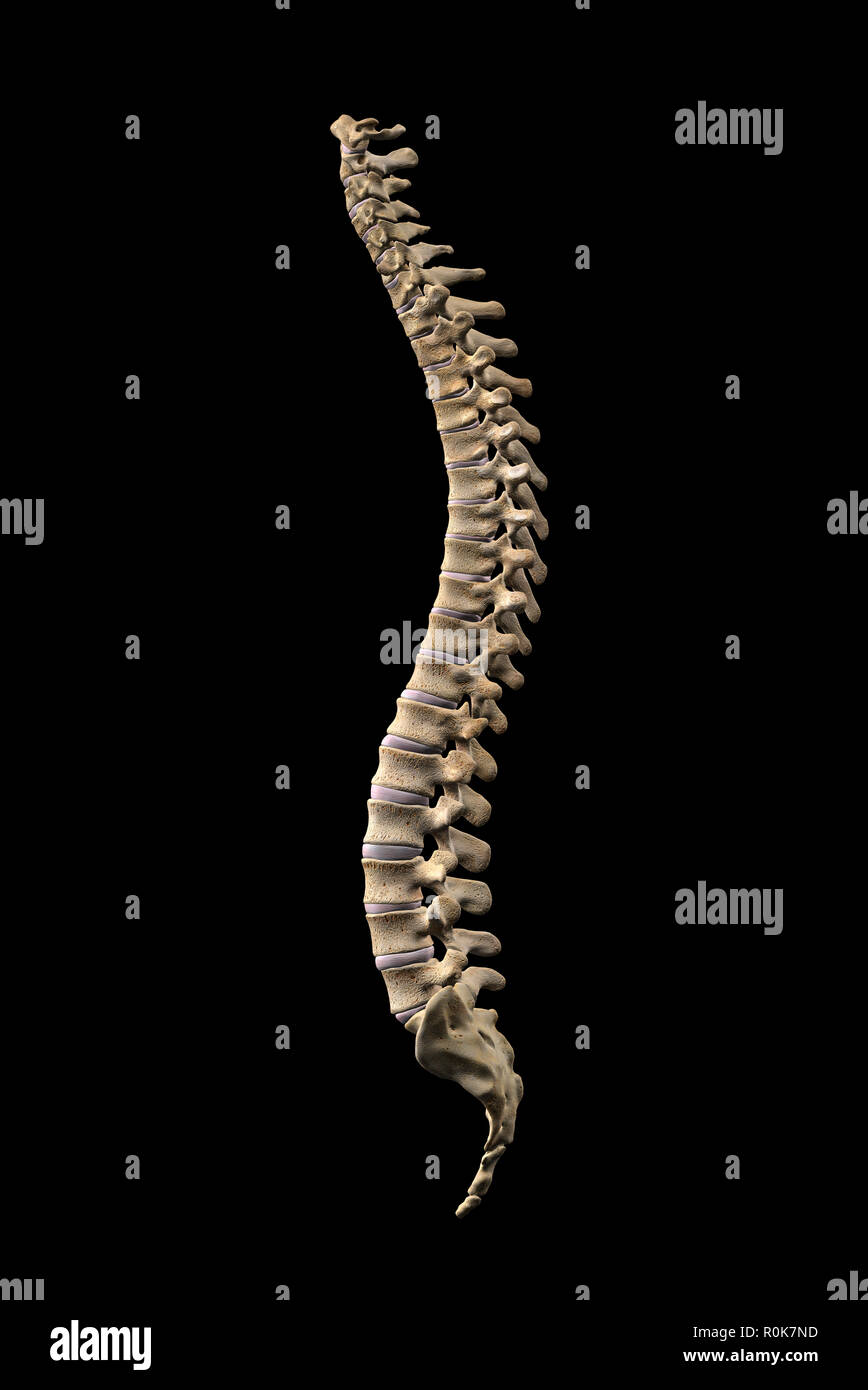 Human vertebral column, side view on black background. Stock Photohttps://www.alamy.com/image-license-details/?v=1https://www.alamy.com/human-vertebral-column-side-view-on-black-background-image224157961.html
RFR0K7ND–Human vertebral column, side view on black background.
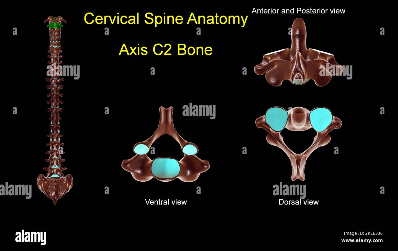 Cervical spine C 2 Axis bone anatomy for medical concept 3D Illustration with anterior and posterior View Stock Photohttps://www.alamy.com/image-license-details/?v=1https://www.alamy.com/cervical-spine-c-2-axis-bone-anatomy-for-medical-concept-3d-illustration-with-anterior-and-posterior-view-image490805258.html
RF2KEE336–Cervical spine C 2 Axis bone anatomy for medical concept 3D Illustration with anterior and posterior View
 3d rendered medically accurate illustration of the axis vertebrae Stock Photohttps://www.alamy.com/image-license-details/?v=1https://www.alamy.com/3d-rendered-medically-accurate-illustration-of-the-axis-vertebrae-image262647300.html
RFW78H8M–3d rendered medically accurate illustration of the axis vertebrae
 Elements of animal physiology, chiefly human . ,, Cerebro-SplnalNervous System. Diaphragm, Alimentary Canal. Ganglia, Alimentary Canal, Fig. 4. Theoretic Longitudinal Section ofHuman Body, showing Dorsal and VentralTube. (After Huxley.) The section is taken perpendiciaarly through the Medianplane, and shows the dorsal (neural) tube containing the dorsalchamber of the skull and the spinal canal, the cut smf aces of theskuU and 33 vertebra, and the ventral (haemal) tube in front ofthe Vertebral Column, containing the Heart, Ijungs, Alimentary 30 ANIMAL PHYSIOLOGY, Cerebro-Spinal Nervous Axis con Stock Photohttps://www.alamy.com/image-license-details/?v=1https://www.alamy.com/elements-of-animal-physiology-chiefly-human-cerebro-splnalnervous-system-diaphragm-alimentary-canal-ganglia-alimentary-canal-fig-4-theoretic-longitudinal-section-ofhuman-body-showing-dorsal-and-ventraltube-after-huxley-the-section-is-taken-perpendiciaarly-through-the-medianplane-and-shows-the-dorsal-neural-tube-containing-the-dorsalchamber-of-the-skull-and-the-spinal-canal-the-cut-smf-aces-of-theskuu-and-33-vertebra-and-the-ventral-haemal-tube-in-front-ofthe-vertebral-column-containing-the-heart-ijungs-alimentary-30-animal-physiology-cerebro-spinal-nervous-axis-con-image339021844.html
RM2AKFNR0–Elements of animal physiology, chiefly human . ,, Cerebro-SplnalNervous System. Diaphragm, Alimentary Canal. Ganglia, Alimentary Canal, Fig. 4. Theoretic Longitudinal Section ofHuman Body, showing Dorsal and VentralTube. (After Huxley.) The section is taken perpendiciaarly through the Medianplane, and shows the dorsal (neural) tube containing the dorsalchamber of the skull and the spinal canal, the cut smf aces of theskuU and 33 vertebra, and the ventral (haemal) tube in front ofthe Vertebral Column, containing the Heart, Ijungs, Alimentary 30 ANIMAL PHYSIOLOGY, Cerebro-Spinal Nervous Axis con
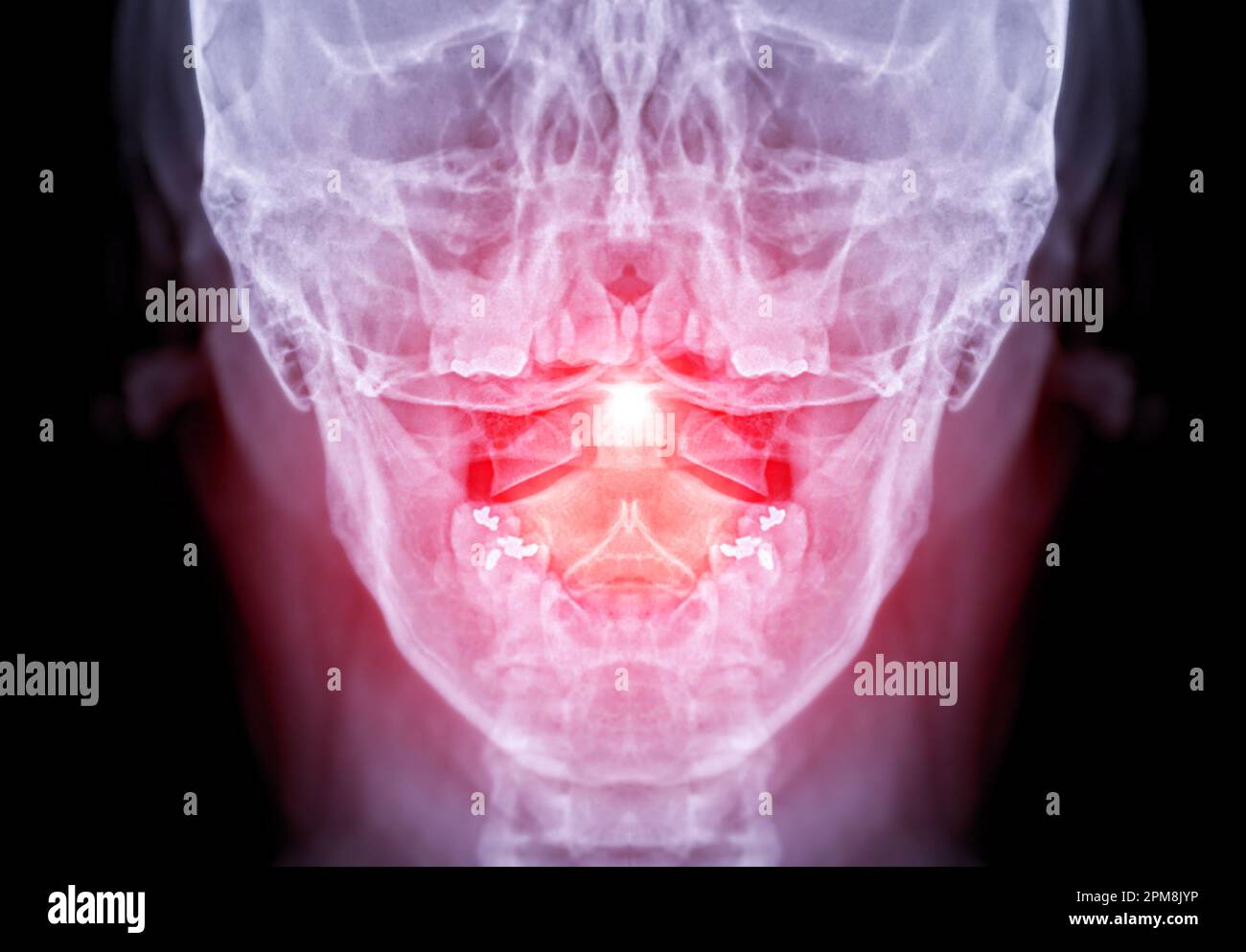 X-ray C-spine or x-ray image of Cervical spine open mount view for fracture of cervical vertebra 2nd ( axis ). Stock Photohttps://www.alamy.com/image-license-details/?v=1https://www.alamy.com/x-ray-c-spine-or-x-ray-image-of-cervical-spine-open-mount-view-for-fracture-of-cervical-vertebra-2nd-axis-image546005034.html
RF2PM8JYP–X-ray C-spine or x-ray image of Cervical spine open mount view for fracture of cervical vertebra 2nd ( axis ).
 The axis bone Stock Photohttps://www.alamy.com/image-license-details/?v=1https://www.alamy.com/stock-photo-the-axis-bone-13206363.html
RFACPB9G–The axis bone
 dens Stock Photohttps://www.alamy.com/image-license-details/?v=1https://www.alamy.com/stock-photo-dens-131859579.html
RFHJEM6K–dens
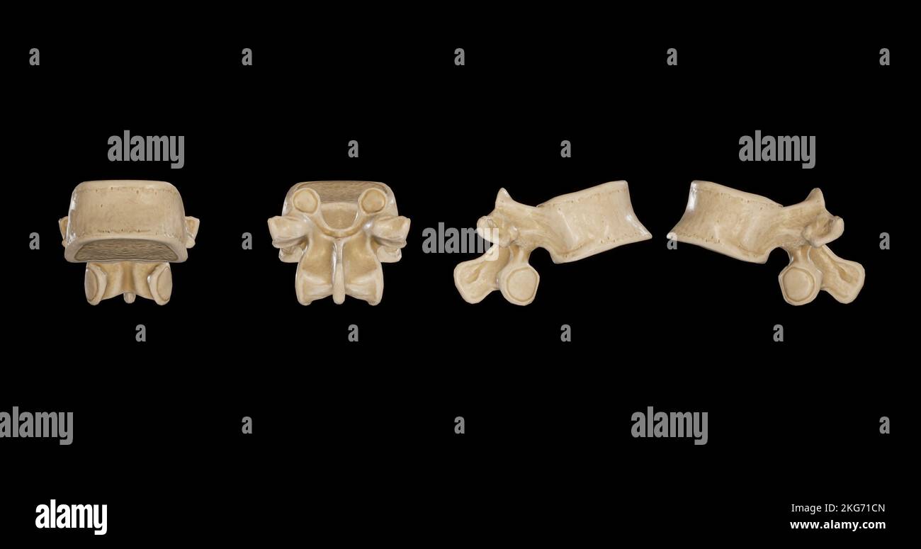 Thoracic Vertebra -Multiple Views Stock Photohttps://www.alamy.com/image-license-details/?v=1https://www.alamy.com/thoracic-vertebra-multiple-views-image491879605.html
RF2KG71CN–Thoracic Vertebra -Multiple Views
 Head and neck MRI scan, patient's and clinic's info removed, toned image Stock Photohttps://www.alamy.com/image-license-details/?v=1https://www.alamy.com/head-and-neck-mri-scan-patients-and-clinics-info-removed-toned-image-image243233698.html
RFT3M71P–Head and neck MRI scan, patient's and clinic's info removed, toned image
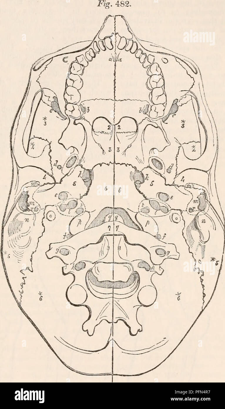 . The cyclopædia of anatomy and physiology. Anatomy; Physiology; Zoology. SKELETON. 661 is altogether obliterated. By this circum- or post sphenoid vertebra, the cranial basis stance of the body of the sixth or occipital is contracted in its longitudinal axis, while cranial vertebra joining the body of the third the cranial vault (j<?g. 481.) fashioned of the. Tlie base of the human cranium, Showing its serial relation with the bodies or centra of the spinal vertebra?, and also the serial homology between the foramina of the cranial vertebra and those of the spinal vertebras. expanded neura Stock Photohttps://www.alamy.com/image-license-details/?v=1https://www.alamy.com/the-cyclopdia-of-anatomy-and-physiology-anatomy-physiology-zoology-skeleton-661-is-altogether-obliterated-by-this-circum-or-post-sphenoid-vertebra-the-cranial-basis-stance-of-the-body-of-the-sixth-or-occipital-is-contracted-in-its-longitudinal-axis-while-cranial-vertebra-joining-the-body-of-the-third-the-cranial-vault-jltg-481-fashioned-of-the-tlie-base-of-the-human-cranium-showing-its-serial-relation-with-the-bodies-or-centra-of-the-spinal-vertebra-and-also-the-serial-homology-between-the-foramina-of-the-cranial-vertebra-and-those-of-the-spinal-vertebras-expanded-neura-image216209035.html
RMPFN4R7–. The cyclopædia of anatomy and physiology. Anatomy; Physiology; Zoology. SKELETON. 661 is altogether obliterated. By this circum- or post sphenoid vertebra, the cranial basis stance of the body of the sixth or occipital is contracted in its longitudinal axis, while cranial vertebra joining the body of the third the cranial vault (j<?g. 481.) fashioned of the. Tlie base of the human cranium, Showing its serial relation with the bodies or centra of the spinal vertebra?, and also the serial homology between the foramina of the cranial vertebra and those of the spinal vertebras. expanded neura
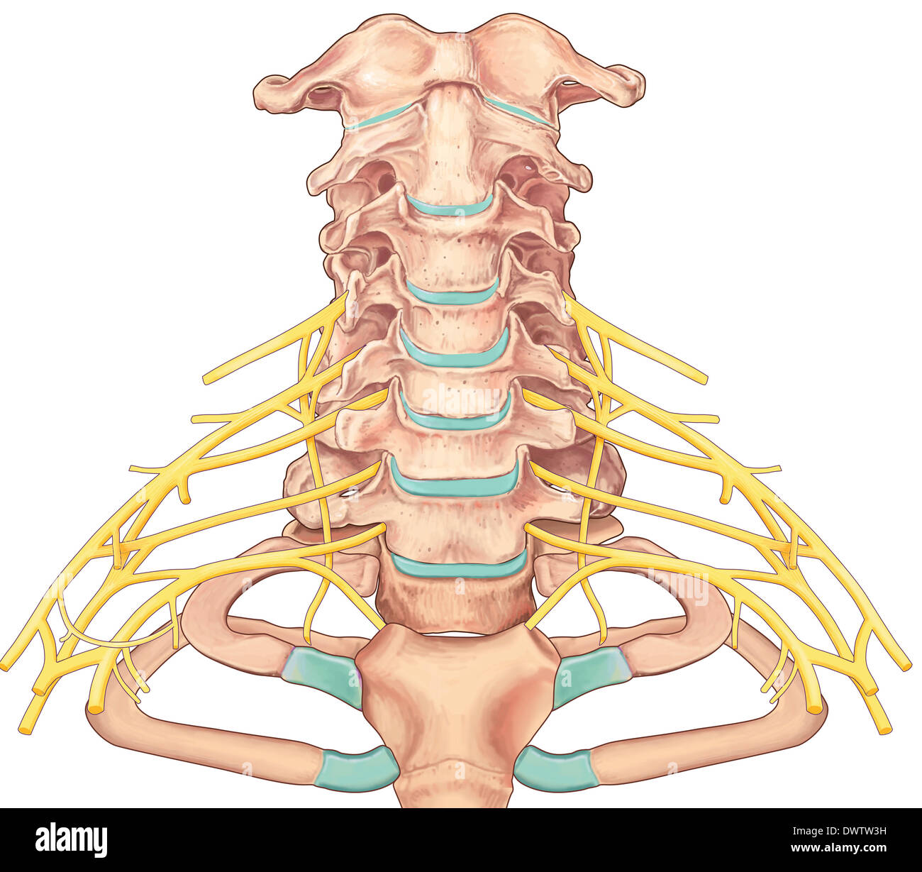 Cervical vertebra drawing Stock Photohttps://www.alamy.com/image-license-details/?v=1https://www.alamy.com/cervical-vertebra-drawing-image67544053.html
RMDWTW3H–Cervical vertebra drawing
 Archive image from page 682 of The cyclopædia of anatomy and. The cyclopædia of anatomy and physiology cyclopdiaofana0401todd Year: 1847 SKELETON. 661 is altogether obliterated. By this circum- or post sphenoid vertebra, the cranial basis stance of the body of the sixth or occipital is contracted in its longitudinal axis, while cranial vertebra joining the body of the third the cranial vault (j<?g. 481.) fashioned of the Tlie base of the human cranium, Showing its serial relation with the bodies or centra of the spinal vertebra?, and also the serial homology between the foramina of the Stock Photohttps://www.alamy.com/image-license-details/?v=1https://www.alamy.com/archive-image-from-page-682-of-the-cyclopdia-of-anatomy-and-the-cyclopdia-of-anatomy-and-physiology-cyclopdiaofana0401todd-year-1847-skeleton-661-is-altogether-obliterated-by-this-circum-or-post-sphenoid-vertebra-the-cranial-basis-stance-of-the-body-of-the-sixth-or-occipital-is-contracted-in-its-longitudinal-axis-while-cranial-vertebra-joining-the-body-of-the-third-the-cranial-vault-jltg-481-fashioned-of-the-tlie-base-of-the-human-cranium-showing-its-serial-relation-with-the-bodies-or-centra-of-the-spinal-vertebra-and-also-the-serial-homology-between-the-foramina-of-the-image259328398.html
RMW1WC0E–Archive image from page 682 of The cyclopædia of anatomy and. The cyclopædia of anatomy and physiology cyclopdiaofana0401todd Year: 1847 SKELETON. 661 is altogether obliterated. By this circum- or post sphenoid vertebra, the cranial basis stance of the body of the sixth or occipital is contracted in its longitudinal axis, while cranial vertebra joining the body of the third the cranial vault (j<?g. 481.) fashioned of the Tlie base of the human cranium, Showing its serial relation with the bodies or centra of the spinal vertebra?, and also the serial homology between the foramina of the
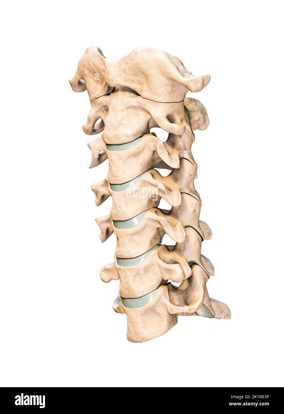 Three-quarter anterior or front view of the seven human cervical vertebrae isolated on white background 3D rendering illustration. Anatomy, osteology, Stock Photohttps://www.alamy.com/image-license-details/?v=1https://www.alamy.com/three-quarter-anterior-or-front-view-of-the-seven-human-cervical-vertebrae-isolated-on-white-background-3d-rendering-illustration-anatomy-osteology-image483020938.html
RF2K1RE3P–Three-quarter anterior or front view of the seven human cervical vertebrae isolated on white background 3D rendering illustration. Anatomy, osteology,
RFRCA33M–Anatomy spine icon, simple style
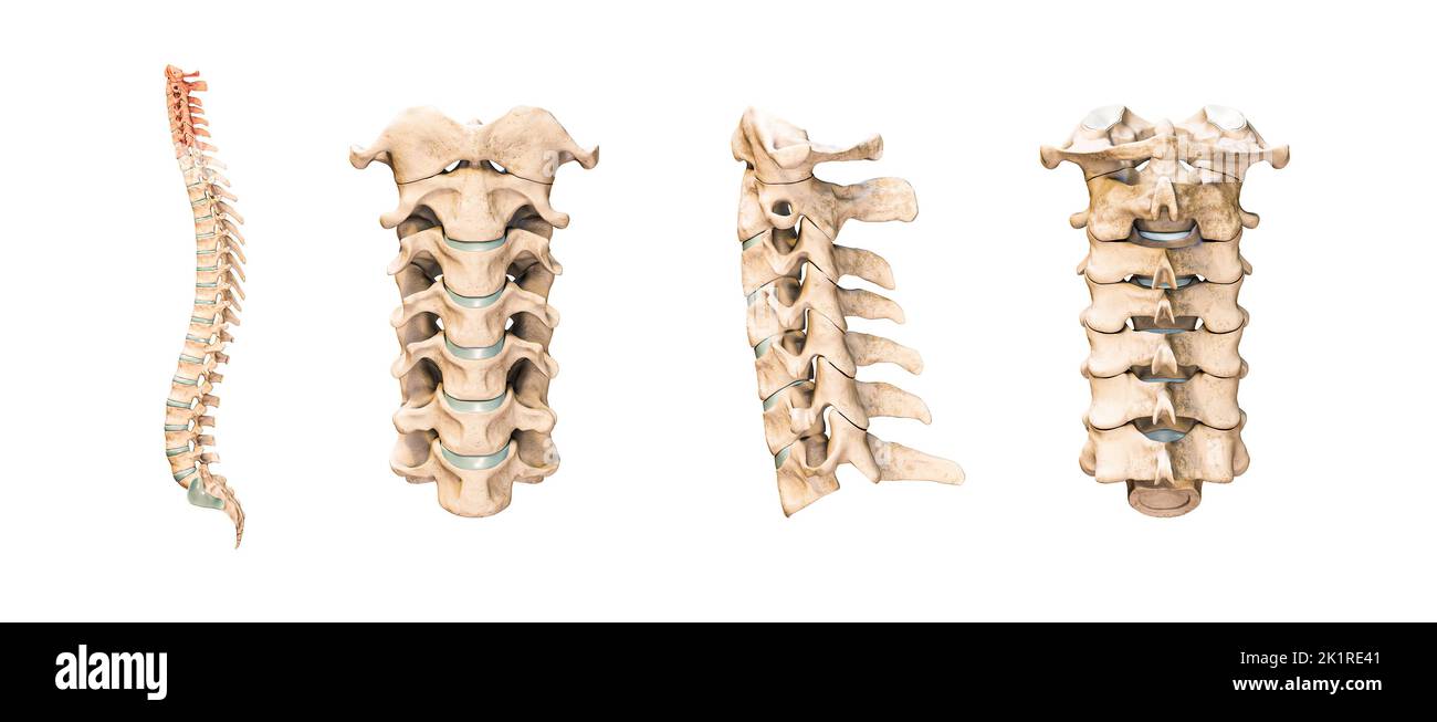 Accurate human cervical vertebrae or bones isolated on white background 3D rendering illustration. Anterior, lateral and posterior views. Anatomy, med Stock Photohttps://www.alamy.com/image-license-details/?v=1https://www.alamy.com/accurate-human-cervical-vertebrae-or-bones-isolated-on-white-background-3d-rendering-illustration-anterior-lateral-and-posterior-views-anatomy-med-image483020945.html
RF2K1RE41–Accurate human cervical vertebrae or bones isolated on white background 3D rendering illustration. Anterior, lateral and posterior views. Anatomy, med
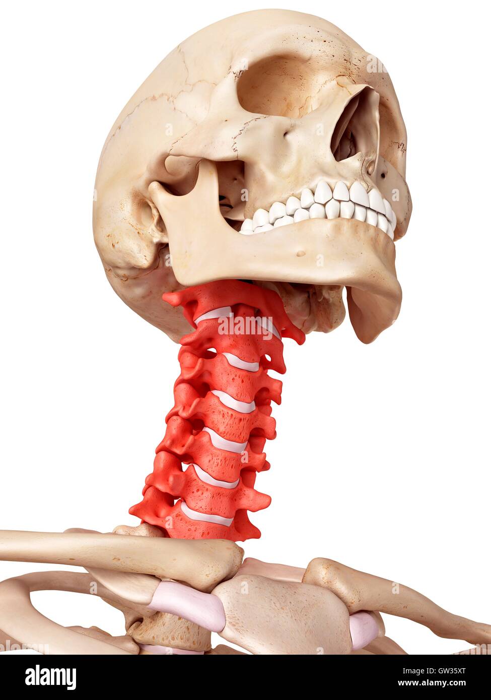 Human cervical spine, illustration. Stock Photohttps://www.alamy.com/image-license-details/?v=1https://www.alamy.com/stock-photo-human-cervical-spine-illustration-118699136.html
RFGW35XT–Human cervical spine, illustration.
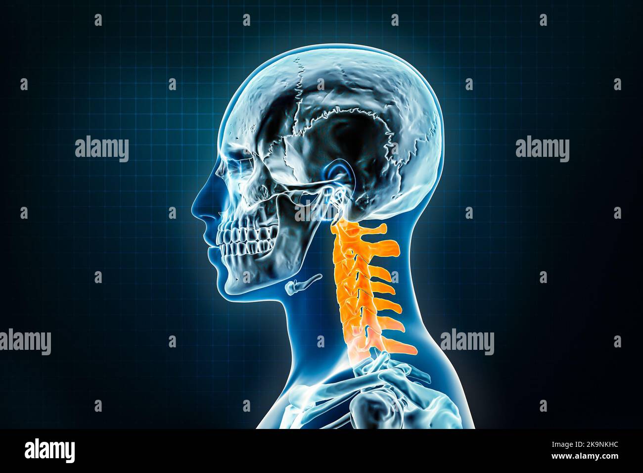 Cervical vertebrae x-ray lateral or profile view. Osteology of the human skeleton, spine 3D rendering illustration. Anatomy, medical, science, biology Stock Photohttps://www.alamy.com/image-license-details/?v=1https://www.alamy.com/cervical-vertebrae-x-ray-lateral-or-profile-view-osteology-of-the-human-skeleton-spine-3d-rendering-illustration-anatomy-medical-science-biology-image487898584.html
RF2K9NKHC–Cervical vertebrae x-ray lateral or profile view. Osteology of the human skeleton, spine 3D rendering illustration. Anatomy, medical, science, biology
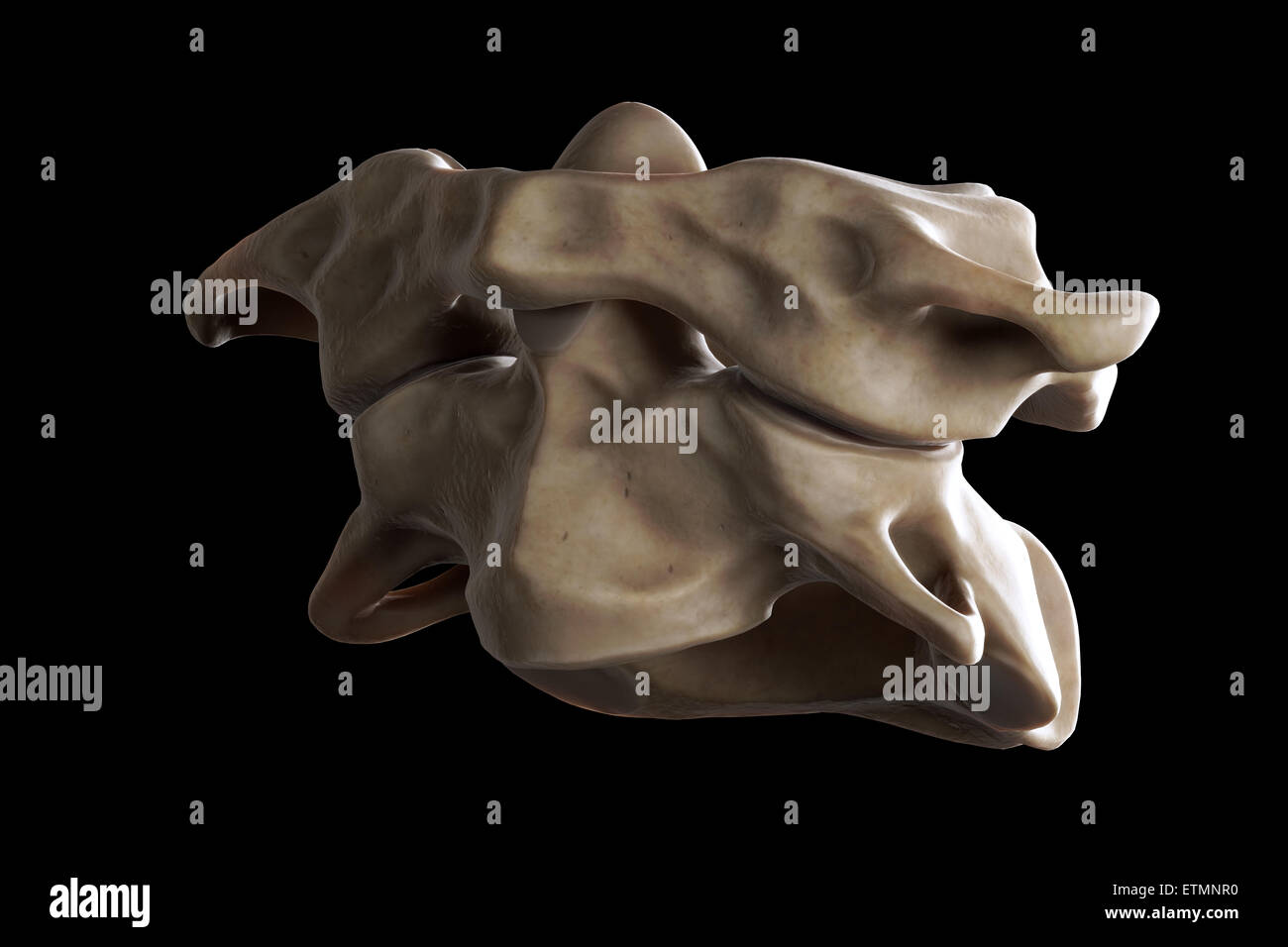 Illustration showing the atlas and axis vertebrae of the neck. Stock Photohttps://www.alamy.com/image-license-details/?v=1https://www.alamy.com/stock-photo-illustration-showing-the-atlas-and-axis-vertebrae-of-the-neck-84049364.html
RMETMNR0–Illustration showing the atlas and axis vertebrae of the neck.
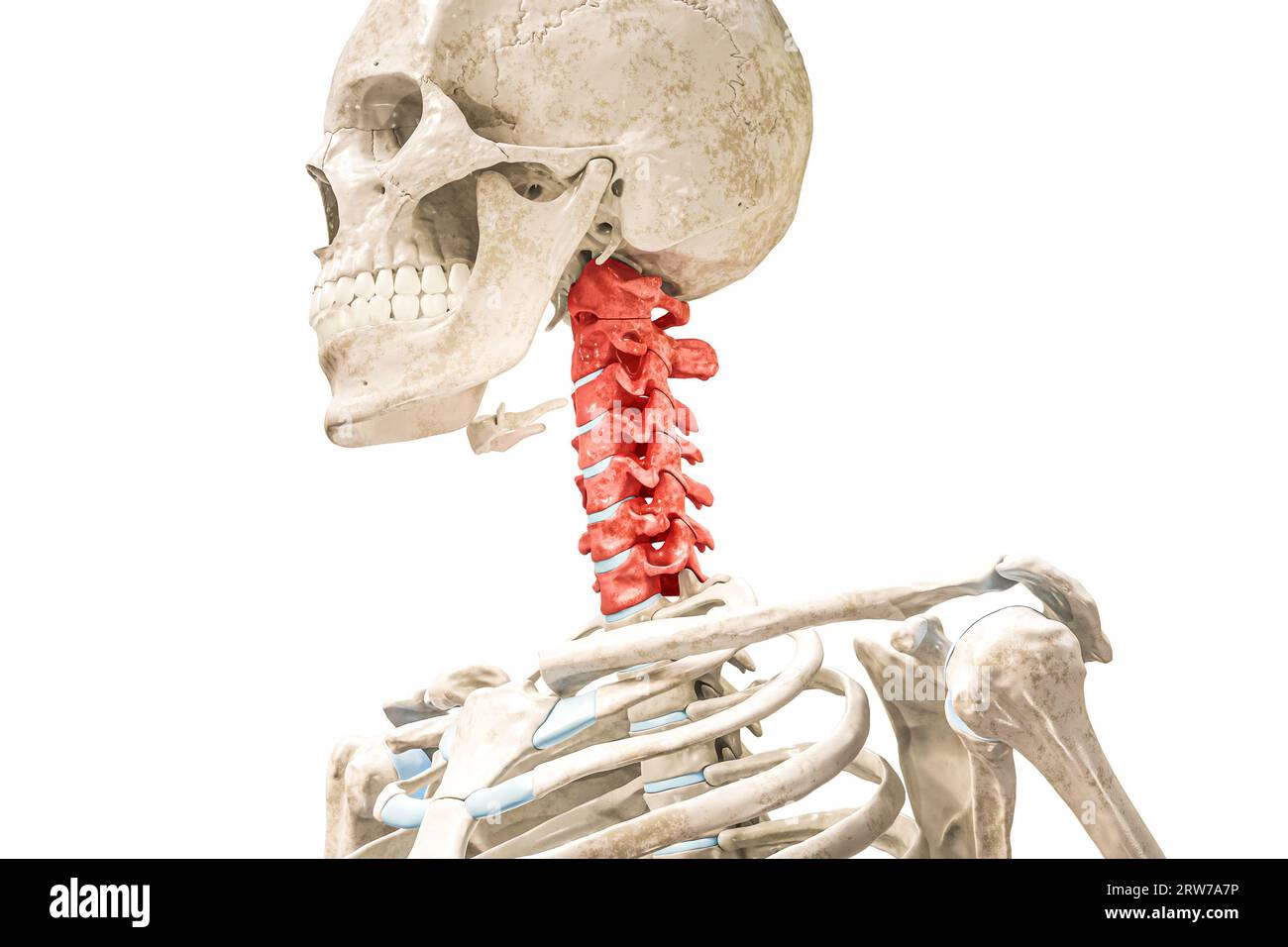 Cervical vertebrae in red color 3D rendering illustration isolated on white. Human skeleton and spine anatomy, medical diagram, osteology, skeletal sy Stock Photohttps://www.alamy.com/image-license-details/?v=1https://www.alamy.com/cervical-vertebrae-in-red-color-3d-rendering-illustration-isolated-on-white-human-skeleton-and-spine-anatomy-medical-diagram-osteology-skeletal-sy-image566259898.html
RF2RW7A7P–Cervical vertebrae in red color 3D rendering illustration isolated on white. Human skeleton and spine anatomy, medical diagram, osteology, skeletal sy
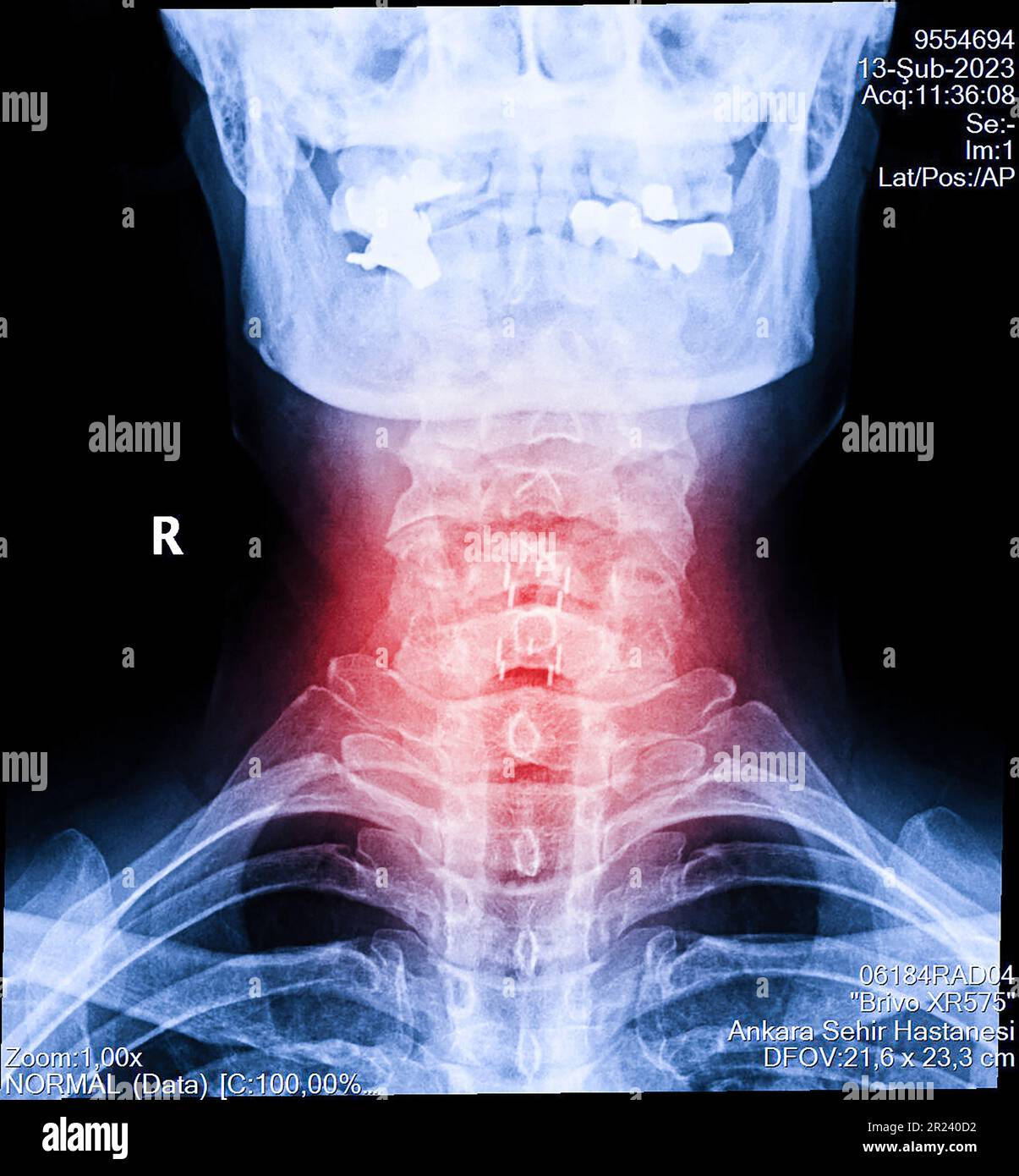 X-ray of neck and cervical spine view. Image of radiography from patient who have neck pain, nerve root compression, numbness at arm hand wrist or fin Stock Photohttps://www.alamy.com/image-license-details/?v=1https://www.alamy.com/x-ray-of-neck-and-cervical-spine-view-image-of-radiography-from-patient-who-have-neck-pain-nerve-root-compression-numbness-at-arm-hand-wrist-or-fin-image552049262.html
RF2R240D2–X-ray of neck and cervical spine view. Image of radiography from patient who have neck pain, nerve root compression, numbness at arm hand wrist or fin
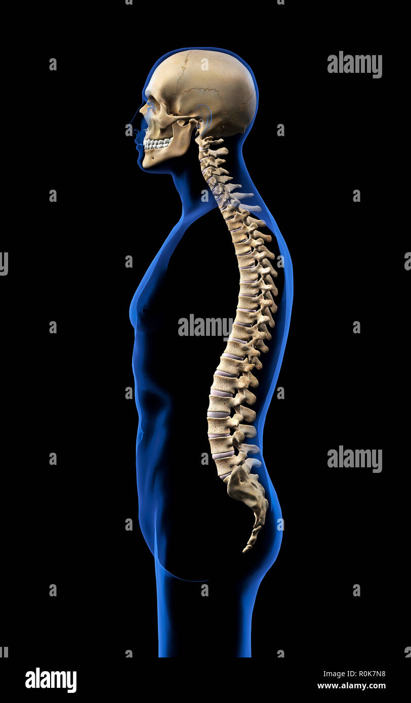 Human skull and vertebral column, side view on black background. Stock Photohttps://www.alamy.com/image-license-details/?v=1https://www.alamy.com/human-skull-and-vertebral-column-side-view-on-black-background-image224157956.html
RFR0K7N8–Human skull and vertebral column, side view on black background.
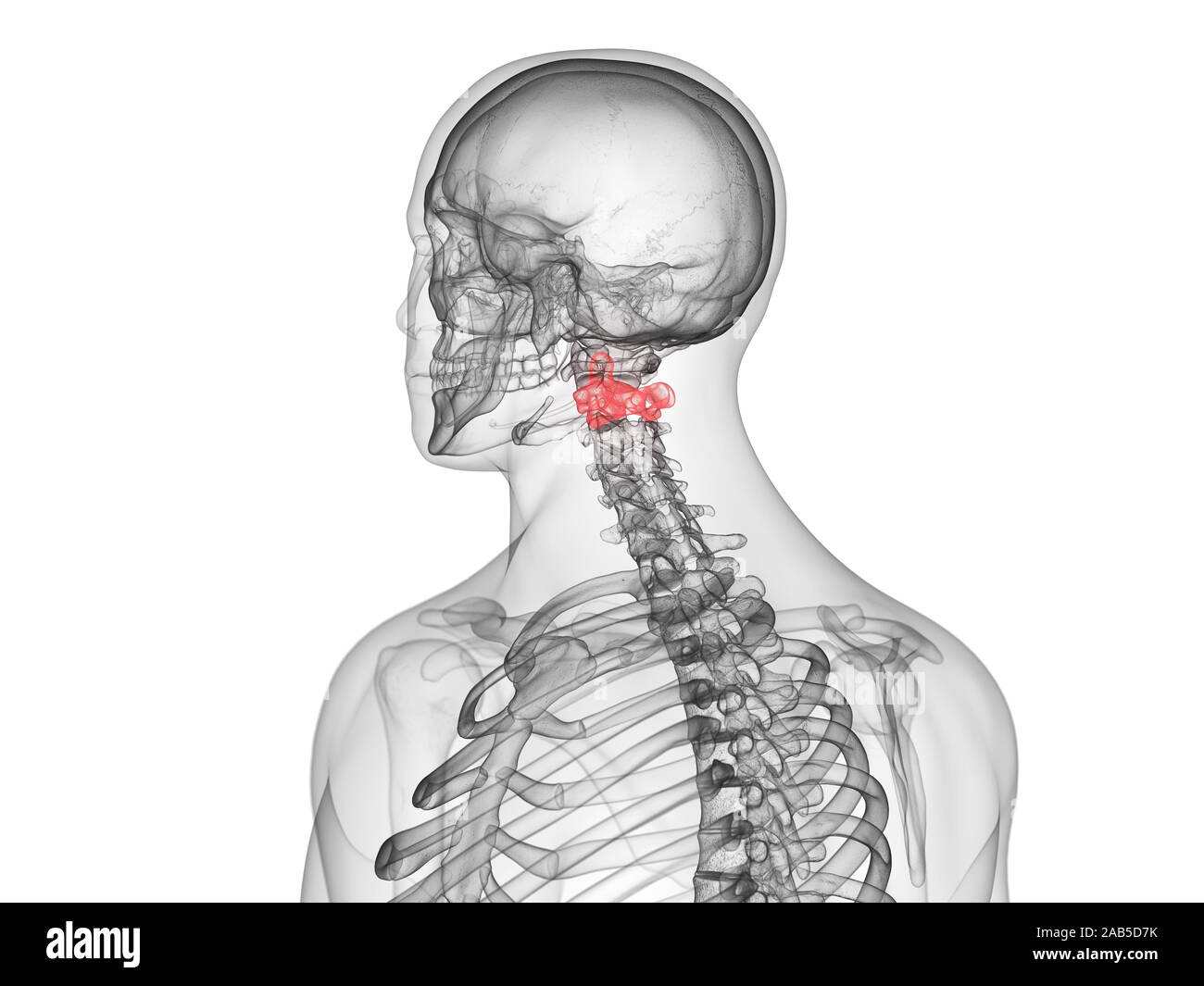 3d rendered medically accurate illustration of the axis vertebrae Stock Photohttps://www.alamy.com/image-license-details/?v=1https://www.alamy.com/3d-rendered-medically-accurate-illustration-of-the-axis-vertebrae-image333878375.html
RF2AB5D7K–3d rendered medically accurate illustration of the axis vertebrae
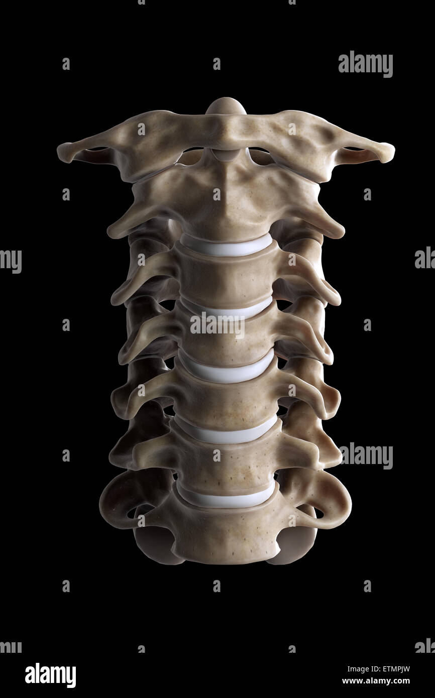 Illustration showing all seven cervical vertebrae. Stock Photohttps://www.alamy.com/image-license-details/?v=1https://www.alamy.com/stock-photo-illustration-showing-all-seven-cervical-vertebrae-84050033.html
RMETMPJW–Illustration showing all seven cervical vertebrae.
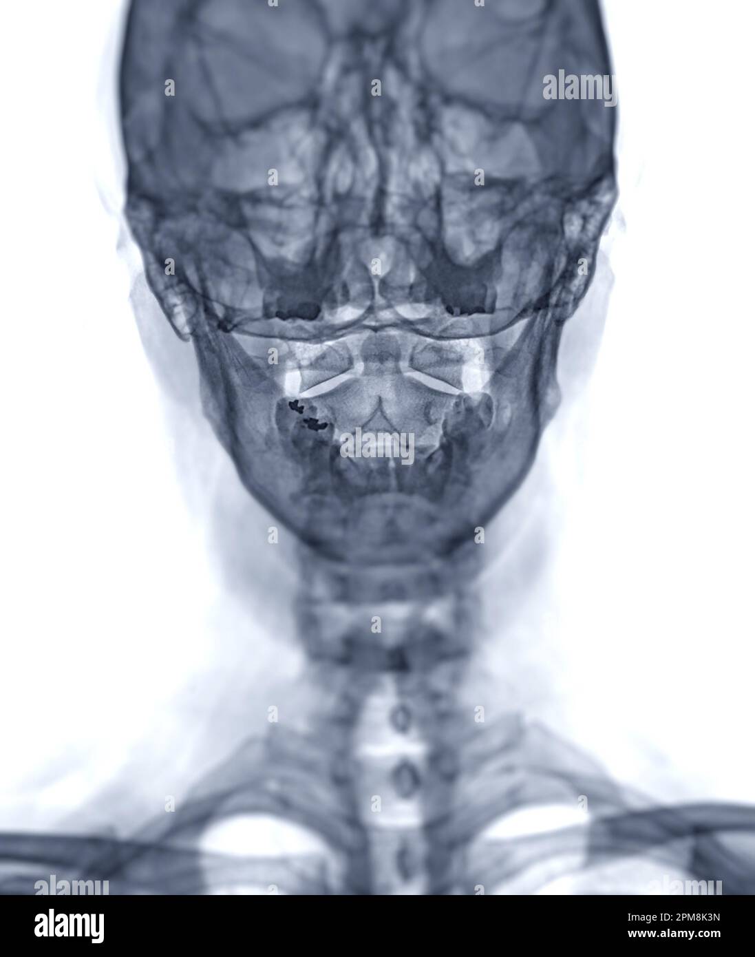 X-ray C-spine or x-ray image of Cervical spine open mount view for fracture of cervical vertebra 2nd ( axis ). Stock Photohttps://www.alamy.com/image-license-details/?v=1https://www.alamy.com/x-ray-c-spine-or-x-ray-image-of-cervical-spine-open-mount-view-for-fracture-of-cervical-vertebra-2nd-axis-image546005145.html
RF2PM8K3N–X-ray C-spine or x-ray image of Cervical spine open mount view for fracture of cervical vertebra 2nd ( axis ).
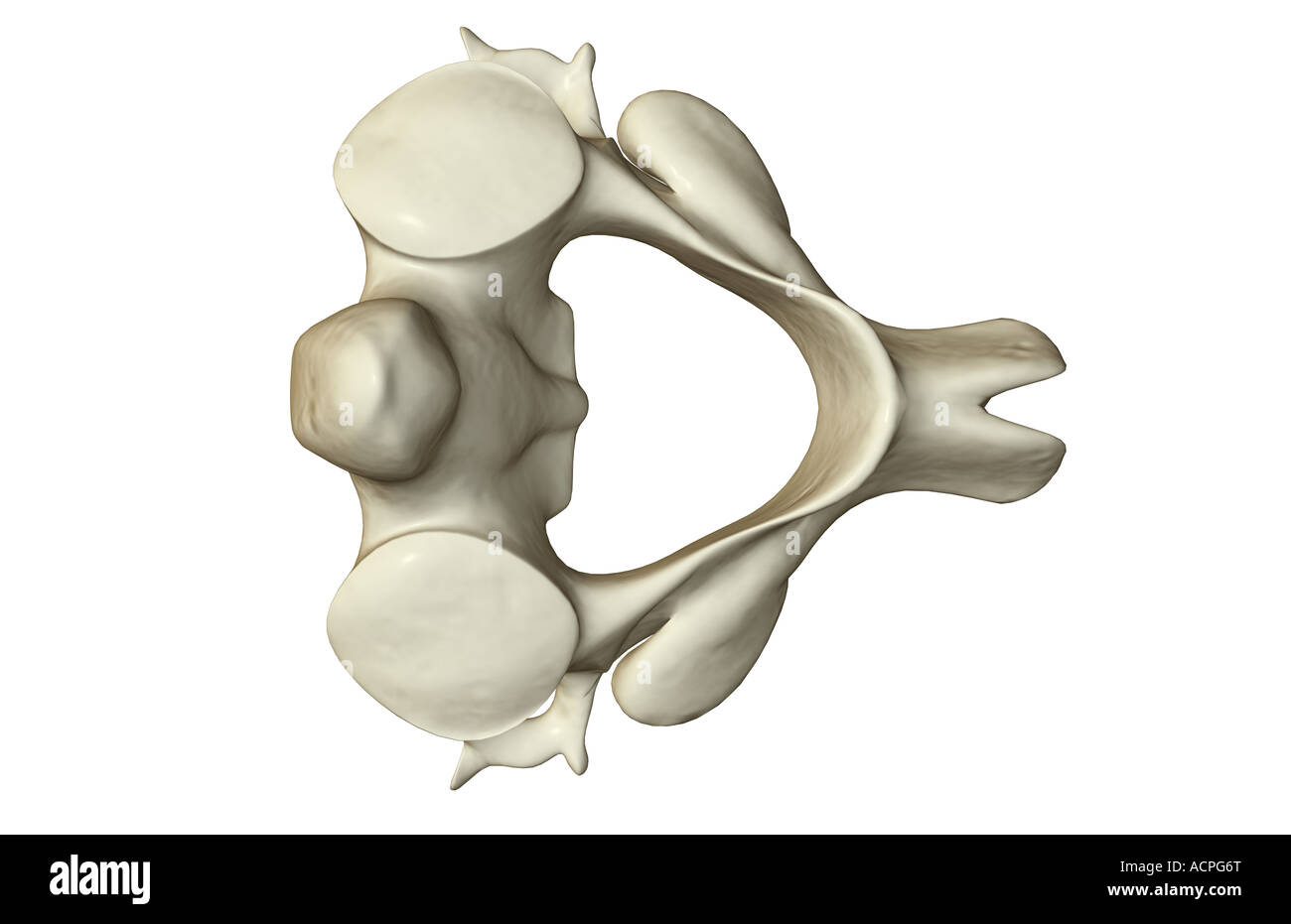 The axis bone Stock Photohttps://www.alamy.com/image-license-details/?v=1https://www.alamy.com/stock-photo-the-axis-bone-13208015.html
RFACPG6T–The axis bone
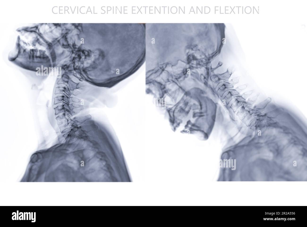 X-ray C-spine or x-ray image of Cervical spine Flexion and Extension viewfor diagnostic intervertebral disc herniation ,Spondylosis and fracture. Stock Photohttps://www.alamy.com/image-license-details/?v=1https://www.alamy.com/x-ray-c-spine-or-x-ray-image-of-cervical-spine-flexion-and-extension-viewfor-diagnostic-intervertebral-disc-herniation-spondylosis-and-fracture-image552184674.html
RF2R2A556–X-ray C-spine or x-ray image of Cervical spine Flexion and Extension viewfor diagnostic intervertebral disc herniation ,Spondylosis and fracture.
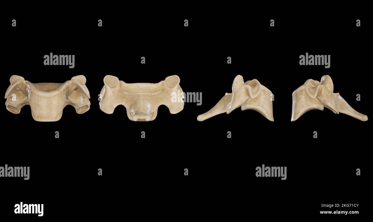 Cervical Vertebra -Multiple Views Stock Photohttps://www.alamy.com/image-license-details/?v=1https://www.alamy.com/cervical-vertebra-multiple-views-image491879611.html
RF2KG71CY–Cervical Vertebra -Multiple Views
 Spine - Cervical Region - lateral v iew. Stock Photohttps://www.alamy.com/image-license-details/?v=1https://www.alamy.com/stock-photo-spine-cervical-region-lateral-v-iew-130806531.html
RFHGPN1R–Spine - Cervical Region - lateral v iew.
 The anatomist's vade mecum : a system of human anatomy . mes the anterior tubercle represents a small butdistinct rib. Dorsal Vertelra.—The hody of a dorsal vertebra is as long frombefore backwards as from side to side, particularly in the middle of thedorsal region; it is thicker behind than before, and marked on eachside by two half-articulating surfaces for the heads of two ribs. The * A lateral view of the axis. 1. The body; the figure is placed on thedepression which gives attachment to the longus colli. 2. The odontoid pro-cess. 3. The smooth facet on the anterior surface of the odontoid Stock Photohttps://www.alamy.com/image-license-details/?v=1https://www.alamy.com/the-anatomists-vade-mecum-a-system-of-human-anatomy-mes-the-anterior-tubercle-represents-a-small-butdistinct-rib-dorsal-vertelrathe-hody-of-a-dorsal-vertebra-is-as-long-frombefore-backwards-as-from-side-to-side-particularly-in-the-middle-of-thedorsal-region-it-is-thicker-behind-than-before-and-marked-on-eachside-by-two-half-articulating-surfaces-for-the-heads-of-two-ribs-the-a-lateral-view-of-the-axis-1-the-body-the-figure-is-placed-on-thedepression-which-gives-attachment-to-the-longus-colli-2-the-odontoid-pro-cess-3-the-smooth-facet-on-the-anterior-surface-of-the-odontoid-image342785019.html
RM2AWK5P3–The anatomist's vade mecum : a system of human anatomy . mes the anterior tubercle represents a small butdistinct rib. Dorsal Vertelra.—The hody of a dorsal vertebra is as long frombefore backwards as from side to side, particularly in the middle of thedorsal region; it is thicker behind than before, and marked on eachside by two half-articulating surfaces for the heads of two ribs. The * A lateral view of the axis. 1. The body; the figure is placed on thedepression which gives attachment to the longus colli. 2. The odontoid pro-cess. 3. The smooth facet on the anterior surface of the odontoid
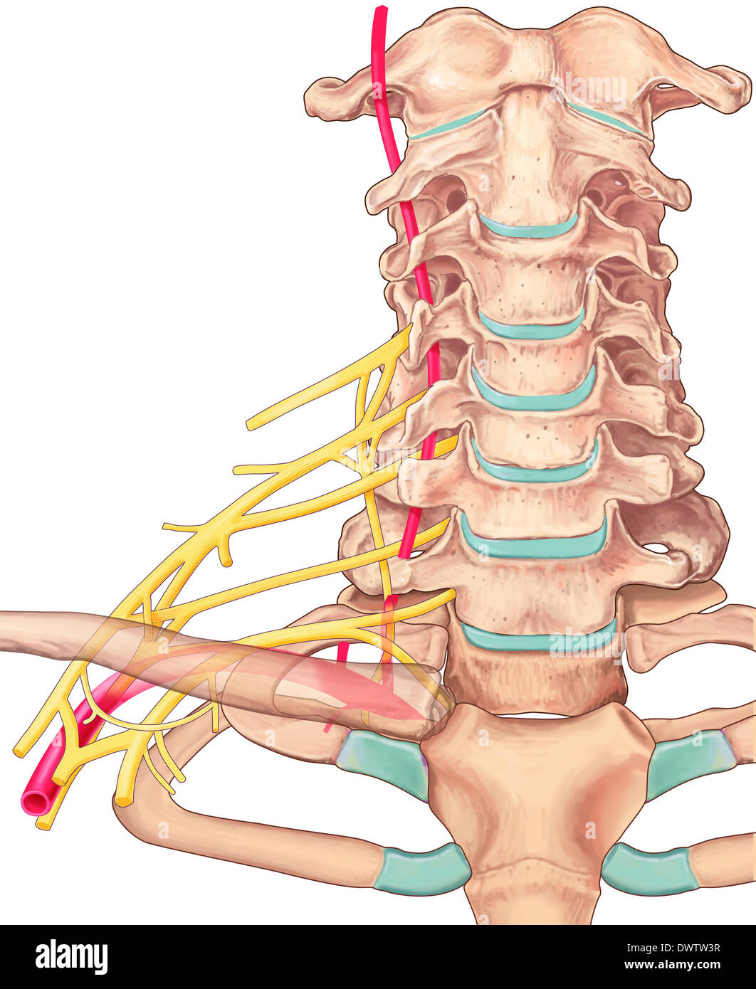 Cervical vertebra drawing Stock Photohttps://www.alamy.com/image-license-details/?v=1https://www.alamy.com/cervical-vertebra-drawing-image67544059.html
RMDWTW3R–Cervical vertebra drawing
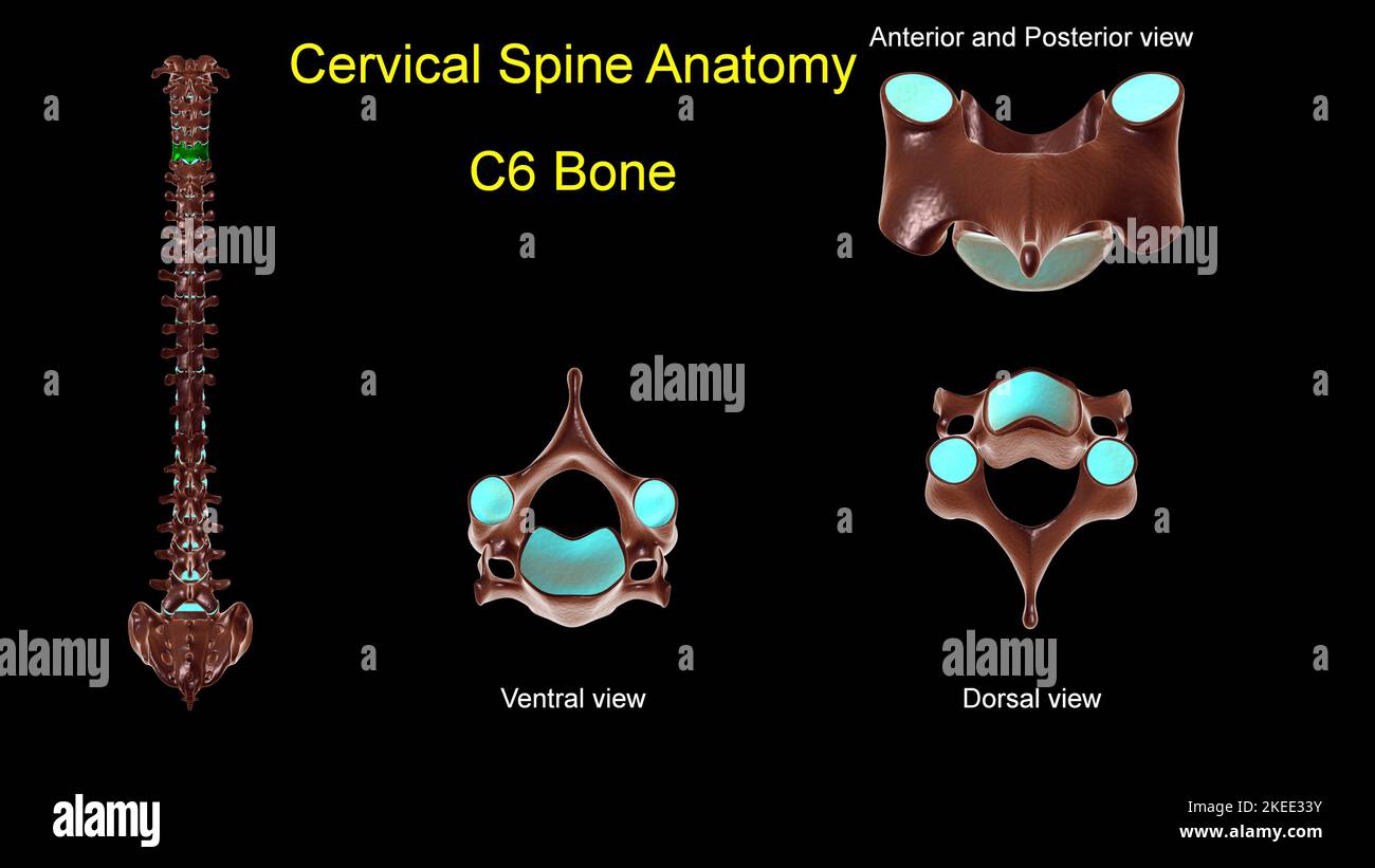 Cervical spine C 6 bone anatomy for medical concept 3D Illustration with anterior and posterior View Stock Photohttps://www.alamy.com/image-license-details/?v=1https://www.alamy.com/cervical-spine-c-6-bone-anatomy-for-medical-concept-3d-illustration-with-anterior-and-posterior-view-image490805279.html
RF2KEE33Y–Cervical spine C 6 bone anatomy for medical concept 3D Illustration with anterior and posterior View
 . The development of the human body : a manual of human embryology. Embryology; Embryo, Non-Mammalian. Fig. ioo.—A, Upper Surface of the First Sacral Vertebra, and B, Ventral View of the Sacrum showing Primary Centers of Ossification. c, Body; na, vertebral arch; r, rib center.—(Sappey.) atlas a double center appears in the persisting hypochordal bar, and the body which corresponds to the atlas, after developing the termi- nal epiphysial disks, fuses with the body of the epistropheus (axis) to form its odontoid process, this vertebra consequently possessing, in addition to the typical centers, Stock Photohttps://www.alamy.com/image-license-details/?v=1https://www.alamy.com/the-development-of-the-human-body-a-manual-of-human-embryology-embryology-embryo-non-mammalian-fig-iooa-upper-surface-of-the-first-sacral-vertebra-and-b-ventral-view-of-the-sacrum-showing-primary-centers-of-ossification-c-body-na-vertebral-arch-r-rib-centersappey-atlas-a-double-center-appears-in-the-persisting-hypochordal-bar-and-the-body-which-corresponds-to-the-atlas-after-developing-the-termi-nal-epiphysial-disks-fuses-with-the-body-of-the-epistropheus-axis-to-form-its-odontoid-process-this-vertebra-consequently-possessing-in-addition-to-the-typical-centers-image215969742.html
RMPFA7H2–. The development of the human body : a manual of human embryology. Embryology; Embryo, Non-Mammalian. Fig. ioo.—A, Upper Surface of the First Sacral Vertebra, and B, Ventral View of the Sacrum showing Primary Centers of Ossification. c, Body; na, vertebral arch; r, rib center.—(Sappey.) atlas a double center appears in the persisting hypochordal bar, and the body which corresponds to the atlas, after developing the termi- nal epiphysial disks, fuses with the body of the epistropheus (axis) to form its odontoid process, this vertebra consequently possessing, in addition to the typical centers,
 Anatomy spine pattern seamless background texture repeat wallpaper geometric vector Stock Vectorhttps://www.alamy.com/image-license-details/?v=1https://www.alamy.com/anatomy-spine-pattern-seamless-background-texture-repeat-wallpaper-geometric-vector-image442765617.html
RF2GM9M1N–Anatomy spine pattern seamless background texture repeat wallpaper geometric vector
 Human cervical spine, illustration. Stock Photohttps://www.alamy.com/image-license-details/?v=1https://www.alamy.com/stock-photo-human-cervical-spine-illustration-118699138.html
RFGW35XX–Human cervical spine, illustration.
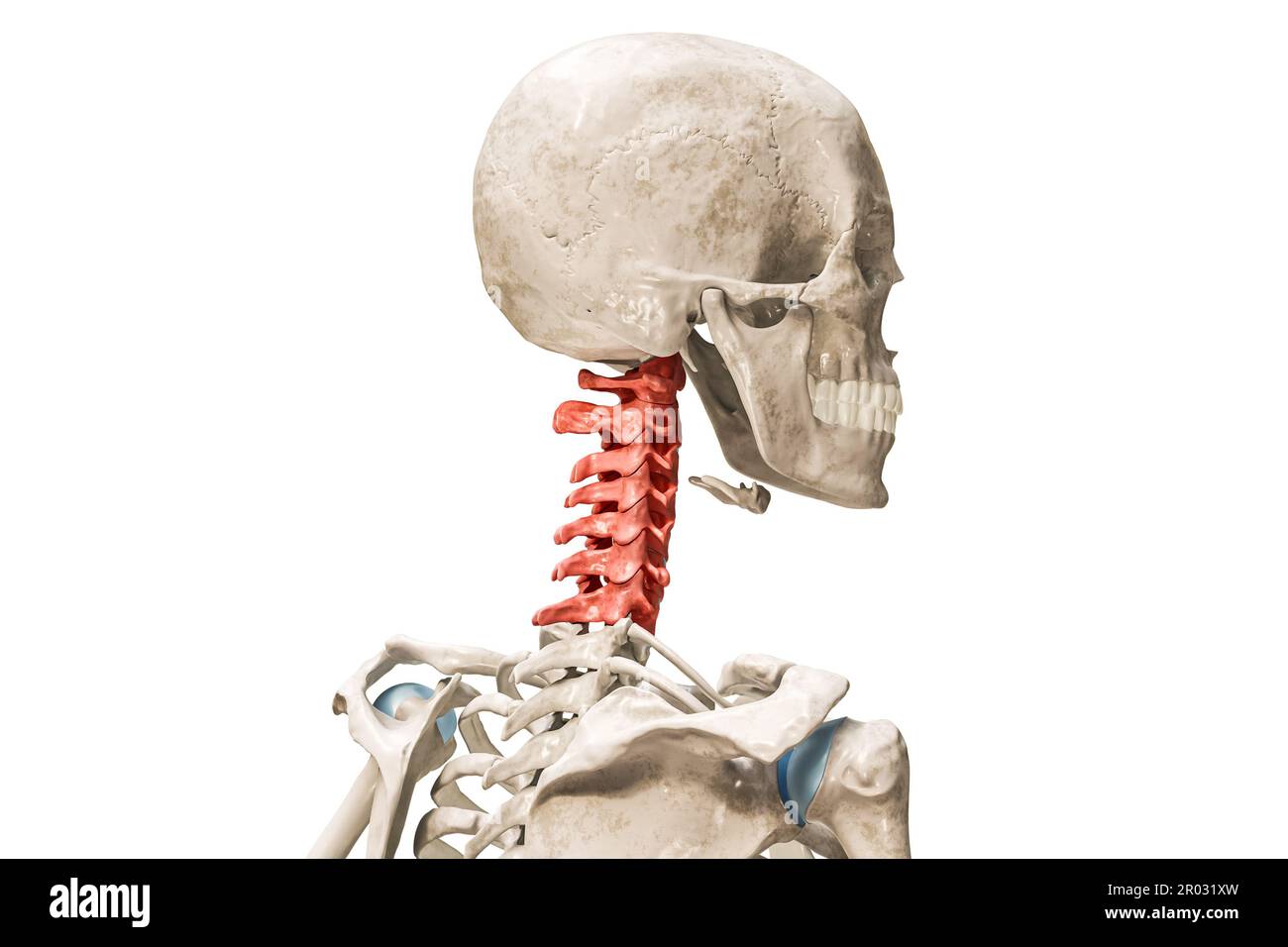 Cervical vertebrae in red color 3D rendering illustration isolated on white with copy space. Human skeleton and spine anatomy, medical diagram, osteol Stock Photohttps://www.alamy.com/image-license-details/?v=1https://www.alamy.com/cervical-vertebrae-in-red-color-3d-rendering-illustration-isolated-on-white-with-copy-space-human-skeleton-and-spine-anatomy-medical-diagram-osteol-image550799169.html
RF2R031XW–Cervical vertebrae in red color 3D rendering illustration isolated on white with copy space. Human skeleton and spine anatomy, medical diagram, osteol
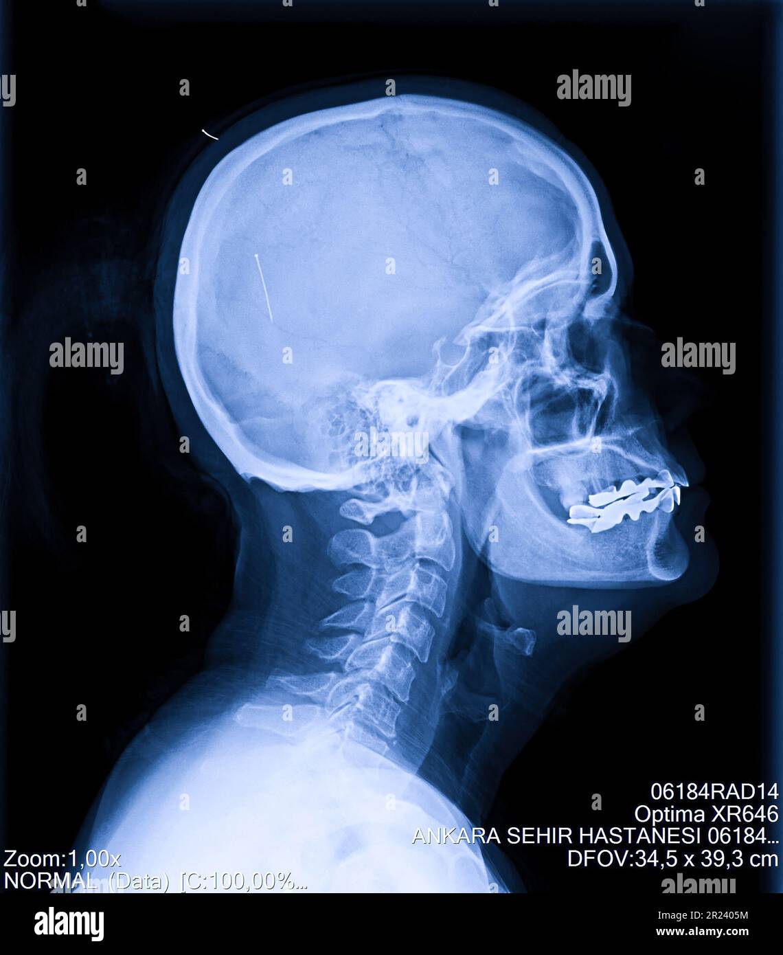 Human cervical spine x-ray, neck radiography Stock Photohttps://www.alamy.com/image-license-details/?v=1https://www.alamy.com/human-cervical-spine-x-ray-neck-radiography-image552049056.html
RF2R2405M–Human cervical spine x-ray, neck radiography
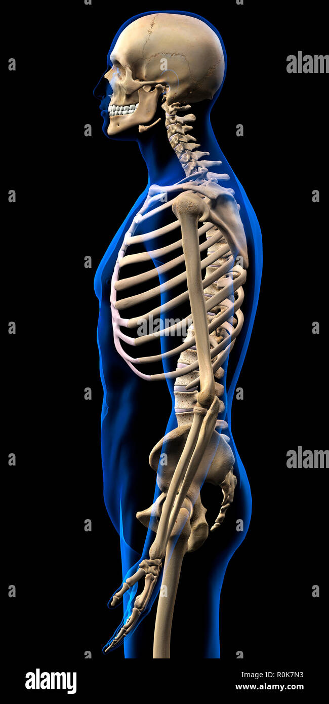 Human skeleton, side view with blue x-ray body outline. Stock Photohttps://www.alamy.com/image-license-details/?v=1https://www.alamy.com/human-skeleton-side-view-with-blue-x-ray-body-outline-image224157951.html
RFR0K7N3–Human skeleton, side view with blue x-ray body outline.
 3d rendered medically accurate illustration of the axis vertebrae Stock Photohttps://www.alamy.com/image-license-details/?v=1https://www.alamy.com/3d-rendered-medically-accurate-illustration-of-the-axis-vertebrae-image333878471.html
RF2AB5DB3–3d rendered medically accurate illustration of the axis vertebrae
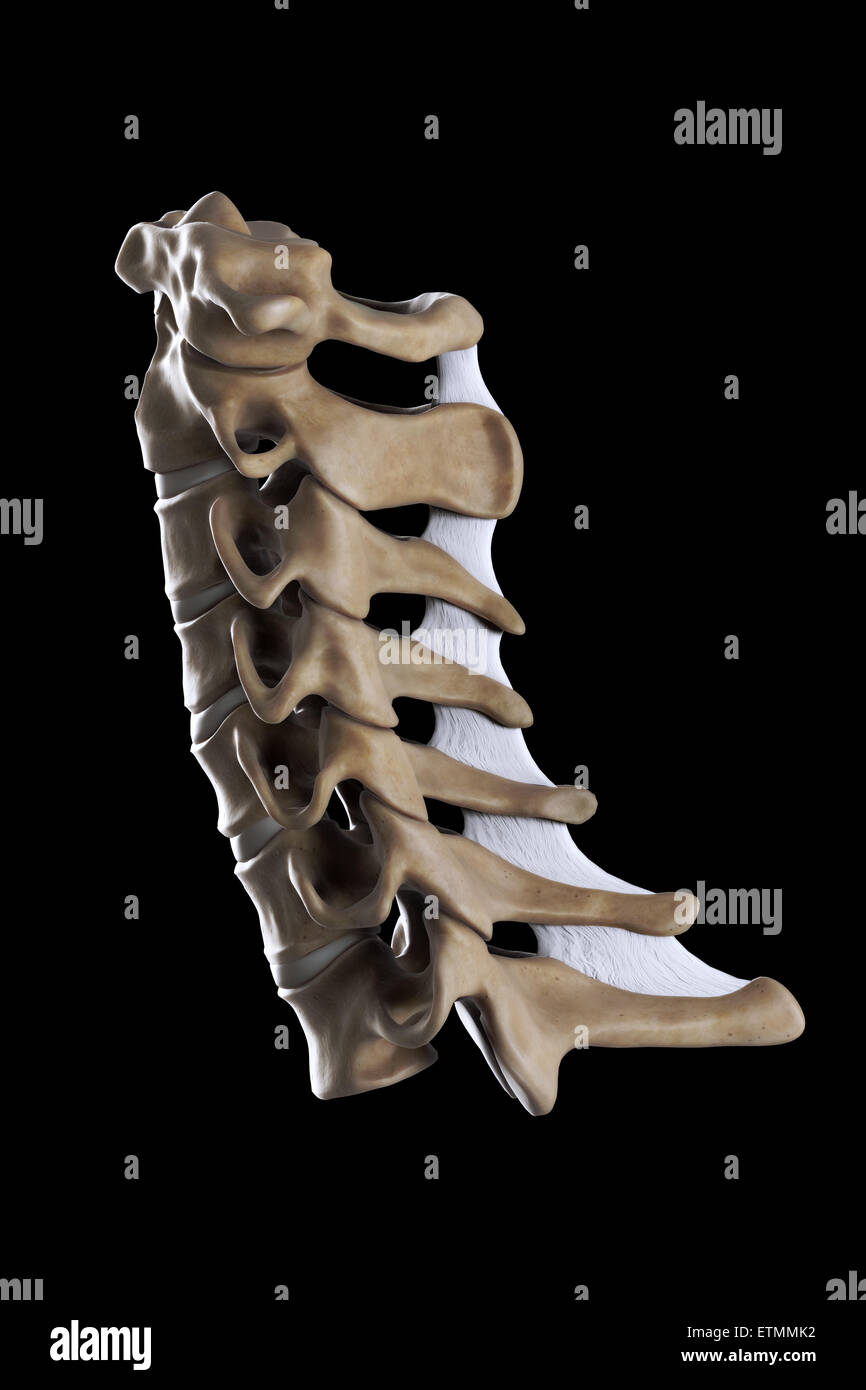 Illustration showing all seven cervical vertebrae. Stock Photohttps://www.alamy.com/image-license-details/?v=1https://www.alamy.com/stock-photo-illustration-showing-all-seven-cervical-vertebrae-84048470.html
RMETMMK2–Illustration showing all seven cervical vertebrae.
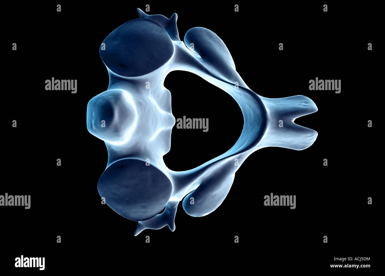 The axis bone Stock Photohttps://www.alamy.com/image-license-details/?v=1https://www.alamy.com/stock-photo-the-axis-bone-13166767.html
RFACJ5DM–The axis bone
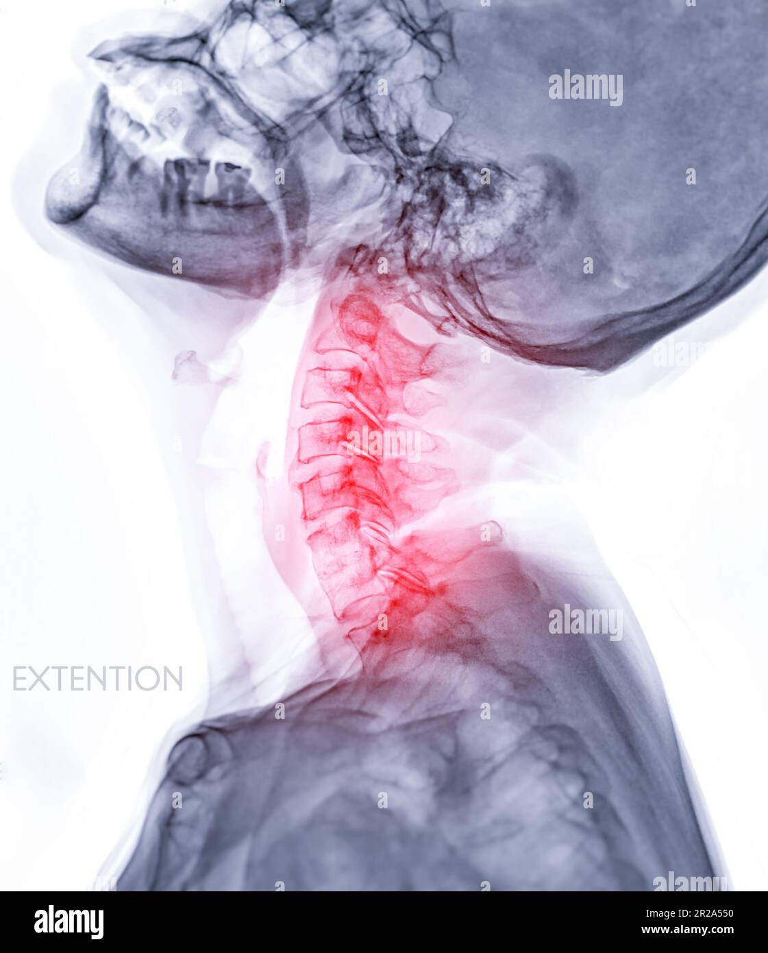 X-ray C-spine or x-ray image of Cervical spine Extension viewfor diagnostic intervertebral disc herniation ,Spondylosis and fracture. Stock Photohttps://www.alamy.com/image-license-details/?v=1https://www.alamy.com/x-ray-c-spine-or-x-ray-image-of-cervical-spine-extension-viewfor-diagnostic-intervertebral-disc-herniation-spondylosis-and-fracture-image552184668.html
RF2R2A550–X-ray C-spine or x-ray image of Cervical spine Extension viewfor diagnostic intervertebral disc herniation ,Spondylosis and fracture.
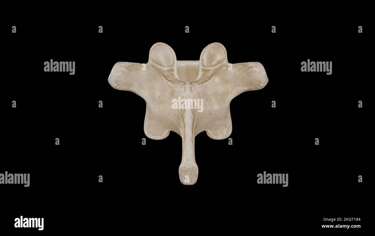 Posterior view of Tenth Thoracic Vertebra (T10) Stock Photohttps://www.alamy.com/image-license-details/?v=1https://www.alamy.com/posterior-view-of-tenth-thoracic-vertebra-t10-image491879476.html
RF2KG7184–Posterior view of Tenth Thoracic Vertebra (T10)
 The anatomist's vade mecum : a system of human anatomy . ar vertebrae in the cervical region:—Thefirst, or atlas; the second, or axis; and the seventh, or vertebraprominens. The Atlas (named from supporting the head) is a simple ring ofbone, without body, and composed of arches and processes. The * A central cervical vertebra, seen upon its upper surface. 1. The body,concave in the middle, and rising on each side into a sharp ridge. 2. Thelamina. 3. The pedicle, rendered concave by the superior intervertebralnotch. 4. The bifid spinous process. 5. The bifid transverse process. Thefigure is pla Stock Photohttps://www.alamy.com/image-license-details/?v=1https://www.alamy.com/the-anatomists-vade-mecum-a-system-of-human-anatomy-ar-vertebrae-in-the-cervical-regionthefirst-or-atlas-the-second-or-axis-and-the-seventh-or-vertebraprominens-the-atlas-named-from-supporting-the-head-is-a-simple-ring-ofbone-without-body-and-composed-of-arches-and-processes-the-a-central-cervical-vertebra-seen-upon-its-upper-surface-1-the-bodyconcave-in-the-middle-and-rising-on-each-side-into-a-sharp-ridge-2-thelamina-3-the-pedicle-rendered-concave-by-the-superior-intervertebralnotch-4-the-bifid-spinous-process-5-the-bifid-transverse-process-thefigure-is-pla-image342785511.html
RM2AWK6BK–The anatomist's vade mecum : a system of human anatomy . ar vertebrae in the cervical region:—Thefirst, or atlas; the second, or axis; and the seventh, or vertebraprominens. The Atlas (named from supporting the head) is a simple ring ofbone, without body, and composed of arches and processes. The * A central cervical vertebra, seen upon its upper surface. 1. The body,concave in the middle, and rising on each side into a sharp ridge. 2. Thelamina. 3. The pedicle, rendered concave by the superior intervertebralnotch. 4. The bifid spinous process. 5. The bifid transverse process. Thefigure is pla
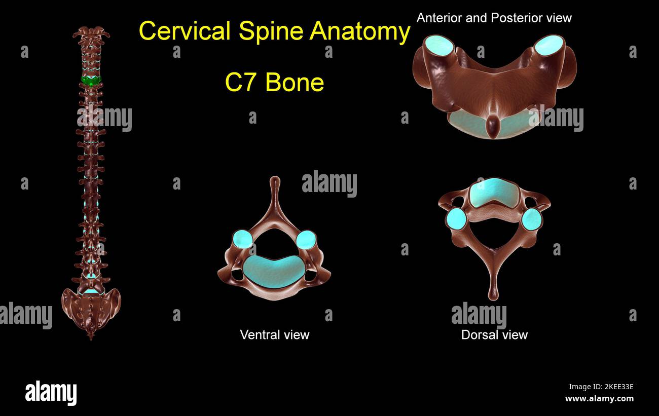 Cervical spine C 7 bone anatomy for medical concept 3D Illustration with anterior and posterior View Stock Photohttps://www.alamy.com/image-license-details/?v=1https://www.alamy.com/cervical-spine-c-7-bone-anatomy-for-medical-concept-3d-illustration-with-anterior-and-posterior-view-image490805266.html
RF2KEE33E–Cervical spine C 7 bone anatomy for medical concept 3D Illustration with anterior and posterior View
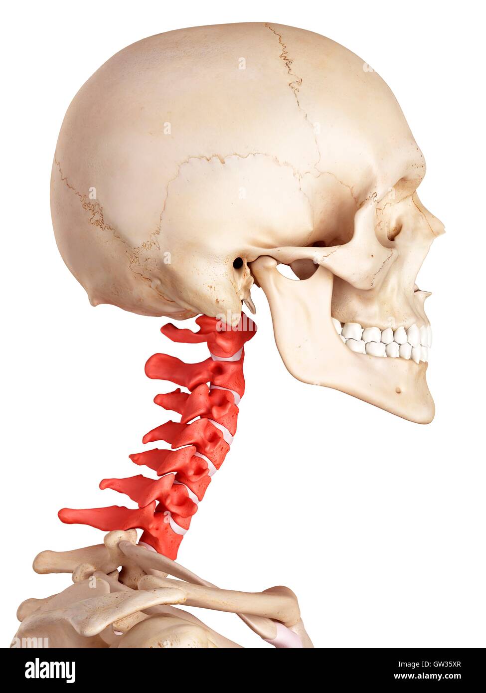 Human cervical spine, illustration. Stock Photohttps://www.alamy.com/image-license-details/?v=1https://www.alamy.com/stock-photo-human-cervical-spine-illustration-118699135.html
RFGW35XR–Human cervical spine, illustration.
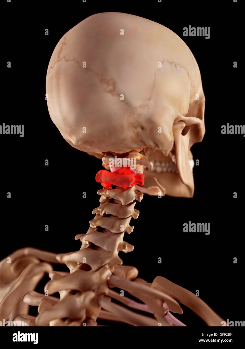 Human upper neck pain, illustration. Stock Photohttps://www.alamy.com/image-license-details/?v=1https://www.alamy.com/stock-photo-human-upper-neck-pain-illustration-112681511.html
RFGF92BK–Human upper neck pain, illustration.
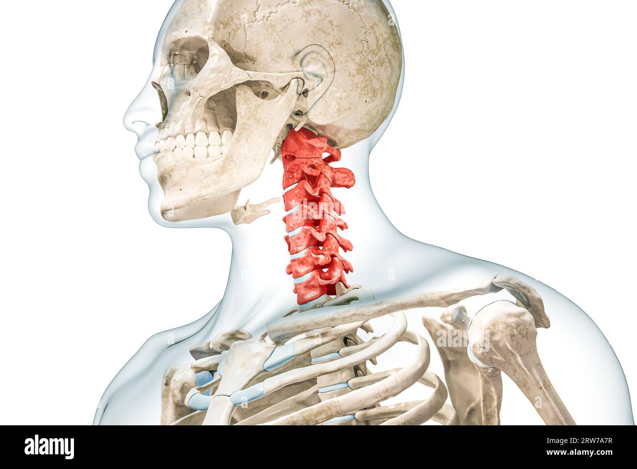 Cervical vertebrae in red color with body 3D rendering illustration isolated on white. Human skeleton and spine anatomy, medical diagram, osteology, s Stock Photohttps://www.alamy.com/image-license-details/?v=1https://www.alamy.com/cervical-vertebrae-in-red-color-with-body-3d-rendering-illustration-isolated-on-white-human-skeleton-and-spine-anatomy-medical-diagram-osteology-s-image566259899.html
RF2RW7A7R–Cervical vertebrae in red color with body 3D rendering illustration isolated on white. Human skeleton and spine anatomy, medical diagram, osteology, s
 Male cervical spine, illustration Stock Photohttps://www.alamy.com/image-license-details/?v=1https://www.alamy.com/male-cervical-spine-illustration-image561718398.html
RF2RHTDFA–Male cervical spine, illustration
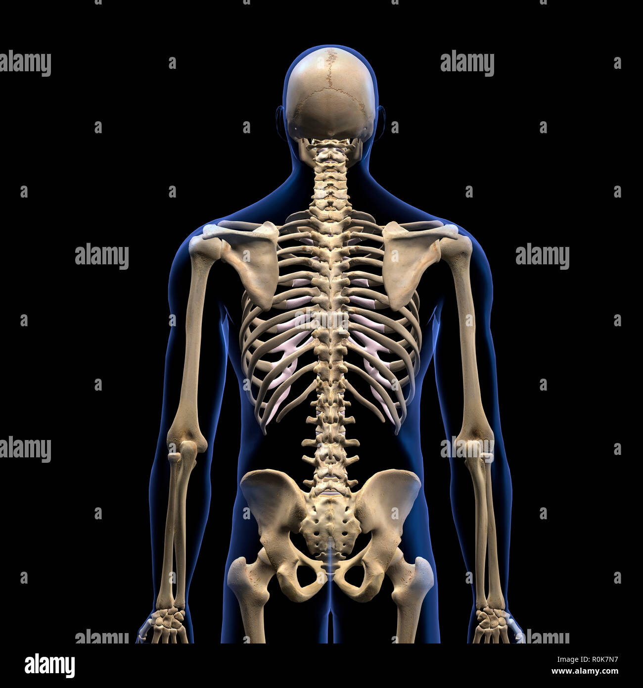 Human vertebral column, rear view on black background. Stock Photohttps://www.alamy.com/image-license-details/?v=1https://www.alamy.com/human-vertebral-column-rear-view-on-black-background-image224157955.html
RFR0K7N7–Human vertebral column, rear view on black background.
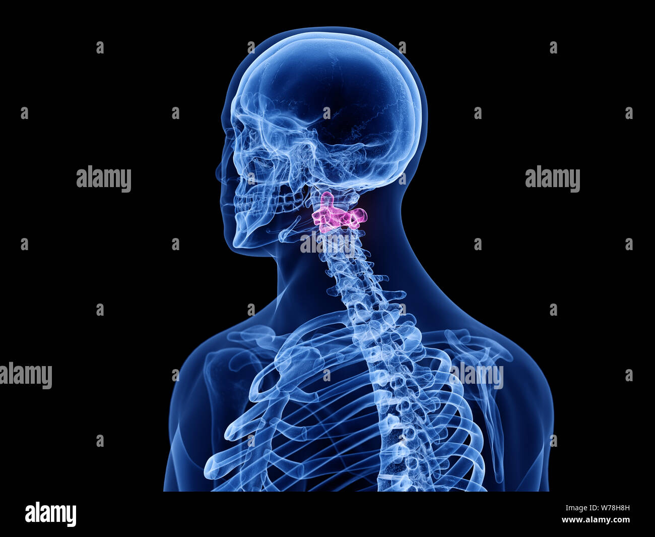 3d rendered medically accurate illustration of the axis vertebrae Stock Photohttps://www.alamy.com/image-license-details/?v=1https://www.alamy.com/3d-rendered-medically-accurate-illustration-of-the-axis-vertebrae-image262647297.html
RFW78H8H–3d rendered medically accurate illustration of the axis vertebrae
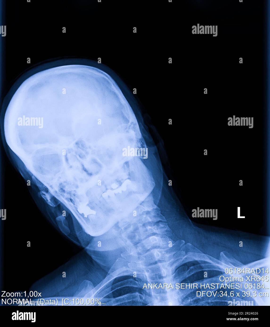 Human cervical spine x-ray, neck radiography Stock Photohttps://www.alamy.com/image-license-details/?v=1https://www.alamy.com/human-cervical-spine-x-ray-neck-radiography-image552048958.html
RF2R24026–Human cervical spine x-ray, neck radiography
