Quick filters:
Bacteria plate Stock Photos and Images
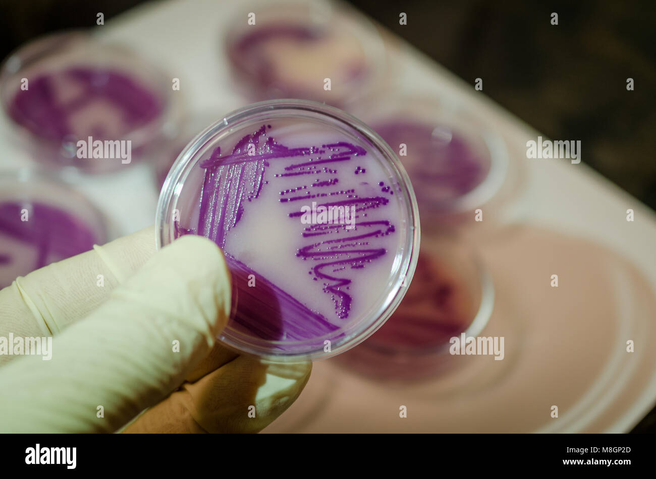 Bacterial culture plate holding in hand Stock Photohttps://www.alamy.com/image-license-details/?v=1https://www.alamy.com/stock-photo-bacterial-culture-plate-holding-in-hand-177389477.html
RFM8GP2D–Bacterial culture plate holding in hand
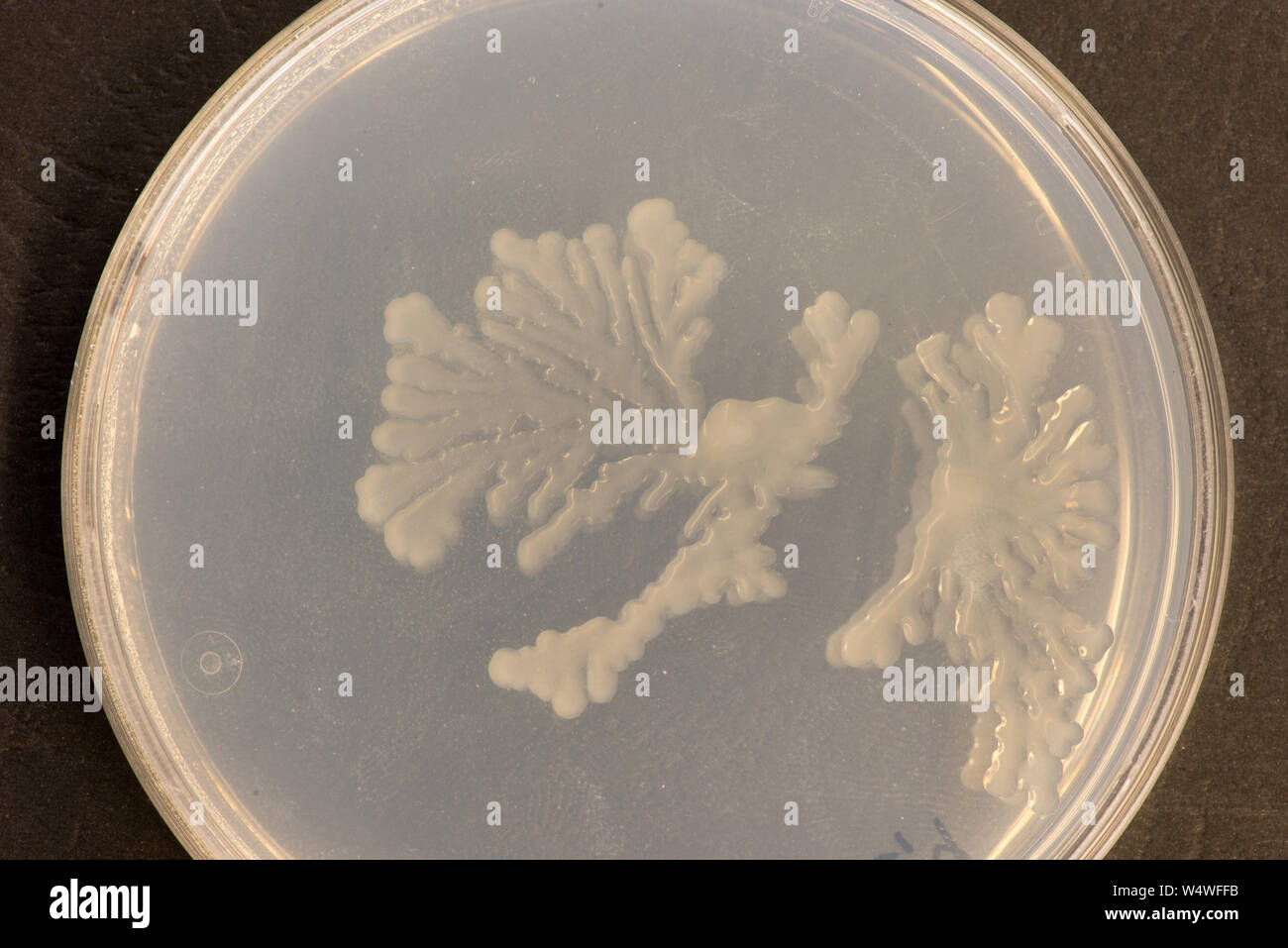 Colony of soil bacteria on an agar plate with rhizoid form Stock Photohttps://www.alamy.com/image-license-details/?v=1https://www.alamy.com/colony-of-soil-bacteria-on-an-agar-plate-with-rhizoid-form-image261175135.html
RFW4WFFB–Colony of soil bacteria on an agar plate with rhizoid form
 Gloved hand holding petri dish with bacterial colony isolated on a white background with space for text or cutout Stock Photohttps://www.alamy.com/image-license-details/?v=1https://www.alamy.com/stock-photo-gloved-hand-holding-petri-dish-with-bacterial-colony-isolated-on-a-37648504.html
RMC5711C–Gloved hand holding petri dish with bacterial colony isolated on a white background with space for text or cutout
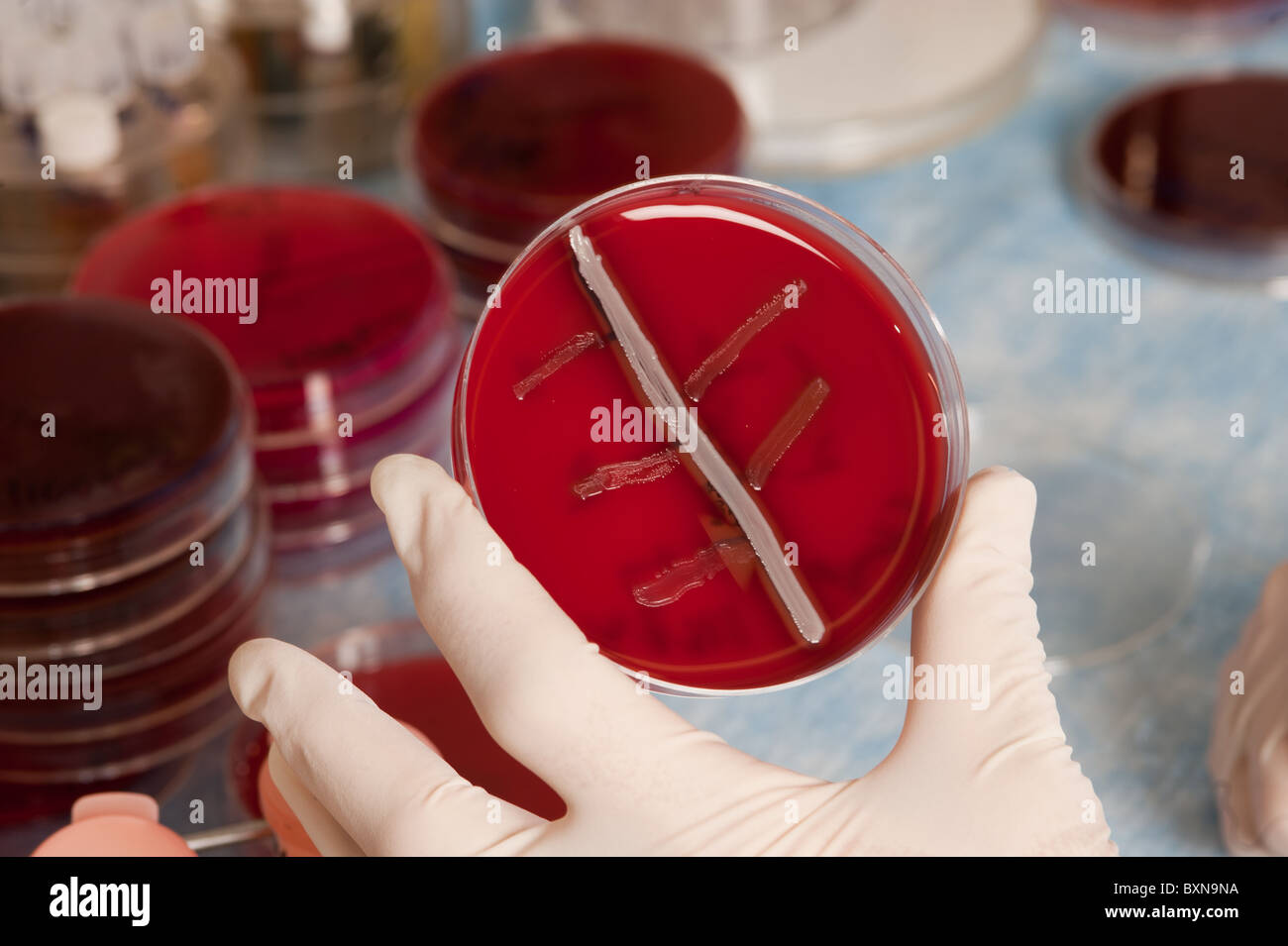 Lab technician and experiment Stock Photohttps://www.alamy.com/image-license-details/?v=1https://www.alamy.com/stock-photo-lab-technician-and-experiment-33660070.html
RMBXN9NA–Lab technician and experiment
 Petri dishes with blood agar. Stock Photohttps://www.alamy.com/image-license-details/?v=1https://www.alamy.com/petri-dishes-with-blood-agar-image182144572.html
RFMG9B78–Petri dishes with blood agar.
 Plate with bacteria colony Stock Vectorhttps://www.alamy.com/image-license-details/?v=1https://www.alamy.com/plate-with-bacteria-colony-image234361657.html
RFRH82K5–Plate with bacteria colony
 A technician transferring a small sample of blood onto an agar plate. Stock Photohttps://www.alamy.com/image-license-details/?v=1https://www.alamy.com/stock-photo-a-technician-transferring-a-small-sample-of-blood-onto-an-agar-plate-49249091.html
RMCT3DM3–A technician transferring a small sample of blood onto an agar plate.
 bacteria on agar plate Stock Photohttps://www.alamy.com/image-license-details/?v=1https://www.alamy.com/stock-photo-bacteria-on-agar-plate-55383495.html
RMD62X5Y–bacteria on agar plate
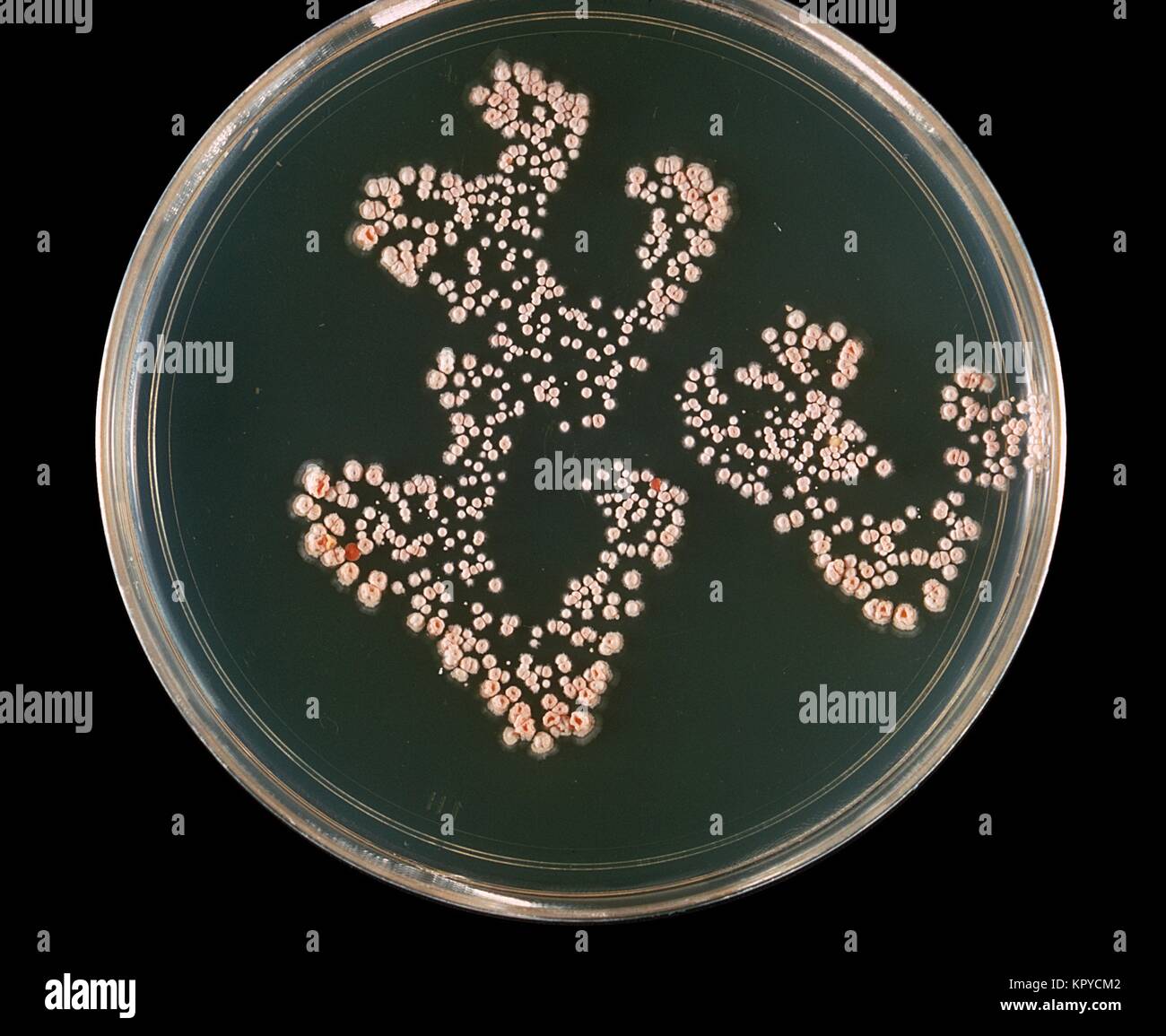 This is a plate culture of the bacteria Nocardia asteroides grown on 7H10 agar plates at 37{degrees} C. The bacterial complex Nocardia asteroids is a serious threat to immunosuppressed individuals, especially those with organ transplants, lung disease, and AIDS, 1969. Nocardia asteroids causes at least 50% of invasive infections of Nocardiosis. Image courtesy CDC/Dr. William Kaplan. Stock Photohttps://www.alamy.com/image-license-details/?v=1https://www.alamy.com/stock-image-this-is-a-plate-culture-of-the-bacteria-nocardia-asteroides-grown-169018418.html
RMKPYCM2–This is a plate culture of the bacteria Nocardia asteroides grown on 7H10 agar plates at 37{degrees} C. The bacterial complex Nocardia asteroids is a serious threat to immunosuppressed individuals, especially those with organ transplants, lung disease, and AIDS, 1969. Nocardia asteroids causes at least 50% of invasive infections of Nocardiosis. Image courtesy CDC/Dr. William Kaplan.
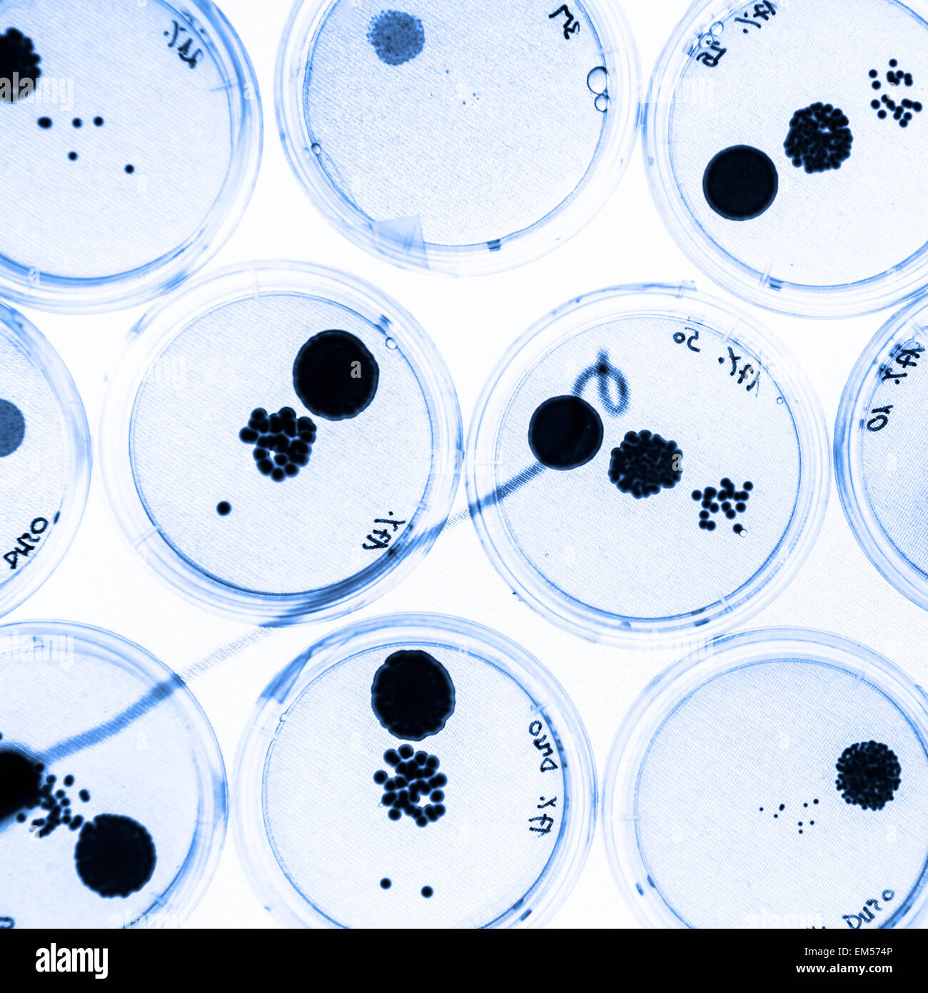 Growing Bacteria in Petri Dishes. Stock Photohttps://www.alamy.com/image-license-details/?v=1https://www.alamy.com/stock-photo-growing-bacteria-in-petri-dishes-81249974.html
RFEM574P–Growing Bacteria in Petri Dishes.
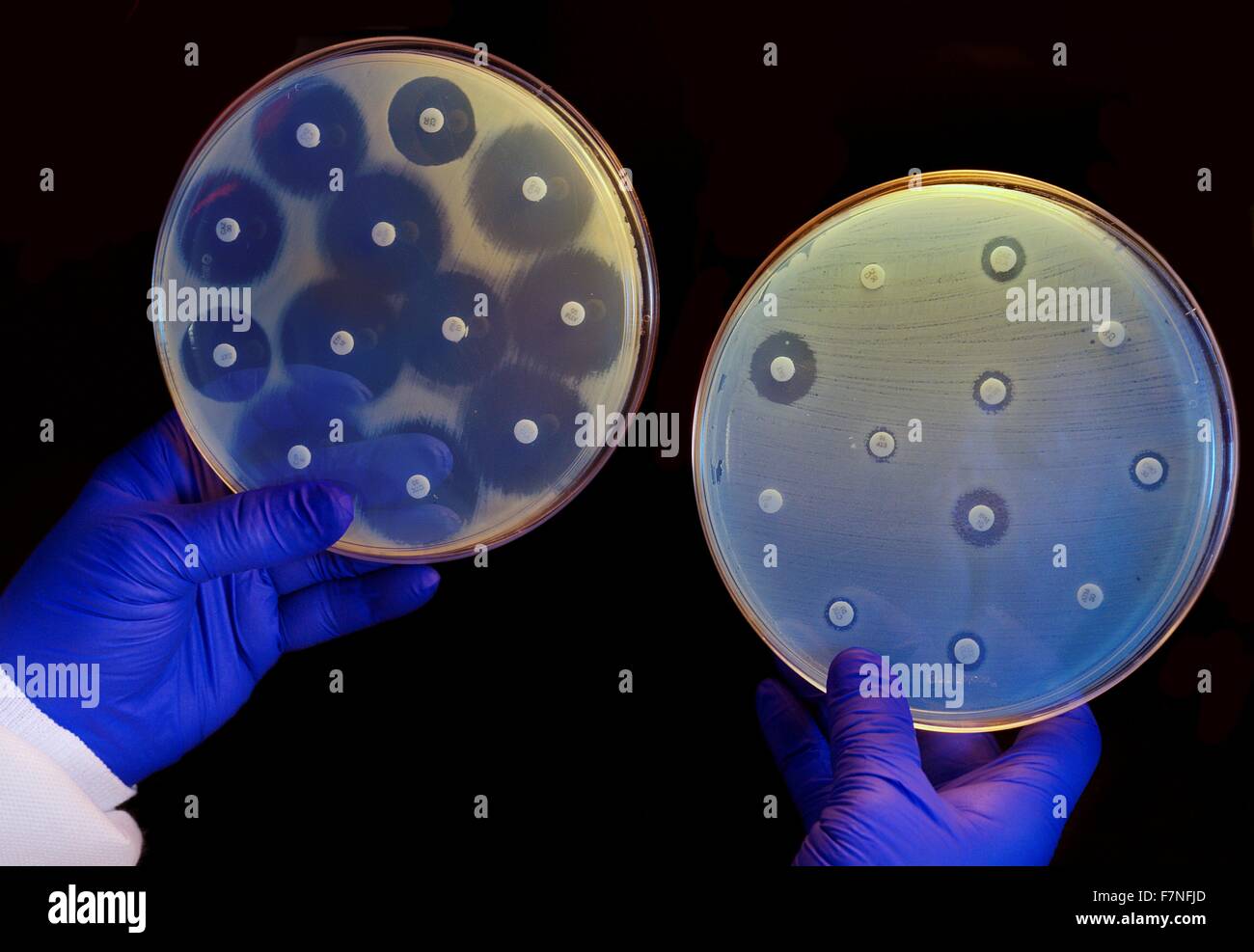 two plates growing bacteria in the presence of discs containing various antibiotics. The isolate on the left plate is susceptible to the antibiotics on the discs and is unable to grow around the discs. The one on the right has a CRE that is resistant to all of the antibiotics tested and is able to grow near the disks. Stock Photohttps://www.alamy.com/image-license-details/?v=1https://www.alamy.com/stock-photo-two-plates-growing-bacteria-in-the-presence-of-discs-containing-various-90827701.html
RMF7NFJD–two plates growing bacteria in the presence of discs containing various antibiotics. The isolate on the left plate is susceptible to the antibiotics on the discs and is unable to grow around the discs. The one on the right has a CRE that is resistant to all of the antibiotics tested and is able to grow near the disks.
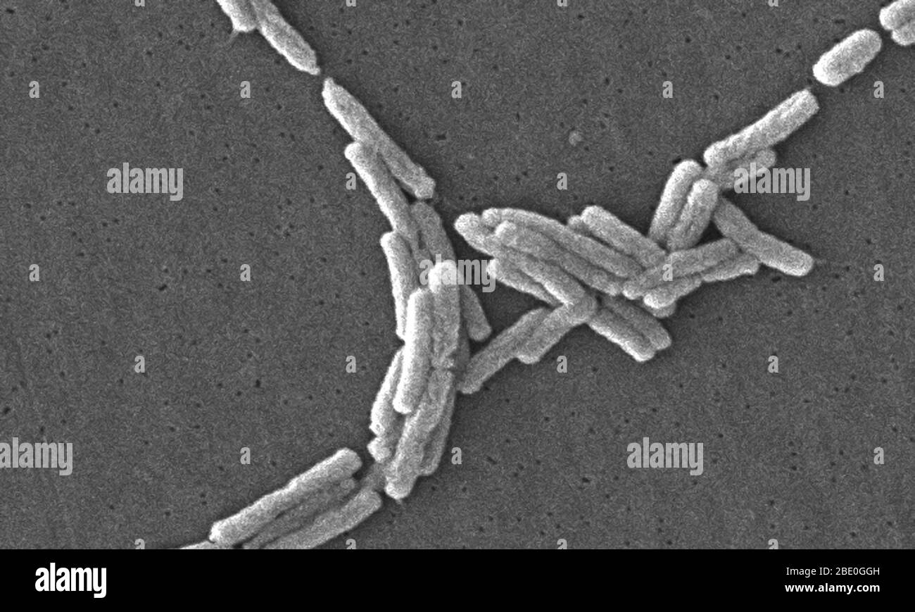 Scanning electron micrograph SEM) of a number of a large grouping of Gram-negative Legionella pneumophila bacteria. Note the presence of polar flagella, and pili, or long streamers. a number of these bacteria seem to display an elongated-rod morphology. L. pneumophila are known to most frequently exhibit this configuration when grown in broth, however, they can also elongate when plate-grown cells age, as it was in this case, especially when they've been refrigerated. The usual L. pneumophila morphology consists of stout, 'fat' bacilli, which is the case for the vast majority of the organisms Stock Photohttps://www.alamy.com/image-license-details/?v=1https://www.alamy.com/scanning-electron-micrograph-sem-of-a-number-of-a-large-grouping-of-gram-negative-legionella-pneumophila-bacteria-note-the-presence-of-polar-flagella-and-pili-or-long-streamers-a-number-of-these-bacteria-seem-to-display-an-elongated-rod-morphology-l-pneumophila-are-known-to-most-frequently-exhibit-this-configuration-when-grown-in-broth-however-they-can-also-elongate-when-plate-grown-cells-age-as-it-was-in-this-case-especially-when-theyve-been-refrigerated-the-usual-l-pneumophila-morphology-consists-of-stout-fat-bacilli-which-is-the-case-for-the-vast-majority-of-the-organisms-image352825553.html
RM2BE0GGH–Scanning electron micrograph SEM) of a number of a large grouping of Gram-negative Legionella pneumophila bacteria. Note the presence of polar flagella, and pili, or long streamers. a number of these bacteria seem to display an elongated-rod morphology. L. pneumophila are known to most frequently exhibit this configuration when grown in broth, however, they can also elongate when plate-grown cells age, as it was in this case, especially when they've been refrigerated. The usual L. pneumophila morphology consists of stout, 'fat' bacilli, which is the case for the vast majority of the organisms
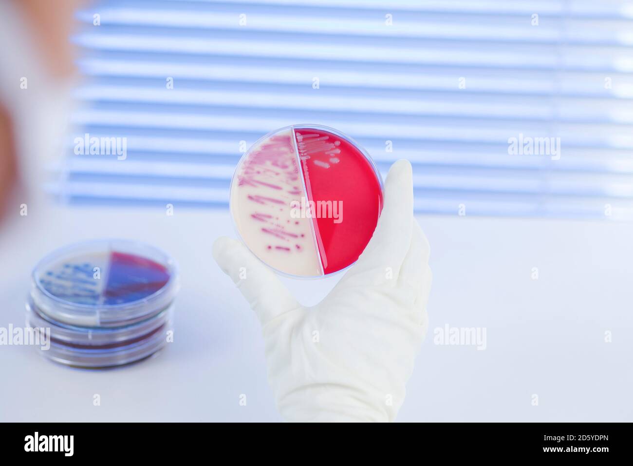 Laboratory technician examining agar plate with bacteria Stock Photohttps://www.alamy.com/image-license-details/?v=1https://www.alamy.com/laboratory-technician-examining-agar-plate-with-bacteria-image382304909.html
RF2D5YDPN–Laboratory technician examining agar plate with bacteria
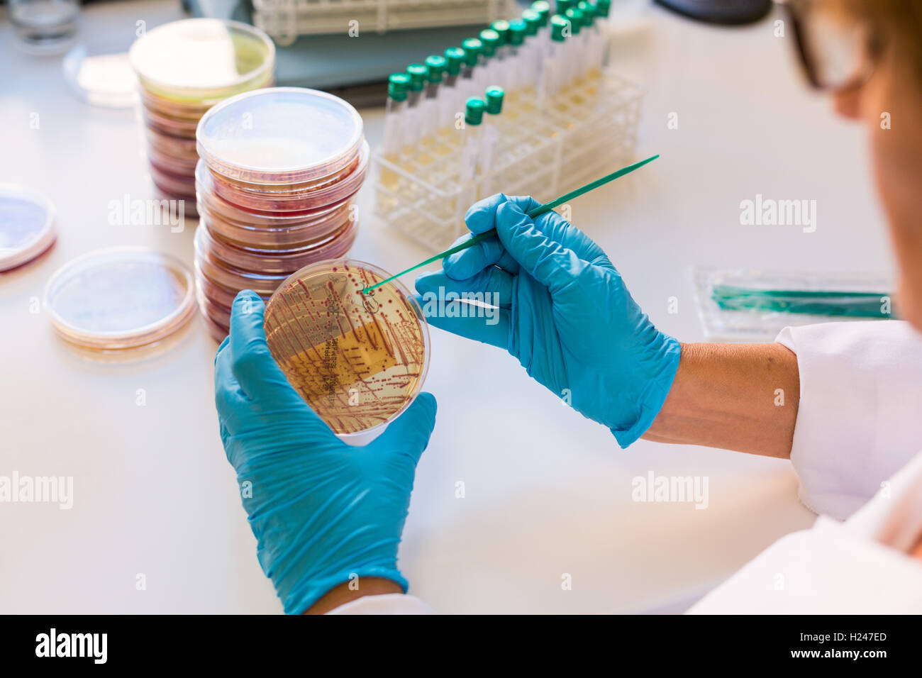 Hands holding a culture plate testing for the presence of Escherichia coli bacteria by looking at antibiotic resistance. Stock Photohttps://www.alamy.com/image-license-details/?v=1https://www.alamy.com/stock-photo-hands-holding-a-culture-plate-testing-for-the-presence-of-escherichia-121795589.html
RMH247ED–Hands holding a culture plate testing for the presence of Escherichia coli bacteria by looking at antibiotic resistance.
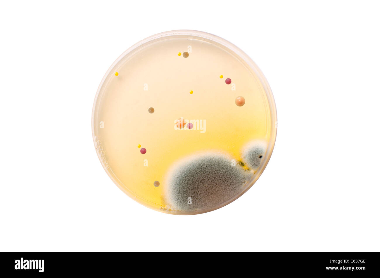 Microbiological plate with bacteria and fungi over white background Stock Photohttps://www.alamy.com/image-license-details/?v=1https://www.alamy.com/stock-photo-microbiological-plate-with-bacteria-and-fungi-over-white-background-38180478.html
RFC637GE–Microbiological plate with bacteria and fungi over white background
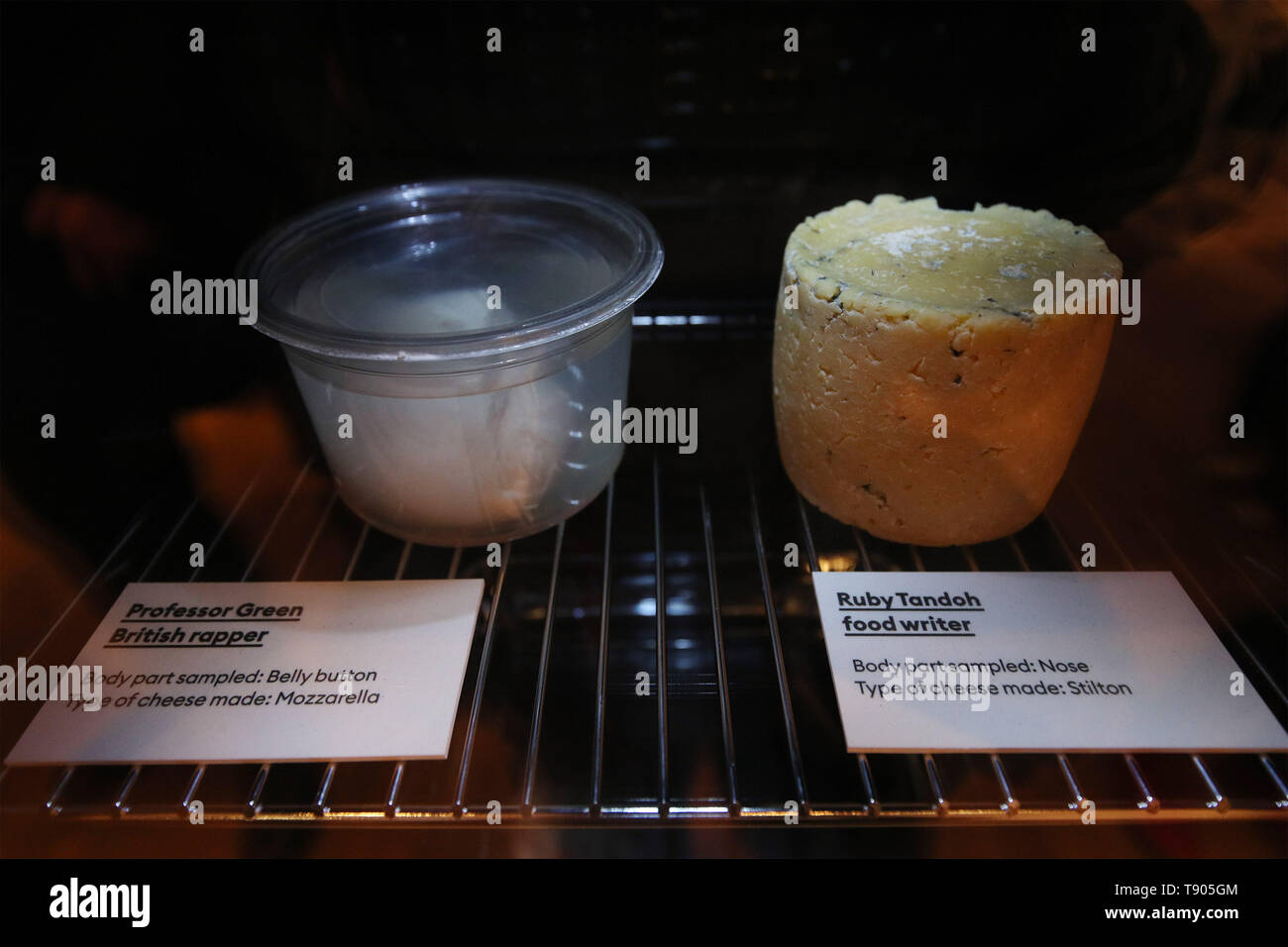 A Mozzarella cheese (left) made with bacteria sampled from the belly button of Professor Green, and a Stilton cheese made with bacteria sampled from the nose of food writer Ruby Tandoh, part of 'Selfmade', a project by Christina Agapakis and Sissel Tolaas, which involves cheese produced with microbes cultured from human bacteria taken from people's ears, toes and armpits, at the FOOD: Bigger than the Plate exhibition at the Victoria and Albert Museum in London. Stock Photohttps://www.alamy.com/image-license-details/?v=1https://www.alamy.com/a-mozzarella-cheese-left-made-with-bacteria-sampled-from-the-belly-button-of-professor-green-and-a-stilton-cheese-made-with-bacteria-sampled-from-the-nose-of-food-writer-ruby-tandoh-part-of-selfmade-a-project-by-christina-agapakis-and-sissel-tolaas-which-involves-cheese-produced-with-microbes-cultured-from-human-bacteria-taken-from-peoples-ears-toes-and-armpits-at-the-food-bigger-than-the-plate-exhibition-at-the-victoria-and-albert-museum-in-london-image246481444.html
RMT905GM–A Mozzarella cheese (left) made with bacteria sampled from the belly button of Professor Green, and a Stilton cheese made with bacteria sampled from the nose of food writer Ruby Tandoh, part of 'Selfmade', a project by Christina Agapakis and Sissel Tolaas, which involves cheese produced with microbes cultured from human bacteria taken from people's ears, toes and armpits, at the FOOD: Bigger than the Plate exhibition at the Victoria and Albert Museum in London.
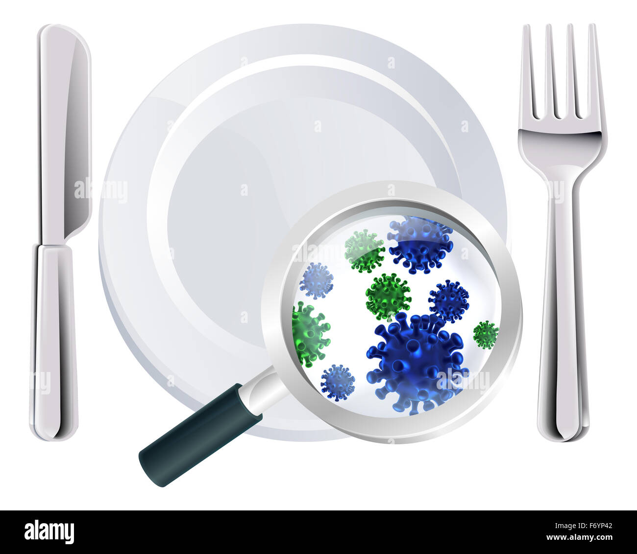 Microscopic bacteria cutlery concept of a plate, knife and fork place setting with a magnifying glass showing microscopic bacter Stock Photohttps://www.alamy.com/image-license-details/?v=1https://www.alamy.com/stock-photo-microscopic-bacteria-cutlery-concept-of-a-plate-knife-and-fork-place-90349842.html
RFF6YP42–Microscopic bacteria cutlery concept of a plate, knife and fork place setting with a magnifying glass showing microscopic bacter
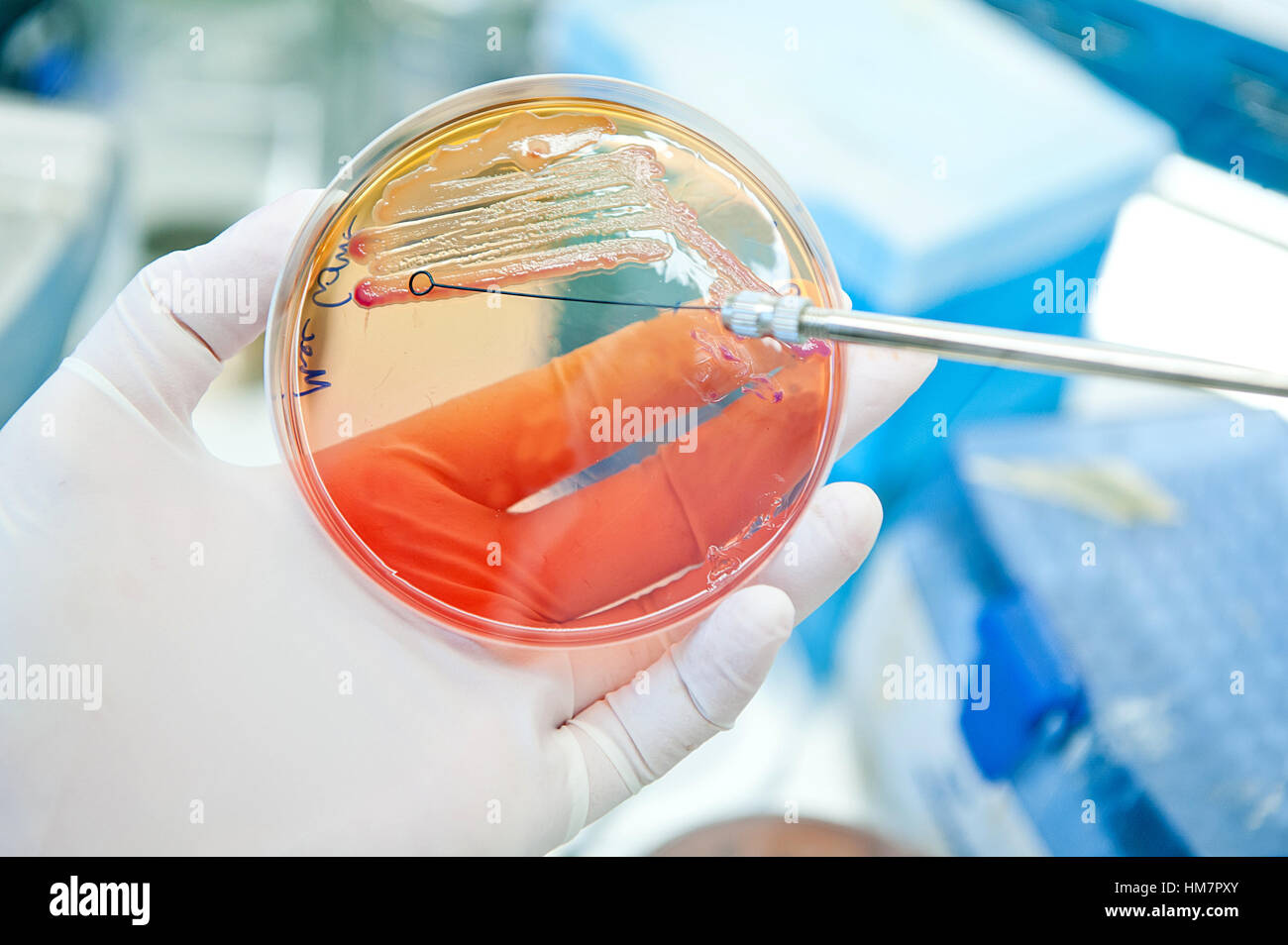 escherichia coli bacteria at petri plate Stock Photohttps://www.alamy.com/image-license-details/?v=1https://www.alamy.com/stock-photo-escherichia-coli-bacteria-at-petri-plate-132937363.html
RFHM7PXY–escherichia coli bacteria at petri plate
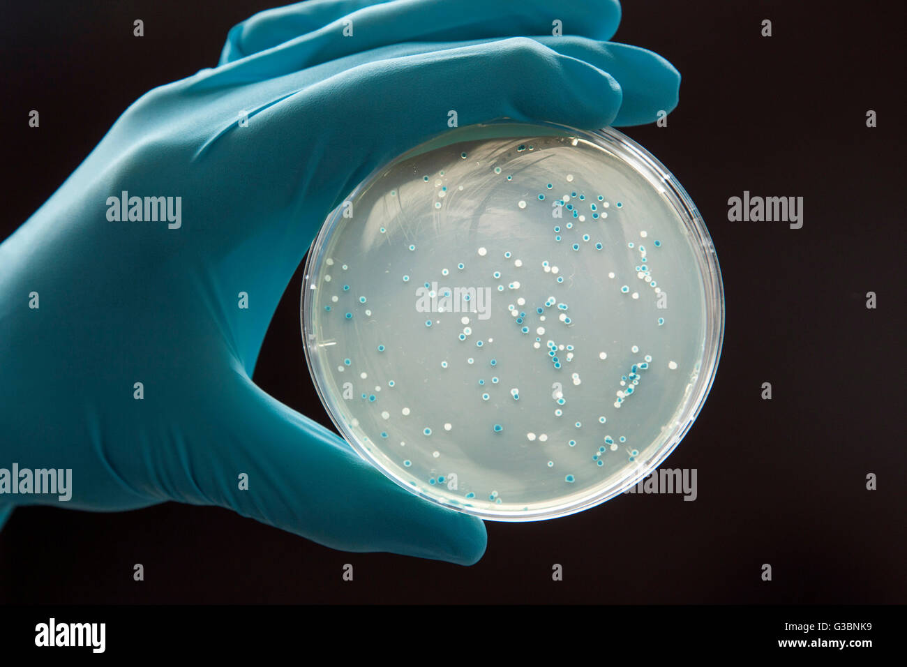 Bacteria culture in a Petri dish Stock Photohttps://www.alamy.com/image-license-details/?v=1https://www.alamy.com/stock-photo-bacteria-culture-in-a-petri-dish-105364653.html
RMG3BNK9–Bacteria culture in a Petri dish
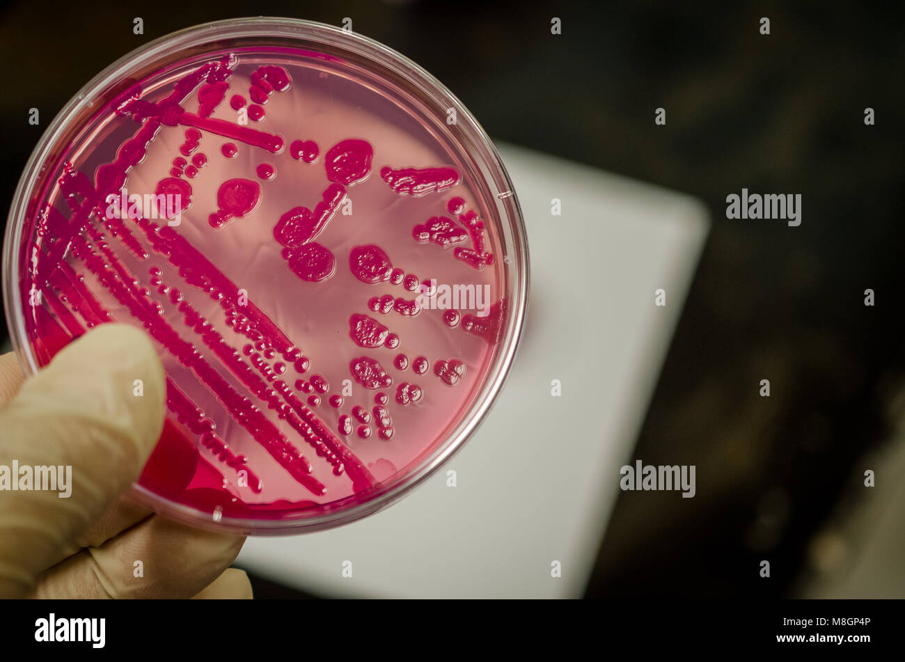 Bacterial culture plate holding in hand Stock Photohttps://www.alamy.com/image-license-details/?v=1https://www.alamy.com/stock-photo-bacterial-culture-plate-holding-in-hand-177389542.html
RFM8GP4P–Bacterial culture plate holding in hand
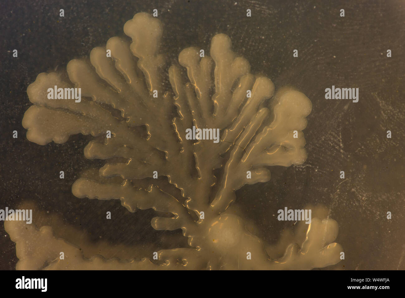 Colony of soil bacteria on an agar plate with rhizoid form Stock Photohttps://www.alamy.com/image-license-details/?v=1https://www.alamy.com/colony-of-soil-bacteria-on-an-agar-plate-with-rhizoid-form-image261175218.html
RFW4WFJA–Colony of soil bacteria on an agar plate with rhizoid form
 Bacteria culture plates petri dishes with blue transformation colonies in incubator Stock Photohttps://www.alamy.com/image-license-details/?v=1https://www.alamy.com/bacteria-culture-plates-petri-dishes-with-blue-transformation-colonies-image6349431.html
RMA4RMR8–Bacteria culture plates petri dishes with blue transformation colonies in incubator
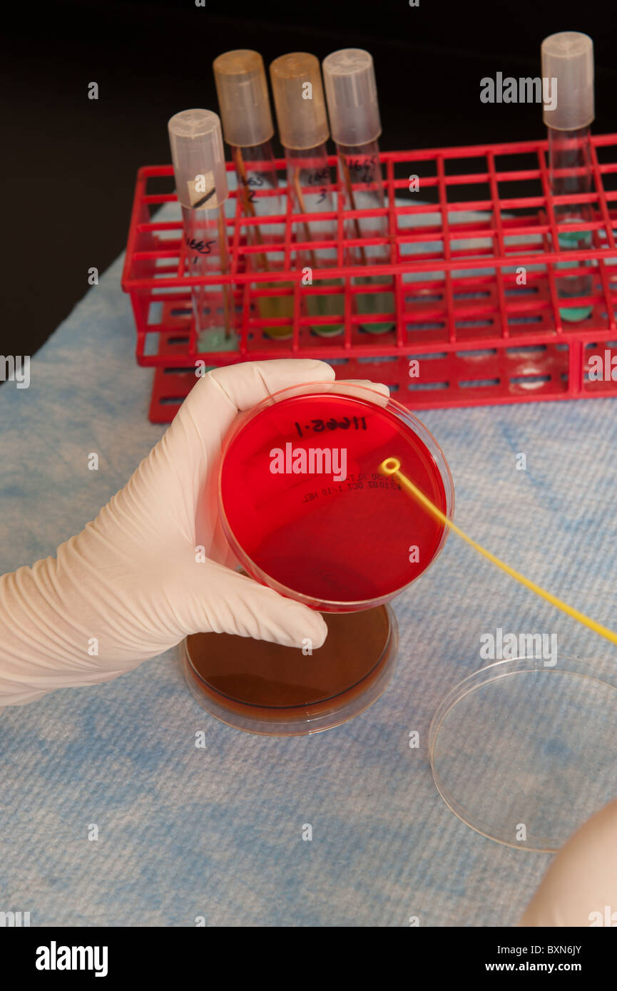 Lab technician and experiment Stock Photohttps://www.alamy.com/image-license-details/?v=1https://www.alamy.com/stock-photo-lab-technician-and-experiment-33657651.html
RMBXN6JY–Lab technician and experiment
 Petri dishes with blood agar. Stock Photohttps://www.alamy.com/image-license-details/?v=1https://www.alamy.com/petri-dishes-with-blood-agar-image182144570.html
RFMG9B76–Petri dishes with blood agar.
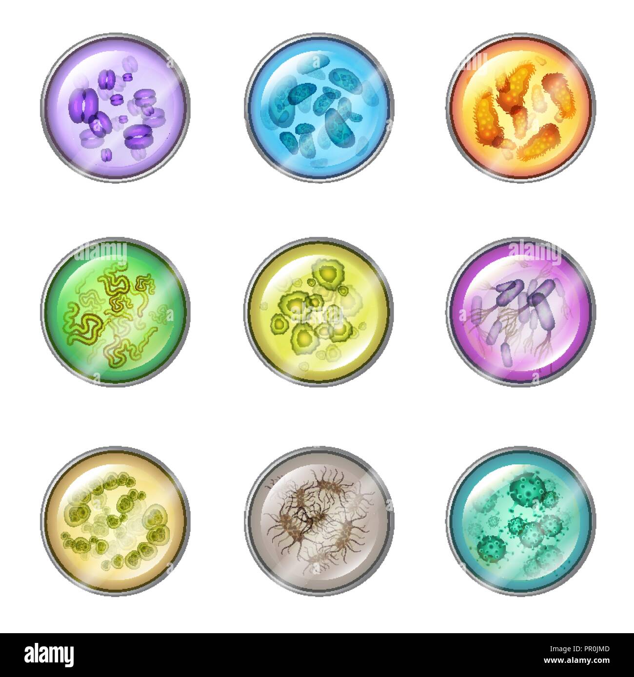 Set of isolated glassware plates with bacteria or petri dish with virus colony, laboratory bowl for bacterial organisms samples. Science and medicine, laboratory or lab, chemical and biology theme Stock Vectorhttps://www.alamy.com/image-license-details/?v=1https://www.alamy.com/set-of-isolated-glassware-plates-with-bacteria-or-petri-dish-with-virus-colony-laboratory-bowl-for-bacterial-organisms-samples-science-and-medicine-laboratory-or-lab-chemical-and-biology-theme-image220676189.html
RFPR0JMD–Set of isolated glassware plates with bacteria or petri dish with virus colony, laboratory bowl for bacterial organisms samples. Science and medicine, laboratory or lab, chemical and biology theme
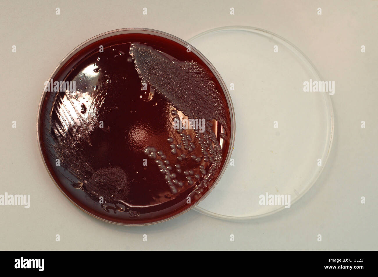 A sample of cultured blood-poisoning bacteria on an agar plate. Stock Photohttps://www.alamy.com/image-license-details/?v=1https://www.alamy.com/stock-photo-a-sample-of-cultured-blood-poisoning-bacteria-on-an-agar-plate-49249371.html
RMCT3E23–A sample of cultured blood-poisoning bacteria on an agar plate.
 bacteria mutants on agar plate in laboratory Stock Photohttps://www.alamy.com/image-license-details/?v=1https://www.alamy.com/stock-photo-bacteria-mutants-on-agar-plate-in-laboratory-55371593.html
RMD62B0W–bacteria mutants on agar plate in laboratory
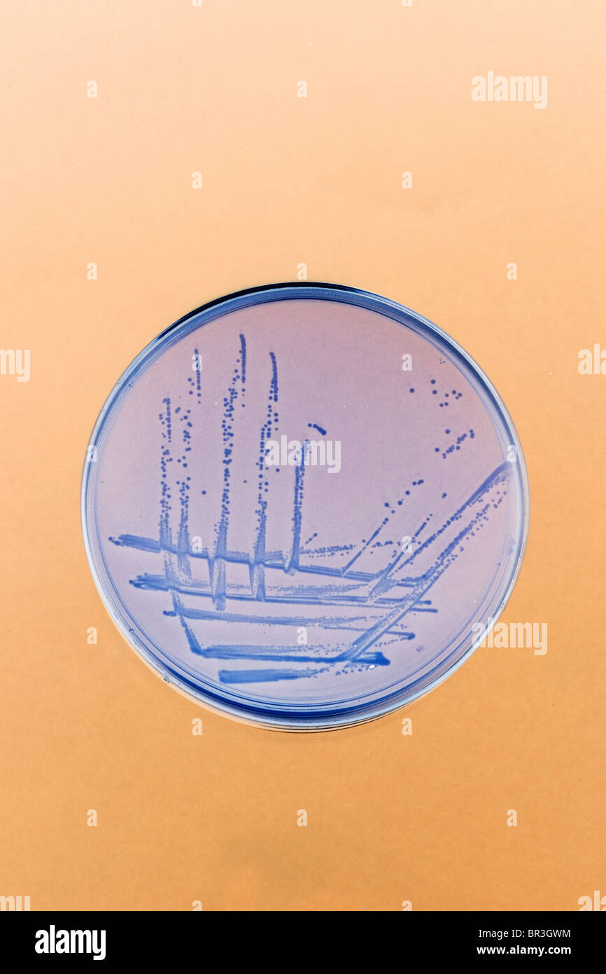 bacteria streaked and grows on an agar plate in the lab Stock Photohttps://www.alamy.com/image-license-details/?v=1https://www.alamy.com/stock-photo-bacteria-streaked-and-grows-on-an-agar-plate-in-the-lab-31426576.html
RMBR3GWM–bacteria streaked and grows on an agar plate in the lab
 Vector bacteria and viruses on the plate under a microscope in a laboratory. Stock Vectorhttps://www.alamy.com/image-license-details/?v=1https://www.alamy.com/vector-bacteria-and-viruses-on-the-plate-under-a-microscope-in-a-laboratory-image64654199.html
RFDN572F–Vector bacteria and viruses on the plate under a microscope in a laboratory.
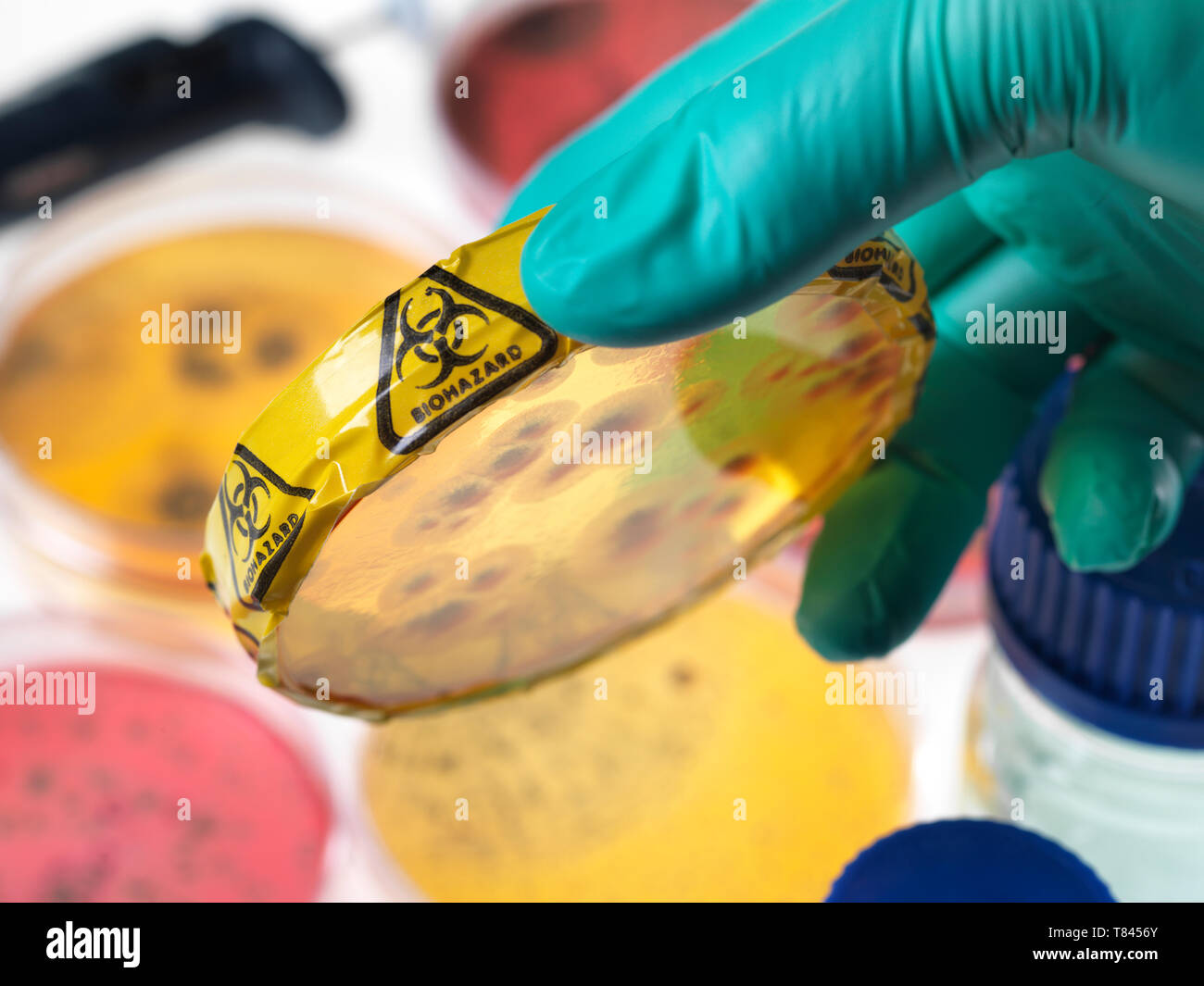 Microbiology Experiment, Scientist viewing microorganisms in bacterial cultures growing in petri dishes in the laboratory, close up of hand Stock Photohttps://www.alamy.com/image-license-details/?v=1https://www.alamy.com/microbiology-experiment-scientist-viewing-microorganisms-in-bacterial-cultures-growing-in-petri-dishes-in-the-laboratory-close-up-of-hand-image245954323.html
RFT8456Y–Microbiology Experiment, Scientist viewing microorganisms in bacterial cultures growing in petri dishes in the laboratory, close up of hand
 Scanning electron micrograph (SEM) of a number of a large grouping of Gram-negative Legionella pneumophila bacteria. Note the presence of polar flagella, and pili, or long streamers. a number of these bacteria seem to display an elongated-rod morphology. L. pneumophila are known to most frequently exhibit this configuration when grown in broth, however, they can also elongate when plate-grown cells age, as it was in this case, especially when they've been refrigerated. The usual L. pneumophila morphology consists of stout, 'fat' bacilli, which is the case for the vast majority of the organisms Stock Photohttps://www.alamy.com/image-license-details/?v=1https://www.alamy.com/scanning-electron-micrograph-sem-of-a-number-of-a-large-grouping-of-gram-negative-legionella-pneumophila-bacteria-note-the-presence-of-polar-flagella-and-pili-or-long-streamers-a-number-of-these-bacteria-seem-to-display-an-elongated-rod-morphology-l-pneumophila-are-known-to-most-frequently-exhibit-this-configuration-when-grown-in-broth-however-they-can-also-elongate-when-plate-grown-cells-age-as-it-was-in-this-case-especially-when-theyve-been-refrigerated-the-usual-l-pneumophila-morphology-consists-of-stout-fat-bacilli-which-is-the-case-for-the-vast-majority-of-the-organisms-image352825581.html
RM2BE0GHH–Scanning electron micrograph (SEM) of a number of a large grouping of Gram-negative Legionella pneumophila bacteria. Note the presence of polar flagella, and pili, or long streamers. a number of these bacteria seem to display an elongated-rod morphology. L. pneumophila are known to most frequently exhibit this configuration when grown in broth, however, they can also elongate when plate-grown cells age, as it was in this case, especially when they've been refrigerated. The usual L. pneumophila morphology consists of stout, 'fat' bacilli, which is the case for the vast majority of the organisms
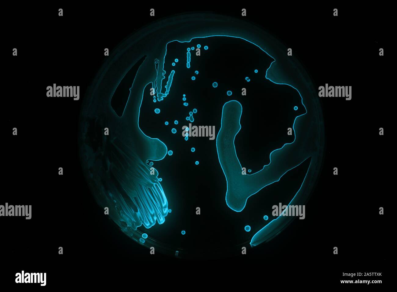 Aliivibrio fisheri are performing bioluminescence, a blueish glowing in the dark. The bacteria have been growing on a agar plate. Stock Photohttps://www.alamy.com/image-license-details/?v=1https://www.alamy.com/aliivibrio-fisheri-are-performing-bioluminescence-a-blueish-glowing-in-the-dark-the-bacteria-have-been-growing-on-a-agar-plate-image330616683.html
RF2A5TTXK–Aliivibrio fisheri are performing bioluminescence, a blueish glowing in the dark. The bacteria have been growing on a agar plate.
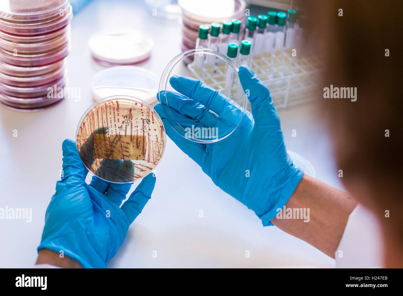 Hands holding a culture plate testing for the presence of Escherichia coli bacteria by looking at antibiotic resistance. Stock Photohttps://www.alamy.com/image-license-details/?v=1https://www.alamy.com/stock-photo-hands-holding-a-culture-plate-testing-for-the-presence-of-escherichia-121795587.html
RMH247EB–Hands holding a culture plate testing for the presence of Escherichia coli bacteria by looking at antibiotic resistance.
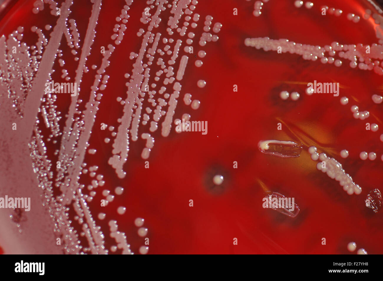 Bacterial colonies growing on culture of sheep's blood Stock Photohttps://www.alamy.com/image-license-details/?v=1https://www.alamy.com/stock-photo-bacterial-colonies-growing-on-culture-of-sheeps-blood-87456468.html
RFF27YH8–Bacterial colonies growing on culture of sheep's blood
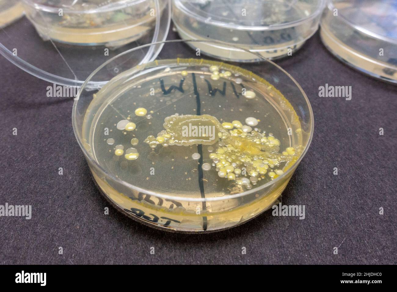 Agar plates petri dishes with bacteria spore growths after UK secondary school biology lesson investigating hand washing. Stock Photohttps://www.alamy.com/image-license-details/?v=1https://www.alamy.com/agar-plates-petri-dishes-with-bacteria-spore-growths-after-uk-secondary-school-biology-lesson-investigating-hand-washing-image458832416.html
RM2HJDHC0–Agar plates petri dishes with bacteria spore growths after UK secondary school biology lesson investigating hand washing.
 microorganisms mutants on agar plate in laboratory Stock Photohttps://www.alamy.com/image-license-details/?v=1https://www.alamy.com/stock-photo-microorganisms-mutants-on-agar-plate-in-laboratory-49786098.html
RFCTYXJX–microorganisms mutants on agar plate in laboratory
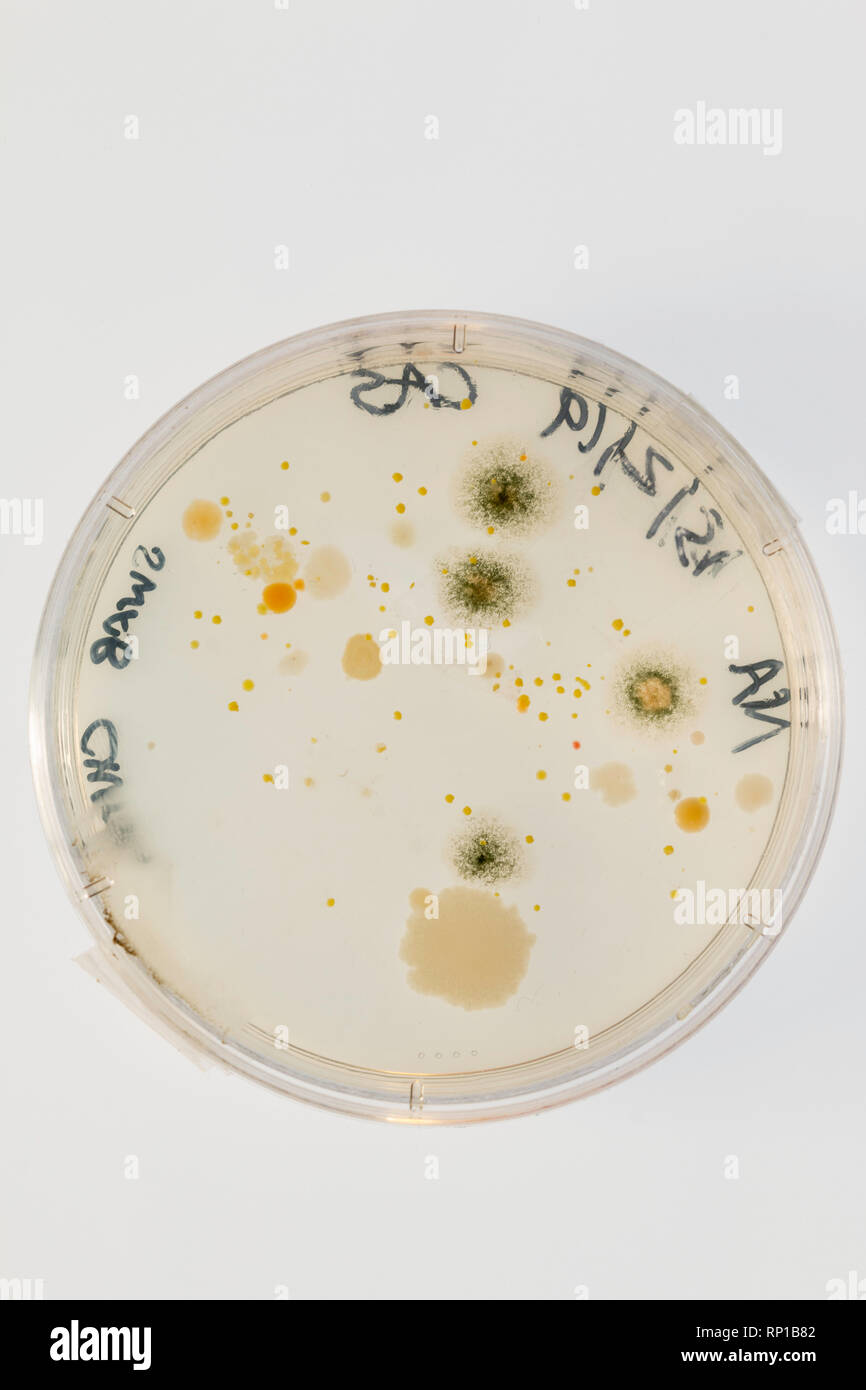 Microbes growing on an agar plate after taking a swab from a school chair Stock Photohttps://www.alamy.com/image-license-details/?v=1https://www.alamy.com/microbes-growing-on-an-agar-plate-after-taking-a-swab-from-a-school-chair-image237288018.html
RFRP1B82–Microbes growing on an agar plate after taking a swab from a school chair
 Bacteria culture in a Petri dish Stock Photohttps://www.alamy.com/image-license-details/?v=1https://www.alamy.com/stock-photo-bacteria-culture-in-a-petri-dish-105364646.html
RMG3BNK2–Bacteria culture in a Petri dish
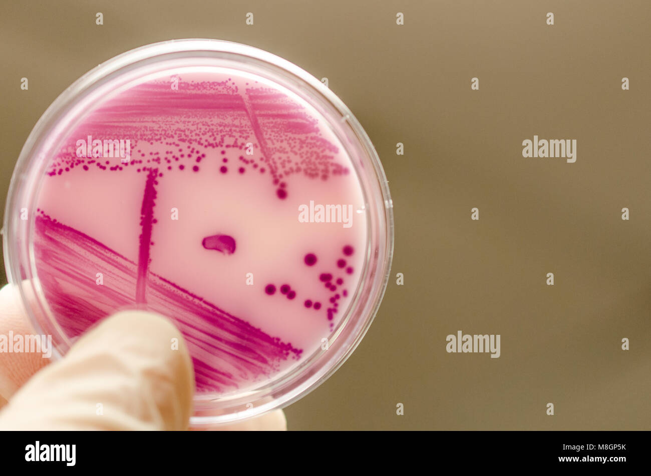 Bacterial culture plate holding in hand Stock Photohttps://www.alamy.com/image-license-details/?v=1https://www.alamy.com/stock-photo-bacterial-culture-plate-holding-in-hand-177389567.html
RFM8GP5K–Bacterial culture plate holding in hand
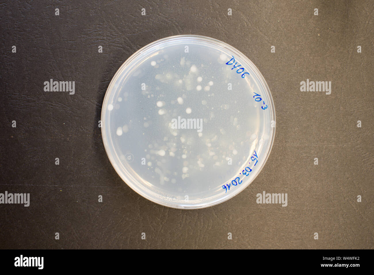 Bacteria colonies on agar plate in a research laboratory Stock Photohttps://www.alamy.com/image-license-details/?v=1https://www.alamy.com/bacteria-colonies-on-agar-plate-in-a-research-laboratory-image261175238.html
RFW4WFK2–Bacteria colonies on agar plate in a research laboratory
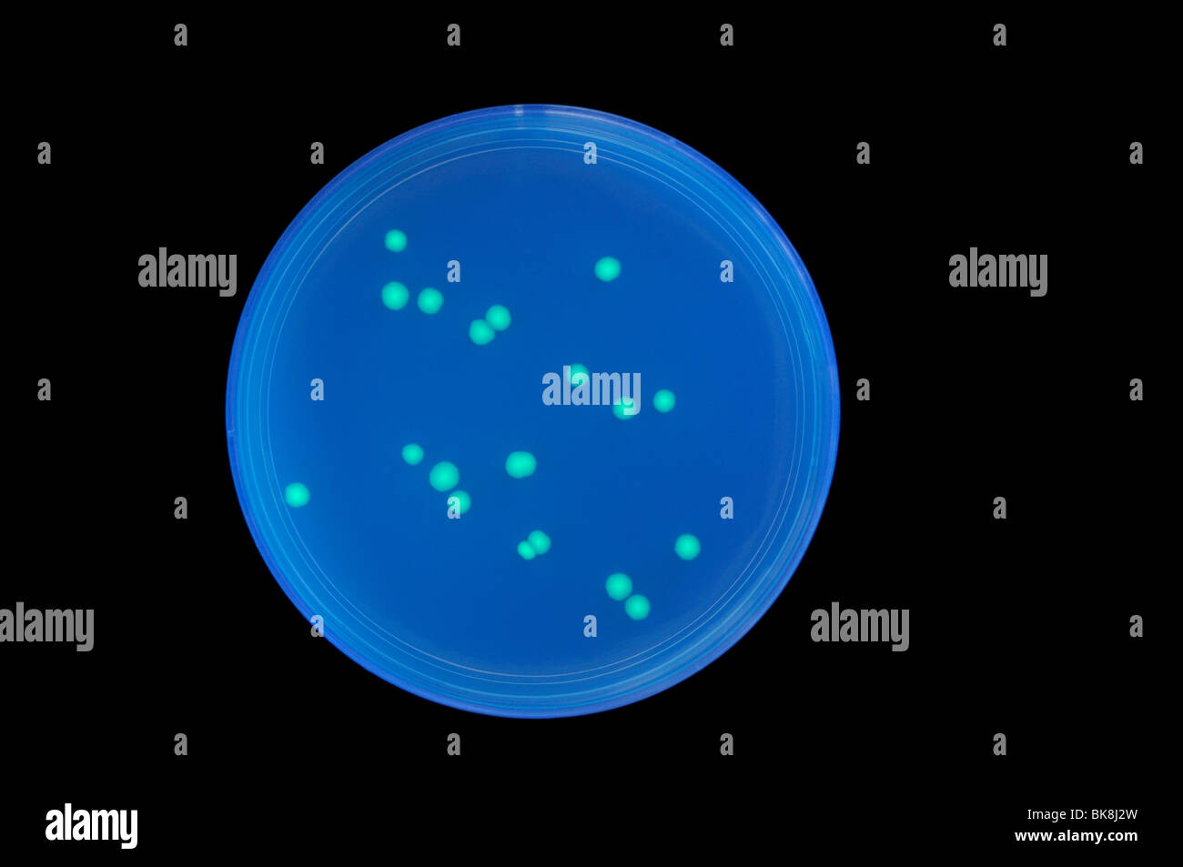 Transformed bacteria colonies containing a jellyfish gene for GFP (green fluorescent protein) causing green bioluminescence. Stock Photohttps://www.alamy.com/image-license-details/?v=1https://www.alamy.com/stock-photo-transformed-bacteria-colonies-containing-a-jellyfish-gene-for-gfp-29078641.html
RMBK8J2W–Transformed bacteria colonies containing a jellyfish gene for GFP (green fluorescent protein) causing green bioluminescence.
 Lab technician and experiment Stock Photohttps://www.alamy.com/image-license-details/?v=1https://www.alamy.com/stock-photo-lab-technician-and-experiment-33657669.html
RMBXN6KH–Lab technician and experiment
 Petri dishes with blood agar. Stock Photohttps://www.alamy.com/image-license-details/?v=1https://www.alamy.com/petri-dishes-with-blood-agar-image182144564.html
RFMG9B70–Petri dishes with blood agar.
 Petri dish Stock Photohttps://www.alamy.com/image-license-details/?v=1https://www.alamy.com/petri-dish-image231975735.html
RFRDBBBK–Petri dish
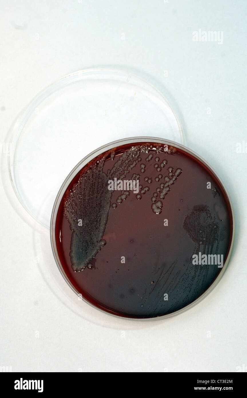 A sample of cultured blood-poisoning bacteria on an agar plate. Stock Photohttps://www.alamy.com/image-license-details/?v=1https://www.alamy.com/stock-photo-a-sample-of-cultured-blood-poisoning-bacteria-on-an-agar-plate-49249388.html
RMCT3E2M–A sample of cultured blood-poisoning bacteria on an agar plate.
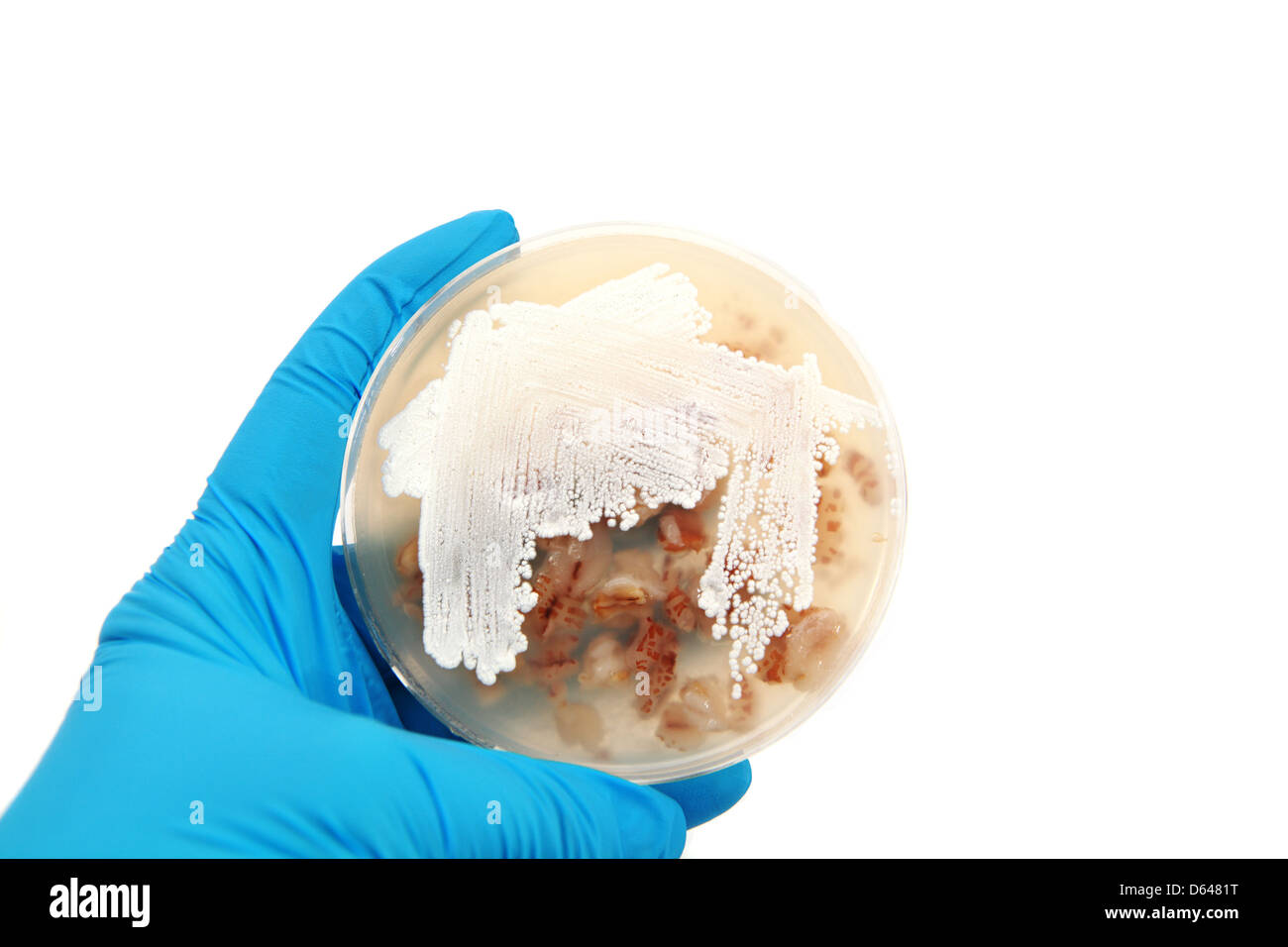 Streptomyces bacteria on agar plate Stock Photohttps://www.alamy.com/image-license-details/?v=1https://www.alamy.com/stock-photo-streptomyces-bacteria-on-agar-plate-55413172.html
RMD6481T–Streptomyces bacteria on agar plate
 A Comte cheese made with bacteria sampled from the nostrils and pubic hair of Heston Blumenthal, part of 'Selfmade', a project by Christina Agapakis and Sissel Tolaas, which involves cheese produced with microbes cultured from human bacteria taken from people's ears, toes and armpits, at the FOOD: Bigger than the Plate exhibition at the Victoria and Albert Museum in London. Stock Photohttps://www.alamy.com/image-license-details/?v=1https://www.alamy.com/a-comte-cheese-made-with-bacteria-sampled-from-the-nostrils-and-pubic-hair-of-heston-blumenthal-part-of-selfmade-a-project-by-christina-agapakis-and-sissel-tolaas-which-involves-cheese-produced-with-microbes-cultured-from-human-bacteria-taken-from-peoples-ears-toes-and-armpits-at-the-food-bigger-than-the-plate-exhibition-at-the-victoria-and-albert-museum-in-london-image246481435.html
RMT905GB–A Comte cheese made with bacteria sampled from the nostrils and pubic hair of Heston Blumenthal, part of 'Selfmade', a project by Christina Agapakis and Sissel Tolaas, which involves cheese produced with microbes cultured from human bacteria taken from people's ears, toes and armpits, at the FOOD: Bigger than the Plate exhibition at the Victoria and Albert Museum in London.
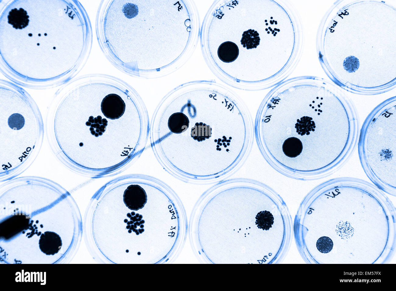 Growing Bacteria in Petri Dishes. Stock Photohttps://www.alamy.com/image-license-details/?v=1https://www.alamy.com/stock-photo-growing-bacteria-in-petri-dishes-81250286.html
RFEM57FX–Growing Bacteria in Petri Dishes.
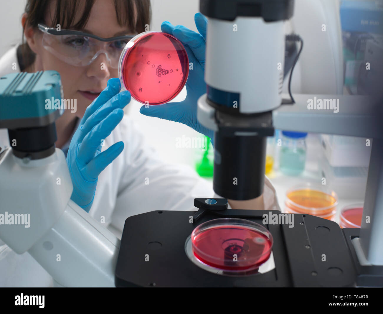 Female scientist examining cultures growing in petri dishes using inverted microscope in laboratory Stock Photohttps://www.alamy.com/image-license-details/?v=1https://www.alamy.com/female-scientist-examining-cultures-growing-in-petri-dishes-using-inverted-microscope-in-laboratory-image245956699.html
RFT8487R–Female scientist examining cultures growing in petri dishes using inverted microscope in laboratory
 Scanning electron micrograph (SEM) of a number of a large grouping of Gram-negative Legionella pneumophila bacteria. Note the presence of polar flagella, and pili, or long streamers. a number of these bacteria seem to display an elongated-rod morphology. L. pneumophila are known to most frequently exhibit this configuration when grown in broth, however, they can also elongate when plate-grown cells age, as it was in this case, especially when they've been refrigerated. The usual L. pneumophila morphology consists of stout, 'fat' bacilli, which is the case for the vast majority of the organisms Stock Photohttps://www.alamy.com/image-license-details/?v=1https://www.alamy.com/scanning-electron-micrograph-sem-of-a-number-of-a-large-grouping-of-gram-negative-legionella-pneumophila-bacteria-note-the-presence-of-polar-flagella-and-pili-or-long-streamers-a-number-of-these-bacteria-seem-to-display-an-elongated-rod-morphology-l-pneumophila-are-known-to-most-frequently-exhibit-this-configuration-when-grown-in-broth-however-they-can-also-elongate-when-plate-grown-cells-age-as-it-was-in-this-case-especially-when-theyve-been-refrigerated-the-usual-l-pneumophila-morphology-consists-of-stout-fat-bacilli-which-is-the-case-for-the-vast-majority-of-the-organisms-image352825582.html
RM2BE0GHJ–Scanning electron micrograph (SEM) of a number of a large grouping of Gram-negative Legionella pneumophila bacteria. Note the presence of polar flagella, and pili, or long streamers. a number of these bacteria seem to display an elongated-rod morphology. L. pneumophila are known to most frequently exhibit this configuration when grown in broth, however, they can also elongate when plate-grown cells age, as it was in this case, especially when they've been refrigerated. The usual L. pneumophila morphology consists of stout, 'fat' bacilli, which is the case for the vast majority of the organisms
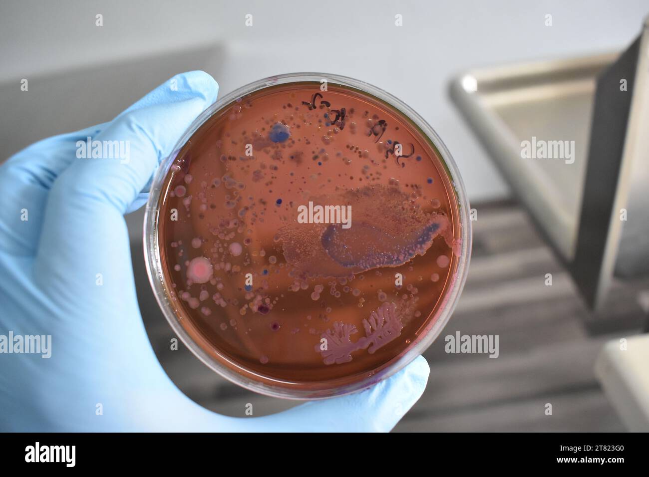 Bacteria colonies or bacteria growth on eosin methylene blue agar plate. Stock Photohttps://www.alamy.com/image-license-details/?v=1https://www.alamy.com/bacteria-colonies-or-bacteria-growth-on-eosin-methylene-blue-agar-plate-image572906096.html
RF2T823G0–Bacteria colonies or bacteria growth on eosin methylene blue agar plate.
 Hands holding a culture plate testing for the presence of Escherichia coli bacteria by looking at antibiotic resistance. Stock Photohttps://www.alamy.com/image-license-details/?v=1https://www.alamy.com/stock-photo-hands-holding-a-culture-plate-testing-for-the-presence-of-escherichia-83055841.html
RMER3EG1–Hands holding a culture plate testing for the presence of Escherichia coli bacteria by looking at antibiotic resistance.
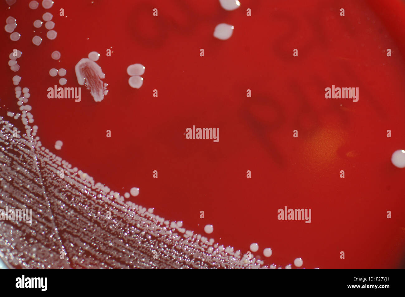 bacterial colonies growing in sheep's blood medium on a petri dish Stock Photohttps://www.alamy.com/image-license-details/?v=1https://www.alamy.com/stock-photo-bacterial-colonies-growing-in-sheeps-blood-medium-on-a-petri-dish-87456489.html
RFF27YJ1–bacterial colonies growing in sheep's blood medium on a petri dish
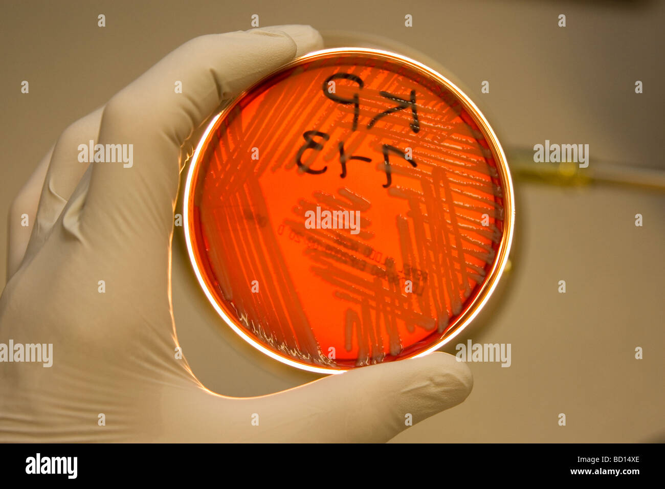 Microbiologist holds a petri dish growimg bacteria, Klebsiella pneumoniae. Stock Photohttps://www.alamy.com/image-license-details/?v=1https://www.alamy.com/stock-photo-microbiologist-holds-a-petri-dish-growimg-bacteria-klebsiella-pneumoniae-25226726.html
RMBD14XE–Microbiologist holds a petri dish growimg bacteria, Klebsiella pneumoniae.
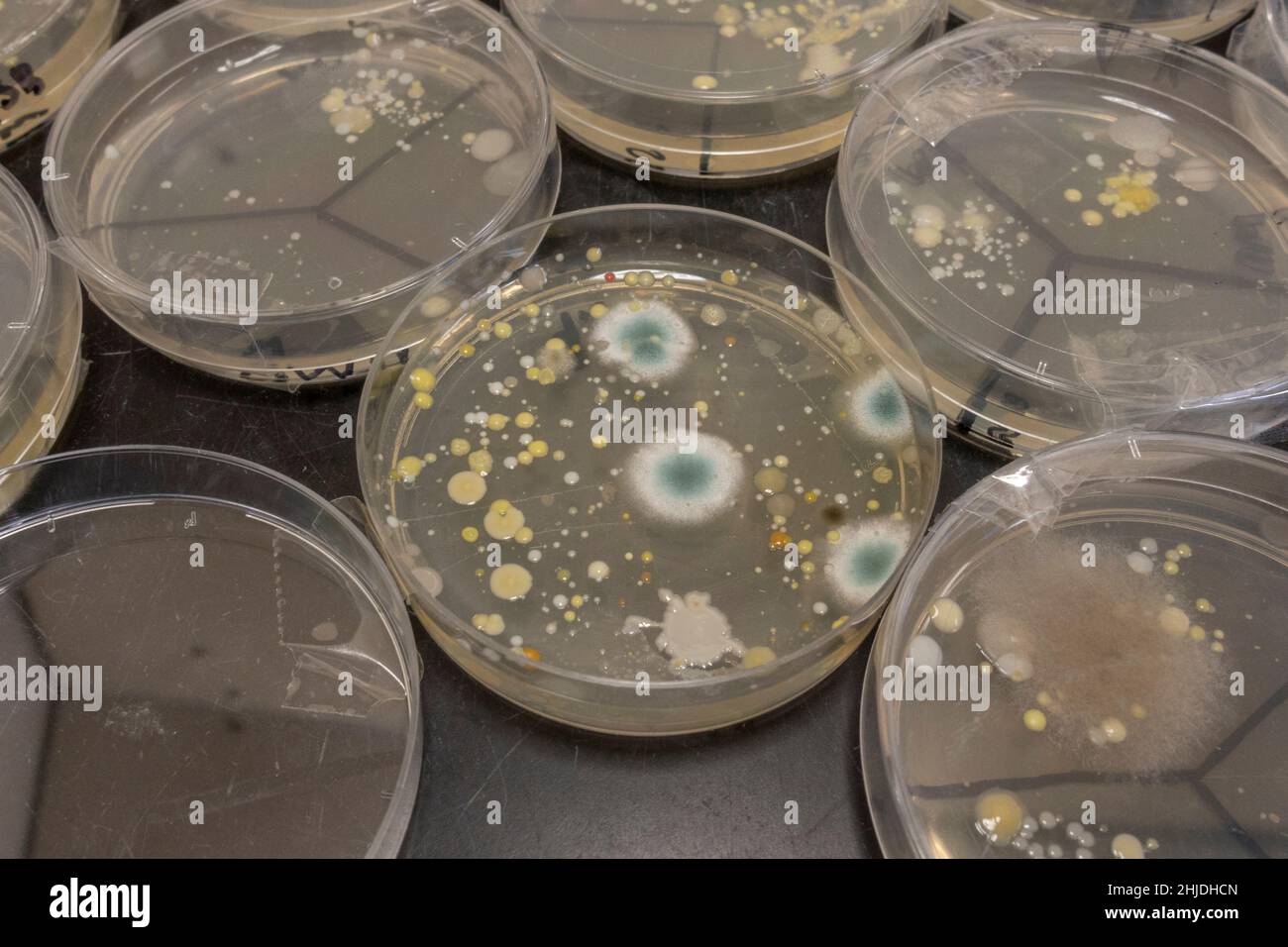 Agar plates petri dishes with bacteria spore growths after UK secondary school biology lesson investigating hand washing. Stock Photohttps://www.alamy.com/image-license-details/?v=1https://www.alamy.com/agar-plates-petri-dishes-with-bacteria-spore-growths-after-uk-secondary-school-biology-lesson-investigating-hand-washing-image458832437.html
RM2HJDHCN–Agar plates petri dishes with bacteria spore growths after UK secondary school biology lesson investigating hand washing.
 Image of a petri plate used to culture cells. Stock Photohttps://www.alamy.com/image-license-details/?v=1https://www.alamy.com/stock-photo-image-of-a-petri-plate-used-to-culture-cells-73789327.html
RFE81B13–Image of a petri plate used to culture cells.
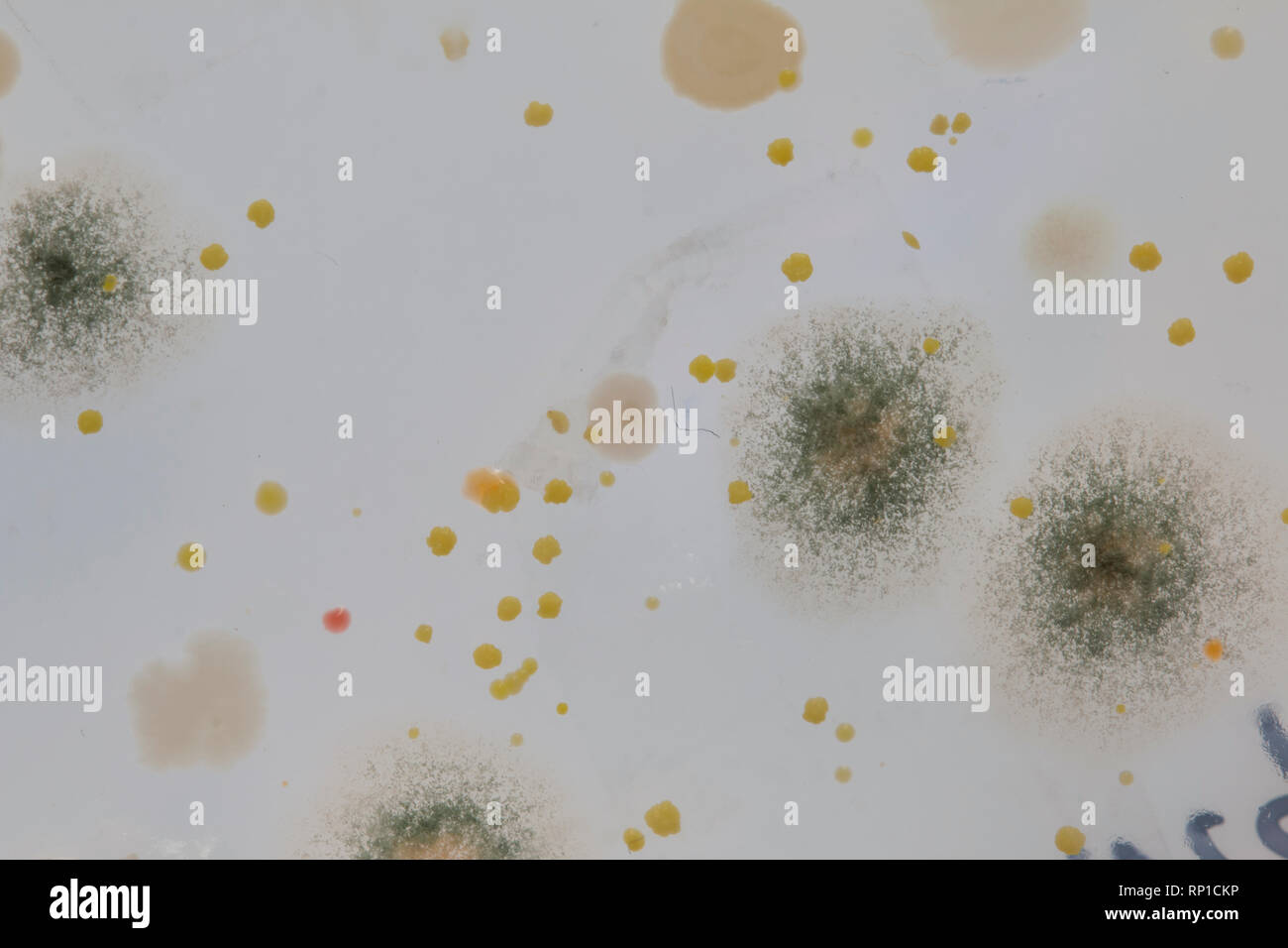 Microbes growing on an agar plate after taking a swab from a school chair Stock Photohttps://www.alamy.com/image-license-details/?v=1https://www.alamy.com/microbes-growing-on-an-agar-plate-after-taking-a-swab-from-a-school-chair-image237289130.html
RFRP1CKP–Microbes growing on an agar plate after taking a swab from a school chair
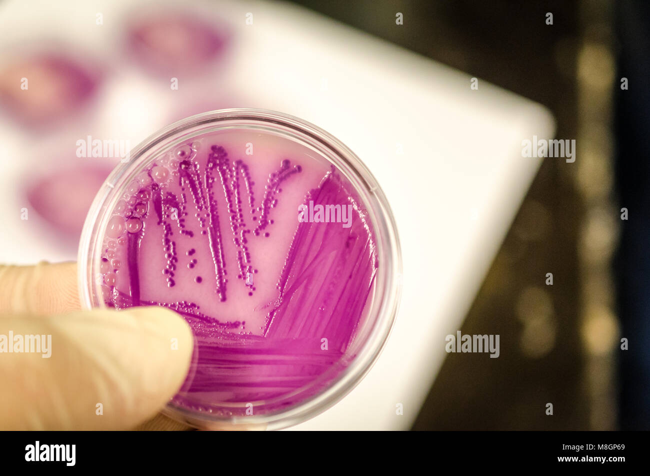 Bacterial culture plate holding in hand Stock Photohttps://www.alamy.com/image-license-details/?v=1https://www.alamy.com/stock-photo-bacterial-culture-plate-holding-in-hand-177389585.html
RFM8GP69–Bacterial culture plate holding in hand
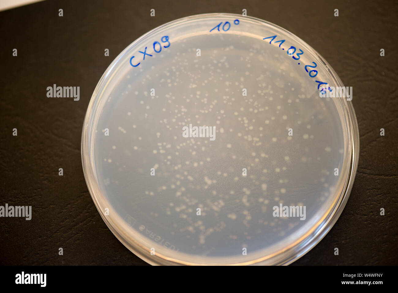 Bacteria colonies on agar plate in a research laboratory Stock Photohttps://www.alamy.com/image-license-details/?v=1https://www.alamy.com/bacteria-colonies-on-agar-plate-in-a-research-laboratory-image261175319.html
RFW4WFNY–Bacteria colonies on agar plate in a research laboratory
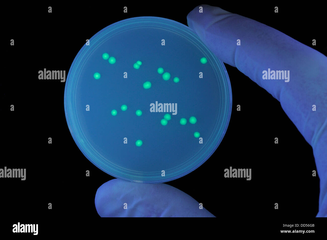 Transformed bacteria colonies containing a jellyfish gene for GFP (green fluorescent protein) causing green bioluminescence. Stock Photohttps://www.alamy.com/image-license-details/?v=1https://www.alamy.com/stock-photo-transformed-bacteria-colonies-containing-a-jellyfish-gene-for-gfp-59736555.html
RMDD56GB–Transformed bacteria colonies containing a jellyfish gene for GFP (green fluorescent protein) causing green bioluminescence.
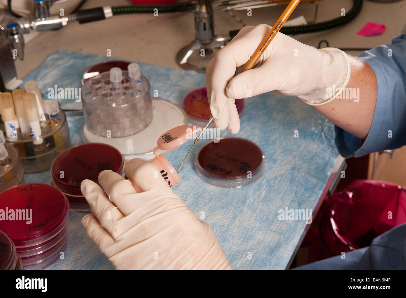 Lab technician and experiment in the diagnostic lab at University of Maine Stock Photohttps://www.alamy.com/image-license-details/?v=1https://www.alamy.com/stock-photo-lab-technician-and-experiment-in-the-diagnostic-lab-at-university-33657695.html
RMBXN6MF–Lab technician and experiment in the diagnostic lab at University of Maine
 Petri dishes with blood agar. Stock Photohttps://www.alamy.com/image-license-details/?v=1https://www.alamy.com/petri-dishes-with-blood-agar-image182144568.html
RFMG9B74–Petri dishes with blood agar.
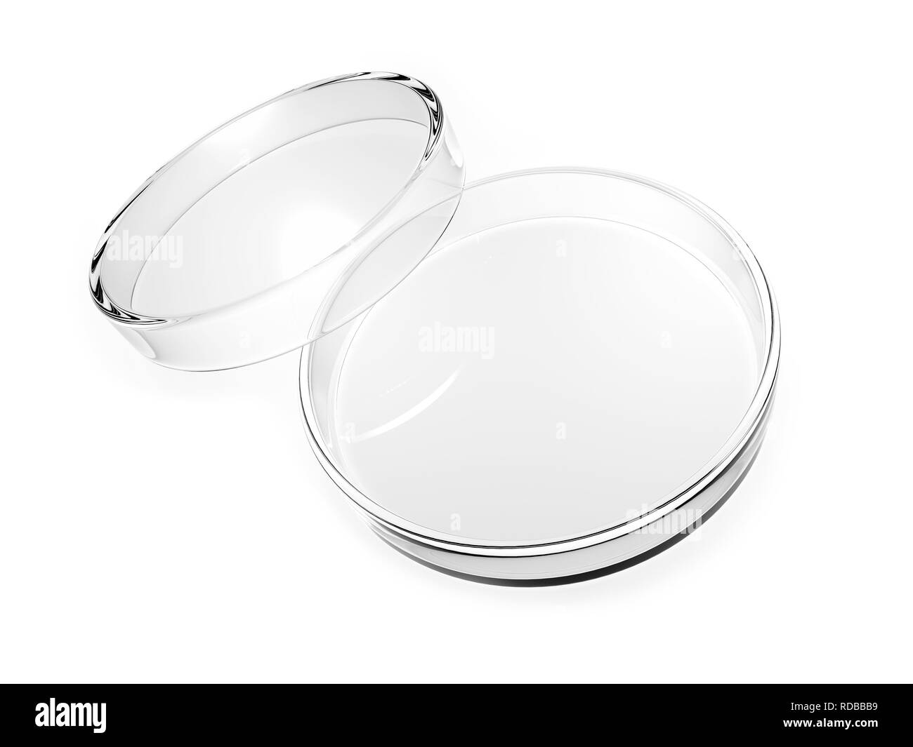 Petri dish Stock Photohttps://www.alamy.com/image-license-details/?v=1https://www.alamy.com/petri-dish-image231975725.html
RFRDBBB9–Petri dish
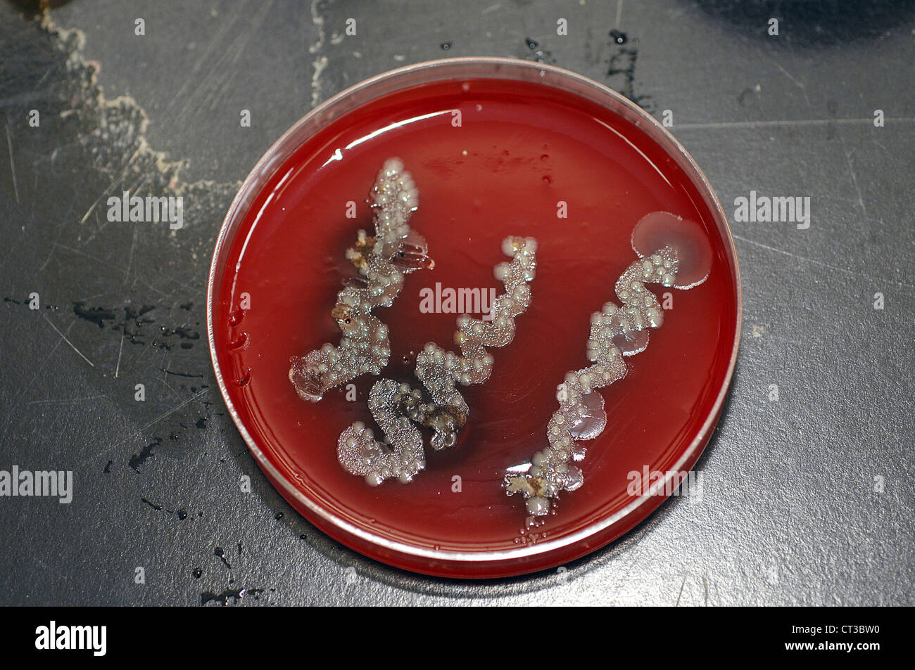 Bacterial culture growing in a petri dish on agar jelly. Stock Photohttps://www.alamy.com/image-license-details/?v=1https://www.alamy.com/stock-photo-bacterial-culture-growing-in-a-petri-dish-on-agar-jelly-49247660.html
RMCT3BW0–Bacterial culture growing in a petri dish on agar jelly.
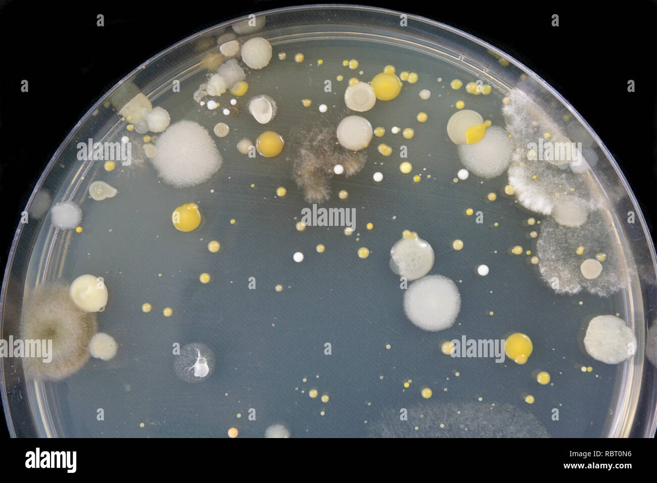 Close-up of bacteria and mold growing in a Petri dish. Stock Photohttps://www.alamy.com/image-license-details/?v=1https://www.alamy.com/close-up-of-bacteria-and-mold-growing-in-a-petri-dish-image231023442.html
RFRBT0N6–Close-up of bacteria and mold growing in a Petri dish.
 Scientist pipetting a specimen into medium containing bacteria for scientific research in laboratory Stock Photohttps://www.alamy.com/image-license-details/?v=1https://www.alamy.com/scientist-pipetting-a-specimen-into-medium-containing-bacteria-for-image154123463.html
RFJXMX1B–Scientist pipetting a specimen into medium containing bacteria for scientific research in laboratory
 bacteria on agar plate Stock Photohttps://www.alamy.com/image-license-details/?v=1https://www.alamy.com/stock-photo-bacteria-on-agar-plate-59488066.html
RFDCNWHP–bacteria on agar plate
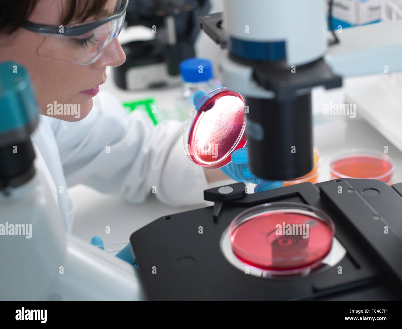 Female scientist examining cultures growing in petri dishes using inverted microscope in laboratory Stock Photohttps://www.alamy.com/image-license-details/?v=1https://www.alamy.com/female-scientist-examining-cultures-growing-in-petri-dishes-using-inverted-microscope-in-laboratory-image245956698.html
RFT8487P–Female scientist examining cultures growing in petri dishes using inverted microscope in laboratory
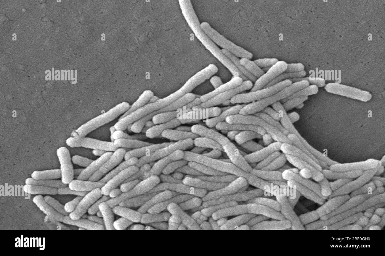 Scanning electron micrograph (SEM) of a number of a large grouping of Gram-negative Legionella pneumophila bacteria. Note the presence of polar flagella, and pili, or long streamers. a number of these bacteria seem to display an elongated-rod morphology. L. pneumophila are known to most frequently exhibit this configuration when grown in broth, however, they can also elongate when plate-grown cells age, as it was in this case, especially when they've been refrigerated. The usual L. pneumophila morphology consists of stout, 'fat' bacilli, which is the case for the vast majority of the organisms Stock Photohttps://www.alamy.com/image-license-details/?v=1https://www.alamy.com/scanning-electron-micrograph-sem-of-a-number-of-a-large-grouping-of-gram-negative-legionella-pneumophila-bacteria-note-the-presence-of-polar-flagella-and-pili-or-long-streamers-a-number-of-these-bacteria-seem-to-display-an-elongated-rod-morphology-l-pneumophila-are-known-to-most-frequently-exhibit-this-configuration-when-grown-in-broth-however-they-can-also-elongate-when-plate-grown-cells-age-as-it-was-in-this-case-especially-when-theyve-been-refrigerated-the-usual-l-pneumophila-morphology-consists-of-stout-fat-bacilli-which-is-the-case-for-the-vast-majority-of-the-organisms-image352825564.html
RM2BE0GH0–Scanning electron micrograph (SEM) of a number of a large grouping of Gram-negative Legionella pneumophila bacteria. Note the presence of polar flagella, and pili, or long streamers. a number of these bacteria seem to display an elongated-rod morphology. L. pneumophila are known to most frequently exhibit this configuration when grown in broth, however, they can also elongate when plate-grown cells age, as it was in this case, especially when they've been refrigerated. The usual L. pneumophila morphology consists of stout, 'fat' bacilli, which is the case for the vast majority of the organisms
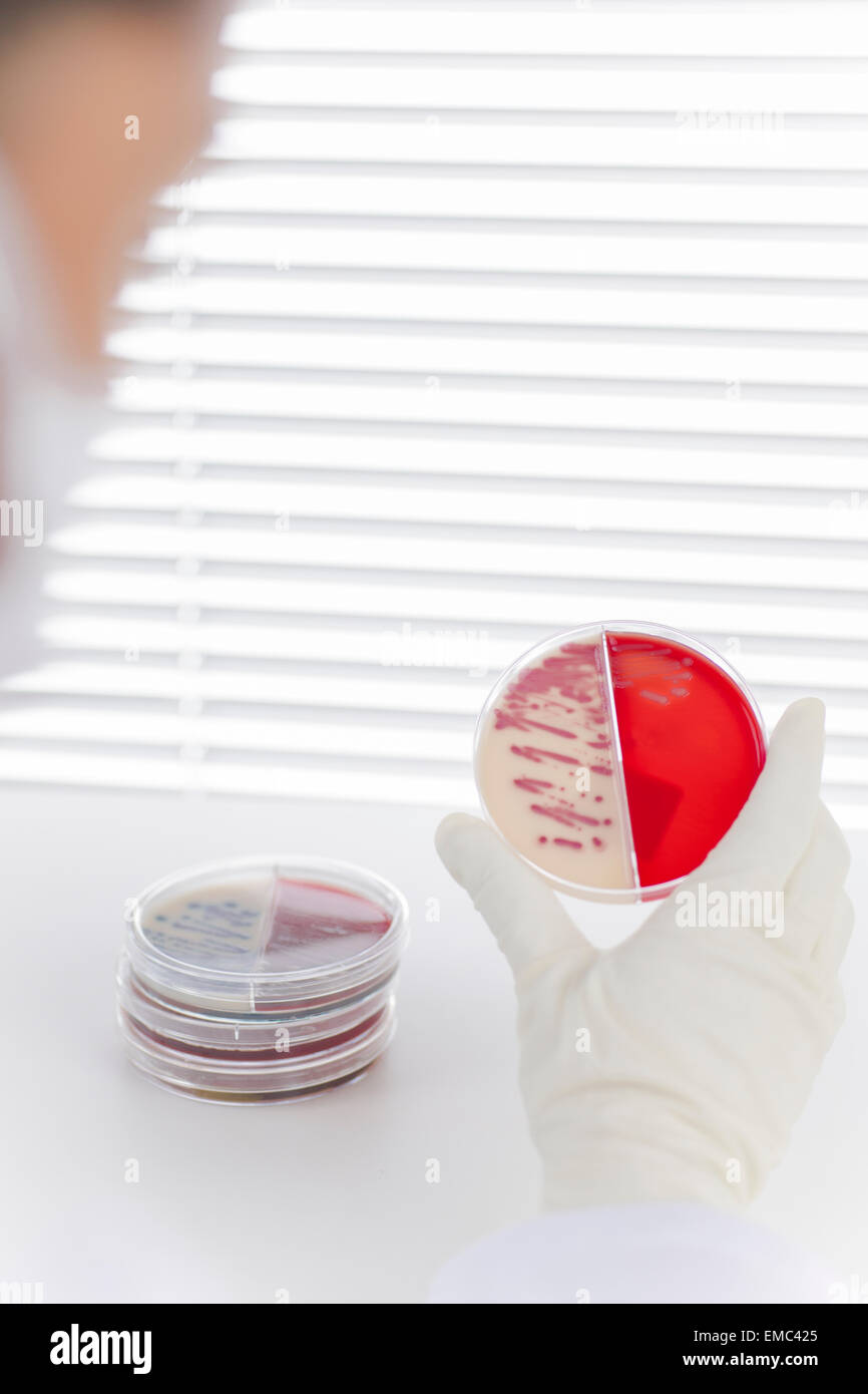 Laboratory technician examining agar plate with bacteria Stock Photohttps://www.alamy.com/image-license-details/?v=1https://www.alamy.com/stock-photo-laboratory-technician-examining-agar-plate-with-bacteria-81401213.html
RFEMC425–Laboratory technician examining agar plate with bacteria
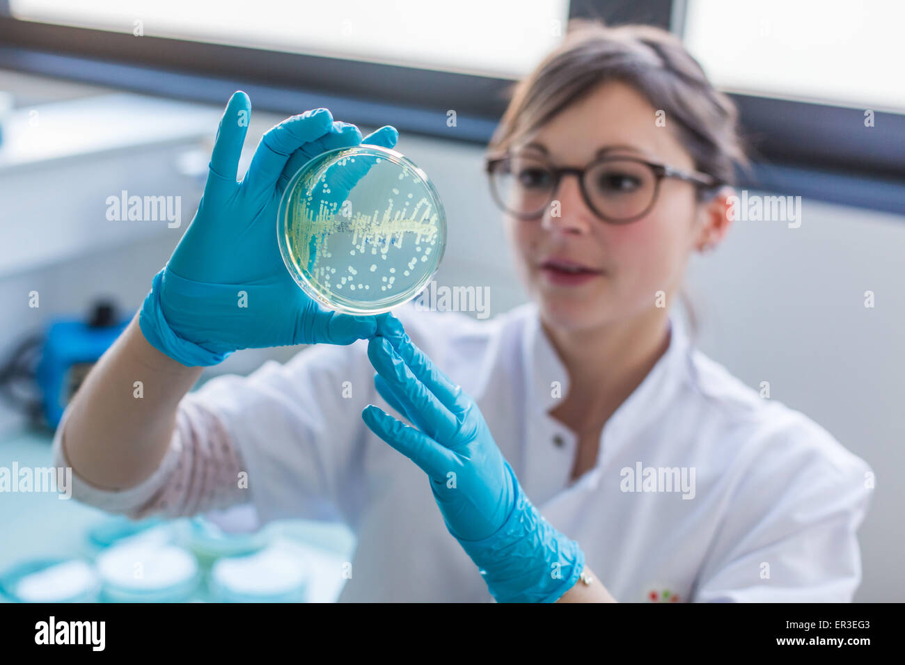 Hands holding a culture plate testing for the presence of Escherichia coli bacteria by looking at antibiotic resistance, Biology and Research Center in University Hospital Health, Limoges, France. Stock Photohttps://www.alamy.com/image-license-details/?v=1https://www.alamy.com/stock-photo-hands-holding-a-culture-plate-testing-for-the-presence-of-escherichia-83055843.html
RMER3EG3–Hands holding a culture plate testing for the presence of Escherichia coli bacteria by looking at antibiotic resistance, Biology and Research Center in University Hospital Health, Limoges, France.
 bacterial colonies growing in sheep blood medium on a petri dish Stock Photohttps://www.alamy.com/image-license-details/?v=1https://www.alamy.com/stock-photo-bacterial-colonies-growing-in-sheep-blood-medium-on-a-petri-dish-87456485.html
RFF27YHW–bacterial colonies growing in sheep blood medium on a petri dish
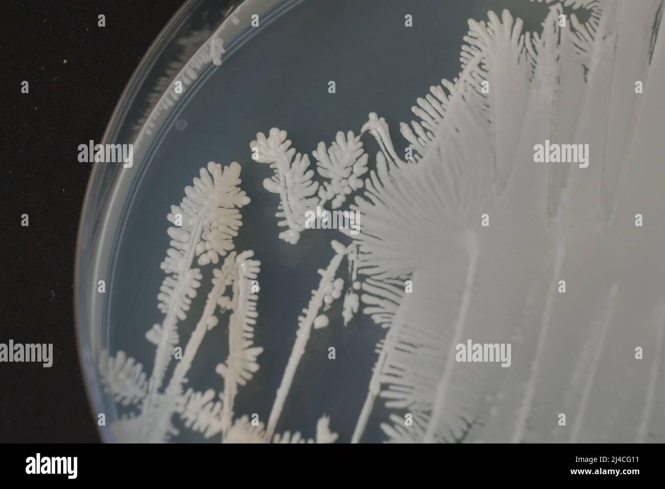 CLOSEUP PHOTO OF BACTERIA AND FUNGI GRWOTH ON AGAR MEDIA IN A PLASTIC PLATE Stock Photohttps://www.alamy.com/image-license-details/?v=1https://www.alamy.com/closeup-photo-of-bacteria-and-fungi-grwoth-on-agar-media-in-a-plastic-plate-image467414557.html
RF2J4CG11–CLOSEUP PHOTO OF BACTERIA AND FUNGI GRWOTH ON AGAR MEDIA IN A PLASTIC PLATE
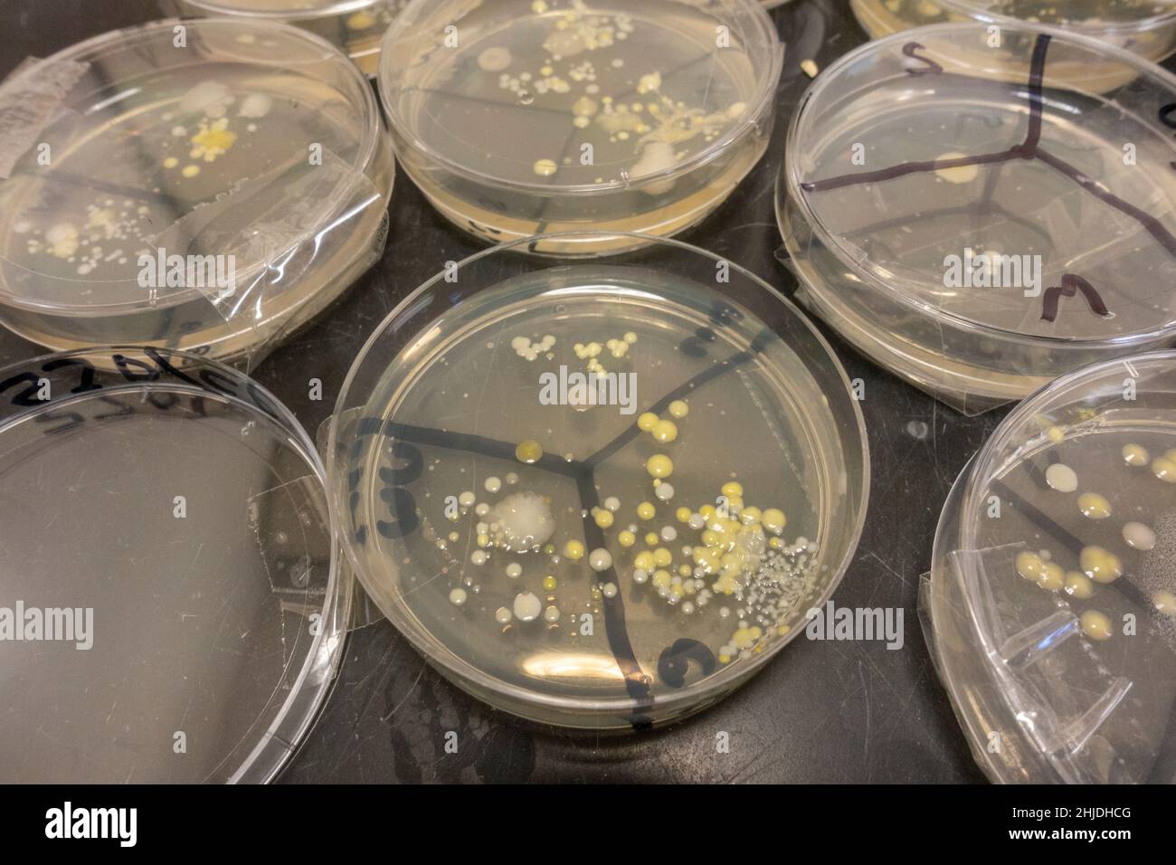 Agar plates petri dishes with bacteria spore growths after UK secondary school biology lesson investigating hand washing. Stock Photohttps://www.alamy.com/image-license-details/?v=1https://www.alamy.com/agar-plates-petri-dishes-with-bacteria-spore-growths-after-uk-secondary-school-biology-lesson-investigating-hand-washing-image458832432.html
RM2HJDHCG–Agar plates petri dishes with bacteria spore growths after UK secondary school biology lesson investigating hand washing.
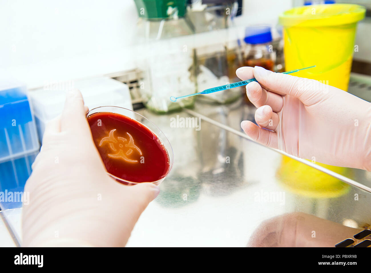 hand working with inoculating loop for spreading bacteria. Petri plate with agar and bacteria escherichia coli in shape of biohazard Stock Photohttps://www.alamy.com/image-license-details/?v=1https://www.alamy.com/hand-working-with-inoculating-loop-for-spreading-bacteria-petri-plate-with-agar-and-bacteria-escherichia-coli-in-shape-of-biohazard-image213874679.html
RFPBXR9B–hand working with inoculating loop for spreading bacteria. Petri plate with agar and bacteria escherichia coli in shape of biohazard
 Microbiologist in a laboratory analysing bacteria samples on a plate. Scientist working in a biology lab Stock Photohttps://www.alamy.com/image-license-details/?v=1https://www.alamy.com/microbiologist-in-a-laboratory-analysing-bacteria-samples-on-a-plate-scientist-working-in-a-biology-lab-image370723530.html
RF2CF3WJ2–Microbiologist in a laboratory analysing bacteria samples on a plate. Scientist working in a biology lab
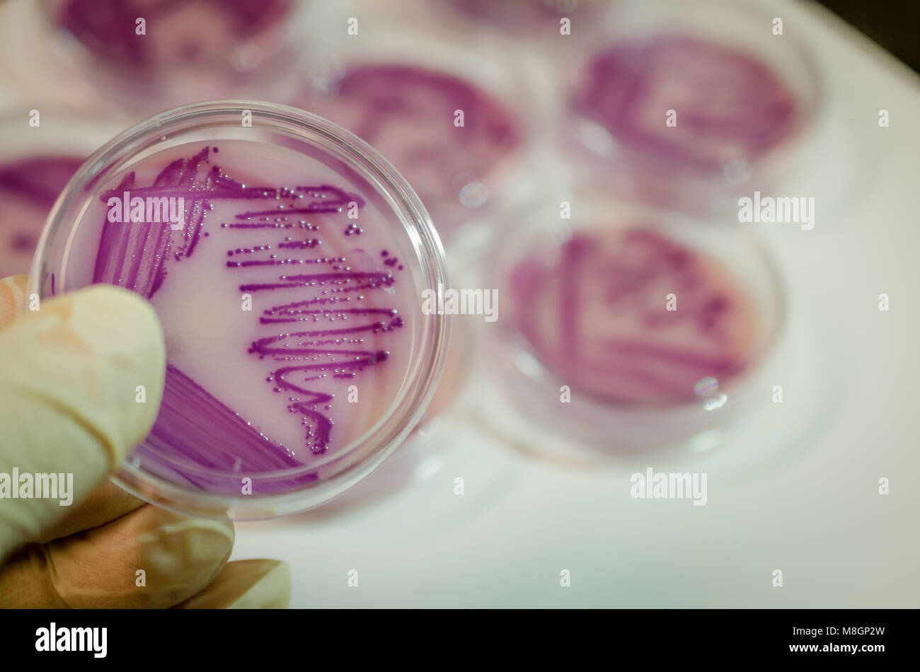 Bacterial culture plate holding in hand Stock Photohttps://www.alamy.com/image-license-details/?v=1https://www.alamy.com/stock-photo-bacterial-culture-plate-holding-in-hand-177389489.html
RFM8GP2W–Bacterial culture plate holding in hand
 Growing Bacteria in Petri Dishes. Stock Photohttps://www.alamy.com/image-license-details/?v=1https://www.alamy.com/stock-photo-growing-bacteria-in-petri-dishes-83917337.html
RFETENBN–Growing Bacteria in Petri Dishes.
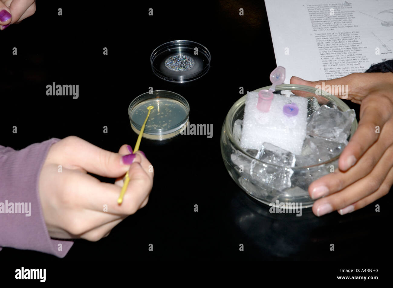 High school girl transfers bacteria cells in a biology science lab biotechnology microbiology experiment Stock Photohttps://www.alamy.com/image-license-details/?v=1https://www.alamy.com/high-school-girl-transfers-bacteria-cells-in-a-biology-science-lab-image6349519.html
RMA4RNH0–High school girl transfers bacteria cells in a biology science lab biotechnology microbiology experiment
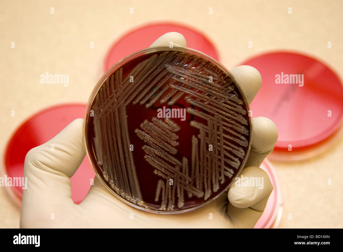 Microbiologist holds a petri dish growimg bacteria, Klebsiella pneumoniae. Stock Photohttps://www.alamy.com/image-license-details/?v=1https://www.alamy.com/stock-photo-microbiologist-holds-a-petri-dish-growimg-bacteria-klebsiella-pneumoniae-25226733.html
RMBD14XN–Microbiologist holds a petri dish growimg bacteria, Klebsiella pneumoniae.
 Petri dishes with blood agar. Stock Photohttps://www.alamy.com/image-license-details/?v=1https://www.alamy.com/petri-dishes-with-blood-agar-image182144571.html
RFMG9B77–Petri dishes with blood agar.
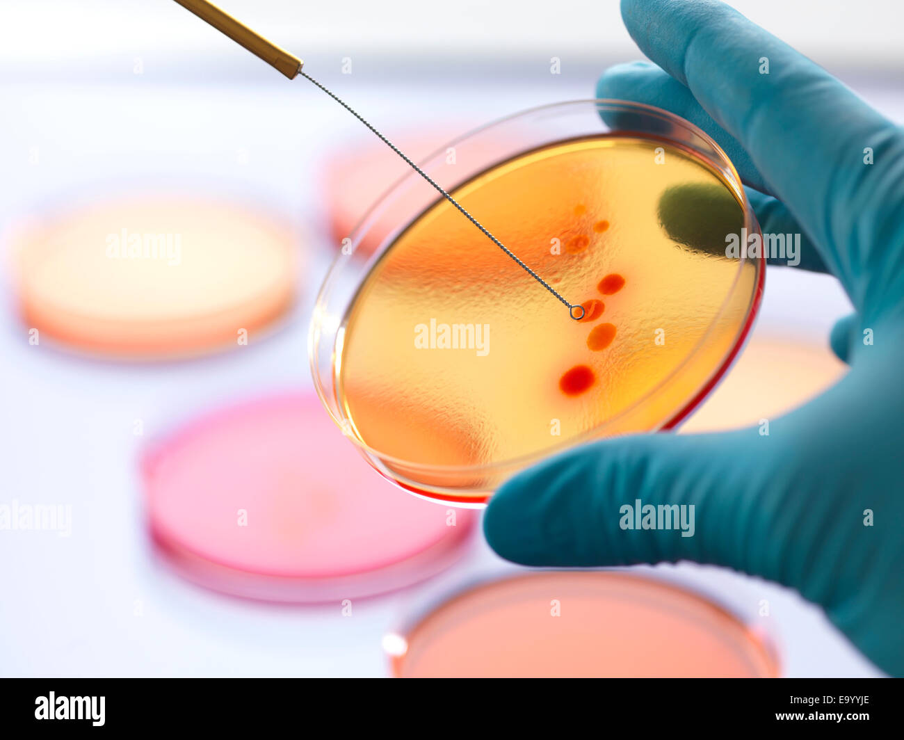 Close up of male scientist hand inoculating an agar plates with bacteria in microbiology lab Stock Photohttps://www.alamy.com/image-license-details/?v=1https://www.alamy.com/stock-photo-close-up-of-male-scientist-hand-inoculating-an-agar-plates-with-bacteria-74987766.html
RFE9YYJE–Close up of male scientist hand inoculating an agar plates with bacteria in microbiology lab
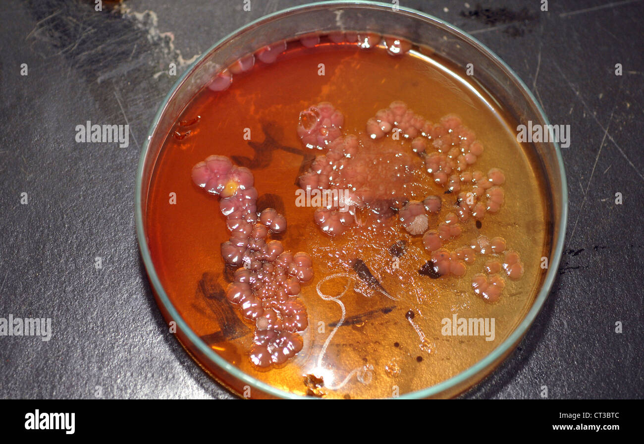 Bacterial culture growing in a petri dish on agar jelly. Stock Photohttps://www.alamy.com/image-license-details/?v=1https://www.alamy.com/stock-photo-bacterial-culture-growing-in-a-petri-dish-on-agar-jelly-49247644.html
RMCT3BTC–Bacterial culture growing in a petri dish on agar jelly.
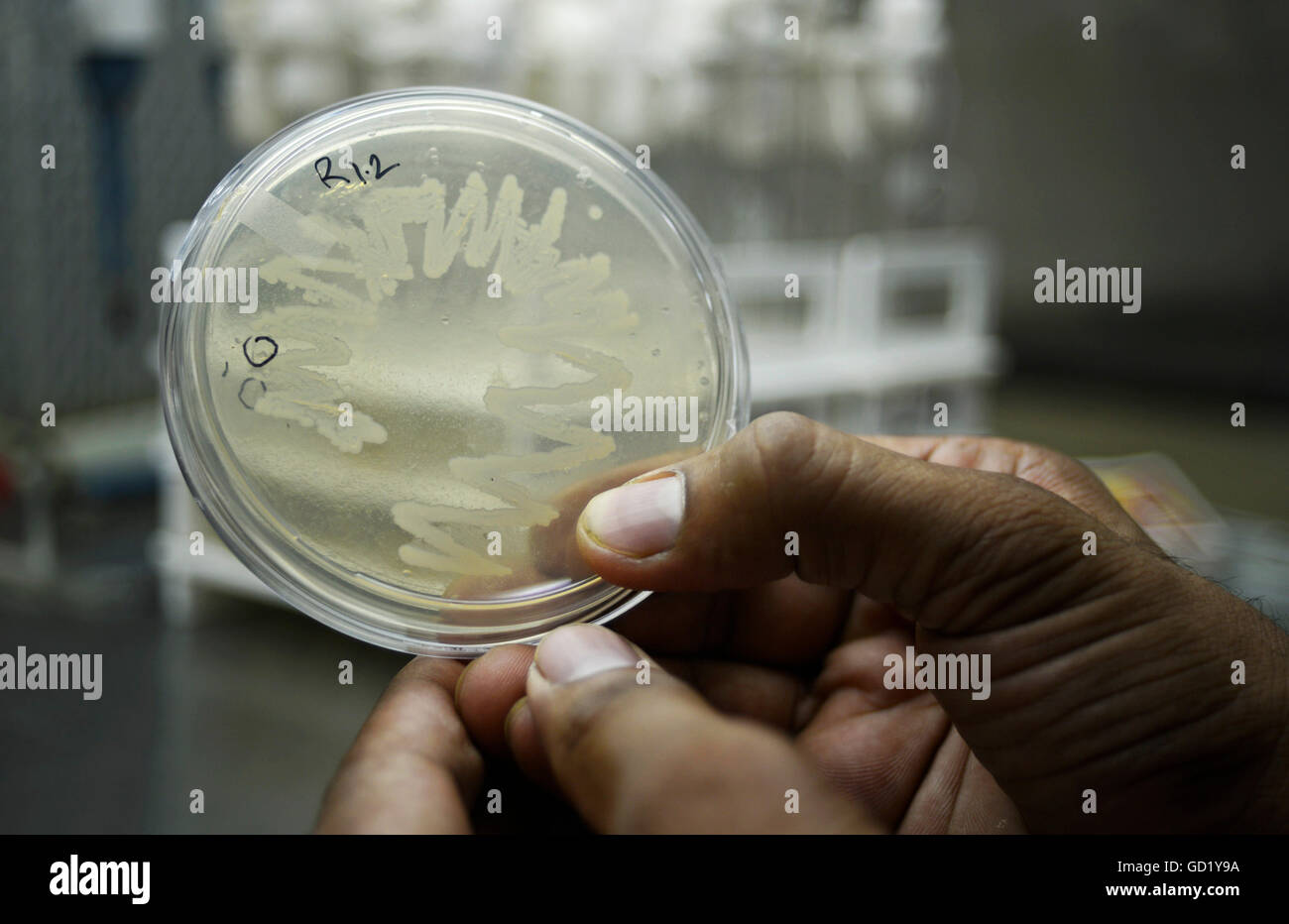 Bacteria Culture Plate - Stock Image Stock Photohttps://www.alamy.com/image-license-details/?v=1https://www.alamy.com/stock-photo-bacteria-culture-plate-stock-image-111296118.html
RFGD1Y9A–Bacteria Culture Plate - Stock Image
 Scientist pipetting a specimen into medium containing bacteria for scientific research in laboratory Stock Photohttps://www.alamy.com/image-license-details/?v=1https://www.alamy.com/scientist-pipetting-a-specimen-into-medium-containing-bacteria-for-image154123775.html
RFJXMXCF–Scientist pipetting a specimen into medium containing bacteria for scientific research in laboratory
 bacteria mutants on agar plate in laboratory Stock Photohttps://www.alamy.com/image-license-details/?v=1https://www.alamy.com/stock-photo-bacteria-mutants-on-agar-plate-in-laboratory-59487808.html
RFDCNW8G–bacteria mutants on agar plate in laboratory
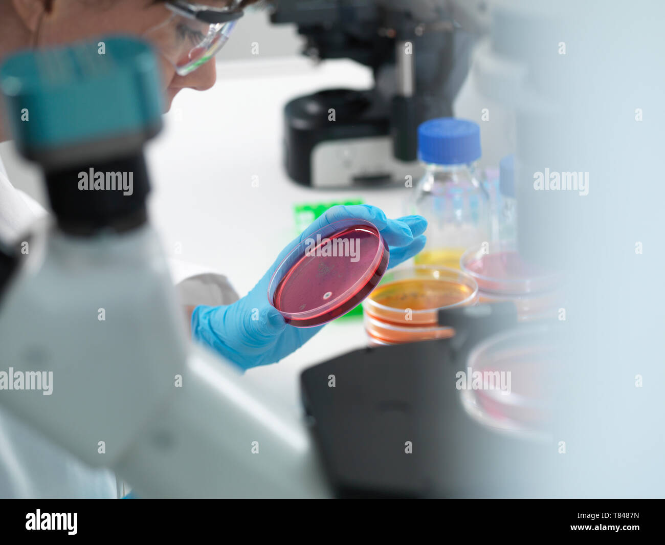 Female scientist examining cultures growing in petri dishes using inverted microscope in laboratory Stock Photohttps://www.alamy.com/image-license-details/?v=1https://www.alamy.com/female-scientist-examining-cultures-growing-in-petri-dishes-using-inverted-microscope-in-laboratory-image245956697.html
RFT8487N–Female scientist examining cultures growing in petri dishes using inverted microscope in laboratory
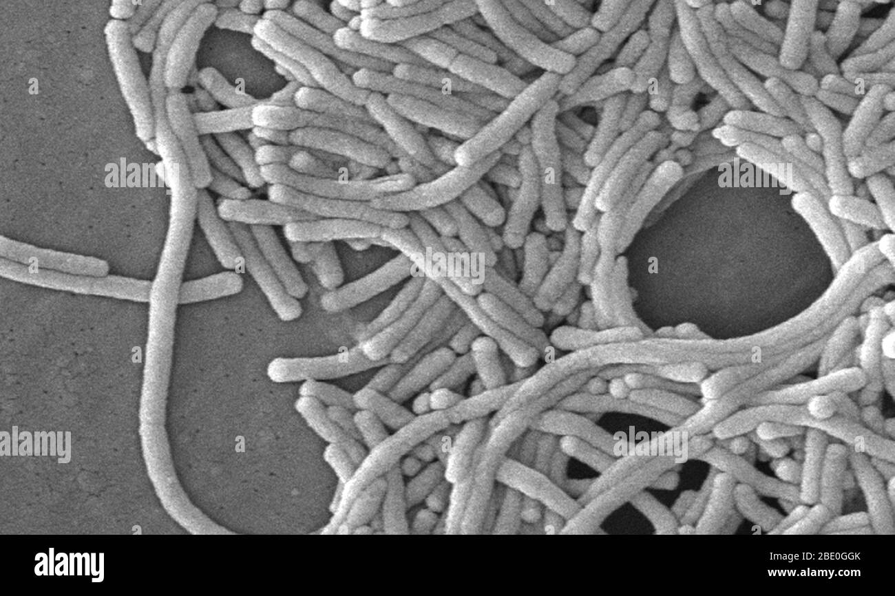 Scanning electron micrograph (SEM) of a number of a large grouping of Gram-negative Legionella pneumophila bacteria. Note the presence of polar flagella, and pili, or long streamers. a number of these bacteria seem to display an elongated-rod morphology. L. pneumophila are known to most frequently exhibit this configuration when grown in broth, however, they can also elongate when plate-grown cells age, as it was in this case, especially when they've been refrigerated. The usual L. pneumophila morphology consists of stout, 'fat' bacilli, which is the case for the vast majority of the organisms Stock Photohttps://www.alamy.com/image-license-details/?v=1https://www.alamy.com/scanning-electron-micrograph-sem-of-a-number-of-a-large-grouping-of-gram-negative-legionella-pneumophila-bacteria-note-the-presence-of-polar-flagella-and-pili-or-long-streamers-a-number-of-these-bacteria-seem-to-display-an-elongated-rod-morphology-l-pneumophila-are-known-to-most-frequently-exhibit-this-configuration-when-grown-in-broth-however-they-can-also-elongate-when-plate-grown-cells-age-as-it-was-in-this-case-especially-when-theyve-been-refrigerated-the-usual-l-pneumophila-morphology-consists-of-stout-fat-bacilli-which-is-the-case-for-the-vast-majority-of-the-organisms-image352825555.html
RM2BE0GGK–Scanning electron micrograph (SEM) of a number of a large grouping of Gram-negative Legionella pneumophila bacteria. Note the presence of polar flagella, and pili, or long streamers. a number of these bacteria seem to display an elongated-rod morphology. L. pneumophila are known to most frequently exhibit this configuration when grown in broth, however, they can also elongate when plate-grown cells age, as it was in this case, especially when they've been refrigerated. The usual L. pneumophila morphology consists of stout, 'fat' bacilli, which is the case for the vast majority of the organisms
 Vector illustration of petri dish with microbes. Laboratory research bacteria Stock Vectorhttps://www.alamy.com/image-license-details/?v=1https://www.alamy.com/vector-illustration-of-petri-dish-with-microbes-laboratory-research-bacteria-image353022295.html
RF2BE9FF3–Vector illustration of petri dish with microbes. Laboratory research bacteria
 Hands holding a culture plate testing for the presence of Escherichia coli bacteria by looking at antibiotic resistance, Biology and Research Center in University Hospital Health, Limoges, France. Stock Photohttps://www.alamy.com/image-license-details/?v=1https://www.alamy.com/stock-photo-hands-holding-a-culture-plate-testing-for-the-presence-of-escherichia-83055844.html
RMER3EG4–Hands holding a culture plate testing for the presence of Escherichia coli bacteria by looking at antibiotic resistance, Biology and Research Center in University Hospital Health, Limoges, France.
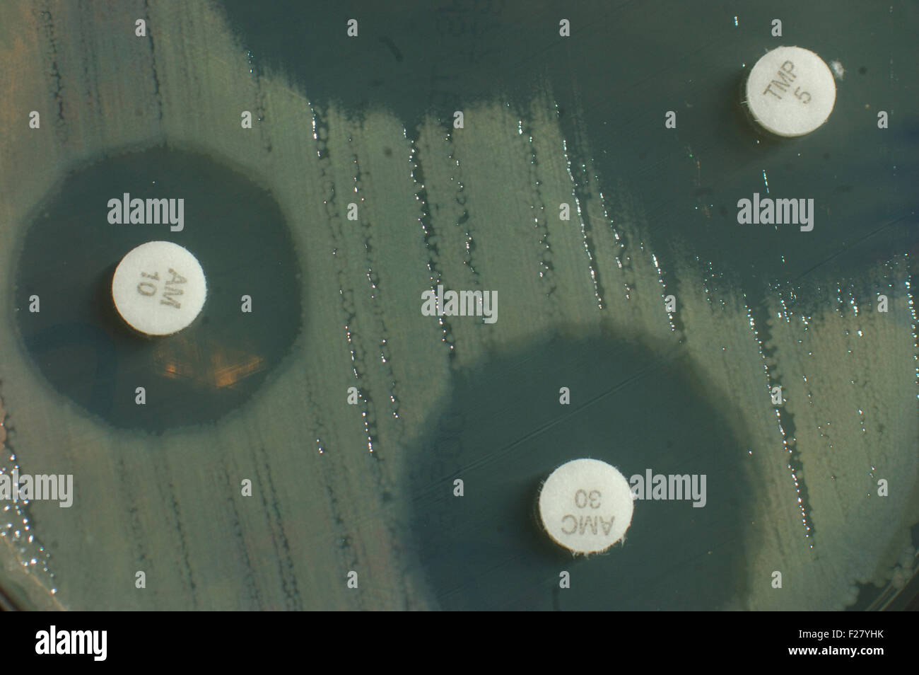 petri dish with antibiotic sensitivity discs showing inhibition zones for bacterial colonies Stock Photohttps://www.alamy.com/image-license-details/?v=1https://www.alamy.com/stock-photo-petri-dish-with-antibiotic-sensitivity-discs-showing-inhibition-zones-87456479.html
RFF27YHK–petri dish with antibiotic sensitivity discs showing inhibition zones for bacterial colonies
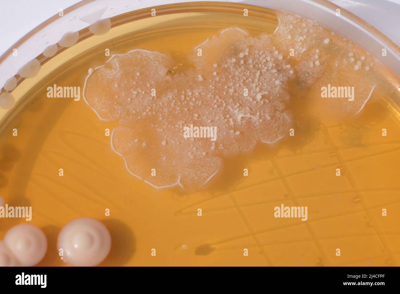 CLOSEUP PHOTO OF BACTERIA AND FUNGI GRWOTH ON AGAR MEDIA IN A PLASTIC PLATE Stock Photohttps://www.alamy.com/image-license-details/?v=1https://www.alamy.com/closeup-photo-of-bacteria-and-fungi-grwoth-on-agar-media-in-a-plastic-plate-image467414375.html
RF2J4CFPF–CLOSEUP PHOTO OF BACTERIA AND FUNGI GRWOTH ON AGAR MEDIA IN A PLASTIC PLATE
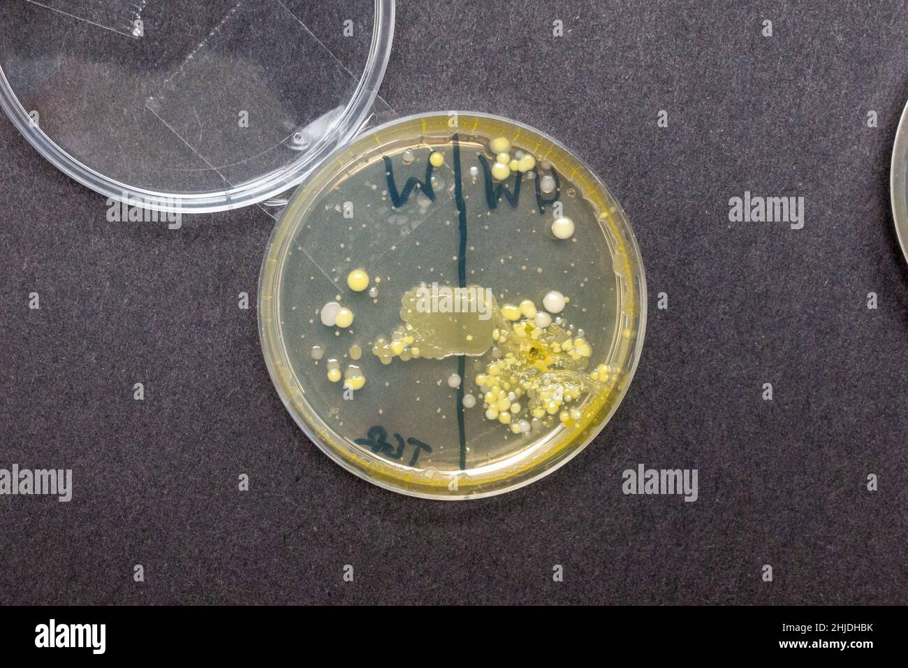 Agar plates petri dishes with bacteria spore growths after UK secondary school biology lesson investigating hand washing. Stock Photohttps://www.alamy.com/image-license-details/?v=1https://www.alamy.com/agar-plates-petri-dishes-with-bacteria-spore-growths-after-uk-secondary-school-biology-lesson-investigating-hand-washing-image458832407.html
RM2HJDHBK–Agar plates petri dishes with bacteria spore growths after UK secondary school biology lesson investigating hand washing.
 Plate with a Kombucha tea mushroom, scoby to produce Kombucha drink isolated on white background close up Stock Photohttps://www.alamy.com/image-license-details/?v=1https://www.alamy.com/plate-with-a-kombucha-tea-mushroom-scoby-to-produce-kombucha-drink-isolated-on-white-background-close-up-image573898006.html
RF2T9K8NA–Plate with a Kombucha tea mushroom, scoby to produce Kombucha drink isolated on white background close up
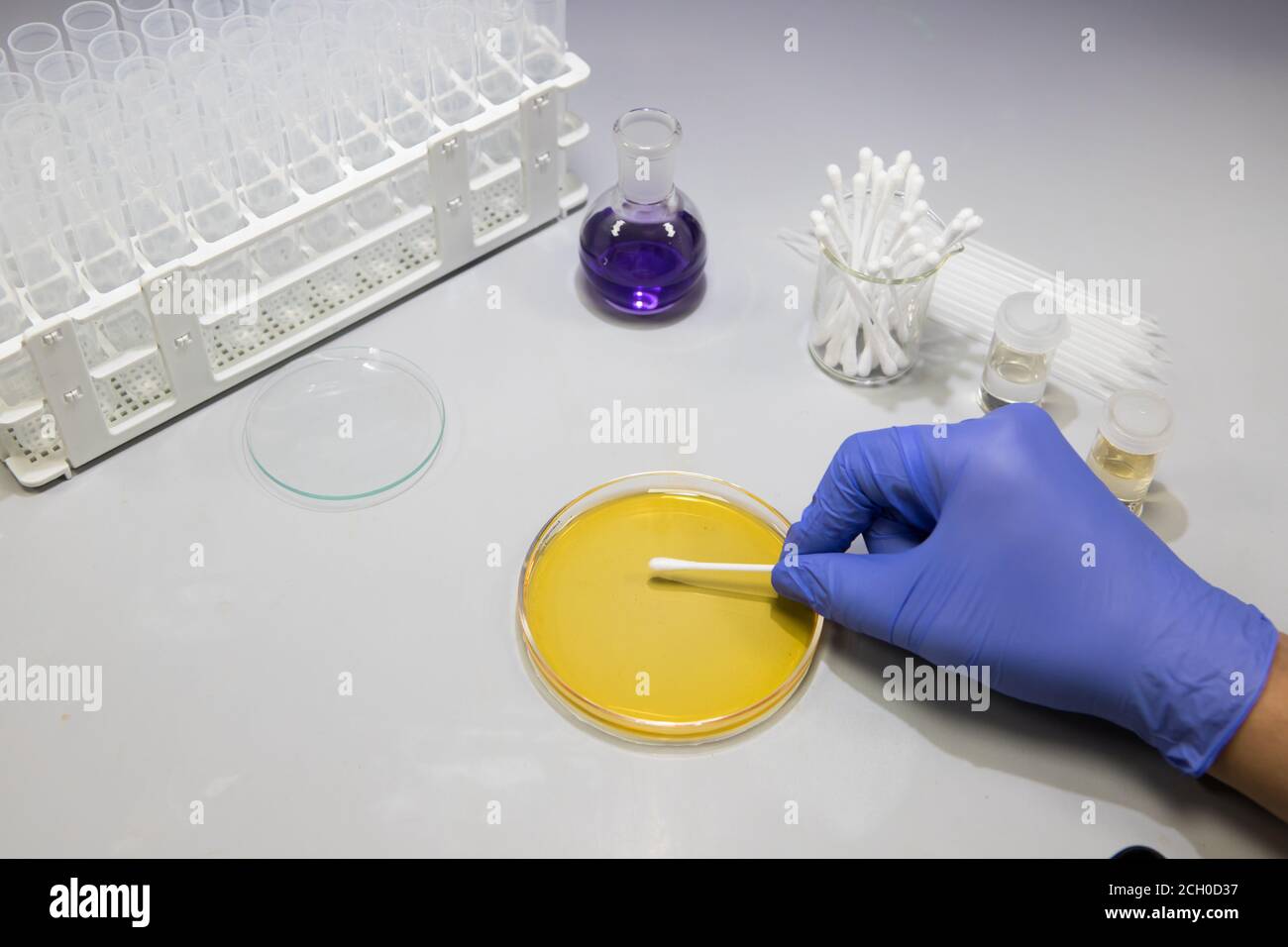 Microbiologist in a laboratory analysing bacteria samples on a plate. Scientist working in a biology lab Stock Photohttps://www.alamy.com/image-license-details/?v=1https://www.alamy.com/microbiologist-in-a-laboratory-analysing-bacteria-samples-on-a-plate-scientist-working-in-a-biology-lab-image371877163.html
RF2CH0D37–Microbiologist in a laboratory analysing bacteria samples on a plate. Scientist working in a biology lab
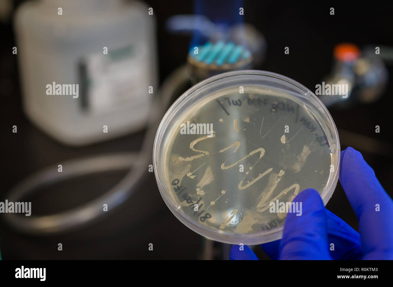 E coli bacterial culture on culture plate Stock Photohttps://www.alamy.com/image-license-details/?v=1https://www.alamy.com/e-coli-bacterial-culture-on-culture-plate-image224171251.html
RFR0KTM3–E coli bacterial culture on culture plate
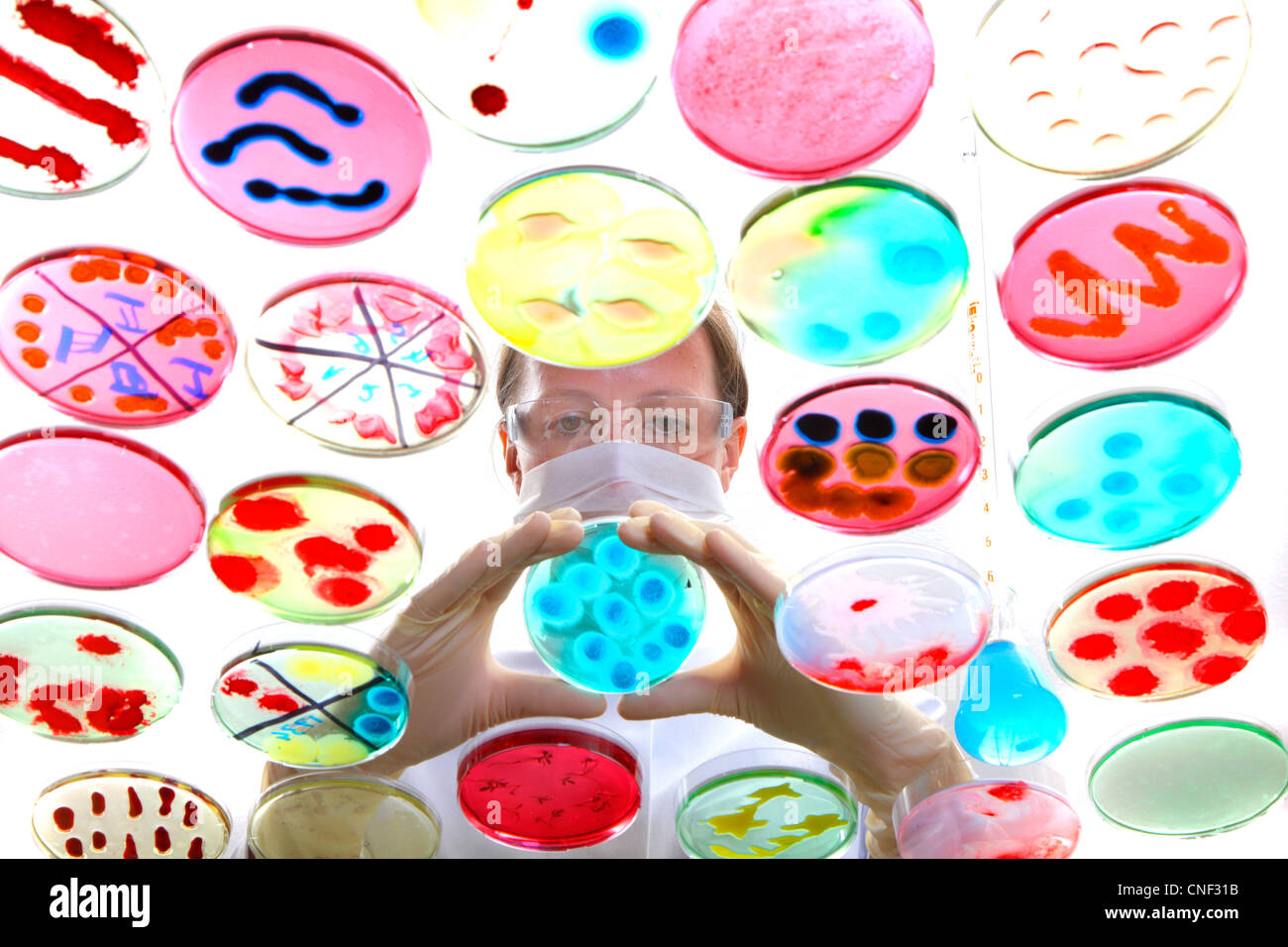 Laboratory, biological, chemical. Analysis of bacterial cultures of bacteria growing in petri dishes. Stock Photohttps://www.alamy.com/image-license-details/?v=1https://www.alamy.com/stock-photo-laboratory-biological-chemical-analysis-of-bacterial-cultures-of-bacteria-47660183.html
RMCNF31B–Laboratory, biological, chemical. Analysis of bacterial cultures of bacteria growing in petri dishes.
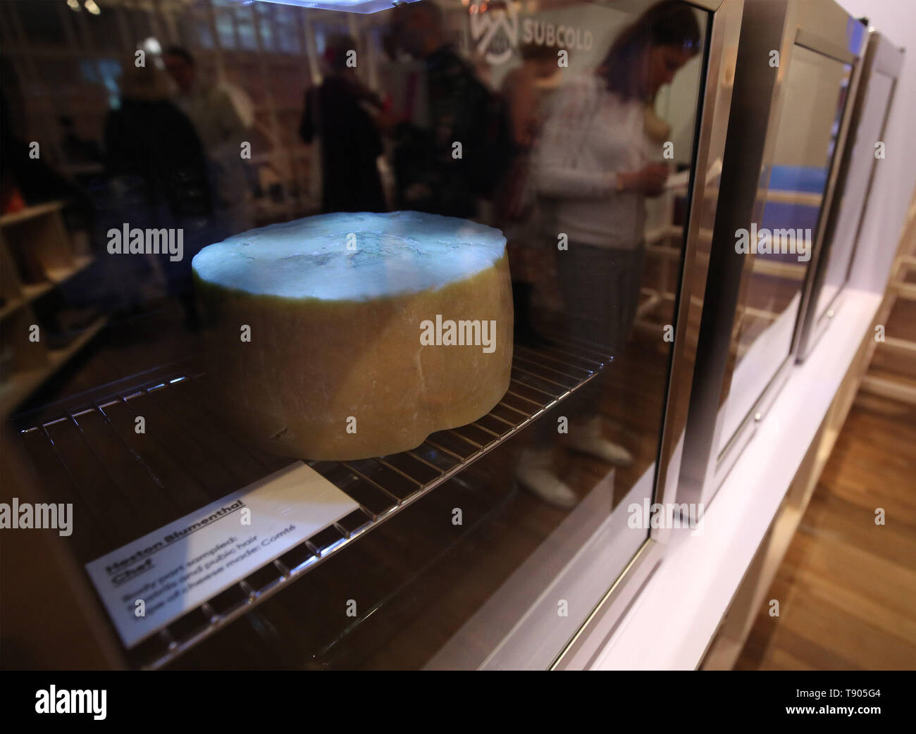 A Comte cheese made with bacteria sampled from the nostrils and pubic hair of Heston Blumenthal, part of 'Selfmade', a project by Christina Agapakis and Sissel Tolaas, which involves cheese produced with microbes cultured from human bacteria taken from people's ears, toes and armpits, at the FOOD: Bigger than the Plate exhibition at the Victoria and Albert Museum in London. Stock Photohttps://www.alamy.com/image-license-details/?v=1https://www.alamy.com/a-comte-cheese-made-with-bacteria-sampled-from-the-nostrils-and-pubic-hair-of-heston-blumenthal-part-of-selfmade-a-project-by-christina-agapakis-and-sissel-tolaas-which-involves-cheese-produced-with-microbes-cultured-from-human-bacteria-taken-from-peoples-ears-toes-and-armpits-at-the-food-bigger-than-the-plate-exhibition-at-the-victoria-and-albert-museum-in-london-image246481428.html
RMT905G4–A Comte cheese made with bacteria sampled from the nostrils and pubic hair of Heston Blumenthal, part of 'Selfmade', a project by Christina Agapakis and Sissel Tolaas, which involves cheese produced with microbes cultured from human bacteria taken from people's ears, toes and armpits, at the FOOD: Bigger than the Plate exhibition at the Victoria and Albert Museum in London.
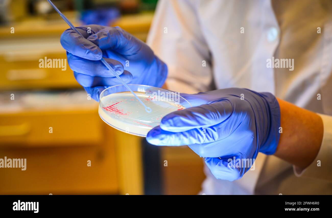 Woman scientist picking up colony of a red bacteria from agar plate in a molecular biology laboratory for the isolation of drug resistant mutants Stock Photohttps://www.alamy.com/image-license-details/?v=1https://www.alamy.com/woman-scientist-picking-up-colony-of-a-red-bacteria-from-agar-plate-in-a-molecular-biology-laboratory-for-the-isolation-of-drug-resistant-mutants-image428793764.html
RF2FWH6R0–Woman scientist picking up colony of a red bacteria from agar plate in a molecular biology laboratory for the isolation of drug resistant mutants
