Quick filters:
Basal sheath Stock Photos and Images
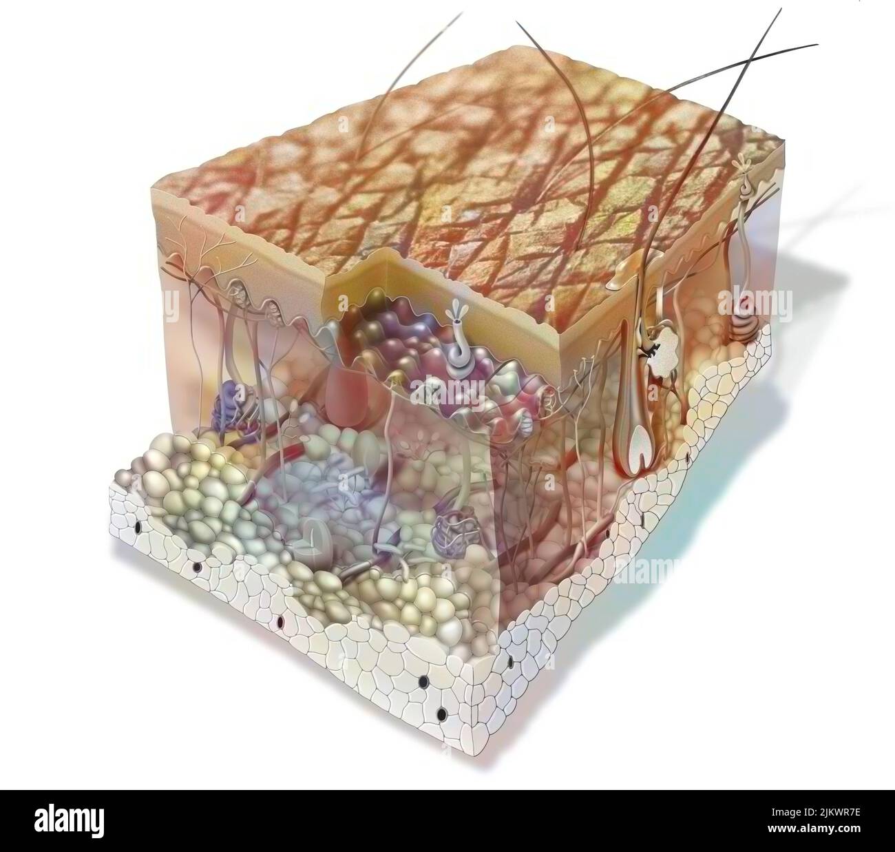 Skin section showing all the structures: the layers of the epidermis, the structure of the dermis. Stock Photohttps://www.alamy.com/image-license-details/?v=1https://www.alamy.com/skin-section-showing-all-the-structures-the-layers-of-the-epidermis-the-structure-of-the-dermis-image476925442.html
RF2JKWR7E–Skin section showing all the structures: the layers of the epidermis, the structure of the dermis.
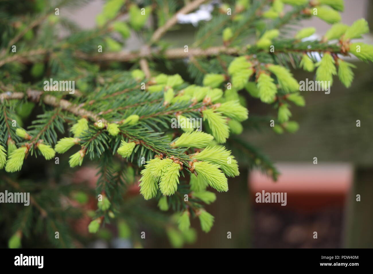 New Growth on Pine Tree, Bright New Pine Needles on Christmas Tree. Stock Photohttps://www.alamy.com/image-license-details/?v=1https://www.alamy.com/new-growth-on-pine-tree-bright-new-pine-needles-on-christmas-tree-image215066900.html
RFPDW40M–New Growth on Pine Tree, Bright New Pine Needles on Christmas Tree.
 Closeup of a Pine sapling growing in Lentiira, Kuhmo, Finland. Stock Photohttps://www.alamy.com/image-license-details/?v=1https://www.alamy.com/stock-photo-closeup-of-a-pine-sapling-growing-in-lentiira-kuhmo-finland-58650323.html
RMDBBN2B–Closeup of a Pine sapling growing in Lentiira, Kuhmo, Finland.
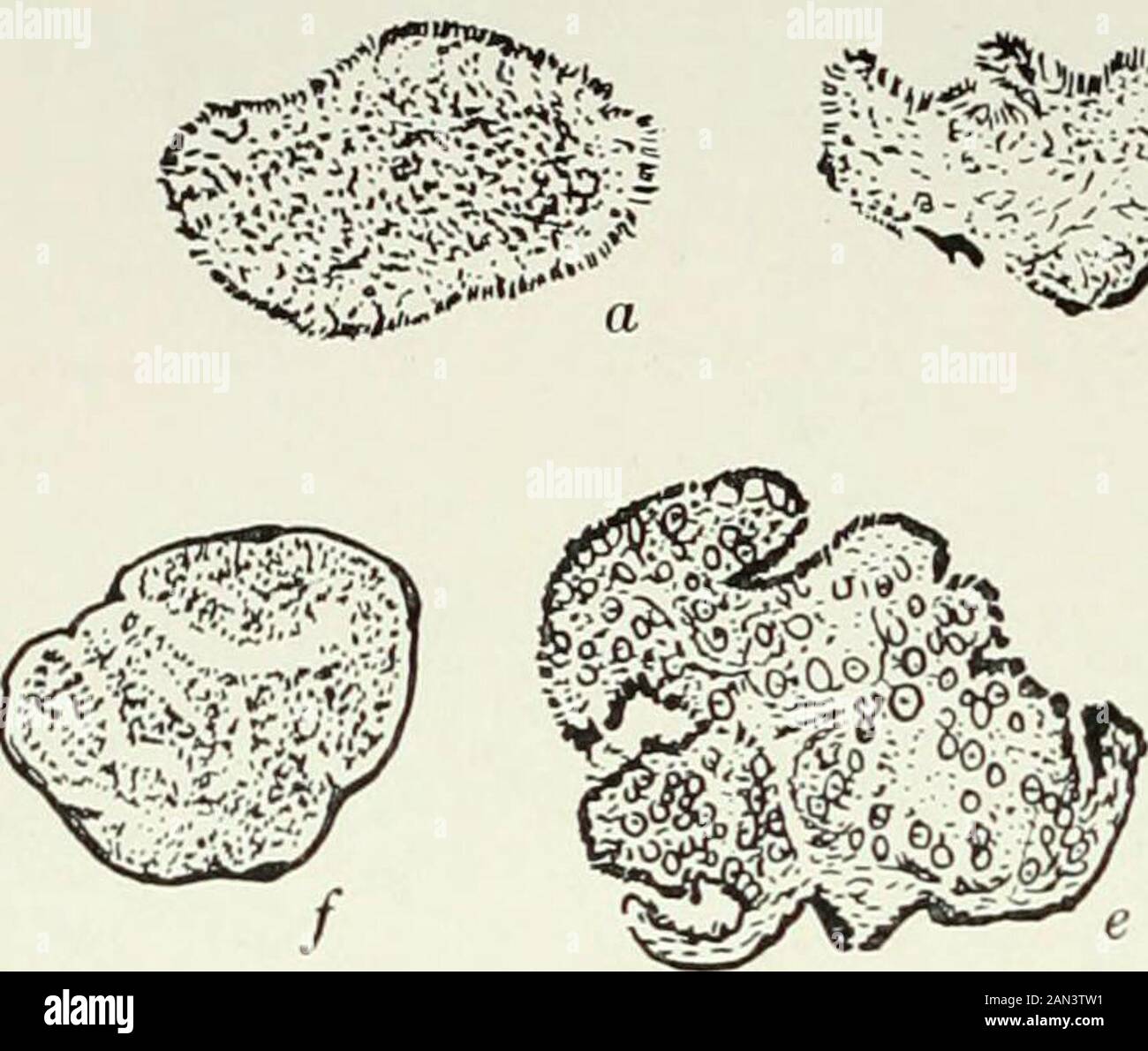 Fungi, Ascomycetes, Ustilaginales, Uredinales . gina-tions of the upper surface, and internally the loose tissue of the sterile veins1 01 nes recognizable. Owing to the rapid growth of the upper portion of the young fruit, thebasal sheath is bent backwards, while at various points along the fertile veinsthe first signs of asci appear. Later the peripheral tissues become thickened,together with the remains of the basal sheath, and form the peridium. Thisultimately closes over the points where the fertile veins are in communicationwith the exterior. Thus the young fruit is open at first, the hym Stock Photohttps://www.alamy.com/image-license-details/?v=1https://www.alamy.com/fungi-ascomycetes-ustilaginales-uredinales-gina-tions-of-the-upper-surface-and-internally-the-loose-tissue-of-the-sterile-veins1-01-nes-recognizable-owing-to-the-rapid-growth-of-the-upper-portion-of-the-young-fruit-thebasal-sheath-is-bent-backwards-while-at-various-points-along-the-fertile-veinsthe-first-signs-of-asci-appear-later-the-peripheral-tissues-become-thickenedtogether-with-the-remains-of-the-basal-sheath-and-form-the-peridium-thisultimately-closes-over-the-points-where-the-fertile-veins-are-in-communicationwith-the-exterior-thus-the-young-fruit-is-open-at-first-the-hym-image339990141.html
RM2AN3TW1–Fungi, Ascomycetes, Ustilaginales, Uredinales . gina-tions of the upper surface, and internally the loose tissue of the sterile veins1 01 nes recognizable. Owing to the rapid growth of the upper portion of the young fruit, thebasal sheath is bent backwards, while at various points along the fertile veinsthe first signs of asci appear. Later the peripheral tissues become thickened,together with the remains of the basal sheath, and form the peridium. Thisultimately closes over the points where the fertile veins are in communicationwith the exterior. Thus the young fruit is open at first, the hym
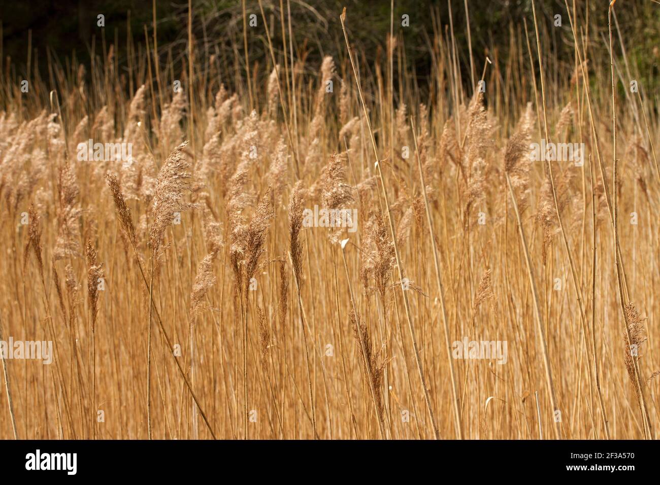 Tallest of the British grass species, the Common Reed spreads using tough creeping rhizomes and stolon's to form dense stands of reed-beds in wetlands Stock Photohttps://www.alamy.com/image-license-details/?v=1https://www.alamy.com/tallest-of-the-british-grass-species-the-common-reed-spreads-using-tough-creeping-rhizomes-and-stolons-to-form-dense-stands-of-reed-beds-in-wetlands-image415116436.html
RM2F3A570–Tallest of the British grass species, the Common Reed spreads using tough creeping rhizomes and stolon's to form dense stands of reed-beds in wetlands
 . Elements of the comparative anatomy of vertebrates. Anatomy, Comparative. 54 COMPARATIVE ANATOMY Ganoidei, Teleostei, and Dipnoi—In these forms the ribs, ahiiost without exception, are connected with the ventral parts of the notochordal sheath (Dipnoans) or with the " basal processes " (Ganoids and Teleosts, see p. 38).-' This is one point of difference between the ribs of these forms and those of other Vertebrates: another is that they are always situated beneath (internal to) the lateral muscles, between these and the peritoneum (Fig. 40a, A, B, C). In Teleosts the ribs are at fi Stock Photohttps://www.alamy.com/image-license-details/?v=1https://www.alamy.com/elements-of-the-comparative-anatomy-of-vertebrates-anatomy-comparative-54-comparative-anatomy-ganoidei-teleostei-and-dipnoiin-these-forms-the-ribs-ahiiost-without-exception-are-connected-with-the-ventral-parts-of-the-notochordal-sheath-dipnoans-or-with-the-quot-basal-processes-quot-ganoids-and-teleosts-see-p-38-this-is-one-point-of-difference-between-the-ribs-of-these-forms-and-those-of-other-vertebrates-another-is-that-they-are-always-situated-beneath-internal-to-the-lateral-muscles-between-these-and-the-peritoneum-fig-40a-a-b-c-in-teleosts-the-ribs-are-at-fi-image216419890.html
RMPG2NNP–. Elements of the comparative anatomy of vertebrates. Anatomy, Comparative. 54 COMPARATIVE ANATOMY Ganoidei, Teleostei, and Dipnoi—In these forms the ribs, ahiiost without exception, are connected with the ventral parts of the notochordal sheath (Dipnoans) or with the " basal processes " (Ganoids and Teleosts, see p. 38).-' This is one point of difference between the ribs of these forms and those of other Vertebrates: another is that they are always situated beneath (internal to) the lateral muscles, between these and the peritoneum (Fig. 40a, A, B, C). In Teleosts the ribs are at fi
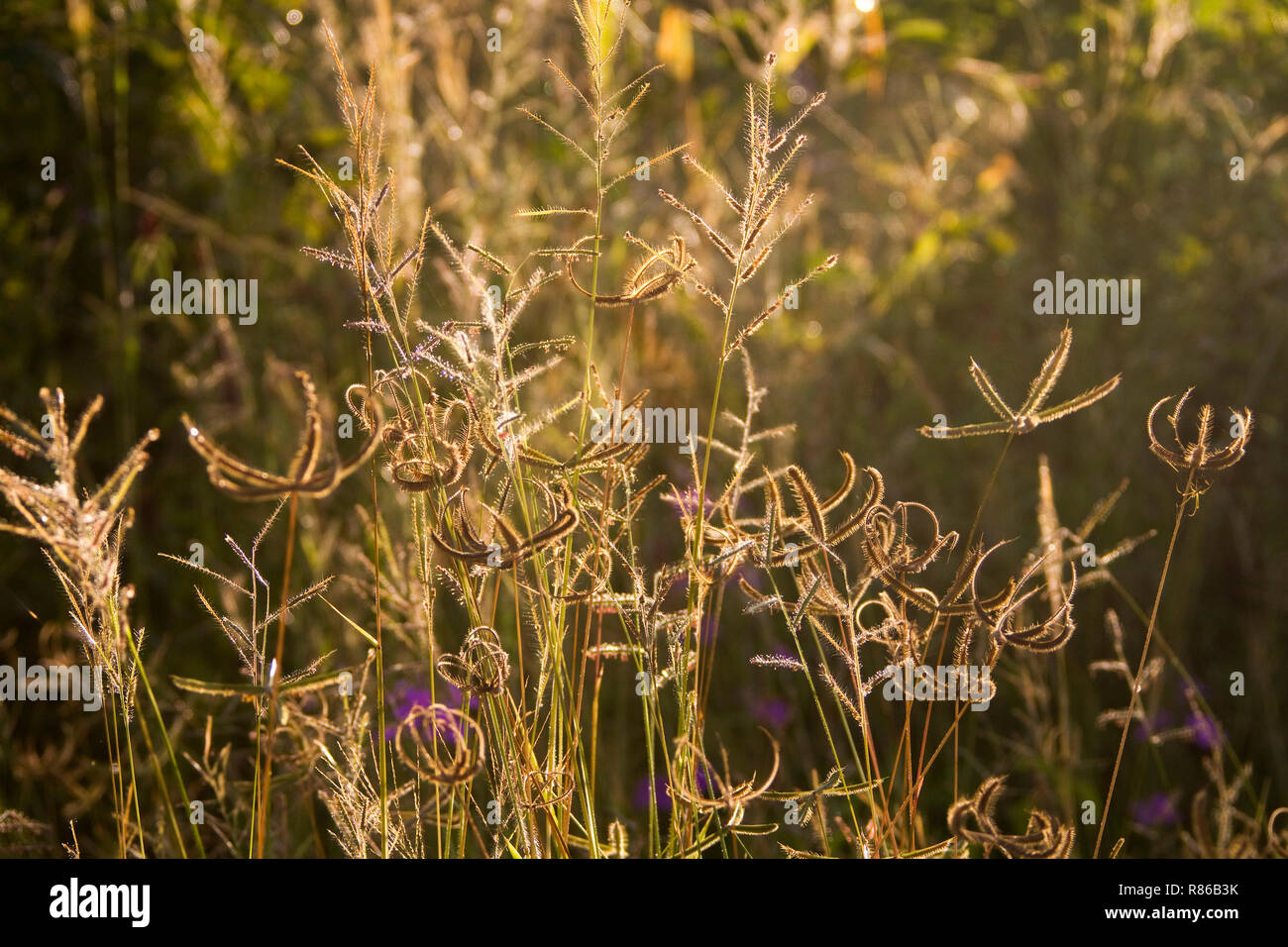 the rains bring a profusion of growth and annual grasses quickly develope seeds stock for the next season provoding a bountiful supply of food in the Stock Photohttps://www.alamy.com/image-license-details/?v=1https://www.alamy.com/the-rains-bring-a-profusion-of-growth-and-annual-grasses-quickly-develope-seeds-stock-for-the-next-season-provoding-a-bountiful-supply-of-food-in-the-image228792471.html
RMR86B3K–the rains bring a profusion of growth and annual grasses quickly develope seeds stock for the next season provoding a bountiful supply of food in the
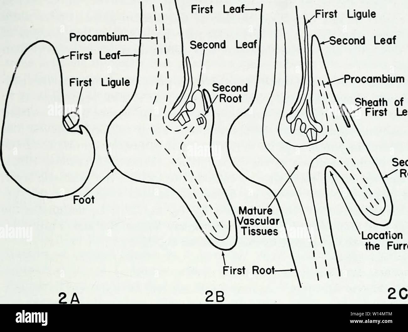 Archive image from page 20 of The developmental anatomy of Isoetes. The developmental anatomy of Isoetes . developmentalana31paol Year: 1963 TERMINOLOGY First Ligule Second Leaf y>—Procambium « Sheath of the ' First Leaf Second Root Location of the Furrow Fig. 2. Three stages in the development of a young sporophyte. A. One leaf. B. Two leaves. C. Three leaves. The plane of sectioning in A, B, C is the same plane that contains all the leaf traces while the plant is in a ¥2 phyllotaxy. The basal furrow forms between the first and second roots in a plane at right angles of the plane Stock Photohttps://www.alamy.com/image-license-details/?v=1https://www.alamy.com/archive-image-from-page-20-of-the-developmental-anatomy-of-isoetes-the-developmental-anatomy-of-isoetes-developmentalana31paol-year-1963-terminology-first-ligule-second-leaf-ygtprocambium-sheath-of-the-first-leaf-second-root-location-of-the-furrow-fig-2-three-stages-in-the-development-of-a-young-sporophyte-a-one-leaf-b-two-leaves-c-three-leaves-the-plane-of-sectioning-in-a-b-c-is-the-same-plane-that-contains-all-the-leaf-traces-while-the-plant-is-in-a-2-phyllotaxy-the-basal-furrow-forms-between-the-first-and-second-roots-in-a-plane-at-right-angles-of-the-plane-image258874356.html
RMW14MTM–Archive image from page 20 of The developmental anatomy of Isoetes. The developmental anatomy of Isoetes . developmentalana31paol Year: 1963 TERMINOLOGY First Ligule Second Leaf y>—Procambium « Sheath of the ' First Leaf Second Root Location of the Furrow Fig. 2. Three stages in the development of a young sporophyte. A. One leaf. B. Two leaves. C. Three leaves. The plane of sectioning in A, B, C is the same plane that contains all the leaf traces while the plant is in a ¥2 phyllotaxy. The basal furrow forms between the first and second roots in a plane at right angles of the plane
 Seed head of Arogrostus grass silhouteed against the dusk during the rainy season in Katavi National Park Stock Photohttps://www.alamy.com/image-license-details/?v=1https://www.alamy.com/seed-head-of-arogrostus-grass-silhouteed-against-the-dusk-during-the-rainy-season-in-katavi-national-park-image228792468.html
RMR86B3G–Seed head of Arogrostus grass silhouteed against the dusk during the rainy season in Katavi National Park
 . Fig. 156. — Basal portion of rye plant showing anthrac- nose upon stem and leaf sheath. After Manns. Stock Photohttps://www.alamy.com/image-license-details/?v=1https://www.alamy.com/fig-156-basal-portion-of-rye-plant-showing-anthrac-nose-upon-stem-and-leaf-sheath-after-manns-image179904443.html
RMMCK9XK–. Fig. 156. — Basal portion of rye plant showing anthrac- nose upon stem and leaf sheath. After Manns.
 Tassel Three-awn grass is a annual pioneer species that quickly germinates in poor soils, often after a fire or erosion. Stock Photohttps://www.alamy.com/image-license-details/?v=1https://www.alamy.com/tassel-three-awn-grass-is-a-annual-pioneer-species-that-quickly-germinates-in-poor-soils-often-after-a-fire-or-erosion-image627736155.html
RM2YD7RTB–Tassel Three-awn grass is a annual pioneer species that quickly germinates in poor soils, often after a fire or erosion.
 dew sits on the seed heads of Tassle Three-awn grass. This grass is a annual pioneer species that quickly germinates in poor soils, often after a fire Stock Photohttps://www.alamy.com/image-license-details/?v=1https://www.alamy.com/dew-sits-on-the-seed-heads-of-tassle-three-awn-grass-this-grass-is-a-annual-pioneer-species-that-quickly-germinates-in-poor-soils-often-after-a-fire-image627736113.html
RM2YD7RPW–dew sits on the seed heads of Tassle Three-awn grass. This grass is a annual pioneer species that quickly germinates in poor soils, often after a fire
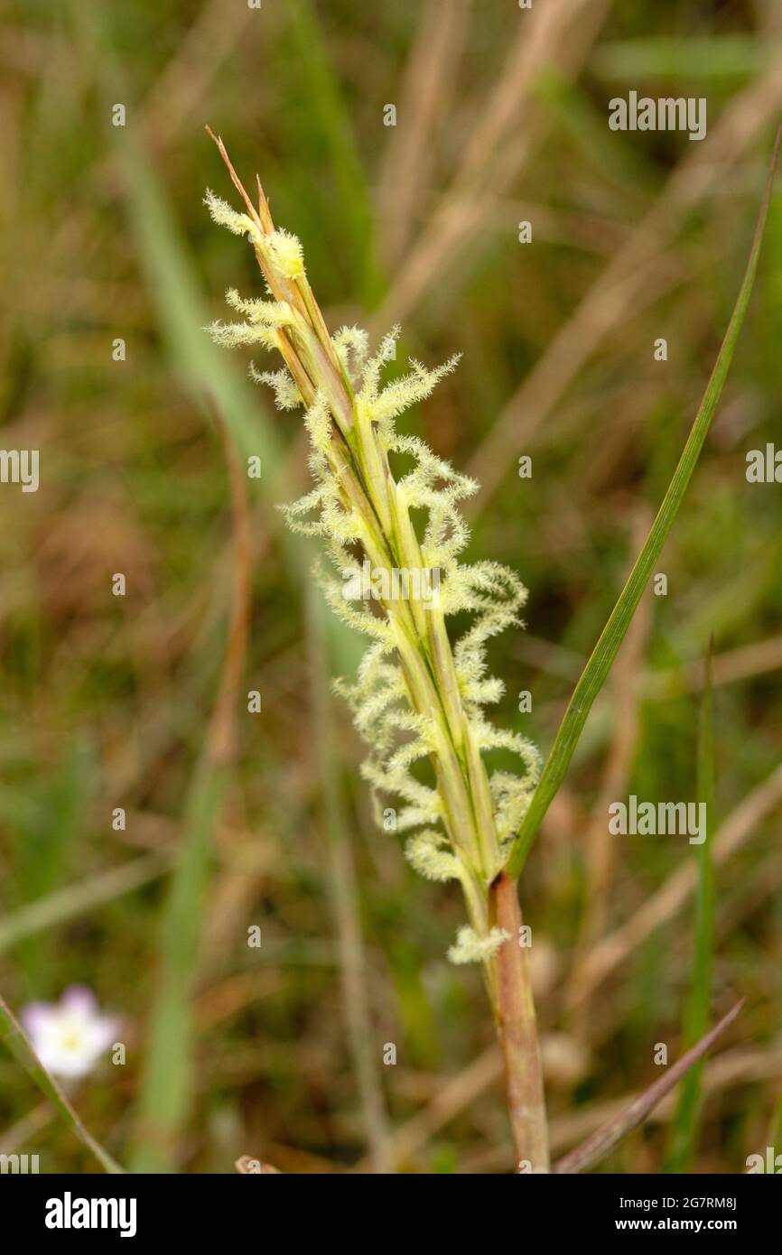 The distinctive spike-like panicles of Sweet Vernal Grass. this plant favours acidic soils and grows well on sand dunes. Also known as Hornwort Stock Photohttps://www.alamy.com/image-license-details/?v=1https://www.alamy.com/the-distinctive-spike-like-panicles-of-sweet-vernal-grass-this-plant-favours-acidic-soils-and-grows-well-on-sand-dunes-also-known-as-hornwort-image435082610.html
RM2G7RM8J–The distinctive spike-like panicles of Sweet Vernal Grass. this plant favours acidic soils and grows well on sand dunes. Also known as Hornwort
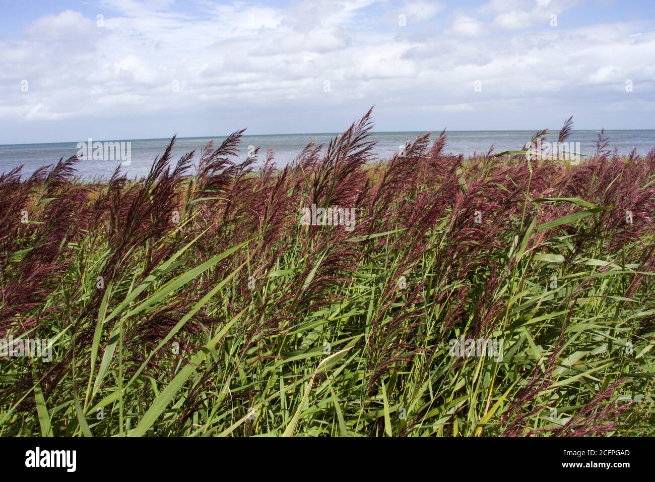 An adaptable and highly productive native of the North American Prairies, Blackwell Switchgrass has been used in the UK to control erosion Stock Photohttps://www.alamy.com/image-license-details/?v=1https://www.alamy.com/an-adaptable-and-highly-productive-native-of-the-north-american-prairies-blackwell-switchgrass-has-been-used-in-the-uk-to-control-erosion-image371133349.html
RM2CFPGAD–An adaptable and highly productive native of the North American Prairies, Blackwell Switchgrass has been used in the UK to control erosion
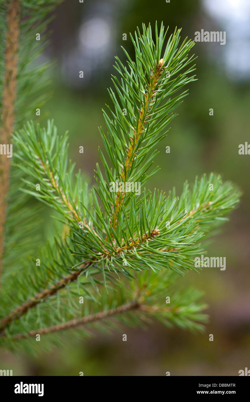 Pine sapling growing in Lentiira, Kuhmo, Finland. Stock Photohttps://www.alamy.com/image-license-details/?v=1https://www.alamy.com/stock-photo-pine-sapling-growing-in-lentiira-kuhmo-finland-58650167.html
RMDBBMTR–Pine sapling growing in Lentiira, Kuhmo, Finland.
 . Smithsonian miscellaneous collections . neath a thor-acic segment; in this section (No. 27, M. C. Z.) the slender arm ofan expodite is shown on the right side and a little of the spiral struc-ture of the arm. It is not at all probable that the spiral arm of the exopodite ofthe thoracic limbs with its basal sheath and slender tubes would be NO. 7 NOTES ON STRUCTURE OF NEOLENUS 4IO, replaced by such a structure as that shown by figures 1-6, plate 100.There is nothing in common between them except the fringing fila-ments. The fringed epipodites of Calymene and Ceraurus (plate ioo) arenot unlike Stock Photohttps://www.alamy.com/image-license-details/?v=1https://www.alamy.com/smithsonian-miscellaneous-collections-neath-a-thor-acic-segment-in-this-section-no-27-m-c-z-the-slender-arm-ofan-expodite-is-shown-on-the-right-side-and-a-little-of-the-spiral-struc-ture-of-the-arm-it-is-not-at-all-probable-that-the-spiral-arm-of-the-exopodite-ofthe-thoracic-limbs-with-its-basal-sheath-and-slender-tubes-would-be-no-7-notes-on-structure-of-neolenus-4io-replaced-by-such-a-structure-as-that-shown-by-figures-1-6-plate-100there-is-nothing-in-common-between-them-except-the-fringing-fila-ments-the-fringed-epipodites-of-calymene-and-ceraurus-plate-ioo-arenot-unlike-image371710974.html
RM2CGMW3X–. Smithsonian miscellaneous collections . neath a thor-acic segment; in this section (No. 27, M. C. Z.) the slender arm ofan expodite is shown on the right side and a little of the spiral struc-ture of the arm. It is not at all probable that the spiral arm of the exopodite ofthe thoracic limbs with its basal sheath and slender tubes would be NO. 7 NOTES ON STRUCTURE OF NEOLENUS 4IO, replaced by such a structure as that shown by figures 1-6, plate 100.There is nothing in common between them except the fringing fila-ments. The fringed epipodites of Calymene and Ceraurus (plate ioo) arenot unlike
 . Beekeeping; a discussion of the life of the honeybee and of the production of honey. Bees; Honey. The Life Processes of the IndividvM 159 bulb {ShB) and is further continued in two arms {ShA) which curve outward. The lancets slide on a grooved track the full length of the sheath, past the bulb and diverge along the two basal arms. The sheath and lancets combine to form a hollow tube (PsnC) through which the poison flows. The arms of the sheath are attached at their anterior ends to oblong plates (Ob) which overlap the sides of the sting. To these plates are attached palpi (StnPlp), soft whit Stock Photohttps://www.alamy.com/image-license-details/?v=1https://www.alamy.com/beekeeping-a-discussion-of-the-life-of-the-honeybee-and-of-the-production-of-honey-bees-honey-the-life-processes-of-the-individvm-159-bulb-shb-and-is-further-continued-in-two-arms-sha-which-curve-outward-the-lancets-slide-on-a-grooved-track-the-full-length-of-the-sheath-past-the-bulb-and-diverge-along-the-two-basal-arms-the-sheath-and-lancets-combine-to-form-a-hollow-tube-psnc-through-which-the-poison-flows-the-arms-of-the-sheath-are-attached-at-their-anterior-ends-to-oblong-plates-ob-which-overlap-the-sides-of-the-sting-to-these-plates-are-attached-palpi-stnplp-soft-whit-image216361574.html
RMPG03B2–. Beekeeping; a discussion of the life of the honeybee and of the production of honey. Bees; Honey. The Life Processes of the IndividvM 159 bulb {ShB) and is further continued in two arms {ShA) which curve outward. The lancets slide on a grooved track the full length of the sheath, past the bulb and diverge along the two basal arms. The sheath and lancets combine to form a hollow tube (PsnC) through which the poison flows. The arms of the sheath are attached at their anterior ends to oblong plates (Ob) which overlap the sides of the sting. To these plates are attached palpi (StnPlp), soft whit
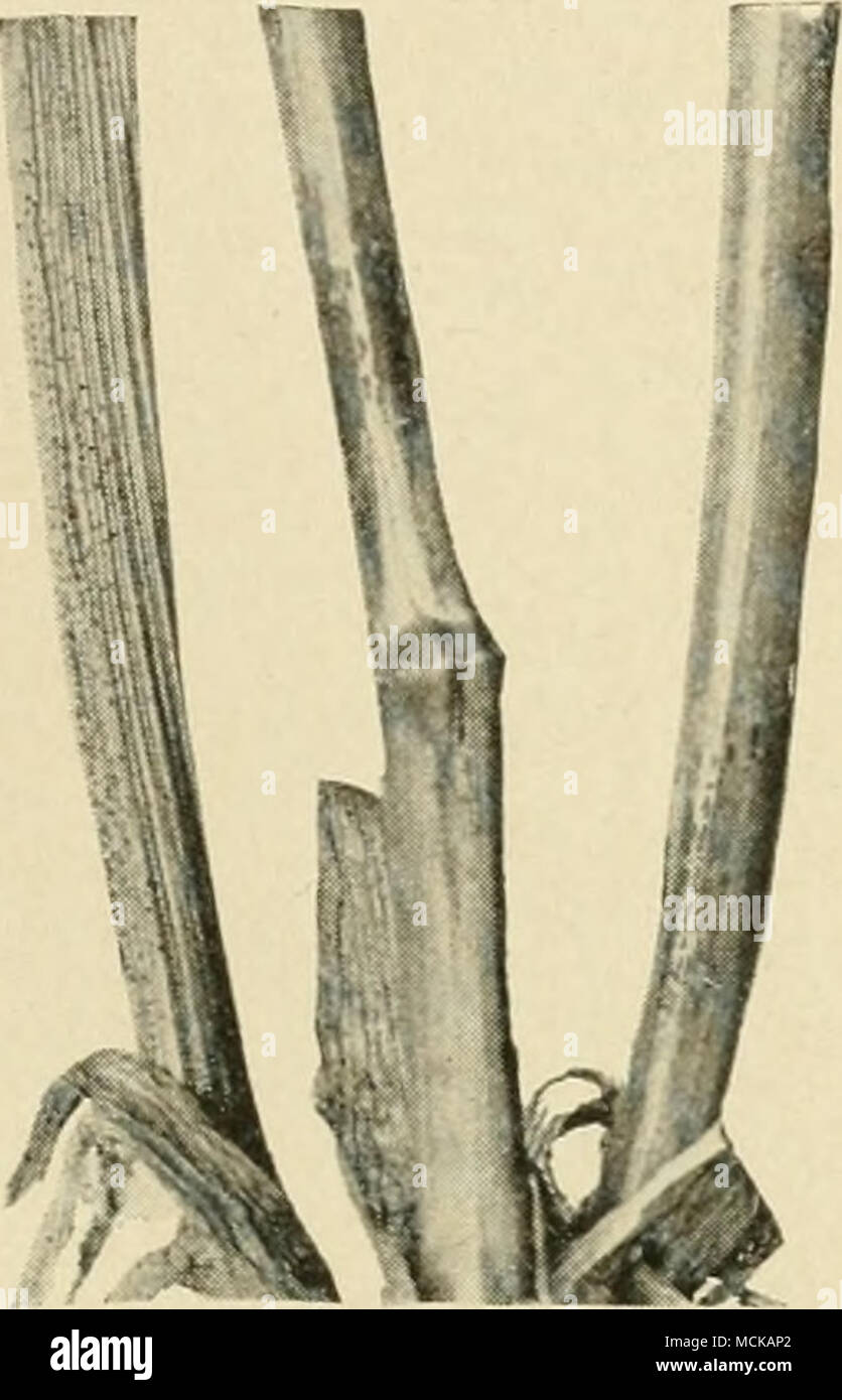 . Fig. loO. — Basal portion of rye plant showing anthrac- nose upon stem and leaf sheath. After Manna. ^^0 «rf Fig. 157. — Normal rye kernels and shriveled ones due to anthraonose. After Manns. Stock Photohttps://www.alamy.com/image-license-details/?v=1https://www.alamy.com/fig-loo-basal-portion-of-rye-plant-showing-anthrac-nose-upon-stem-and-leaf-sheath-after-manna-0-rf-fig-157-normal-rye-kernels-and-shriveled-ones-due-to-anthraonose-after-manns-image179905098.html
RMMCKAP2–. Fig. loO. — Basal portion of rye plant showing anthrac- nose upon stem and leaf sheath. After Manna. ^^0 «rf Fig. 157. — Normal rye kernels and shriveled ones due to anthraonose. After Manns.
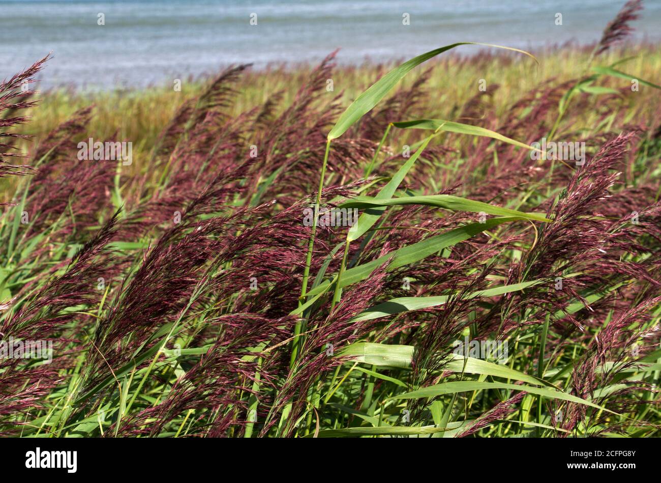 An adaptable and highly productive native of the North American Prairies, Blackwell Switchgrass has been used in the UK to control erosion Stock Photohttps://www.alamy.com/image-license-details/?v=1https://www.alamy.com/an-adaptable-and-highly-productive-native-of-the-north-american-prairies-blackwell-switchgrass-has-been-used-in-the-uk-to-control-erosion-image371133307.html
RM2CFPG8Y–An adaptable and highly productive native of the North American Prairies, Blackwell Switchgrass has been used in the UK to control erosion
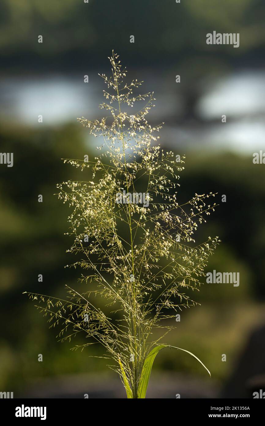 A tall and elegant grass, Buffalo or Tall Guinea Grass has an imposing delicate inflorescence. It can stand over 2m tall and is an important fodder Stock Photohttps://www.alamy.com/image-license-details/?v=1https://www.alamy.com/a-tall-and-elegant-grass-buffalo-or-tall-guinea-grass-has-an-imposing-delicate-inflorescence-it-can-stand-over-2m-tall-and-is-an-important-fodder-image482574914.html
RM2K1356A–A tall and elegant grass, Buffalo or Tall Guinea Grass has an imposing delicate inflorescence. It can stand over 2m tall and is an important fodder
 A solitary Phragmites reed bends in the flow of the rising flood waters of the Great Ruaha River during the rainy season. Stock Photohttps://www.alamy.com/image-license-details/?v=1https://www.alamy.com/a-solitary-phragmites-reed-bends-in-the-flow-of-the-rising-flood-waters-of-the-great-ruaha-river-during-the-rainy-season-image419061155.html
RM2F9NTNR–A solitary Phragmites reed bends in the flow of the rising flood waters of the Great Ruaha River during the rainy season.
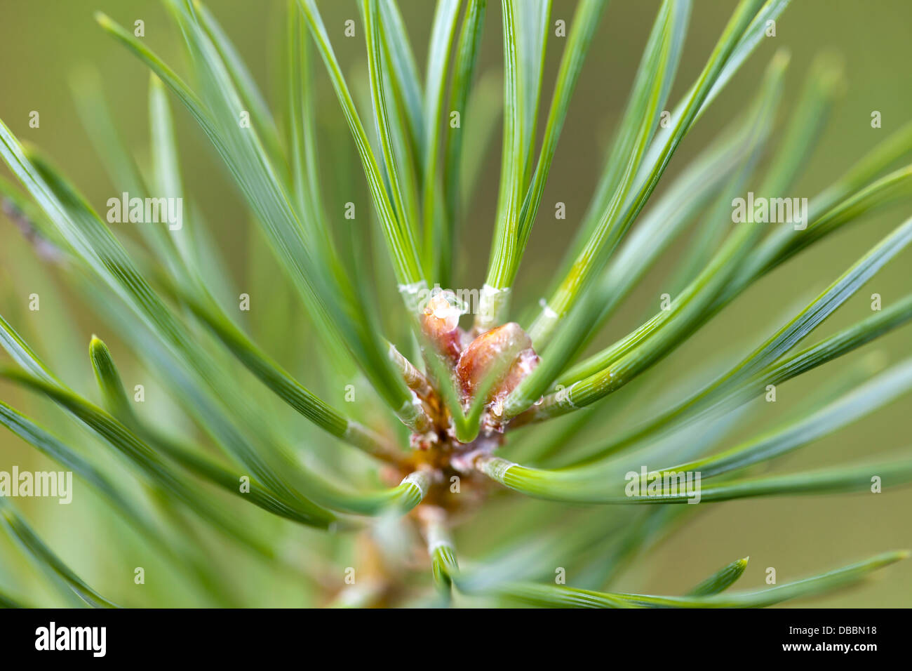 Terminal bud of a Pine sapling growing in Lentiira, Kuhmo, Finland. Stock Photohttps://www.alamy.com/image-license-details/?v=1https://www.alamy.com/stock-photo-terminal-bud-of-a-pine-sapling-growing-in-lentiira-kuhmo-finland-58650292.html
RMDBBN18–Terminal bud of a Pine sapling growing in Lentiira, Kuhmo, Finland.
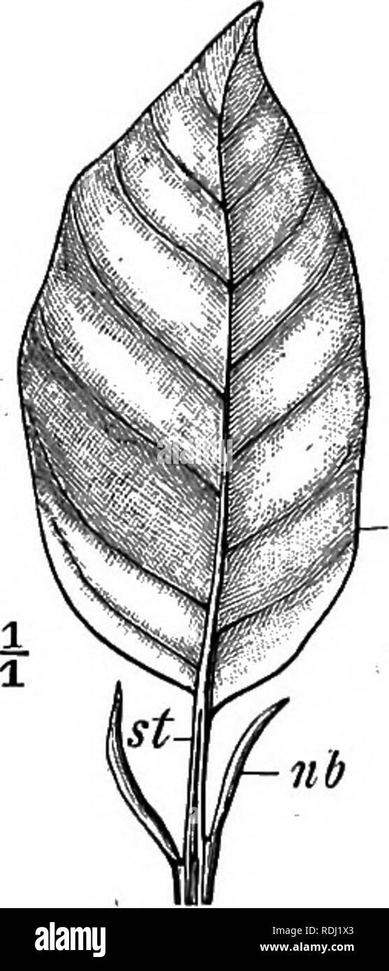 . Elementary botany . Botany. FOLIAGE-LEAVES IS â expanded and flattened basal sheath, by which the leaf is attached to the stem. The sheath frequently bears two lateral outgrowths known as the stipules {nb). (i.) THE SHEATH. The sheath of a leaf may be well developed {e.g. Butter- cup, Carrot, Cowparsnip), but frequently it is not distin- guishable. The stipules (fig. 17 nb) usually take the form of two flattened expansions of the leaf-sheath. They are parts of a single leaf A leaf possessing stipules is described as stipulate; a leaf devoid of stipules is said to be exstipulate. Most frequen Stock Photohttps://www.alamy.com/image-license-details/?v=1https://www.alamy.com/elementary-botany-botany-foliage-leaves-is-expanded-and-flattened-basal-sheath-by-which-the-leaf-is-attached-to-the-stem-the-sheath-frequently-bears-two-lateral-outgrowths-known-as-the-stipules-nb-i-the-sheath-the-sheath-of-a-leaf-may-be-well-developed-eg-butter-cup-carrot-cowparsnip-but-frequently-it-is-not-distin-guishable-the-stipules-fig-17-nb-usually-take-the-form-of-two-flattened-expansions-of-the-leaf-sheath-they-are-parts-of-a-single-leaf-a-leaf-possessing-stipules-is-described-as-stipulate-a-leaf-devoid-of-stipules-is-said-to-be-exstipulate-most-frequen-image232121963.html
RMRDJ1X3–. Elementary botany . Botany. FOLIAGE-LEAVES IS â expanded and flattened basal sheath, by which the leaf is attached to the stem. The sheath frequently bears two lateral outgrowths known as the stipules {nb). (i.) THE SHEATH. The sheath of a leaf may be well developed {e.g. Butter- cup, Carrot, Cowparsnip), but frequently it is not distin- guishable. The stipules (fig. 17 nb) usually take the form of two flattened expansions of the leaf-sheath. They are parts of a single leaf A leaf possessing stipules is described as stipulate; a leaf devoid of stipules is said to be exstipulate. Most frequen
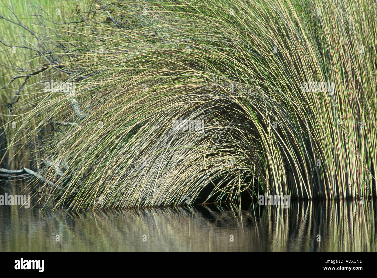 Riverine Grass, Okavango Stock Photohttps://www.alamy.com/image-license-details/?v=1https://www.alamy.com/riverine-grass-okavango-image7719516.html
RMADXGND–Riverine Grass, Okavango
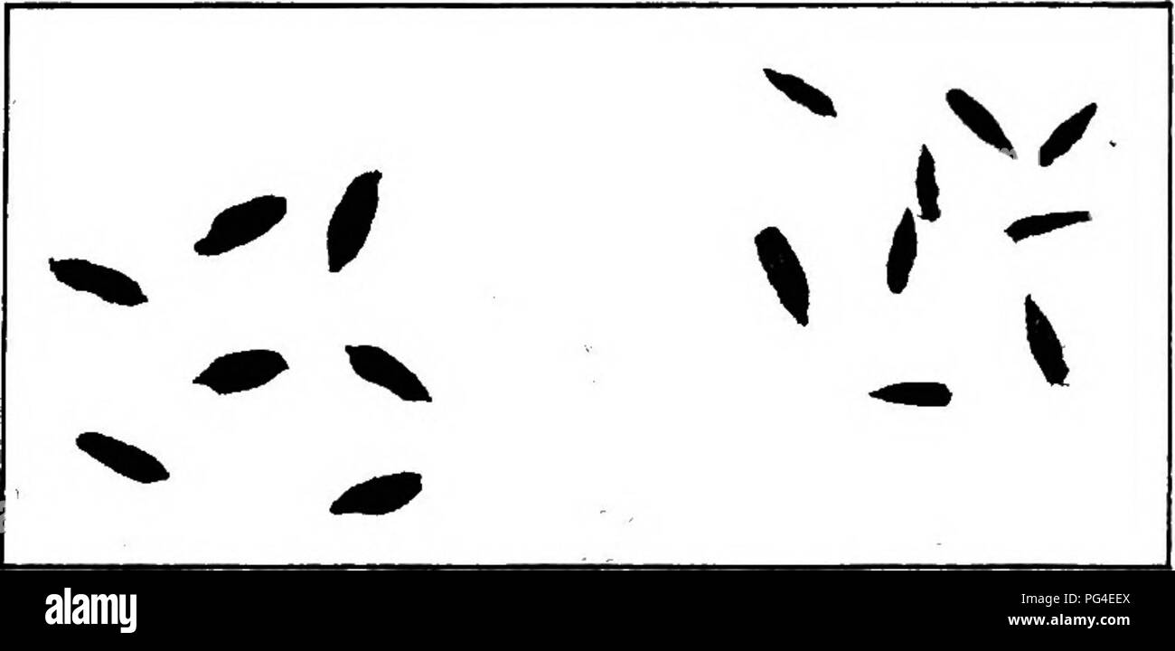 . Diseases of economic plants . Plant diseases. Fig. 156.—Basal portion of rye plant showing anthrac- nose upon stem and leaf sheath. After Manns.. FlQ. 157. — Normal rye kernels and shriveled ones due to antliracnoae. After Manns.. Please note that these images are extracted from scanned page images that may have been digitally enhanced for readability - coloration and appearance of these illustrations may not perfectly resemble the original work.. Stevens, Frank Lincoln, 1871-1934; Hall, John Galentine, 1870-. New York : Macmillan Stock Photohttps://www.alamy.com/image-license-details/?v=1https://www.alamy.com/diseases-of-economic-plants-plant-diseases-fig-156basal-portion-of-rye-plant-showing-anthrac-nose-upon-stem-and-leaf-sheath-after-manns-flq-157-normal-rye-kernels-and-shriveled-ones-due-to-antliracnoae-after-manns-please-note-that-these-images-are-extracted-from-scanned-page-images-that-may-have-been-digitally-enhanced-for-readability-coloration-and-appearance-of-these-illustrations-may-not-perfectly-resemble-the-original-work-stevens-frank-lincoln-1871-1934-hall-john-galentine-1870-new-york-macmillan-image216458114.html
RMPG4EEX–. Diseases of economic plants . Plant diseases. Fig. 156.—Basal portion of rye plant showing anthrac- nose upon stem and leaf sheath. After Manns.. FlQ. 157. — Normal rye kernels and shriveled ones due to antliracnoae. After Manns.. Please note that these images are extracted from scanned page images that may have been digitally enhanced for readability - coloration and appearance of these illustrations may not perfectly resemble the original work.. Stevens, Frank Lincoln, 1871-1934; Hall, John Galentine, 1870-. New York : Macmillan
 Grass seed heads Stock Photohttps://www.alamy.com/image-license-details/?v=1https://www.alamy.com/grass-seed-heads-image4169905.html
RMAB4GB2–Grass seed heads
 . 100 V phot.c.b.c. n.chit.ep. Fig. 29. Longitudinal section through the fifth thoracic Hmb of Systellaspis debilis, to show the structure of the photophore on the propodus. The haemocoele is indicated by mechanical stippling. Mallory's triple stain, chit, chitin; cut. cuticle; f.s. fibrous sheath; m.l. muscles of the limb; n.chit.ep. nucleus of chitogenous epithelium; n.phot.c. nucleus of photogenic cell; phot.c.b.c. basal cap of photogenic cell; phot.c.cl.a. clear area of photogenic cell. 8-2 Stock Photohttps://www.alamy.com/image-license-details/?v=1https://www.alamy.com/100-v-photcbc-nchitep-fig-29-longitudinal-section-through-the-fifth-thoracic-hmb-of-systellaspis-debilis-to-show-the-structure-of-the-photophore-on-the-propodus-the-haemocoele-is-indicated-by-mechanical-stippling-mallorys-triple-stain-chit-chitin-cut-cuticle-fs-fibrous-sheath-ml-muscles-of-the-limb-nchitep-nucleus-of-chitogenous-epithelium-nphotc-nucleus-of-photogenic-cell-photcbc-basal-cap-of-photogenic-cell-photccla-clear-area-of-photogenic-cell-8-2-image179997851.html
RMMCRH2K–. 100 V phot.c.b.c. n.chit.ep. Fig. 29. Longitudinal section through the fifth thoracic Hmb of Systellaspis debilis, to show the structure of the photophore on the propodus. The haemocoele is indicated by mechanical stippling. Mallory's triple stain, chit, chitin; cut. cuticle; f.s. fibrous sheath; m.l. muscles of the limb; n.chit.ep. nucleus of chitogenous epithelium; n.phot.c. nucleus of photogenic cell; phot.c.b.c. basal cap of photogenic cell; phot.c.cl.a. clear area of photogenic cell. 8-2
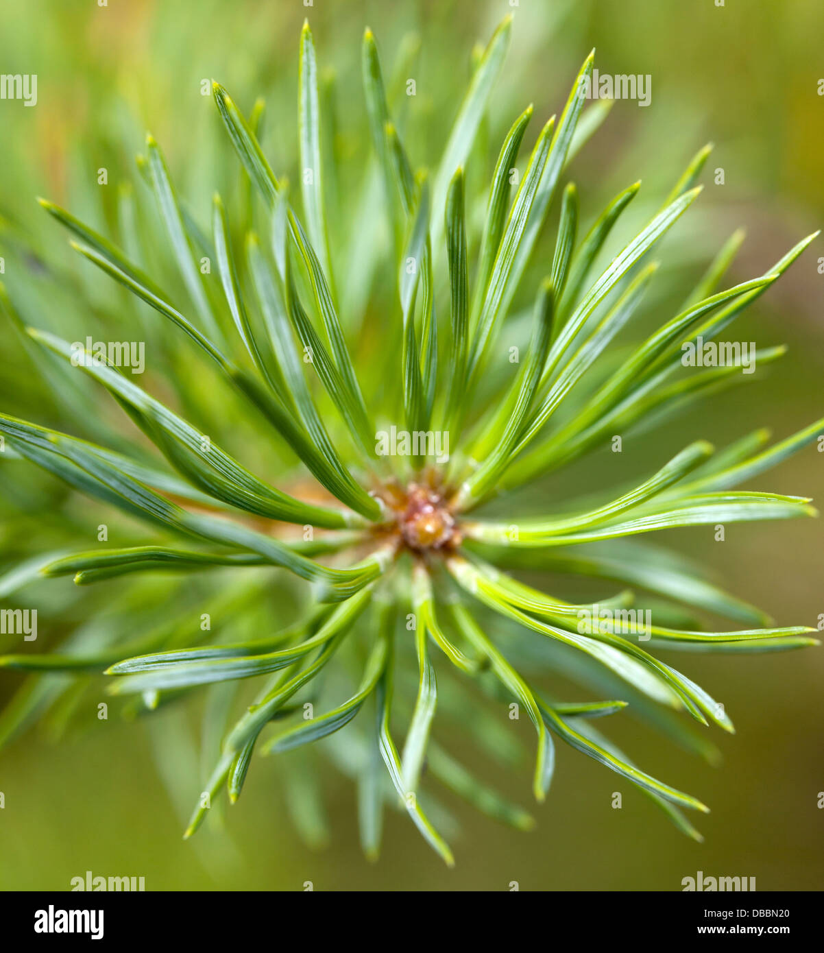 Terminal bud of a Pine sapling growing in Lentiira, Kuhmo, Finland. Stock Photohttps://www.alamy.com/image-license-details/?v=1https://www.alamy.com/stock-photo-terminal-bud-of-a-pine-sapling-growing-in-lentiira-kuhmo-finland-58650312.html
RMDBBN20–Terminal bud of a Pine sapling growing in Lentiira, Kuhmo, Finland.
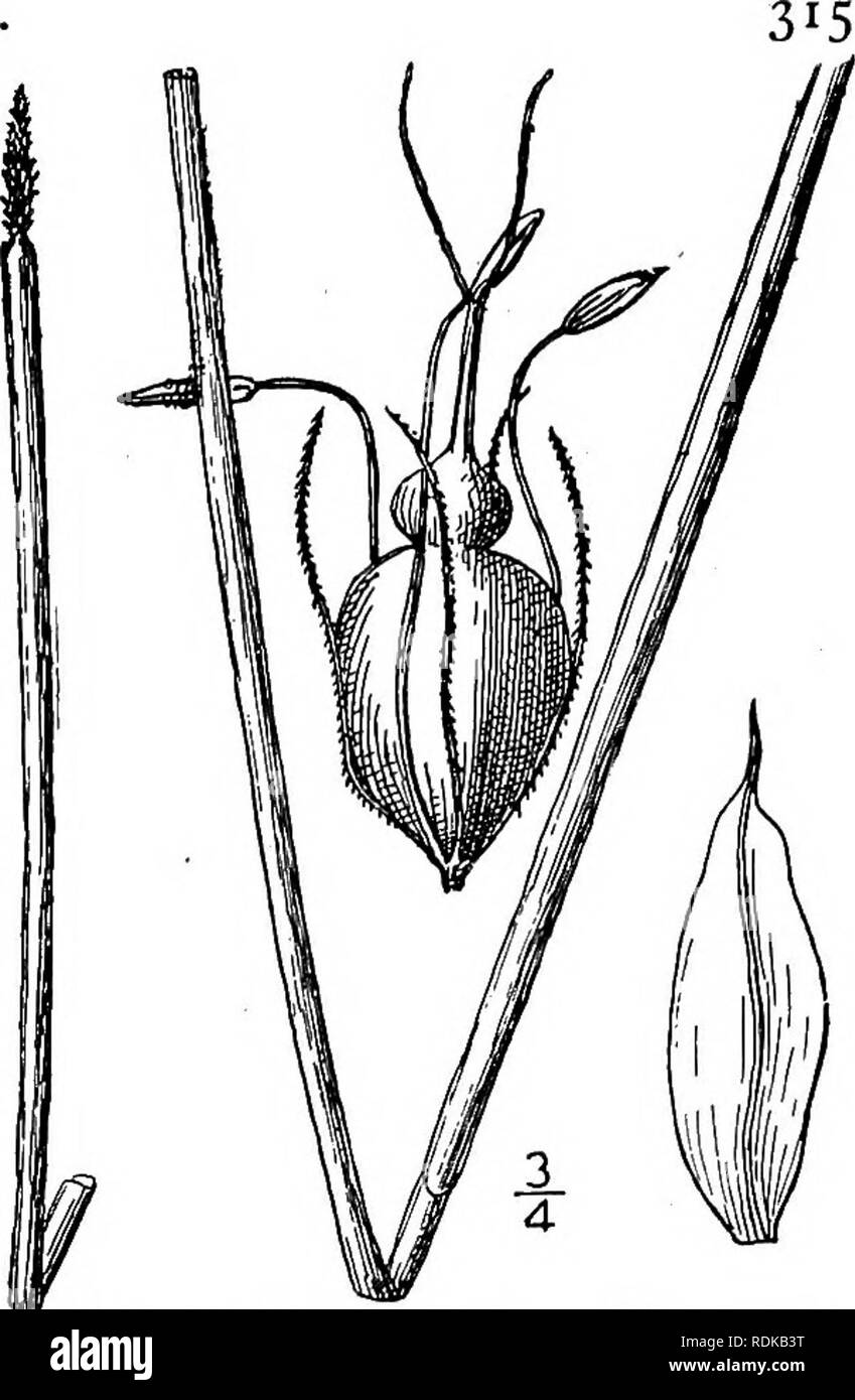 . An illustrated flora of the northern United States, Canada and the British possessions, from Newfoundland to the parallel of the southern boundary of Virginia, and from the Atlantic Ocean westward to the 102d meridian. Botany; Botany. Genus 3. SEDGE FAMILY. 13. Eleocharis Smallii Britton. Small's Spike- rush. Fig. 770. E. Smallii Britton, Torreya 3 : 23. 1903. Perennial by rootstocks; culms rather stout, about 2° high, and i"-ii" thick; top of the basal sheath ob- lique ; spikelet cylindric to conic-cylindric, acute, about 8" long, about as thick as the culm; scales lanceolate Stock Photohttps://www.alamy.com/image-license-details/?v=1https://www.alamy.com/an-illustrated-flora-of-the-northern-united-states-canada-and-the-british-possessions-from-newfoundland-to-the-parallel-of-the-southern-boundary-of-virginia-and-from-the-atlantic-ocean-westward-to-the-102d-meridian-botany-botany-genus-3-sedge-family-13-eleocharis-smallii-britton-smalls-spike-rush-fig-770-e-smallii-britton-torreya-3-23-1903-perennial-by-rootstocks-culms-rather-stout-about-2-high-and-iquot-iiquot-thick-top-of-the-basal-sheath-ob-lique-spikelet-cylindric-to-conic-cylindric-acute-about-8quot-long-about-as-thick-as-the-culm-scales-lanceolate-image232151132.html
RMRDKB3T–. An illustrated flora of the northern United States, Canada and the British possessions, from Newfoundland to the parallel of the southern boundary of Virginia, and from the Atlantic Ocean westward to the 102d meridian. Botany; Botany. Genus 3. SEDGE FAMILY. 13. Eleocharis Smallii Britton. Small's Spike- rush. Fig. 770. E. Smallii Britton, Torreya 3 : 23. 1903. Perennial by rootstocks; culms rather stout, about 2° high, and i"-ii" thick; top of the basal sheath ob- lique ; spikelet cylindric to conic-cylindric, acute, about 8" long, about as thick as the culm; scales lanceolate
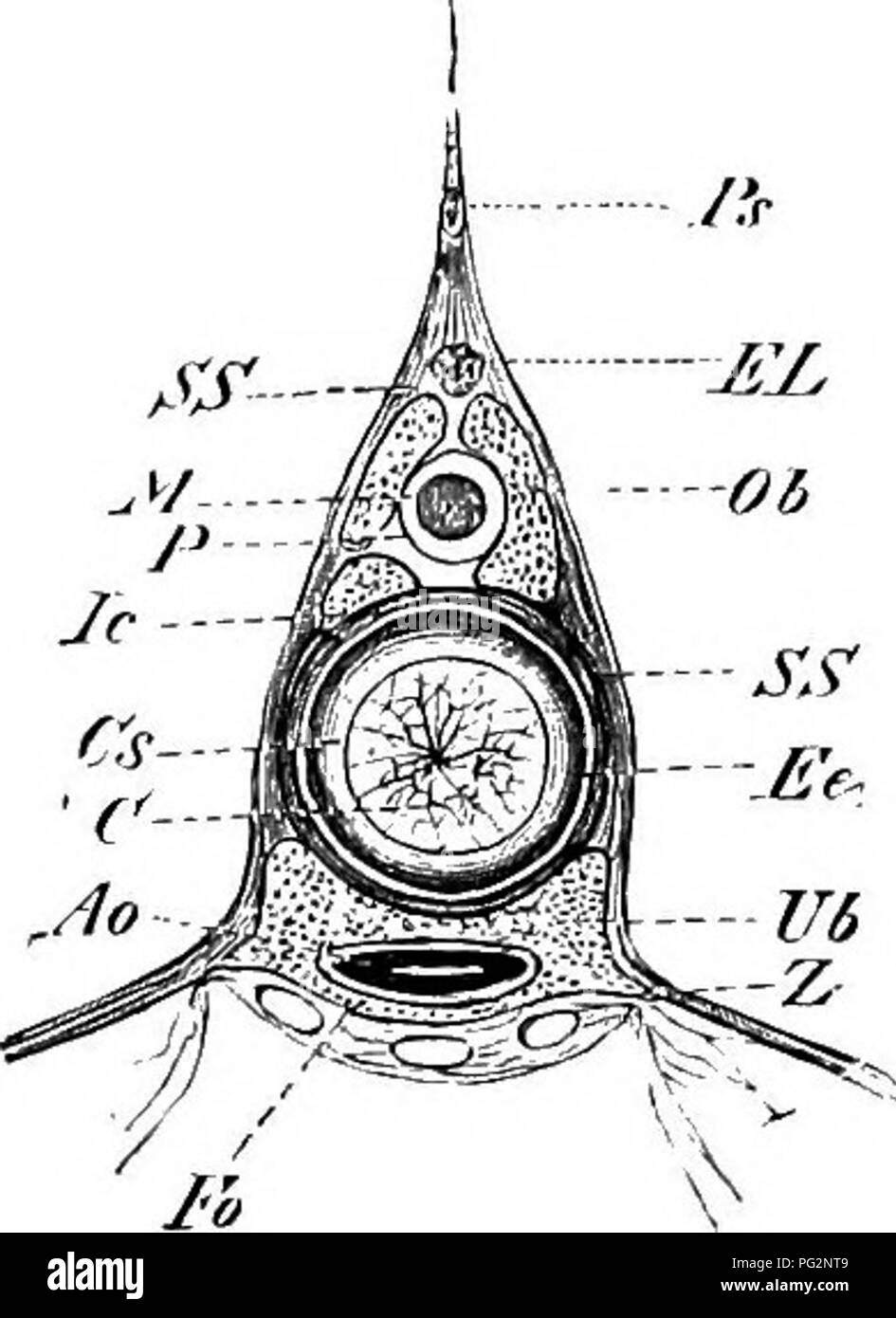 . Elements of the comparative anatomy of vertebrates. Anatomy, Comparative. Fill. 2'ii.—Portion op the Vertebral L'olumx of ,b>'(<H/V(/7V(. (Side view.) Fig. 24.—Transverse Section of the Vertebral Colujin of Aciptuv-r rutheiiux (in the anterior part of the bod}'). Pn, spinous process ; J, lowei' arch ; Ao, aorta ; Po, median parts of the lower arches, which here enclcie the aorta ventrally ; Z, basal processes of the lower arches. is essentially indicated by the neural arches. In the two groups last mentioned, however, skeletogenous cells break through the primary notochordal sheath (el Stock Photohttps://www.alamy.com/image-license-details/?v=1https://www.alamy.com/elements-of-the-comparative-anatomy-of-vertebrates-anatomy-comparative-fill-2iiportion-op-the-vertebral-lolumx-of-bgtlthv7v-side-view-fig-24transverse-section-of-the-vertebral-colujin-of-aciptuv-r-rutheiiux-in-the-anterior-part-of-the-bod-pn-spinous-process-j-lowei-arch-ao-aorta-po-median-parts-of-the-lower-arches-which-here-enclcie-the-aorta-ventrally-z-basal-processes-of-the-lower-arches-is-essentially-indicated-by-the-neural-arches-in-the-two-groups-last-mentioned-however-skeletogenous-cells-break-through-the-primary-notochordal-sheath-el-image216419961.html
RMPG2NT9–. Elements of the comparative anatomy of vertebrates. Anatomy, Comparative. Fill. 2'ii.—Portion op the Vertebral L'olumx of ,b>'(<H/V(/7V(. (Side view.) Fig. 24.—Transverse Section of the Vertebral Colujin of Aciptuv-r rutheiiux (in the anterior part of the bod}'). Pn, spinous process ; J, lowei' arch ; Ao, aorta ; Po, median parts of the lower arches, which here enclcie the aorta ventrally ; Z, basal processes of the lower arches. is essentially indicated by the neural arches. In the two groups last mentioned, however, skeletogenous cells break through the primary notochordal sheath (el
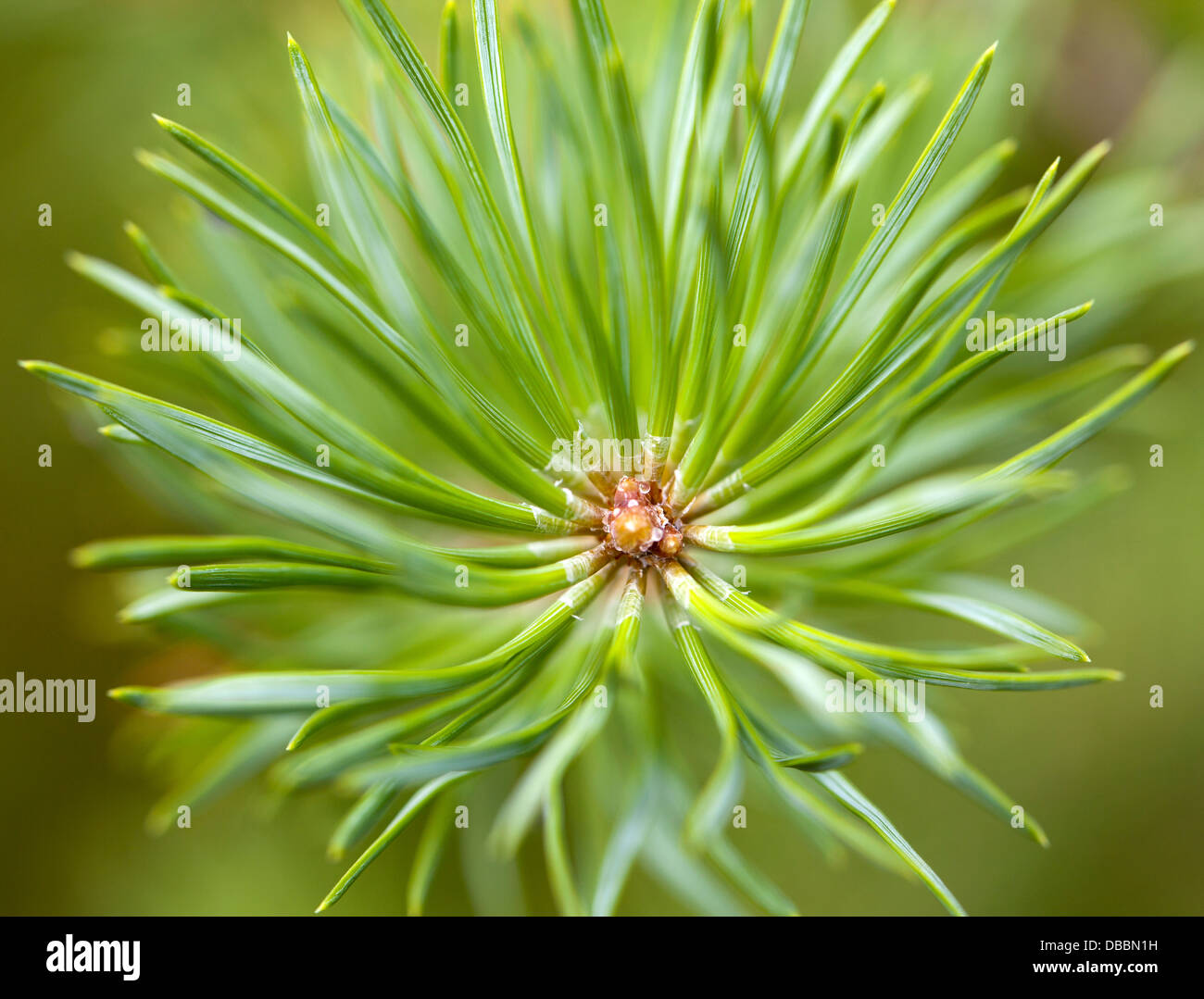 Terminal bud of a Pine sapling growing in Lentiira, Kuhmo, Finland. Stock Photohttps://www.alamy.com/image-license-details/?v=1https://www.alamy.com/stock-photo-terminal-bud-of-a-pine-sapling-growing-in-lentiira-kuhmo-finland-58650301.html
RMDBBN1H–Terminal bud of a Pine sapling growing in Lentiira, Kuhmo, Finland.
 . Comparative morphology of Fungi. Fungi. EUASCOMYCETES 187 of HC1 and pepsin corresponding to the gastric juice. Immature asco- spores and gemmae germinate without this stimulation (Ward, 1899; Brierley, 1917). Trichocomaceae.—The Trichocomaceae are a poorly known family of the warmer regions. Trichocoma paradoxa (E. Fischer, 1890) grows on dead wood and forms fructifications up 1 cm. in diameter and 2 cm. high. Out of a woody-brown, patelliform, basal sheath, resting on the substrate, there arises a more or less columnar tissue. It consists of a system of faveolate tubular chambers which run Stock Photohttps://www.alamy.com/image-license-details/?v=1https://www.alamy.com/comparative-morphology-of-fungi-fungi-euascomycetes-187-of-hc1-and-pepsin-corresponding-to-the-gastric-juice-immature-asco-spores-and-gemmae-germinate-without-this-stimulation-ward-1899-brierley-1917-trichocomaceaethe-trichocomaceae-are-a-poorly-known-family-of-the-warmer-regions-trichocoma-paradoxa-e-fischer-1890-grows-on-dead-wood-and-forms-fructifications-up-1-cm-in-diameter-and-2-cm-high-out-of-a-woody-brown-patelliform-basal-sheath-resting-on-the-substrate-there-arises-a-more-or-less-columnar-tissue-it-consists-of-a-system-of-faveolate-tubular-chambers-which-run-image232676003.html
RMREF8H7–. Comparative morphology of Fungi. Fungi. EUASCOMYCETES 187 of HC1 and pepsin corresponding to the gastric juice. Immature asco- spores and gemmae germinate without this stimulation (Ward, 1899; Brierley, 1917). Trichocomaceae.—The Trichocomaceae are a poorly known family of the warmer regions. Trichocoma paradoxa (E. Fischer, 1890) grows on dead wood and forms fructifications up 1 cm. in diameter and 2 cm. high. Out of a woody-brown, patelliform, basal sheath, resting on the substrate, there arises a more or less columnar tissue. It consists of a system of faveolate tubular chambers which run
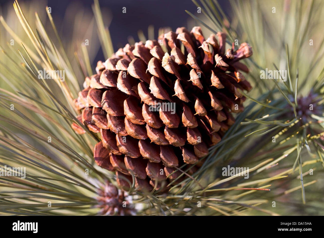 Closeup of a female pine cone at Montecito Sequoia Lodge, Sequoia National Park, California, USA. Stock Photohttps://www.alamy.com/image-license-details/?v=1https://www.alamy.com/stock-photo-closeup-of-a-female-pine-cone-at-montecito-sequoia-lodge-sequoia-national-57804022.html
RMDA15HA–Closeup of a female pine cone at Montecito Sequoia Lodge, Sequoia National Park, California, USA.
 . Animal parasites and human disease. Medical parasitology; Insects as carriers of disease. Fig. 150. Head or capitulum of tick; hyp., hypostome; chel., chelicera; pal., palpus; has. p., basal piece. (Partly after Banks.). Fig. 151. Tip of chelicera of a tick, much enlarged; cut. p., articulated cutting part; shaft, shaft; sh., sheath; fl. t., tendon of flexor muscle; ex. t., tendon of extensor muscle. (After Nuttall, Cooper and Robinson.). Please note that these images are extracted from scanned page images that may have been digitally enhanced for readability - coloration and appearance of t Stock Photohttps://www.alamy.com/image-license-details/?v=1https://www.alamy.com/animal-parasites-and-human-disease-medical-parasitology-insects-as-carriers-of-disease-fig-150-head-or-capitulum-of-tick-hyp-hypostome-chel-chelicera-pal-palpus-has-p-basal-piece-partly-after-banks-fig-151-tip-of-chelicera-of-a-tick-much-enlarged-cut-p-articulated-cutting-part-shaft-shaft-sh-sheath-fl-t-tendon-of-flexor-muscle-ex-t-tendon-of-extensor-muscle-after-nuttall-cooper-and-robinson-please-note-that-these-images-are-extracted-from-scanned-page-images-that-may-have-been-digitally-enhanced-for-readability-coloration-and-appearance-of-t-image216373942.html
RMPG0K4P–. Animal parasites and human disease. Medical parasitology; Insects as carriers of disease. Fig. 150. Head or capitulum of tick; hyp., hypostome; chel., chelicera; pal., palpus; has. p., basal piece. (Partly after Banks.). Fig. 151. Tip of chelicera of a tick, much enlarged; cut. p., articulated cutting part; shaft, shaft; sh., sheath; fl. t., tendon of flexor muscle; ex. t., tendon of extensor muscle. (After Nuttall, Cooper and Robinson.). Please note that these images are extracted from scanned page images that may have been digitally enhanced for readability - coloration and appearance of t
 . Elementary botany . Botany. Fig. 224.—Floral diagram of Orchis, AEACE.S (Arum Family) Smooth herbs with leaves which are often broad and net- veined. Inflorescence a spadix with a spathe; no bracts subtending the separa,te flowers; no prophylls in the inflores- cence. Flowers small, inconspicuous. Perianth small or absent. Fruit, a berry. Type : THE CUCKOO PINT (Arum maculaUcm). Vegetative characters.—Herb with a corm. The leaves are radical, each possessing a basal sheath, a petiole, and a net-veined spotted lamina shaped almost like an arrow-head. Inflorescence (fig. 226).-—A large sheathi Stock Photohttps://www.alamy.com/image-license-details/?v=1https://www.alamy.com/elementary-botany-botany-fig-224floral-diagram-of-orchis-aeaces-arum-family-smooth-herbs-with-leaves-which-are-often-broad-and-net-veined-inflorescence-a-spadix-with-a-spathe-no-bracts-subtending-the-separate-flowers-no-prophylls-in-the-inflores-cence-flowers-small-inconspicuous-perianth-small-or-absent-fruit-a-berry-type-the-cuckoo-pint-arum-maculaucm-vegetative-charactersherb-with-a-corm-the-leaves-are-radical-each-possessing-a-basal-sheath-a-petiole-and-a-net-veined-spotted-lamina-shaped-almost-like-an-arrow-head-inflorescence-fig-226-a-large-sheathi-image232114546.html
RMRDHMD6–. Elementary botany . Botany. Fig. 224.—Floral diagram of Orchis, AEACE.S (Arum Family) Smooth herbs with leaves which are often broad and net- veined. Inflorescence a spadix with a spathe; no bracts subtending the separa,te flowers; no prophylls in the inflores- cence. Flowers small, inconspicuous. Perianth small or absent. Fruit, a berry. Type : THE CUCKOO PINT (Arum maculaUcm). Vegetative characters.—Herb with a corm. The leaves are radical, each possessing a basal sheath, a petiole, and a net-veined spotted lamina shaped almost like an arrow-head. Inflorescence (fig. 226).-—A large sheathi
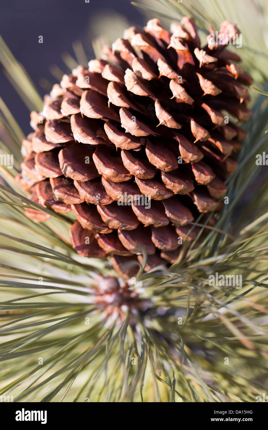 Closeup of a female pine cone at Montecito Sequoia Lodge, Sequoia National Park, California, USA. Stock Photohttps://www.alamy.com/image-license-details/?v=1https://www.alamy.com/stock-photo-closeup-of-a-female-pine-cone-at-montecito-sequoia-lodge-sequoia-national-57804028.html
RMDA15HG–Closeup of a female pine cone at Montecito Sequoia Lodge, Sequoia National Park, California, USA.
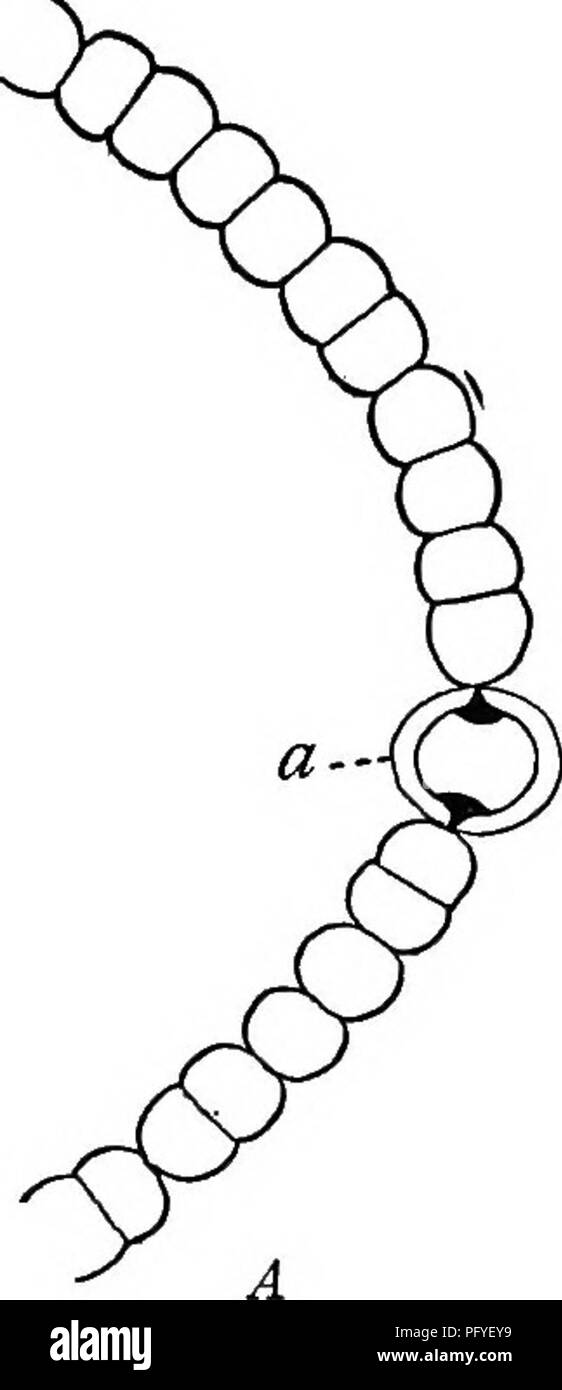 . A text-book of botany for secondary schools. Botany. Fig. 93.—A, Noatoc: showing the chain-like fila- ment and a heterocyst Co) ; B, Glaotrichia: showing mucilage sheath, basal heterocyst, and tapering apex. grows as large as the parent cell and then divides in turn. The mucilaginous walls hold the cells together, and so they are found in groups of various sizes (Fig. 92). This method of reproduction by cell- division is the simplest kind of reproduction. 59. Nostoc.—These plants occur in jelly- like masses in damp places. If the jelly be examined, it will be found to contain em- bedded in i Stock Photohttps://www.alamy.com/image-license-details/?v=1https://www.alamy.com/a-text-book-of-botany-for-secondary-schools-botany-fig-93a-noatoc-showing-the-chain-like-fila-ment-and-a-heterocyst-co-b-glaotrichia-showing-mucilage-sheath-basal-heterocyst-and-tapering-apex-grows-as-large-as-the-parent-cell-and-then-divides-in-turn-the-mucilaginous-walls-hold-the-cells-together-and-so-they-are-found-in-groups-of-various-sizes-fig-92-this-method-of-reproduction-by-cell-division-is-the-simplest-kind-of-reproduction-59-nostocthese-plants-occur-in-jelly-like-masses-in-damp-places-if-the-jelly-be-examined-it-will-be-found-to-contain-em-bedded-in-i-image216348701.html
RMPFYEY9–. A text-book of botany for secondary schools. Botany. Fig. 93.—A, Noatoc: showing the chain-like fila- ment and a heterocyst Co) ; B, Glaotrichia: showing mucilage sheath, basal heterocyst, and tapering apex. grows as large as the parent cell and then divides in turn. The mucilaginous walls hold the cells together, and so they are found in groups of various sizes (Fig. 92). This method of reproduction by cell- division is the simplest kind of reproduction. 59. Nostoc.—These plants occur in jelly- like masses in damp places. If the jelly be examined, it will be found to contain em- bedded in i
![. Elementary botany [microform]. Botany; Botanique. Fiff. 224. —Flcjral diasram of Orchis. Plff. 225. —Fruit of Orchis iiiascula : b = subtending bract; j = seeds. i '; ARACEJE (Arum Family) Smooth herbs with leaves which are often broad and net- venied Inflorescence a spadix with a .sj^athe: no bracts subtending the separate flowers; no prophylls in the inflores- cence. Howers small, inconspicuous. Perianth small or absent. Fruit, a berry. Type THE CUCKOO PINT {Arum maculatum). Vegetative characters.—Herb with a corm. Th- leaves are radical, each possessing a basal sheath, a petiole, and a ne Stock Photo . Elementary botany [microform]. Botany; Botanique. Fiff. 224. —Flcjral diasram of Orchis. Plff. 225. —Fruit of Orchis iiiascula : b = subtending bract; j = seeds. i '; ARACEJE (Arum Family) Smooth herbs with leaves which are often broad and net- venied Inflorescence a spadix with a .sj^athe: no bracts subtending the separate flowers; no prophylls in the inflores- cence. Howers small, inconspicuous. Perianth small or absent. Fruit, a berry. Type THE CUCKOO PINT {Arum maculatum). Vegetative characters.—Herb with a corm. Th- leaves are radical, each possessing a basal sheath, a petiole, and a ne Stock Photo](https://c8.alamy.com/comp/REMRCA/elementary-botany-microform-botany-botanique-fiff-224-flcjral-diasram-of-orchis-plff-225-fruit-of-orchis-iiiascula-b-=-subtending-bract-j-=-seeds-i-araceje-arum-family-smooth-herbs-with-leaves-which-are-often-broad-and-net-venied-inflorescence-a-spadix-with-a-sjathe-no-bracts-subtending-the-separate-flowers-no-prophylls-in-the-inflores-cence-howers-small-inconspicuous-perianth-small-or-absent-fruit-a-berry-type-the-cuckoo-pint-arum-maculatum-vegetative-charactersherb-with-a-corm-th-leaves-are-radical-each-possessing-a-basal-sheath-a-petiole-and-a-ne-REMRCA.jpg) . Elementary botany [microform]. Botany; Botanique. Fiff. 224. —Flcjral diasram of Orchis. Plff. 225. —Fruit of Orchis iiiascula : b = subtending bract; j = seeds. i '; ARACEJE (Arum Family) Smooth herbs with leaves which are often broad and net- venied Inflorescence a spadix with a .sj^athe: no bracts subtending the separate flowers; no prophylls in the inflores- cence. Howers small, inconspicuous. Perianth small or absent. Fruit, a berry. Type THE CUCKOO PINT {Arum maculatum). Vegetative characters.—Herb with a corm. Th- leaves are radical, each possessing a basal sheath, a petiole, and a ne Stock Photohttps://www.alamy.com/image-license-details/?v=1https://www.alamy.com/elementary-botany-microform-botany-botanique-fiff-224-flcjral-diasram-of-orchis-plff-225-fruit-of-orchis-iiiascula-b-=-subtending-bract-j-=-seeds-i-araceje-arum-family-smooth-herbs-with-leaves-which-are-often-broad-and-net-venied-inflorescence-a-spadix-with-a-sjathe-no-bracts-subtending-the-separate-flowers-no-prophylls-in-the-inflores-cence-howers-small-inconspicuous-perianth-small-or-absent-fruit-a-berry-type-the-cuckoo-pint-arum-maculatum-vegetative-charactersherb-with-a-corm-th-leaves-are-radical-each-possessing-a-basal-sheath-a-petiole-and-a-ne-image232797386.html
. Elementary botany [microform]. Botany; Botanique. Fiff. 224. —Flcjral diasram of Orchis. Plff. 225. —Fruit of Orchis iiiascula : b = subtending bract; j = seeds. i '; ARACEJE (Arum Family) Smooth herbs with leaves which are often broad and net- venied Inflorescence a spadix with a .sj^athe: no bracts subtending the separate flowers; no prophylls in the inflores- cence. Howers small, inconspicuous. Perianth small or absent. Fruit, a berry. Type THE CUCKOO PINT {Arum maculatum). Vegetative characters.—Herb with a corm. Th- leaves are radical, each possessing a basal sheath, a petiole, and a ne Stock Photohttps://www.alamy.com/image-license-details/?v=1https://www.alamy.com/elementary-botany-microform-botany-botanique-fiff-224-flcjral-diasram-of-orchis-plff-225-fruit-of-orchis-iiiascula-b-=-subtending-bract-j-=-seeds-i-araceje-arum-family-smooth-herbs-with-leaves-which-are-often-broad-and-net-venied-inflorescence-a-spadix-with-a-sjathe-no-bracts-subtending-the-separate-flowers-no-prophylls-in-the-inflores-cence-howers-small-inconspicuous-perianth-small-or-absent-fruit-a-berry-type-the-cuckoo-pint-arum-maculatum-vegetative-charactersherb-with-a-corm-th-leaves-are-radical-each-possessing-a-basal-sheath-a-petiole-and-a-ne-image232797386.htmlRMREMRCA–. Elementary botany [microform]. Botany; Botanique. Fiff. 224. —Flcjral diasram of Orchis. Plff. 225. —Fruit of Orchis iiiascula : b = subtending bract; j = seeds. i '; ARACEJE (Arum Family) Smooth herbs with leaves which are often broad and net- venied Inflorescence a spadix with a .sj^athe: no bracts subtending the separate flowers; no prophylls in the inflores- cence. Howers small, inconspicuous. Perianth small or absent. Fruit, a berry. Type THE CUCKOO PINT {Arum maculatum). Vegetative characters.—Herb with a corm. Th- leaves are radical, each possessing a basal sheath, a petiole, and a ne
 . Handbook of flower pollination : based upon Hermann Mu?ller's work 'The fertilisation of flowers by insects' . Fertilization of plants. Fig. 66. Head of humbh-bec iafter Herm. Muller). (i) Hcaii of Bombus agroram F. ?, with completely extended and widely separated mouth-parts; seen from above (x 5). (2) Mouth.parts of the honey-bee in the same condition; seen from below (x 12). //', The two basal joints of the labial palps, which are modified to form part of the ligular sheath ; w, the membranous lappets at the tip of the ligula; x^ the piece which cocrs the mouth-opening, which lies betwee Stock Photohttps://www.alamy.com/image-license-details/?v=1https://www.alamy.com/handbook-of-flower-pollination-based-upon-hermann-mullers-work-the-fertilisation-of-flowers-by-insects-fertilization-of-plants-fig-66-head-of-humbh-bec-iafter-herm-muller-i-hcaii-of-bombus-agroram-f-with-completely-extended-and-widely-separated-mouth-parts-seen-from-above-x-5-2-mouthparts-of-the-honey-bee-in-the-same-condition-seen-from-below-x-12-the-two-basal-joints-of-the-labial-palps-which-are-modified-to-form-part-of-the-ligular-sheath-w-the-membranous-lappets-at-the-tip-of-the-ligula-x-the-piece-which-cocrs-the-mouth-opening-which-lies-betwee-image216409589.html
RMPG28HW–. Handbook of flower pollination : based upon Hermann Mu?ller's work 'The fertilisation of flowers by insects' . Fertilization of plants. Fig. 66. Head of humbh-bec iafter Herm. Muller). (i) Hcaii of Bombus agroram F. ?, with completely extended and widely separated mouth-parts; seen from above (x 5). (2) Mouth.parts of the honey-bee in the same condition; seen from below (x 12). //', The two basal joints of the labial palps, which are modified to form part of the ligular sheath ; w, the membranous lappets at the tip of the ligula; x^ the piece which cocrs the mouth-opening, which lies betwee
 . Annual report. New York State Museum; Science; Science. NEW YORK STATE MUSEUM. Fig. 388-93 Climacograptus par- vus Hall. Fig. 388 Type of species in Amer. Mus. Nat. Hist. Fig. 389 Sicular end of specimen showing the basal sheath of virgella. Fig. 390 Portion of rhabdo- some showing subscalariform view. Fig. 391 Perfect rhabdosome with "vesicle." Fig. 3C2 Copy of Lapworth's drawing of a Stockport specimen. Fig. 393 Rhabdo- some which is partly infiltrated with pyrite and preserved in relief. All en- larged x 5, except figure 392, which is enlarged x kl/2. See also text figures 20, 2 Stock Photohttps://www.alamy.com/image-license-details/?v=1https://www.alamy.com/annual-report-new-york-state-museum-science-science-new-york-state-museum-fig-388-93-climacograptus-par-vus-hall-fig-388-type-of-species-in-amer-mus-nat-hist-fig-389-sicular-end-of-specimen-showing-the-basal-sheath-of-virgella-fig-390-portion-of-rhabdo-some-showing-subscalariform-view-fig-391-perfect-rhabdosome-with-quotvesiclequot-fig-3c2-copy-of-lapworths-drawing-of-a-stockport-specimen-fig-393-rhabdo-some-which-is-partly-infiltrated-with-pyrite-and-preserved-in-relief-all-en-larged-x-5-except-figure-392-which-is-enlarged-x-kl2-see-also-text-figures-20-2-image236222583.html
RMRM8T8R–. Annual report. New York State Museum; Science; Science. NEW YORK STATE MUSEUM. Fig. 388-93 Climacograptus par- vus Hall. Fig. 388 Type of species in Amer. Mus. Nat. Hist. Fig. 389 Sicular end of specimen showing the basal sheath of virgella. Fig. 390 Portion of rhabdo- some showing subscalariform view. Fig. 391 Perfect rhabdosome with "vesicle." Fig. 3C2 Copy of Lapworth's drawing of a Stockport specimen. Fig. 393 Rhabdo- some which is partly infiltrated with pyrite and preserved in relief. All en- larged x 5, except figure 392, which is enlarged x kl/2. See also text figures 20, 2
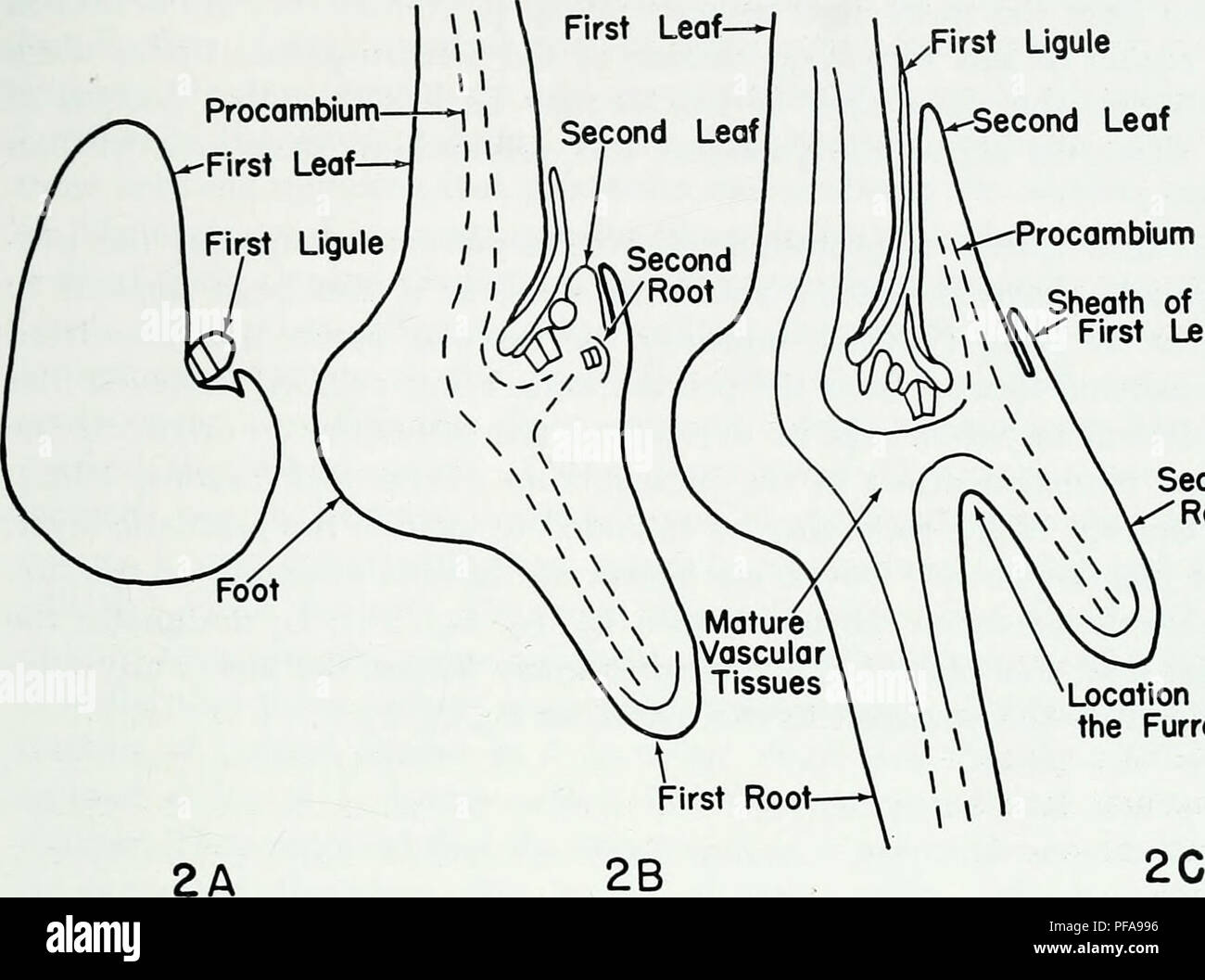 . The developmental anatomy of Isoetes. Isoetes; Botany. TERMINOLOGY First Ligule. Second Leaf ^y>—Procambium « Sheath of the " First Leaf Second Root Location of the Furrow Fig. 2. Three stages in the development of a young sporophyte. A. One leaf. B. Two leaves. C. Three leaves. The plane of sectioning in A, B, C is the same plane that contains all the leaf traces while the plant is in a ¥2 phyllotaxy. The basal furrow forms between the first and second roots in a plane at right angles of the plane of sectioning of the figures. Schematic. TERMINOLOGY Parke (1959) had discussed t Stock Photohttps://www.alamy.com/image-license-details/?v=1https://www.alamy.com/the-developmental-anatomy-of-isoetes-isoetes-botany-terminology-first-ligule-second-leaf-ygtprocambium-sheath-of-the-quot-first-leaf-second-root-location-of-the-furrow-fig-2-three-stages-in-the-development-of-a-young-sporophyte-a-one-leaf-b-two-leaves-c-three-leaves-the-plane-of-sectioning-in-a-b-c-is-the-same-plane-that-contains-all-the-leaf-traces-while-the-plant-is-in-a-2-phyllotaxy-the-basal-furrow-forms-between-the-first-and-second-roots-in-a-plane-at-right-angles-of-the-plane-of-sectioning-of-the-figures-schematic-terminology-parke-1959-had-discussed-t-image215971090.html
RMPFA996–. The developmental anatomy of Isoetes. Isoetes; Botany. TERMINOLOGY First Ligule. Second Leaf ^y>—Procambium « Sheath of the " First Leaf Second Root Location of the Furrow Fig. 2. Three stages in the development of a young sporophyte. A. One leaf. B. Two leaves. C. Three leaves. The plane of sectioning in A, B, C is the same plane that contains all the leaf traces while the plant is in a ¥2 phyllotaxy. The basal furrow forms between the first and second roots in a plane at right angles of the plane of sectioning of the figures. Schematic. TERMINOLOGY Parke (1959) had discussed t
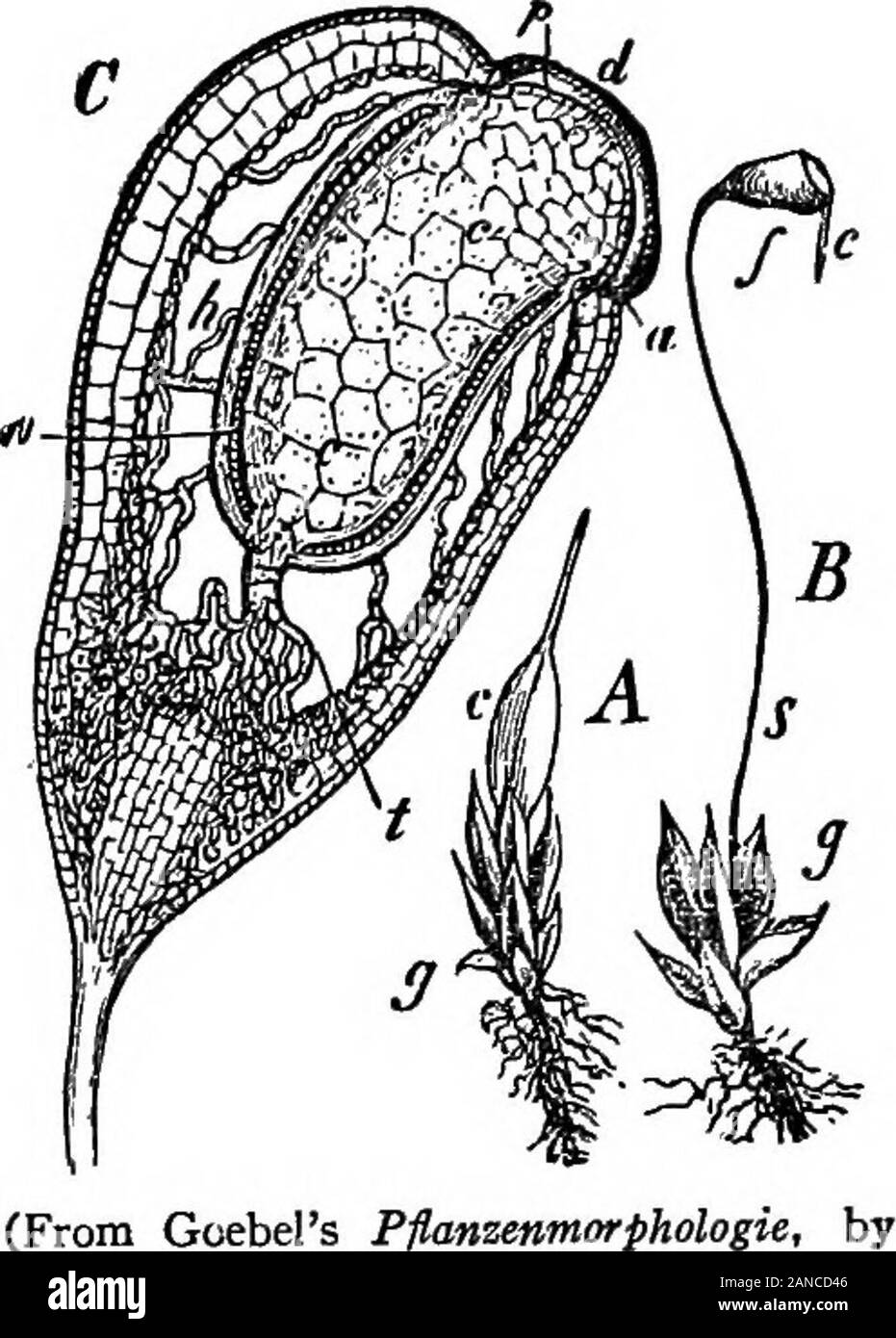 The encyclopdia britannica; a dictionary of arts, sciences, literature and general information . o-sided apical cell (fig. 16 A), and is at first of uniform thickness.After a time the upper region increases in diameter and forms the capsule, while the lower portion forms the long seta and thefoot which is embedded in the end of the stem. With the growthof the sporogonium the archegonial wall, which for a time keptpace with it, is broken through,the larger upper part terminatedby the neck being carried upon the capsule as the cal)rptra.^while the basal portion remainsas a tubular sheath round t Stock Photohttps://www.alamy.com/image-license-details/?v=1https://www.alamy.com/the-encyclopdia-britannica-a-dictionary-of-arts-sciences-literature-and-general-information-o-sided-apical-cell-fig-16-a-and-is-at-first-of-uniform-thicknessafter-a-time-the-upper-region-increases-in-diameter-and-forms-the-capsule-while-the-lower-portion-forms-the-long-seta-and-thefoot-which-is-embedded-in-the-end-of-the-stem-with-the-growthof-the-sporogonium-the-archegonial-wall-which-for-a-time-keptpace-with-it-is-broken-throughthe-larger-upper-part-terminatedby-the-neck-being-carried-upon-the-capsule-as-the-calrptrawhile-the-basal-portion-remainsas-a-tubular-sheath-round-t-image340178502.html
RM2ANCD46–The encyclopdia britannica; a dictionary of arts, sciences, literature and general information . o-sided apical cell (fig. 16 A), and is at first of uniform thickness.After a time the upper region increases in diameter and forms the capsule, while the lower portion forms the long seta and thefoot which is embedded in the end of the stem. With the growthof the sporogonium the archegonial wall, which for a time keptpace with it, is broken through,the larger upper part terminatedby the neck being carried upon the capsule as the cal)rptra.^while the basal portion remainsas a tubular sheath round t
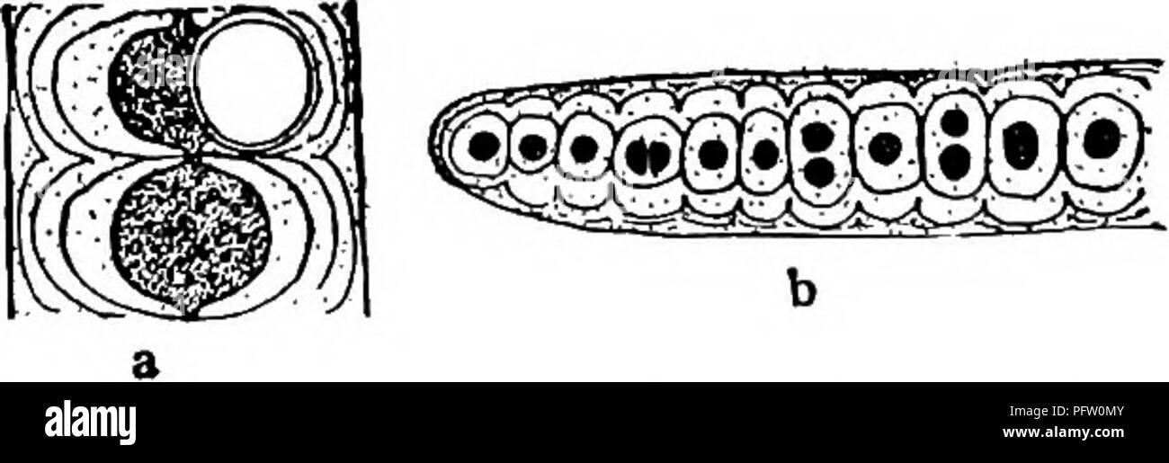 . Fresh-water biology. Freshwater biology. 59 iSSt 56) With basal heterocysts. each sheath. . Two to several filaments enclosed in Desmonema Berkeley and Thwaites. Filaments sometimes slightly branched; heterocysts always basal. On stones, in brooks, and waterfalls. Fig. 61. Desmonema wrangelii Borzi. X 200. (After Borzi.) 60 (54) Filaments usually stout, bearing true branches; cells rounded, dis- posed generally in more than one row; heterocysts present. Family Stigonemaceae . 61 5i (62,63) Sheaths thick; firm SHgonema Agardh. Filaments free-floating or aggregated on the substratum to form le Stock Photohttps://www.alamy.com/image-license-details/?v=1https://www.alamy.com/fresh-water-biology-freshwater-biology-59-isst-56-with-basal-heterocysts-each-sheath-two-to-several-filaments-enclosed-in-desmonema-berkeley-and-thwaites-filaments-sometimes-slightly-branched-heterocysts-always-basal-on-stones-in-brooks-and-waterfalls-fig-61-desmonema-wrangelii-borzi-x-200-after-borzi-60-54-filaments-usually-stout-bearing-true-branches-cells-rounded-dis-posed-generally-in-more-than-one-row-heterocysts-present-family-stigonemaceae-61-5i-6263-sheaths-thick-firm-shgonema-agardh-filaments-free-floating-or-aggregated-on-the-substratum-to-form-le-image216293643.html
RMPFW0MY–. Fresh-water biology. Freshwater biology. 59 iSSt 56) With basal heterocysts. each sheath. . Two to several filaments enclosed in Desmonema Berkeley and Thwaites. Filaments sometimes slightly branched; heterocysts always basal. On stones, in brooks, and waterfalls. Fig. 61. Desmonema wrangelii Borzi. X 200. (After Borzi.) 60 (54) Filaments usually stout, bearing true branches; cells rounded, dis- posed generally in more than one row; heterocysts present. Family Stigonemaceae . 61 5i (62,63) Sheaths thick; firm SHgonema Agardh. Filaments free-floating or aggregated on the substratum to form le
 Text-book of botany, morphological and physiological . erior of the basal tissue of the leaf-sheath, upon radii of the stem which alter-nate with the fibro-vascular bundles, and thus also with the teeth of the sheath.A root may arise beneath the bud of each branch; both break through the leaf-sheath at its base. All the joints of the axis agree in these respects, however theymay be modified as underground rhizomes, tubers, ascending stems, leafy branches,or sporangiferous axes. EQUISETACE^. 36: The end of the stem enveloped by a large number of younger leaf-sheathsterminates in a large apical Stock Photohttps://www.alamy.com/image-license-details/?v=1https://www.alamy.com/text-book-of-botany-morphological-and-physiological-erior-of-the-basal-tissue-of-the-leaf-sheath-upon-radii-of-the-stem-which-alter-nate-with-the-fibro-vascular-bundles-and-thus-also-with-the-teeth-of-the-sheatha-root-may-arise-beneath-the-bud-of-each-branch-both-break-through-the-leaf-sheath-at-its-base-all-the-joints-of-the-axis-agree-in-these-respects-however-theymay-be-modified-as-underground-rhizomes-tubers-ascending-stems-leafy-branchesor-sporangiferous-axes-equisetace-36-the-end-of-the-stem-enveloped-by-a-large-number-of-younger-leaf-sheathsterminates-in-a-large-apical-image342915275.html
RM2AWW3X3–Text-book of botany, morphological and physiological . erior of the basal tissue of the leaf-sheath, upon radii of the stem which alter-nate with the fibro-vascular bundles, and thus also with the teeth of the sheath.A root may arise beneath the bud of each branch; both break through the leaf-sheath at its base. All the joints of the axis agree in these respects, however theymay be modified as underground rhizomes, tubers, ascending stems, leafy branches,or sporangiferous axes. EQUISETACE^. 36: The end of the stem enveloped by a large number of younger leaf-sheathsterminates in a large apical
![. A manual of zoology. Zoology. 400 ARTHROron.V with a tn The slipc outer. Of in tiiany I bears the the most pcndau'cs parts of t basal pan the subiih an.i^ular joint, the cardo (eV whieh is followed by a larger stipes (si). s in turn siqiports two chewini,' lobes, the inner, or hh'iiiui (li), and an i,M/ej (Ic). I'he galea niav either form a .sheath for the laeinia, or, as eetles ^Iil;. 470), it tnav be taeiile and jointed again. The stipes also iiuixUhirv piilpiis (['III), eonsisting of from three to six joints, and is leg-like ]Kn of the appendage. The labium arises as a pair of ap- wiiieh Stock Photo . A manual of zoology. Zoology. 400 ARTHROron.V with a tn The slipc outer. Of in tiiany I bears the the most pcndau'cs parts of t basal pan the subiih an.i^ular joint, the cardo (eV whieh is followed by a larger stipes (si). s in turn siqiports two chewini,' lobes, the inner, or hh'iiiui (li), and an i,M/ej (Ic). I'he galea niav either form a .sheath for the laeinia, or, as eetles ^Iil;. 470), it tnav be taeiile and jointed again. The stipes also iiuixUhirv piilpiis (['III), eonsisting of from three to six joints, and is leg-like ]Kn of the appendage. The labium arises as a pair of ap- wiiieh Stock Photo](https://c8.alamy.com/comp/PG3NY7/a-manual-of-zoology-zoology-400-arthroronv-with-a-tn-the-slipc-outer-of-in-tiiany-i-bears-the-the-most-pcndaucs-parts-of-t-basal-pan-the-subiih-aniular-joint-the-cardo-ev-whieh-is-followed-by-a-larger-stipes-si-s-in-turn-siqiports-two-chewini-lobes-the-inner-or-hhiiiui-li-and-an-imej-ic-ihe-galea-niav-either-form-a-sheath-for-the-laeinia-or-as-eetles-iil-470-it-tnav-be-taeiile-and-jointed-again-the-stipes-also-iiuixuhirv-piilpiis-iii-eonsisting-of-from-three-to-six-joints-and-is-leg-like-kn-of-the-appendage-the-labium-arises-as-a-pair-of-ap-wiiieh-PG3NY7.jpg) . A manual of zoology. Zoology. 400 ARTHROron.V with a tn The slipc outer. Of in tiiany I bears the the most pcndau'cs parts of t basal pan the subiih an.i^ular joint, the cardo (eV whieh is followed by a larger stipes (si). s in turn siqiports two chewini,' lobes, the inner, or hh'iiiui (li), and an i,M/ej (Ic). I'he galea niav either form a .sheath for the laeinia, or, as eetles ^Iil;. 470), it tnav be taeiile and jointed again. The stipes also iiuixUhirv piilpiis (['III), eonsisting of from three to six joints, and is leg-like ]Kn of the appendage. The labium arises as a pair of ap- wiiieh Stock Photohttps://www.alamy.com/image-license-details/?v=1https://www.alamy.com/a-manual-of-zoology-zoology-400-arthroronv-with-a-tn-the-slipc-outer-of-in-tiiany-i-bears-the-the-most-pcndaucs-parts-of-t-basal-pan-the-subiih-aniular-joint-the-cardo-ev-whieh-is-followed-by-a-larger-stipes-si-s-in-turn-siqiports-two-chewini-lobes-the-inner-or-hhiiiui-li-and-an-imej-ic-ihe-galea-niav-either-form-a-sheath-for-the-laeinia-or-as-eetles-iil-470-it-tnav-be-taeiile-and-jointed-again-the-stipes-also-iiuixuhirv-piilpiis-iii-eonsisting-of-from-three-to-six-joints-and-is-leg-like-kn-of-the-appendage-the-labium-arises-as-a-pair-of-ap-wiiieh-image216441995.html
. A manual of zoology. Zoology. 400 ARTHROron.V with a tn The slipc outer. Of in tiiany I bears the the most pcndau'cs parts of t basal pan the subiih an.i^ular joint, the cardo (eV whieh is followed by a larger stipes (si). s in turn siqiports two chewini,' lobes, the inner, or hh'iiiui (li), and an i,M/ej (Ic). I'he galea niav either form a .sheath for the laeinia, or, as eetles ^Iil;. 470), it tnav be taeiile and jointed again. The stipes also iiuixUhirv piilpiis (['III), eonsisting of from three to six joints, and is leg-like ]Kn of the appendage. The labium arises as a pair of ap- wiiieh Stock Photohttps://www.alamy.com/image-license-details/?v=1https://www.alamy.com/a-manual-of-zoology-zoology-400-arthroronv-with-a-tn-the-slipc-outer-of-in-tiiany-i-bears-the-the-most-pcndaucs-parts-of-t-basal-pan-the-subiih-aniular-joint-the-cardo-ev-whieh-is-followed-by-a-larger-stipes-si-s-in-turn-siqiports-two-chewini-lobes-the-inner-or-hhiiiui-li-and-an-imej-ic-ihe-galea-niav-either-form-a-sheath-for-the-laeinia-or-as-eetles-iil-470-it-tnav-be-taeiile-and-jointed-again-the-stipes-also-iiuixuhirv-piilpiis-iii-eonsisting-of-from-three-to-six-joints-and-is-leg-like-kn-of-the-appendage-the-labium-arises-as-a-pair-of-ap-wiiieh-image216441995.htmlRMPG3NY7–. A manual of zoology. Zoology. 400 ARTHROron.V with a tn The slipc outer. Of in tiiany I bears the the most pcndau'cs parts of t basal pan the subiih an.i^ular joint, the cardo (eV whieh is followed by a larger stipes (si). s in turn siqiports two chewini,' lobes, the inner, or hh'iiiui (li), and an i,M/ej (Ic). I'he galea niav either form a .sheath for the laeinia, or, as eetles ^Iil;. 470), it tnav be taeiile and jointed again. The stipes also iiuixUhirv piilpiis (['III), eonsisting of from three to six joints, and is leg-like ]Kn of the appendage. The labium arises as a pair of ap- wiiieh
 Nature and development of plants . into an upper part or pileus and a basal region,the stalk or stipe. As this growth proceeds the mass of hyphaeextending from the margin of the pileus to the stipe becomedrawn out and ultimately form a rather thin membrane knownas the velum (Fig. 166, 2, vl). This entire growth is oftenenveloped by a sheath-like mass of hyphae, the volva (Fig. 167,C, D). At this stage of development, the young mushroom israther spherical in shape and so small that it is quite concealedin the ground or by surrounding vegetation. When conditionsare favorable for further growth, Stock Photohttps://www.alamy.com/image-license-details/?v=1https://www.alamy.com/nature-and-development-of-plants-into-an-upper-part-or-pileus-and-a-basal-regionthe-stalk-or-stipe-as-this-growth-proceeds-the-mass-of-hyphaeextending-from-the-margin-of-the-pileus-to-the-stipe-becomedrawn-out-and-ultimately-form-a-rather-thin-membrane-knownas-the-velum-fig-166-2-vl-this-entire-growth-is-oftenenveloped-by-a-sheath-like-mass-of-hyphae-the-volva-fig-167c-d-at-this-stage-of-development-the-young-mushroom-israther-spherical-in-shape-and-so-small-that-it-is-quite-concealedin-the-ground-or-by-surrounding-vegetation-when-conditionsare-favorable-for-further-growth-image343360324.html
RM2AXHBGM–Nature and development of plants . into an upper part or pileus and a basal region,the stalk or stipe. As this growth proceeds the mass of hyphaeextending from the margin of the pileus to the stipe becomedrawn out and ultimately form a rather thin membrane knownas the velum (Fig. 166, 2, vl). This entire growth is oftenenveloped by a sheath-like mass of hyphae, the volva (Fig. 167,C, D). At this stage of development, the young mushroom israther spherical in shape and so small that it is quite concealedin the ground or by surrounding vegetation. When conditionsare favorable for further growth,
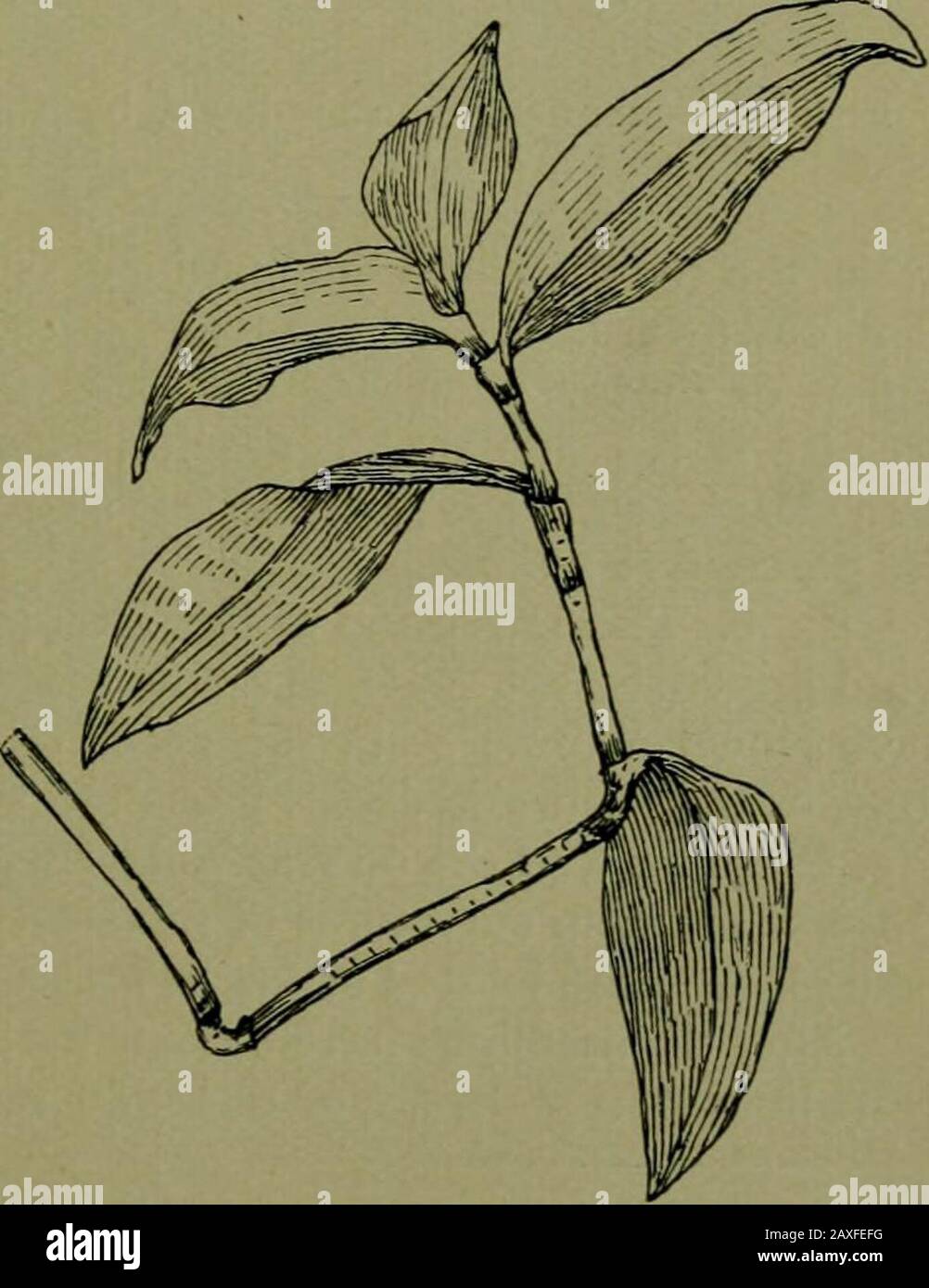 Plants and their ways in South Africa . Fig, 26.—Showing basal growth in Zea mays. needle, and strip from the sheath to expose the base of the leaf within. Mark off lines on the part exposed, carrying them across on to the sheath. When growth has taken place, so that the marks appear above the sheath, it will be seen that the place marked has been pushed up by the growth at the base of the leaf. Commelina ^ will be agood plant for studying thegrowth of a monocotyledon-ousstem. Split the sheathingbase of the leaf and mark thestem off. Mark other plants—Flagellaria, or any of thespecies oiAspara Stock Photohttps://www.alamy.com/image-license-details/?v=1https://www.alamy.com/plants-and-their-ways-in-south-africa-fig-26showing-basal-growth-in-zea-mays-needle-and-strip-from-the-sheath-to-expose-the-base-of-the-leaf-within-mark-off-lines-on-the-part-exposed-carrying-them-across-on-to-the-sheath-when-growth-has-taken-place-so-that-the-marks-appear-above-the-sheath-it-will-be-seen-that-the-place-marked-has-been-pushed-up-by-the-growth-at-the-base-of-the-leaf-commelina-will-be-agood-plant-for-studying-thegrowth-of-a-monocotyledon-ousstem-split-the-sheathingbase-of-the-leaf-and-mark-thestem-off-mark-other-plantsflagellaria-or-any-of-thespecies-oiaspara-image343318740.html
RM2AXFEFG–Plants and their ways in South Africa . Fig, 26.—Showing basal growth in Zea mays. needle, and strip from the sheath to expose the base of the leaf within. Mark off lines on the part exposed, carrying them across on to the sheath. When growth has taken place, so that the marks appear above the sheath, it will be seen that the place marked has been pushed up by the growth at the base of the leaf. Commelina ^ will be agood plant for studying thegrowth of a monocotyledon-ousstem. Split the sheathingbase of the leaf and mark thestem off. Mark other plants—Flagellaria, or any of thespecies oiAspara
 A natural history of British grasses . airy leaves, with theupper sheath more than three times the length of its leaf, andhaving a conspicuous membranous ligule; lower sheaths not solong as their leaves. Joints three or four. Inflorescence com-pound-racemed or simple-panicled. The basal spikelets situatedon lateral branches, whilst those near the apex are on brieffootstalks. Panicle upright. Calyx of two unequal, membra-nous, acute glumes, the basal one destitute of lateral ribs, andshorter than the upper one. Florets of two paleae, the exteriorone of basal floret membranous on the upper porti Stock Photohttps://www.alamy.com/image-license-details/?v=1https://www.alamy.com/a-natural-history-of-british-grasses-airy-leaves-with-theupper-sheath-more-than-three-times-the-length-of-its-leaf-andhaving-a-conspicuous-membranous-ligule-lower-sheaths-not-solong-as-their-leaves-joints-three-or-four-inflorescence-com-pound-racemed-or-simple-panicled-the-basal-spikelets-situatedon-lateral-branches-whilst-those-near-the-apex-are-on-brieffootstalks-panicle-upright-calyx-of-two-unequal-membra-nous-acute-glumes-the-basal-one-destitute-of-lateral-ribs-andshorter-than-the-upper-one-florets-of-two-paleae-the-exteriorone-of-basal-floret-membranous-on-the-upper-porti-image340163362.html
RM2ANBNRE–A natural history of British grasses . airy leaves, with theupper sheath more than three times the length of its leaf, andhaving a conspicuous membranous ligule; lower sheaths not solong as their leaves. Joints three or four. Inflorescence com-pound-racemed or simple-panicled. The basal spikelets situatedon lateral branches, whilst those near the apex are on brieffootstalks. Panicle upright. Calyx of two unequal, membra-nous, acute glumes, the basal one destitute of lateral ribs, andshorter than the upper one. Florets of two paleae, the exteriorone of basal floret membranous on the upper porti
 A natural history of British grasses . inted leaves, with striated sheaths, theupper sheath having an obtuse ragged ligule at its summit.Joints five. Inflorescence usually simple-panicled. Panicle whenyoung upright, when more mature pendant. Branches rough.Spikelets linear-lanceolate, brownish purple, mostly of ten awncdflorets. Calyx consisting of two almost equal, broad acuteglumes; margin membranous. Upper half of the keels dentate.Outer glume three-ribbed; inner glume seven-ribbed. Floretsof two nearly equal-sized palese, the exterior one of basal floretoval, rough, glossy, and somewhat lo Stock Photohttps://www.alamy.com/image-license-details/?v=1https://www.alamy.com/a-natural-history-of-british-grasses-inted-leaves-with-striated-sheaths-theupper-sheath-having-an-obtuse-ragged-ligule-at-its-summitjoints-five-inflorescence-usually-simple-panicled-panicle-whenyoung-upright-when-more-mature-pendant-branches-roughspikelets-linear-lanceolate-brownish-purple-mostly-of-ten-awncdflorets-calyx-consisting-of-two-almost-equal-broad-acuteglumes-margin-membranous-upper-half-of-the-keels-dentateouter-glume-three-ribbed-inner-glume-seven-ribbed-floretsof-two-nearly-equal-sized-palese-the-exterior-one-of-basal-floretoval-rough-glossy-and-somewhat-lo-image340165470.html
RM2ANBTEP–A natural history of British grasses . inted leaves, with striated sheaths, theupper sheath having an obtuse ragged ligule at its summit.Joints five. Inflorescence usually simple-panicled. Panicle whenyoung upright, when more mature pendant. Branches rough.Spikelets linear-lanceolate, brownish purple, mostly of ten awncdflorets. Calyx consisting of two almost equal, broad acuteglumes; margin membranous. Upper half of the keels dentate.Outer glume three-ribbed; inner glume seven-ribbed. Floretsof two nearly equal-sized palese, the exterior one of basal floretoval, rough, glossy, and somewhat lo
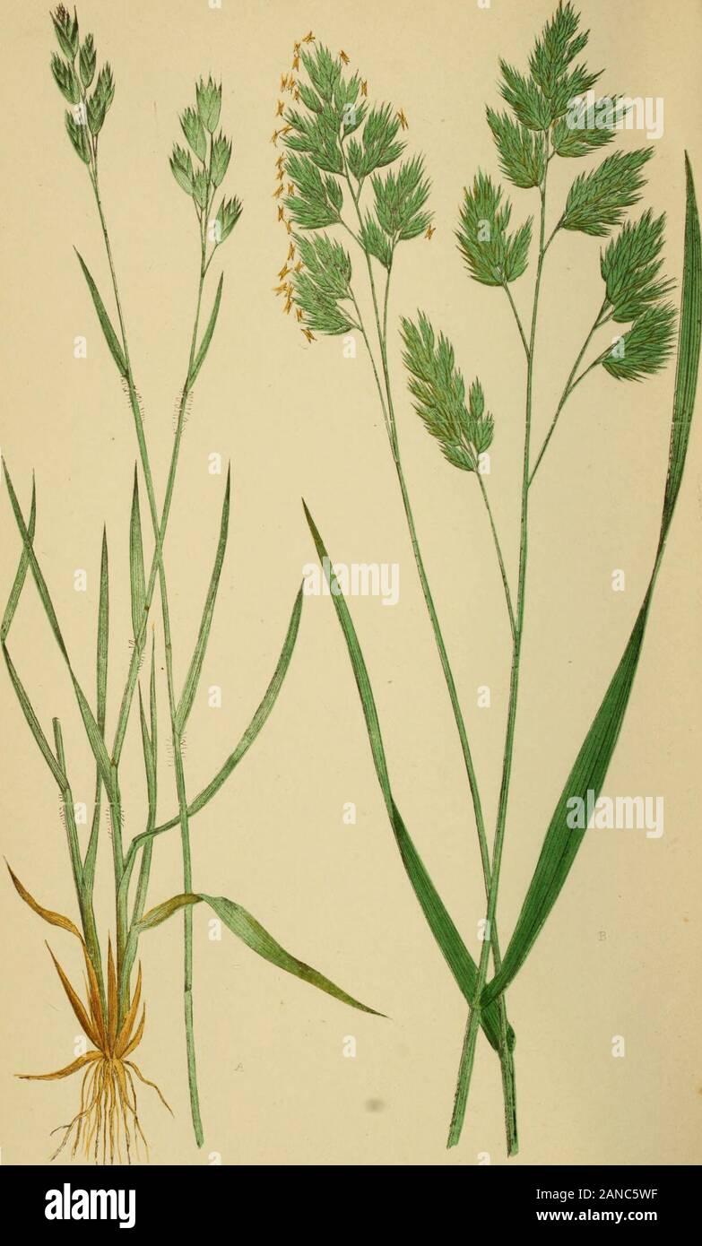 A natural history of British grasses . sheaths; upper sheath longer than its leaf, having athin membranous acute ligule at the apex. Inflorescence com-pound-panicled. Panicle upright, outline triangular and spreading.Branches smooth, mostly in pairs. Spikelets ovate-oblong,mostly of five to eight aAvnlcss florets, commonly tinged withgreen, white, and purple; apex of basal floret stretching beyondthe large glume of the calyx. Calyx of two unequal acuteglumes, three-ribbed, dorsal rib dentate above. Florets of two 130 POA ANNUA. palese, not webbed; exterior one of basal floret five-ribbed; ribs Stock Photohttps://www.alamy.com/image-license-details/?v=1https://www.alamy.com/a-natural-history-of-british-grasses-sheaths-upper-sheath-longer-than-its-leaf-having-athin-membranous-acute-ligule-at-the-apex-inflorescence-com-pound-panicled-panicle-upright-outline-triangular-and-spreadingbranches-smooth-mostly-in-pairs-spikelets-ovate-oblongmostly-of-five-to-eight-aavnlcss-florets-commonly-tinged-withgreen-white-and-purple-apex-of-basal-floret-stretching-beyondthe-large-glume-of-the-calyx-calyx-of-two-unequal-acuteglumes-three-ribbed-dorsal-rib-dentate-above-florets-of-two-130-poa-annua-palese-not-webbed-exterior-one-of-basal-floret-five-ribbed-ribs-image340172827.html
RM2ANC5WF–A natural history of British grasses . sheaths; upper sheath longer than its leaf, having athin membranous acute ligule at the apex. Inflorescence com-pound-panicled. Panicle upright, outline triangular and spreading.Branches smooth, mostly in pairs. Spikelets ovate-oblong,mostly of five to eight aAvnlcss florets, commonly tinged withgreen, white, and purple; apex of basal floret stretching beyondthe large glume of the calyx. Calyx of two unequal acuteglumes, three-ribbed, dorsal rib dentate above. Florets of two 130 POA ANNUA. palese, not webbed; exterior one of basal floret five-ribbed; ribs
 Bulletin - United States National Museum . ^, first ray % to 2K; caudal 1 to %,forked; least depth of caudal peduncle 2% to 2K; ventral V/^ to 1%;pectoral 2% to 3 in body without caudal.. Figure 15.—G«rre« poidi Cuvier, young Back pale brown to ohvaceous, sides and below white, all with sil-very white sheen. Each row of scales on back, also at least 2 or 3below lateral line, with slightly darker spot on each scale exposuremedially. Iris silvery white. Lips pale. Dorsal membranes darkerterminally and each also with dark basal spot, usually just abovebasal scaly sheath. Fins otherwise all pale Stock Photohttps://www.alamy.com/image-license-details/?v=1https://www.alamy.com/bulletin-united-states-national-museum-first-ray-to-2k-caudal-1-to-forked-least-depth-of-caudal-peduncle-2-to-2k-ventral-v-to-1pectoral-2-to-3-in-body-without-caudal-figure-15grre-poidi-cuvier-young-back-pale-brown-to-ohvaceous-sides-and-below-white-all-with-sil-very-white-sheen-each-row-of-scales-on-back-also-at-least-2-or-3below-lateral-line-with-slightly-darker-spot-on-each-scale-exposuremedially-iris-silvery-white-lips-pale-dorsal-membranes-darkerterminally-and-each-also-with-dark-basal-spot-usually-just-abovebasal-scaly-sheath-fins-otherwise-all-pale-image342825153.html
RM2AWN0YD–Bulletin - United States National Museum . ^, first ray % to 2K; caudal 1 to %,forked; least depth of caudal peduncle 2% to 2K; ventral V/^ to 1%;pectoral 2% to 3 in body without caudal.. Figure 15.—G«rre« poidi Cuvier, young Back pale brown to ohvaceous, sides and below white, all with sil-very white sheen. Each row of scales on back, also at least 2 or 3below lateral line, with slightly darker spot on each scale exposuremedially. Iris silvery white. Lips pale. Dorsal membranes darkerterminally and each also with dark basal spot, usually just abovebasal scaly sheath. Fins otherwise all pale
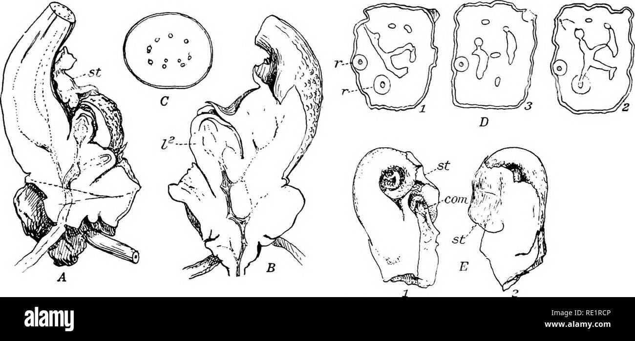 . The Eusporangiatae; the comparative morphology of the Ophioglossaceae and Marattiaceae. Ophioglossaceae; Marattiaceae. J92 THE MARATTIALES younger leaf. Fig. 175, E, shows a young leaf from another plant, in which one side has been cut away so as to show the relation of the stipules to the leaf base. This leaf was still coiled up and its apex was quite concealed within the large overlapping stipules. Both as to its position and its relation to the stipules, the commissure exactly resembles the lip-like basal extension of the stipular sheath m Botrychium and Helminthostachys, and there is no Stock Photohttps://www.alamy.com/image-license-details/?v=1https://www.alamy.com/the-eusporangiatae-the-comparative-morphology-of-the-ophioglossaceae-and-marattiaceae-ophioglossaceae-marattiaceae-j92-the-marattiales-younger-leaf-fig-175-e-shows-a-young-leaf-from-another-plant-in-which-one-side-has-been-cut-away-so-as-to-show-the-relation-of-the-stipules-to-the-leaf-base-this-leaf-was-still-coiled-up-and-its-apex-was-quite-concealed-within-the-large-overlapping-stipules-both-as-to-its-position-and-its-relation-to-the-stipules-the-commissure-exactly-resembles-the-lip-like-basal-extension-of-the-stipular-sheath-m-botrychium-and-helminthostachys-and-there-is-no-image232380310.html
RMRE1RCP–. The Eusporangiatae; the comparative morphology of the Ophioglossaceae and Marattiaceae. Ophioglossaceae; Marattiaceae. J92 THE MARATTIALES younger leaf. Fig. 175, E, shows a young leaf from another plant, in which one side has been cut away so as to show the relation of the stipules to the leaf base. This leaf was still coiled up and its apex was quite concealed within the large overlapping stipules. Both as to its position and its relation to the stipules, the commissure exactly resembles the lip-like basal extension of the stipular sheath m Botrychium and Helminthostachys, and there is no
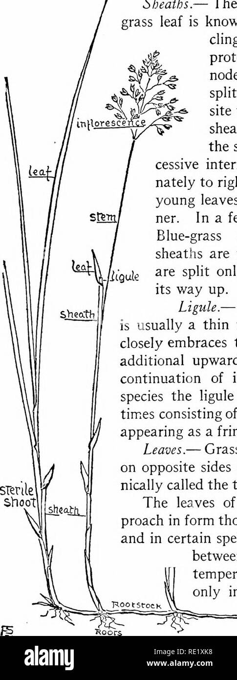 . The book of grasses; an illustrated guide to the common grasses, and the most common of the rushes and sedges. Grasses; Juncaceae; Cyperaceae. The Book of Grasses no part in such service, but if one notices grasses that have been beaten to earth by heavy showers, it will be seen that the lower nodes have lengthened on the side turned earthward, and that the stems are thereby bent upward at sharp angles. Sheaths.— The broad, basal portion of each grass leaf is known as the sheath, and, encir- cling the stem, is an important protection to the growing inter- node. Each sheath is usually split, Stock Photohttps://www.alamy.com/image-license-details/?v=1https://www.alamy.com/the-book-of-grasses-an-illustrated-guide-to-the-common-grasses-and-the-most-common-of-the-rushes-and-sedges-grasses-juncaceae-cyperaceae-the-book-of-grasses-no-part-in-such-service-but-if-one-notices-grasses-that-have-been-beaten-to-earth-by-heavy-showers-it-will-be-seen-that-the-lower-nodes-have-lengthened-on-the-side-turned-earthward-and-that-the-stems-are-thereby-bent-upward-at-sharp-angles-sheaths-the-broad-basal-portion-of-each-grass-leaf-is-known-as-the-sheath-and-encir-cling-the-stem-is-an-important-protection-to-the-growing-inter-node-each-sheath-is-usually-split-image232382844.html
RMRE1XK8–. The book of grasses; an illustrated guide to the common grasses, and the most common of the rushes and sedges. Grasses; Juncaceae; Cyperaceae. The Book of Grasses no part in such service, but if one notices grasses that have been beaten to earth by heavy showers, it will be seen that the lower nodes have lengthened on the side turned earthward, and that the stems are thereby bent upward at sharp angles. Sheaths.— The broad, basal portion of each grass leaf is known as the sheath, and, encir- cling the stem, is an important protection to the growing inter- node. Each sheath is usually split,
 . Elements of the comparative anatomy of vertebrates. Anatomy, Comparative. 54 COMPARATIVE ANATOMY Ganoidei, Teleostei, and Dipnoi—In these forms the ribs, ahiiost without exception, are connected with the ventral parts of the notochordal sheath (Dipnoans) or with the " basal processes " (Ganoids and Teleosts, see p. 38).-' This is one point of difference between the ribs of these forms and those of other Vertebrates: another is that they are always situated beneath (internal to) the lateral muscles, between these and the peritoneum (Fig. 40a, A, B, C). In Teleosts the ribs are at fi Stock Photohttps://www.alamy.com/image-license-details/?v=1https://www.alamy.com/elements-of-the-comparative-anatomy-of-vertebrates-anatomy-comparative-54-comparative-anatomy-ganoidei-teleostei-and-dipnoiin-these-forms-the-ribs-ahiiost-without-exception-are-connected-with-the-ventral-parts-of-the-notochordal-sheath-dipnoans-or-with-the-quot-basal-processes-quot-ganoids-and-teleosts-see-p-38-this-is-one-point-of-difference-between-the-ribs-of-these-forms-and-those-of-other-vertebrates-another-is-that-they-are-always-situated-beneath-internal-to-the-lateral-muscles-between-these-and-the-peritoneum-fig-40a-a-b-c-in-teleosts-the-ribs-are-at-fi-image232075803.html
RMRDFY1F–. Elements of the comparative anatomy of vertebrates. Anatomy, Comparative. 54 COMPARATIVE ANATOMY Ganoidei, Teleostei, and Dipnoi—In these forms the ribs, ahiiost without exception, are connected with the ventral parts of the notochordal sheath (Dipnoans) or with the " basal processes " (Ganoids and Teleosts, see p. 38).-' This is one point of difference between the ribs of these forms and those of other Vertebrates: another is that they are always situated beneath (internal to) the lateral muscles, between these and the peritoneum (Fig. 40a, A, B, C). In Teleosts the ribs are at fi
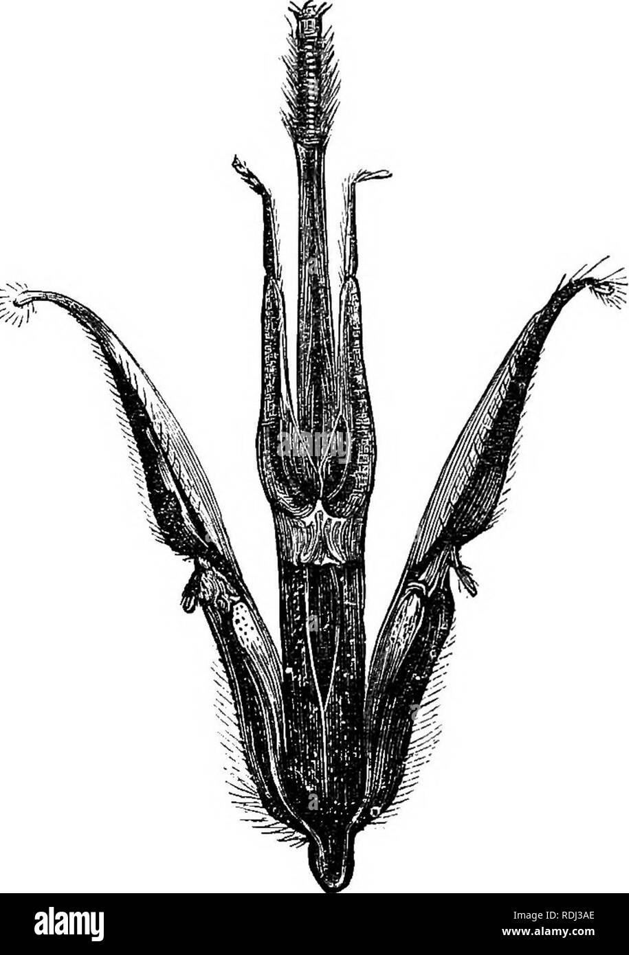 . Evenings at the microscope : or, researches among the minuter organs and forms of animal life. Microscopy; Microscopes; Medical microscopy. 164 EVENINGS AT THE MICEOSCOPE. Tient parts :—the thick opaque chin, at its basal end ; then the two labial palpi, each consisting of four joints, of which the two terminal ones are minute, while the two basal are large and greatly lengthened so as to resemble in appearance the maxillce, whose function they imitate also; for the pair of palpi when closed. JA"WS OF BEE. form an inner sheath for the tongue (Jiguld). Finally you see this organ, which i Stock Photohttps://www.alamy.com/image-license-details/?v=1https://www.alamy.com/evenings-at-the-microscope-or-researches-among-the-minuter-organs-and-forms-of-animal-life-microscopy-microscopes-medical-microscopy-164-evenings-at-the-miceoscope-tient-parts-the-thick-opaque-chin-at-its-basal-end-then-the-two-labial-palpi-each-consisting-of-four-joints-of-which-the-two-terminal-ones-are-minute-while-the-two-basal-are-large-and-greatly-lengthened-so-as-to-resemble-in-appearance-the-maxillce-whose-function-they-imitate-also-for-the-pair-of-palpi-when-closed-jaquotws-of-bee-form-an-inner-sheath-for-the-tongue-jiguld-finally-you-see-this-organ-which-i-image232123094.html
RMRDJ3AE–. Evenings at the microscope : or, researches among the minuter organs and forms of animal life. Microscopy; Microscopes; Medical microscopy. 164 EVENINGS AT THE MICEOSCOPE. Tient parts :—the thick opaque chin, at its basal end ; then the two labial palpi, each consisting of four joints, of which the two terminal ones are minute, while the two basal are large and greatly lengthened so as to resemble in appearance the maxillce, whose function they imitate also; for the pair of palpi when closed. JA"WS OF BEE. form an inner sheath for the tongue (Jiguld). Finally you see this organ, which i
 . Principles of economic zoo?logy. Zoology, Economic. HEMIPTERA 141 of plants. The sucking beak consists of the lahiuin, which, to- gether with the labial palpi, is modified into a jointed sheath. This incloses the mandibles and maxillae, which are changed into long, piercing stylets.^ The labrum or upper lip is small or rudimentary. There are usually four wings. In the typical Hemiptera, as exemplified in the sub-order Heterop'tera, the character of the anterior wings is a distinguishing feature. The basal portions of these wings are thickened and parch- ment-like, while the terminal portions Stock Photohttps://www.alamy.com/image-license-details/?v=1https://www.alamy.com/principles-of-economic-zoology-zoology-economic-hemiptera-141-of-plants-the-sucking-beak-consists-of-the-lahiuin-which-to-gether-with-the-labial-palpi-is-modified-into-a-jointed-sheath-this-incloses-the-mandibles-and-maxillae-which-are-changed-into-long-piercing-stylets-the-labrum-or-upper-lip-is-small-or-rudimentary-there-are-usually-four-wings-in-the-typical-hemiptera-as-exemplified-in-the-sub-order-heteroptera-the-character-of-the-anterior-wings-is-a-distinguishing-feature-the-basal-portions-of-these-wings-are-thickened-and-parch-ment-like-while-the-terminal-portions-image232253990.html
RMRDT29A–. Principles of economic zoo?logy. Zoology, Economic. HEMIPTERA 141 of plants. The sucking beak consists of the lahiuin, which, to- gether with the labial palpi, is modified into a jointed sheath. This incloses the mandibles and maxillae, which are changed into long, piercing stylets.^ The labrum or upper lip is small or rudimentary. There are usually four wings. In the typical Hemiptera, as exemplified in the sub-order Heterop'tera, the character of the anterior wings is a distinguishing feature. The basal portions of these wings are thickened and parch- ment-like, while the terminal portions
 . Bulletin - United States National Museum. Science. THE SESSILE BARNACLES. 231 The sheath is short, about half the length of the carina and sides, one-fourth that of the rostrum, with fine horizontal ridges bearing short fringes of golden bristles. Its lower edge is continuous with the interior of the walls. The parietes are nearly smooth within, but sometimes very weak ribs are visible. The basal edges of the com- partments are smooth and beveled. The base of the rostrum is broadly rounded, the upper end tapering a little. Sides nearly in a plane. The labruni has three teeth and is minutely Stock Photohttps://www.alamy.com/image-license-details/?v=1https://www.alamy.com/bulletin-united-states-national-museum-science-the-sessile-barnacles-231-the-sheath-is-short-about-half-the-length-of-the-carina-and-sides-one-fourth-that-of-the-rostrum-with-fine-horizontal-ridges-bearing-short-fringes-of-golden-bristles-its-lower-edge-is-continuous-with-the-interior-of-the-walls-the-parietes-are-nearly-smooth-within-but-sometimes-very-weak-ribs-are-visible-the-basal-edges-of-the-com-partments-are-smooth-and-beveled-the-base-of-the-rostrum-is-broadly-rounded-the-upper-end-tapering-a-little-sides-nearly-in-a-plane-the-labruni-has-three-teeth-and-is-minutely-image233749490.html
RMRG85T2–. Bulletin - United States National Museum. Science. THE SESSILE BARNACLES. 231 The sheath is short, about half the length of the carina and sides, one-fourth that of the rostrum, with fine horizontal ridges bearing short fringes of golden bristles. Its lower edge is continuous with the interior of the walls. The parietes are nearly smooth within, but sometimes very weak ribs are visible. The basal edges of the com- partments are smooth and beveled. The base of the rostrum is broadly rounded, the upper end tapering a little. Sides nearly in a plane. The labruni has three teeth and is minutely
 . A handbook of cryptogamic botany. Cryptogams. CYANOPHYCEM only temporary duration ; when it disappears it leaves the membra- nous sheath open at the extremity. This is especially well seen in Calothrix (Ag.). The ordinary mode of multi- plication of the Rivulariacese is by means of hormogones, frag- ments of the green portion which become detached from the rest of the filament, escape from the gelatinous envelope, move about with a creeping motion, eventually come to rest, invest themselves with a gelatinous sheath, and develop into a new filament in which the differenti- ation of the basal Stock Photohttps://www.alamy.com/image-license-details/?v=1https://www.alamy.com/a-handbook-of-cryptogamic-botany-cryptogams-cyanophycem-only-temporary-duration-when-it-disappears-it-leaves-the-membra-nous-sheath-open-at-the-extremity-this-is-especially-well-seen-in-calothrix-ag-the-ordinary-mode-of-multi-plication-of-the-rivulariacese-is-by-means-of-hormogones-frag-ments-of-the-green-portion-which-become-detached-from-the-rest-of-the-filament-escape-from-the-gelatinous-envelope-move-about-with-a-creeping-motion-eventually-come-to-rest-invest-themselves-with-a-gelatinous-sheath-and-develop-into-a-new-filament-in-which-the-differenti-ation-of-the-basal-image232400892.html
RMRE2NKT–. A handbook of cryptogamic botany. Cryptogams. CYANOPHYCEM only temporary duration ; when it disappears it leaves the membra- nous sheath open at the extremity. This is especially well seen in Calothrix (Ag.). The ordinary mode of multi- plication of the Rivulariacese is by means of hormogones, frag- ments of the green portion which become detached from the rest of the filament, escape from the gelatinous envelope, move about with a creeping motion, eventually come to rest, invest themselves with a gelatinous sheath, and develop into a new filament in which the differenti- ation of the basal
 . The book of grasses; an illustrated guide to the common grasses, and the most common of the rushes and sedges. The Book of Grasses no part in such service, but if one notices grasses that have been beaten to earth by heavy showers, it will be seen that the lower nodes have lengthened on the side turned earthward, and that the stems are thereby bent upward at sharp angles. Sheaths.— The broad, basal portion of each ^rass leaf is known as the sheath, and, encir- cling the stem, is an important protection to the growing inter- node. Each sheath is usually split, or open, on the side oppo- site Stock Photohttps://www.alamy.com/image-license-details/?v=1https://www.alamy.com/the-book-of-grasses-an-illustrated-guide-to-the-common-grasses-and-the-most-common-of-the-rushes-and-sedges-the-book-of-grasses-no-part-in-such-service-but-if-one-notices-grasses-that-have-been-beaten-to-earth-by-heavy-showers-it-will-be-seen-that-the-lower-nodes-have-lengthened-on-the-side-turned-earthward-and-that-the-stems-are-thereby-bent-upward-at-sharp-angles-sheaths-the-broad-basal-portion-of-each-rass-leaf-is-known-as-the-sheath-and-encir-cling-the-stem-is-an-important-protection-to-the-growing-inter-node-each-sheath-is-usually-split-or-open-on-the-side-oppo-site-image234475519.html
RMRHD7WK–. The book of grasses; an illustrated guide to the common grasses, and the most common of the rushes and sedges. The Book of Grasses no part in such service, but if one notices grasses that have been beaten to earth by heavy showers, it will be seen that the lower nodes have lengthened on the side turned earthward, and that the stems are thereby bent upward at sharp angles. Sheaths.— The broad, basal portion of each ^rass leaf is known as the sheath, and, encir- cling the stem, is an important protection to the growing inter- node. Each sheath is usually split, or open, on the side oppo- site
 . Bulletin - United States National Museum. Science. THE SESSILE BARNACLES. 381 wall, its lower margin not in the least overhanging. The interior is not in the least grooved below the sheath, and at the basal edge the compartments are very narrowly inflexed or have a slight rim around the membranous basis. The carina is longer than the rostrum, longer than wide, triangular in outline. The carinolateral compartments have very narioAv, band- like parietes, hardly one-fourth as wide as tliat of the rostrum. The rostrum is normally about as high as wide, and triangular in shape. The base is modifi Stock Photohttps://www.alamy.com/image-license-details/?v=1https://www.alamy.com/bulletin-united-states-national-museum-science-the-sessile-barnacles-381-wall-its-lower-margin-not-in-the-least-overhanging-the-interior-is-not-in-the-least-grooved-below-the-sheath-and-at-the-basal-edge-the-compartments-are-very-narrowly-inflexed-or-have-a-slight-rim-around-the-membranous-basis-the-carina-is-longer-than-the-rostrum-longer-than-wide-triangular-in-outline-the-carinolateral-compartments-have-very-narioav-band-like-parietes-hardly-one-fourth-as-wide-as-tliat-of-the-rostrum-the-rostrum-is-normally-about-as-high-as-wide-and-triangular-in-shape-the-base-is-modifi-image233749115.html
RMRG85AK–. Bulletin - United States National Museum. Science. THE SESSILE BARNACLES. 381 wall, its lower margin not in the least overhanging. The interior is not in the least grooved below the sheath, and at the basal edge the compartments are very narrowly inflexed or have a slight rim around the membranous basis. The carina is longer than the rostrum, longer than wide, triangular in outline. The carinolateral compartments have very narioAv, band- like parietes, hardly one-fourth as wide as tliat of the rostrum. The rostrum is normally about as high as wide, and triangular in shape. The base is modifi
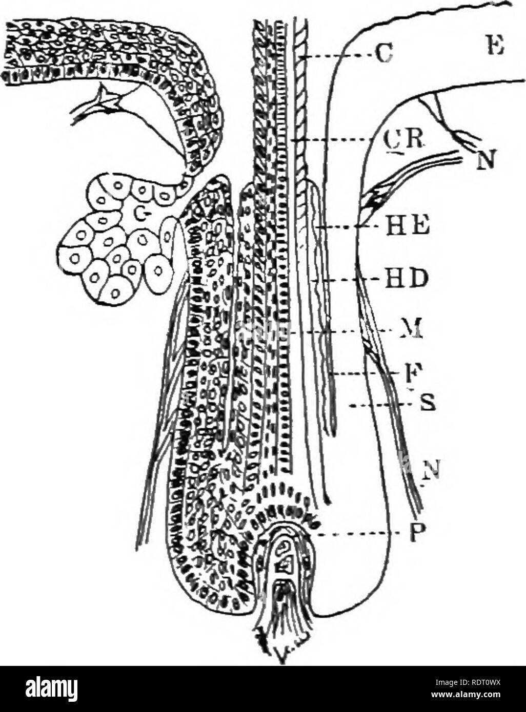 . Text book of vertebrate zoology. Vertebrates; Anatomy, Comparative. gS MORPHOLOGY OF THE ORGANS OF VERTEBRATES. hair follicle. In both hair and follicle several layers may be distinguished. In the follicle there is the basal layer and the more superficial layers of the epidermis, without, however, an}- clear differentiation of strata corneum and lucidum. At the bottom of the follicle (root of the hair) these pass directly into the hair itself, on the out- side of which is the so-called inner root-sheath (the walls of the follicle forming the outer root-sheath) composed of two layers of cells Stock Photohttps://www.alamy.com/image-license-details/?v=1https://www.alamy.com/text-book-of-vertebrate-zoology-vertebrates-anatomy-comparative-gs-morphology-of-the-organs-of-vertebrates-hair-follicle-in-both-hair-and-follicle-several-layers-may-be-distinguished-in-the-follicle-there-is-the-basal-layer-and-the-more-superficial-layers-of-the-epidermis-without-however-an-clear-differentiation-of-strata-corneum-and-lucidum-at-the-bottom-of-the-follicle-root-of-the-hair-these-pass-directly-into-the-hair-itself-on-the-out-side-of-which-is-the-so-called-inner-root-sheath-the-walls-of-the-follicle-forming-the-outer-root-sheath-composed-of-two-layers-of-cells-image232252886.html
RMRDT0WX–. Text book of vertebrate zoology. Vertebrates; Anatomy, Comparative. gS MORPHOLOGY OF THE ORGANS OF VERTEBRATES. hair follicle. In both hair and follicle several layers may be distinguished. In the follicle there is the basal layer and the more superficial layers of the epidermis, without, however, an}- clear differentiation of strata corneum and lucidum. At the bottom of the follicle (root of the hair) these pass directly into the hair itself, on the out- side of which is the so-called inner root-sheath (the walls of the follicle forming the outer root-sheath) composed of two layers of cells
 . Beekeeping; a discussion of the life of the honeybee and of the production of honey. Bees; Honey. The Life Processes of the IndividvM 159 bulb {ShB) and is further continued in two arms {ShA) which curve outward. The lancets slide on a grooved track the full length of the sheath, past the bulb and diverge along the two basal arms. The sheath and lancets combine to form a hollow tube (PsnC) through which the poison flows. The arms of the sheath are attached at their anterior ends to oblong plates (Ob) which overlap the sides of the sting. To these plates are attached palpi (StnPlp), soft whit Stock Photohttps://www.alamy.com/image-license-details/?v=1https://www.alamy.com/beekeeping-a-discussion-of-the-life-of-the-honeybee-and-of-the-production-of-honey-bees-honey-the-life-processes-of-the-individvm-159-bulb-shb-and-is-further-continued-in-two-arms-sha-which-curve-outward-the-lancets-slide-on-a-grooved-track-the-full-length-of-the-sheath-past-the-bulb-and-diverge-along-the-two-basal-arms-the-sheath-and-lancets-combine-to-form-a-hollow-tube-psnc-through-which-the-poison-flows-the-arms-of-the-sheath-are-attached-at-their-anterior-ends-to-oblong-plates-ob-which-overlap-the-sides-of-the-sting-to-these-plates-are-attached-palpi-stnplp-soft-whit-image232061714.html
RMRDF92A–. Beekeeping; a discussion of the life of the honeybee and of the production of honey. Bees; Honey. The Life Processes of the IndividvM 159 bulb {ShB) and is further continued in two arms {ShA) which curve outward. The lancets slide on a grooved track the full length of the sheath, past the bulb and diverge along the two basal arms. The sheath and lancets combine to form a hollow tube (PsnC) through which the poison flows. The arms of the sheath are attached at their anterior ends to oblong plates (Ob) which overlap the sides of the sting. To these plates are attached palpi (StnPlp), soft whit
 . Chordate morphology. Morphology (Animals); Chordata. sheath and constriction of the notochord in the basal plate, notochord reaching dorsal margin of dorsum sellae and pro- jecting from it (at least in younger stages), dorsum sellae forming between ends of parachordals, epihyal not ex- tending up lateral to head vein (no laterohyal involved), presence of orbital margin of cartilages, presence of nasal and labial cartilages, and separate posterior superficial ophthalmic foramen. Acanthodians The acanthodians are represented by spines and plates in Upper Silurian deposits, but it is not until Stock Photohttps://www.alamy.com/image-license-details/?v=1https://www.alamy.com/chordate-morphology-morphology-animals-chordata-sheath-and-constriction-of-the-notochord-in-the-basal-plate-notochord-reaching-dorsal-margin-of-dorsum-sellae-and-pro-jecting-from-it-at-least-in-younger-stages-dorsum-sellae-forming-between-ends-of-parachordals-epihyal-not-ex-tending-up-lateral-to-head-vein-no-laterohyal-involved-presence-of-orbital-margin-of-cartilages-presence-of-nasal-and-labial-cartilages-and-separate-posterior-superficial-ophthalmic-foramen-acanthodians-the-acanthodians-are-represented-by-spines-and-plates-in-upper-silurian-deposits-but-it-is-not-until-image234908567.html
RMRJ507K–. Chordate morphology. Morphology (Animals); Chordata. sheath and constriction of the notochord in the basal plate, notochord reaching dorsal margin of dorsum sellae and pro- jecting from it (at least in younger stages), dorsum sellae forming between ends of parachordals, epihyal not ex- tending up lateral to head vein (no laterohyal involved), presence of orbital margin of cartilages, presence of nasal and labial cartilages, and separate posterior superficial ophthalmic foramen. Acanthodians The acanthodians are represented by spines and plates in Upper Silurian deposits, but it is not until
 . The Biological bulletin. Biology; Zoology; Biology; Marine Biology. STUDIES ON THE ACROSOME. IV. 55 3. In the large Mytilns acrosome, three differentiated regions can be dis- tinguished, consisting of a basal structure which seems to be concerned with the extrusion of the filament; a distal region containing what is believed to be an egg- membrane lysin; and an axial structure which appears to be a tubular sheath, possibly surrounding a precursor of the filament. The same regions can be seen in the spermatozoa of Pctricola, Spondylus and Mactra. In the smaller acrosomes of Lithophaga. Cliain Stock Photohttps://www.alamy.com/image-license-details/?v=1https://www.alamy.com/the-biological-bulletin-biology-zoology-biology-marine-biology-studies-on-the-acrosome-iv-55-3-in-the-large-mytilns-acrosome-three-differentiated-regions-can-be-dis-tinguished-consisting-of-a-basal-structure-which-seems-to-be-concerned-with-the-extrusion-of-the-filament-a-distal-region-containing-what-is-believed-to-be-an-egg-membrane-lysin-and-an-axial-structure-which-appears-to-be-a-tubular-sheath-possibly-surrounding-a-precursor-of-the-filament-the-same-regions-can-be-seen-in-the-spermatozoa-of-pctricola-spondylus-and-mactra-in-the-smaller-acrosomes-of-lithophaga-cliain-image234622884.html
RMRHKYTM–. The Biological bulletin. Biology; Zoology; Biology; Marine Biology. STUDIES ON THE ACROSOME. IV. 55 3. In the large Mytilns acrosome, three differentiated regions can be dis- tinguished, consisting of a basal structure which seems to be concerned with the extrusion of the filament; a distal region containing what is believed to be an egg- membrane lysin; and an axial structure which appears to be a tubular sheath, possibly surrounding a precursor of the filament. The same regions can be seen in the spermatozoa of Pctricola, Spondylus and Mactra. In the smaller acrosomes of Lithophaga. Cliain
 . Comparative anatomy of the vegetative organs of the phanerogams and ferns. Plant anatomy; Phanerogams; Ferns. STRUCTURE OF COLLATERAL BUNDLES. 33^ the leaf and the upper part of the leaf-sheath the bundles are though on the average smaller. Fig. 151 (145), on the other hand, is taken from the place where the bundle passes through the basal portion of the leaf-sheath of a young plant. The bundle itself, with its group of vessels at g, is in all parts smallerthan that in the precedin figure, but otherwise similar to it, as is clear without explanation, even in the finer points of structure not Stock Photohttps://www.alamy.com/image-license-details/?v=1https://www.alamy.com/comparative-anatomy-of-the-vegetative-organs-of-the-phanerogams-and-ferns-plant-anatomy-phanerogams-ferns-structure-of-collateral-bundles-33-the-leaf-and-the-upper-part-of-the-leaf-sheath-the-bundles-are-though-on-the-average-smaller-fig-151-145-on-the-other-hand-is-taken-from-the-place-where-the-bundle-passes-through-the-basal-portion-of-the-leaf-sheath-of-a-young-plant-the-bundle-itself-with-its-group-of-vessels-at-g-is-in-all-parts-smallerthan-that-in-the-precedin-figure-but-otherwise-similar-to-it-as-is-clear-without-explanation-even-in-the-finer-points-of-structure-not-image232681038.html
RMREFF12–. Comparative anatomy of the vegetative organs of the phanerogams and ferns. Plant anatomy; Phanerogams; Ferns. STRUCTURE OF COLLATERAL BUNDLES. 33^ the leaf and the upper part of the leaf-sheath the bundles are though on the average smaller. Fig. 151 (145), on the other hand, is taken from the place where the bundle passes through the basal portion of the leaf-sheath of a young plant. The bundle itself, with its group of vessels at g, is in all parts smallerthan that in the precedin figure, but otherwise similar to it, as is clear without explanation, even in the finer points of structure not
 . The Eusporangiatae; the comparative morphology of the Ophioglossaceae and Marattiaceae. Ophioglossaceae; Marattiaceae. THE ADULT SPOROPHYTE 91 ctWm O.moluccanummzy therefore be described as a three-sided or four-sided prism or a truncate pyramid. As m the young plant, the stipular sheath of each new leaf is formed mainly from the basal tissue of the leaf itself, but includes also tissue from the margin of the stem apex. The sheath is open above by a narrow pore through which it communicates with the space between it and the next stipular sheath. In 0. moluccanum, as in 0. vulgatum, three lea Stock Photohttps://www.alamy.com/image-license-details/?v=1https://www.alamy.com/the-eusporangiatae-the-comparative-morphology-of-the-ophioglossaceae-and-marattiaceae-ophioglossaceae-marattiaceae-the-adult-sporophyte-91-ctwm-omoluccanummzy-therefore-be-described-as-a-three-sided-or-four-sided-prism-or-a-truncate-pyramid-as-m-the-young-plant-the-stipular-sheath-of-each-new-leaf-is-formed-mainly-from-the-basal-tissue-of-the-leaf-itself-but-includes-also-tissue-from-the-margin-of-the-stem-apex-the-sheath-is-open-above-by-a-narrow-pore-through-which-it-communicates-with-the-space-between-it-and-the-next-stipular-sheath-in-0-moluccanum-as-in-0-vulgatum-three-lea-image232358246.html
RMRE0R8P–. The Eusporangiatae; the comparative morphology of the Ophioglossaceae and Marattiaceae. Ophioglossaceae; Marattiaceae. THE ADULT SPOROPHYTE 91 ctWm O.moluccanummzy therefore be described as a three-sided or four-sided prism or a truncate pyramid. As m the young plant, the stipular sheath of each new leaf is formed mainly from the basal tissue of the leaf itself, but includes also tissue from the margin of the stem apex. The sheath is open above by a narrow pore through which it communicates with the space between it and the next stipular sheath. In 0. moluccanum, as in 0. vulgatum, three lea
![. Bulletin of the U.S. Department of Agriculture. Agriculture; Agriculture. Bui. 545, U. S. Dept. of Agriculture. Plate XVI11.. Short-awned Bromegrass (Bronius marginatus). The sheath on the basal portion of the culm was accidentally removed before the photograph was taken. 19. Please note that these images are extracted from scanned page images that may have been digitally enhanced for readability - coloration and appearance of these illustrations may not perfectly resemble the original work.. United States. Dept. of Agriculture. [Washington, D. C. ?] : The Dept. : Supt. of Docs. , G. P. O. Stock Photo . Bulletin of the U.S. Department of Agriculture. Agriculture; Agriculture. Bui. 545, U. S. Dept. of Agriculture. Plate XVI11.. Short-awned Bromegrass (Bronius marginatus). The sheath on the basal portion of the culm was accidentally removed before the photograph was taken. 19. Please note that these images are extracted from scanned page images that may have been digitally enhanced for readability - coloration and appearance of these illustrations may not perfectly resemble the original work.. United States. Dept. of Agriculture. [Washington, D. C. ?] : The Dept. : Supt. of Docs. , G. P. O. Stock Photo](https://c8.alamy.com/comp/RGBPXJ/bulletin-of-the-us-department-of-agriculture-agriculture-agriculture-bui-545-u-s-dept-of-agriculture-plate-xvi11-short-awned-bromegrass-bronius-marginatus-the-sheath-on-the-basal-portion-of-the-culm-was-accidentally-removed-before-the-photograph-was-taken-19-please-note-that-these-images-are-extracted-from-scanned-page-images-that-may-have-been-digitally-enhanced-for-readability-coloration-and-appearance-of-these-illustrations-may-not-perfectly-resemble-the-original-work-united-states-dept-of-agriculture-washington-d-c-the-dept-supt-of-docs-g-p-o-RGBPXJ.jpg) . Bulletin of the U.S. Department of Agriculture. Agriculture; Agriculture. Bui. 545, U. S. Dept. of Agriculture. Plate XVI11.. Short-awned Bromegrass (Bronius marginatus). The sheath on the basal portion of the culm was accidentally removed before the photograph was taken. 19. Please note that these images are extracted from scanned page images that may have been digitally enhanced for readability - coloration and appearance of these illustrations may not perfectly resemble the original work.. United States. Dept. of Agriculture. [Washington, D. C. ?] : The Dept. : Supt. of Docs. , G. P. O. Stock Photohttps://www.alamy.com/image-license-details/?v=1https://www.alamy.com/bulletin-of-the-us-department-of-agriculture-agriculture-agriculture-bui-545-u-s-dept-of-agriculture-plate-xvi11-short-awned-bromegrass-bronius-marginatus-the-sheath-on-the-basal-portion-of-the-culm-was-accidentally-removed-before-the-photograph-was-taken-19-please-note-that-these-images-are-extracted-from-scanned-page-images-that-may-have-been-digitally-enhanced-for-readability-coloration-and-appearance-of-these-illustrations-may-not-perfectly-resemble-the-original-work-united-states-dept-of-agriculture-washington-d-c-the-dept-supt-of-docs-g-p-o-image233828746.html
. Bulletin of the U.S. Department of Agriculture. Agriculture; Agriculture. Bui. 545, U. S. Dept. of Agriculture. Plate XVI11.. Short-awned Bromegrass (Bronius marginatus). The sheath on the basal portion of the culm was accidentally removed before the photograph was taken. 19. Please note that these images are extracted from scanned page images that may have been digitally enhanced for readability - coloration and appearance of these illustrations may not perfectly resemble the original work.. United States. Dept. of Agriculture. [Washington, D. C. ?] : The Dept. : Supt. of Docs. , G. P. O. Stock Photohttps://www.alamy.com/image-license-details/?v=1https://www.alamy.com/bulletin-of-the-us-department-of-agriculture-agriculture-agriculture-bui-545-u-s-dept-of-agriculture-plate-xvi11-short-awned-bromegrass-bronius-marginatus-the-sheath-on-the-basal-portion-of-the-culm-was-accidentally-removed-before-the-photograph-was-taken-19-please-note-that-these-images-are-extracted-from-scanned-page-images-that-may-have-been-digitally-enhanced-for-readability-coloration-and-appearance-of-these-illustrations-may-not-perfectly-resemble-the-original-work-united-states-dept-of-agriculture-washington-d-c-the-dept-supt-of-docs-g-p-o-image233828746.htmlRMRGBPXJ–. Bulletin of the U.S. Department of Agriculture. Agriculture; Agriculture. Bui. 545, U. S. Dept. of Agriculture. Plate XVI11.. Short-awned Bromegrass (Bronius marginatus). The sheath on the basal portion of the culm was accidentally removed before the photograph was taken. 19. Please note that these images are extracted from scanned page images that may have been digitally enhanced for readability - coloration and appearance of these illustrations may not perfectly resemble the original work.. United States. Dept. of Agriculture. [Washington, D. C. ?] : The Dept. : Supt. of Docs. , G. P. O.
 . Catalogue of the fossil plants of the Glossopteris flora in the Department of geology. Paleobotany -- Carboniferous; Paleobotany -- Catalogs and collections; Plants, Fossil -- Catalogs and collections. 37 from Natal, on which occur numerous fronds of Glossopteris. In addition, there is a small and imperfect fragment (Text-fig. 11), which is in all probability referable to Sphenophyllum, a genus which so far has not been found in South Africa. The plant appears to consist of a whorl of leaves, the basal portions of five leaves being seen, which seem to be united into a short sheath. Fig. 11.— Stock Photohttps://www.alamy.com/image-license-details/?v=1https://www.alamy.com/catalogue-of-the-fossil-plants-of-the-glossopteris-flora-in-the-department-of-geology-paleobotany-carboniferous-paleobotany-catalogs-and-collections-plants-fossil-catalogs-and-collections-37-from-natal-on-which-occur-numerous-fronds-of-glossopteris-in-addition-there-is-a-small-and-imperfect-fragment-text-fig-11-which-is-in-all-probability-referable-to-sphenophyllum-a-genus-which-so-far-has-not-been-found-in-south-africa-the-plant-appears-to-consist-of-a-whorl-of-leaves-the-basal-portions-of-five-leaves-being-seen-which-seem-to-be-united-into-a-short-sheath-fig-11-image233005482.html
RMRF28TA–. Catalogue of the fossil plants of the Glossopteris flora in the Department of geology. Paleobotany -- Carboniferous; Paleobotany -- Catalogs and collections; Plants, Fossil -- Catalogs and collections. 37 from Natal, on which occur numerous fronds of Glossopteris. In addition, there is a small and imperfect fragment (Text-fig. 11), which is in all probability referable to Sphenophyllum, a genus which so far has not been found in South Africa. The plant appears to consist of a whorl of leaves, the basal portions of five leaves being seen, which seem to be united into a short sheath. Fig. 11.—
 . Circular. Agriculture; Agriculture; Entomology. Insect Enemies of Greenhouse and Ornamental Plants 19 the plants, but usually in the curled up, basal part of the leaf or in the sheath surrounding the flower stalk. Advantage is taken of this habit by the orchid-grower who sometimes sends a man daily through the house to hunt out and destroy them. Up to the present I have been unsuccessful in my search for larva? and pupa? and know nothing concerning the early stages. Mr. G. C. Champion writes that the eight known species of diorymelius are all from Central America and that this new species is Stock Photohttps://www.alamy.com/image-license-details/?v=1https://www.alamy.com/circular-agriculture-agriculture-entomology-insect-enemies-of-greenhouse-and-ornamental-plants-19-the-plants-but-usually-in-the-curled-up-basal-part-of-the-leaf-or-in-the-sheath-surrounding-the-flower-stalk-advantage-is-taken-of-this-habit-by-the-orchid-grower-who-sometimes-sends-a-man-daily-through-the-house-to-hunt-out-and-destroy-them-up-to-the-present-i-have-been-unsuccessful-in-my-search-for-larva-and-pupa-and-know-nothing-concerning-the-early-stages-mr-g-c-champion-writes-that-the-eight-known-species-of-diorymelius-are-all-from-central-america-and-that-this-new-species-is-image232780344.html
RMREM1KM–. Circular. Agriculture; Agriculture; Entomology. Insect Enemies of Greenhouse and Ornamental Plants 19 the plants, but usually in the curled up, basal part of the leaf or in the sheath surrounding the flower stalk. Advantage is taken of this habit by the orchid-grower who sometimes sends a man daily through the house to hunt out and destroy them. Up to the present I have been unsuccessful in my search for larva? and pupa? and know nothing concerning the early stages. Mr. G. C. Champion writes that the eight known species of diorymelius are all from Central America and that this new species is
 . Beekeeping; a discussion of the life of the honeybee and of the production of honey. Bee culture; Honey. The Life Processes of the Individual 159 bulb (ShB) and is further continued in two arms (ShA) which curve outward. The lancets slide on a grooved track the full length of the sheath, past the bulb and diverge along the two basal arms. The sheath and lancets combine to form a hollow tube (PsnC) through which the poison flows. The arms of the sheath are attached at their anterior ends to oblong plates (Ob) which overlap the sides of the sting. To these plates are attached palpi (StnPlp), s Stock Photohttps://www.alamy.com/image-license-details/?v=1https://www.alamy.com/beekeeping-a-discussion-of-the-life-of-the-honeybee-and-of-the-production-of-honey-bee-culture-honey-the-life-processes-of-the-individual-159-bulb-shb-and-is-further-continued-in-two-arms-sha-which-curve-outward-the-lancets-slide-on-a-grooved-track-the-full-length-of-the-sheath-past-the-bulb-and-diverge-along-the-two-basal-arms-the-sheath-and-lancets-combine-to-form-a-hollow-tube-psnc-through-which-the-poison-flows-the-arms-of-the-sheath-are-attached-at-their-anterior-ends-to-oblong-plates-ob-which-overlap-the-sides-of-the-sting-to-these-plates-are-attached-palpi-stnplp-s-image235166008.html
RMRJGMJ0–. Beekeeping; a discussion of the life of the honeybee and of the production of honey. Bee culture; Honey. The Life Processes of the Individual 159 bulb (ShB) and is further continued in two arms (ShA) which curve outward. The lancets slide on a grooved track the full length of the sheath, past the bulb and diverge along the two basal arms. The sheath and lancets combine to form a hollow tube (PsnC) through which the poison flows. The arms of the sheath are attached at their anterior ends to oblong plates (Ob) which overlap the sides of the sting. To these plates are attached palpi (StnPlp), s
 . Buccinum : (the whelk). Buccinidae. 53 external surface of the sac at this point forms a perfect circle in transverse sections (Text-fig. 5). The cells of the lateral and basal walls of this circular sheath are of medium length. The cells of the dorsal wall are of extraordinary size and extend down into the cavity, forming a deep ridge, which extends for some distance from the blind end. This odontoblast ridge lies, of course, in the gutter formed by the radula. Thus one sees that the cells of the lateral and ventral walls are directed towards the basal ventral plate of the radula, whilst th Stock Photohttps://www.alamy.com/image-license-details/?v=1https://www.alamy.com/buccinum-the-whelk-buccinidae-53-external-surface-of-the-sac-at-this-point-forms-a-perfect-circle-in-transverse-sections-text-fig-5-the-cells-of-the-lateral-and-basal-walls-of-this-circular-sheath-are-of-medium-length-the-cells-of-the-dorsal-wall-are-of-extraordinary-size-and-extend-down-into-the-cavity-forming-a-deep-ridge-which-extends-for-some-distance-from-the-blind-end-this-odontoblast-ridge-lies-of-course-in-the-gutter-formed-by-the-radula-thus-one-sees-that-the-cells-of-the-lateral-and-ventral-walls-are-directed-towards-the-basal-ventral-plate-of-the-radula-whilst-th-image234244541.html
RMRH2N8D–. Buccinum : (the whelk). Buccinidae. 53 external surface of the sac at this point forms a perfect circle in transverse sections (Text-fig. 5). The cells of the lateral and basal walls of this circular sheath are of medium length. The cells of the dorsal wall are of extraordinary size and extend down into the cavity, forming a deep ridge, which extends for some distance from the blind end. This odontoblast ridge lies, of course, in the gutter formed by the radula. Thus one sees that the cells of the lateral and ventral walls are directed towards the basal ventral plate of the radula, whilst th
 . Nature study and agriculture. Nature study; Agriculture. CHAPTER VII A FEW IMPORTANT PLANT FAMILIES The Grass Family. — This is perhaps our most important family of plants as it furnishes us the cereal grains and much other food for man and beast. The plants have stems that are solid at the joints and usually hollow be- tween ; in Indian corn and a few other species the stem is not hollow, but pithy. The leaves are alternate and parallel veined; the basal part envelops the stem, forming a sheath that is open on the side opposite the blade. A good example for studying the structure of the flo Stock Photohttps://www.alamy.com/image-license-details/?v=1https://www.alamy.com/nature-study-and-agriculture-nature-study-agriculture-chapter-vii-a-few-important-plant-families-the-grass-family-this-is-perhaps-our-most-important-family-of-plants-as-it-furnishes-us-the-cereal-grains-and-much-other-food-for-man-and-beast-the-plants-have-stems-that-are-solid-at-the-joints-and-usually-hollow-be-tween-in-indian-corn-and-a-few-other-species-the-stem-is-not-hollow-but-pithy-the-leaves-are-alternate-and-parallel-veined-the-basal-part-envelops-the-stem-forming-a-sheath-that-is-open-on-the-side-opposite-the-blade-a-good-example-for-studying-the-structure-of-the-flo-image232343690.html
RMRE04MX–. Nature study and agriculture. Nature study; Agriculture. CHAPTER VII A FEW IMPORTANT PLANT FAMILIES The Grass Family. — This is perhaps our most important family of plants as it furnishes us the cereal grains and much other food for man and beast. The plants have stems that are solid at the joints and usually hollow be- tween ; in Indian corn and a few other species the stem is not hollow, but pithy. The leaves are alternate and parallel veined; the basal part envelops the stem, forming a sheath that is open on the side opposite the blade. A good example for studying the structure of the flo
 . Studies on the vegetation of the Transcaspian lowlands. Botany. — 255 — have long triangular and hairy leaves, the upper ones sub- tending the solitary flowers. H. inllosa and macranthera, probably also H. pilosa, have leaves constructed after the ordinary centric type. Since the leaf is thick the aqueous tissue in the middle is large; the midrib has a bast-sheath; the hairs consist of a basal cell and a long fllamentous cell; the stomata are not sunk. The leaves of H. Karelini (fig. 78), a bracteole-succulent, are similar in structure; they have very short hairs and the stomata are slightly Stock Photohttps://www.alamy.com/image-license-details/?v=1https://www.alamy.com/studies-on-the-vegetation-of-the-transcaspian-lowlands-botany-255-have-long-triangular-and-hairy-leaves-the-upper-ones-sub-tending-the-solitary-flowers-h-inllosa-and-macranthera-probably-also-h-pilosa-have-leaves-constructed-after-the-ordinary-centric-type-since-the-leaf-is-thick-the-aqueous-tissue-in-the-middle-is-large-the-midrib-has-a-bast-sheath-the-hairs-consist-of-a-basal-cell-and-a-long-fllamentous-cell-the-stomata-are-not-sunk-the-leaves-of-h-karelini-fig-78-a-bracteole-succulent-are-similar-in-structure-they-have-very-short-hairs-and-the-stomata-are-slightly-image232240631.html
RMRDRD87–. Studies on the vegetation of the Transcaspian lowlands. Botany. — 255 — have long triangular and hairy leaves, the upper ones sub- tending the solitary flowers. H. inllosa and macranthera, probably also H. pilosa, have leaves constructed after the ordinary centric type. Since the leaf is thick the aqueous tissue in the middle is large; the midrib has a bast-sheath; the hairs consist of a basal cell and a long fllamentous cell; the stomata are not sunk. The leaves of H. Karelini (fig. 78), a bracteole-succulent, are similar in structure; they have very short hairs and the stomata are slightly
 . Diseases of economic plants . Plant diseases. Fig. 156.—Basal portion of rye plant showing anthrac- nose upon stem and leaf sheath. After Manns.. FlQ. 157. — Normal rye kernels and shriveled ones due to antliracnoae. After Manns.. Please note that these images are extracted from scanned page images that may have been digitally enhanced for readability - coloration and appearance of these illustrations may not perfectly resemble the original work.. Stevens, Frank Lincoln, 1871-1934; Hall, John Galentine, 1870-. New York : Macmillan Stock Photohttps://www.alamy.com/image-license-details/?v=1https://www.alamy.com/diseases-of-economic-plants-plant-diseases-fig-156basal-portion-of-rye-plant-showing-anthrac-nose-upon-stem-and-leaf-sheath-after-manns-flq-157-normal-rye-kernels-and-shriveled-ones-due-to-antliracnoae-after-manns-please-note-that-these-images-are-extracted-from-scanned-page-images-that-may-have-been-digitally-enhanced-for-readability-coloration-and-appearance-of-these-illustrations-may-not-perfectly-resemble-the-original-work-stevens-frank-lincoln-1871-1934-hall-john-galentine-1870-new-york-macmillan-image232034947.html
RMRDE2XB–. Diseases of economic plants . Plant diseases. Fig. 156.—Basal portion of rye plant showing anthrac- nose upon stem and leaf sheath. After Manns.. FlQ. 157. — Normal rye kernels and shriveled ones due to antliracnoae. After Manns.. Please note that these images are extracted from scanned page images that may have been digitally enhanced for readability - coloration and appearance of these illustrations may not perfectly resemble the original work.. Stevens, Frank Lincoln, 1871-1934; Hall, John Galentine, 1870-. New York : Macmillan
 . Comparative anatomy of the vegetative organs of the phanerogams and ferns;. Plant anatomy; Ferns. STRUCTURE OF COLLATERAL BUNDLES. 33^ ^^^^^^S the leaf and the upper part of the leaf-sheath the bundles are similar to that in Fig. 130, though on the average smaller. Fig. 151 (i4s), on the other hand, is taken from the place where the bundle passes through the basal portion of the leaf-sheath of a young plant. The bundle itself, with its group of vessels at g, is in all parts smallerthan that in the preceding figure, but otherwise similar to it, as is clear without explanation, even in the fin Stock Photohttps://www.alamy.com/image-license-details/?v=1https://www.alamy.com/comparative-anatomy-of-the-vegetative-organs-of-the-phanerogams-and-ferns-plant-anatomy-ferns-structure-of-collateral-bundles-33-s-the-leaf-and-the-upper-part-of-the-leaf-sheath-the-bundles-are-similar-to-that-in-fig-130-though-on-the-average-smaller-fig-151-i4s-on-the-other-hand-is-taken-from-the-place-where-the-bundle-passes-through-the-basal-portion-of-the-leaf-sheath-of-a-young-plant-the-bundle-itself-with-its-group-of-vessels-at-g-is-in-all-parts-smallerthan-that-in-the-preceding-figure-but-otherwise-similar-to-it-as-is-clear-without-explanation-even-in-the-fin-image232400806.html
RMRE2NGP–. Comparative anatomy of the vegetative organs of the phanerogams and ferns;. Plant anatomy; Ferns. STRUCTURE OF COLLATERAL BUNDLES. 33^ ^^^^^^S the leaf and the upper part of the leaf-sheath the bundles are similar to that in Fig. 130, though on the average smaller. Fig. 151 (i4s), on the other hand, is taken from the place where the bundle passes through the basal portion of the leaf-sheath of a young plant. The bundle itself, with its group of vessels at g, is in all parts smallerthan that in the preceding figure, but otherwise similar to it, as is clear without explanation, even in the fin
 . American bee journal. Bee culture; Bees. between the two. Reaumur, Newport, Kirby, Spence, Carpenter, Shuckard, Bevan, and Hunter all state that the tongue is solid, and that the honey is sopped up, or taken through a tube, formed by the close approxi- mation of the maxillae labium, and labial palpi. Newport speaks of a haiiy sheath along the under side of the basal two-thirds of the organ. Neighbour says there is a gutter throughout the entire length of the tongue. While Swammerdam, Lamarck, Burmeister, Wildman and Munn claim that the organ is. tubular. Newport and Carpenter assert that the Stock Photohttps://www.alamy.com/image-license-details/?v=1https://www.alamy.com/american-bee-journal-bee-culture-bees-between-the-two-reaumur-newport-kirby-spence-carpenter-shuckard-bevan-and-hunter-all-state-that-the-tongue-is-solid-and-that-the-honey-is-sopped-up-or-taken-through-a-tube-formed-by-the-close-approxi-mation-of-the-maxillae-labium-and-labial-palpi-newport-speaks-of-a-haiiy-sheath-along-the-under-side-of-the-basal-two-thirds-of-the-organ-neighbour-says-there-is-a-gutter-throughout-the-entire-length-of-the-tongue-while-swammerdam-lamarck-burmeister-wildman-and-munn-claim-that-the-organ-is-tubular-newport-and-carpenter-assert-that-the-image237719939.html
RMRPN25R–. American bee journal. Bee culture; Bees. between the two. Reaumur, Newport, Kirby, Spence, Carpenter, Shuckard, Bevan, and Hunter all state that the tongue is solid, and that the honey is sopped up, or taken through a tube, formed by the close approxi- mation of the maxillae labium, and labial palpi. Newport speaks of a haiiy sheath along the under side of the basal two-thirds of the organ. Neighbour says there is a gutter throughout the entire length of the tongue. While Swammerdam, Lamarck, Burmeister, Wildman and Munn claim that the organ is. tubular. Newport and Carpenter assert that the
 . Catalogue of the fossil plants of the Glossopteris flora in the Department of geology. Paleobotany -- Carboniferous; Paleobotany -- Catalogs and collections; Plants, Fossil -- Catalogs and collections. 37 from Natal, on which occur numerous fronds of Glossopteris. In addition, there is a small and imperfect fragment (Text-fig. 11), which is in all probability referable to Sphenophyllum, a genus which so far has not been found in South Africa. The plant appears to consist of a whorl of leaves, the basal portions of five leaves being seen, which seem to be united into a short sheath. Fig. 11.— Stock Photohttps://www.alamy.com/image-license-details/?v=1https://www.alamy.com/catalogue-of-the-fossil-plants-of-the-glossopteris-flora-in-the-department-of-geology-paleobotany-carboniferous-paleobotany-catalogs-and-collections-plants-fossil-catalogs-and-collections-37-from-natal-on-which-occur-numerous-fronds-of-glossopteris-in-addition-there-is-a-small-and-imperfect-fragment-text-fig-11-which-is-in-all-probability-referable-to-sphenophyllum-a-genus-which-so-far-has-not-been-found-in-south-africa-the-plant-appears-to-consist-of-a-whorl-of-leaves-the-basal-portions-of-five-leaves-being-seen-which-seem-to-be-united-into-a-short-sheath-fig-11-image233177031.html
RMRFA3K3–. Catalogue of the fossil plants of the Glossopteris flora in the Department of geology. Paleobotany -- Carboniferous; Paleobotany -- Catalogs and collections; Plants, Fossil -- Catalogs and collections. 37 from Natal, on which occur numerous fronds of Glossopteris. In addition, there is a small and imperfect fragment (Text-fig. 11), which is in all probability referable to Sphenophyllum, a genus which so far has not been found in South Africa. The plant appears to consist of a whorl of leaves, the basal portions of five leaves being seen, which seem to be united into a short sheath. Fig. 11.—
 . The biology of the protozoa. Protozoa; Protozoa. DERIVED ORGANIZATION—TAX0N0M1C STRUCTURES 153 layer containing trichocysts if the latter are present; (3) a contrac- tile zone containing the basal bodies of cilia, myonemes and coordin- ating fibers; (4) a denser zone which shades off into the endoplasin and supplies an anchorage for nuclei and contractile vacuoles. A single cilium is constructed on much the same plan as a flagel- lum, consisting of a central axial filament or fiber, and an elastic sheath of protoplasm. Movement is due to the active contraction. Fig. 83.—Cyclidium glaucoma. C Stock Photohttps://www.alamy.com/image-license-details/?v=1https://www.alamy.com/the-biology-of-the-protozoa-protozoa-protozoa-derived-organizationtax0n0m1c-structures-153-layer-containing-trichocysts-if-the-latter-are-present-3-a-contrac-tile-zone-containing-the-basal-bodies-of-cilia-myonemes-and-coordin-ating-fibers-4-a-denser-zone-which-shades-off-into-the-endoplasin-and-supplies-an-anchorage-for-nuclei-and-contractile-vacuoles-a-single-cilium-is-constructed-on-much-the-same-plan-as-a-flagel-lum-consisting-of-a-central-axial-filament-or-fiber-and-an-elastic-sheath-of-protoplasm-movement-is-due-to-the-active-contraction-fig-83cyclidium-glaucoma-c-image234603558.html
RMRHK36E–. The biology of the protozoa. Protozoa; Protozoa. DERIVED ORGANIZATION—TAX0N0M1C STRUCTURES 153 layer containing trichocysts if the latter are present; (3) a contrac- tile zone containing the basal bodies of cilia, myonemes and coordin- ating fibers; (4) a denser zone which shades off into the endoplasin and supplies an anchorage for nuclei and contractile vacuoles. A single cilium is constructed on much the same plan as a flagel- lum, consisting of a central axial filament or fiber, and an elastic sheath of protoplasm. Movement is due to the active contraction. Fig. 83.—Cyclidium glaucoma. C
 . Carnegie Institution of Washington publication. V)2 I III MARAT! I.M.I S younger leaf. Fig. 175, E, shows .1 young leaf from anothei plant, in which one siilc has been cut away so as to show the relation of the stipules to the leaf base. This leal was still coiled up and its apex was quite concealed within the large overlapping stipules. Both as to its position and its relation to the stipules, the commissure exactly resembles the lip-like basal extension of the stipular sheath in Botrychium and Helminthostachys, and there is no reason to suppose that it is not exactly homologous with this. Stock Photohttps://www.alamy.com/image-license-details/?v=1https://www.alamy.com/carnegie-institution-of-washington-publication-v2-i-iii-marat!-imi-s-younger-leaf-fig-175-e-shows-1-young-leaf-from-anothei-plant-in-which-one-siilc-has-been-cut-away-so-as-to-show-the-relation-of-the-stipules-to-the-leaf-base-this-leal-was-still-coiled-up-and-its-apex-was-quite-concealed-within-the-large-overlapping-stipules-both-as-to-its-position-and-its-relation-to-the-stipules-the-commissure-exactly-resembles-the-lip-like-basal-extension-of-the-stipular-sheath-in-botrychium-and-helminthostachys-and-there-is-no-reason-to-suppose-that-it-is-not-exactly-homologous-with-this-image233465557.html
RMRFR7KH–. Carnegie Institution of Washington publication. V)2 I III MARAT! I.M.I S younger leaf. Fig. 175, E, shows .1 young leaf from anothei plant, in which one siilc has been cut away so as to show the relation of the stipules to the leaf base. This leal was still coiled up and its apex was quite concealed within the large overlapping stipules. Both as to its position and its relation to the stipules, the commissure exactly resembles the lip-like basal extension of the stipular sheath in Botrychium and Helminthostachys, and there is no reason to suppose that it is not exactly homologous with this.
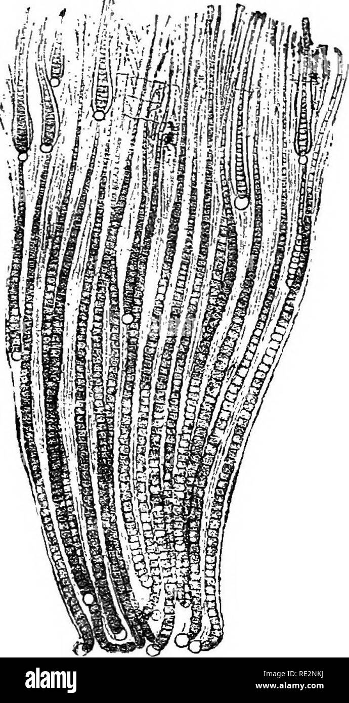 . A handbook of cryptogamic botany. Cryptogams. 436 PROTOPHYTA (Thur.) while still within the sheath, the cell-contents passing from a heterocyst into the basal cell of a hormogone. Multiplication by quiescent resting-spores has been observed in some species of Rivulariaceae. The lower portion of the green part of a fila- ment immediately above the basilar cell is transformed into an elliptical thick-walled spore, which escapes from its investing membrane, and, after a period of rest, either develops directly into a new filament, or breaks up into a number of hormo- gones. The spores of Gloeot Stock Photohttps://www.alamy.com/image-license-details/?v=1https://www.alamy.com/a-handbook-of-cryptogamic-botany-cryptogams-436-protophyta-thur-while-still-within-the-sheath-the-cell-contents-passing-from-a-heterocyst-into-the-basal-cell-of-a-hormogone-multiplication-by-quiescent-resting-spores-has-been-observed-in-some-species-of-rivulariaceae-the-lower-portion-of-the-green-part-of-a-fila-ment-immediately-above-the-basilar-cell-is-transformed-into-an-elliptical-thick-walled-spore-which-escapes-from-its-investing-membrane-and-after-a-period-of-rest-either-develops-directly-into-a-new-filament-or-breaks-up-into-a-number-of-hormo-gones-the-spores-of-gloeot-image232400886.html
RMRE2NKJ–. A handbook of cryptogamic botany. Cryptogams. 436 PROTOPHYTA (Thur.) while still within the sheath, the cell-contents passing from a heterocyst into the basal cell of a hormogone. Multiplication by quiescent resting-spores has been observed in some species of Rivulariaceae. The lower portion of the green part of a fila- ment immediately above the basilar cell is transformed into an elliptical thick-walled spore, which escapes from its investing membrane, and, after a period of rest, either develops directly into a new filament, or breaks up into a number of hormo- gones. The spores of Gloeot
 . Bulletin of the U.S. Department of Agriculture. Agriculture; Agriculture. IDENTIFICATION OP GRASSES. 3 Leaves (fig. 3).—A grass leaf consists of two principal parts, the sheath or tubular basal portion which incloses the stem, and the blade, nearly always long, narrow, and most commonly flat. The sheaths are usually cylindrical in form, but in some grasses they are laterally compressed, forming a keel at the back, in which case they are described as compressed. In some grasses the nerves of the sheaths are prominent, in others scarcely noticeable. A few grasses have distinctively colored she Stock Photohttps://www.alamy.com/image-license-details/?v=1https://www.alamy.com/bulletin-of-the-us-department-of-agriculture-agriculture-agriculture-identification-op-grasses-3-leaves-fig-3a-grass-leaf-consists-of-two-principal-parts-the-sheath-or-tubular-basal-portion-which-incloses-the-stem-and-the-blade-nearly-always-long-narrow-and-most-commonly-flat-the-sheaths-are-usually-cylindrical-in-form-but-in-some-grasses-they-are-laterally-compressed-forming-a-keel-at-the-back-in-which-case-they-are-described-as-compressed-in-some-grasses-the-nerves-of-the-sheaths-are-prominent-in-others-scarcely-noticeable-a-few-grasses-have-distinctively-colored-she-image233830649.html
RMRGBWAH–. Bulletin of the U.S. Department of Agriculture. Agriculture; Agriculture. IDENTIFICATION OP GRASSES. 3 Leaves (fig. 3).—A grass leaf consists of two principal parts, the sheath or tubular basal portion which incloses the stem, and the blade, nearly always long, narrow, and most commonly flat. The sheaths are usually cylindrical in form, but in some grasses they are laterally compressed, forming a keel at the back, in which case they are described as compressed. In some grasses the nerves of the sheaths are prominent, in others scarcely noticeable. A few grasses have distinctively colored she
![. Flies in relation to disease: bloodsucking flies. Flies; Flies as carriers of disease; Diptera. xvi] DESCRIPTION 245 contain the stylet-like epipharynx and the tubular hypopharynx which, by apposition, form a suctorial tube. The maxillary palps are slender and as long as the proboscis, round which they form a loose sheath when the insect is resting. The wings possess a very striking venation which is quite sufficient in itself to distinguish the genus. The fourth longi- tudinal vein is strongly curved in its proximal portions. The anterior basal transverse vein, at the base of the discal cel Stock Photo . Flies in relation to disease: bloodsucking flies. Flies; Flies as carriers of disease; Diptera. xvi] DESCRIPTION 245 contain the stylet-like epipharynx and the tubular hypopharynx which, by apposition, form a suctorial tube. The maxillary palps are slender and as long as the proboscis, round which they form a loose sheath when the insect is resting. The wings possess a very striking venation which is quite sufficient in itself to distinguish the genus. The fourth longi- tudinal vein is strongly curved in its proximal portions. The anterior basal transverse vein, at the base of the discal cel Stock Photo](https://c8.alamy.com/comp/RE3J4A/flies-in-relation-to-disease-bloodsucking-flies-flies-flies-as-carriers-of-disease-diptera-xvi-description-245-contain-the-stylet-like-epipharynx-and-the-tubular-hypopharynx-which-by-apposition-form-a-suctorial-tube-the-maxillary-palps-are-slender-and-as-long-as-the-proboscis-round-which-they-form-a-loose-sheath-when-the-insect-is-resting-the-wings-possess-a-very-striking-venation-which-is-quite-sufficient-in-itself-to-distinguish-the-genus-the-fourth-longi-tudinal-vein-is-strongly-curved-in-its-proximal-portions-the-anterior-basal-transverse-vein-at-the-base-of-the-discal-cel-RE3J4A.jpg) . Flies in relation to disease: bloodsucking flies. Flies; Flies as carriers of disease; Diptera. xvi] DESCRIPTION 245 contain the stylet-like epipharynx and the tubular hypopharynx which, by apposition, form a suctorial tube. The maxillary palps are slender and as long as the proboscis, round which they form a loose sheath when the insect is resting. The wings possess a very striking venation which is quite sufficient in itself to distinguish the genus. The fourth longi- tudinal vein is strongly curved in its proximal portions. The anterior basal transverse vein, at the base of the discal cel Stock Photohttps://www.alamy.com/image-license-details/?v=1https://www.alamy.com/flies-in-relation-to-disease-bloodsucking-flies-flies-flies-as-carriers-of-disease-diptera-xvi-description-245-contain-the-stylet-like-epipharynx-and-the-tubular-hypopharynx-which-by-apposition-form-a-suctorial-tube-the-maxillary-palps-are-slender-and-as-long-as-the-proboscis-round-which-they-form-a-loose-sheath-when-the-insect-is-resting-the-wings-possess-a-very-striking-venation-which-is-quite-sufficient-in-itself-to-distinguish-the-genus-the-fourth-longi-tudinal-vein-is-strongly-curved-in-its-proximal-portions-the-anterior-basal-transverse-vein-at-the-base-of-the-discal-cel-image232420058.html
. Flies in relation to disease: bloodsucking flies. Flies; Flies as carriers of disease; Diptera. xvi] DESCRIPTION 245 contain the stylet-like epipharynx and the tubular hypopharynx which, by apposition, form a suctorial tube. The maxillary palps are slender and as long as the proboscis, round which they form a loose sheath when the insect is resting. The wings possess a very striking venation which is quite sufficient in itself to distinguish the genus. The fourth longi- tudinal vein is strongly curved in its proximal portions. The anterior basal transverse vein, at the base of the discal cel Stock Photohttps://www.alamy.com/image-license-details/?v=1https://www.alamy.com/flies-in-relation-to-disease-bloodsucking-flies-flies-flies-as-carriers-of-disease-diptera-xvi-description-245-contain-the-stylet-like-epipharynx-and-the-tubular-hypopharynx-which-by-apposition-form-a-suctorial-tube-the-maxillary-palps-are-slender-and-as-long-as-the-proboscis-round-which-they-form-a-loose-sheath-when-the-insect-is-resting-the-wings-possess-a-very-striking-venation-which-is-quite-sufficient-in-itself-to-distinguish-the-genus-the-fourth-longi-tudinal-vein-is-strongly-curved-in-its-proximal-portions-the-anterior-basal-transverse-vein-at-the-base-of-the-discal-cel-image232420058.htmlRMRE3J4A–. Flies in relation to disease: bloodsucking flies. Flies; Flies as carriers of disease; Diptera. xvi] DESCRIPTION 245 contain the stylet-like epipharynx and the tubular hypopharynx which, by apposition, form a suctorial tube. The maxillary palps are slender and as long as the proboscis, round which they form a loose sheath when the insect is resting. The wings possess a very striking venation which is quite sufficient in itself to distinguish the genus. The fourth longi- tudinal vein is strongly curved in its proximal portions. The anterior basal transverse vein, at the base of the discal cel
 . Bulletin of the U.S. Department of Agriculture. Agriculture. 8 BULLETIN 438, U. S. DEPARTMENT OF AGRICULTURE. THE ADULT. Female.—Length 4.5 mm., very short and robust, shiny; head densely punctured, rather opaque; clypeus very slightly emarginate; frontal wanting or very slightly indi- cated; antennae very short, not as long as head and thorax, filiform, third joint longest; intercostal nearly at right angles with costa, interstitial with basal; venation otherwise normal; stigma short, broadly ovate at base;apex of costa strongly thickened; sheath broad, slightly emarginate beneath and acumi Stock Photohttps://www.alamy.com/image-license-details/?v=1https://www.alamy.com/bulletin-of-the-us-department-of-agriculture-agriculture-8-bulletin-438-u-s-department-of-agriculture-the-adult-femalelength-45-mm-very-short-and-robust-shiny-head-densely-punctured-rather-opaque-clypeus-very-slightly-emarginate-frontal-wanting-or-very-slightly-indi-cated-antennae-very-short-not-as-long-as-head-and-thorax-filiform-third-joint-longest-intercostal-nearly-at-right-angles-with-costa-interstitial-with-basal-venation-otherwise-normal-stigma-short-broadly-ovate-at-baseapex-of-costa-strongly-thickened-sheath-broad-slightly-emarginate-beneath-and-acumi-image233814331.html
RMRGB4FR–. Bulletin of the U.S. Department of Agriculture. Agriculture. 8 BULLETIN 438, U. S. DEPARTMENT OF AGRICULTURE. THE ADULT. Female.—Length 4.5 mm., very short and robust, shiny; head densely punctured, rather opaque; clypeus very slightly emarginate; frontal wanting or very slightly indi- cated; antennae very short, not as long as head and thorax, filiform, third joint longest; intercostal nearly at right angles with costa, interstitial with basal; venation otherwise normal; stigma short, broadly ovate at base;apex of costa strongly thickened; sheath broad, slightly emarginate beneath and acumi
 . An illustrated flora of the northern United States, Canada and the British possessions, from Newfoundland to the parallel of the southern boundary of Virginia, and from the Atlantic Ocean westward to the 102d meridian. Botany; Botany. 19. Juncus setaceus Rostk. Awl-leaved Rush. Fig. 1184. Juncus setaceus Rostk. Monog. June. 13. pi. 1. f. 2. 1801. Densely tufted from stout branching rootstsocks. Stems terete, spreading and recurved above, ii°-3° long; leaves all basal except those of the inflorescence, the uppermost sheath usually bearing a long terete blade similar to the stem, but channeled Stock Photohttps://www.alamy.com/image-license-details/?v=1https://www.alamy.com/an-illustrated-flora-of-the-northern-united-states-canada-and-the-british-possessions-from-newfoundland-to-the-parallel-of-the-southern-boundary-of-virginia-and-from-the-atlantic-ocean-westward-to-the-102d-meridian-botany-botany-19-juncus-setaceus-rostk-awl-leaved-rush-fig-1184-juncus-setaceus-rostk-monog-june-13-pi-1-f-2-1801-densely-tufted-from-stout-branching-rootstsocks-stems-terete-spreading-and-recurved-above-ii-3-long-leaves-all-basal-except-those-of-the-inflorescence-the-uppermost-sheath-usually-bearing-a-long-terete-blade-similar-to-the-stem-but-channeled-image232120013.html
RMRDHYCD–. An illustrated flora of the northern United States, Canada and the British possessions, from Newfoundland to the parallel of the southern boundary of Virginia, and from the Atlantic Ocean westward to the 102d meridian. Botany; Botany. 19. Juncus setaceus Rostk. Awl-leaved Rush. Fig. 1184. Juncus setaceus Rostk. Monog. June. 13. pi. 1. f. 2. 1801. Densely tufted from stout branching rootstsocks. Stems terete, spreading and recurved above, ii°-3° long; leaves all basal except those of the inflorescence, the uppermost sheath usually bearing a long terete blade similar to the stem, but channeled
 . Bulletin of the U.S. Department of Agriculture. Agriculture; Agriculture. 8 BULLETIISr 438, U. S. DEPARTMENT OF AGRICULTURE. THE ADULT. Female.—Length 4.5 mm., very short and robust, shiny; head densely punctured, rather opaque; clypeus very slightly emarginate; frontal wanting or very slightly indi- cated; antennae very short, not as long as head and thorax, filiform, third joint longest; intercostal nfearly at right angles with costa, interstitial with basal; venation otherwise normal; stigma short, broadly ovate at base; apex of costa strongly thickened; sheath broad, slightly emarginate Stock Photohttps://www.alamy.com/image-license-details/?v=1https://www.alamy.com/bulletin-of-the-us-department-of-agriculture-agriculture-agriculture-8-bulletiisr-438-u-s-department-of-agriculture-the-adult-femalelength-45-mm-very-short-and-robust-shiny-head-densely-punctured-rather-opaque-clypeus-very-slightly-emarginate-frontal-wanting-or-very-slightly-indi-cated-antennae-very-short-not-as-long-as-head-and-thorax-filiform-third-joint-longest-intercostal-nfearly-at-right-angles-with-costa-interstitial-with-basal-venation-otherwise-normal-stigma-short-broadly-ovate-at-base-apex-of-costa-strongly-thickened-sheath-broad-slightly-emarginate-image233819162.html
RMRGBAMA–. Bulletin of the U.S. Department of Agriculture. Agriculture; Agriculture. 8 BULLETIISr 438, U. S. DEPARTMENT OF AGRICULTURE. THE ADULT. Female.—Length 4.5 mm., very short and robust, shiny; head densely punctured, rather opaque; clypeus very slightly emarginate; frontal wanting or very slightly indi- cated; antennae very short, not as long as head and thorax, filiform, third joint longest; intercostal nfearly at right angles with costa, interstitial with basal; venation otherwise normal; stigma short, broadly ovate at base; apex of costa strongly thickened; sheath broad, slightly emarginate
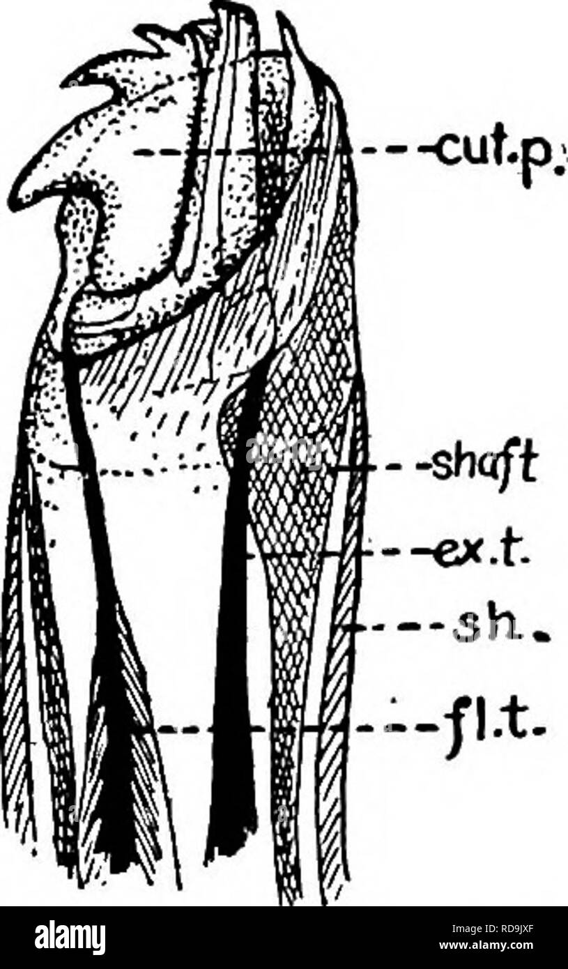 . Animal parasites and human disease. Medical parasitology; Insects as carriers of disease. Fig. 150. Head or capitulum of tick; hyp., hypostome; chel., chelicera; pal., palpus; has. p., basal piece. (Partly after Banks.). Fig. 151. Tip of chelicera of a tick, much enlarged; cut. p., articulated cutting part; shaft, shaft; sh., sheath; fl. t., tendon of flexor muscle; ex. t., tendon of extensor muscle. (After Nuttall, Cooper and Robinson.). Please note that these images are extracted from scanned page images that may have been digitally enhanced for readability - coloration and appearance of t Stock Photohttps://www.alamy.com/image-license-details/?v=1https://www.alamy.com/animal-parasites-and-human-disease-medical-parasitology-insects-as-carriers-of-disease-fig-150-head-or-capitulum-of-tick-hyp-hypostome-chel-chelicera-pal-palpus-has-p-basal-piece-partly-after-banks-fig-151-tip-of-chelicera-of-a-tick-much-enlarged-cut-p-articulated-cutting-part-shaft-shaft-sh-sheath-fl-t-tendon-of-flexor-muscle-ex-t-tendon-of-extensor-muscle-after-nuttall-cooper-and-robinson-please-note-that-these-images-are-extracted-from-scanned-page-images-that-may-have-been-digitally-enhanced-for-readability-coloration-and-appearance-of-t-image231937735.html
RMRD9JXF–. Animal parasites and human disease. Medical parasitology; Insects as carriers of disease. Fig. 150. Head or capitulum of tick; hyp., hypostome; chel., chelicera; pal., palpus; has. p., basal piece. (Partly after Banks.). Fig. 151. Tip of chelicera of a tick, much enlarged; cut. p., articulated cutting part; shaft, shaft; sh., sheath; fl. t., tendon of flexor muscle; ex. t., tendon of extensor muscle. (After Nuttall, Cooper and Robinson.). Please note that these images are extracted from scanned page images that may have been digitally enhanced for readability - coloration and appearance of t
 . Bulletin of the Museum of Comparative Zoology at Harvard College. Zoology. Bembidion Systematics • Maddison 257 anterior •dorsal basal orifice ostial microtrichial patch brush sclerite flagellum. sperm duct dorsal plate CSC right lobe CSC left lobe brush sclerite flagellar sheath CSC flagellum dorsal field. Please note that these images are extracted from scanned page images that may have been digitally enhanced for readability - coloration and appearance of these illustrations may not perfectly resemble the original work.. Harvard University. Museum of Comparative Zoology. Cambridge, Mass. Stock Photohttps://www.alamy.com/image-license-details/?v=1https://www.alamy.com/bulletin-of-the-museum-of-comparative-zoology-at-harvard-college-zoology-bembidion-systematics-maddison-257-anterior-dorsal-basal-orifice-ostial-microtrichial-patch-brush-sclerite-flagellum-sperm-duct-dorsal-plate-csc-right-lobe-csc-left-lobe-brush-sclerite-flagellar-sheath-csc-flagellum-dorsal-field-please-note-that-these-images-are-extracted-from-scanned-page-images-that-may-have-been-digitally-enhanced-for-readability-coloration-and-appearance-of-these-illustrations-may-not-perfectly-resemble-the-original-work-harvard-university-museum-of-comparative-zoology-cambridge-mass-image233898093.html
RMRGEYB9–. Bulletin of the Museum of Comparative Zoology at Harvard College. Zoology. Bembidion Systematics • Maddison 257 anterior •dorsal basal orifice ostial microtrichial patch brush sclerite flagellum. sperm duct dorsal plate CSC right lobe CSC left lobe brush sclerite flagellar sheath CSC flagellum dorsal field. Please note that these images are extracted from scanned page images that may have been digitally enhanced for readability - coloration and appearance of these illustrations may not perfectly resemble the original work.. Harvard University. Museum of Comparative Zoology. Cambridge, Mass.
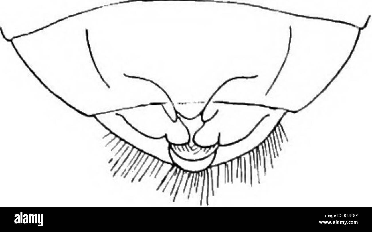 . Entomology for medical officers. Insect pests; Insects as carriers of disease. 204 ENTOMOLOGY FOR MEDICAL OFFICERS ClMEX LECTULARIUS, L. The insect, which is of a reddish-brown colour, is about one-fifth of an inch long, or rather less. The outline of its broad head is shaped something like a crown, the eyes projecting strongly. The 2 distal segments of the antennae are much slenderer than the 2 basal segments. The beak {cf. Fig. 86) forms a stout three-jointed sheath for the bristle-like mandibles and maxillae. The prothorax (Fig. 87) is large, its lateral margins are thin, and its Fig. 87. Stock Photohttps://www.alamy.com/image-license-details/?v=1https://www.alamy.com/entomology-for-medical-officers-insect-pests-insects-as-carriers-of-disease-204-entomology-for-medical-officers-clmex-lectularius-l-the-insect-which-is-of-a-reddish-brown-colour-is-about-one-fifth-of-an-inch-long-or-rather-less-the-outline-of-its-broad-head-is-shaped-something-like-a-crown-the-eyes-projecting-strongly-the-2-distal-segments-of-the-antennae-are-much-slenderer-than-the-2-basal-segments-the-beak-cf-fig-86-forms-a-stout-three-jointed-sheath-for-the-bristle-like-mandibles-and-maxillae-the-prothorax-fig-87-is-large-its-lateral-margins-are-thin-and-its-fig-87-image232427238.html
RMRE3Y8P–. Entomology for medical officers. Insect pests; Insects as carriers of disease. 204 ENTOMOLOGY FOR MEDICAL OFFICERS ClMEX LECTULARIUS, L. The insect, which is of a reddish-brown colour, is about one-fifth of an inch long, or rather less. The outline of its broad head is shaped something like a crown, the eyes projecting strongly. The 2 distal segments of the antennae are much slenderer than the 2 basal segments. The beak {cf. Fig. 86) forms a stout three-jointed sheath for the bristle-like mandibles and maxillae. The prothorax (Fig. 87) is large, its lateral margins are thin, and its Fig. 87.
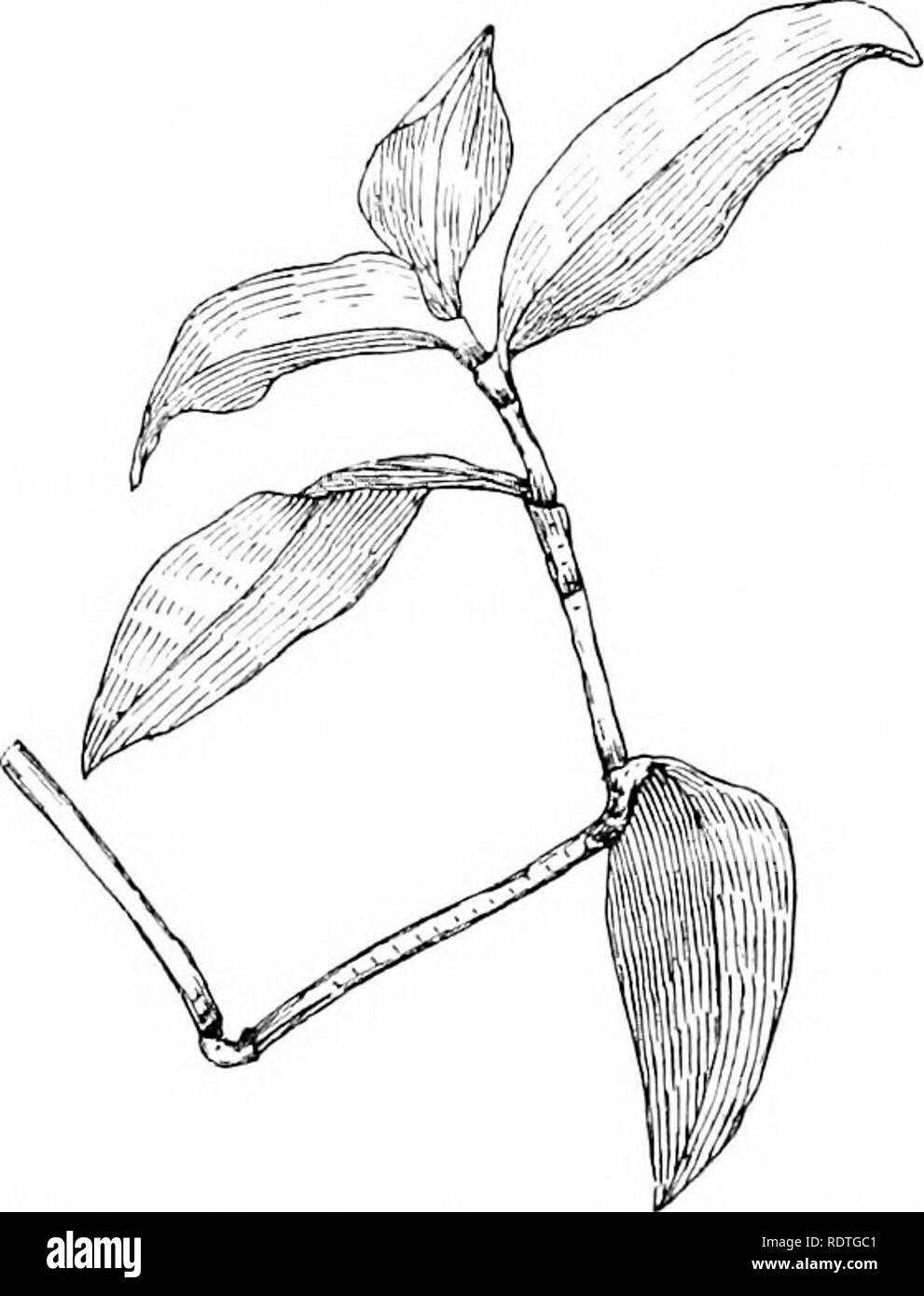 . Plants and their ways in South Africa. Botany; Botany. Fig. 26.—Showing basal growth in /.ca mays. needle, and strip from the sheath to expose the base of the leaf within. Mark off lines on the part exposed, carrying them across on to the sheath. When growth has taken place, so that the marks appear above the sheath, it will be seen that the place marked has been pushed up by the growth at the base of the leaf. Coumielina ' will be a good plant for studying the growth of a monocotyiedon- ousstem. Split the sheathing base of the leaf and mark the stem off. Mark other plants —Flagellaria, or a Stock Photohttps://www.alamy.com/image-license-details/?v=1https://www.alamy.com/plants-and-their-ways-in-south-africa-botany-botany-fig-26showing-basal-growth-in-ca-mays-needle-and-strip-from-the-sheath-to-expose-the-base-of-the-leaf-within-mark-off-lines-on-the-part-exposed-carrying-them-across-on-to-the-sheath-when-growth-has-taken-place-so-that-the-marks-appear-above-the-sheath-it-will-be-seen-that-the-place-marked-has-been-pushed-up-by-the-growth-at-the-base-of-the-leaf-coumielina-will-be-a-good-plant-for-studying-the-growth-of-a-monocotyiedon-ousstem-split-the-sheathing-base-of-the-leaf-and-mark-the-stem-off-mark-other-plants-flagellaria-or-a-image232265041.html
RMRDTGC1–. Plants and their ways in South Africa. Botany; Botany. Fig. 26.—Showing basal growth in /.ca mays. needle, and strip from the sheath to expose the base of the leaf within. Mark off lines on the part exposed, carrying them across on to the sheath. When growth has taken place, so that the marks appear above the sheath, it will be seen that the place marked has been pushed up by the growth at the base of the leaf. Coumielina ' will be a good plant for studying the growth of a monocotyiedon- ousstem. Split the sheathing base of the leaf and mark the stem off. Mark other plants —Flagellaria, or a
 . Brigham Young University science bulletin. Biology -- Periodicals. Bbicjiam Young University Science Buixetin CHELICERAL SHEATH ARTICLE III ARTICLE II LATERAL POINt SCAPULA CERVICAL GROOVE FESTOONS,. JHYPOSTOME DENTICLE ARTICLE I BASAL SPUR JNTERNAL SPUR EXTERNAL SPUR EXTERNAL SPUR 5PIRACULAR PLATE .GOBLET CELLS J>OSTANAL GROOVE Fig. 3. Dorsal and ventral views, njmphal Dennaccntor.. Please note that these images are extracted from scanned page images that may have been digitally enhanced for readability - coloration and appearance of these illustrations may not perfectly resemble the ori Stock Photohttps://www.alamy.com/image-license-details/?v=1https://www.alamy.com/brigham-young-university-science-bulletin-biology-periodicals-bbicjiam-young-university-science-buixetin-cheliceral-sheath-article-iii-article-ii-lateral-point-scapula-cervical-groove-festoons-jhypostome-denticle-article-i-basal-spur-jnternal-spur-external-spur-external-spur-5piracular-plate-goblet-cells-jgtostanal-groove-fig-3-dorsal-and-ventral-views-njmphal-dennaccntor-please-note-that-these-images-are-extracted-from-scanned-page-images-that-may-have-been-digitally-enhanced-for-readability-coloration-and-appearance-of-these-illustrations-may-not-perfectly-resemble-the-ori-image234276287.html
RMRH45P7–. Brigham Young University science bulletin. Biology -- Periodicals. Bbicjiam Young University Science Buixetin CHELICERAL SHEATH ARTICLE III ARTICLE II LATERAL POINt SCAPULA CERVICAL GROOVE FESTOONS,. JHYPOSTOME DENTICLE ARTICLE I BASAL SPUR JNTERNAL SPUR EXTERNAL SPUR EXTERNAL SPUR 5PIRACULAR PLATE .GOBLET CELLS J>OSTANAL GROOVE Fig. 3. Dorsal and ventral views, njmphal Dennaccntor.. Please note that these images are extracted from scanned page images that may have been digitally enhanced for readability - coloration and appearance of these illustrations may not perfectly resemble the ori
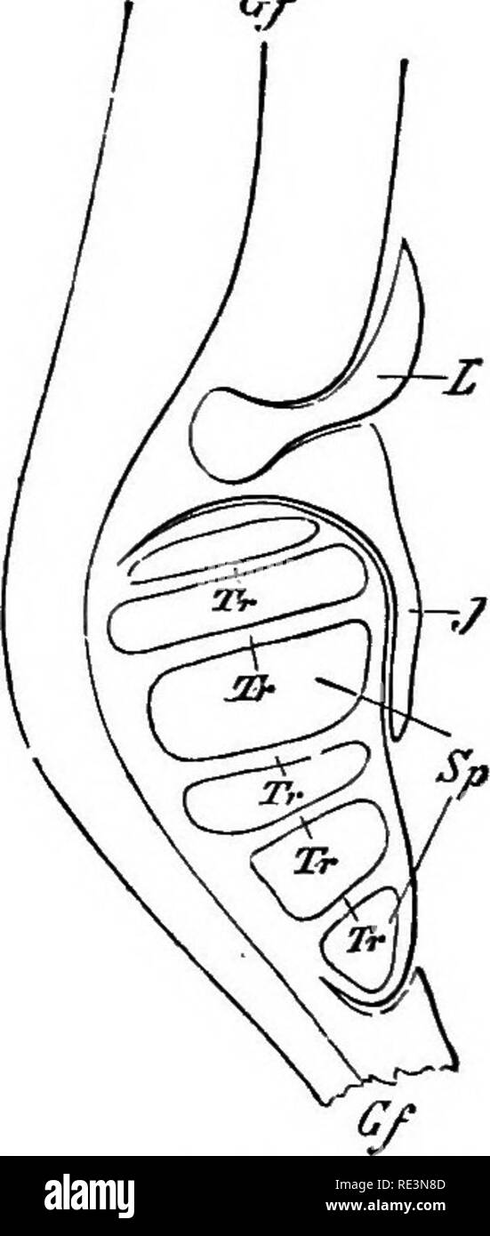 . A handbook of cryptogamic botany. Cryptogams. Fig. 31.—Longitudinal section of stem of/. lacitstris. h—b^, leaves; r^—7-^% roots: the ligules are shaded (X30). (After Hofmeister.) Fig. 32.—Longitudinal section through lower portion of leaf of/. /acwj^rri (diagram- matic). L, ligule ; y, inda sium; S^, microsporange Tr, trabecules; Gf^ vascu- lar bundle of sporophyll. (After Goebel.) The leaves of Isoetes are very elongated, cylindrical, and quill-shaped, and are arranged in a complicated phyllotaxis on the very short stem. They are segmented into a basal portion, the sheath or glossqpode, an Stock Photohttps://www.alamy.com/image-license-details/?v=1https://www.alamy.com/a-handbook-of-cryptogamic-botany-cryptogams-fig-31longitudinal-section-of-stem-of-lacitstris-hb-leaves-r7-roots-the-ligules-are-shaded-x30-after-hofmeister-fig-32longitudinal-section-through-lower-portion-of-leaf-of-acwjrri-diagram-matic-l-ligule-y-inda-sium-s-microsporange-tr-trabecules-gf-vascu-lar-bundle-of-sporophyll-after-goebel-the-leaves-of-isoetes-are-very-elongated-cylindrical-and-quill-shaped-and-are-arranged-in-a-complicated-phyllotaxis-on-the-very-short-stem-they-are-segmented-into-a-basal-portion-the-sheath-or-glossqpode-an-image232422525.html
RMRE3N8D–. A handbook of cryptogamic botany. Cryptogams. Fig. 31.—Longitudinal section of stem of/. lacitstris. h—b^, leaves; r^—7-^% roots: the ligules are shaded (X30). (After Hofmeister.) Fig. 32.—Longitudinal section through lower portion of leaf of/. /acwj^rri (diagram- matic). L, ligule ; y, inda sium; S^, microsporange Tr, trabecules; Gf^ vascu- lar bundle of sporophyll. (After Goebel.) The leaves of Isoetes are very elongated, cylindrical, and quill-shaped, and are arranged in a complicated phyllotaxis on the very short stem. They are segmented into a basal portion, the sheath or glossqpode, an
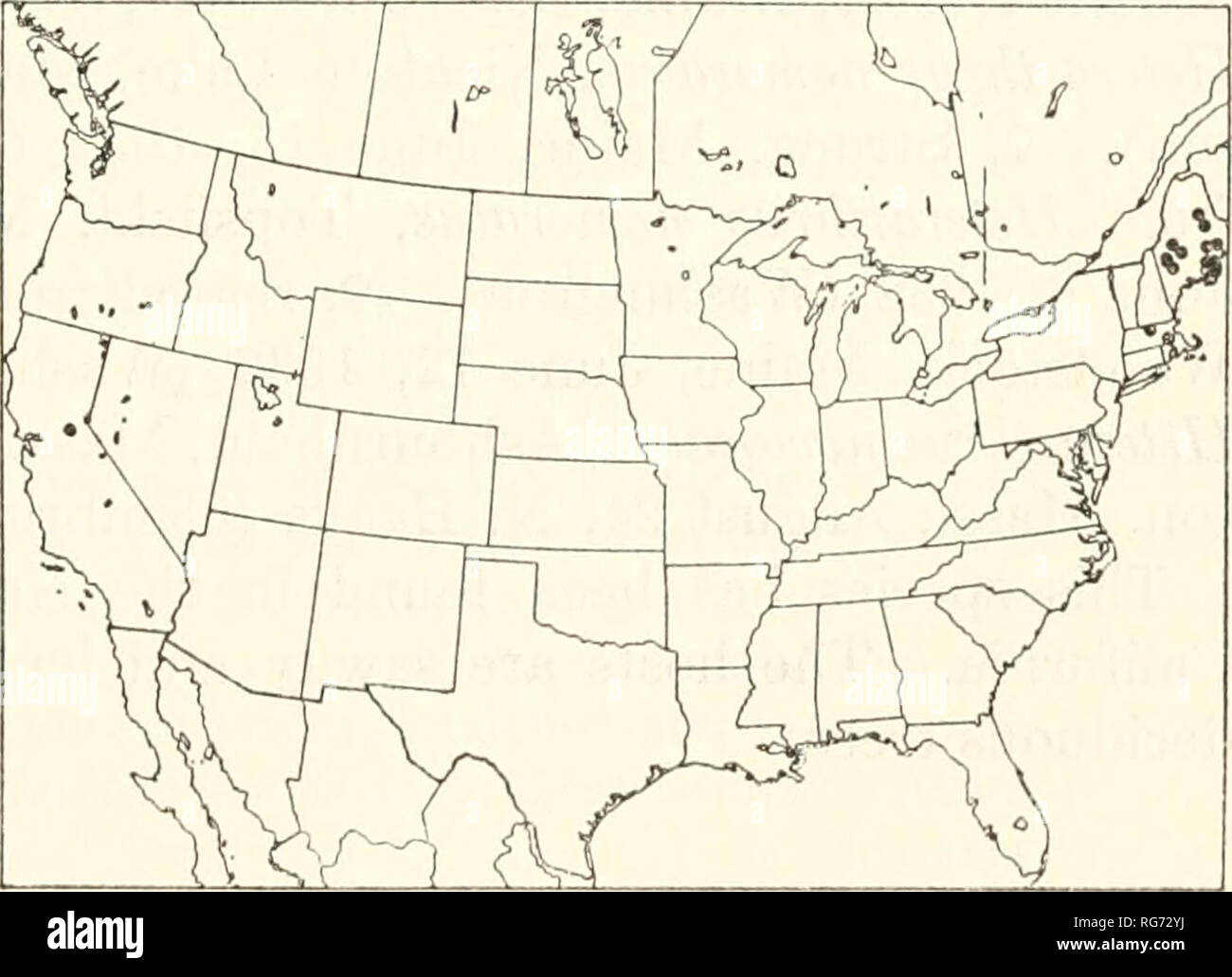 . Bulletin - United States National Museum. Science. ICHNEUMON-FLIES, PART 2: EPHIALTINAE 207 their length; female subgenital plate completely but weakly sclero- tized, or with a large median basal membranous area; ovipositor weakly compressed, decurved but its apical part straight; ovipositor sheath about 0.33 as long as front wing. This genus includes only the genotype, which is transcontinental in the Canadian and Transition zones of North America, and an undescribed species in Japan. 1. Alophosternuin foliicola Cushman Figure 370 Alophosternum foliicola Cushman, 1933, Proc. U. S. Nat. Mus. Stock Photohttps://www.alamy.com/image-license-details/?v=1https://www.alamy.com/bulletin-united-states-national-museum-science-ichneumon-flies-part-2-ephialtinae-207-their-length-female-subgenital-plate-completely-but-weakly-sclero-tized-or-with-a-large-median-basal-membranous-area-ovipositor-weakly-compressed-decurved-but-its-apical-part-straight-ovipositor-sheath-about-033-as-long-as-front-wing-this-genus-includes-only-the-genotype-which-is-transcontinental-in-the-canadian-and-transition-zones-of-north-america-and-an-undescribed-species-in-japan-1-alophosternuin-foliicola-cushman-figure-370-alophosternum-foliicola-cushman-1933-proc-u-s-nat-mus-image233725286.html
RMRG72YJ–. Bulletin - United States National Museum. Science. ICHNEUMON-FLIES, PART 2: EPHIALTINAE 207 their length; female subgenital plate completely but weakly sclero- tized, or with a large median basal membranous area; ovipositor weakly compressed, decurved but its apical part straight; ovipositor sheath about 0.33 as long as front wing. This genus includes only the genotype, which is transcontinental in the Canadian and Transition zones of North America, and an undescribed species in Japan. 1. Alophosternuin foliicola Cushman Figure 370 Alophosternum foliicola Cushman, 1933, Proc. U. S. Nat. Mus.
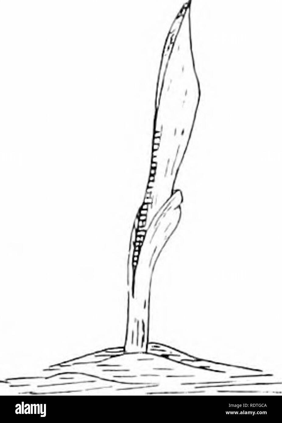 . Plants and their ways in South Africa. Botany; Botany. Fig. 26.—Showing basal growth in /.ca mays. needle, and strip from the sheath to expose the base of the leaf within. Mark off lines on the part exposed, carrying them across on to the sheath. When growth has taken place, so that the marks appear above the sheath, it will be seen that the place marked has been pushed up by the growth at the base of the leaf. Coumielina ' will be a good plant for studying the growth of a monocotyiedon- ousstem. Split the sheathing base of the leaf and mark the stem off. Mark other plants —Flagellaria, or a Stock Photohttps://www.alamy.com/image-license-details/?v=1https://www.alamy.com/plants-and-their-ways-in-south-africa-botany-botany-fig-26showing-basal-growth-in-ca-mays-needle-and-strip-from-the-sheath-to-expose-the-base-of-the-leaf-within-mark-off-lines-on-the-part-exposed-carrying-them-across-on-to-the-sheath-when-growth-has-taken-place-so-that-the-marks-appear-above-the-sheath-it-will-be-seen-that-the-place-marked-has-been-pushed-up-by-the-growth-at-the-base-of-the-leaf-coumielina-will-be-a-good-plant-for-studying-the-growth-of-a-monocotyiedon-ousstem-split-the-sheathing-base-of-the-leaf-and-mark-the-stem-off-mark-other-plants-flagellaria-or-a-image232265050.html
RMRDTGCA–. Plants and their ways in South Africa. Botany; Botany. Fig. 26.—Showing basal growth in /.ca mays. needle, and strip from the sheath to expose the base of the leaf within. Mark off lines on the part exposed, carrying them across on to the sheath. When growth has taken place, so that the marks appear above the sheath, it will be seen that the place marked has been pushed up by the growth at the base of the leaf. Coumielina ' will be a good plant for studying the growth of a monocotyiedon- ousstem. Split the sheathing base of the leaf and mark the stem off. Mark other plants —Flagellaria, or a
 . Elements of the comparative anatomy of vertebrates. Anatomy, Comparative. Fill. 2'ii.—Portion op the Vertebral L'olumx of ,b>'(<H/V(/7V(. (Side view.) Fig. 24.—Transverse Section of the Vertebral Colujin of Aciptuv-r rutheiiux (in the anterior part of the bod}'). Pn, spinous process ; J, lowei' arch ; Ao, aorta ; Po, median parts of the lower arches, which here enclcie the aorta ventrally ; Z, basal processes of the lower arches. is essentially indicated by the neural arches. In the two groups last mentioned, however, skeletogenous cells break through the primary notochordal sheath (el Stock Photohttps://www.alamy.com/image-license-details/?v=1https://www.alamy.com/elements-of-the-comparative-anatomy-of-vertebrates-anatomy-comparative-fill-2iiportion-op-the-vertebral-lolumx-of-bgtlthv7v-side-view-fig-24transverse-section-of-the-vertebral-colujin-of-aciptuv-r-rutheiiux-in-the-anterior-part-of-the-bod-pn-spinous-process-j-lowei-arch-ao-aorta-po-median-parts-of-the-lower-arches-which-here-enclcie-the-aorta-ventrally-z-basal-processes-of-the-lower-arches-is-essentially-indicated-by-the-neural-arches-in-the-two-groups-last-mentioned-however-skeletogenous-cells-break-through-the-primary-notochordal-sheath-el-image232075877.html
RMRDFY45–. Elements of the comparative anatomy of vertebrates. Anatomy, Comparative. Fill. 2'ii.—Portion op the Vertebral L'olumx of ,b>'(<H/V(/7V(. (Side view.) Fig. 24.—Transverse Section of the Vertebral Colujin of Aciptuv-r rutheiiux (in the anterior part of the bod}'). Pn, spinous process ; J, lowei' arch ; Ao, aorta ; Po, median parts of the lower arches, which here enclcie the aorta ventrally ; Z, basal processes of the lower arches. is essentially indicated by the neural arches. In the two groups last mentioned, however, skeletogenous cells break through the primary notochordal sheath (el
 . Bulletin. Natural history; Natural history. c? Genitalia Fig. 21. .-/gray/ea sa/leseti sinuate appendages (probably cerci) bearing a number of well separated setae at their apex; these extend con- siderably beyond the claspers. Above these and below the oedagus is a sclero- tized rod which (seen from lateral view) appears bent downward at almost a right angle; this has practically no setae on it and forms a sheath for the oedagus. Oedagus with the basal portion shorter than the apical portion; its margins irregular and sinuate and gradually tapering to the neck; apical portion somewhat bulbo Stock Photohttps://www.alamy.com/image-license-details/?v=1https://www.alamy.com/bulletin-natural-history-natural-history-c-genitalia-fig-21-grayea-saleseti-sinuate-appendages-probably-cerci-bearing-a-number-of-well-separated-setae-at-their-apex-these-extend-con-siderably-beyond-the-claspers-above-these-and-below-the-oedagus-is-a-sclero-tized-rod-which-seen-from-lateral-view-appears-bent-downward-at-almost-a-right-angle-this-has-practically-no-setae-on-it-and-forms-a-sheath-for-the-oedagus-oedagus-with-the-basal-portion-shorter-than-the-apical-portion-its-margins-irregular-and-sinuate-and-gradually-tapering-to-the-neck-apical-portion-somewhat-bulbo-image234129923.html
RMRGWF2Y–. Bulletin. Natural history; Natural history. c? Genitalia Fig. 21. .-/gray/ea sa/leseti sinuate appendages (probably cerci) bearing a number of well separated setae at their apex; these extend con- siderably beyond the claspers. Above these and below the oedagus is a sclero- tized rod which (seen from lateral view) appears bent downward at almost a right angle; this has practically no setae on it and forms a sheath for the oedagus. Oedagus with the basal portion shorter than the apical portion; its margins irregular and sinuate and gradually tapering to the neck; apical portion somewhat bulbo
 . A manual of structural botany; an introductory textbook for students of science and pharmacy. Plant morphology. DEVELOPMENT OP THE LEAP 171 grasses (a, in Fig. 465 A). Instead of passing around the stem, the edges may come together between the leaf and the stem, so as to produce a hollow tube, as in the Sarracenia. Let it next be assumed that the apical portion, as well as the central-basal, enlarges, with little enlarge- ment of the axial or lateral portions. We shall then get a form in which a Lamina, or Leaf-blade, is superposed directly upon a Leaf-sheath. Such a leaf, expanded, would ap Stock Photohttps://www.alamy.com/image-license-details/?v=1https://www.alamy.com/a-manual-of-structural-botany-an-introductory-textbook-for-students-of-science-and-pharmacy-plant-morphology-development-op-the-leap-171-grasses-a-in-fig-465-a-instead-of-passing-around-the-stem-the-edges-may-come-together-between-the-leaf-and-the-stem-so-as-to-produce-a-hollow-tube-as-in-the-sarracenia-let-it-next-be-assumed-that-the-apical-portion-as-well-as-the-central-basal-enlarges-with-little-enlarge-ment-of-the-axial-or-lateral-portions-we-shall-then-get-a-form-in-which-a-lamina-or-leaf-blade-is-superposed-directly-upon-a-leaf-sheath-such-a-leaf-expanded-would-ap-image232347279.html
RMRE0993–. A manual of structural botany; an introductory textbook for students of science and pharmacy. Plant morphology. DEVELOPMENT OP THE LEAP 171 grasses (a, in Fig. 465 A). Instead of passing around the stem, the edges may come together between the leaf and the stem, so as to produce a hollow tube, as in the Sarracenia. Let it next be assumed that the apical portion, as well as the central-basal, enlarges, with little enlarge- ment of the axial or lateral portions. We shall then get a form in which a Lamina, or Leaf-blade, is superposed directly upon a Leaf-sheath. Such a leaf, expanded, would ap
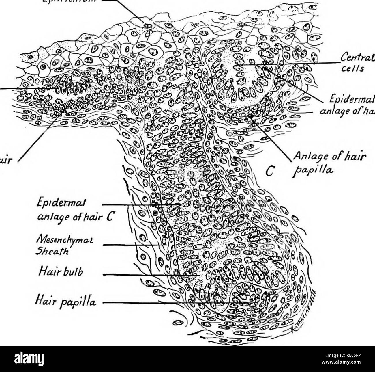 . A laboratory manual and text-book of embryology. Embryology. 306 HISTOGENESIS The hair is differentiated from the basal epidermal cells surrounding the hair papilla. These cells elongate and grow centrally toward the surface distinct Epitricbium Epidermal anlage of hair A Anlaqe of hair hapilla *, Epidermaf. anlage of hair B. Stieath Hair bulb flair papilla Fig. 293.—Section through the integument of the face of a 65 mm. embryo showing three stages in the early development of the hair. X 330. from the peripheral cells which form the outer sheath of the hair (Fig. 294). The central core of ce Stock Photohttps://www.alamy.com/image-license-details/?v=1https://www.alamy.com/a-laboratory-manual-and-text-book-of-embryology-embryology-306-histogenesis-the-hair-is-differentiated-from-the-basal-epidermal-cells-surrounding-the-hair-papilla-these-cells-elongate-and-grow-centrally-toward-the-surface-distinct-epitricbium-epidermal-anlage-of-hair-a-anlaqe-of-hair-hapilla-epidermaf-anlage-of-hair-b-stieath-hair-bulb-flair-papilla-fig-293section-through-the-integument-of-the-face-of-a-65-mm-embryo-showing-three-stages-in-the-early-development-of-the-hair-x-330-from-the-peripheral-cells-which-form-the-outer-sheath-of-the-hair-fig-294-the-central-core-of-ce-image232344526.html
RMRE05PP–. A laboratory manual and text-book of embryology. Embryology. 306 HISTOGENESIS The hair is differentiated from the basal epidermal cells surrounding the hair papilla. These cells elongate and grow centrally toward the surface distinct Epitricbium Epidermal anlage of hair A Anlaqe of hair hapilla *, Epidermaf. anlage of hair B. Stieath Hair bulb flair papilla Fig. 293.—Section through the integument of the face of a 65 mm. embryo showing three stages in the early development of the hair. X 330. from the peripheral cells which form the outer sheath of the hair (Fig. 294). The central core of ce
 . Animal parasites and human disease. Insect Vectors; Parasites; Parasitic Diseases; Medical parasitology; Insects as carriers of disease. rj^-bas.p. Fio. 150. Head or capitulum of tick; hyp., hypostome; chel., chelicera; pal., palpus; bas. p., basal piece. (Partly after Banks.). Fig. 151. Tip of chelicera of a tick, much enlarged; cut. p., articulated cutting part; shaft, shaft; sh., sheath; fl. t., tendon of flexor muscle; ex. t., tendon of extensor muscle. (After Nuttall, Cooper and Robinson.). Please note that these images are extracted from scanned page images that may have been digitally Stock Photohttps://www.alamy.com/image-license-details/?v=1https://www.alamy.com/animal-parasites-and-human-disease-insect-vectors-parasites-parasitic-diseases-medical-parasitology-insects-as-carriers-of-disease-rj-basp-fio-150-head-or-capitulum-of-tick-hyp-hypostome-chel-chelicera-pal-palpus-bas-p-basal-piece-partly-after-banks-fig-151-tip-of-chelicera-of-a-tick-much-enlarged-cut-p-articulated-cutting-part-shaft-shaft-sh-sheath-fl-t-tendon-of-flexor-muscle-ex-t-tendon-of-extensor-muscle-after-nuttall-cooper-and-robinson-please-note-that-these-images-are-extracted-from-scanned-page-images-that-may-have-been-digitally-image236732997.html
RMRN439W–. Animal parasites and human disease. Insect Vectors; Parasites; Parasitic Diseases; Medical parasitology; Insects as carriers of disease. rj^-bas.p. Fio. 150. Head or capitulum of tick; hyp., hypostome; chel., chelicera; pal., palpus; bas. p., basal piece. (Partly after Banks.). Fig. 151. Tip of chelicera of a tick, much enlarged; cut. p., articulated cutting part; shaft, shaft; sh., sheath; fl. t., tendon of flexor muscle; ex. t., tendon of extensor muscle. (After Nuttall, Cooper and Robinson.). Please note that these images are extracted from scanned page images that may have been digitally
 . Bulletin of the British Museum (Natural History) Entom Supp. OF THE FAMILY COCCIDAE 67 narrowly joined anterior to anus ; length of basal rod §—$ as long as aedeagus and extending anteriorly from the base of aedeagus for about § of the length to the apex of the basal mem- branous area ; apex of sheath without membranous extension. The area from base of sheath to tip of aedeagus with 17-26 (average 20) small setae ; a cluster of small sensilla occurring ventrally near apex of sheath. Aedeagus of medium length (101-120, average 114 (x), penial sheath and basisternum longer, the ratios being 1 Stock Photohttps://www.alamy.com/image-license-details/?v=1https://www.alamy.com/bulletin-of-the-british-museum-natural-history-entom-supp-of-the-family-coccidae-67-narrowly-joined-anterior-to-anus-length-of-basal-rod-as-long-as-aedeagus-and-extending-anteriorly-from-the-base-of-aedeagus-for-about-of-the-length-to-the-apex-of-the-basal-mem-branous-area-apex-of-sheath-without-membranous-extension-the-area-from-base-of-sheath-to-tip-of-aedeagus-with-17-26-average-20-small-setae-a-cluster-of-small-sensilla-occurring-ventrally-near-apex-of-sheath-aedeagus-of-medium-length-101-120-average-114-x-penial-sheath-and-basisternum-longer-the-ratios-being-1-image233951744.html
RMRGHBRC–. Bulletin of the British Museum (Natural History) Entom Supp. OF THE FAMILY COCCIDAE 67 narrowly joined anterior to anus ; length of basal rod §—$ as long as aedeagus and extending anteriorly from the base of aedeagus for about § of the length to the apex of the basal mem- branous area ; apex of sheath without membranous extension. The area from base of sheath to tip of aedeagus with 17-26 (average 20) small setae ; a cluster of small sensilla occurring ventrally near apex of sheath. Aedeagus of medium length (101-120, average 114 (x), penial sheath and basisternum longer, the ratios being 1
 . Bulletin. Natural history; Natural history. March 1938 ROSS: NEARCTIC CADDIS FLIES 115. c? Genitalia Fig. 21. .-/gray/ea sa/leseti sinuate appendages (probably cerci) bearing a number of well separated setae at their apex; these extend con- siderably beyond the claspers. Above these and below the oedagus is a sclero- tized rod which (seen from lateral view) appears bent downward at almost a right angle; this has practically no setae on it and forms a sheath for the oedagus. Oedagus with the basal portion shorter than the apical portion; its margins irregular and sinuate and gradually taperin Stock Photohttps://www.alamy.com/image-license-details/?v=1https://www.alamy.com/bulletin-natural-history-natural-history-march-1938-ross-nearctic-caddis-flies-115-c-genitalia-fig-21-grayea-saleseti-sinuate-appendages-probably-cerci-bearing-a-number-of-well-separated-setae-at-their-apex-these-extend-con-siderably-beyond-the-claspers-above-these-and-below-the-oedagus-is-a-sclero-tized-rod-which-seen-from-lateral-view-appears-bent-downward-at-almost-a-right-angle-this-has-practically-no-setae-on-it-and-forms-a-sheath-for-the-oedagus-oedagus-with-the-basal-portion-shorter-than-the-apical-portion-its-margins-irregular-and-sinuate-and-gradually-taperin-image234129932.html
RMRGWF38–. Bulletin. Natural history; Natural history. March 1938 ROSS: NEARCTIC CADDIS FLIES 115. c? Genitalia Fig. 21. .-/gray/ea sa/leseti sinuate appendages (probably cerci) bearing a number of well separated setae at their apex; these extend con- siderably beyond the claspers. Above these and below the oedagus is a sclero- tized rod which (seen from lateral view) appears bent downward at almost a right angle; this has practically no setae on it and forms a sheath for the oedagus. Oedagus with the basal portion shorter than the apical portion; its margins irregular and sinuate and gradually taperin