Quick filters:
Bipolar neuron Stock Photos and Images
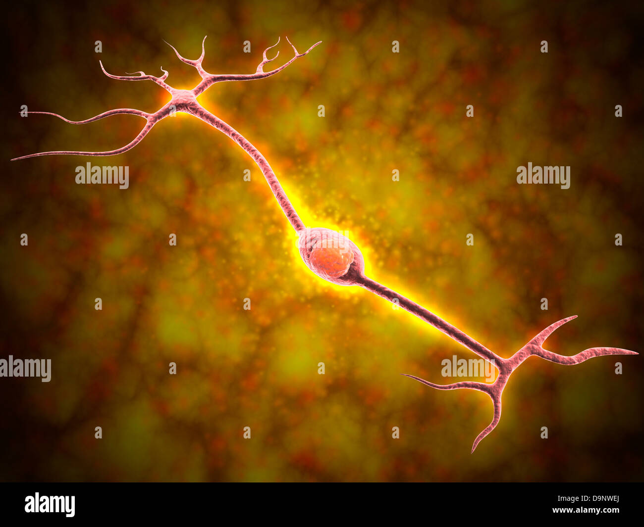 Microscopic view of a bipolar neuron Stock Photohttps://www.alamy.com/image-license-details/?v=1https://www.alamy.com/stock-photo-microscopic-view-of-a-bipolar-neuron-57644010.html
RFD9NWEJ–Microscopic view of a bipolar neuron
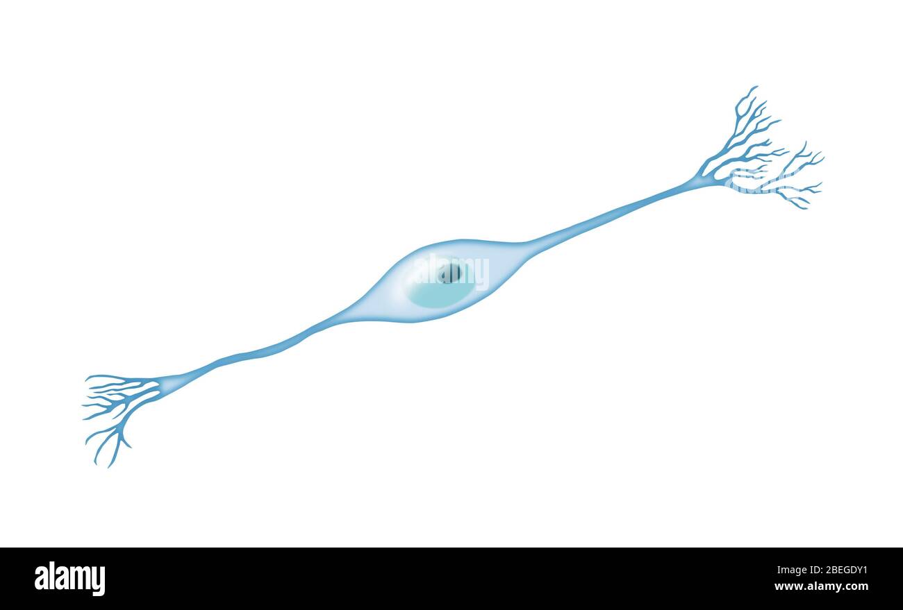 Bipolar Neuron Stock Photohttps://www.alamy.com/image-license-details/?v=1https://www.alamy.com/bipolar-neuron-image353174725.html
RM2BEGDY1–Bipolar Neuron
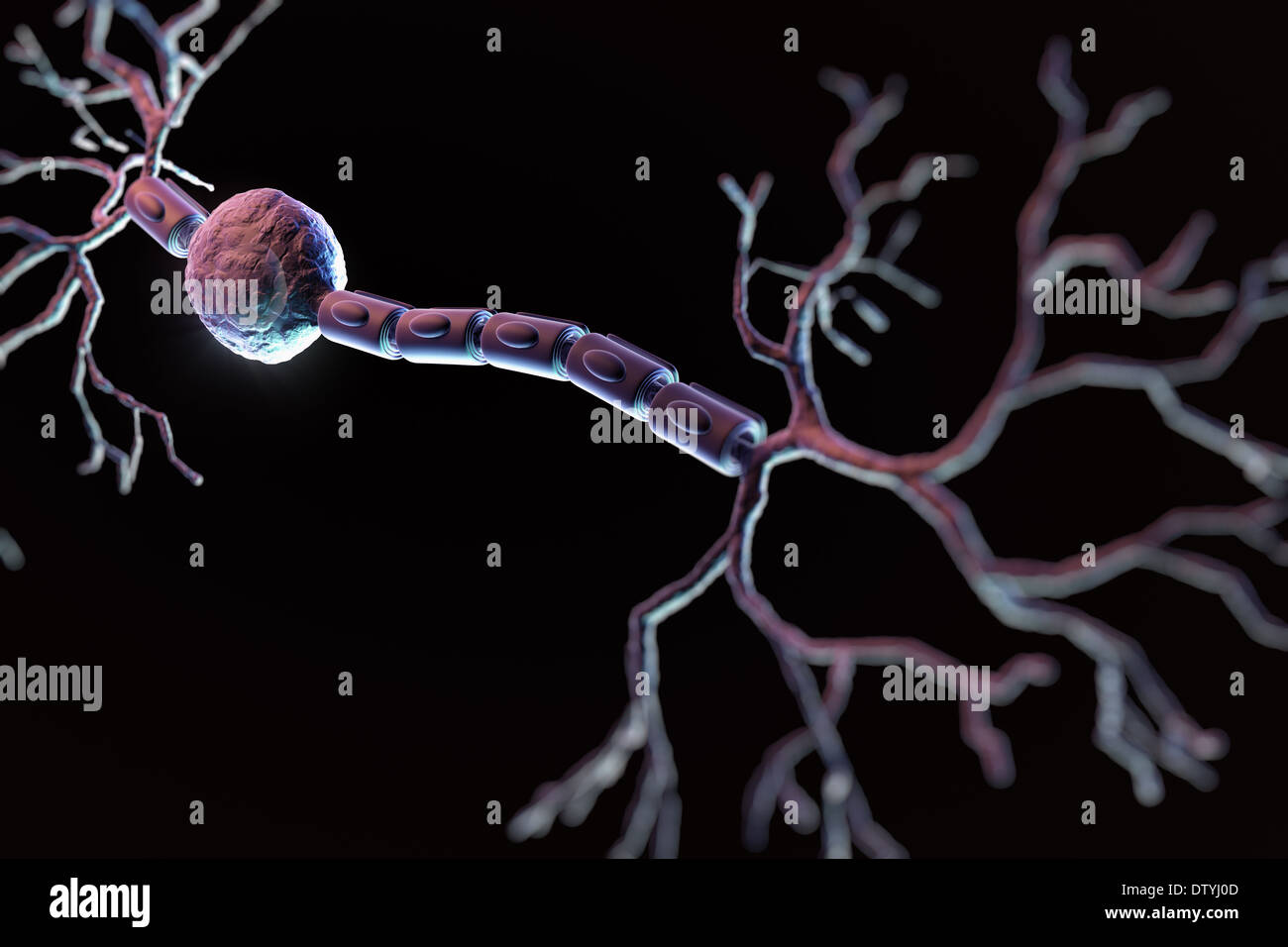 Bipolar Neuron Stock Photohttps://www.alamy.com/image-license-details/?v=1https://www.alamy.com/bipolar-neuron-image66989677.html
RMDTYJ0D–Bipolar Neuron
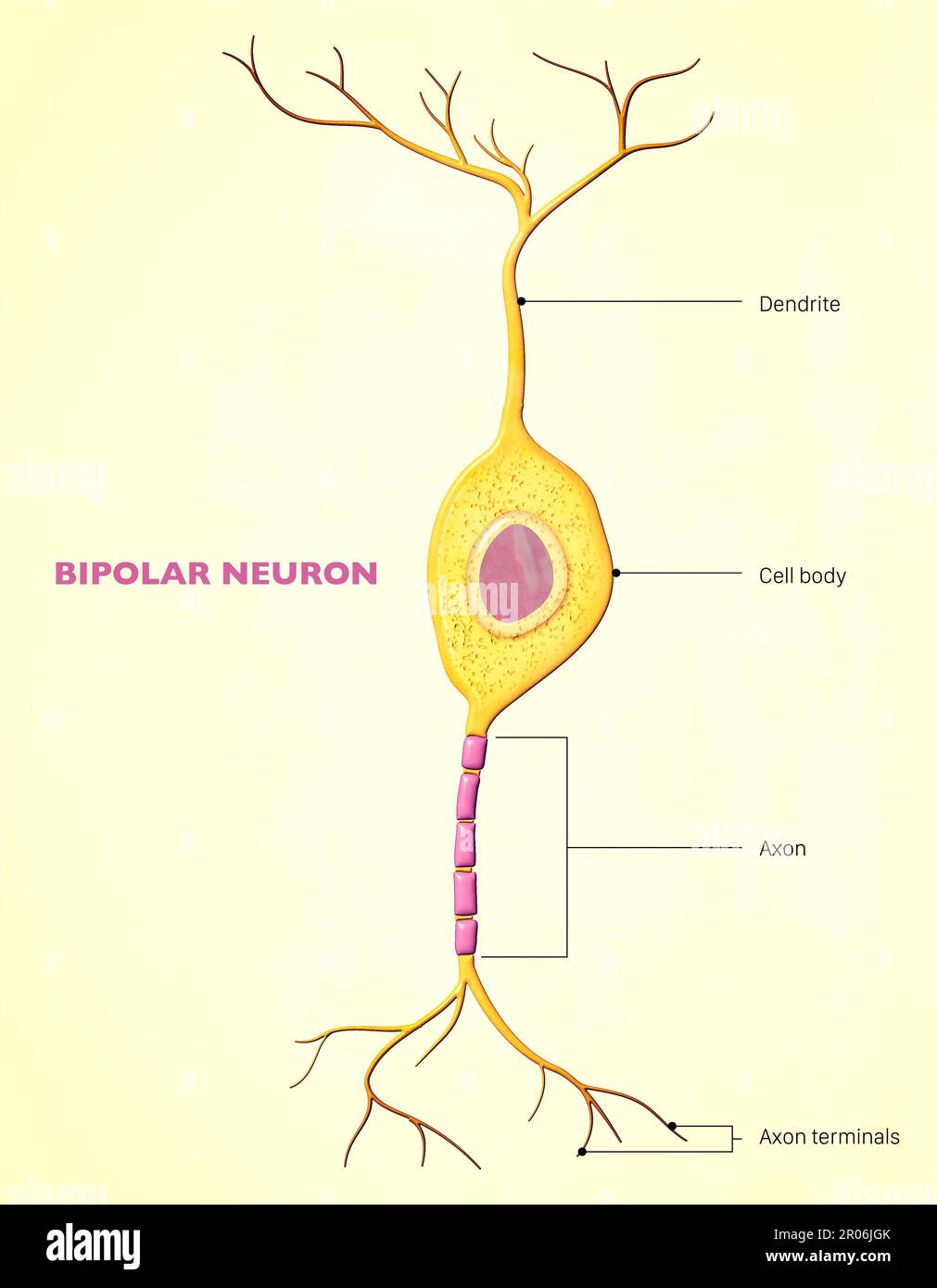 A bipolar neuron, or bipolar cell, is a type of neuron that has two extensions (one axon and one dendrite). Many bipolar cells are specialized sensory Stock Photohttps://www.alamy.com/image-license-details/?v=1https://www.alamy.com/a-bipolar-neuron-or-bipolar-cell-is-a-type-of-neuron-that-has-two-extensions-one-axon-and-one-dendrite-many-bipolar-cells-are-specialized-sensory-image550878067.html
RF2R06JGK–A bipolar neuron, or bipolar cell, is a type of neuron that has two extensions (one axon and one dendrite). Many bipolar cells are specialized sensory
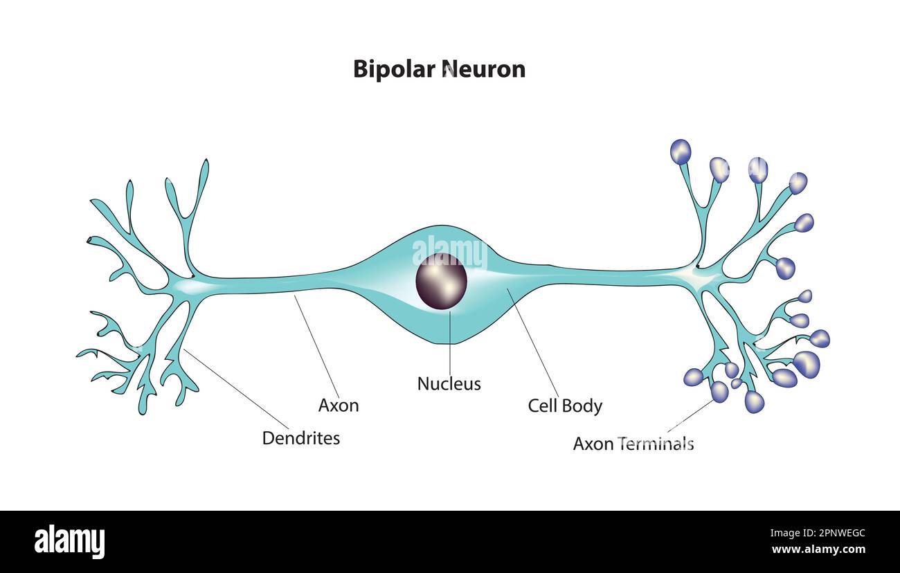 bipolar neuron Stock Vectorhttps://www.alamy.com/image-license-details/?v=1https://www.alamy.com/bipolar-neuron-image546989420.html
RF2PNWEGC–bipolar neuron
 Neural circuits in the mouse retina. Cone photoreceptors (red) allow color vision bipolar neurons (magenta) to relay information further down the circuit and a subtype of bipolar neuron (green) helps process signals detected by other photoreceptors in the penumbra. Stock Photohttps://www.alamy.com/image-license-details/?v=1https://www.alamy.com/neural-circuits-in-the-mouse-retina-cone-photoreceptors-red-allow-color-vision-bipolar-neurons-magenta-to-relay-information-further-down-the-circuit-and-a-subtype-of-bipolar-neuron-green-helps-process-signals-detected-by-other-photoreceptors-in-the-penumbra-image476707004.html
RM2JKFTJ4–Neural circuits in the mouse retina. Cone photoreceptors (red) allow color vision bipolar neurons (magenta) to relay information further down the circuit and a subtype of bipolar neuron (green) helps process signals detected by other photoreceptors in the penumbra.
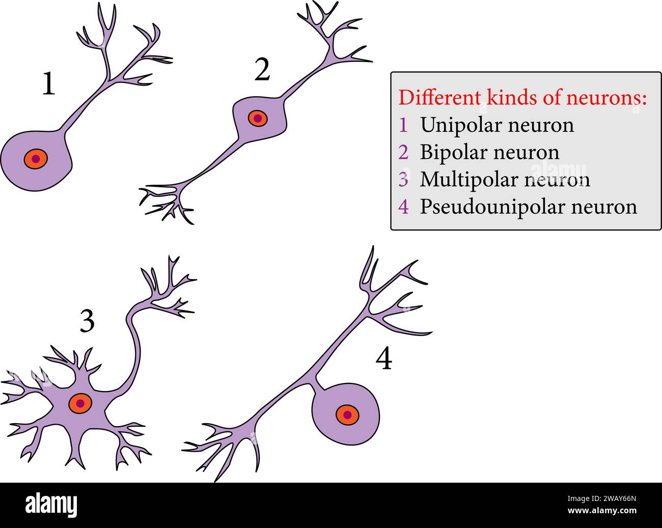 kinds of neurons: Unipolar neuron , Bipolar neuron , Multipolar neuron ,Pseudounipolar neuron.Vector illustration. Stock Vectorhttps://www.alamy.com/image-license-details/?v=1https://www.alamy.com/kinds-of-neurons-unipolar-neuron-bipolar-neuron-multipolar-neuron-pseudounipolar-neuronvector-illustration-image591896669.html
RF2WAY66N–kinds of neurons: Unipolar neuron , Bipolar neuron , Multipolar neuron ,Pseudounipolar neuron.Vector illustration.
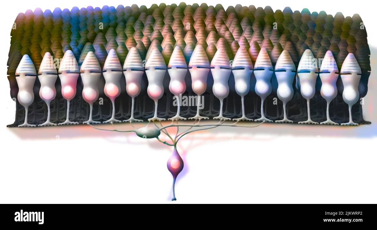 Organization of photoreceptors into receptor fields transmitting nerve impulses to bipolar cells. Stock Photohttps://www.alamy.com/image-license-details/?v=1https://www.alamy.com/organization-of-photoreceptors-into-receptor-fields-transmitting-nerve-impulses-to-bipolar-cells-image476925850.html
RF2JKWRP2–Organization of photoreceptors into receptor fields transmitting nerve impulses to bipolar cells.
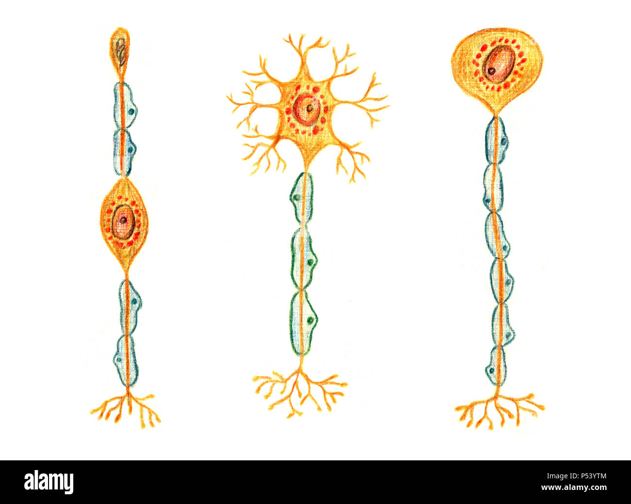 Different kinds of neurons: Bipolar neuron, Multipolar neuron, Unipolar neuron, hand drawn medical illustration, color pencils drawing with imitation Stock Photohttps://www.alamy.com/image-license-details/?v=1https://www.alamy.com/different-kinds-of-neurons-bipolar-neuron-multipolar-neuron-unipolar-neuron-hand-drawn-medical-illustration-color-pencils-drawing-with-imitation-image209685412.html
RFP53YTM–Different kinds of neurons: Bipolar neuron, Multipolar neuron, Unipolar neuron, hand drawn medical illustration, color pencils drawing with imitation
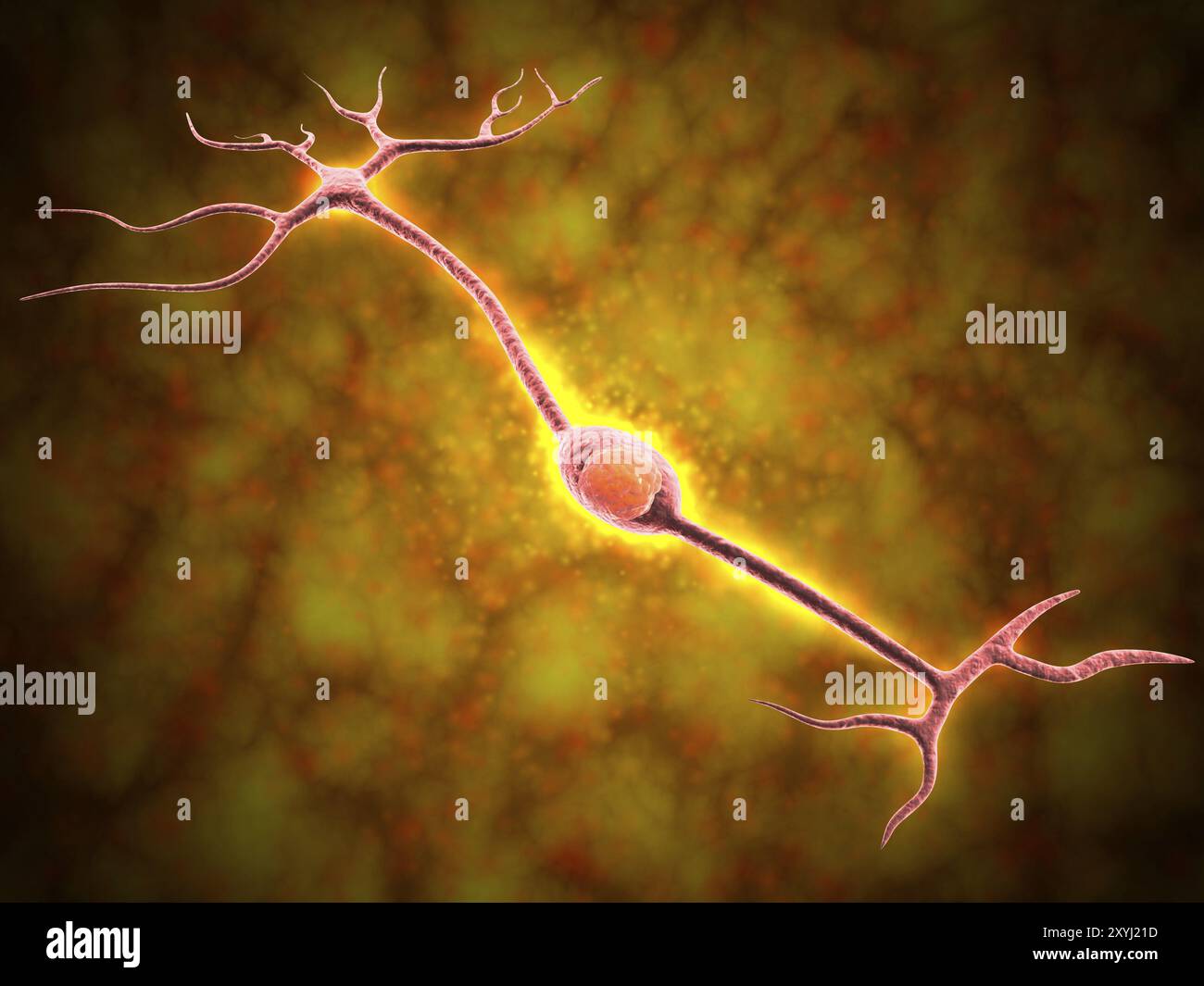 Microscopic view of a bipolar neuron. A bipolar cell is a type of neuron which has two extensions. Bipolar cells are specialized sensory neurons for t Stock Photohttps://www.alamy.com/image-license-details/?v=1https://www.alamy.com/microscopic-view-of-a-bipolar-neuron-a-bipolar-cell-is-a-type-of-neuron-which-has-two-extensions-bipolar-cells-are-specialized-sensory-neurons-for-t-image619355337.html
RM2XYJ21D–Microscopic view of a bipolar neuron. A bipolar cell is a type of neuron which has two extensions. Bipolar cells are specialized sensory neurons for t
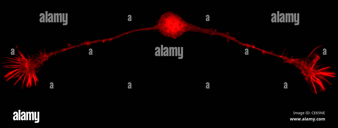 Bipolar neuron Stock Photohttps://www.alamy.com/image-license-details/?v=1https://www.alamy.com/stock-photo-bipolar-neuron-43162154.html
RMCE65NE–Bipolar neuron
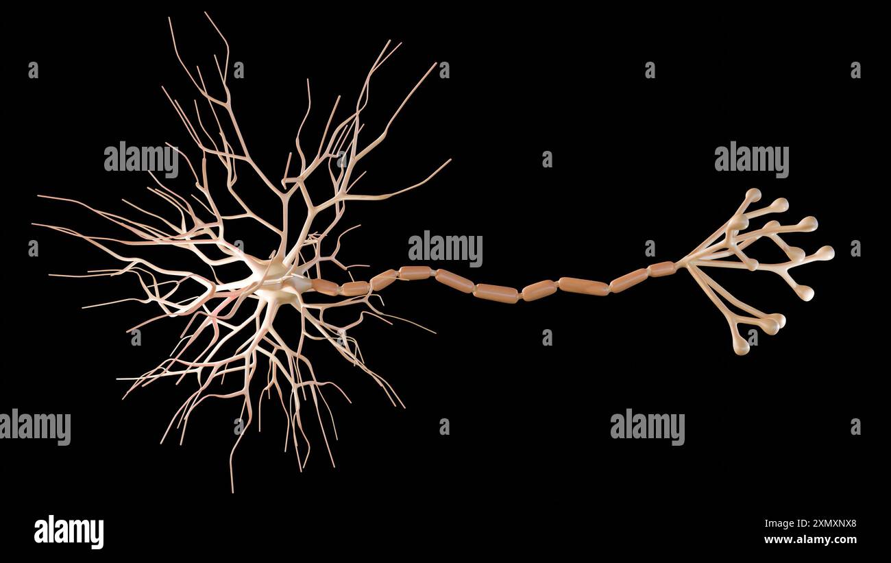 3d rendering of Multipolar neurons. Multipolar neurons are the most common type of neuron Stock Photohttps://www.alamy.com/image-license-details/?v=1https://www.alamy.com/3d-rendering-of-multipolar-neurons-multipolar-neurons-are-the-most-common-type-of-neuron-image615243952.html
RF2XMXNX8–3d rendering of Multipolar neurons. Multipolar neurons are the most common type of neuron
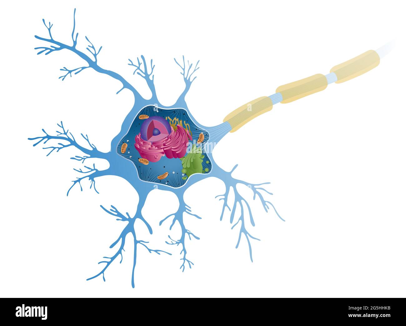 Anatomy of a multipolar neuron. Nerve cells, also known as a neurons, are the active component of the nervous system Stock Photohttps://www.alamy.com/image-license-details/?v=1https://www.alamy.com/anatomy-of-a-multipolar-neuron-nerve-cells-also-known-as-a-neurons-are-the-active-component-of-the-nervous-system-image433719535.html
RF2G5HHKB–Anatomy of a multipolar neuron. Nerve cells, also known as a neurons, are the active component of the nervous system
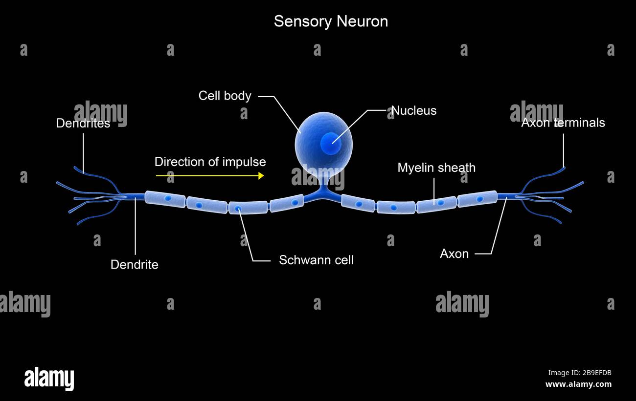 Conceptual image of a sensory neuron. Stock Photohttps://www.alamy.com/image-license-details/?v=1https://www.alamy.com/conceptual-image-of-a-sensory-neuron-image350058727.html
RF2B9EFDB–Conceptual image of a sensory neuron.
 Neuron Stock Photohttps://www.alamy.com/image-license-details/?v=1https://www.alamy.com/neuron-image226217493.html
RFR412M5–Neuron
 Pseudounipolar neurons. Pseudounipolar neurons of rounded soma of a dorsal root ganglion.Сell body is surrounded by glia cells called satellite cells. Stock Photohttps://www.alamy.com/image-license-details/?v=1https://www.alamy.com/pseudounipolar-neurons-pseudounipolar-neurons-of-rounded-soma-of-a-dorsal-root-ganglionell-body-is-surrounded-by-glia-cells-called-satellite-cells-image471432281.html
RF2JAYGK5–Pseudounipolar neurons. Pseudounipolar neurons of rounded soma of a dorsal root ganglion.Сell body is surrounded by glia cells called satellite cells.
 Neuron Stock Photohttps://www.alamy.com/image-license-details/?v=1https://www.alamy.com/neuron-image226259528.html
RFR4309C–Neuron
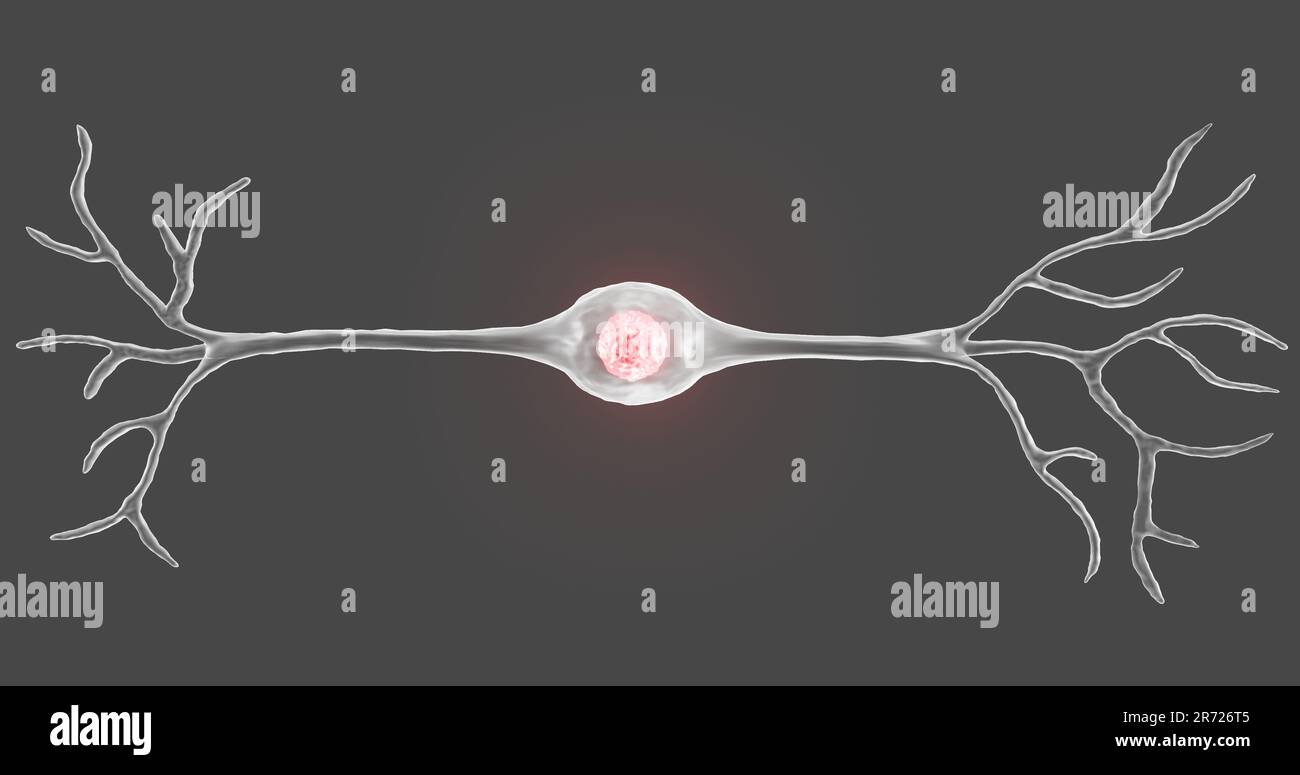 A bipolar neuron or nerve cell in 3D illustration Stock Photohttps://www.alamy.com/image-license-details/?v=1https://www.alamy.com/a-bipolar-neuron-or-nerve-cell-in-3d-illustration-image555083653.html
RF2R726T5–A bipolar neuron or nerve cell in 3D illustration
 Neuron Stock Photohttps://www.alamy.com/image-license-details/?v=1https://www.alamy.com/neuron-image226241433.html
RFR42575–Neuron
RF2YJ2NRH–High-Quality Vector Icons Collection with Editable Stroke. Ideal for Professional and Creative Projects.
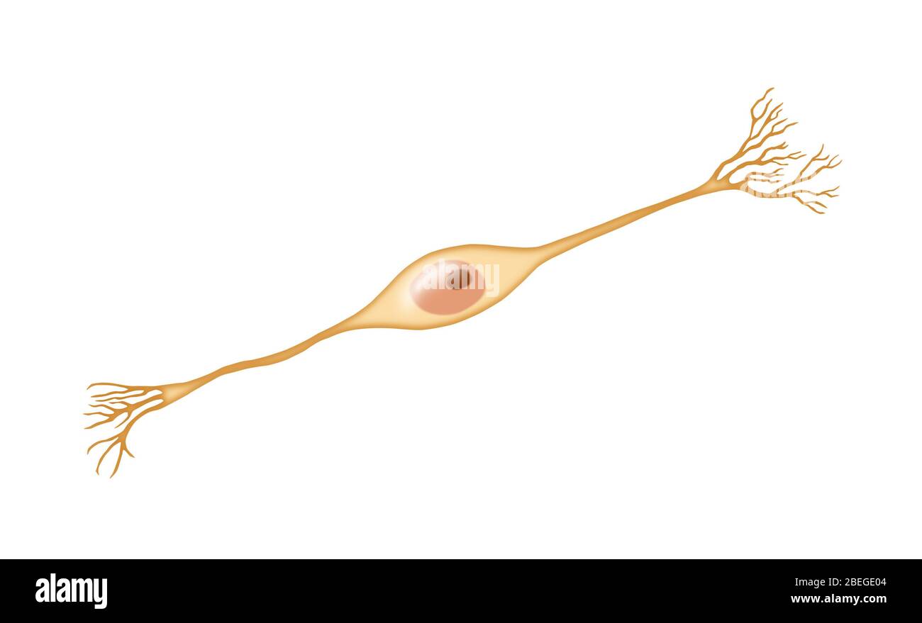 Bipolar Neuron Stock Photohttps://www.alamy.com/image-license-details/?v=1https://www.alamy.com/bipolar-neuron-image353174756.html
RM2BEGE04–Bipolar Neuron
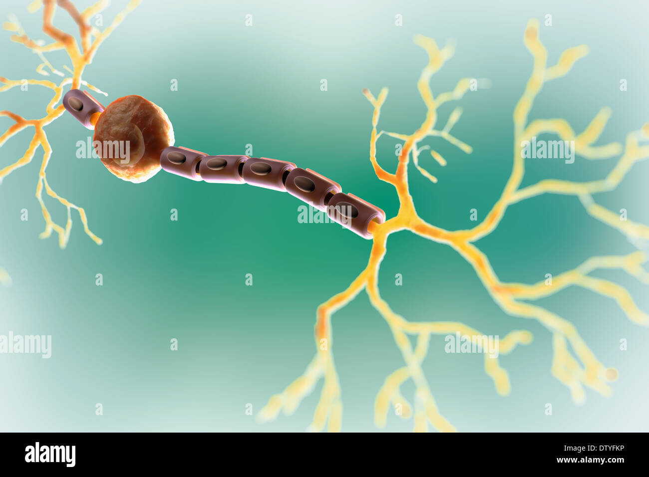 Bipolar Neuron Stock Photohttps://www.alamy.com/image-license-details/?v=1https://www.alamy.com/bipolar-neuron-image66987866.html
RMDTYFKP–Bipolar Neuron
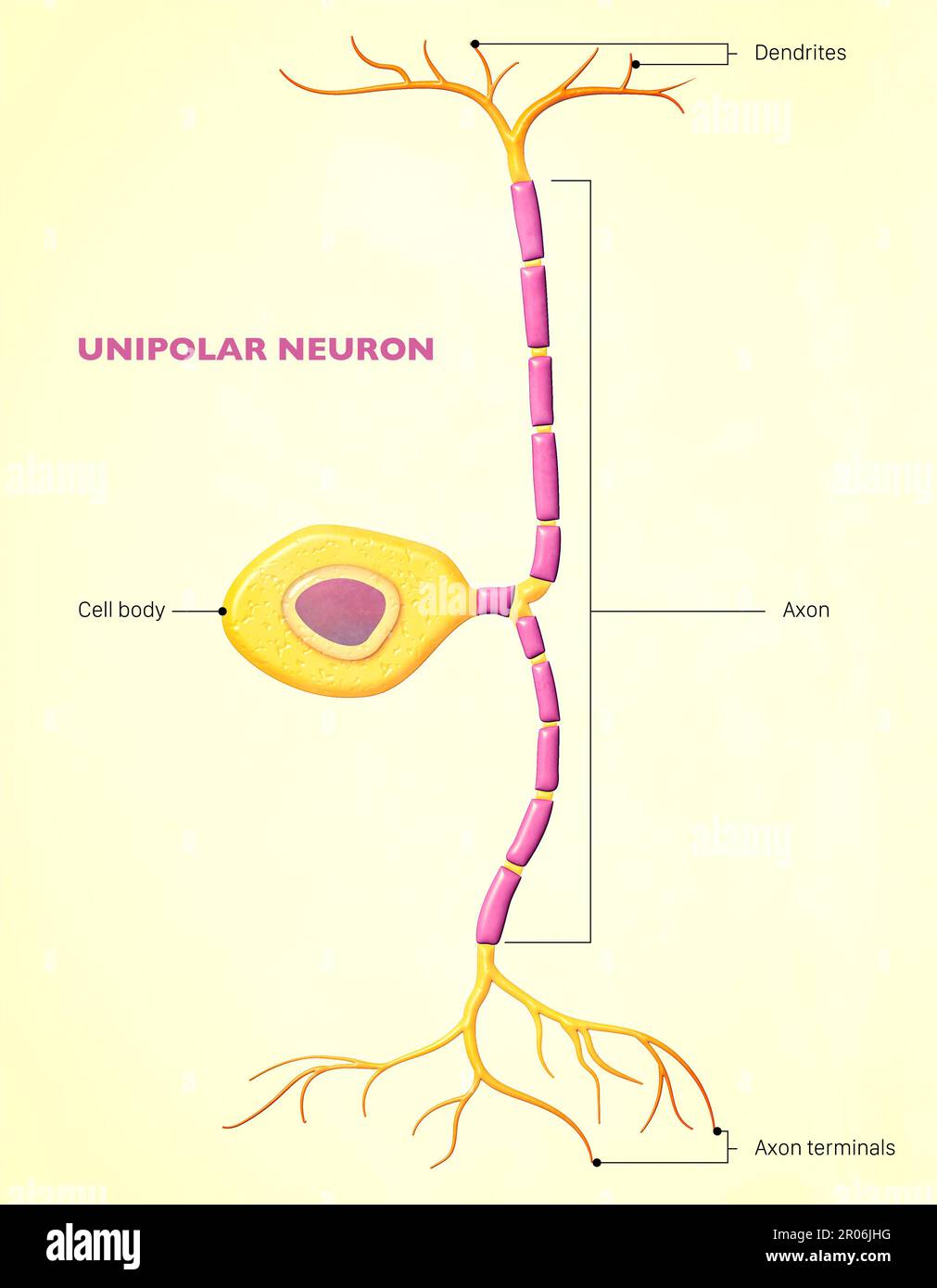 A bipolar neuron, or bipolar cell, is a type of neuron that has two extensions ( axon and dendrite). Sensory neurons for the transmission of sense Stock Photohttps://www.alamy.com/image-license-details/?v=1https://www.alamy.com/a-bipolar-neuron-or-bipolar-cell-is-a-type-of-neuron-that-has-two-extensions-axon-and-dendrite-sensory-neurons-for-the-transmission-of-sense-image550878092.html
RF2R06JHG–A bipolar neuron, or bipolar cell, is a type of neuron that has two extensions ( axon and dendrite). Sensory neurons for the transmission of sense
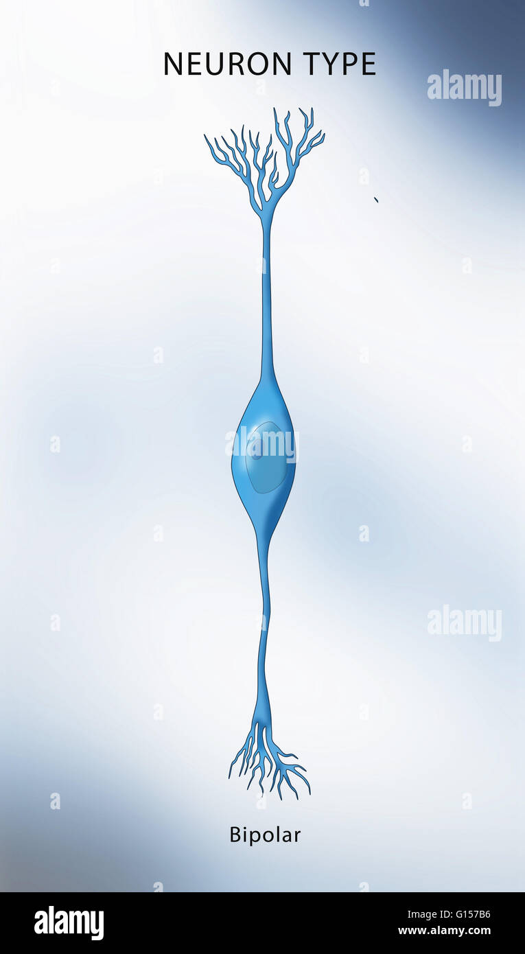 Illustration of a bipolar neuron, a neuron which has two extensions. Bipolar cells are specialized sensory neurons and part of the sensory pathways for smell, sight, taste, hearing and vestibular functions. Stock Photohttps://www.alamy.com/image-license-details/?v=1https://www.alamy.com/stock-photo-illustration-of-a-bipolar-neuron-a-neuron-which-has-two-extensions-103992426.html
RMG157B6–Illustration of a bipolar neuron, a neuron which has two extensions. Bipolar cells are specialized sensory neurons and part of the sensory pathways for smell, sight, taste, hearing and vestibular functions.
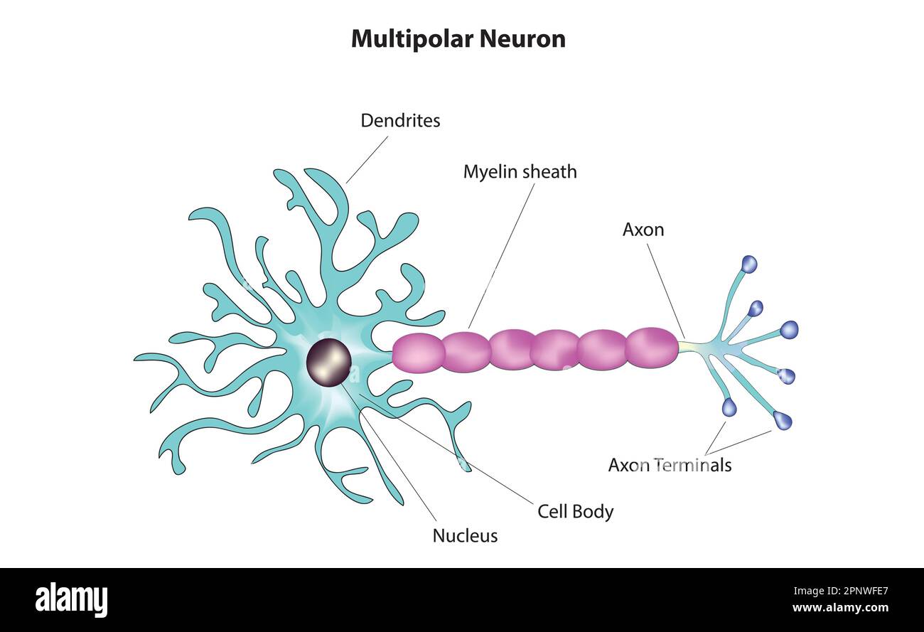 multipolar neuron Stock Vectorhttps://www.alamy.com/image-license-details/?v=1https://www.alamy.com/multipolar-neuron-image546990143.html
RF2PNWFE7–multipolar neuron
 creative human brain colorful wires technology logo vector logo symbol Stock Vectorhttps://www.alamy.com/image-license-details/?v=1https://www.alamy.com/creative-human-brain-colorful-wires-technology-logo-vector-logo-symbol-image248614391.html
RFTCDA5B–creative human brain colorful wires technology logo vector logo symbol
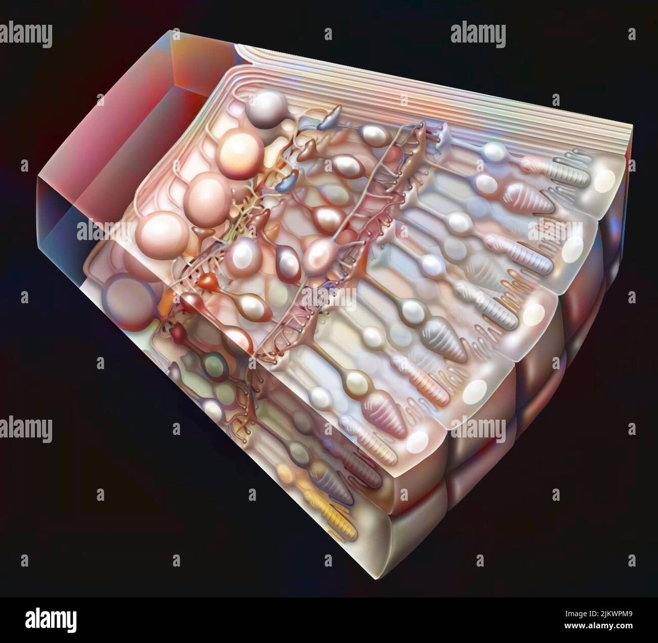 Cross-section of the retina with the photoreceptors: cones, rods, and also the optic nerve. Stock Photohttps://www.alamy.com/image-license-details/?v=1https://www.alamy.com/cross-section-of-the-retina-with-the-photoreceptors-cones-rods-and-also-the-optic-nerve-image476925017.html
RF2JKWPM9–Cross-section of the retina with the photoreceptors: cones, rods, and also the optic nerve.
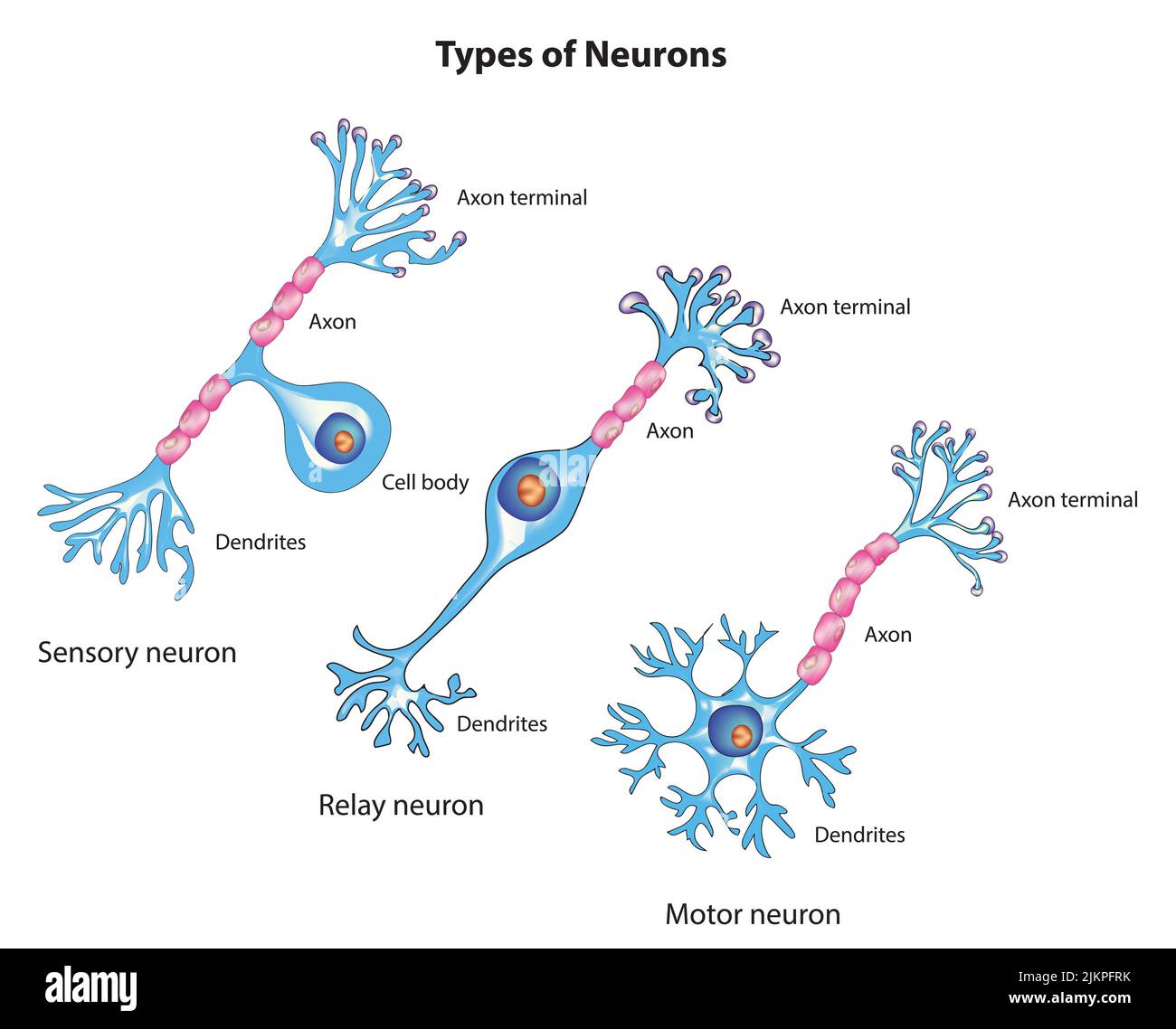 Types of neuron Stock Photohttps://www.alamy.com/image-license-details/?v=1https://www.alamy.com/types-of-neuron-image476853767.html
RF2JKPFRK–Types of neuron
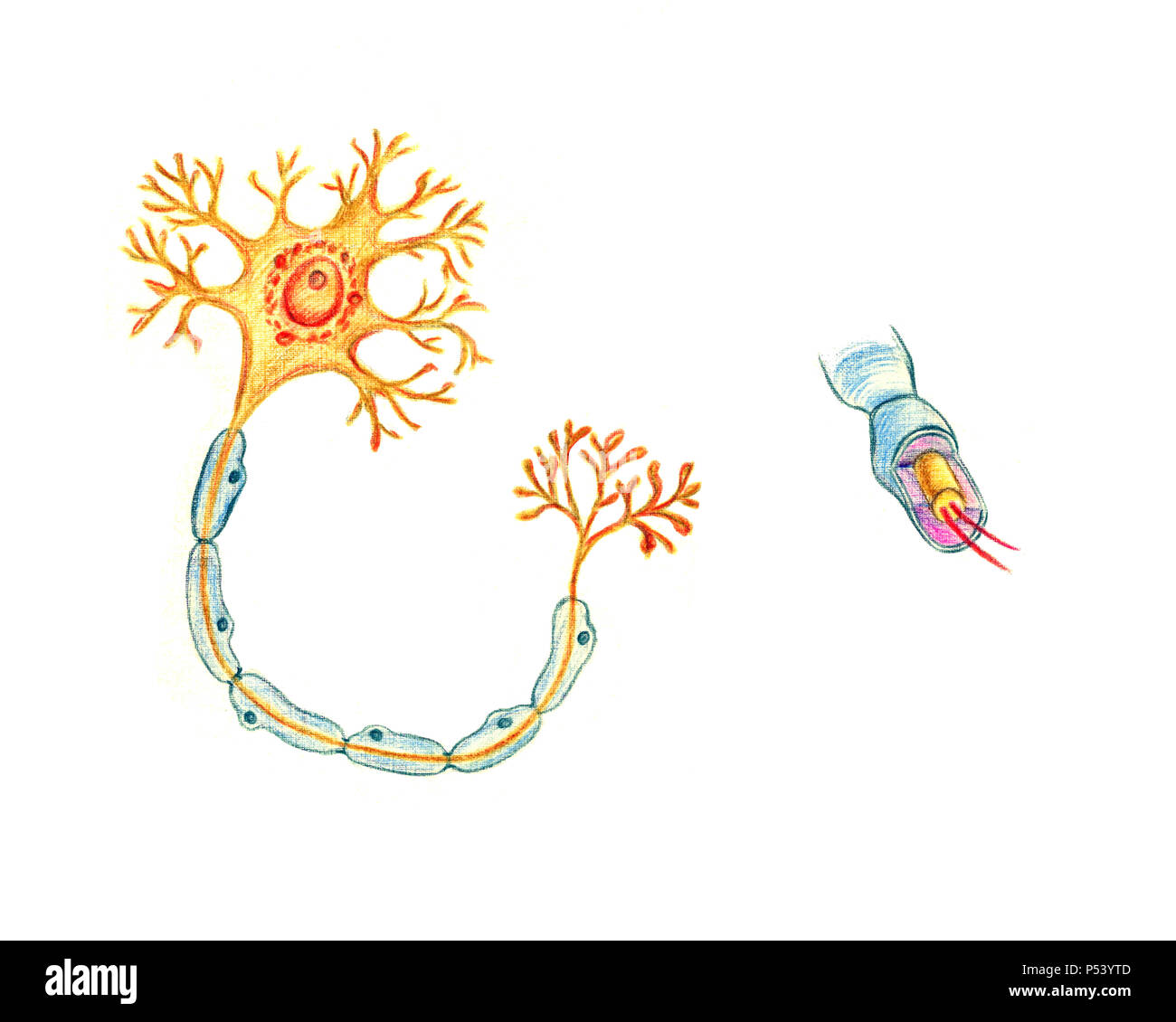 Structure of a typical neuron, hand drawn medical illustration, color pencils drawing with imitation of lithography Stock Photohttps://www.alamy.com/image-license-details/?v=1https://www.alamy.com/structure-of-a-typical-neuron-hand-drawn-medical-illustration-color-pencils-drawing-with-imitation-of-lithography-image209685405.html
RFP53YTD–Structure of a typical neuron, hand drawn medical illustration, color pencils drawing with imitation of lithography
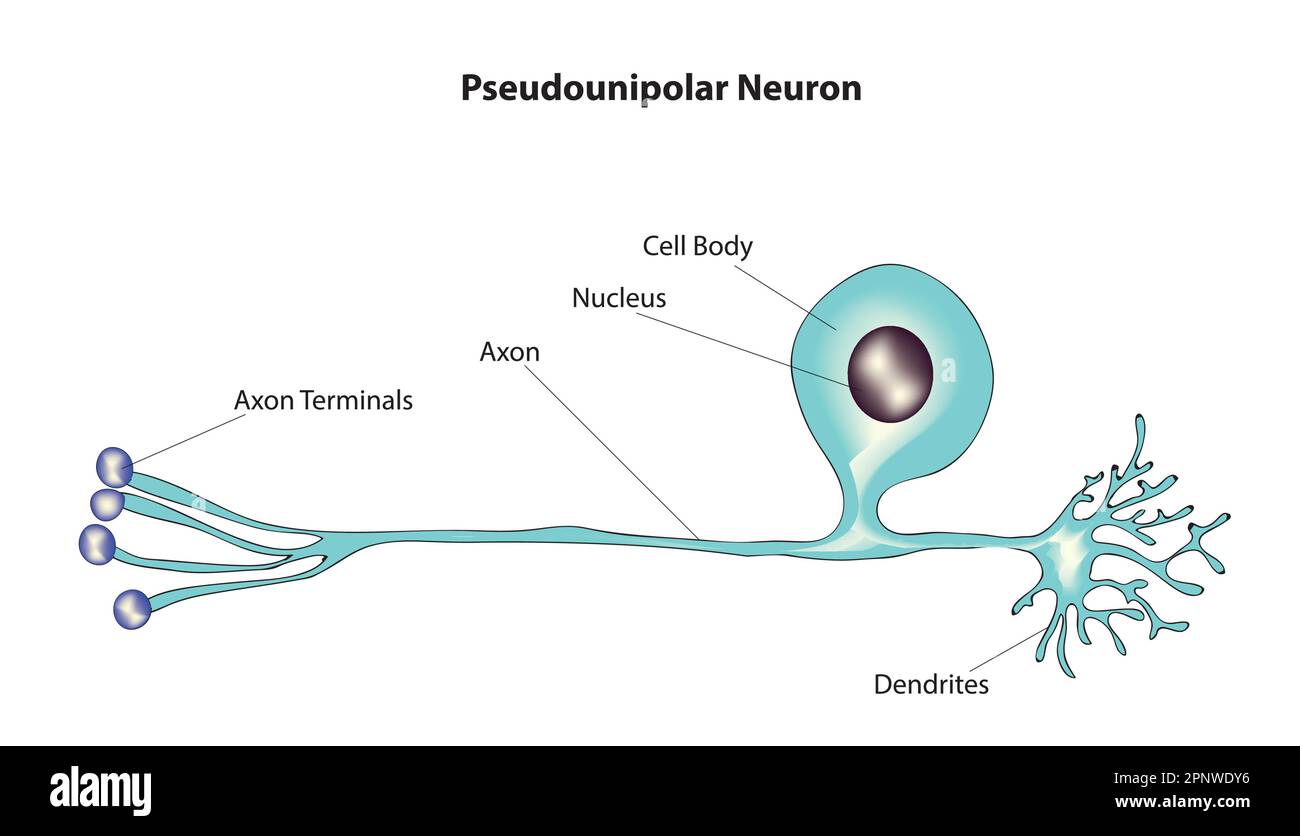 pseudounipolar neuron Stock Vectorhttps://www.alamy.com/image-license-details/?v=1https://www.alamy.com/pseudounipolar-neuron-image546988938.html
RF2PNWDY6–pseudounipolar neuron
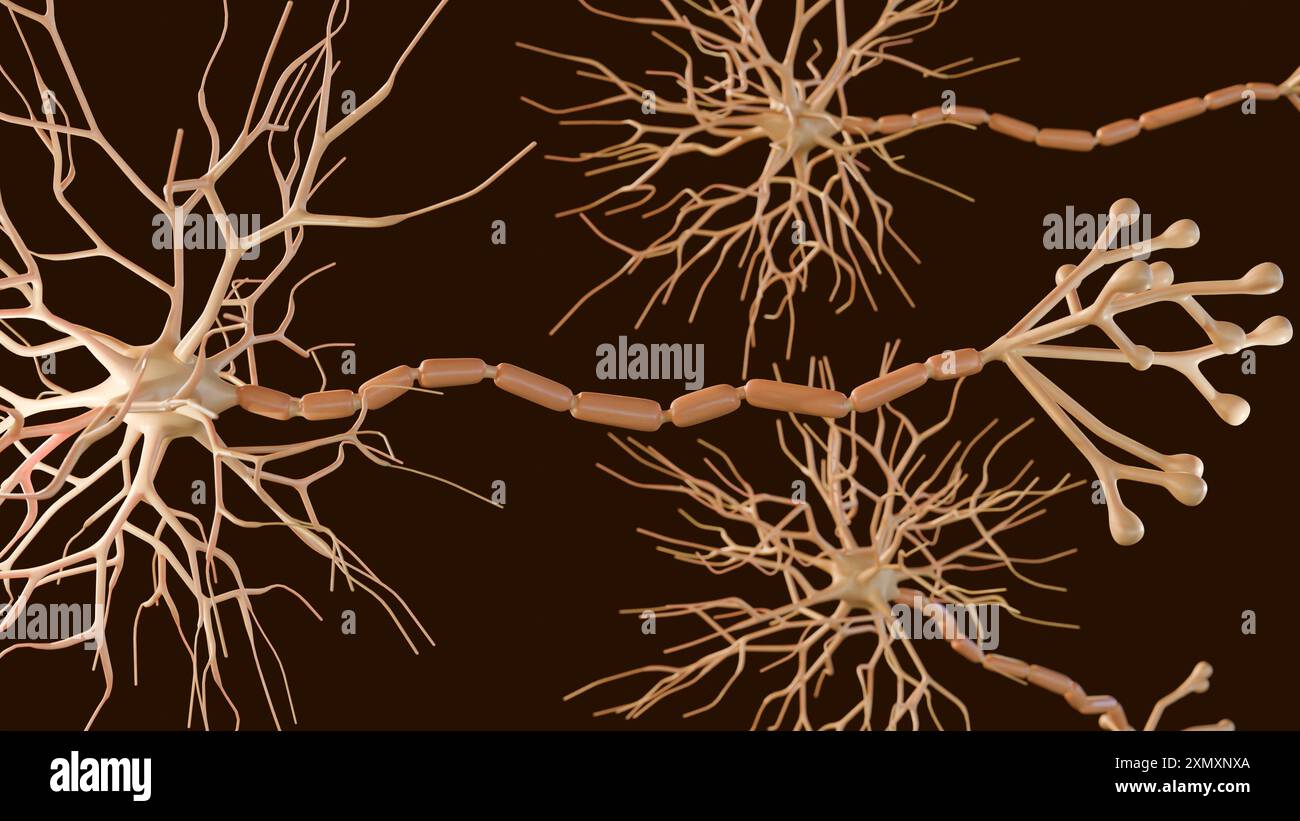 3d rendering of Multipolar neurons. Multipolar neurons are the most common type of neuron Stock Photohttps://www.alamy.com/image-license-details/?v=1https://www.alamy.com/3d-rendering-of-multipolar-neurons-multipolar-neurons-are-the-most-common-type-of-neuron-image615243954.html
RF2XMXNXA–3d rendering of Multipolar neurons. Multipolar neurons are the most common type of neuron
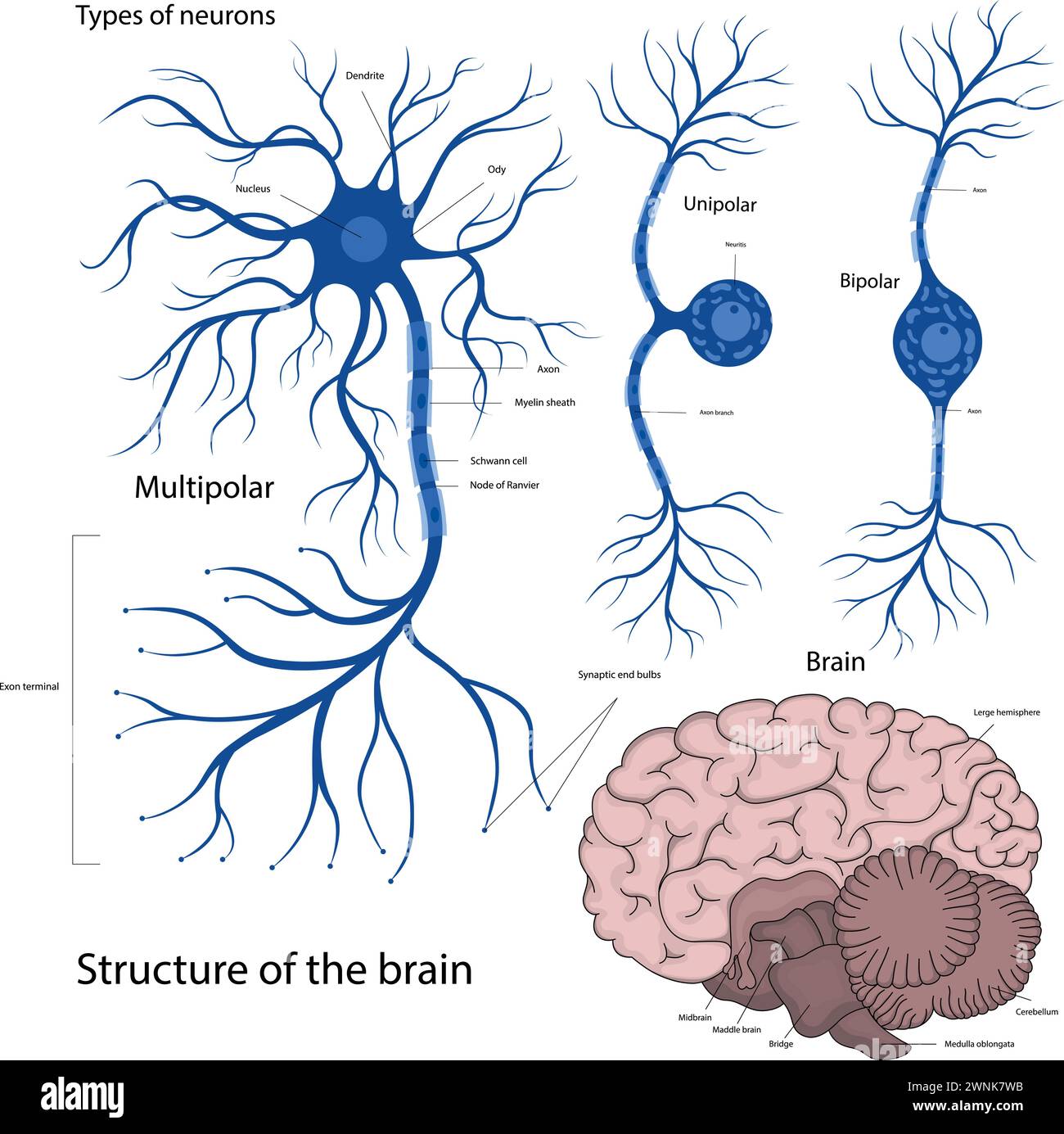 Types of neurons bipolar, unipolar, multipolar. The structure of a neuron in the brain. The structure of the human brain. Stock Vectorhttps://www.alamy.com/image-license-details/?v=1https://www.alamy.com/types-of-neurons-bipolar-unipolar-multipolar-the-structure-of-a-neuron-in-the-brain-the-structure-of-the-human-brain-image598483575.html
RF2WNK7WB–Types of neurons bipolar, unipolar, multipolar. The structure of a neuron in the brain. The structure of the human brain.
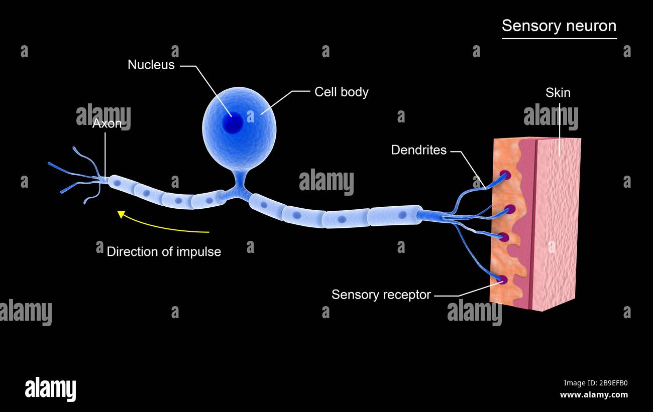 Conceptual image of a sensory neuron. Stock Photohttps://www.alamy.com/image-license-details/?v=1https://www.alamy.com/conceptual-image-of-a-sensory-neuron-image350058660.html
RF2B9EFB0–Conceptual image of a sensory neuron.
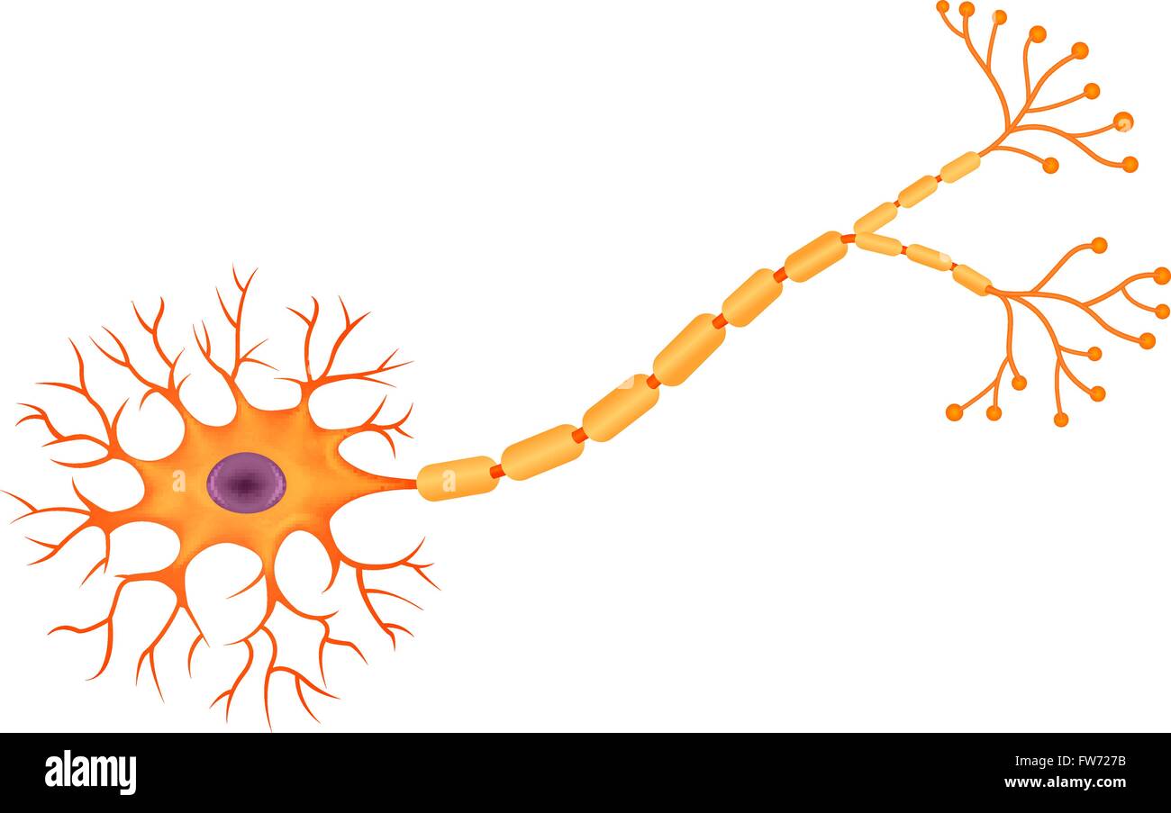 Illustration of Human Neuron Anatomy Stock Vectorhttps://www.alamy.com/image-license-details/?v=1https://www.alamy.com/stock-photo-illustration-of-human-neuron-anatomy-101573679.html
RFFW727B–Illustration of Human Neuron Anatomy
 Dorsal root ganglion. Pseudounipolar neurons of a dorsal root ganglion. Hematoxlyn and eosin stain. Magnification: x40. Stock Photohttps://www.alamy.com/image-license-details/?v=1https://www.alamy.com/dorsal-root-ganglion-pseudounipolar-neurons-of-a-dorsal-root-ganglion-hematoxlyn-and-eosin-stain-magnification-x40-image471432248.html
RF2JAYGJ0–Dorsal root ganglion. Pseudounipolar neurons of a dorsal root ganglion. Hematoxlyn and eosin stain. Magnification: x40.
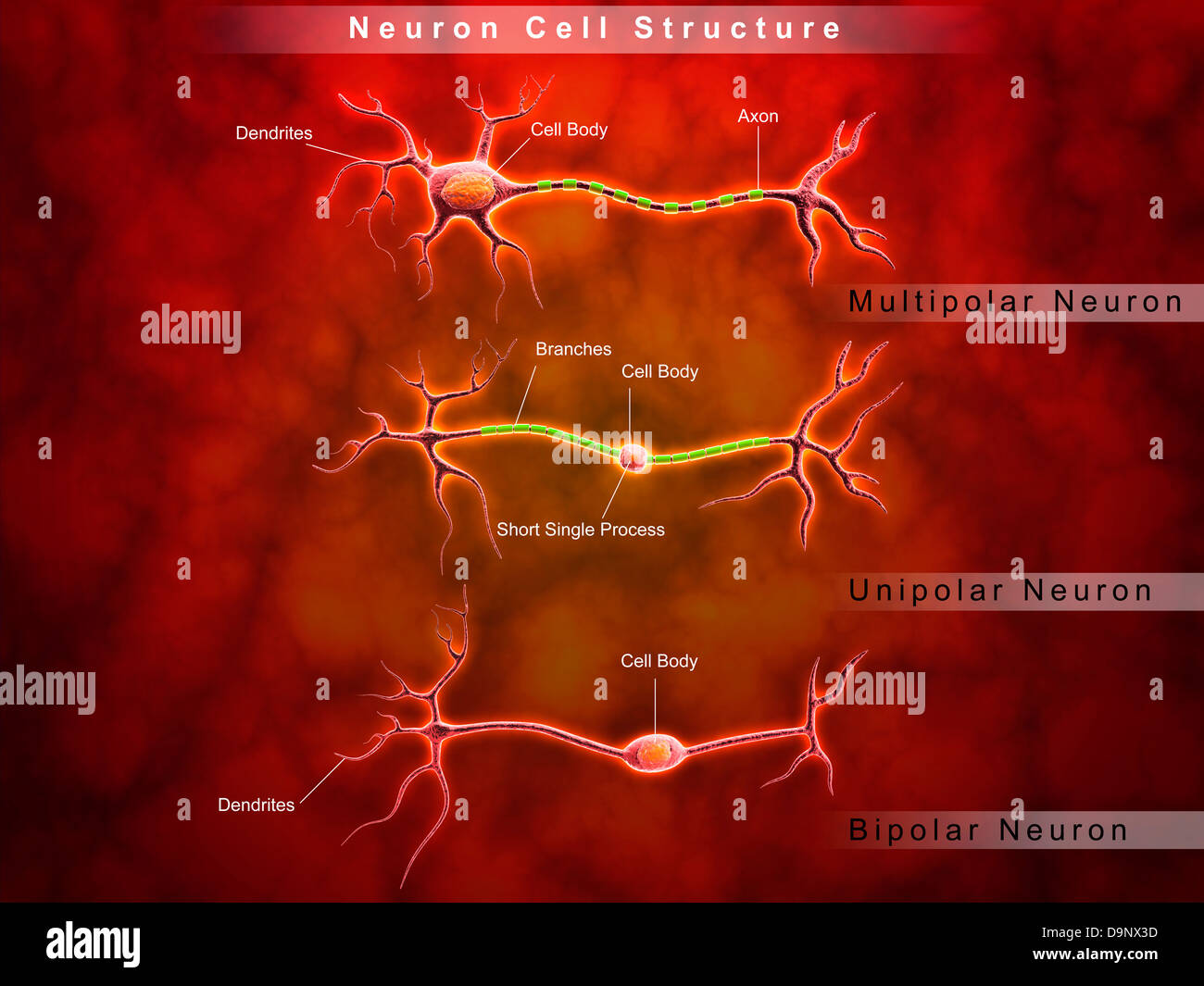 Anatomy structure of neurons. Stock Photohttps://www.alamy.com/image-license-details/?v=1https://www.alamy.com/stock-photo-anatomy-structure-of-neurons-57644481.html
RFD9NX3D–Anatomy structure of neurons.
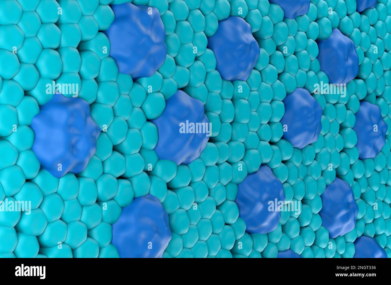 Retina surface (cones and rods) in the human eye isometric view 3d illustration Stock Photohttps://www.alamy.com/image-license-details/?v=1https://www.alamy.com/retina-surface-cones-and-rods-in-the-human-eye-isometric-view-3d-illustration-image526674826.html
RF2NGT336–Retina surface (cones and rods) in the human eye isometric view 3d illustration
 Neuron Stock Photohttps://www.alamy.com/image-license-details/?v=1https://www.alamy.com/neuron-image226241403.html
RFR42563–Neuron
 Schematic of the hypothalamus receiving nerve impulses from the body and sending messages to the circulatory and nervous system. Stock Photohttps://www.alamy.com/image-license-details/?v=1https://www.alamy.com/stock-photo-schematic-of-the-hypothalamus-receiving-nerve-impulses-from-the-body-72785292.html
RME6BJAM–Schematic of the hypothalamus receiving nerve impulses from the body and sending messages to the circulatory and nervous system.
 Long term Neurodegenerative Motor System Disorder or Parkinson's disease Stock Photohttps://www.alamy.com/image-license-details/?v=1https://www.alamy.com/long-term-neurodegenerative-motor-system-disorder-or-parkinsons-disease-image631329370.html
RF2YK3F1E–Long term Neurodegenerative Motor System Disorder or Parkinson's disease
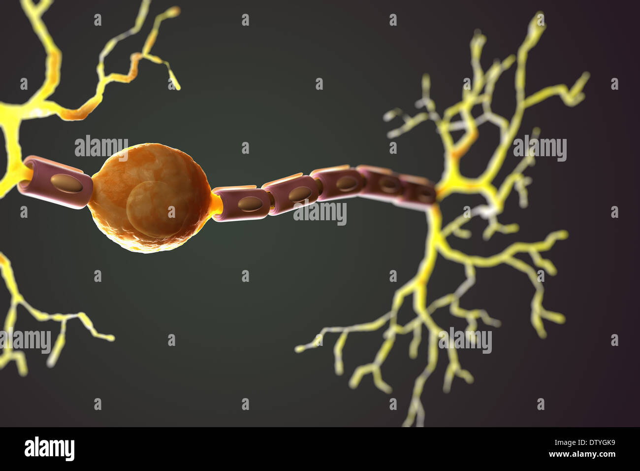 Bipolar Neuron Stock Photohttps://www.alamy.com/image-license-details/?v=1https://www.alamy.com/bipolar-neuron-image66988637.html
RMDTYGK9–Bipolar Neuron
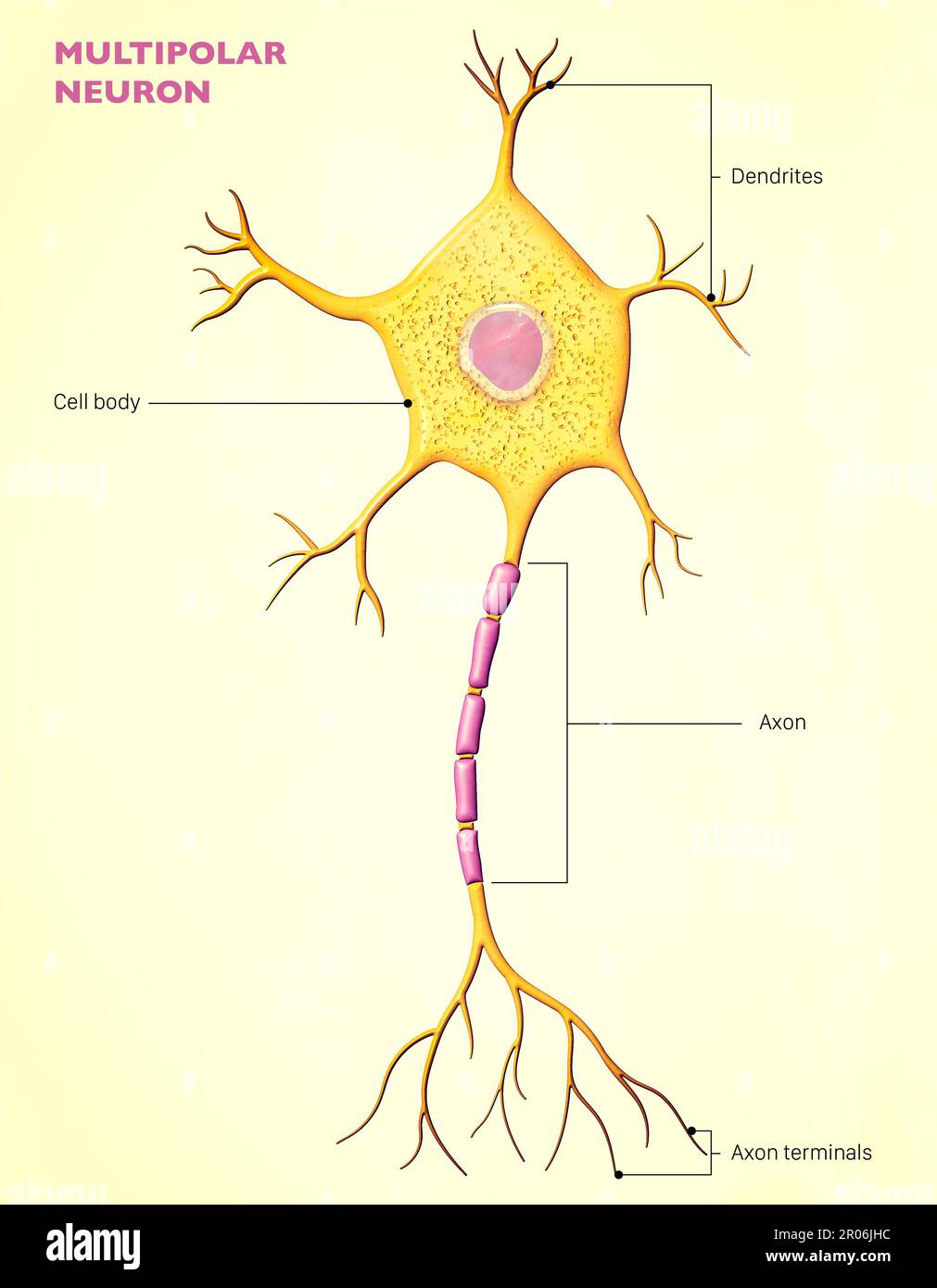 A multipolar neuron is a type of neuron that possesses a single axon and many dendrites, allowing for the integration of information from other neuron Stock Photohttps://www.alamy.com/image-license-details/?v=1https://www.alamy.com/a-multipolar-neuron-is-a-type-of-neuron-that-possesses-a-single-axon-and-many-dendrites-allowing-for-the-integration-of-information-from-other-neuron-image550878088.html
RF2R06JHC–A multipolar neuron is a type of neuron that possesses a single axon and many dendrites, allowing for the integration of information from other neuron
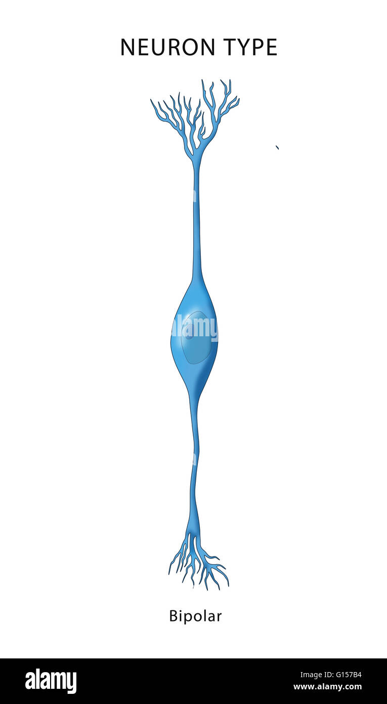 Illustration of a bipolar neuron, a neuron which has two extensions. Bipolar cells are specialized sensory neurons and part of the sensory pathways for smell, sight, taste, hearing and vestibular functions. Stock Photohttps://www.alamy.com/image-license-details/?v=1https://www.alamy.com/stock-photo-illustration-of-a-bipolar-neuron-a-neuron-which-has-two-extensions-103992424.html
RMG157B4–Illustration of a bipolar neuron, a neuron which has two extensions. Bipolar cells are specialized sensory neurons and part of the sensory pathways for smell, sight, taste, hearing and vestibular functions.
 businessman with emotions, mad, calm and happy. Emotional controlling, mental health concept. Stock Photohttps://www.alamy.com/image-license-details/?v=1https://www.alamy.com/businessman-with-emotions-mad-calm-and-happy-emotional-controlling-mental-health-concept-image185295722.html
RFMNCXGA–businessman with emotions, mad, calm and happy. Emotional controlling, mental health concept.
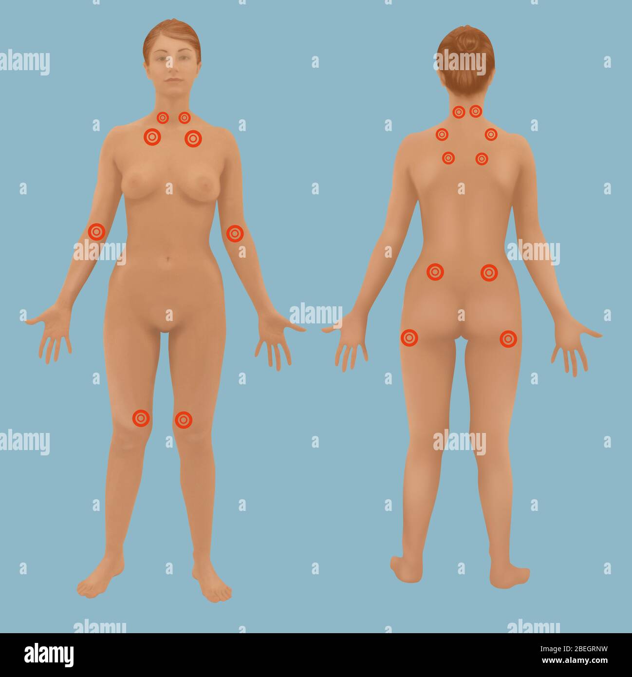 Fibromyalgia Stock Photohttps://www.alamy.com/image-license-details/?v=1https://www.alamy.com/fibromyalgia-image353182421.html
RF2BEGRNW–Fibromyalgia
 Retina at the level of the bowl drawn by the fovea. Stock Photohttps://www.alamy.com/image-license-details/?v=1https://www.alamy.com/retina-at-the-level-of-the-bowl-drawn-by-the-fovea-image476925818.html
RF2JKWRMX–Retina at the level of the bowl drawn by the fovea.
 World Bipolar Day set. Illustrates depressive phase, personalized treatment, and genetic research. Mental health, tailor-made therapy, scientific study. Vector illustration. Stock Vectorhttps://www.alamy.com/image-license-details/?v=1https://www.alamy.com/world-bipolar-day-set-illustrates-depressive-phase-personalized-treatment-and-genetic-research-mental-health-tailor-made-therapy-scientific-study-vector-illustration-image637596021.html
RF2S1906D–World Bipolar Day set. Illustrates depressive phase, personalized treatment, and genetic research. Mental health, tailor-made therapy, scientific study. Vector illustration.
 Types of Neurons Stock Photohttps://www.alamy.com/image-license-details/?v=1https://www.alamy.com/stock-photo-types-of-neurons-49222114.html
RFCT278J–Types of Neurons
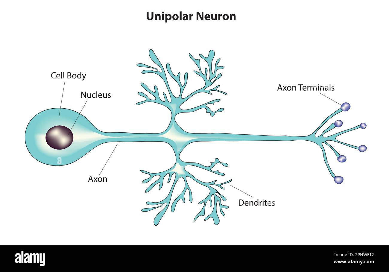 unipolar neuron Stock Vectorhttps://www.alamy.com/image-license-details/?v=1https://www.alamy.com/unipolar-neuron-image546989774.html
RF2PNWF12–unipolar neuron
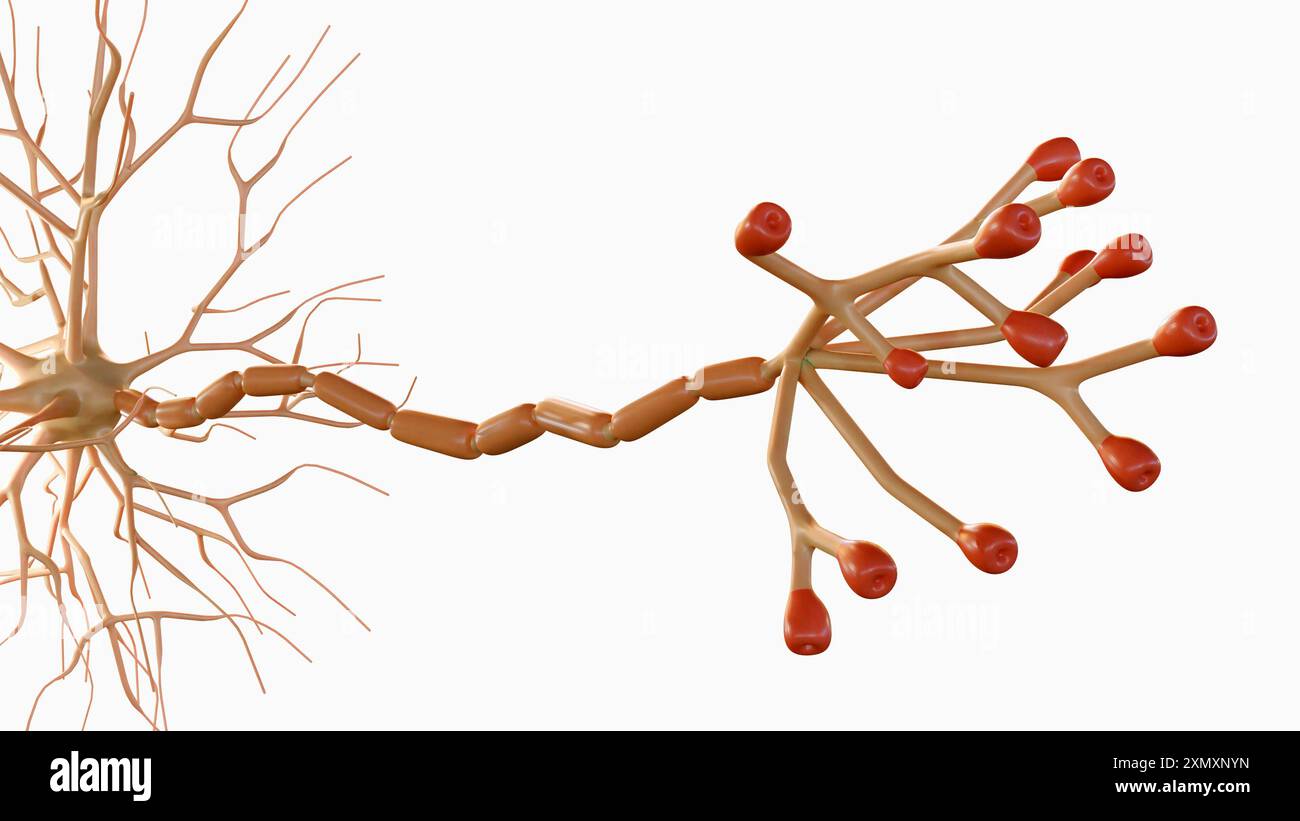 3d rendering of Multipolar neurons. Multipolar neurons are the most common type of neuron Stock Photohttps://www.alamy.com/image-license-details/?v=1https://www.alamy.com/3d-rendering-of-multipolar-neurons-multipolar-neurons-are-the-most-common-type-of-neuron-image615243993.html
RF2XMXNYN–3d rendering of Multipolar neurons. Multipolar neurons are the most common type of neuron
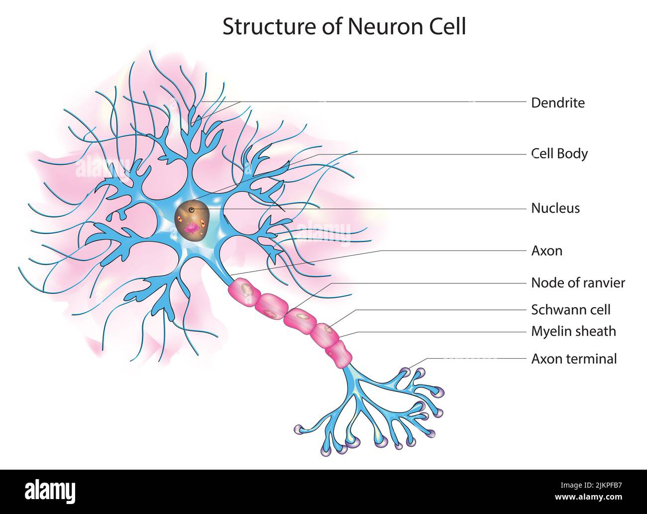 Structure of neuron cell Stock Photohttps://www.alamy.com/image-license-details/?v=1https://www.alamy.com/structure-of-neuron-cell-image476853419.html
RF2JKPFB7–Structure of neuron cell
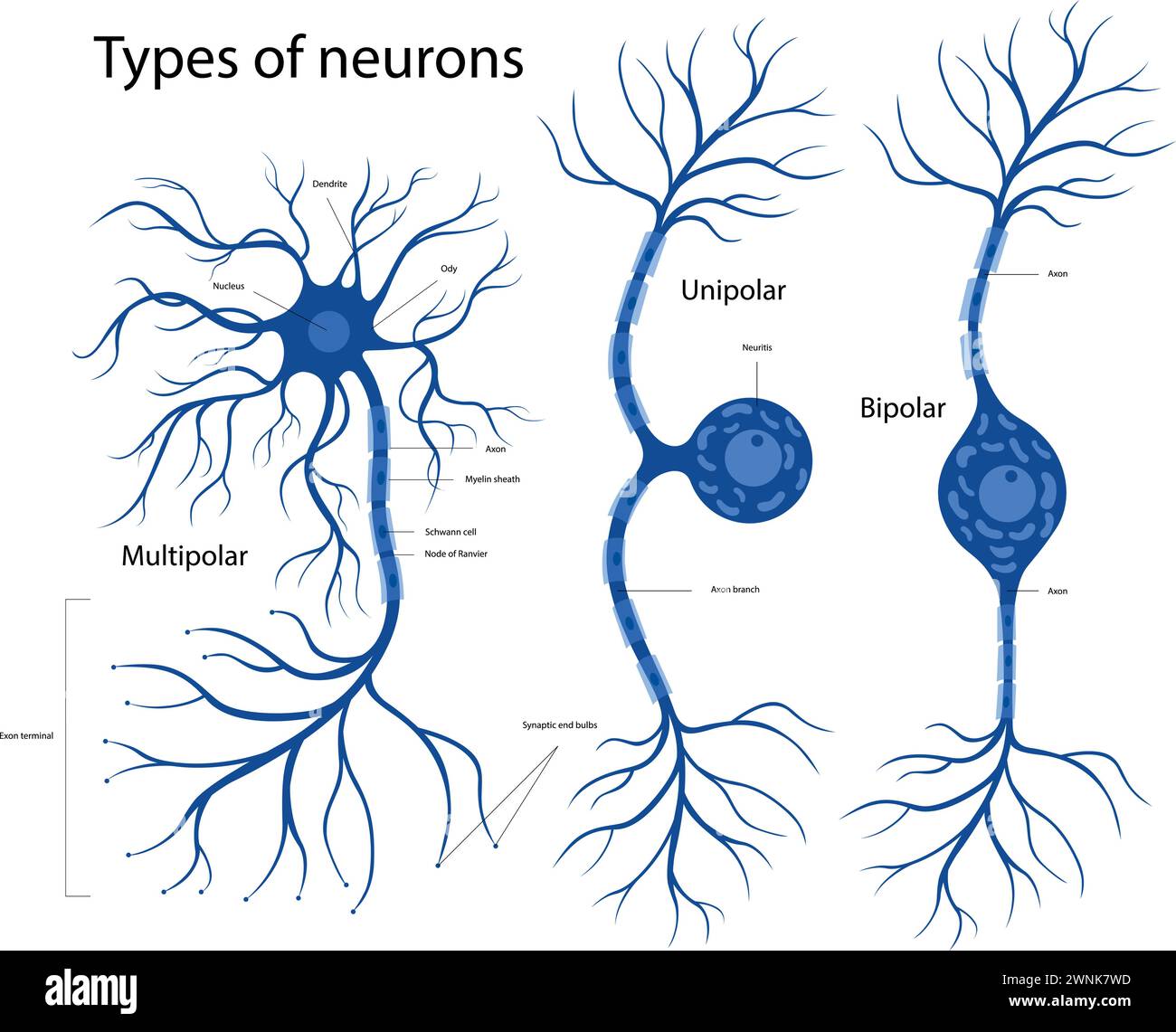 Types of neurons. The structure of a neuron in the brain. Stock Vectorhttps://www.alamy.com/image-license-details/?v=1https://www.alamy.com/types-of-neurons-the-structure-of-a-neuron-in-the-brain-image598483577.html
RF2WNK7WD–Types of neurons. The structure of a neuron in the brain.
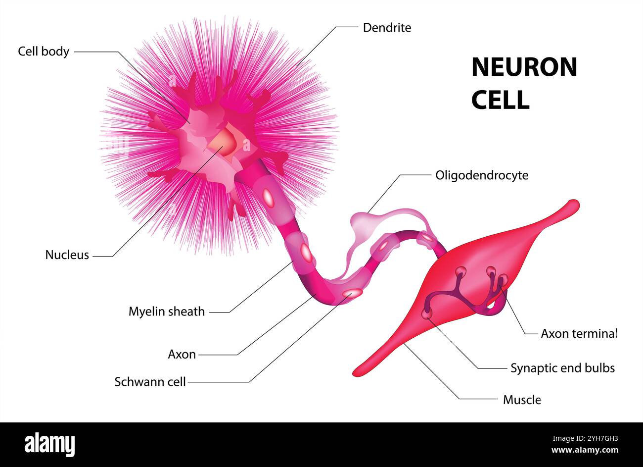 Human neuron structure. Brain neuron cell illustration. Synapses, myelin sheath, cell body, nucleus, axon and dendrites scheme. Neurology illustration Stock Vectorhttps://www.alamy.com/image-license-details/?v=1https://www.alamy.com/human-neuron-structure-brain-neuron-cell-illustration-synapses-myelin-sheath-cell-body-nucleus-axon-and-dendrites-scheme-neurology-illustration-image630189087.html
RF2YH7GH3–Human neuron structure. Brain neuron cell illustration. Synapses, myelin sheath, cell body, nucleus, axon and dendrites scheme. Neurology illustration
RF2K0X2B6–Neuron icon on white background. Human neuron cell sign. Brain neuron symbol. flat style.
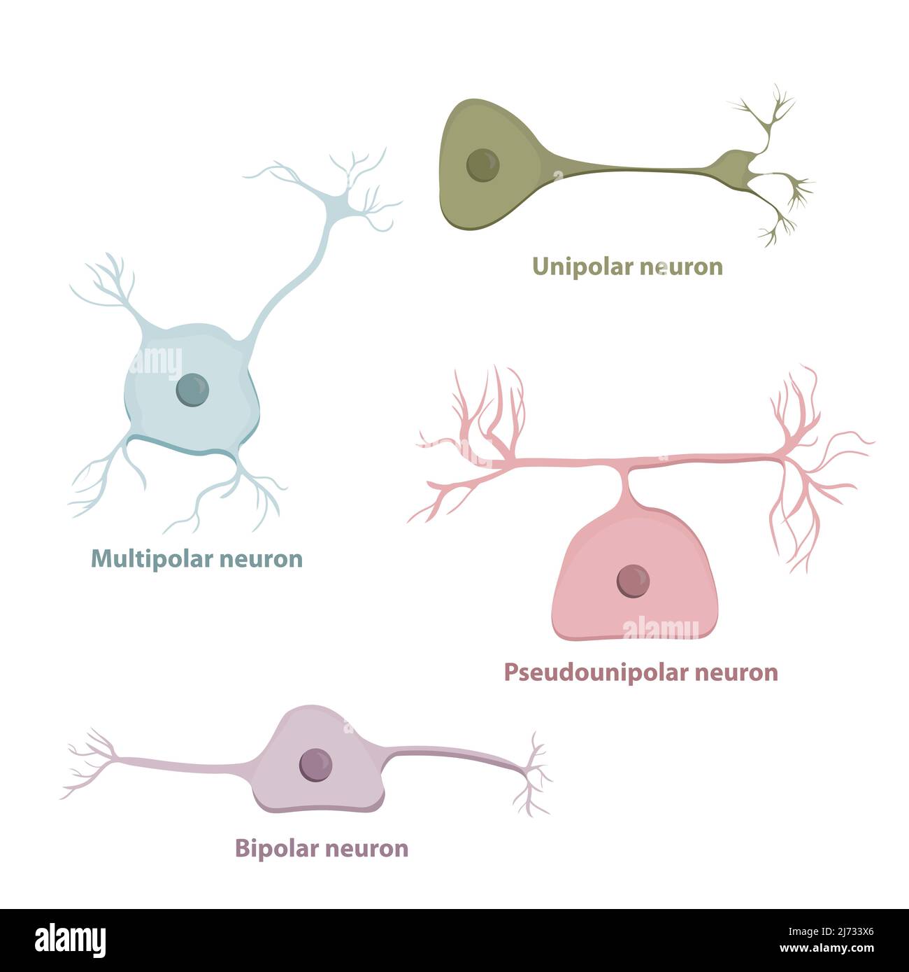 Basic neurons types, based on the number and placement of axons Stock Vectorhttps://www.alamy.com/image-license-details/?v=1https://www.alamy.com/basic-neurons-types-based-on-the-number-and-placement-of-axons-image469051470.html
RF2J733X6–Basic neurons types, based on the number and placement of axons
 Retina surface (cones and rods) in the human eye closeup view 3d illustration Stock Photohttps://www.alamy.com/image-license-details/?v=1https://www.alamy.com/retina-surface-cones-and-rods-in-the-human-eye-closeup-view-3d-illustration-image526674761.html
RF2NGT30W–Retina surface (cones and rods) in the human eye closeup view 3d illustration
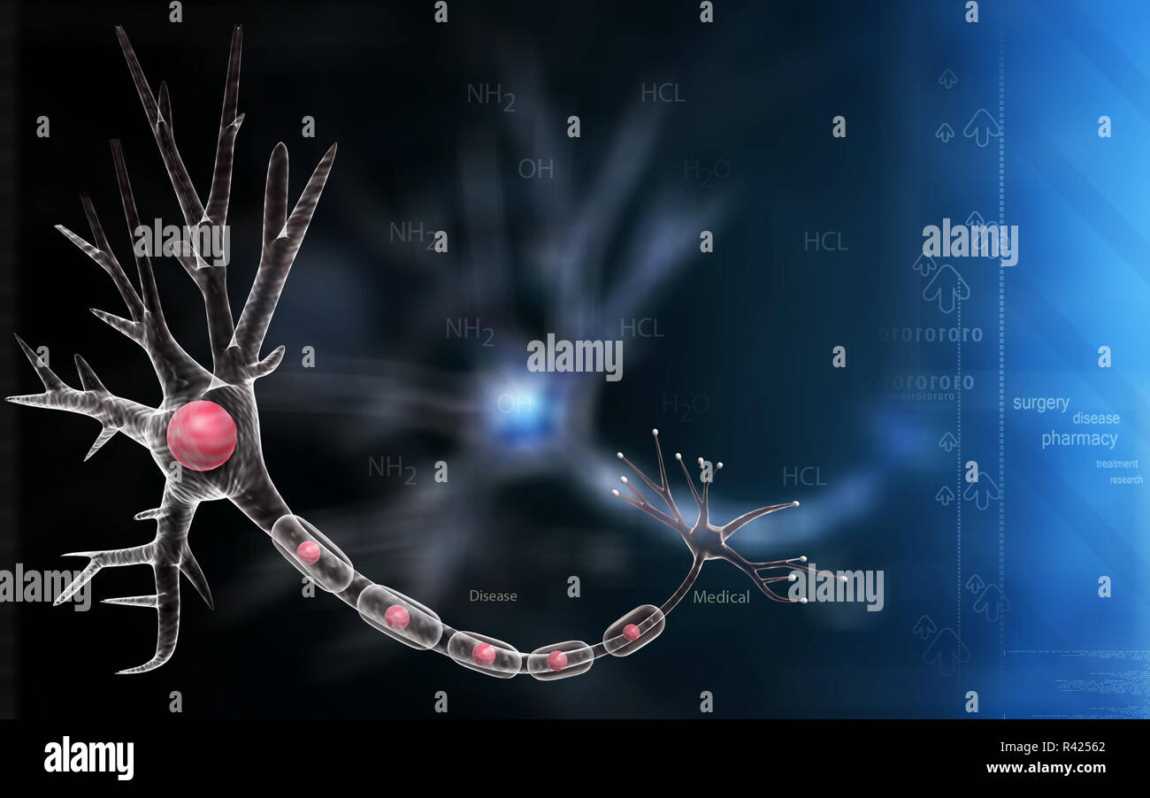 Neuron Stock Photohttps://www.alamy.com/image-license-details/?v=1https://www.alamy.com/neuron-image226241402.html
RFR42562–Neuron
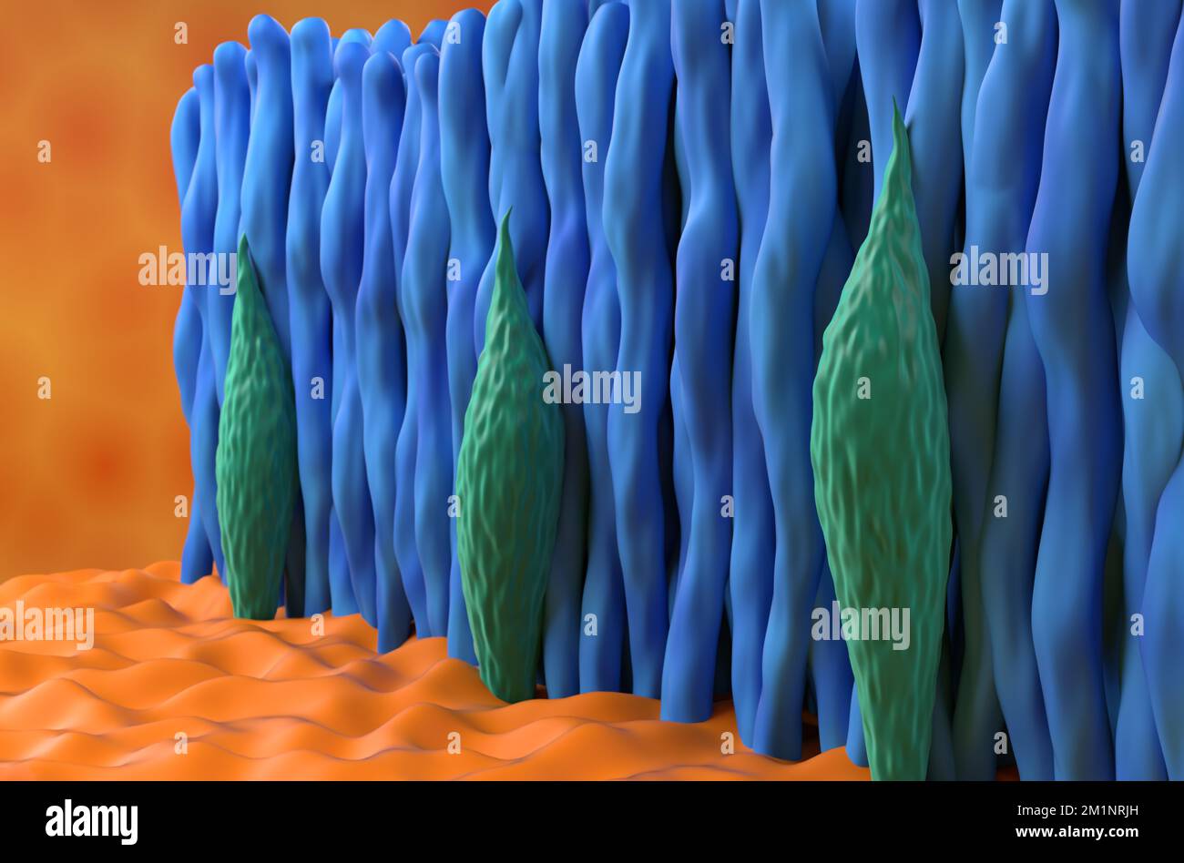 Retina cone and rod in the human eye - closeup view 3d illustration Stock Photohttps://www.alamy.com/image-license-details/?v=1https://www.alamy.com/retina-cone-and-rod-in-the-human-eye-closeup-view-3d-illustration-image500194873.html
RF2M1NRJH–Retina cone and rod in the human eye - closeup view 3d illustration
 Neuron Stock Photohttps://www.alamy.com/image-license-details/?v=1https://www.alamy.com/neuron-image226184825.html
RFR3YH1D–Neuron
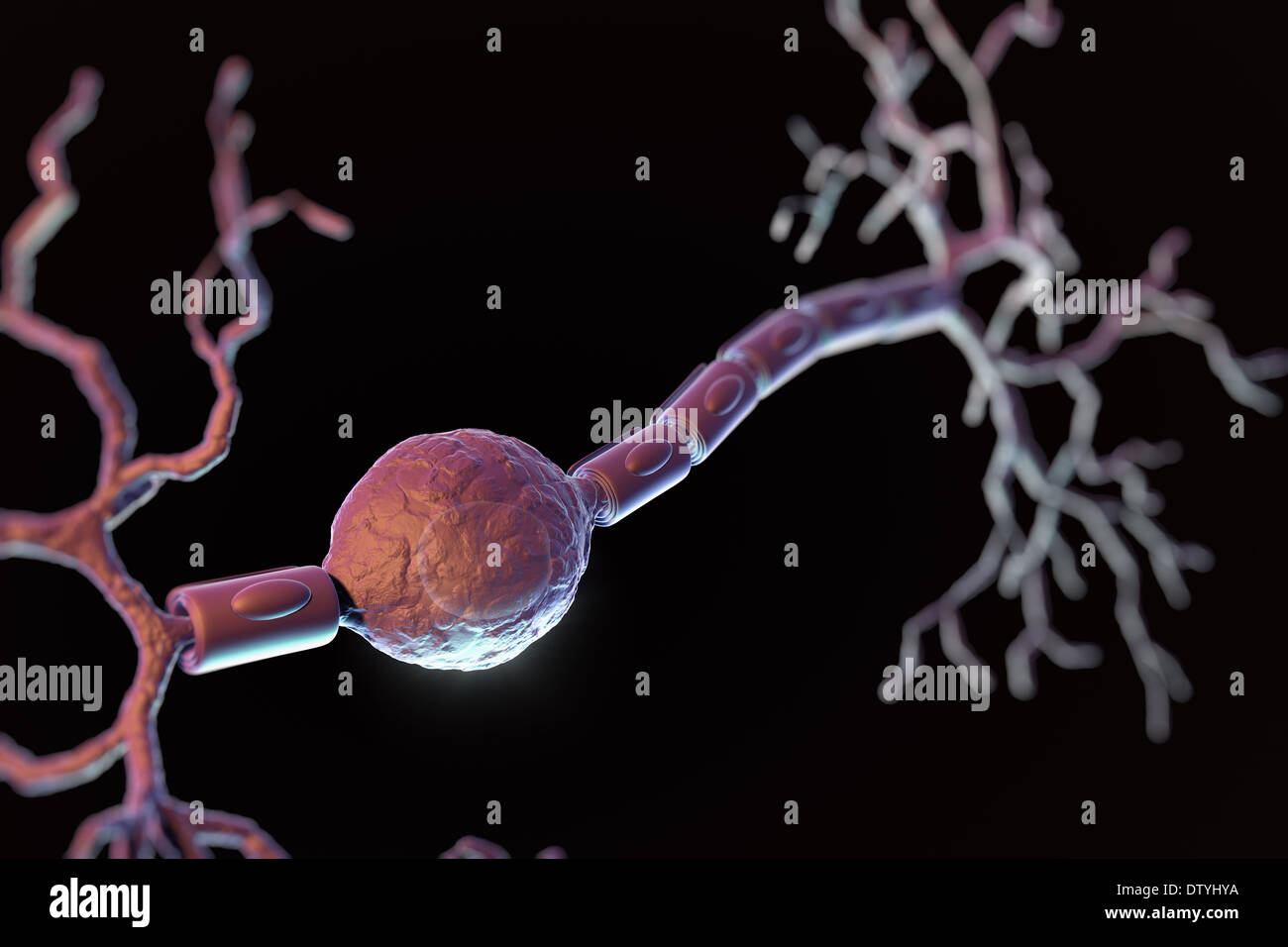 Bipolar Neuron Stock Photohttps://www.alamy.com/image-license-details/?v=1https://www.alamy.com/bipolar-neuron-image66989646.html
RMDTYHYA–Bipolar Neuron
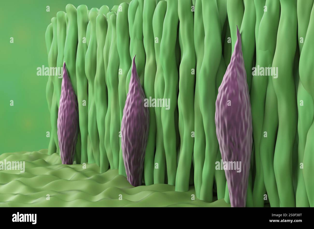 Retina photoreceptor (cone and rod cells) closeup view 3d illustration Stock Photohttps://www.alamy.com/image-license-details/?v=1https://www.alamy.com/retina-photoreceptor-cone-and-rod-cells-closeup-view-3d-illustration-image637115496.html
RF2S0F38T–Retina photoreceptor (cone and rod cells) closeup view 3d illustration
 Neurodegenerative Disorder with Motor Dysfunction or Parkinson's disease Stock Photohttps://www.alamy.com/image-license-details/?v=1https://www.alamy.com/neurodegenerative-disorder-with-motor-dysfunction-or-parkinsons-disease-image631329379.html
RF2YK3F1R–Neurodegenerative Disorder with Motor Dysfunction or Parkinson's disease
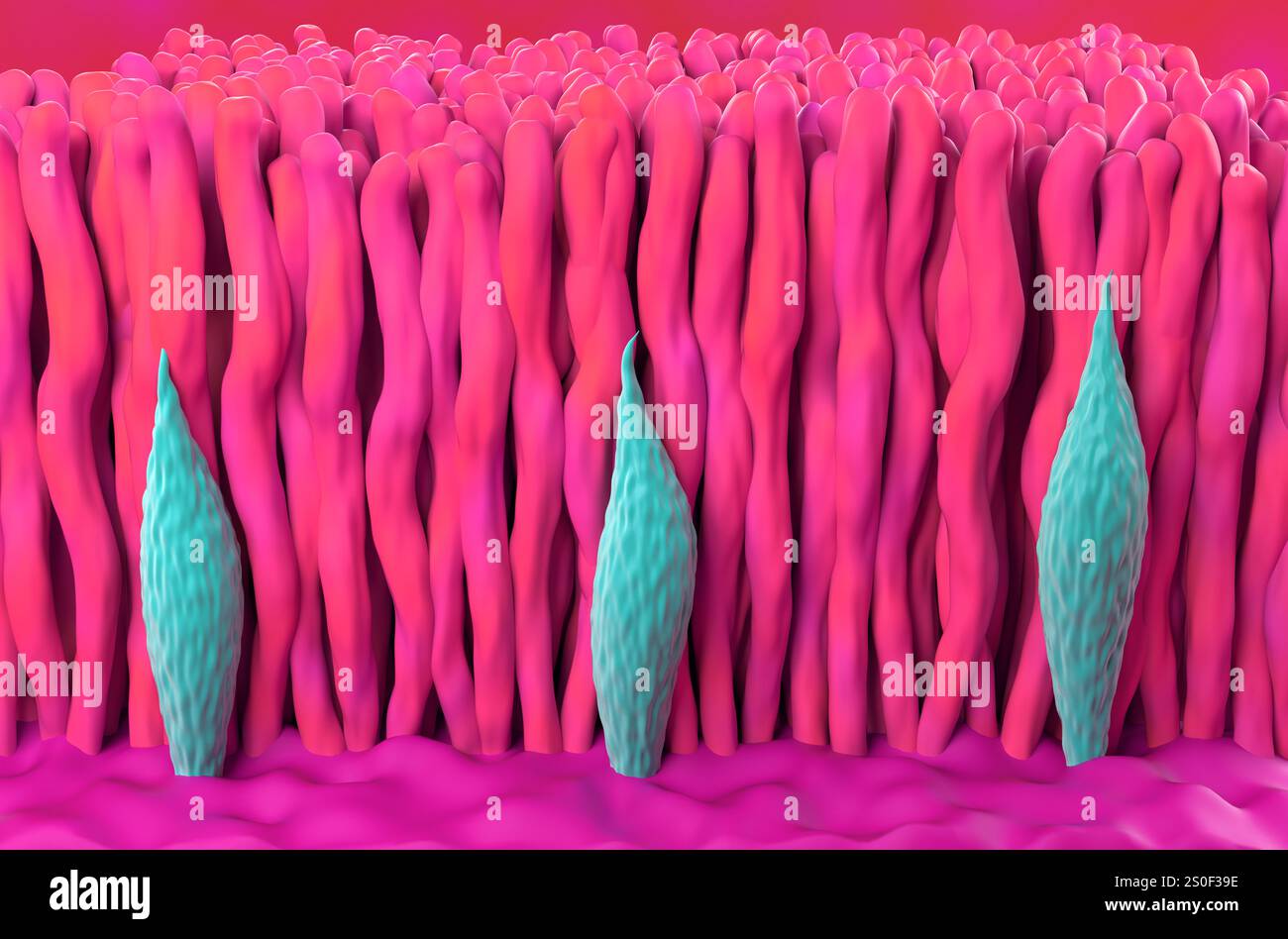 Retina photoreceptor (cone and rod cells) isometric view 3d illustration Stock Photohttps://www.alamy.com/image-license-details/?v=1https://www.alamy.com/retina-photoreceptor-cone-and-rod-cells-isometric-view-3d-illustration-image637115514.html
RF2S0F39E–Retina photoreceptor (cone and rod cells) isometric view 3d illustration
 Fibromyalgia Stock Photohttps://www.alamy.com/image-license-details/?v=1https://www.alamy.com/fibromyalgia-image353182327.html
RF2BEGRJF–Fibromyalgia
 Cross-section of the retina with the photoreceptors: cones, rods, and also the optic nerve. Stock Photohttps://www.alamy.com/image-license-details/?v=1https://www.alamy.com/cross-section-of-the-retina-with-the-photoreceptors-cones-rods-and-also-the-optic-nerve-image476925005.html
RF2JKWPKW–Cross-section of the retina with the photoreceptors: cones, rods, and also the optic nerve.
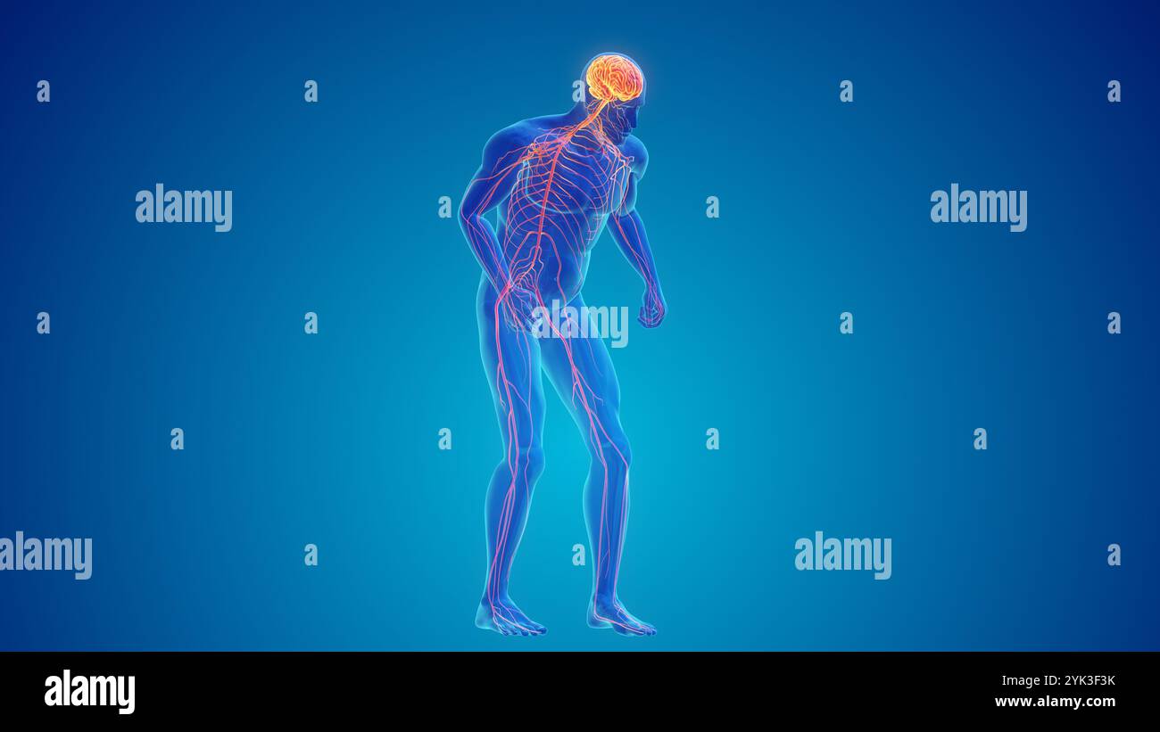 Progressive Central Nervous System Degeneration or Parkinson's disease Stock Photohttps://www.alamy.com/image-license-details/?v=1https://www.alamy.com/progressive-central-nervous-system-degeneration-or-parkinsons-disease-image631329431.html
RF2YK3F3K–Progressive Central Nervous System Degeneration or Parkinson's disease
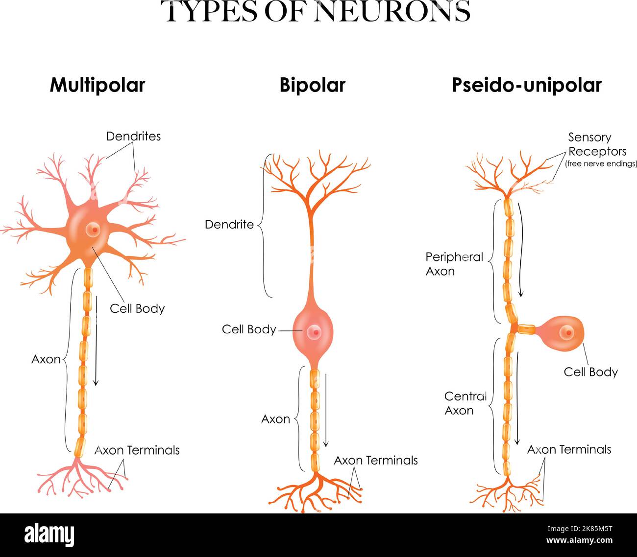 Types of neurons- multipolar, pseudounipolar, bipolar - structure anatomy colorful illustration. Stock Vectorhttps://www.alamy.com/image-license-details/?v=1https://www.alamy.com/types-of-neurons-multipolar-pseudounipolar-bipolar-structure-anatomy-colorful-illustration-image486933156.html
RF2K85M5T–Types of neurons- multipolar, pseudounipolar, bipolar - structure anatomy colorful illustration.
 Degenerative Disease of the Nervous System or Parkinson's disease Stock Photohttps://www.alamy.com/image-license-details/?v=1https://www.alamy.com/degenerative-disease-of-the-nervous-system-or-parkinsons-disease-image631329170.html
RF2YK3EPA–Degenerative Disease of the Nervous System or Parkinson's disease
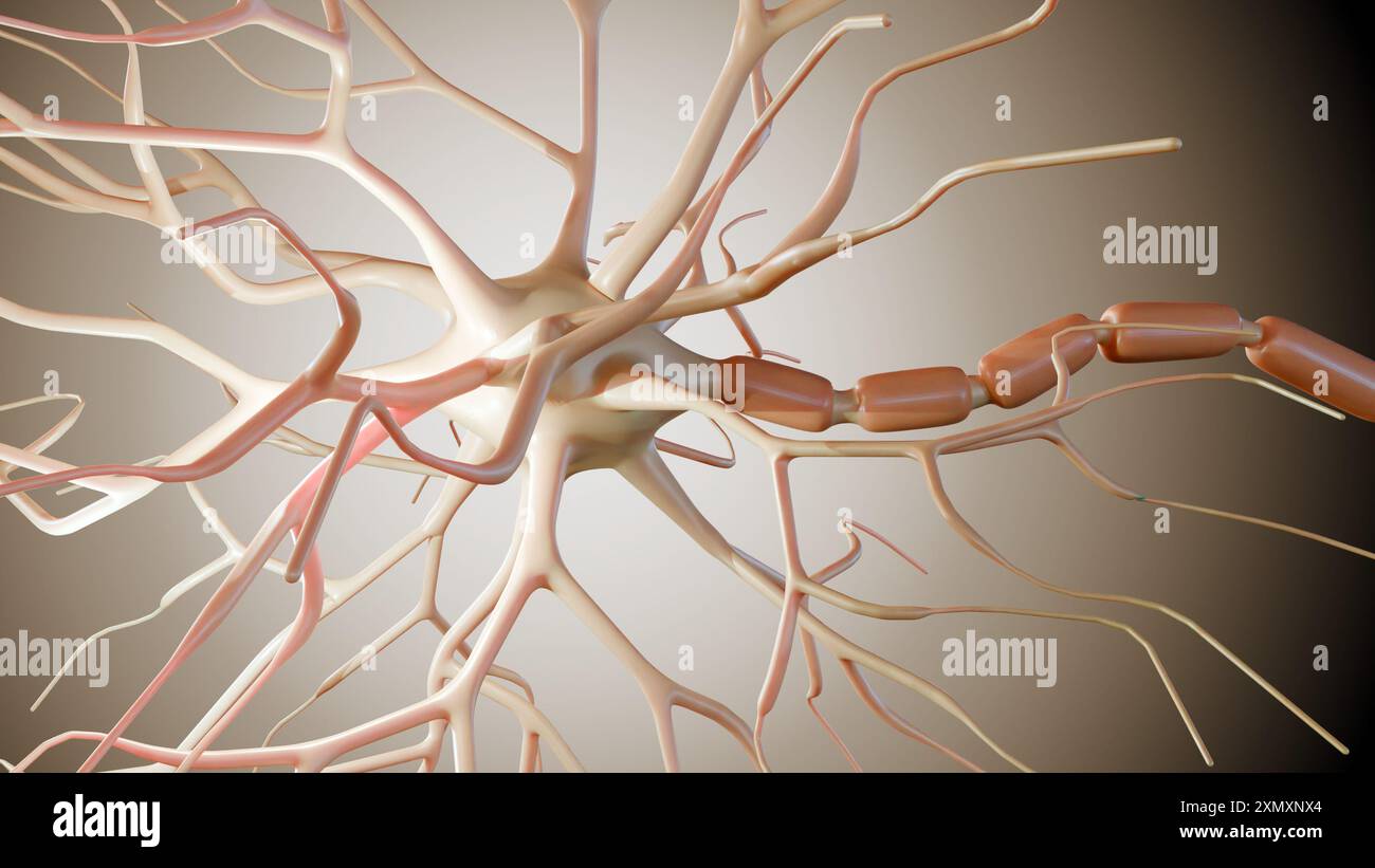 3d rendering of Multipolar neurons. Multipolar neurons are the most common type of neuron Stock Photohttps://www.alamy.com/image-license-details/?v=1https://www.alamy.com/3d-rendering-of-multipolar-neurons-multipolar-neurons-are-the-most-common-type-of-neuron-image615243948.html
RF2XMXNX4–3d rendering of Multipolar neurons. Multipolar neurons are the most common type of neuron
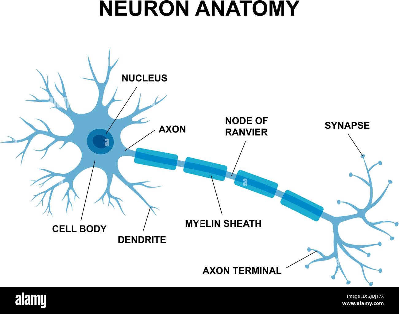 Vector infographic of neuron anatomy. Medical chart human neuron structure illustration. Synapses, cell body, nucleus, axon and dendrites scheme. Stock Vectorhttps://www.alamy.com/image-license-details/?v=1https://www.alamy.com/vector-infographic-of-neuron-anatomy-medical-chart-human-neuron-structure-illustration-synapses-cell-body-nucleus-axon-and-dendrites-scheme-image473084638.html
RF2JDJT7X–Vector infographic of neuron anatomy. Medical chart human neuron structure illustration. Synapses, cell body, nucleus, axon and dendrites scheme.
 Illustration of a woman head with brain Stock Vectorhttps://www.alamy.com/image-license-details/?v=1https://www.alamy.com/illustration-of-a-woman-head-with-brain-image342208217.html
RF2ATMX21–Illustration of a woman head with brain
 . The Biological bulletin. Biology; Zoology; Biology; Marine Biology. 264 ARLAX L. EDGAR. FIGURE 1. Lyriform organ at distal margin of trochanter (arrow). The autotomy plane which separates trochanter from femur is shown under the other end of the arrow. A cluster of I- and L-shaped slit organs may he seen on the proximal portion of the femur fright). FIGURE 2. Several slit organs clustered at base of femur (see Figure 1). The expanded area in the middle of the slit probably receives the attachment of a bipolar neuron. FIGURE 3. Large, thick-bordered slit organ on femur (arrow) where femur art Stock Photohttps://www.alamy.com/image-license-details/?v=1https://www.alamy.com/the-biological-bulletin-biology-zoology-biology-marine-biology-264-arlax-l-edgar-figure-1-lyriform-organ-at-distal-margin-of-trochanter-arrow-the-autotomy-plane-which-separates-trochanter-from-femur-is-shown-under-the-other-end-of-the-arrow-a-cluster-of-i-and-l-shaped-slit-organs-may-he-seen-on-the-proximal-portion-of-the-femur-fright-figure-2-several-slit-organs-clustered-at-base-of-femur-see-figure-1-the-expanded-area-in-the-middle-of-the-slit-probably-receives-the-attachment-of-a-bipolar-neuron-figure-3-large-thick-bordered-slit-organ-on-femur-arrow-where-femur-art-image234638984.html
RMRHMMBM–. The Biological bulletin. Biology; Zoology; Biology; Marine Biology. 264 ARLAX L. EDGAR. FIGURE 1. Lyriform organ at distal margin of trochanter (arrow). The autotomy plane which separates trochanter from femur is shown under the other end of the arrow. A cluster of I- and L-shaped slit organs may he seen on the proximal portion of the femur fright). FIGURE 2. Several slit organs clustered at base of femur (see Figure 1). The expanded area in the middle of the slit probably receives the attachment of a bipolar neuron. FIGURE 3. Large, thick-bordered slit organ on femur (arrow) where femur art
 creative human brain colorful wires technology logo vector logo symbol Stock Vectorhttps://www.alamy.com/image-license-details/?v=1https://www.alamy.com/creative-human-brain-colorful-wires-technology-logo-vector-logo-symbol-image455176393.html
RF2HCF23N–creative human brain colorful wires technology logo vector logo symbol
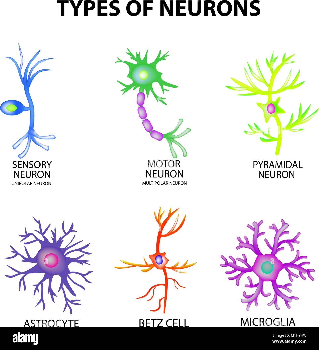 Types of neurons. Structure sensory, motor neuron, astrocyte, pyromidal, Betz cell, microglia. Set. Infographics Vector illustration on isolated backg Stock Vectorhttps://www.alamy.com/image-license-details/?v=1https://www.alamy.com/stock-photo-types-of-neurons-structure-sensory-motor-neuron-astrocyte-pyromidal-173113189.html
RFM1HYHW–Types of neurons. Structure sensory, motor neuron, astrocyte, pyromidal, Betz cell, microglia. Set. Infographics Vector illustration on isolated backg
 Illustration of a woman head with brain Stock Vectorhttps://www.alamy.com/image-license-details/?v=1https://www.alamy.com/illustration-of-a-woman-head-with-brain-image350510296.html
RF2BA73CT–Illustration of a woman head with brain
 Neuron Stock Photohttps://www.alamy.com/image-license-details/?v=1https://www.alamy.com/neuron-image226184821.html
RFR3YH19–Neuron
 Bipolar Neuron and Brain Stock Photohttps://www.alamy.com/image-license-details/?v=1https://www.alamy.com/bipolar-neuron-and-brain-image66988047.html
RMDTYFX7–Bipolar Neuron and Brain
 Retina cone and rod in the human eye - isometric view 3d illustration Stock Photohttps://www.alamy.com/image-license-details/?v=1https://www.alamy.com/retina-cone-and-rod-in-the-human-eye-isometric-view-3d-illustration-image499289375.html
RF2M08GKB–Retina cone and rod in the human eye - isometric view 3d illustration
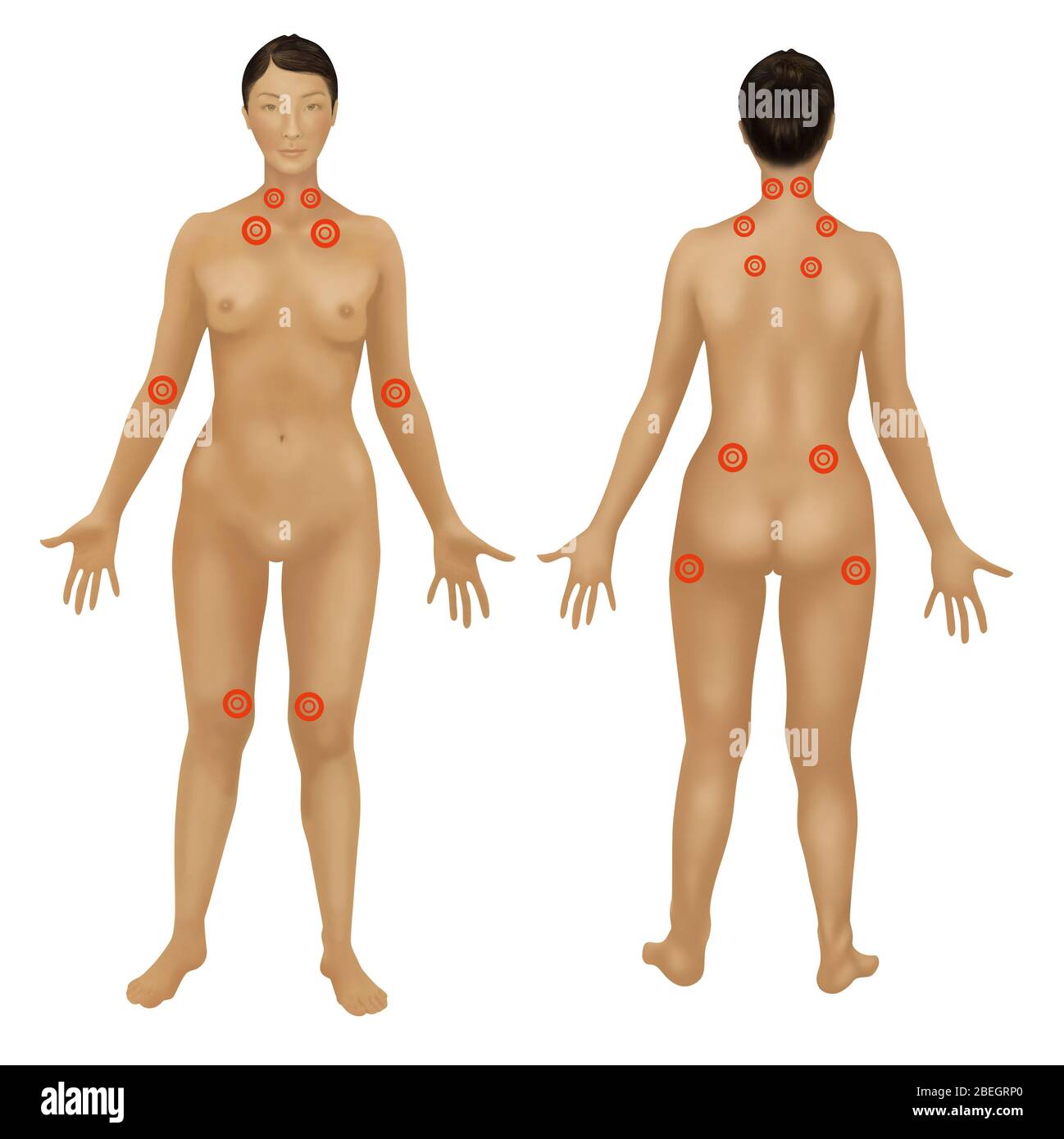 Fibromyalgia Stock Photohttps://www.alamy.com/image-license-details/?v=1https://www.alamy.com/fibromyalgia-image353182424.html
RF2BEGRP0–Fibromyalgia
 Eye showing the retina and the fovea. Stock Photohttps://www.alamy.com/image-license-details/?v=1https://www.alamy.com/eye-showing-the-retina-and-the-fovea-image476925436.html
RF2JKWR78–Eye showing the retina and the fovea.
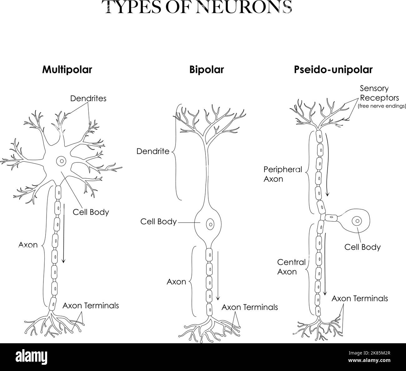 Types of neurons- multipolar, pseudounipolar, bipolar anatomy black and white line art illustration. Can be used as a worksheet for coloring and learn Stock Vectorhttps://www.alamy.com/image-license-details/?v=1https://www.alamy.com/types-of-neurons-multipolar-pseudounipolar-bipolar-anatomy-black-and-white-line-art-illustration-can-be-used-as-a-worksheet-for-coloring-and-learn-image486933071.html
RF2K85M2R–Types of neurons- multipolar, pseudounipolar, bipolar anatomy black and white line art illustration. Can be used as a worksheet for coloring and learn
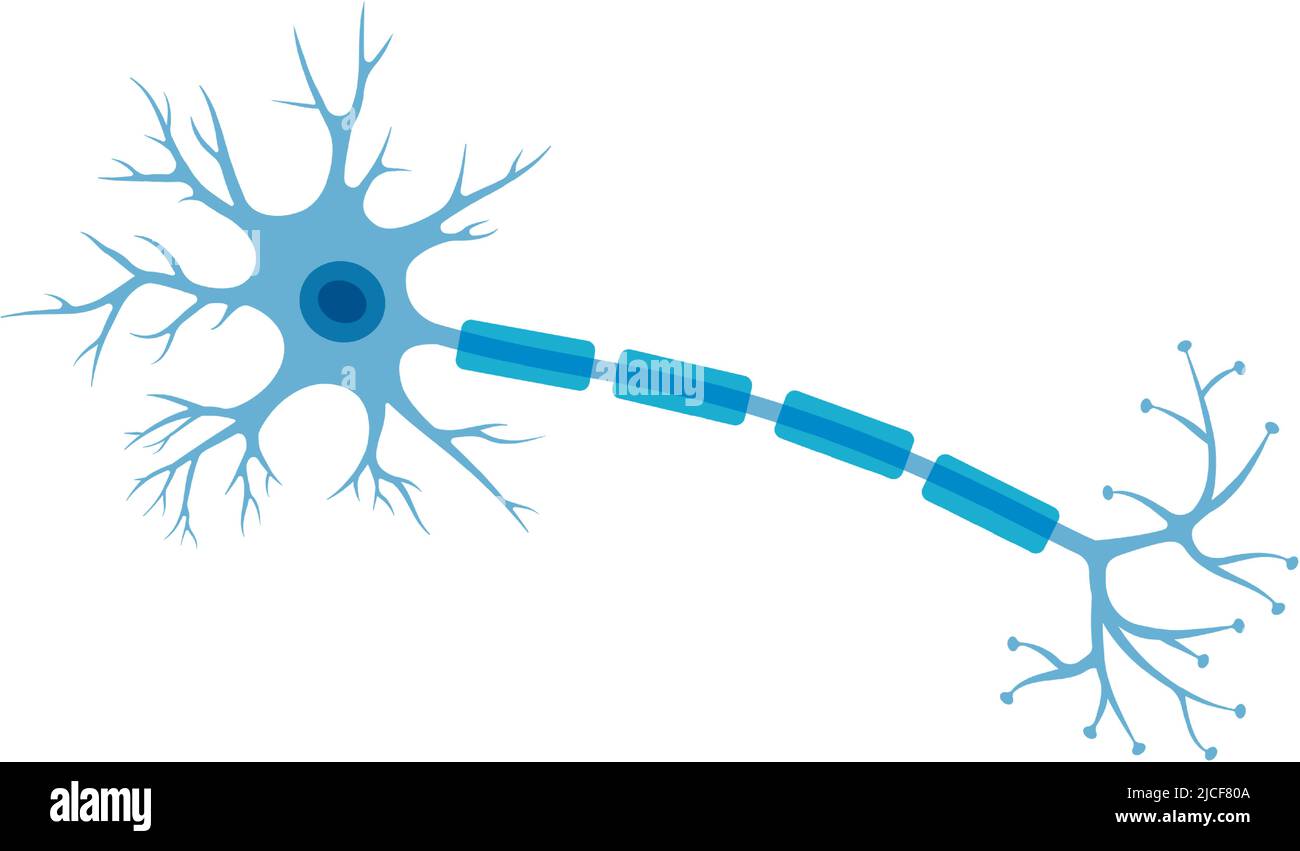 Human neuron structure. Brain neuron cell illustration. Synapses, myelin sheat, cell body, nucleus, axon and dendrites scheme. Neurology illustration Stock Vectorhttps://www.alamy.com/image-license-details/?v=1https://www.alamy.com/human-neuron-structure-brain-neuron-cell-illustration-synapses-myelin-sheat-cell-body-nucleus-axon-and-dendrites-scheme-neurology-illustration-image472391370.html
RF2JCF80A–Human neuron structure. Brain neuron cell illustration. Synapses, myelin sheat, cell body, nucleus, axon and dendrites scheme. Neurology illustration
 . The Biological bulletin. Biology; Zoology; Biology; Marine Biology. 272 A.-J. BEER ET AL.. Figure 2. Observations of live larvae. (A) Multipolar cells of the blastocoelar network in the oral hood of an early 4-armed pluteus (arrows). (B) Higher magnification of (A) showing a flask-shaped bipolar neuron with a tapering apical process (arrowhead) and basal varicose axon contributing to the ciliary nerve (arrows). (C) Diagram of (B) with the neuron shown in black (no! to scale). Bars: A = 25 /xm, B = 10 ju.ni.. Please note that these images are extracted from scanned page images that may have b Stock Photohttps://www.alamy.com/image-license-details/?v=1https://www.alamy.com/the-biological-bulletin-biology-zoology-biology-marine-biology-272-a-j-beer-et-al-figure-2-observations-of-live-larvae-a-multipolar-cells-of-the-blastocoelar-network-in-the-oral-hood-of-an-early-4-armed-pluteus-arrows-b-higher-magnification-of-a-showing-a-flask-shaped-bipolar-neuron-with-a-tapering-apical-process-arrowhead-and-basal-varicose-axon-contributing-to-the-ciliary-nerve-arrows-c-diagram-of-b-with-the-neuron-shown-in-black-no!-to-scale-bars-a-=-25-xm-b-=-10-juni-please-note-that-these-images-are-extracted-from-scanned-page-images-that-may-have-b-image234616677.html
RMRHKKY1–. The Biological bulletin. Biology; Zoology; Biology; Marine Biology. 272 A.-J. BEER ET AL.. Figure 2. Observations of live larvae. (A) Multipolar cells of the blastocoelar network in the oral hood of an early 4-armed pluteus (arrows). (B) Higher magnification of (A) showing a flask-shaped bipolar neuron with a tapering apical process (arrowhead) and basal varicose axon contributing to the ciliary nerve (arrows). (C) Diagram of (B) with the neuron shown in black (no! to scale). Bars: A = 25 /xm, B = 10 ju.ni.. Please note that these images are extracted from scanned page images that may have b
 creative human brain wires technology logo vector logo symbol Stock Vectorhttps://www.alamy.com/image-license-details/?v=1https://www.alamy.com/creative-human-brain-wires-technology-logo-vector-logo-symbol-image455176352.html
RF2HCF228–creative human brain wires technology logo vector logo symbol
 Illustration of a head with brain Stock Photohttps://www.alamy.com/image-license-details/?v=1https://www.alamy.com/illustration-of-a-head-with-brain-image245847284.html
RFT7Y8M4–Illustration of a head with brain
 Neuron Stock Photohttps://www.alamy.com/image-license-details/?v=1https://www.alamy.com/neuron-image226184829.html
RFR3YH1H–Neuron
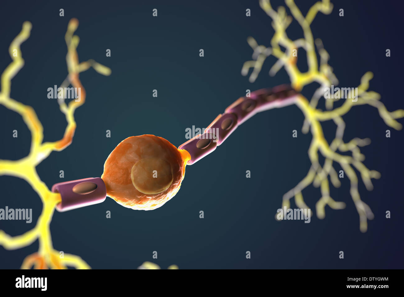 Bipolar Neuron Stock Photohttps://www.alamy.com/image-license-details/?v=1https://www.alamy.com/bipolar-neuron-image66988816.html
RMDTYGWM–Bipolar Neuron
 Neuron Stock Photohttps://www.alamy.com/image-license-details/?v=1https://www.alamy.com/neuron-image226264328.html
RFR436CT–Neuron
 Neuron Stock Photohttps://www.alamy.com/image-license-details/?v=1https://www.alamy.com/neuron-image226217495.html
RFR412M7–Neuron
 Fibromyalgia Stock Photohttps://www.alamy.com/image-license-details/?v=1https://www.alamy.com/fibromyalgia-image353182429.html
RF2BEGRP5–Fibromyalgia
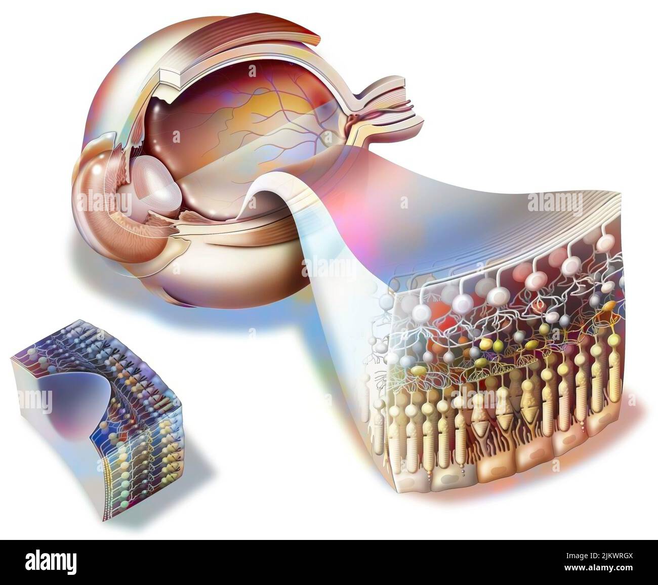 Structure of an eye with zoom on the retina and fovea. Stock Photohttps://www.alamy.com/image-license-details/?v=1https://www.alamy.com/structure-of-an-eye-with-zoom-on-the-retina-and-fovea-image476925706.html
RF2JKWRGX–Structure of an eye with zoom on the retina and fovea.
 Neuron Stock Photohttps://www.alamy.com/image-license-details/?v=1https://www.alamy.com/neuron-image226184837.html
RFR3YH1W–Neuron
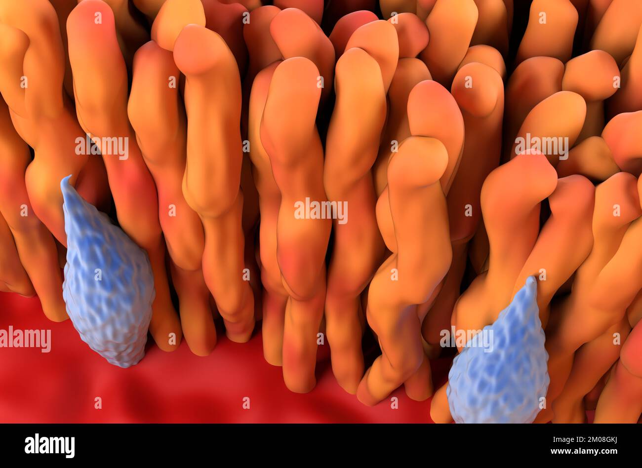 Retina cone and rod in the human eye - top view 3d illustration Stock Photohttps://www.alamy.com/image-license-details/?v=1https://www.alamy.com/retina-cone-and-rod-in-the-human-eye-top-view-3d-illustration-image499289382.html
RF2M08GKJ–Retina cone and rod in the human eye - top view 3d illustration
 effects bipolar Stock Photohttps://www.alamy.com/image-license-details/?v=1https://www.alamy.com/stock-photo-effects-bipolar-146400976.html
RFJE53X8–effects bipolar
RF2JBKRGR–Brain neuron symbol. Human neuron cell sign. Synapses, myelin sheat, cell body, nucleus, axon and dendrites icon. Neurology illustration
 An American text-book of physiology . Fig. 147.—Spinal ganglion of an embryo duck ; composed of dineuric nerve-cells (van Gehuchten). are but two in number and both are medullated. They pass in opposite direc-tions and in this type there are no dendrons. To understand the arrangementin these cases, recourse must be had to the facts of development. The second. CENTRAL NERVOUS SYSTEM. 613 type begins its development as :i bipolar cell, a neuron growing from each pole(Fig. 147). In the adult spinal ganglion of tlie higher mannnuls, however,no such bii)olar cells are to be found, but only cells ha Stock Photohttps://www.alamy.com/image-license-details/?v=1https://www.alamy.com/an-american-text-book-of-physiology-fig-147spinal-ganglion-of-an-embryo-duck-composed-of-dineuric-nerve-cells-van-gehuchten-are-but-two-in-number-and-both-are-medullated-they-pass-in-opposite-direc-tions-and-in-this-type-there-are-no-dendrons-to-understand-the-arrangementin-these-cases-recourse-must-be-had-to-the-facts-of-development-the-second-central-nervous-system-613-type-begins-its-development-as-i-bipolar-cell-a-neuron-growing-from-each-polefig-147-in-the-adult-spinal-ganglion-of-tlie-higher-mannnuls-howeverno-such-biiolar-cells-are-to-be-found-but-only-cells-ha-image338376165.html
RM2AJEA71–An American text-book of physiology . Fig. 147.—Spinal ganglion of an embryo duck ; composed of dineuric nerve-cells (van Gehuchten). are but two in number and both are medullated. They pass in opposite direc-tions and in this type there are no dendrons. To understand the arrangementin these cases, recourse must be had to the facts of development. The second. CENTRAL NERVOUS SYSTEM. 613 type begins its development as :i bipolar cell, a neuron growing from each pole(Fig. 147). In the adult spinal ganglion of tlie higher mannnuls, however,no such bii)olar cells are to be found, but only cells ha
 Abstract illustration of a human head with brain. Woman face silhouette. Medical theme creative concept. Connected lines with dots. Schizophrenia dise Stock Photohttps://www.alamy.com/image-license-details/?v=1https://www.alamy.com/abstract-illustration-of-a-human-head-with-brain-woman-face-silhouette-medical-theme-creative-concept-connected-lines-with-dots-schizophrenia-dise-image340445115.html
RF2ANTH63–Abstract illustration of a human head with brain. Woman face silhouette. Medical theme creative concept. Connected lines with dots. Schizophrenia dise
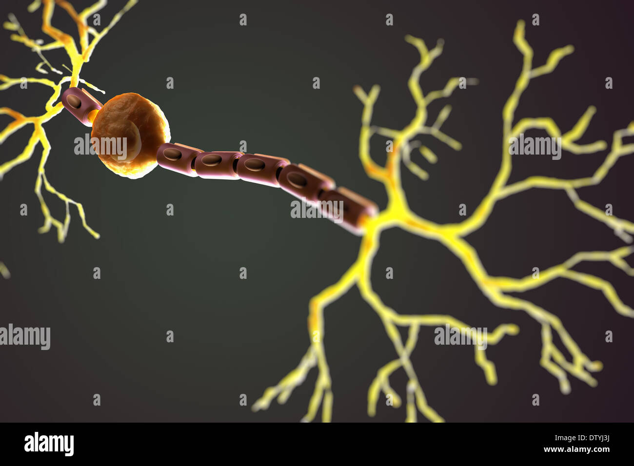 Bipolar Neuron Stock Photohttps://www.alamy.com/image-license-details/?v=1https://www.alamy.com/bipolar-neuron-image66989766.html
RMDTYJ3J–Bipolar Neuron

