Quick filters:
Blood vessels sem Stock Photos and Images
RM2BE0GFT–Colored scanning electron micrograph (SEM) of human red blood cells (erythrocytes). Red blood cells are biconcave, giving them a large surface area for gas exchange, and highly elastic, enabling them to pass through narrow capillary vessels. The nucleus and other organelles are lost as the cells mature. The cell's interior is packed with hemoglobin, a red iron-containing pigment that has an oxygen-carrying capacity. The main function of red blood cells is to distribute oxygen to body tissues and to carry waste carbon dioxide back to the lungs.
 Fossilised dinosaur bone, coloured scanning electron micrograph (SEM). This specimen is from a Tyrannosaurus rex fossil. This dinosaur was a carnivore from the Cretaceous Period. The circular holes are the Haversian canals that carry blood vessels. Magnification: x20 when printed at 10 centimetres across. Stock Photohttps://www.alamy.com/image-license-details/?v=1https://www.alamy.com/fossilised-dinosaur-bone-coloured-scanning-electron-micrograph-sem-this-specimen-is-from-a-tyrannosaurus-rex-fossil-this-dinosaur-was-a-carnivore-from-the-cretaceous-period-the-circular-holes-are-the-haversian-canals-that-carry-blood-vessels-magnification-x20-when-printed-at-10-centimetres-across-image623982675.html
RF2Y74T7F–Fossilised dinosaur bone, coloured scanning electron micrograph (SEM). This specimen is from a Tyrannosaurus rex fossil. This dinosaur was a carnivore from the Cretaceous Period. The circular holes are the Haversian canals that carry blood vessels. Magnification: x20 when printed at 10 centimetres across.
RM2BE0H23–This scanning electron micrograph (SEM) depicted a number of red blood cells found enmeshed in a fibrinous matrix on the luminal surface of an indwelling vascular catheter; Magnified 2858x. Note the biconcave cytomorphologic shape of each erythrocyte, which increases the surface area of these hemoglobin-filled cells, thereby, promoting a greater degree of gas exchange, which is their primary function in an in vivo setting. In their adult phase, these cells possess no nucleus. What appears to be irregularly-shaped chunks of debris, are actually fibrin clumps, which when inside the living organi
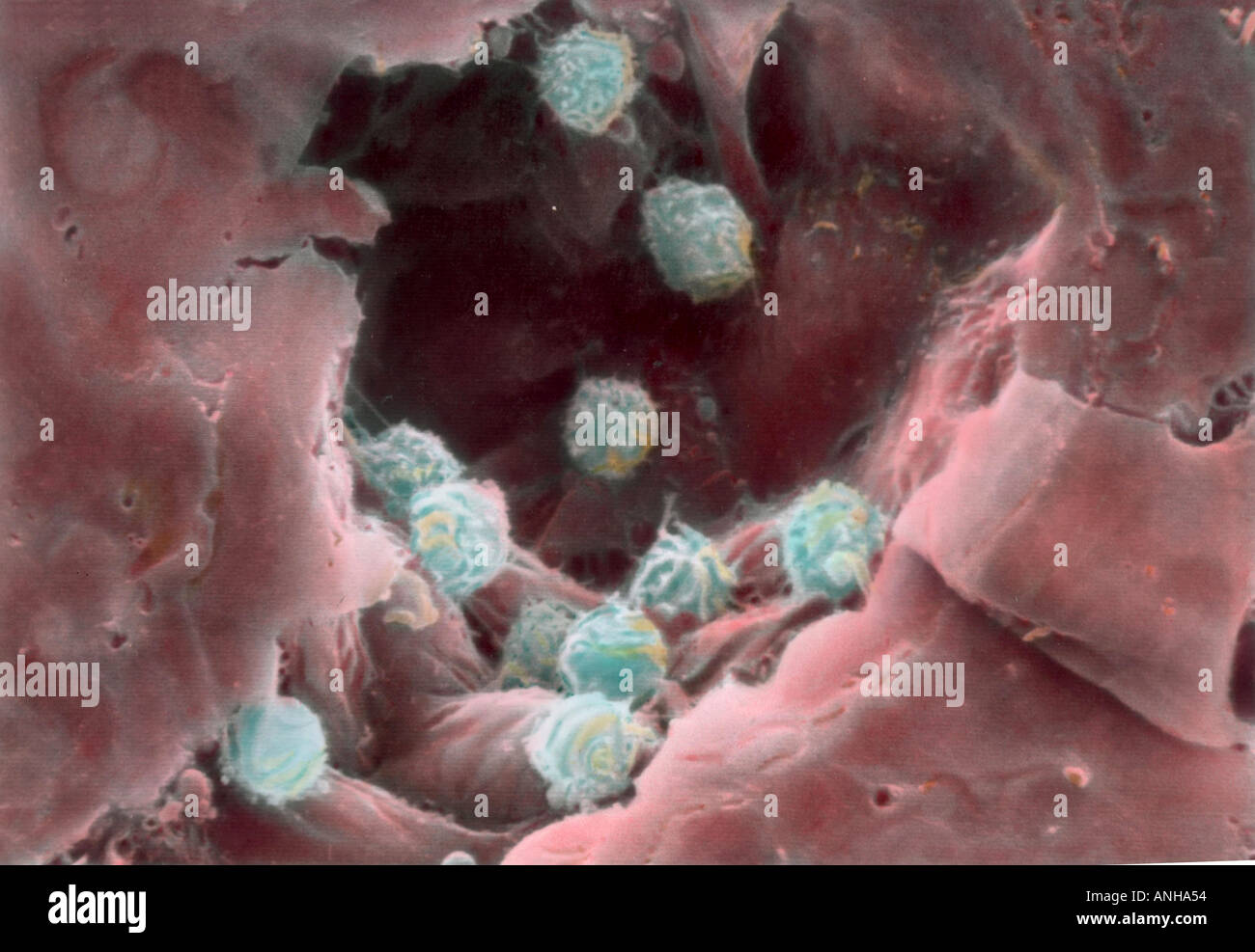 White blood cells lymphocytes in human vein A lymphocyte is a white blood cell that is formed in lymphoid tissue A venule is Stock Photohttps://www.alamy.com/image-license-details/?v=1https://www.alamy.com/white-blood-cells-lymphocytes-in-human-vein-a-lymphocyte-is-a-white-image2873939.html
RMANHA54–White blood cells lymphocytes in human vein A lymphocyte is a white blood cell that is formed in lymphoid tissue A venule is
 ENDOTHELIUM RABBIT, SEM Stock Photohttps://www.alamy.com/image-license-details/?v=1https://www.alamy.com/stock-photo-endothelium-rabbit-sem-25562509.html
RFBDGD6N–ENDOTHELIUM RABBIT, SEM
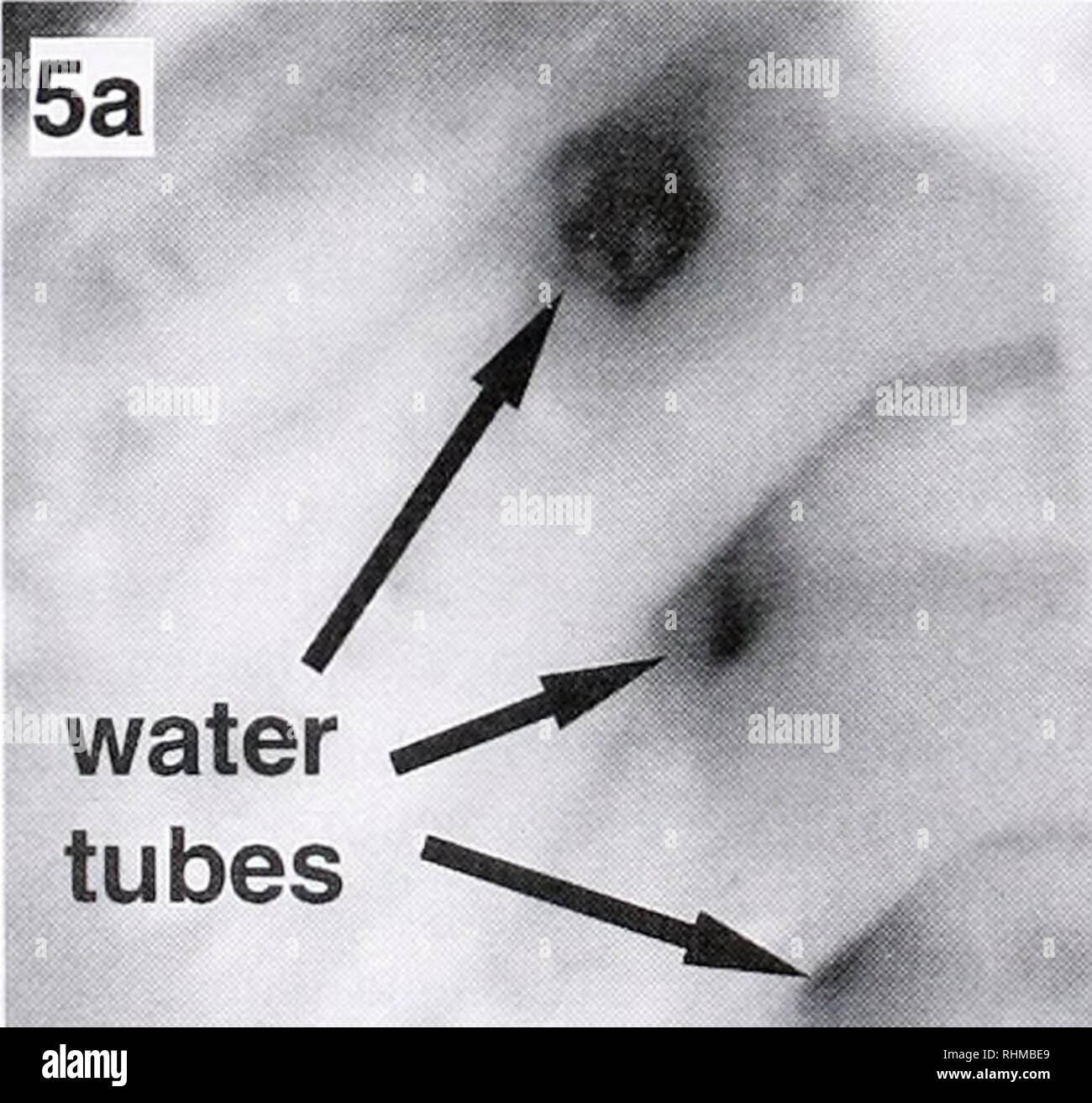 . The Biological bulletin. Biology; Zoology; Biology; Marine Biology. relaxed contracted Figure 4. Water-tube dimension, (a) Cross-sectional area of the water tubes per unit gill length significantly decreased upon muscular contraction tor each of the species (Corbicula fluminea, Dreissena polymorpha, and Mercenaria mercenaria) examined (mean ± SE; n = 5). (b. c) M. mercenaria gill (SEM) in cross-section: (b) relaxed gill; (c) gill from the same animal after the gill muscles contracted. Blood vessels (bv) are visible, with only the vessel at the top left appearing to be open. the pressure drop Stock Photohttps://www.alamy.com/image-license-details/?v=1https://www.alamy.com/the-biological-bulletin-biology-zoology-biology-marine-biology-relaxed-contracted-figure-4-water-tube-dimension-a-cross-sectional-area-of-the-water-tubes-per-unit-gill-length-significantly-decreased-upon-muscular-contraction-tor-each-of-the-species-corbicula-fluminea-dreissena-polymorpha-and-mercenaria-mercenaria-examined-mean-se-n-=-5-b-c-m-mercenaria-gill-sem-in-cross-section-b-relaxed-gill-c-gill-from-the-same-animal-after-the-gill-muscles-contracted-blood-vessels-bv-are-visible-with-only-the-vessel-at-the-top-left-appearing-to-be-open-the-pressure-drop-image234632001.html
RMRHMBE9–. The Biological bulletin. Biology; Zoology; Biology; Marine Biology. relaxed contracted Figure 4. Water-tube dimension, (a) Cross-sectional area of the water tubes per unit gill length significantly decreased upon muscular contraction tor each of the species (Corbicula fluminea, Dreissena polymorpha, and Mercenaria mercenaria) examined (mean ± SE; n = 5). (b. c) M. mercenaria gill (SEM) in cross-section: (b) relaxed gill; (c) gill from the same animal after the gill muscles contracted. Blood vessels (bv) are visible, with only the vessel at the top left appearing to be open. the pressure drop
RF2CGGAW0–Platelet-1 vector icon. Modern vector illustration concepts. Easy to edit and customize.
 Fossilised dinosaur bone, coloured scanning electron micrograph (SEM). This specimen is from a Tyrannosaurus rex fossil. This dinosaur was a carnivore from the Cretaceous Period. The circular holes are the Haversian canals that carry blood vessels. Magnification: x20 when printed at 10 centimetres across. Stock Photohttps://www.alamy.com/image-license-details/?v=1https://www.alamy.com/fossilised-dinosaur-bone-coloured-scanning-electron-micrograph-sem-this-specimen-is-from-a-tyrannosaurus-rex-fossil-this-dinosaur-was-a-carnivore-from-the-cretaceous-period-the-circular-holes-are-the-haversian-canals-that-carry-blood-vessels-magnification-x20-when-printed-at-10-centimetres-across-image623982804.html
RF2Y74TC4–Fossilised dinosaur bone, coloured scanning electron micrograph (SEM). This specimen is from a Tyrannosaurus rex fossil. This dinosaur was a carnivore from the Cretaceous Period. The circular holes are the Haversian canals that carry blood vessels. Magnification: x20 when printed at 10 centimetres across.
RM2BE0H0H–This scanning electron micrograph (SEM) depicted a number of red blood cells found enmeshed in a fibrinous matrix on the luminal surface of an indwelling vascular; Magnified 2849x. In this instance, the indwelling catheter was a tube that was left in place creating a patent portal directly into a blood vessel. Note the biconcave cytomorphologic shape of each erythrocyte, which increases the surface area of these hemoglobin-filled cells, thereby, promoting a greater degree of gas exchange, which is their primary function in an in vivo setting. In their adult phase, these cells possess no nucleu
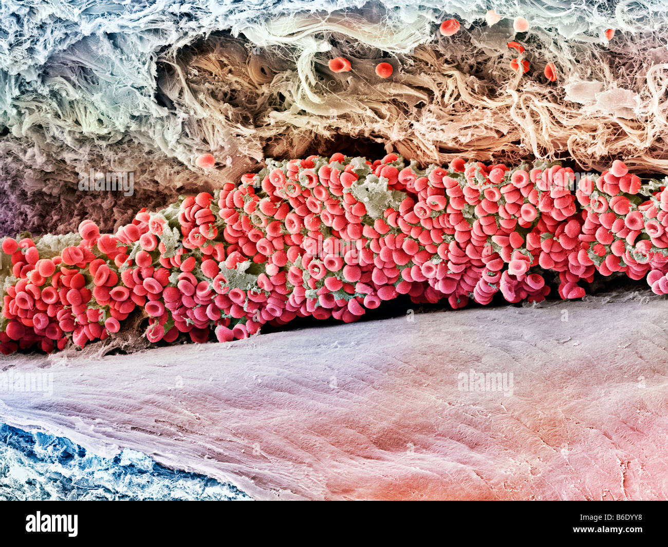 Blood clot, Coloured scanning electron micrograph (SEM) of blood clotting in an ovarian follicle. Stock Photohttps://www.alamy.com/image-license-details/?v=1https://www.alamy.com/stock-photo-blood-clot-coloured-scanning-electron-micrograph-sem-of-blood-clotting-21205612.html
RFB6DYY8–Blood clot, Coloured scanning electron micrograph (SEM) of blood clotting in an ovarian follicle.
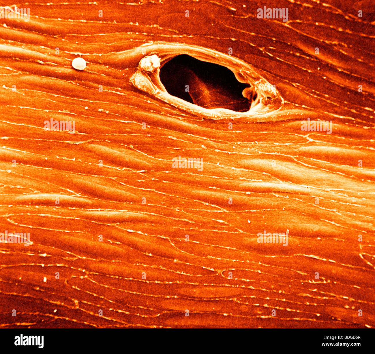 ENDOTHELIUM RABBIT, SEM Stock Photohttps://www.alamy.com/image-license-details/?v=1https://www.alamy.com/stock-photo-endothelium-rabbit-sem-25562511.html
RFBDGD6R–ENDOTHELIUM RABBIT, SEM
 . The Cephalopoda. Cephalopoda. Oegopsida sacc; v. etl prost. can cil. ves. sem. 2 figure 14. Cross section of the genitalia of a male Illex (at the level of y in Figure 12). The descending branch of the 3rd part of the seminal vesicle is cut at the left, the ascending branch at the right. The swelling (>v.) is recognizable in the second part of the seminal vesicle (w. sem. 2), as in the 3rd part. can. cil. ciliated canal; v. blood vessels .sacc' diverticulum of genital pockets between vas efferens and spermatophore sac. Other lettering as in Figure 13. A dashed line indicates the epitheli Stock Photohttps://www.alamy.com/image-license-details/?v=1https://www.alamy.com/the-cephalopoda-cephalopoda-oegopsida-sacc-v-etl-prost-can-cil-ves-sem-2-figure-14-cross-section-of-the-genitalia-of-a-male-illex-at-the-level-of-y-in-figure-12-the-descending-branch-of-the-3rd-part-of-the-seminal-vesicle-is-cut-at-the-left-the-ascending-branch-at-the-right-the-swelling-gtv-is-recognizable-in-the-second-part-of-the-seminal-vesicle-w-sem-2-as-in-the-3rd-part-can-cil-ciliated-canal-v-blood-vessels-sacc-diverticulum-of-genital-pockets-between-vas-efferens-and-spermatophore-sac-other-lettering-as-in-figure-13-a-dashed-line-indicates-the-epitheli-image235079439.html
RMRJCP67–. The Cephalopoda. Cephalopoda. Oegopsida sacc; v. etl prost. can cil. ves. sem. 2 figure 14. Cross section of the genitalia of a male Illex (at the level of y in Figure 12). The descending branch of the 3rd part of the seminal vesicle is cut at the left, the ascending branch at the right. The swelling (>v.) is recognizable in the second part of the seminal vesicle (w. sem. 2), as in the 3rd part. can. cil. ciliated canal; v. blood vessels .sacc' diverticulum of genital pockets between vas efferens and spermatophore sac. Other lettering as in Figure 13. A dashed line indicates the epitheli
RM2BE0H14–SEM depicting red blood cells found enmeshed in a fibrinous matrix on the luminal surface of an indwelling vascular. The erythrocyte in the center had undergone the process of crenation, whereupon, it developed a number of cell wall projections, thereby, transforming it into what is termed an acanthocyte. Acanthocytosis could be indicative of a number of hematologic disease processes, but in this instance, was probably due to the fixation procedure carried out on this specimen prior to electron micrographic viewing. Note the normally appearing, biconcave cytomorphologic shape of the other eryt
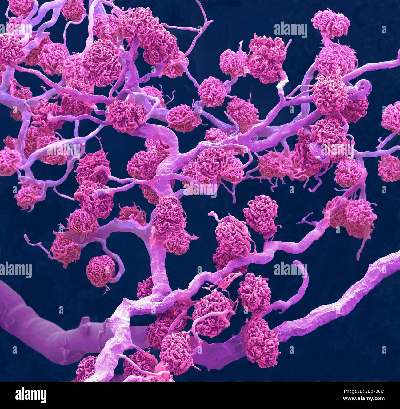 Kidney glomeruli. Coloured scanning electron micrograph (SEM) of a resin cast of glomeruli capillaries and the larger blood vessels supplying them wit Stock Photohttps://www.alamy.com/image-license-details/?v=1https://www.alamy.com/kidney-glomeruli-coloured-scanning-electron-micrograph-sem-of-a-resin-cast-of-glomeruli-capillaries-and-the-larger-blood-vessels-supplying-them-wit-image378784356.html
RF2D0738M–Kidney glomeruli. Coloured scanning electron micrograph (SEM) of a resin cast of glomeruli capillaries and the larger blood vessels supplying them wit
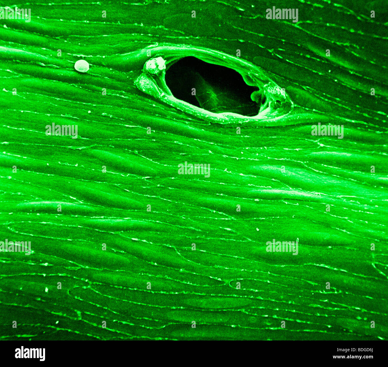 ENDOTHELIUM RABBIT, SEM Stock Photohttps://www.alamy.com/image-license-details/?v=1https://www.alamy.com/stock-photo-endothelium-rabbit-sem-25562506.html
RFBDGD6J–ENDOTHELIUM RABBIT, SEM
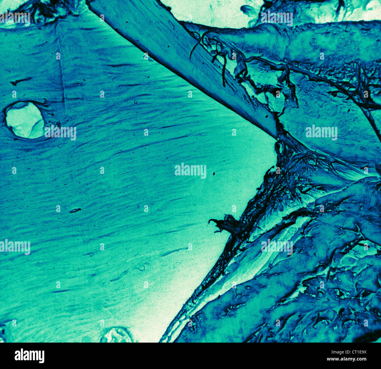 BLOOD CAPILLARY, SEM Stock Photohttps://www.alamy.com/image-license-details/?v=1https://www.alamy.com/stock-photo-blood-capillary-sem-49205686.html
RMCT1E9X–BLOOD CAPILLARY, SEM
RM2BE0H0B–This scanning electron micrograph (SEM) depicted a number of red blood cells found enmeshed in a fibrinous matrix on the luminal surface of an indwelling vascular catheter; Magnified 11432x Note the biconcave cytomorphologic shape of each erythrocyte, which increases the surface area of these hemoglobin-filled cells, thereby, promoting a greater degree of gas exchange, which is their primary function in an in vivo setting. In their adult phase, these cells possess no nucleus. What appears to be irregularly-shaped chunks of debris, are actually fibrin clumps, which when inside the living organi
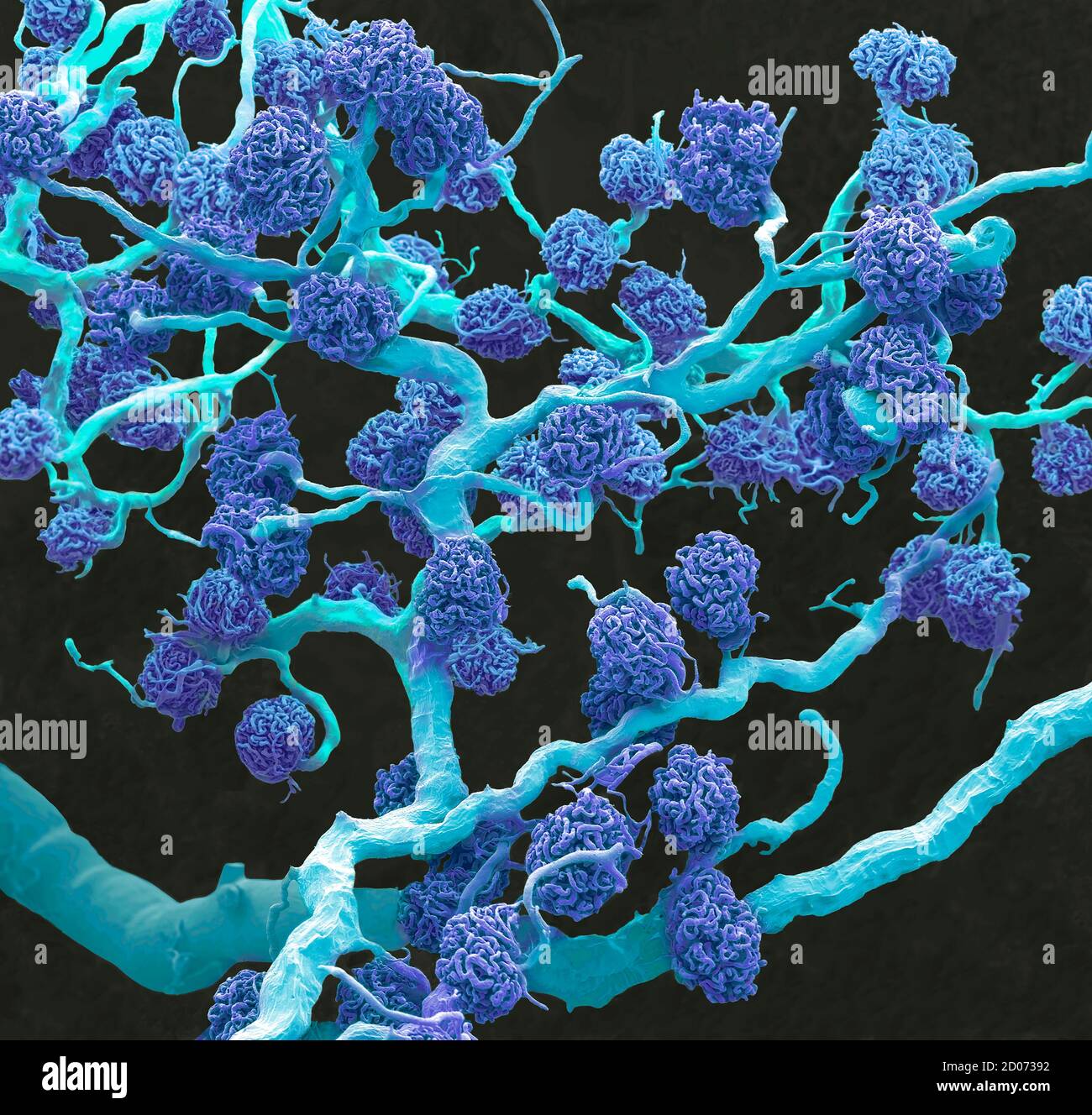 Kidney glomeruli. Coloured scanning electron micrograph (SEM) of a resin cast of glomeruli capillaries and the larger blood vessels supplying them wit Stock Photohttps://www.alamy.com/image-license-details/?v=1https://www.alamy.com/kidney-glomeruli-coloured-scanning-electron-micrograph-sem-of-a-resin-cast-of-glomeruli-capillaries-and-the-larger-blood-vessels-supplying-them-wit-image378784366.html
RF2D07392–Kidney glomeruli. Coloured scanning electron micrograph (SEM) of a resin cast of glomeruli capillaries and the larger blood vessels supplying them wit
 BLOOD CAPILLARY, SEM Stock Photohttps://www.alamy.com/image-license-details/?v=1https://www.alamy.com/stock-photo-blood-capillary-sem-49205435.html
RMCT1E0Y–BLOOD CAPILLARY, SEM
RM2BE0H0J–This scanning electron micrograph (SEM) depicted a number of red blood cells found enmeshed in a fibrinous matrix on the luminal surface of an indwelling vascular catheter; Magnified 2858x Note the biconcave cytomorphologic shape of each erythrocyte, which increases the surface area of these hemoglobin-filled cells, thereby, promoting a greater degree of gas exchange, which is their primary function in an in vivo setting. In their adult phase, these cells possess no nucleus. What appears to be irregularly-shaped chunks of debris, are actually fibrin clumps, which when inside the living organis
 Artery, Coloured scanning electron micrograph (SEM) of sectioned artery containing red bloodcells (erythrocytes, red). Stock Photohttps://www.alamy.com/image-license-details/?v=1https://www.alamy.com/stock-photo-artery-coloured-scanning-electron-micrograph-sem-of-sectioned-artery-21205589.html
RFB6DYXD–Artery, Coloured scanning electron micrograph (SEM) of sectioned artery containing red bloodcells (erythrocytes, red).
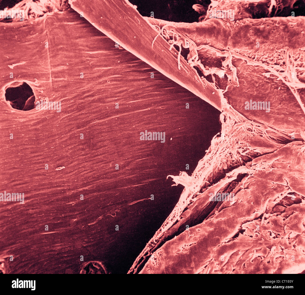
 BLOOD CAPILLARY, SEM Stock Photohttps://www.alamy.com/image-license-details/?v=1https://www.alamy.com/stock-photo-blood-capillary-sem-49205730.html
RMCT1EBE–BLOOD CAPILLARY, SEM
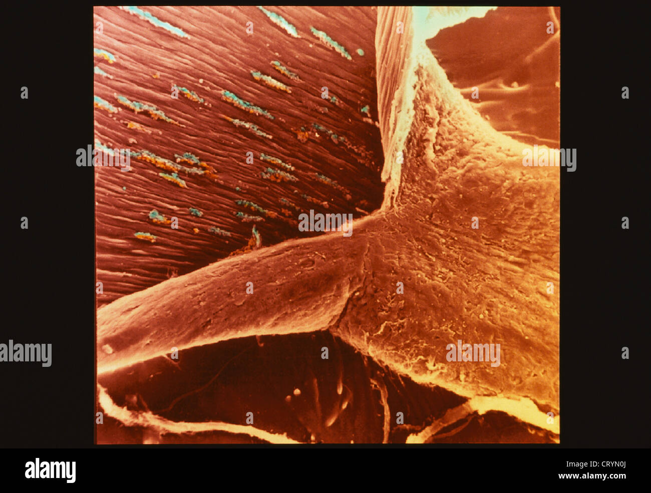 BLOOD CAPILLARY, SEM Stock Photohttps://www.alamy.com/image-license-details/?v=1https://www.alamy.com/stock-photo-blood-capillary-sem-49167010.html
RMCRYN0J–BLOOD CAPILLARY, SEM
 CEREBRAL ARTERIOGRAPHY Stock Photohttps://www.alamy.com/image-license-details/?v=1https://www.alamy.com/stock-photo-cerebral-arteriography-49286139.html
RMCT54Y7–CEREBRAL ARTERIOGRAPHY
RM2BE0H18–This scanning electron micrograph (SEM) depicted a number of red blood cells found enmeshed in a fibrinous matrix on the luminal surface of an indwelling vascular catheter; Magnified 5716x Note the biconcave cytomorphologic shape of each erythrocyte, which increases the surface area of these hemoglobin-filled cells, thereby, promoting a greater degree of gas exchange, which is their primary function in an in vivo setting. In their adult phase, these cells possess no nucleus. What appears to be irregularly-shaped chunks of debris, are actually fibrin clumps, which when inside the living organis
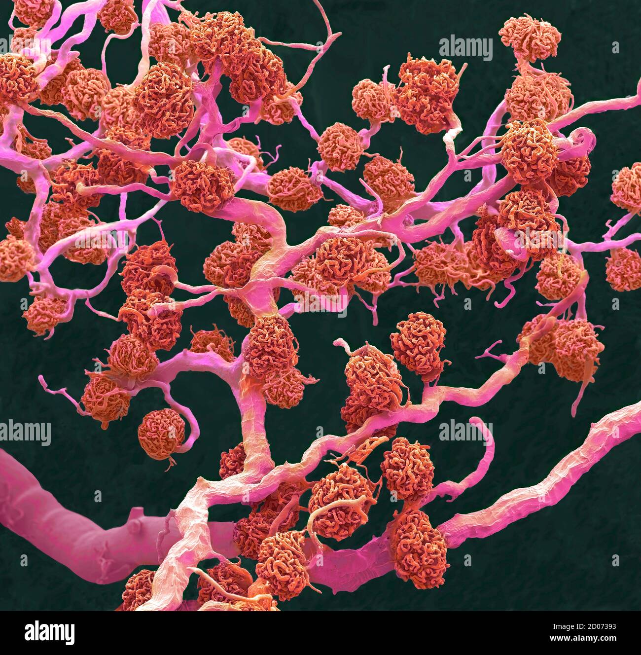 Kidney glomeruli. Coloured scanning electron micrograph (SEM) of a resin cast of glomeruli capillaries and the larger blood vessels supplying them wit Stock Photohttps://www.alamy.com/image-license-details/?v=1https://www.alamy.com/kidney-glomeruli-coloured-scanning-electron-micrograph-sem-of-a-resin-cast-of-glomeruli-capillaries-and-the-larger-blood-vessels-supplying-them-wit-image378784367.html
RF2D07393–Kidney glomeruli. Coloured scanning electron micrograph (SEM) of a resin cast of glomeruli capillaries and the larger blood vessels supplying them wit
RM2BE0GCG–Blood cells and platelets. Scanning electron micrograph (SEM) of human blood showing red and white cells and platelets. Red blood cells (erythrocytes) have a characteristic biconcave-disc shape and are numerous. These large cells contain hemoglobin, a red pigment by which oxygen is transported around the body. They are more numerous than white blood cells one of which is visible in this sample. White blood cells (leukocytes) are rounded cells with microvilli projections from the cell surface. Leucocytes play an important role in the immune response of the body. Platelets are smaller cells that
RM2BE0H0K–This scanning electron micrograph (SEM) depicted a closer view of number of red blood cells found enmeshed in a fibrinous matrix on the luminal surface of an indwelling vascular; Magnified 7766x. In this instance, the indwelling catheter was a tube that was left in place creating a patent portal directly into a blood vessel. Note the biconcave cytomorphologic shape of each erythrocyte, which increases the surface area of these hemoglobin-filled cells, thereby, promoting a greater degree of gas exchange, which is their primary function in an in vivo setting. In their adult phase, these cells po
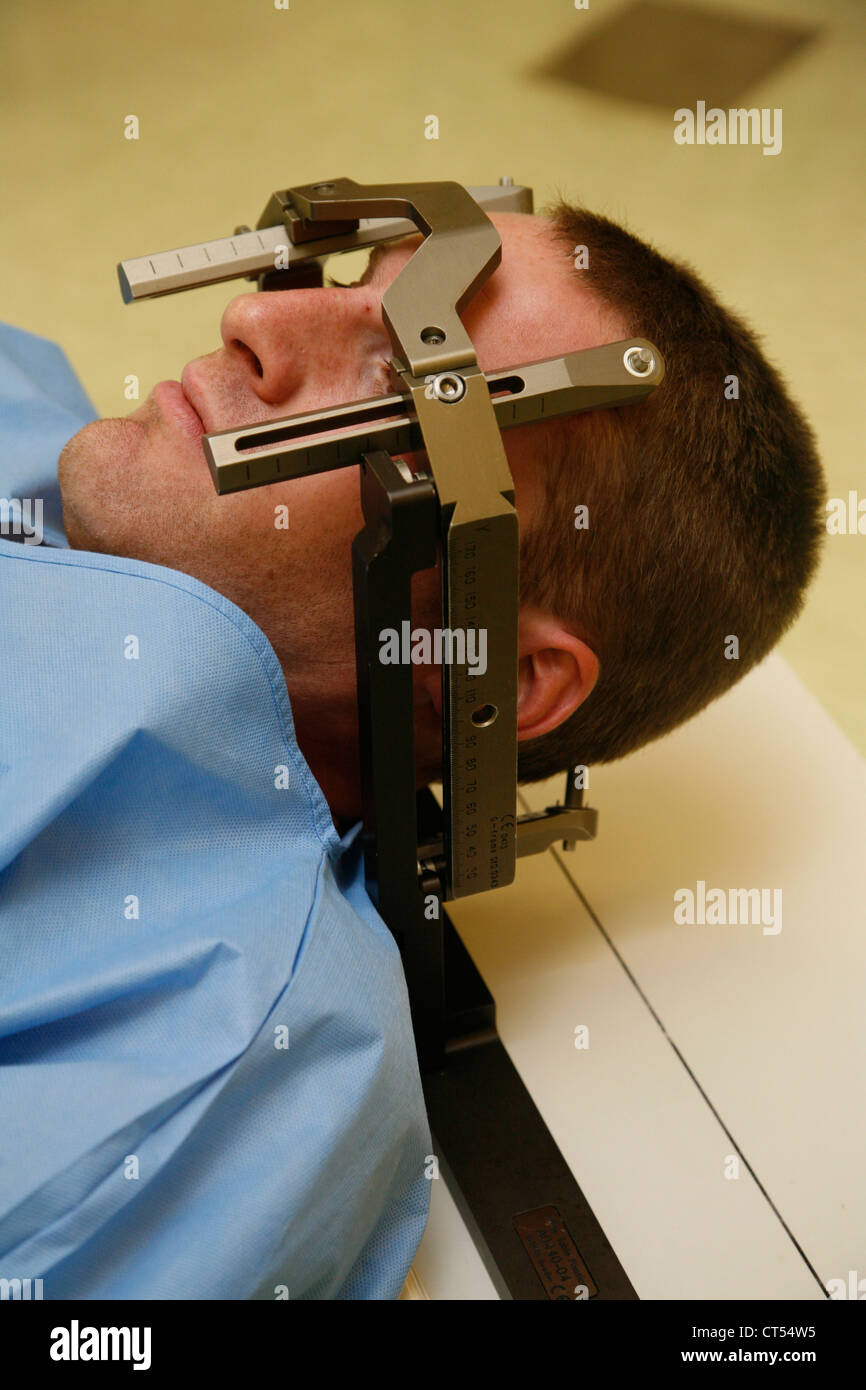 CEREBRAL ARTERIOGRAPHY Stock Photohttps://www.alamy.com/image-license-details/?v=1https://www.alamy.com/stock-photo-cerebral-arteriography-49286081.html
RMCT54W5–CEREBRAL ARTERIOGRAPHY
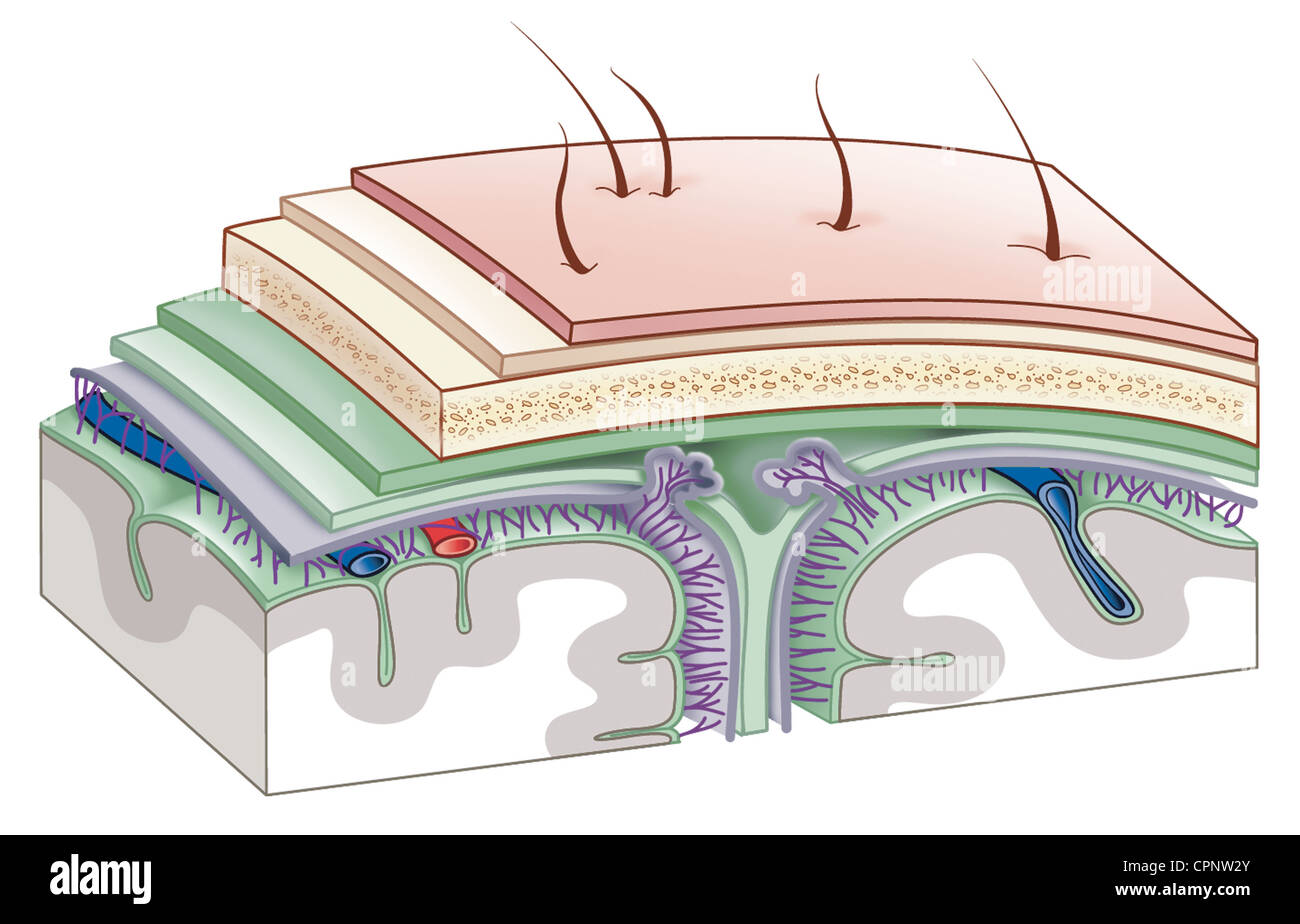 MENINGES, DRAWING Stock Photohttps://www.alamy.com/image-license-details/?v=1https://www.alamy.com/stock-photo-meninges-drawing-48423843.html
RMCPNW2Y–MENINGES, DRAWING
 HEAD, MRI Stock Photohttps://www.alamy.com/image-license-details/?v=1https://www.alamy.com/stock-photo-head-mri-53867172.html
RFD3HT3G–HEAD, MRI
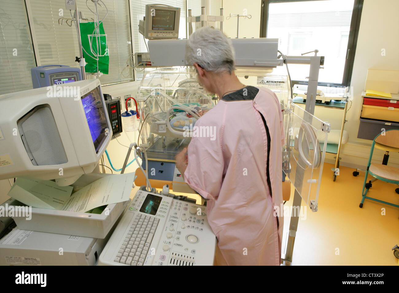 TRANSFONTANELLAR ULTRASONOGRAPHY Stock Photohttps://www.alamy.com/image-license-details/?v=1https://www.alamy.com/stock-photo-transfontanellar-ultrasonography-49258798.html
RMCT3X2P–TRANSFONTANELLAR ULTRASONOGRAPHY
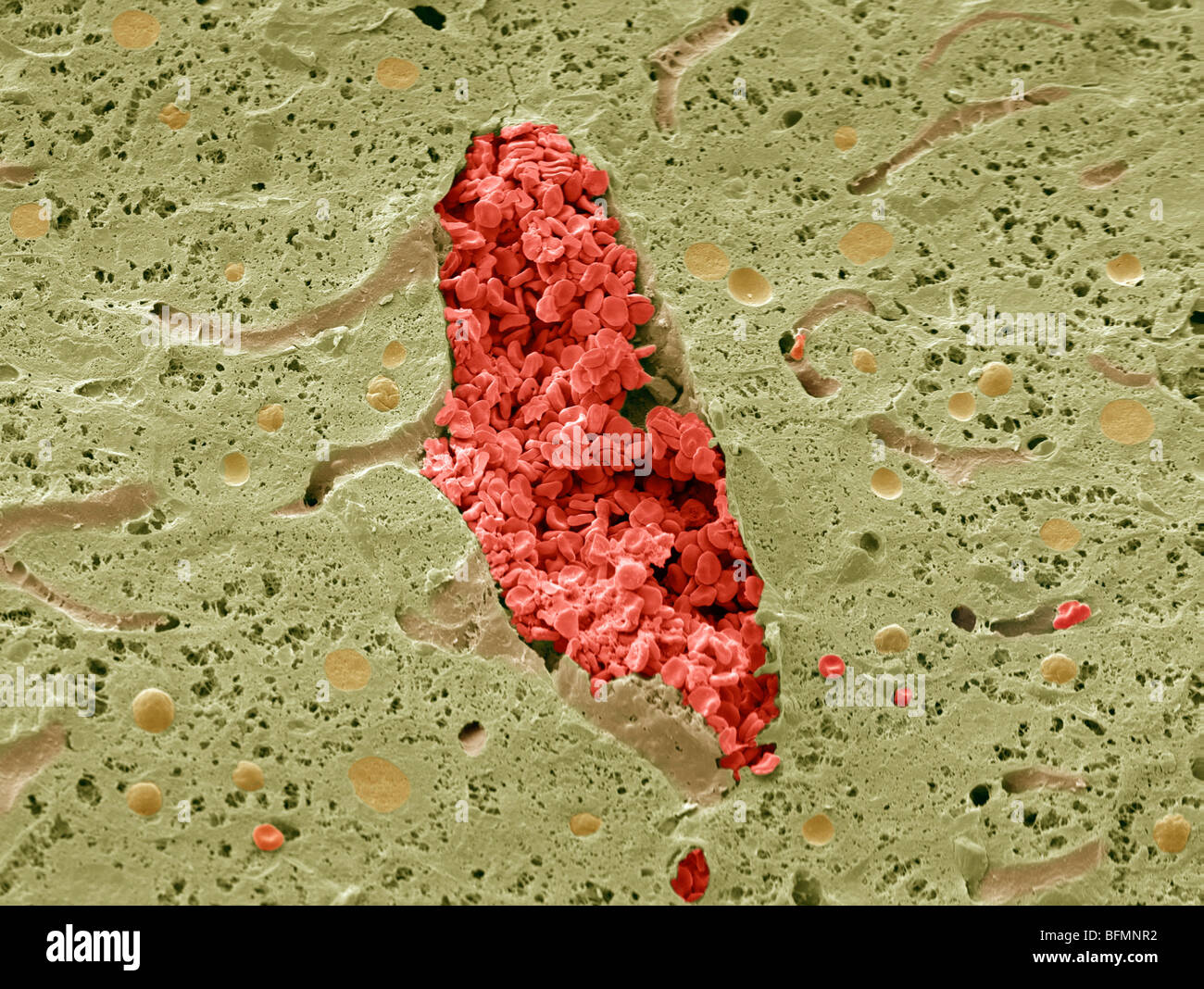 Liver vein, SEM Stock Photohttps://www.alamy.com/image-license-details/?v=1https://www.alamy.com/stock-photo-liver-vein-sem-26886358.html
RFBFMNR2–Liver vein, SEM
RM2BE0H1Y–This highly enlarged scanning electron micrograph (SEM) depicted a closer look at the details exhibited by of number of red blood cells found enmeshed in a fibrinous matrix on the luminal surface of an indwelling vascular; Magnified 11397x. In this instance, the indwelling catheter was a tube that was left in place creating a patent portal directly into a blood vessel. Note the biconcave cytomorphologic shape of each erythrocyte, which increases the surface area of these hemoglobin-filled cells, thereby, promoting a greater degree of gas exchange, which is their primary function in an in vivo
 TRANSFONTANELLAR ULTRASONOGRAPHY Stock Photohttps://www.alamy.com/image-license-details/?v=1https://www.alamy.com/stock-photo-transfontanellar-ultrasonography-49258967.html
RMCT3X8R–TRANSFONTANELLAR ULTRASONOGRAPHY
 Liver vein, SEM Stock Photohttps://www.alamy.com/image-license-details/?v=1https://www.alamy.com/stock-photo-liver-vein-sem-26886362.html
RFBFMNR6–Liver vein, SEM
RM2BE0H1A–This scanning electron micrograph (SEM) depicted a closer view of number of red blood cells found enmeshed in a fibrinous matrix on the luminal surface of an indwelling vascular; Magnified 5698x. In this instance, the indwelling catheter was a tube that was left in place creating a patent portal directly into a blood vessel. Note the biconcave cytomorphologic shape of each erythrocyte, which increases the surface area of these hemoglobin-filled cells, thereby, promoting a greater degree of gas exchange, which is their primary function in an in vivo setting. In their adult phase, these cells po
 TRANSFONTANELLAR ULTRASONOGRAPHY Stock Photohttps://www.alamy.com/image-license-details/?v=1https://www.alamy.com/stock-photo-transfontanellar-ultrasonography-49258954.html
RMCT3X8A–TRANSFONTANELLAR ULTRASONOGRAPHY
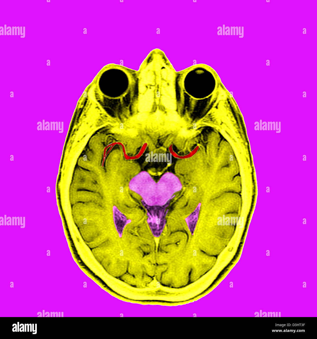 HEAD, MRI Stock Photohttps://www.alamy.com/image-license-details/?v=1https://www.alamy.com/stock-photo-head-mri-53867171.html
RFD3HT3F–HEAD, MRI
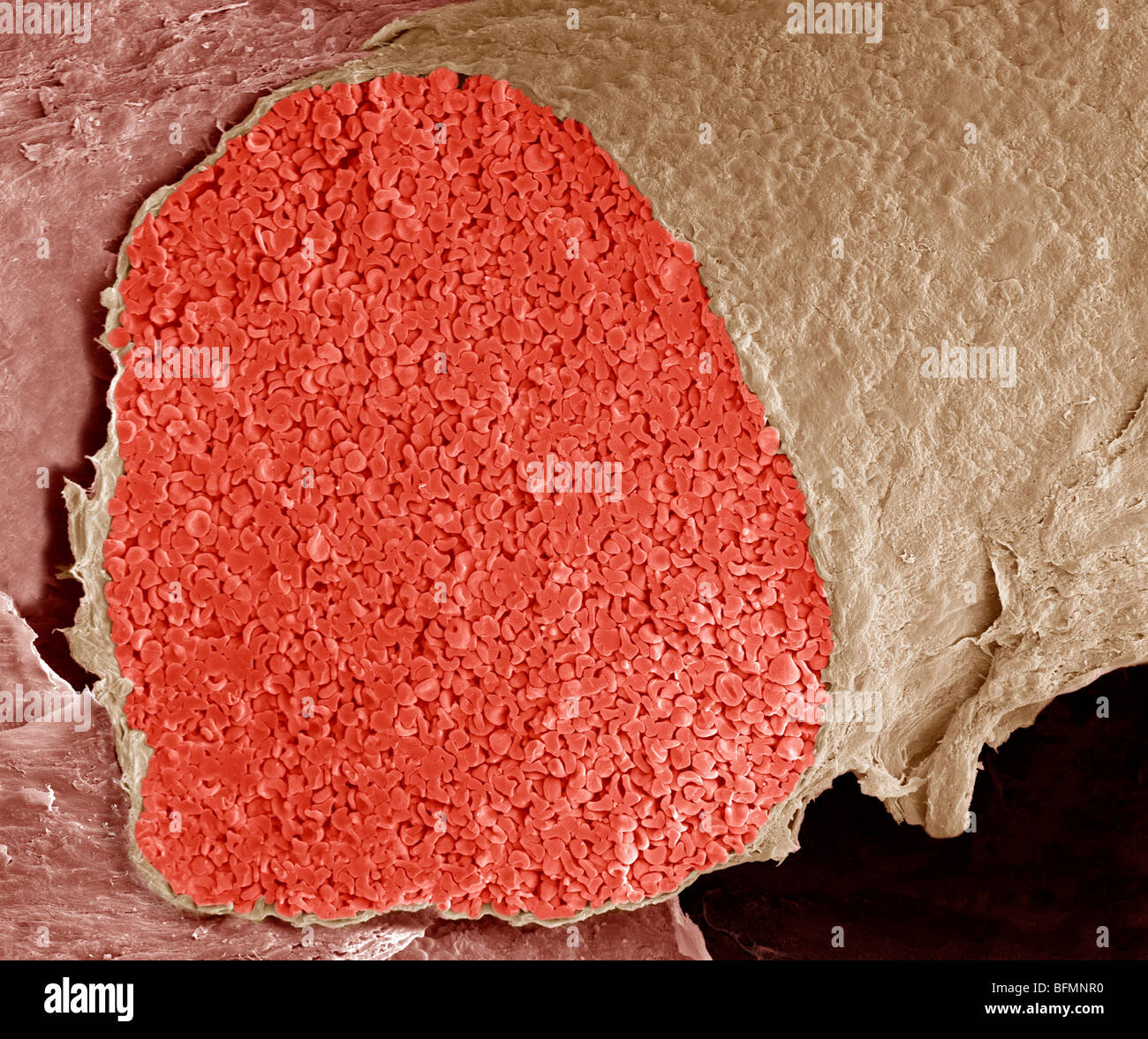 Foetal vein, SEM Stock Photohttps://www.alamy.com/image-license-details/?v=1https://www.alamy.com/stock-photo-foetal-vein-sem-26886356.html
RFBFMNR0–Foetal vein, SEM
RM2BE0H07–This highly enlarged scanning electron micrograph (SEM) depicted a closer look at the details exhibited by of number of red blood cells found enmeshed in a fibrinous matrix on the luminal surface of an indwelling vascular; Magnified 11397x. In this instance, the indwelling catheter was a tube that was left in place creating a patent portal directly into a blood vessel. Note the biconcave cytomorphologic shape of each erythrocyte, which increases the surface area of these hemoglobin-filled cells, thereby, promoting a greater degree of gas exchange, which is their primary function in an in vivo
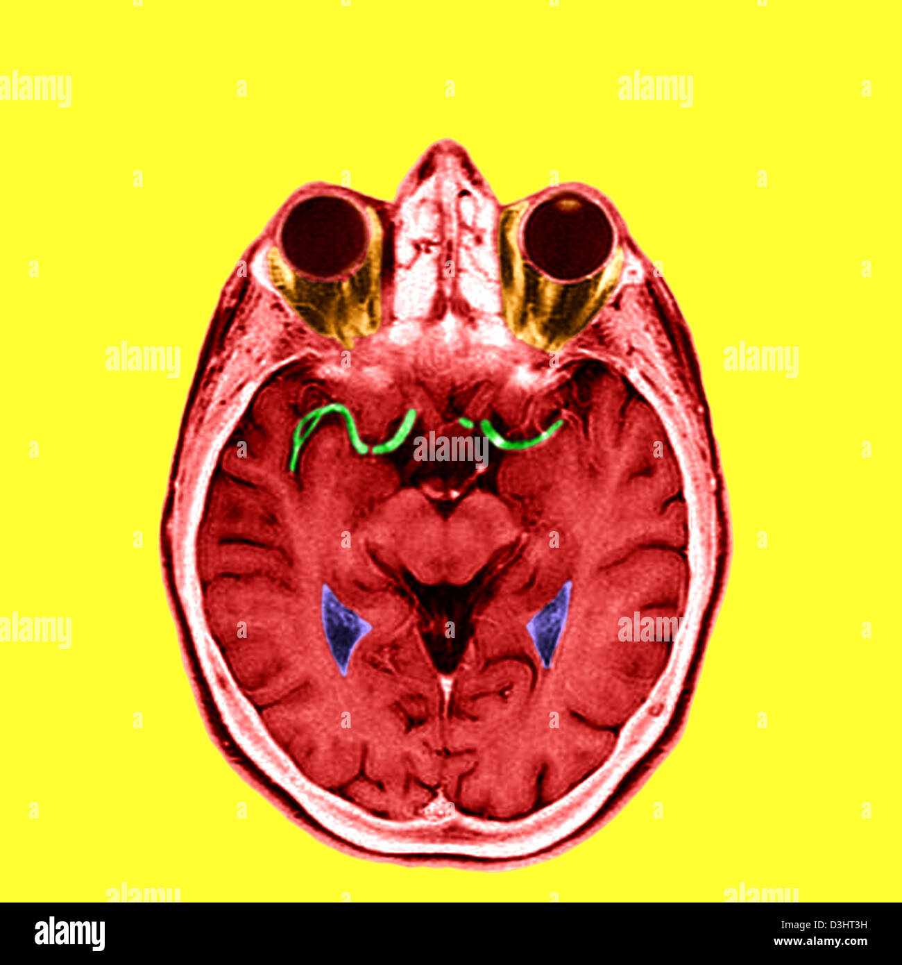 HEAD, MRI Stock Photohttps://www.alamy.com/image-license-details/?v=1https://www.alamy.com/stock-photo-head-mri-53867173.html
RFD3HT3H–HEAD, MRI
 BLOOD CIRCULATION, ILLUSTRATION Stock Photohttps://www.alamy.com/image-license-details/?v=1https://www.alamy.com/stock-photo-blood-circulation-illustration-53865162.html
RMD3HNFP–BLOOD CIRCULATION, ILLUSTRATION
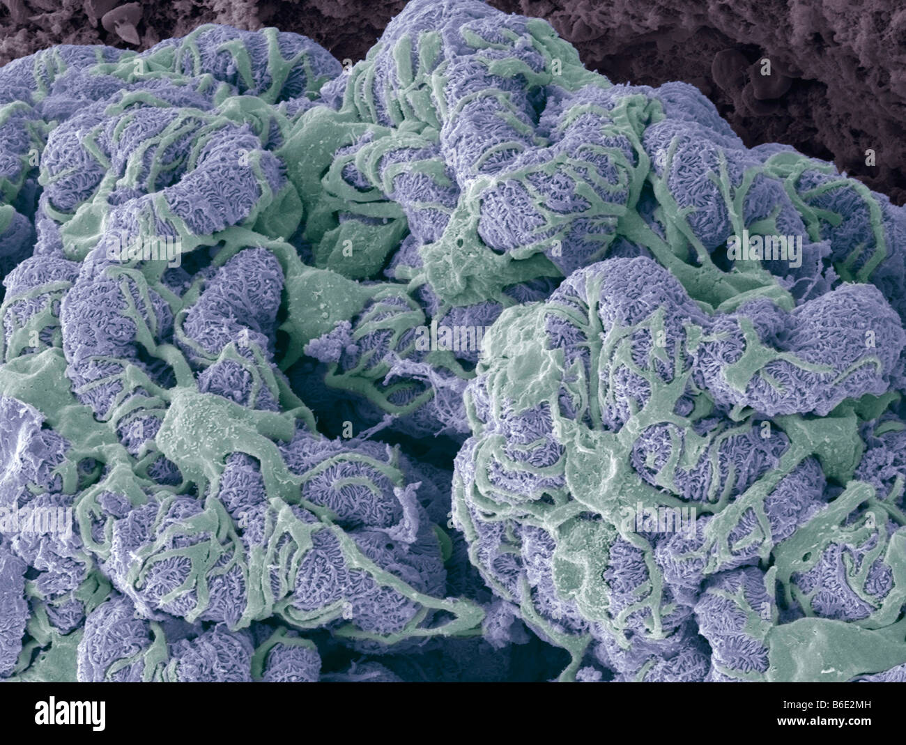 Kidney glomerulus. Coloured scanning electronmicrograph (SEM) showing the surface of aglomerulus. Stock Photohttps://www.alamy.com/image-license-details/?v=1https://www.alamy.com/stock-photo-kidney-glomerulus-coloured-scanning-electronmicrograph-sem-showing-21207777.html
RFB6E2MH–Kidney glomerulus. Coloured scanning electronmicrograph (SEM) showing the surface of aglomerulus.
RM2BE0H0W–This scanning electron micrograph (SEM) depicted a number of red blood cells found enmeshed in a fibrinous matrix on the luminal surface of an indwelling vascular; Magnified 1425x. In this instance, the indwelling catheter was a tube that was left in place creating a patent portal directly into a blood vessel. Note the biconcave cytomorphologic shape of each erythrocyte, which increases the surface area of these hemoglobin-filled cells, thereby, promoting a greater degree of gas exchange, which is their primary function in an in vivo setting. In their adult phase, these cells possess no nucleu
 BLOOD CIRCULATION, ILLUSTRATION Stock Photohttps://www.alamy.com/image-license-details/?v=1https://www.alamy.com/stock-photo-blood-circulation-illustration-53865152.html
RMD3HNFC–BLOOD CIRCULATION, ILLUSTRATION
 Kidney glomeruli, coloured scanning electron micrograph (SEM) Stock Photohttps://www.alamy.com/image-license-details/?v=1https://www.alamy.com/stock-photo-kidney-glomeruli-coloured-scanning-electron-micrograph-sem-21205690.html
RFB6E022–Kidney glomeruli, coloured scanning electron micrograph (SEM)
RM2BE0H29–This scanning electron micrograph (SEM) depicted a closer view of a number of red blood cells found enmeshed in a fibrinous matrix on the luminal surface of an indwelling vascular; Magnified 7766x. In this instance, the indwelling catheter was a tube that was left in place creating a patent portal directly into a blood vessel. Some of the erythrocytes are grouped in a stack known as a Rouleaux formation. Note the biconcave cytomorphologic shape of each erythrocyte, which increases the surface area of these hemoglobin-filled cells, thereby, promoting a greater degree of gas exchange, which is t
 BLOOD CIRCULATION, ILLUSTRATION Stock Photohttps://www.alamy.com/image-license-details/?v=1https://www.alamy.com/stock-photo-blood-circulation-illustration-53865153.html
RMD3HNFD–BLOOD CIRCULATION, ILLUSTRATION
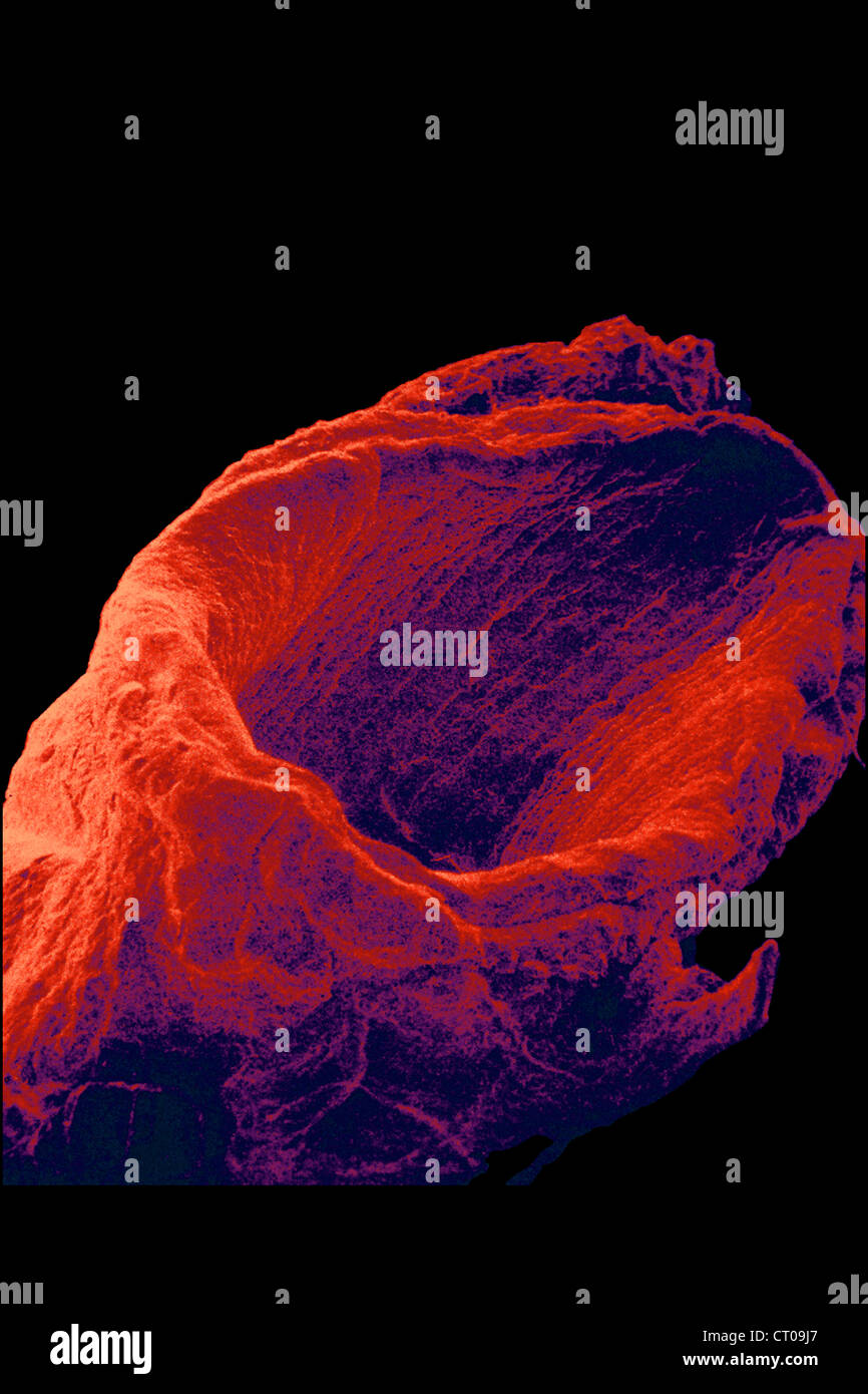 VEIN, SEM Stock Photohttps://www.alamy.com/image-license-details/?v=1https://www.alamy.com/stock-photo-vein-sem-49180047.html
RMCT09J7–VEIN, SEM
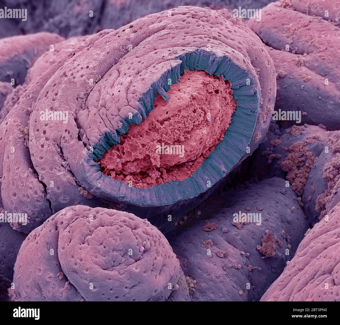 Small intestine. Coloured scanning electron micrograph (SEM) of a freeze-fractured of the small intestine. The surface consists of deep folds, called Stock Photohttps://www.alamy.com/image-license-details/?v=1https://www.alamy.com/small-intestine-coloured-scanning-electron-micrograph-sem-of-a-freeze-fractured-of-the-small-intestine-the-surface-consists-of-deep-folds-called-image359042796.html
RF2BT3PN0–Small intestine. Coloured scanning electron micrograph (SEM) of a freeze-fractured of the small intestine. The surface consists of deep folds, called
RM2BE0H1D–This scanning electron micrograph (SEM) depicted a number of red blood cells found enmeshed in a fibrinous matrix on the luminal surface of an indwelling vascular; Magnified 2849x. In this instance, the indwelling catheter was a tube that was left in place creating a patent portal directly into a blood vessel. Some of the erythrocytes are grouped in stacks known as a Rouleaux formation. Note the biconcave cytomorphologic shape of each erythrocyte, which increases the surface area of these hemoglobin-filled cells, thereby, promoting a greater degree of gas exchange, which is their primary funct
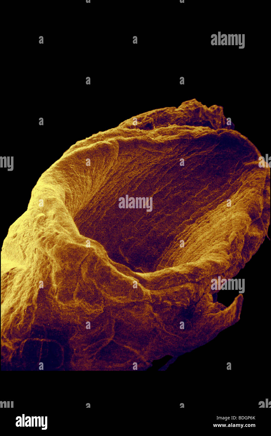 VEIN, SEM Stock Photohttps://www.alamy.com/image-license-details/?v=1https://www.alamy.com/stock-photo-vein-sem-25569563.html
RFBDGP6K–VEIN, SEM
 Intestinal lining. Coloured scanning electron micrograph (SEM) of a freeze-fractured of the small intestine. The surface consists of deep folds, calle Stock Photohttps://www.alamy.com/image-license-details/?v=1https://www.alamy.com/intestinal-lining-coloured-scanning-electron-micrograph-sem-of-a-freeze-fractured-of-the-small-intestine-the-surface-consists-of-deep-folds-calle-image359042736.html
RF2BT3PJT–Intestinal lining. Coloured scanning electron micrograph (SEM) of a freeze-fractured of the small intestine. The surface consists of deep folds, calle
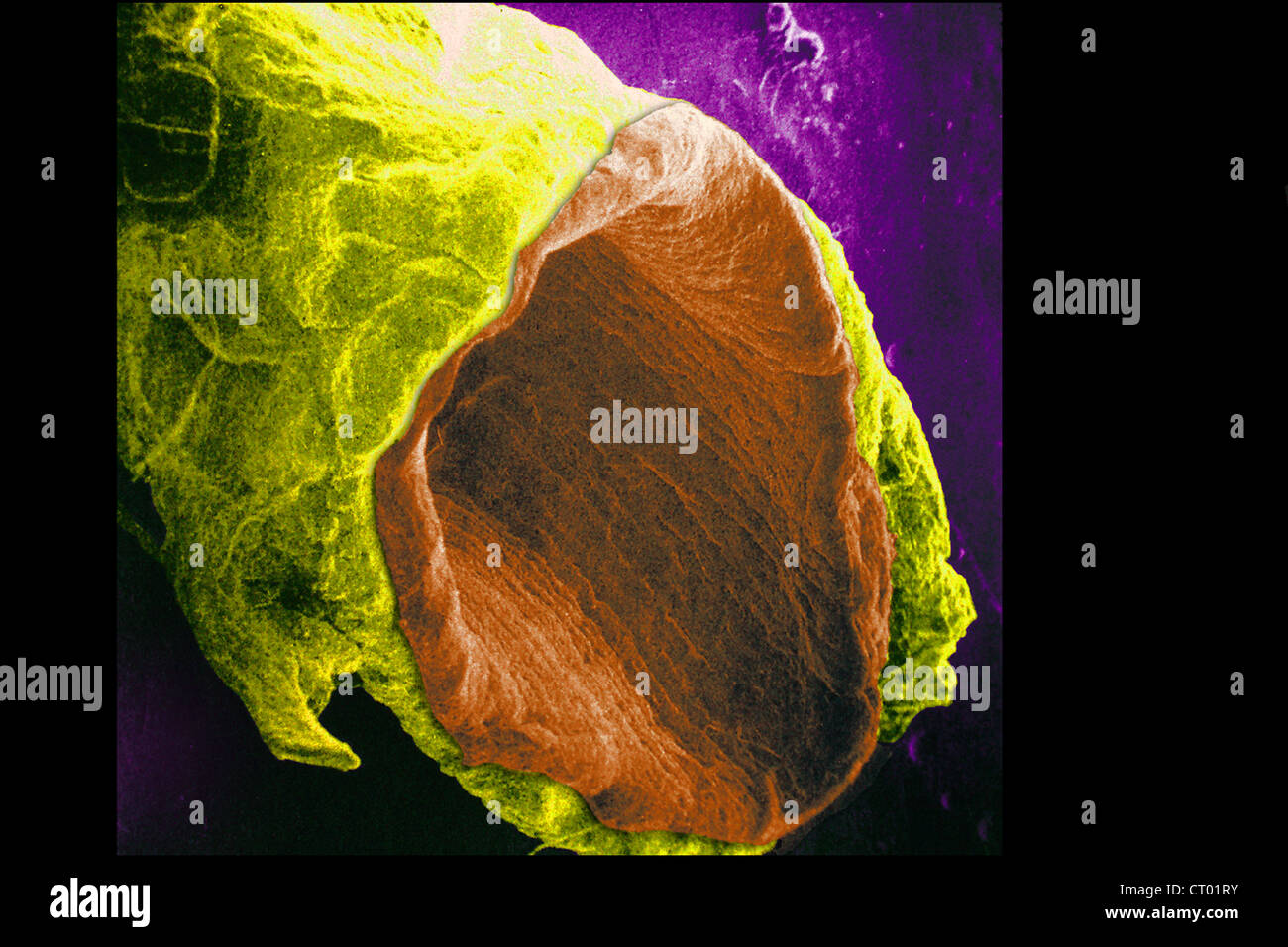 VEIN, SEM Stock Photohttps://www.alamy.com/image-license-details/?v=1https://www.alamy.com/stock-photo-vein-sem-49173935.html
RMCT01RY–VEIN, SEM
RM2BE0H47–This scanning electron micrograph (SEM) depicted a number of red blood cells found enmeshed in a fibrinous matrix on the luminal surface of an indwelling vascular catheter; Magnified 7766x. In this instance, the indwelling catheter was a tube that was left in place creating a patent portal directly into a blood vessel. The cell in the center was a white blood cell, also known as a leucocyte. The biconcave cytomorphologic shape of the red blood cell, or erythrocyte, increases its surface area of this hemoglobin-filled cell, thereby, promoting a greater degree of gas exchange, which is its prima
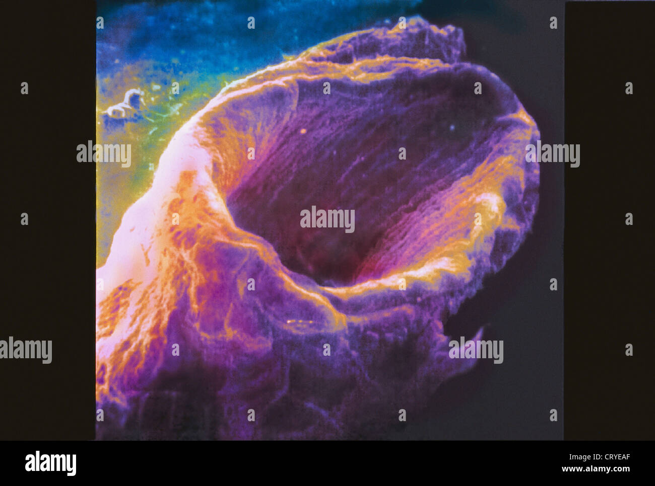 VEIN, SEM Stock Photohttps://www.alamy.com/image-license-details/?v=1https://www.alamy.com/stock-photo-vein-sem-49161799.html
RMCRYEAF–VEIN, SEM
 Fat Cells and Blood Vessels, SEM Stock Photohttps://www.alamy.com/image-license-details/?v=1https://www.alamy.com/stock-photo-fat-cells-and-blood-vessels-sem-135017892.html
RMHRJGKG–Fat Cells and Blood Vessels, SEM
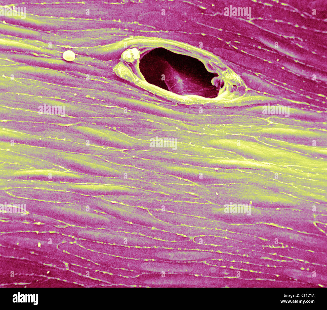 ENDOTHELIUM RABBIT, SEM Stock Photohttps://www.alamy.com/image-license-details/?v=1https://www.alamy.com/stock-photo-endothelium-rabbit-sem-49205390.html
RMCT1DYA–ENDOTHELIUM RABBIT, SEM
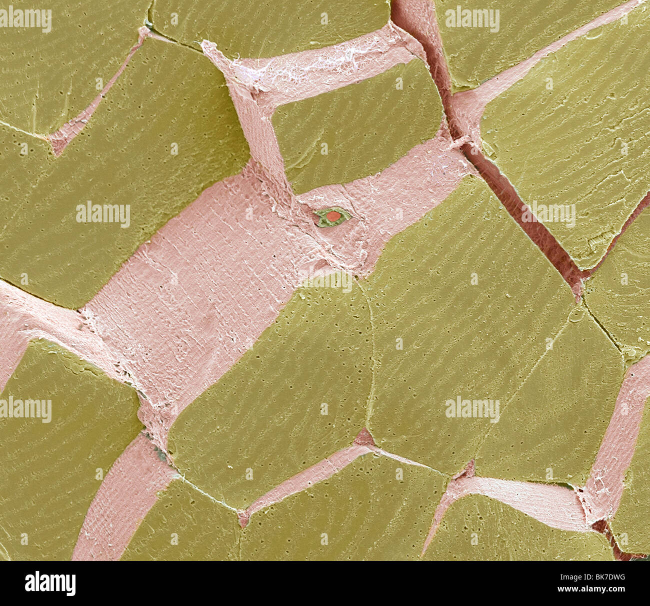 Skeletal muscle fibres, SEM Stock Photohttps://www.alamy.com/image-license-details/?v=1https://www.alamy.com/stock-photo-skeletal-muscle-fibres-sem-29053404.html
RFBK7DWG–Skeletal muscle fibres, SEM
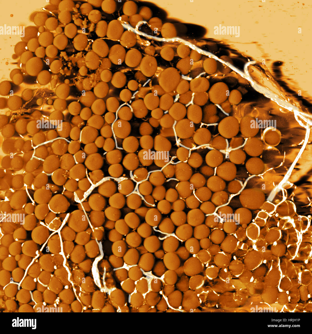 Fat Cells and Blood Vessels, SEM Stock Photohttps://www.alamy.com/image-license-details/?v=1https://www.alamy.com/stock-photo-fat-cells-and-blood-vessels-sem-135018178.html
RMHRJH1P–Fat Cells and Blood Vessels, SEM
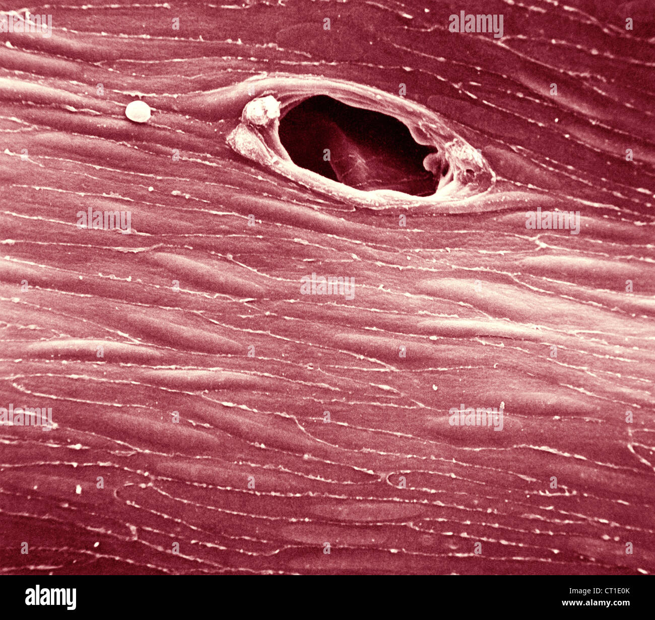 ENDOTHELIUM RABBIT, SEM Stock Photohttps://www.alamy.com/image-license-details/?v=1https://www.alamy.com/stock-photo-endothelium-rabbit-sem-49205427.html
RMCT1E0K–ENDOTHELIUM RABBIT, SEM
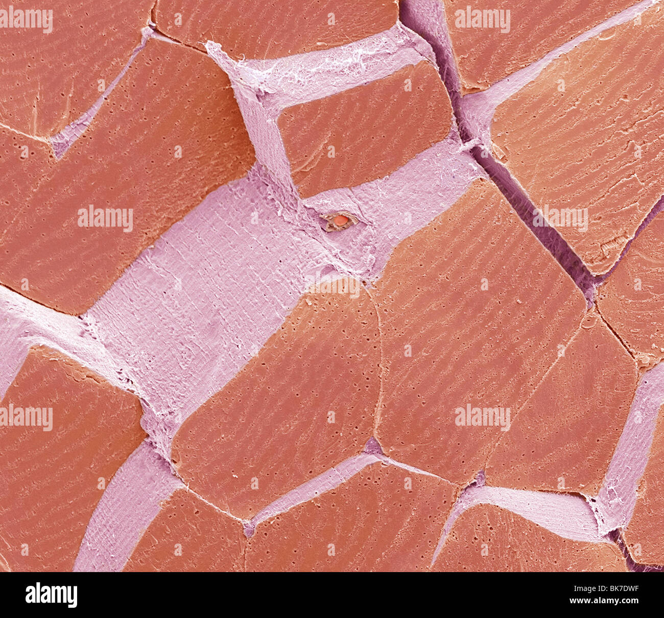 Skeletal muscle fibres, SEM Stock Photohttps://www.alamy.com/image-license-details/?v=1https://www.alamy.com/stock-photo-skeletal-muscle-fibres-sem-29053403.html
RFBK7DWF–Skeletal muscle fibres, SEM
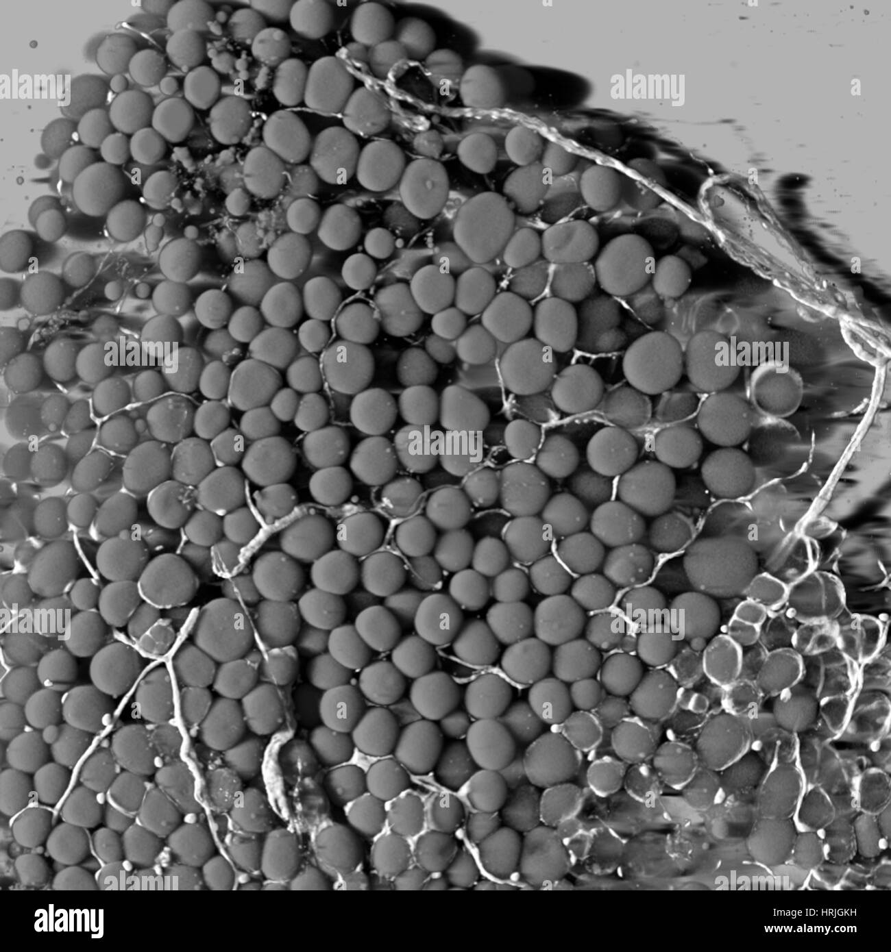 Fat Cells and Blood Vessels, SEM Stock Photohttps://www.alamy.com/image-license-details/?v=1https://www.alamy.com/stock-photo-fat-cells-and-blood-vessels-sem-135017893.html
RMHRJGKH–Fat Cells and Blood Vessels, SEM
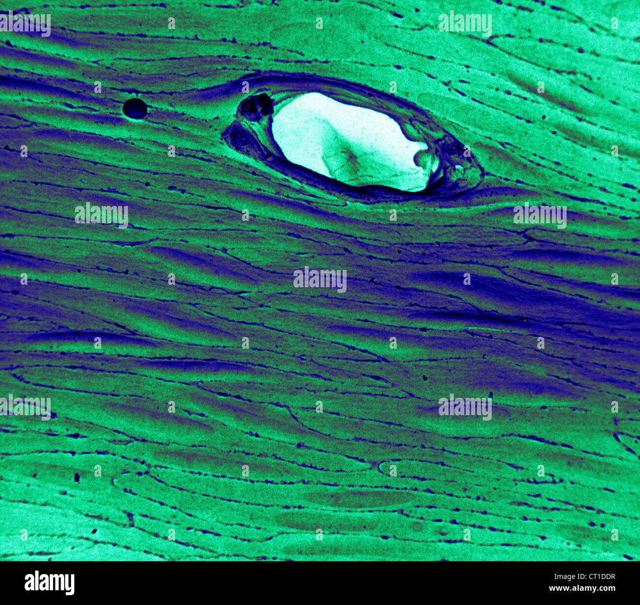 ENDOTHELIUM RABBIT, SEM Stock Photohttps://www.alamy.com/image-license-details/?v=1https://www.alamy.com/stock-photo-endothelium-rabbit-sem-49205011.html
RMCT1DDR–ENDOTHELIUM RABBIT, SEM
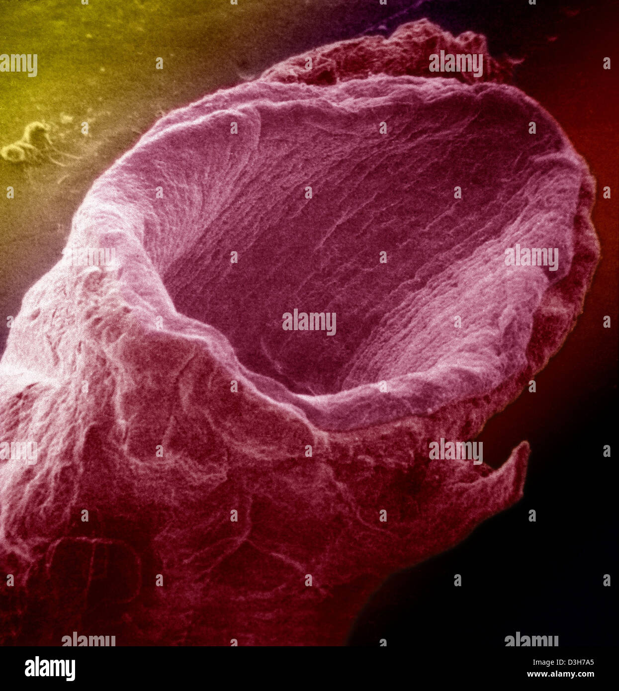 VEIN, SEM Stock Photohttps://www.alamy.com/image-license-details/?v=1https://www.alamy.com/stock-photo-vein-sem-53854029.html
RMD3H7A5–VEIN, SEM
 Kidney glomerulus. Coloured scanning electronmicrograph (SEM) showing the surface of aglomerulus. Stock Photohttps://www.alamy.com/image-license-details/?v=1https://www.alamy.com/stock-photo-kidney-glomerulus-coloured-scanning-electronmicrograph-sem-showing-21207775.html
RFB6E2MF–Kidney glomerulus. Coloured scanning electronmicrograph (SEM) showing the surface of aglomerulus.
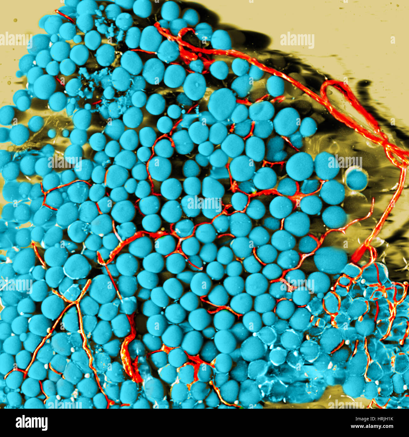 Fat Cells and Blood Vessels, SEM Stock Photohttps://www.alamy.com/image-license-details/?v=1https://www.alamy.com/stock-photo-fat-cells-and-blood-vessels-sem-135018175.html
RMHRJH1K–Fat Cells and Blood Vessels, SEM
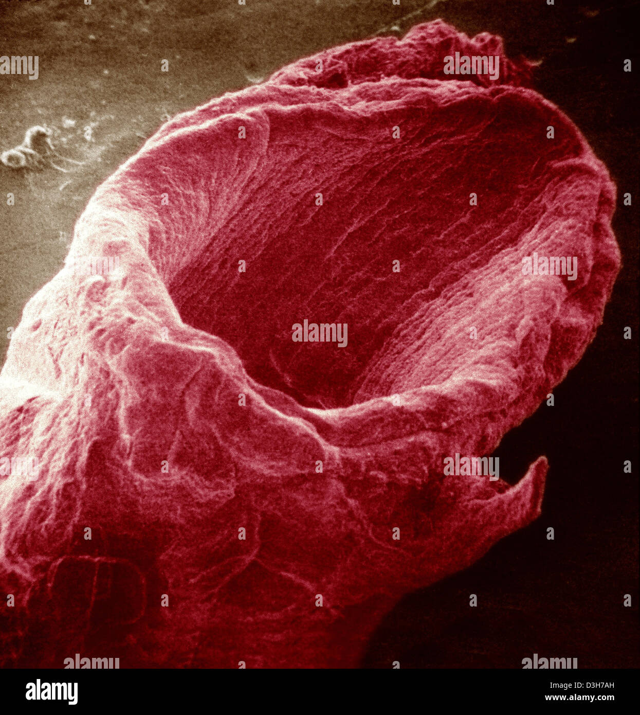 VEIN, SEM Stock Photohttps://www.alamy.com/image-license-details/?v=1https://www.alamy.com/stock-photo-vein-sem-53854041.html
RMD3H7AH–VEIN, SEM
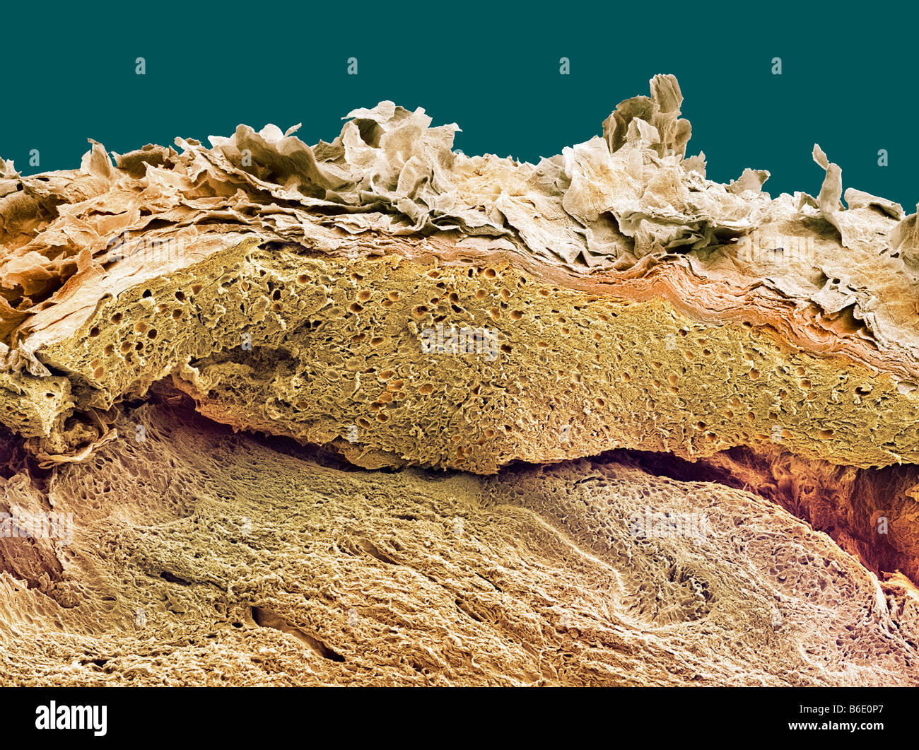 Skin. Coloured scanning electron micrograph(SEM) of a section through healthy skin. Stock Photohttps://www.alamy.com/image-license-details/?v=1https://www.alamy.com/stock-photo-skin-coloured-scanning-electron-micrographsem-of-a-section-through-21206255.html
RFB6E0P7–Skin. Coloured scanning electron micrograph(SEM) of a section through healthy skin.
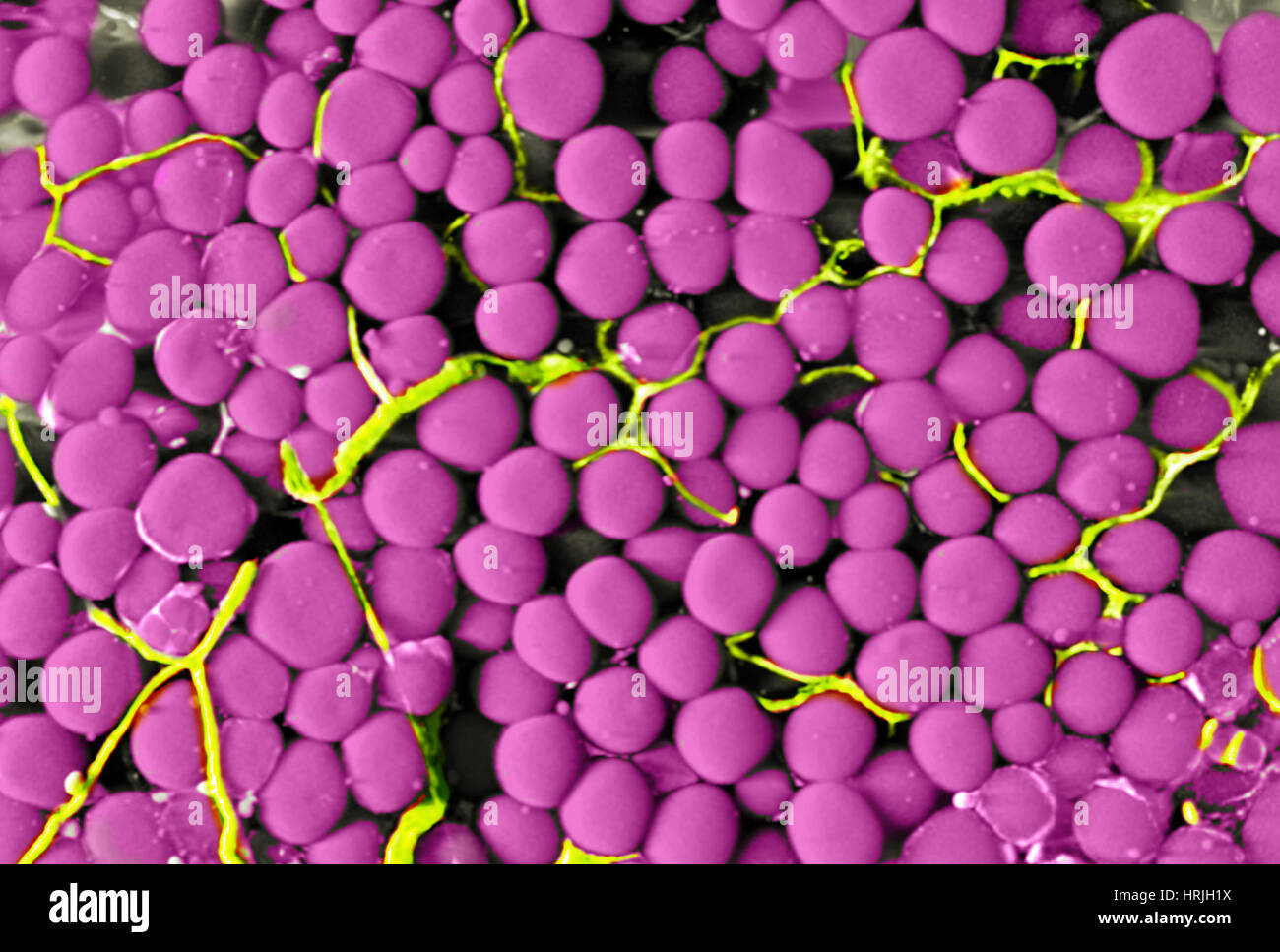 Fat Cells and Blood Vessels, SEM Stock Photohttps://www.alamy.com/image-license-details/?v=1https://www.alamy.com/stock-photo-fat-cells-and-blood-vessels-sem-135018182.html
RMHRJH1X–Fat Cells and Blood Vessels, SEM
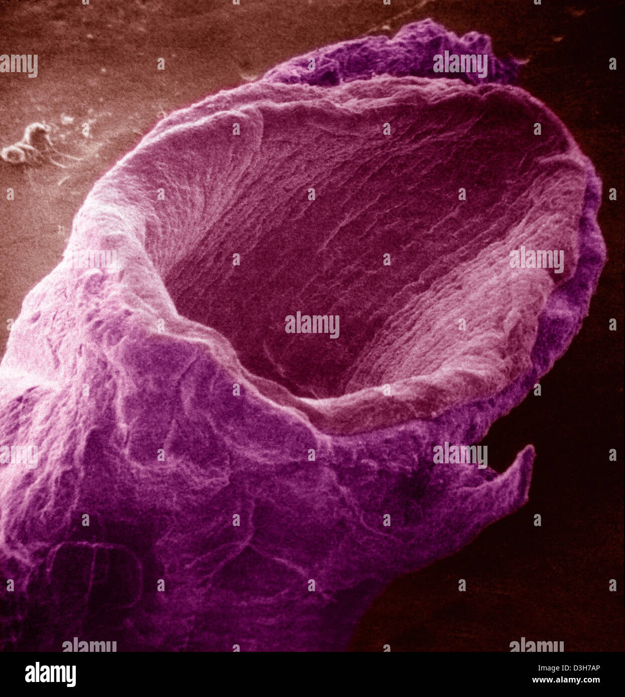 VEIN, SEM Stock Photohttps://www.alamy.com/image-license-details/?v=1https://www.alamy.com/stock-photo-vein-sem-53854046.html
RMD3H7AP–VEIN, SEM
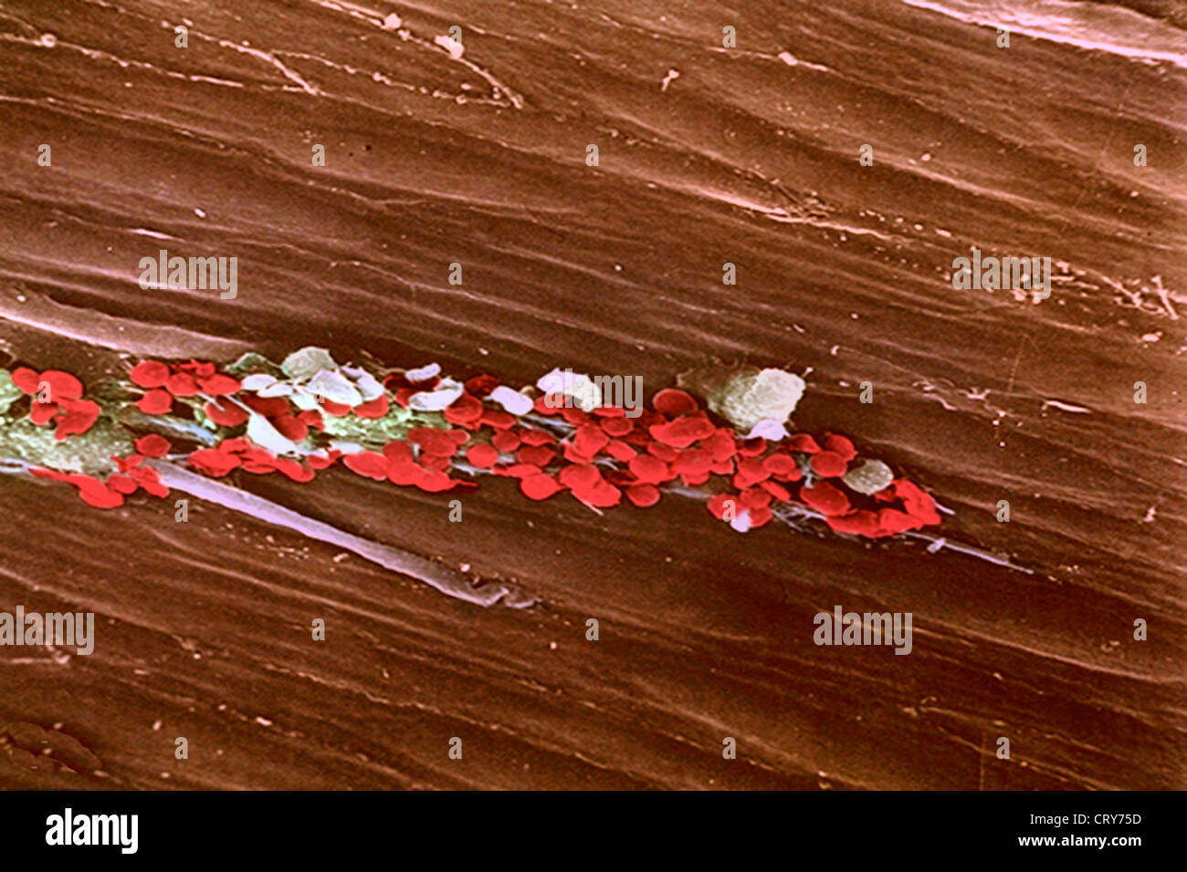 PLATELET, SEM Stock Photohttps://www.alamy.com/image-license-details/?v=1https://www.alamy.com/stock-photo-platelet-sem-49156169.html
RMCRY75D–PLATELET, SEM
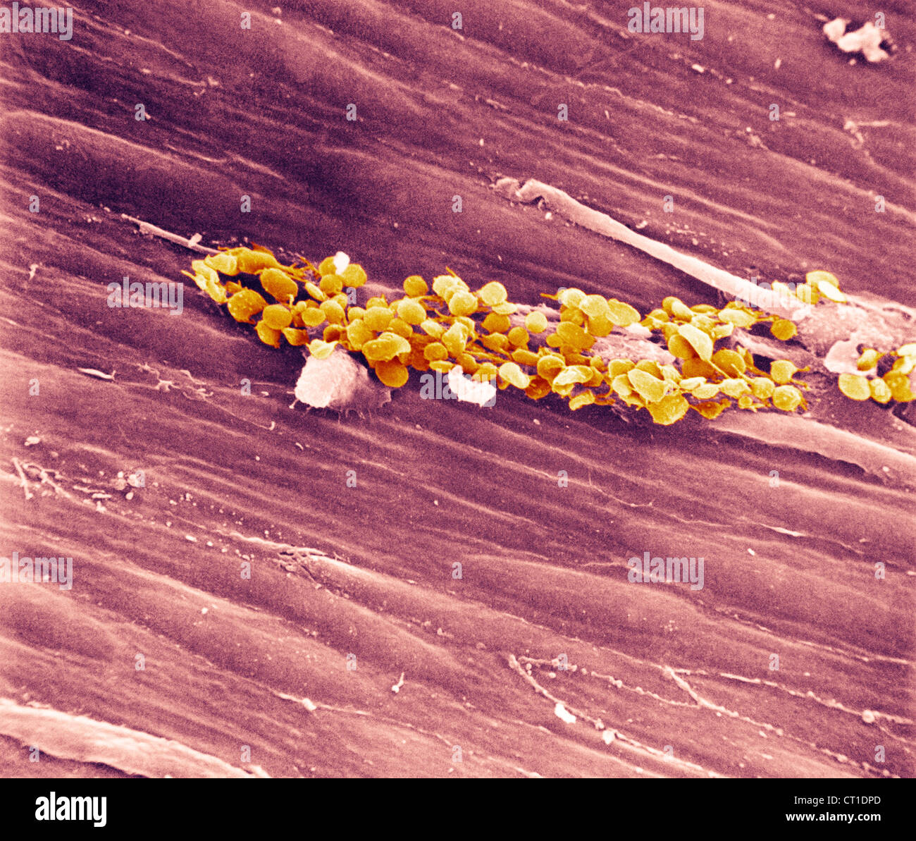 RABBIT PLATELET, SEM Stock Photohttps://www.alamy.com/image-license-details/?v=1https://www.alamy.com/stock-photo-rabbit-platelet-sem-49205253.html
RMCT1DPD–RABBIT PLATELET, SEM
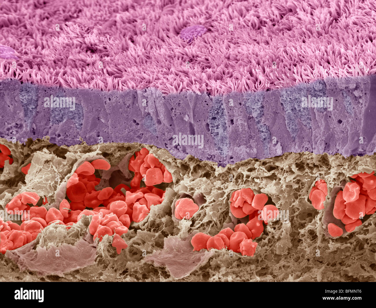 Trachea mucous membrane, SEM Stock Photohttps://www.alamy.com/image-license-details/?v=1https://www.alamy.com/stock-photo-trachea-mucous-membrane-sem-26886390.html
RFBFMNT6–Trachea mucous membrane, SEM
 Fat Cells and Blood Vessels, SEM Stock Photohttps://www.alamy.com/image-license-details/?v=1https://www.alamy.com/stock-photo-fat-cells-and-blood-vessels-sem-135018177.html
RMHRJH1N–Fat Cells and Blood Vessels, SEM
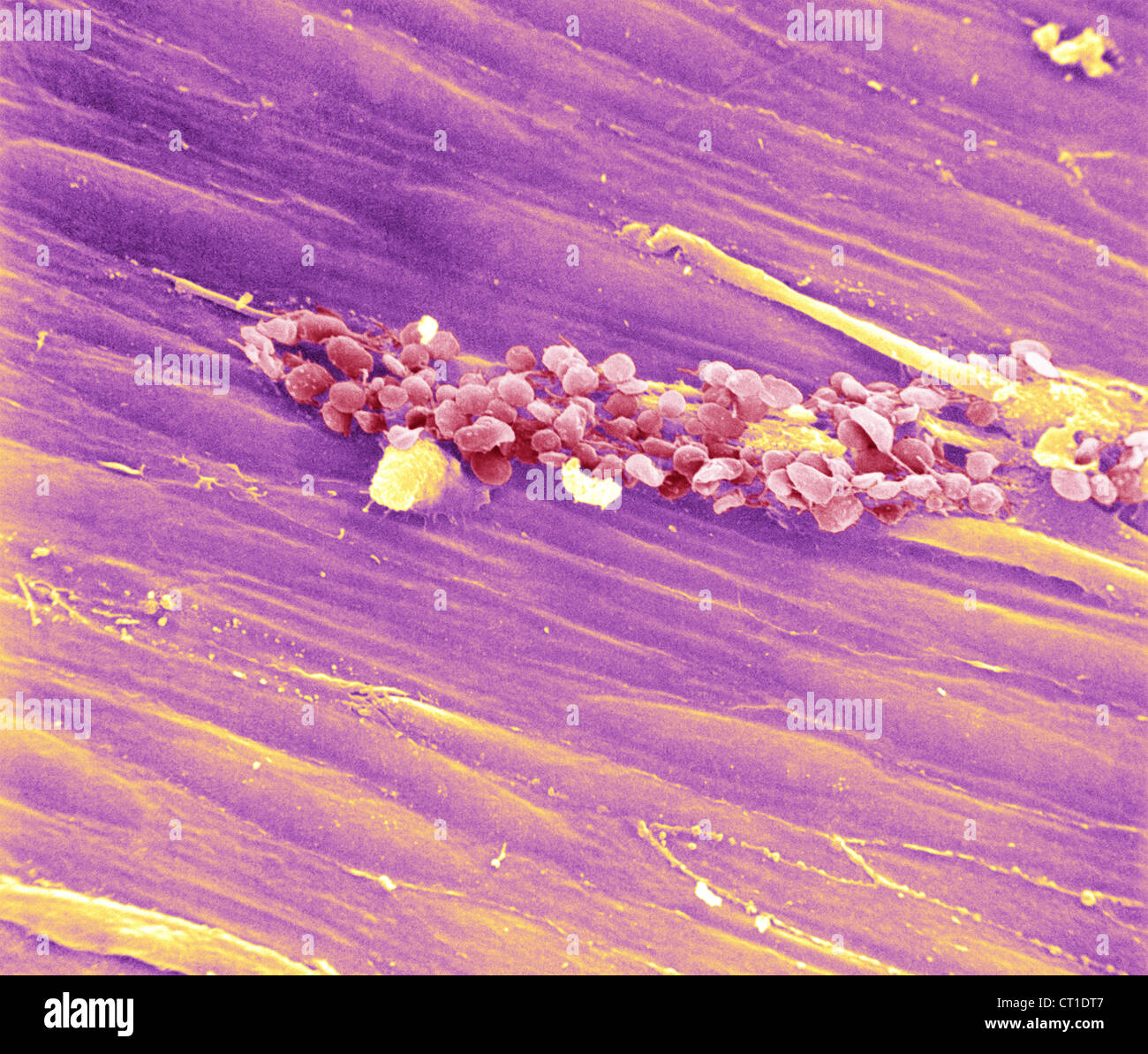 RABBIT PLATELET, SEM Stock Photohttps://www.alamy.com/image-license-details/?v=1https://www.alamy.com/stock-photo-rabbit-platelet-sem-49205303.html
RMCT1DT7–RABBIT PLATELET, SEM
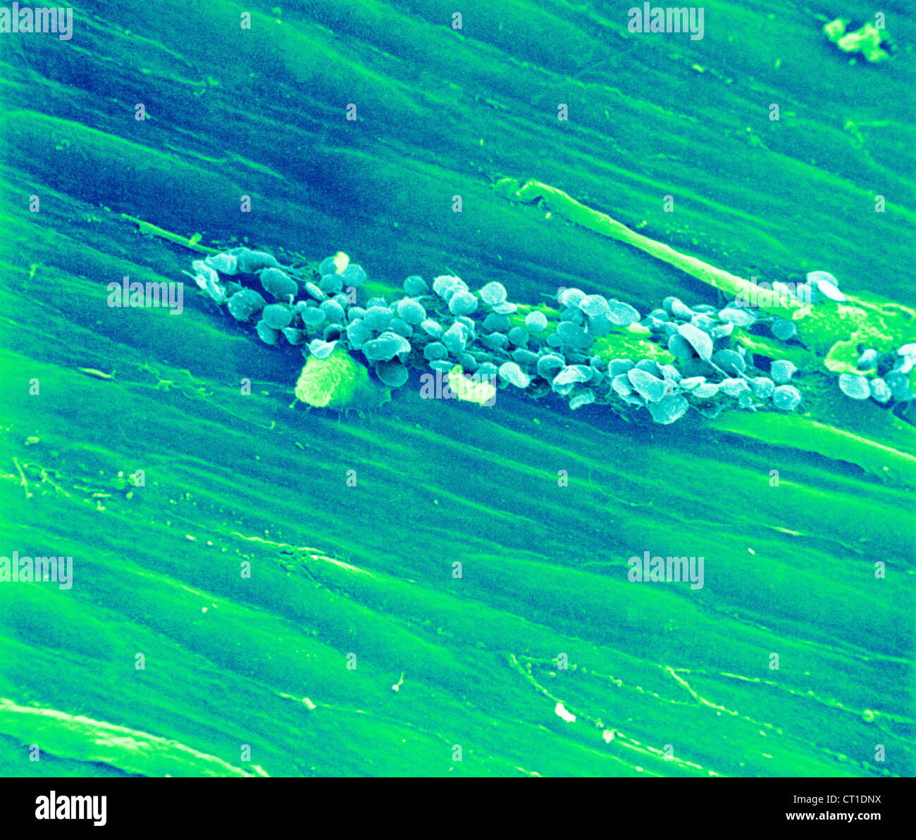 RABBIT PLATELET, SEM Stock Photohttps://www.alamy.com/image-license-details/?v=1https://www.alamy.com/stock-photo-rabbit-platelet-sem-49205238.html
RMCT1DNX–RABBIT PLATELET, SEM
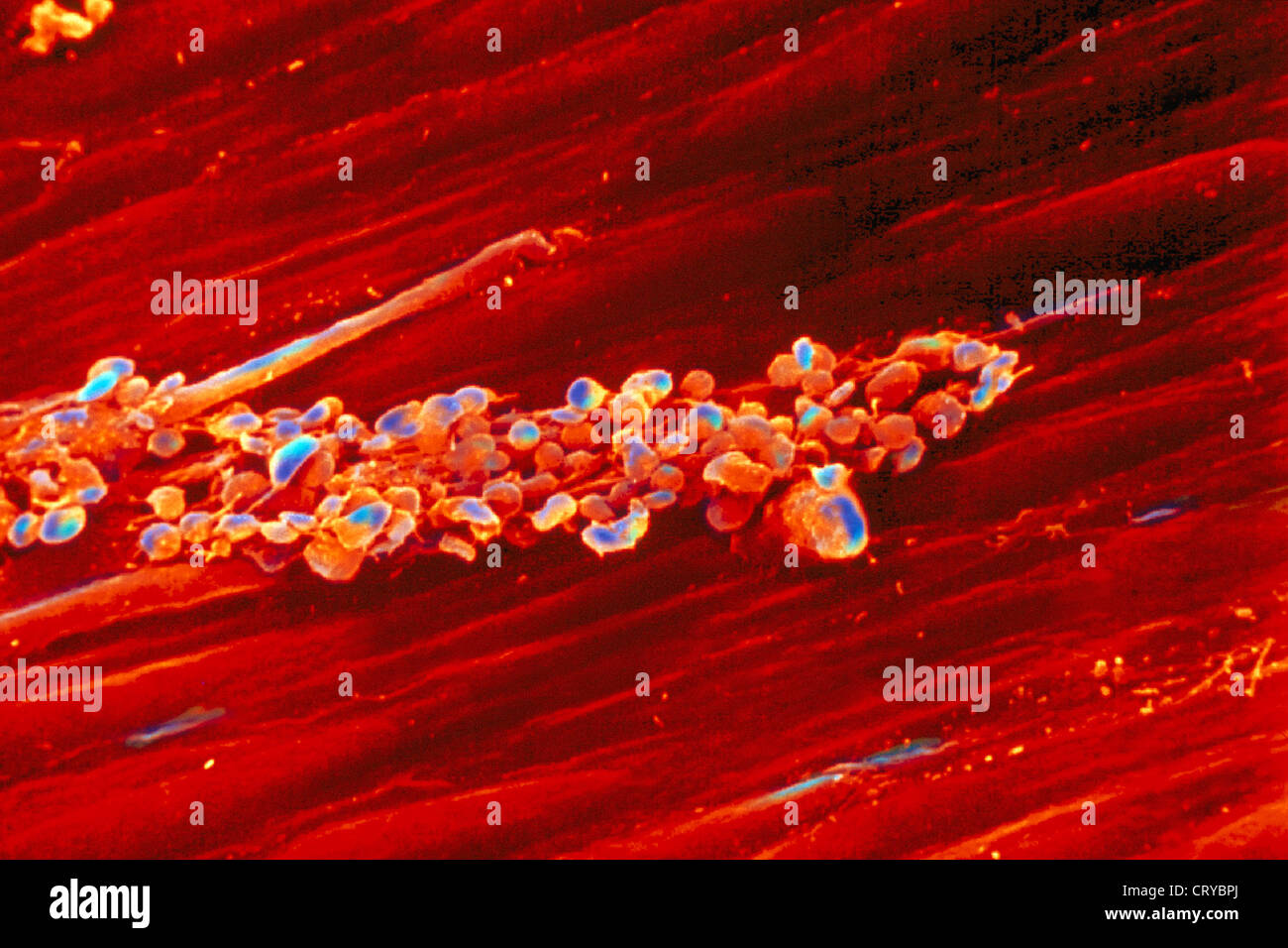 RABBIT PLATELET, SEM Stock Photohttps://www.alamy.com/image-license-details/?v=1https://www.alamy.com/stock-photo-rabbit-platelet-sem-49159786.html
RMCRYBPJ–RABBIT PLATELET, SEM
 ARTERIOLE, SEM Stock Photohttps://www.alamy.com/image-license-details/?v=1https://www.alamy.com/stock-photo-arteriole-sem-49179024.html
RMCT089M–ARTERIOLE, SEM
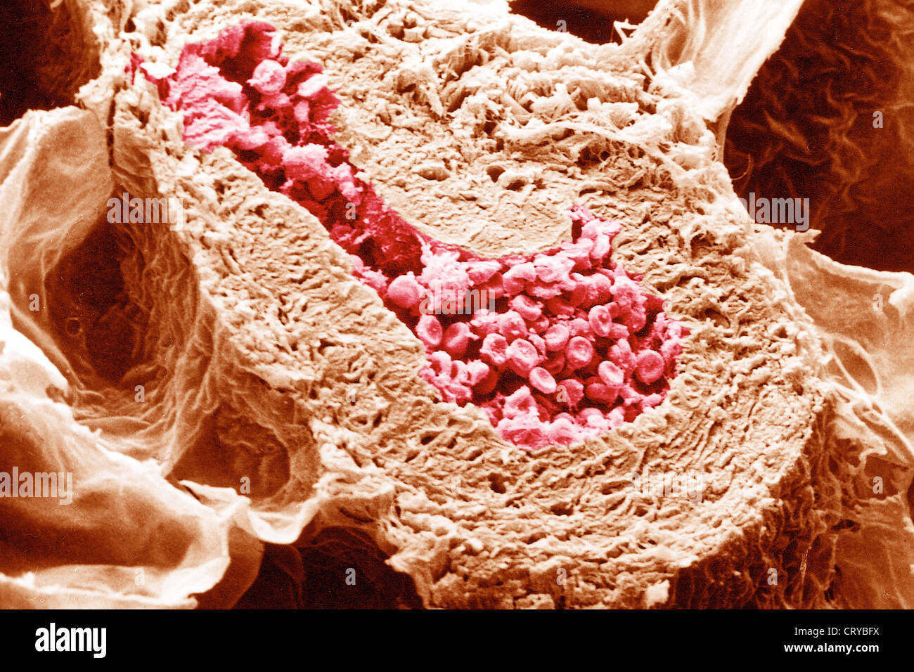 ARTERY, SEM Stock Photohttps://www.alamy.com/image-license-details/?v=1https://www.alamy.com/stock-photo-artery-sem-49159598.html
RMCRYBFX–ARTERY, SEM
 CEREBRAL ARTERIOGRAPHY Stock Photohttps://www.alamy.com/image-license-details/?v=1https://www.alamy.com/stock-photo-cerebral-arteriography-49163464.html
RMCRYGE0–CEREBRAL ARTERIOGRAPHY
 CEREBRAL ARTERIOGRAPHY Stock Photohttps://www.alamy.com/image-license-details/?v=1https://www.alamy.com/stock-photo-cerebral-arteriography-49163456.html
RMCRYGDM–CEREBRAL ARTERIOGRAPHY
 CEREBRAL CIRCULATION, DRAWING Stock Photohttps://www.alamy.com/image-license-details/?v=1https://www.alamy.com/stock-photo-cerebral-circulation-drawing-49165935.html
RMCRYKJ7–CEREBRAL CIRCULATION, DRAWING
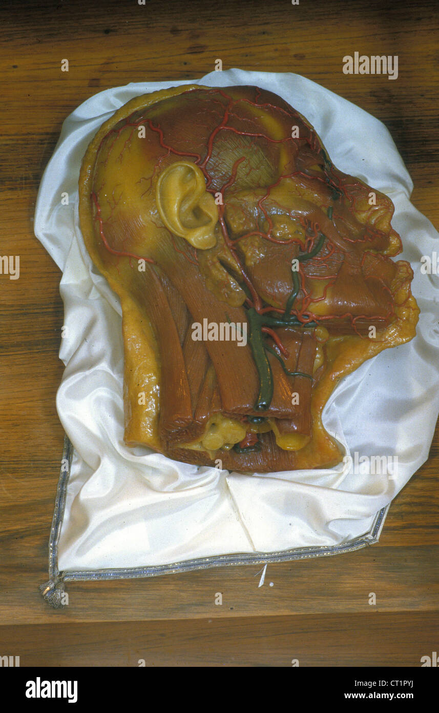 MUSEUM OF MEDICINE Stock Photohttps://www.alamy.com/image-license-details/?v=1https://www.alamy.com/stock-photo-museum-of-medicine-49212454.html
RMCT1PYJ–MUSEUM OF MEDICINE
 CEREBRAL ARTERIOGRAPHY Stock Photohttps://www.alamy.com/image-license-details/?v=1https://www.alamy.com/stock-photo-cerebral-arteriography-49163449.html
RMCRYGDD–CEREBRAL ARTERIOGRAPHY
 CEREBRAL ARTERIOGRAPHY Stock Photohttps://www.alamy.com/image-license-details/?v=1https://www.alamy.com/stock-photo-cerebral-arteriography-49163452.html
RMCRYGDG–CEREBRAL ARTERIOGRAPHY
 VENOUS INSUFFICIENCY Stock Photohttps://www.alamy.com/image-license-details/?v=1https://www.alamy.com/stock-photo-venous-insufficiency-49161803.html
RMCRYEAK–VENOUS INSUFFICIENCY
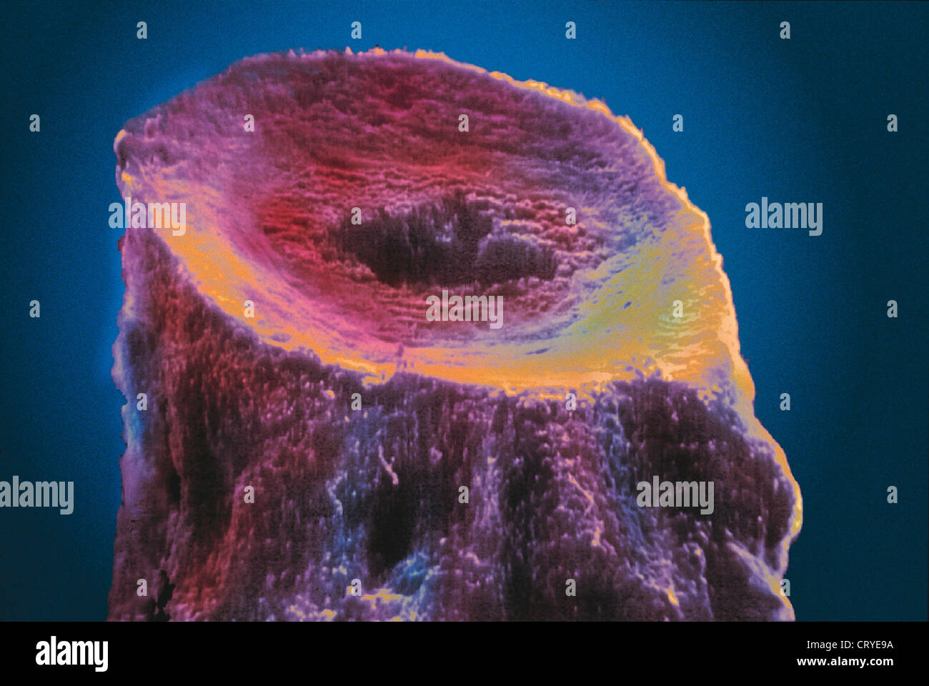 VENOUS INSUFFICIENCY Stock Photohttps://www.alamy.com/image-license-details/?v=1https://www.alamy.com/stock-photo-venous-insufficiency-49161766.html
RMCRYE9A–VENOUS INSUFFICIENCY
 CEREBRAL ARTERIOGRAPHY Stock Photohttps://www.alamy.com/image-license-details/?v=1https://www.alamy.com/stock-photo-cerebral-arteriography-49161534.html
RMCRYE12–CEREBRAL ARTERIOGRAPHY
 CEREBRAL ARTERIOGRAPHY Stock Photohttps://www.alamy.com/image-license-details/?v=1https://www.alamy.com/stock-photo-cerebral-arteriography-49161533.html
RMCRYE11–CEREBRAL ARTERIOGRAPHY
 CEREBRAL ARTERIOGRAPHY Stock Photohttps://www.alamy.com/image-license-details/?v=1https://www.alamy.com/stock-photo-cerebral-arteriography-49159632.html
RMCRYBH4–CEREBRAL ARTERIOGRAPHY
 CEREBRAL ARTERIOGRAPHY Stock Photohttps://www.alamy.com/image-license-details/?v=1https://www.alamy.com/stock-photo-cerebral-arteriography-49159636.html
RMCRYBH8–CEREBRAL ARTERIOGRAPHY
 ANATOMY Stock Photohttps://www.alamy.com/image-license-details/?v=1https://www.alamy.com/stock-photo-anatomy-49202916.html
RMCT1AR0–ANATOMY
 ANATOMY Stock Photohttps://www.alamy.com/image-license-details/?v=1https://www.alamy.com/stock-photo-anatomy-53853674.html
RMD3H6WE–ANATOMY
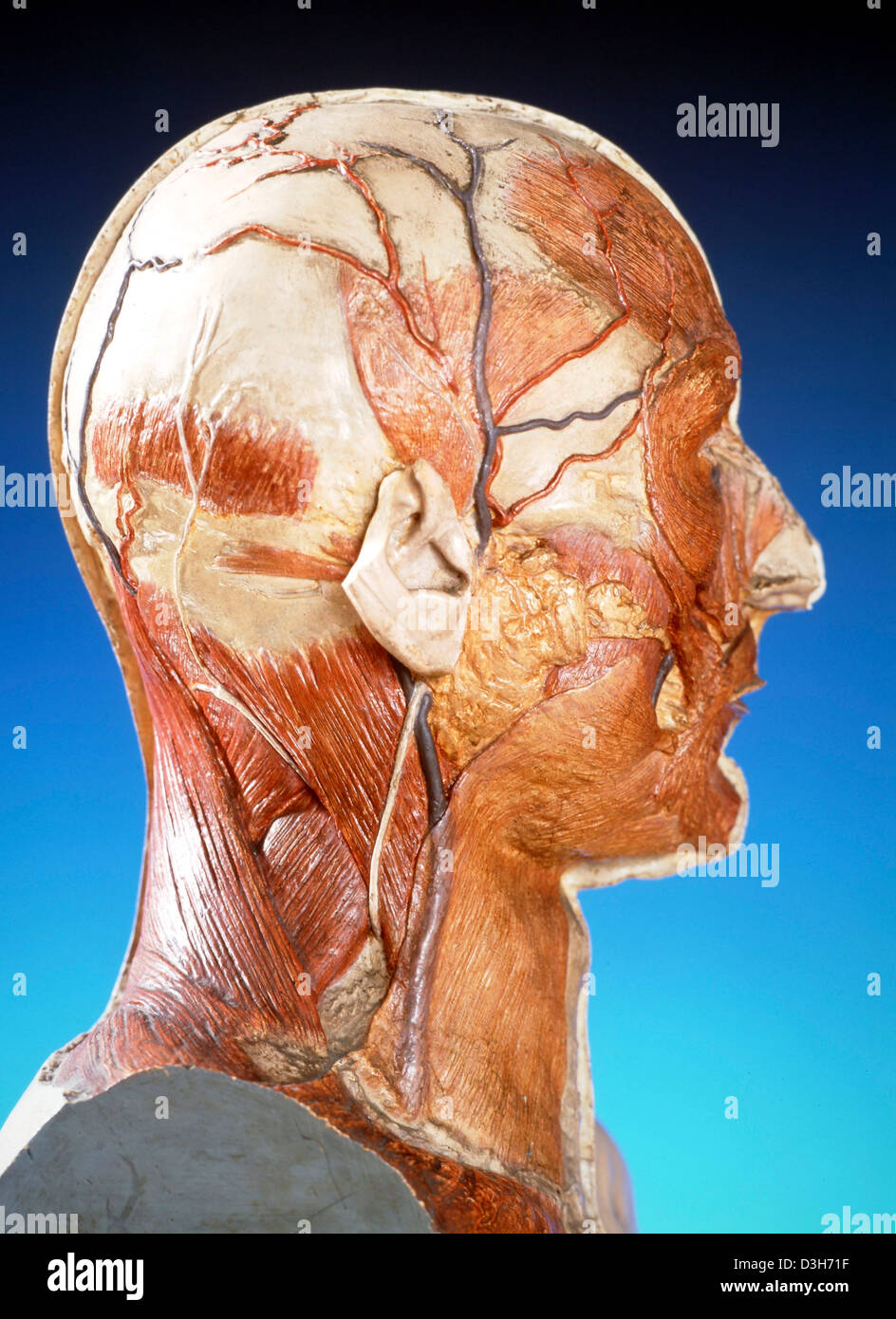 ANATOMY Stock Photohttps://www.alamy.com/image-license-details/?v=1https://www.alamy.com/stock-photo-anatomy-53853787.html
RMD3H71F–ANATOMY
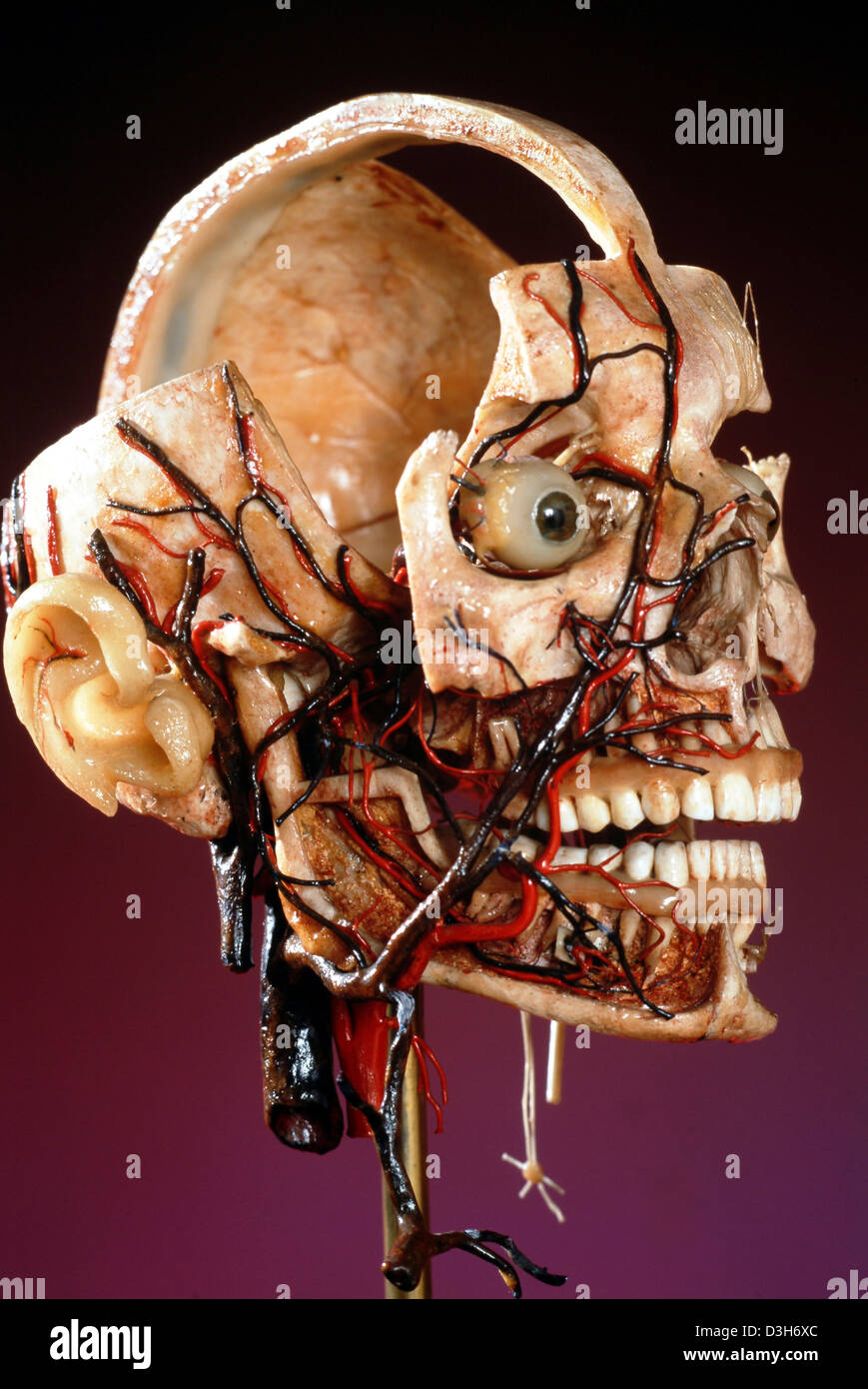 ANATOMY Stock Photohttps://www.alamy.com/image-license-details/?v=1https://www.alamy.com/stock-photo-anatomy-53853700.html
RMD3H6XC–ANATOMY
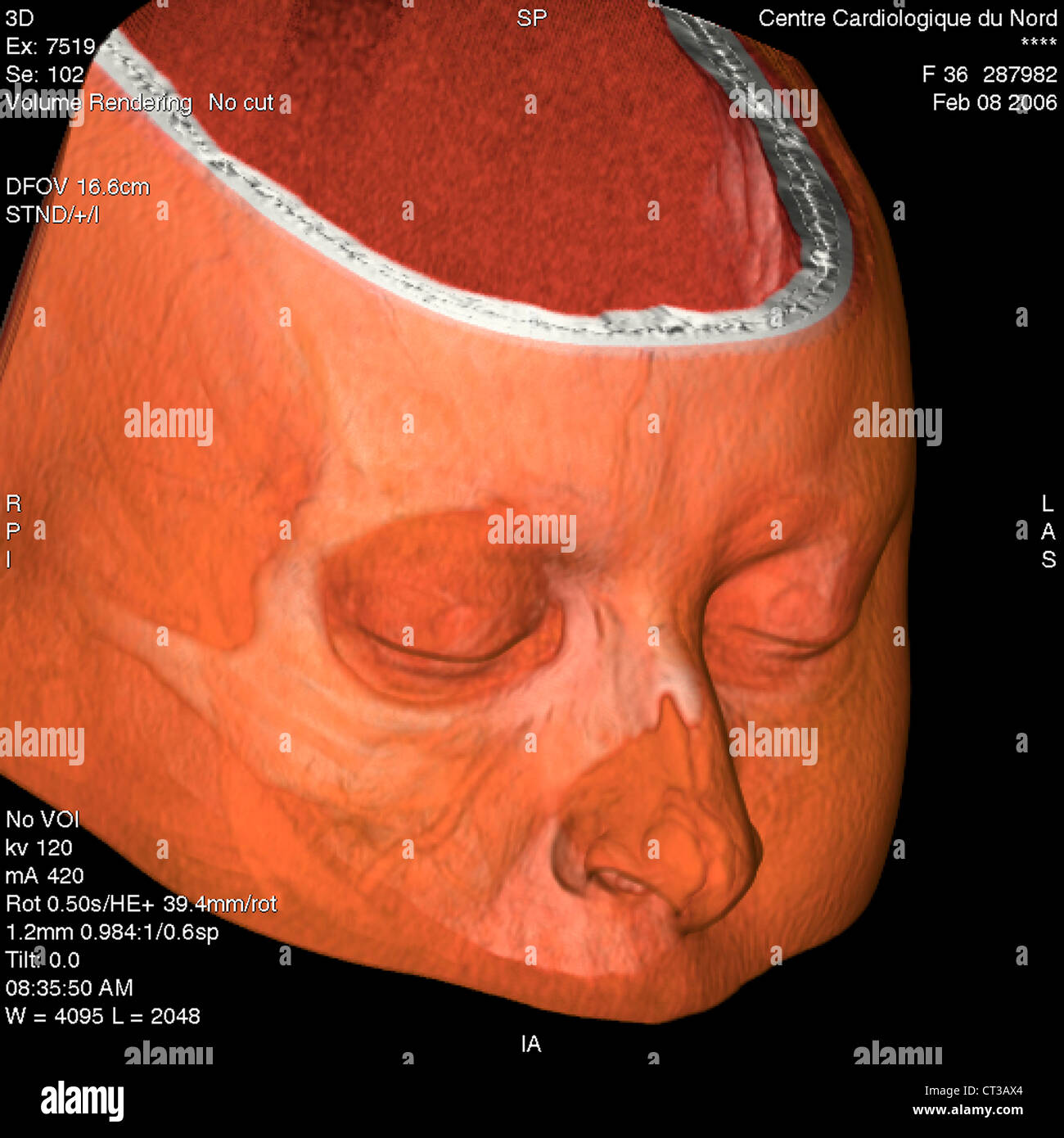 HEAD, 3D SCAN Stock Photohttps://www.alamy.com/image-license-details/?v=1https://www.alamy.com/stock-photo-head-3d-scan-49246908.html
RMCT3AX4–HEAD, 3D SCAN
