Quick filters:
Bonehuman Stock Photos and Images
 medical pattern with elements of tooth, shield, magnifier, broken bonehuman, thermometer and bowel Stock Vectorhttps://www.alamy.com/image-license-details/?v=1https://www.alamy.com/medical-pattern-with-elements-of-tooth-shield-magnifier-broken-bonehuman-thermometer-and-bowel-image551933838.html
RF2R1XN6P–medical pattern with elements of tooth, shield, magnifier, broken bonehuman, thermometer and bowel
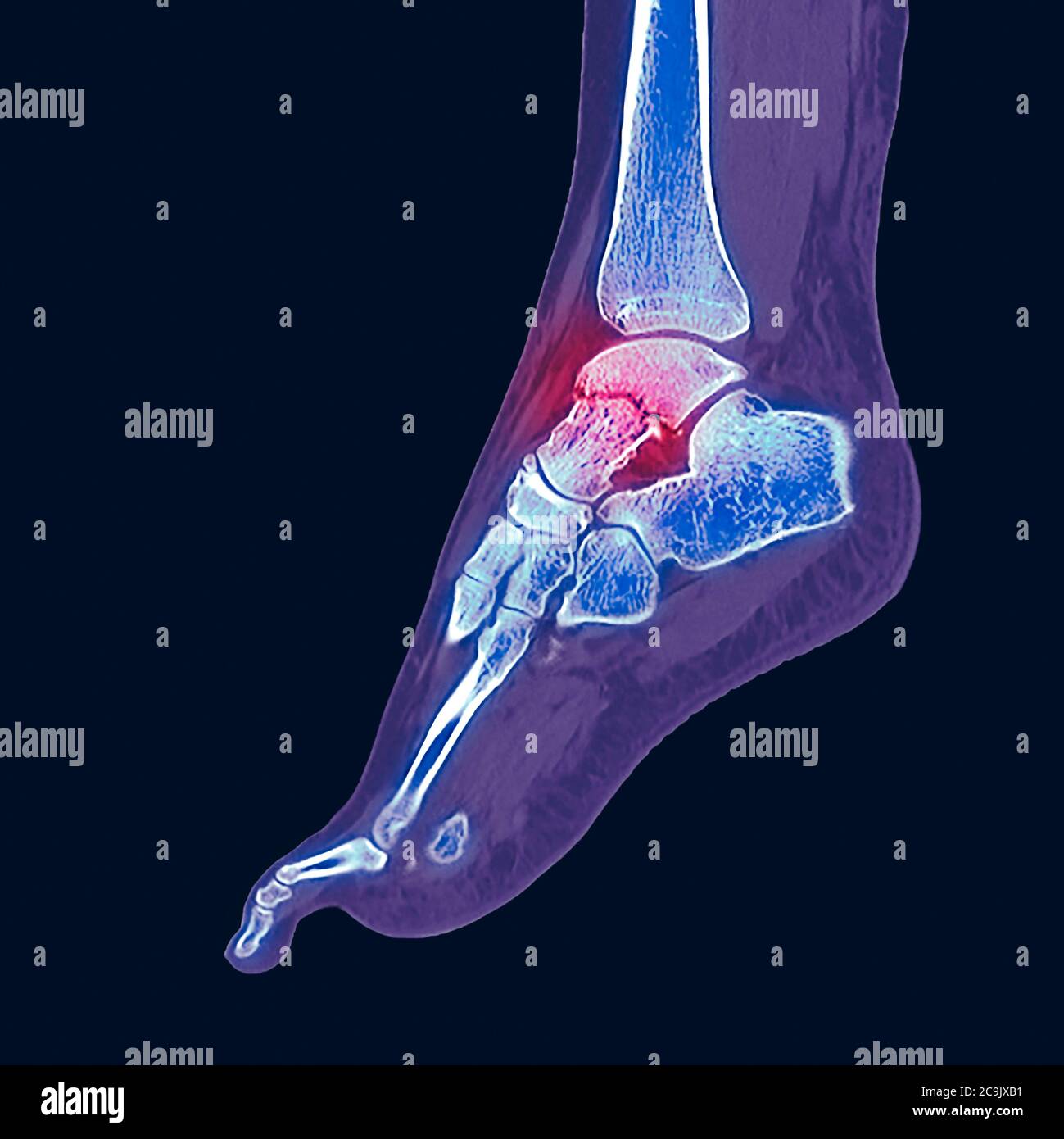 Fractured ankle bone. Coloured computed tomography (CT) scan of the bones of the foot and ankle of a 23-year-old woman with a comminuted (splintered) Stock Photohttps://www.alamy.com/image-license-details/?v=1https://www.alamy.com/fractured-ankle-bone-coloured-computed-tomography-ct-scan-of-the-bones-of-the-foot-and-ankle-of-a-23-year-old-woman-with-a-comminuted-splintered-image367365461.html
RF2C9JXB1–Fractured ankle bone. Coloured computed tomography (CT) scan of the bones of the foot and ankle of a 23-year-old woman with a comminuted (splintered)
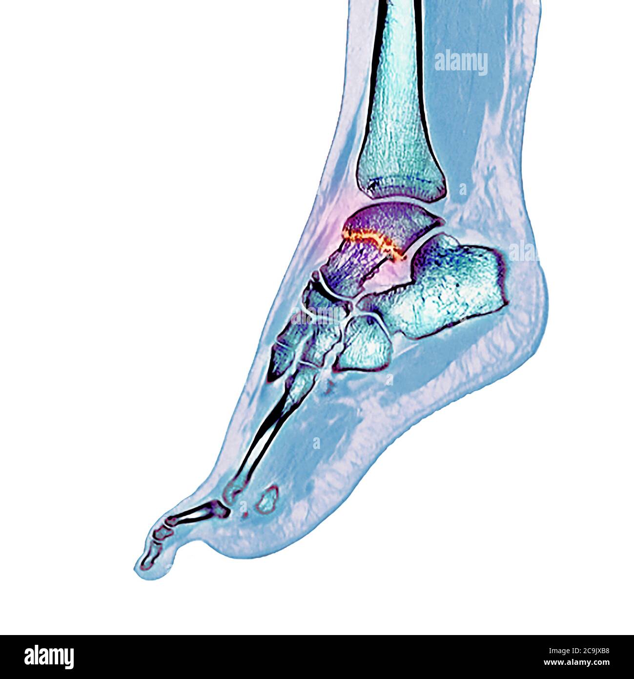 Fractured ankle bone. Coloured computed tomography (CT) scan of the bones of the foot and ankle of a 23-year-old woman with a comminuted (splintered) Stock Photohttps://www.alamy.com/image-license-details/?v=1https://www.alamy.com/fractured-ankle-bone-coloured-computed-tomography-ct-scan-of-the-bones-of-the-foot-and-ankle-of-a-23-year-old-woman-with-a-comminuted-splintered-image367365468.html
RF2C9JXB8–Fractured ankle bone. Coloured computed tomography (CT) scan of the bones of the foot and ankle of a 23-year-old woman with a comminuted (splintered)
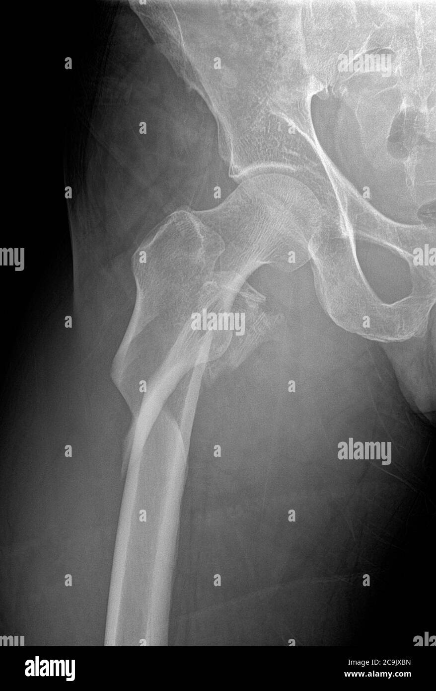 Hip fracture. Frontal X-ray of the right hip of a 53-year-old man, showing a comminuted (splintered) fracture of the neck of the femur (centre left). Stock Photohttps://www.alamy.com/image-license-details/?v=1https://www.alamy.com/hip-fracture-frontal-x-ray-of-the-right-hip-of-a-53-year-old-man-showing-a-comminuted-splintered-fracture-of-the-neck-of-the-femur-centre-left-image367365481.html
RF2C9JXBN–Hip fracture. Frontal X-ray of the right hip of a 53-year-old man, showing a comminuted (splintered) fracture of the neck of the femur (centre left).
 Hip fracture. Coloured frontal X-ray of the right hip of a 53-year-old man, showing a comminuted (splintered) fracture of the neck of the femur (centr Stock Photohttps://www.alamy.com/image-license-details/?v=1https://www.alamy.com/hip-fracture-coloured-frontal-x-ray-of-the-right-hip-of-a-53-year-old-man-showing-a-comminuted-splintered-fracture-of-the-neck-of-the-femur-centr-image367365485.html
RF2C9JXBW–Hip fracture. Coloured frontal X-ray of the right hip of a 53-year-old man, showing a comminuted (splintered) fracture of the neck of the femur (centr
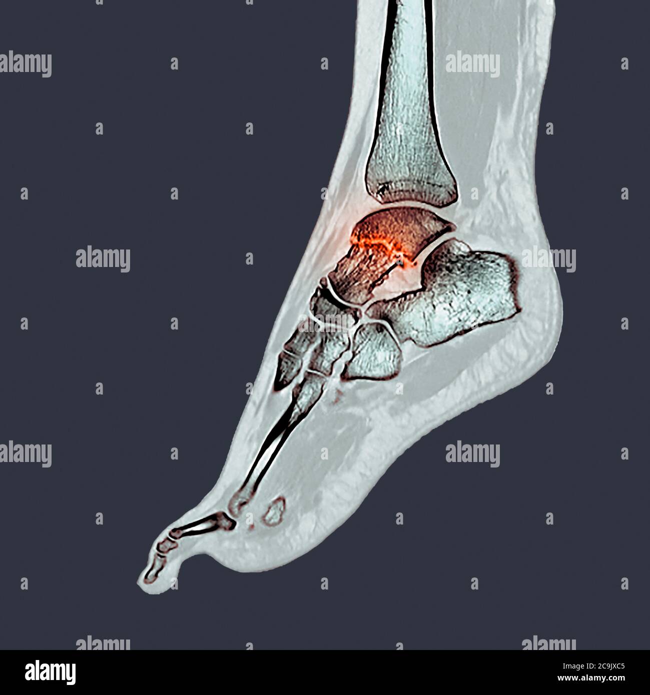 Fractured ankle bone. Coloured computed tomography (CT) scan of the bones of the foot and ankle of a 23-year-old woman with a comminuted (splintered) Stock Photohttps://www.alamy.com/image-license-details/?v=1https://www.alamy.com/fractured-ankle-bone-coloured-computed-tomography-ct-scan-of-the-bones-of-the-foot-and-ankle-of-a-23-year-old-woman-with-a-comminuted-splintered-image367365493.html
RF2C9JXC5–Fractured ankle bone. Coloured computed tomography (CT) scan of the bones of the foot and ankle of a 23-year-old woman with a comminuted (splintered)
 Hip fracture. Coloured frontal X-ray of the right hip of a 53-year-old man, showing a comminuted (splintered) fracture of the neck of the femur (centr Stock Photohttps://www.alamy.com/image-license-details/?v=1https://www.alamy.com/hip-fracture-coloured-frontal-x-ray-of-the-right-hip-of-a-53-year-old-man-showing-a-comminuted-splintered-fracture-of-the-neck-of-the-femur-centr-image367365498.html
RF2C9JXCA–Hip fracture. Coloured frontal X-ray of the right hip of a 53-year-old man, showing a comminuted (splintered) fracture of the neck of the femur (centr
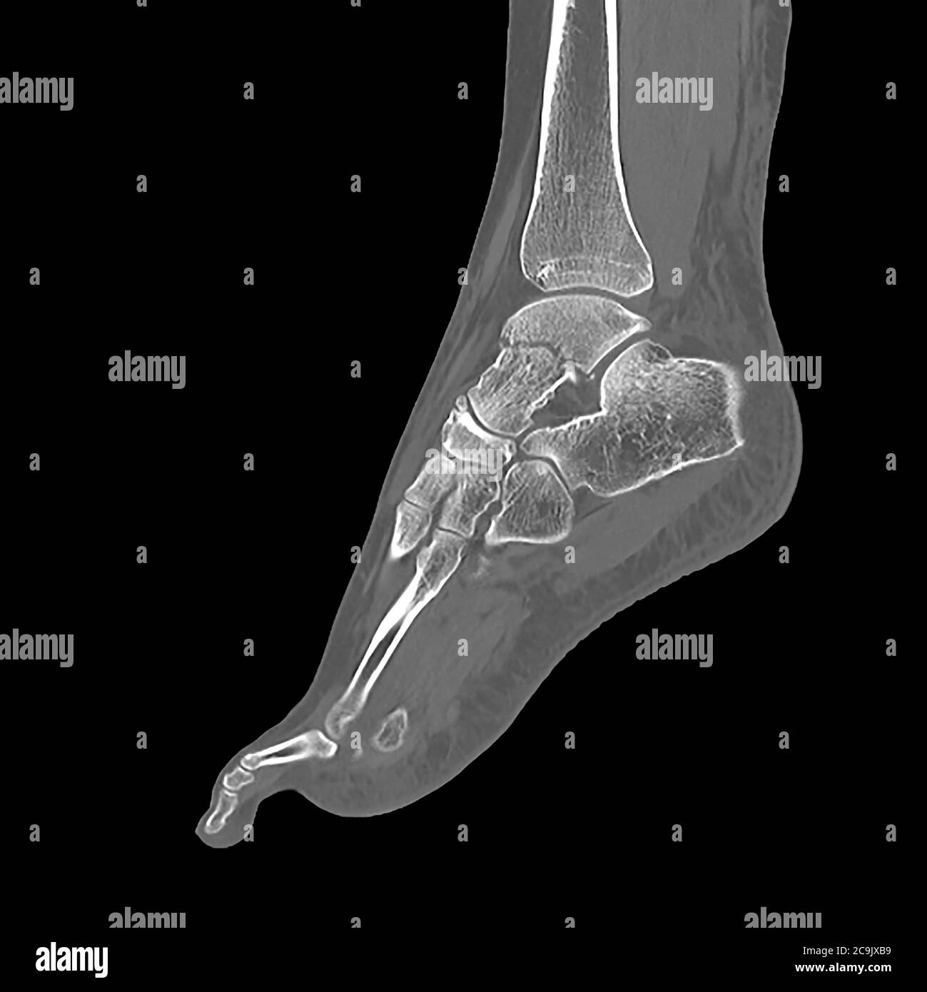 Fractured ankle bone. Computed tomography (CT) scan of the bones of the foot and ankle of a 23-year-old woman with a comminuted (splintered) fracture Stock Photohttps://www.alamy.com/image-license-details/?v=1https://www.alamy.com/fractured-ankle-bone-computed-tomography-ct-scan-of-the-bones-of-the-foot-and-ankle-of-a-23-year-old-woman-with-a-comminuted-splintered-fracture-image367365469.html
RF2C9JXB9–Fractured ankle bone. Computed tomography (CT) scan of the bones of the foot and ankle of a 23-year-old woman with a comminuted (splintered) fracture
 Hip fracture. Coloured frontal X-ray of the right hip of a 53-year-old man, showing a comminuted (splintered) fracture of the neck of the femur (centr Stock Photohttps://www.alamy.com/image-license-details/?v=1https://www.alamy.com/hip-fracture-coloured-frontal-x-ray-of-the-right-hip-of-a-53-year-old-man-showing-a-comminuted-splintered-fracture-of-the-neck-of-the-femur-centr-image367365506.html
RF2C9JXCJ–Hip fracture. Coloured frontal X-ray of the right hip of a 53-year-old man, showing a comminuted (splintered) fracture of the neck of the femur (centr
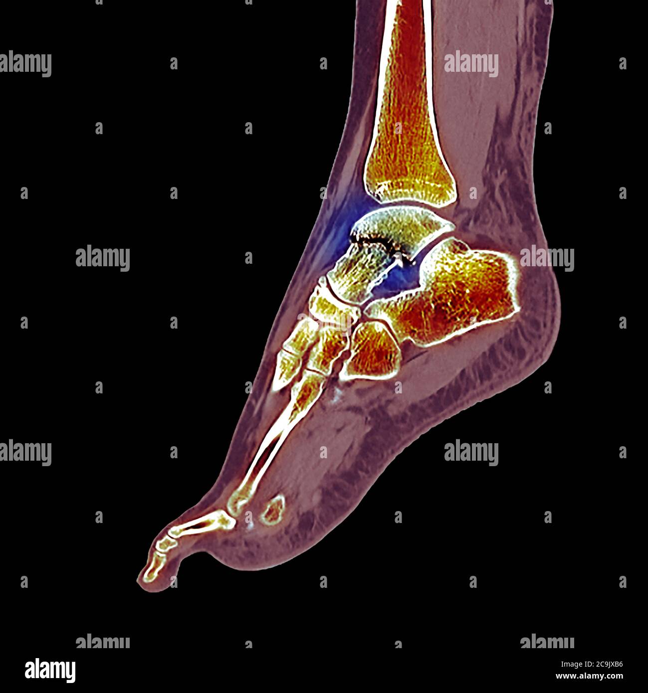 Fractured ankle bone. Coloured computed tomography (CT) scan of the bones of the foot and ankle of a 23-year-old woman with a comminuted (splintered) Stock Photohttps://www.alamy.com/image-license-details/?v=1https://www.alamy.com/fractured-ankle-bone-coloured-computed-tomography-ct-scan-of-the-bones-of-the-foot-and-ankle-of-a-23-year-old-woman-with-a-comminuted-splintered-image367365466.html
RF2C9JXB6–Fractured ankle bone. Coloured computed tomography (CT) scan of the bones of the foot and ankle of a 23-year-old woman with a comminuted (splintered)
 Hip fracture. Coloured frontal X-ray of the right hip of a 53-year-old man, showing a comminuted (splintered) fracture of the neck of the femur (centr Stock Photohttps://www.alamy.com/image-license-details/?v=1https://www.alamy.com/hip-fracture-coloured-frontal-x-ray-of-the-right-hip-of-a-53-year-old-man-showing-a-comminuted-splintered-fracture-of-the-neck-of-the-femur-centr-image367365482.html
RF2C9JXBP–Hip fracture. Coloured frontal X-ray of the right hip of a 53-year-old man, showing a comminuted (splintered) fracture of the neck of the femur (centr