Quick filters:
Brain cortex Stock Photos and Images
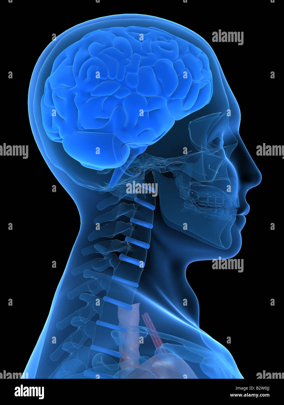 x-ray head with brain Stock Photohttps://www.alamy.com/image-license-details/?v=1https://www.alamy.com/stock-photo-x-ray-head-with-brain-18989002.html
RFB2W0JJ–x-ray head with brain
 Multiple realizability in human brain - dozens of terms describing its properties painted over the brain cortex to symbolize its connection to the min Stock Photohttps://www.alamy.com/image-license-details/?v=1https://www.alamy.com/multiple-realizability-in-human-brain-dozens-of-terms-describing-its-properties-painted-over-the-brain-cortex-to-symbolize-its-connection-to-the-min-image472069047.html
RF2JC0GTR–Multiple realizability in human brain - dozens of terms describing its properties painted over the brain cortex to symbolize its connection to the min
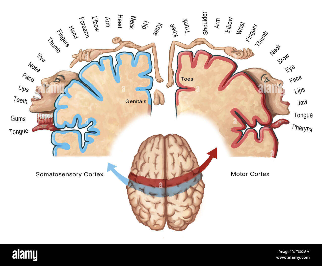 Cortical Homunculus Illustration Stock Photohttps://www.alamy.com/image-license-details/?v=1https://www.alamy.com/cortical-homunculus-illustration-image245864436.html
RMT802GM–Cortical Homunculus Illustration
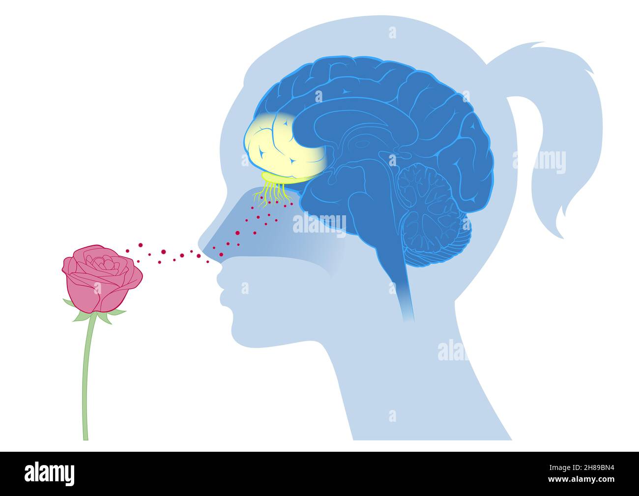 Brain cortex smell Stock Photohttps://www.alamy.com/image-license-details/?v=1https://www.alamy.com/brain-cortex-smell-image452593600.html
RM2H89BN4–Brain cortex smell
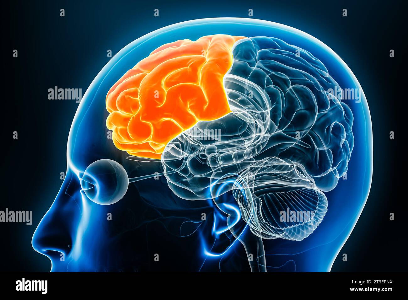 Frontal lobe of the cerebral cortex profile view close-up 3D rendering illustration. Human brain anatomy, neurology, neuroscience, medical and healthc Stock Photohttps://www.alamy.com/image-license-details/?v=1https://www.alamy.com/frontal-lobe-of-the-cerebral-cortex-profile-view-close-up-3d-rendering-illustration-human-brain-anatomy-neurology-neuroscience-medical-and-healthc-image570111302.html
RF2T3EPNX–Frontal lobe of the cerebral cortex profile view close-up 3D rendering illustration. Human brain anatomy, neurology, neuroscience, medical and healthc
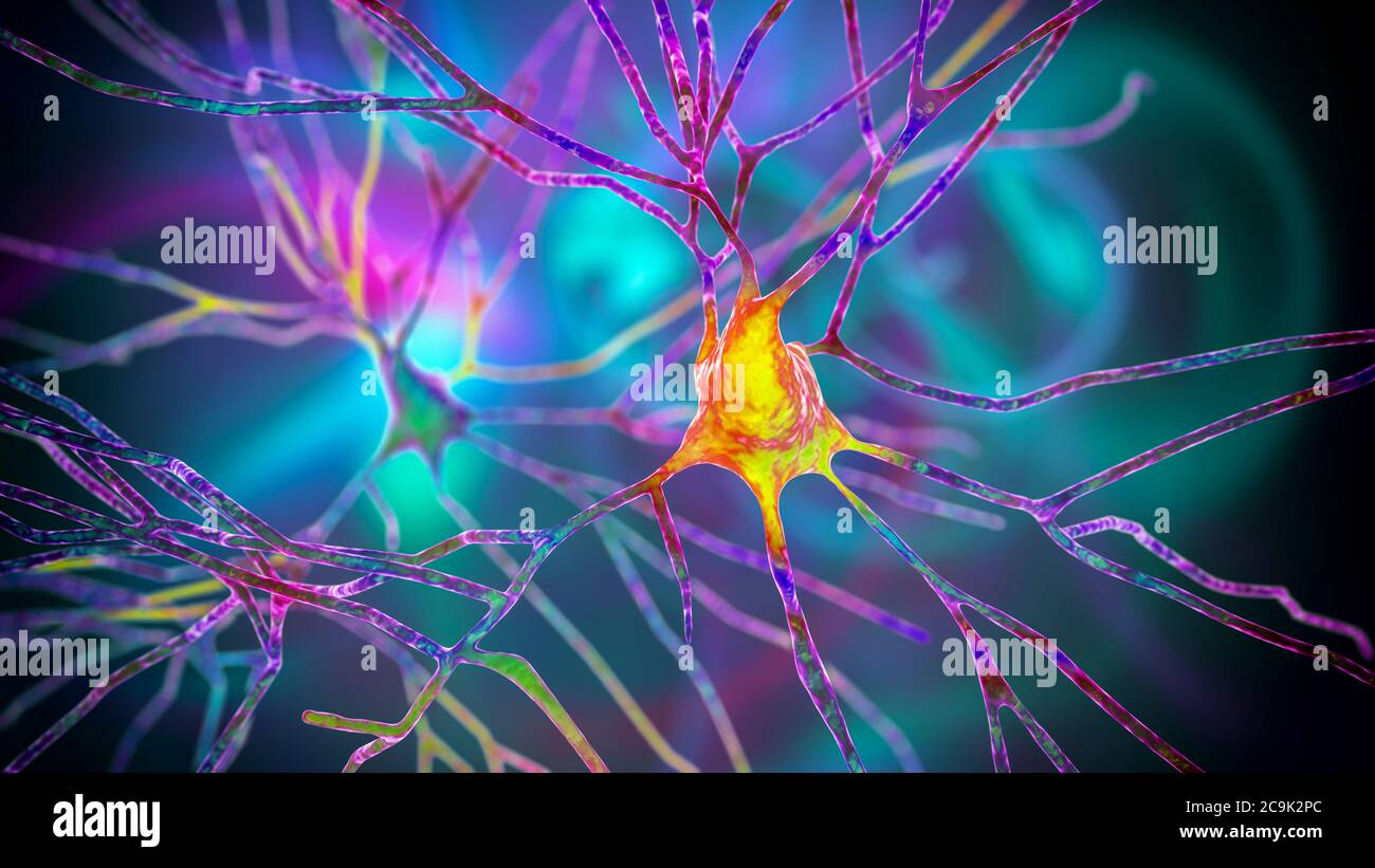 Pyramidal neurons of human brain cortex, computer illustration. Stock Photohttps://www.alamy.com/image-license-details/?v=1https://www.alamy.com/pyramidal-neurons-of-human-brain-cortex-computer-illustration-image367368916.html
RF2C9K2PC–Pyramidal neurons of human brain cortex, computer illustration.
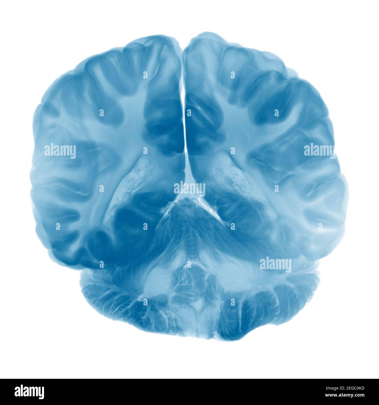 Cross Section Of Brain Cortex On White Background Stock Photohttps://www.alamy.com/image-license-details/?v=1https://www.alamy.com/cross-section-of-brain-cortex-on-white-background-image405936929.html
RF2EGC0KD–Cross Section Of Brain Cortex On White Background
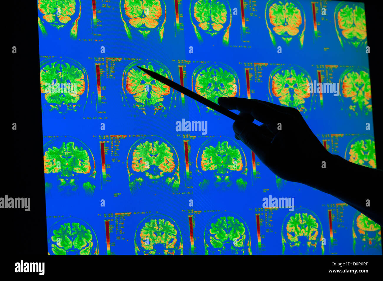 colored x-rays of cerebral cortex cerebellum human brain MRI Stock Photohttps://www.alamy.com/image-license-details/?v=1https://www.alamy.com/stock-photo-colored-x-rays-of-cerebral-cortex-cerebellum-human-brain-mri-52136666.html
RMD0R0RP–colored x-rays of cerebral cortex cerebellum human brain MRI
 A clever shot of two halves of a Walnut positioned to look like or to form a brain shape. Stock Photohttps://www.alamy.com/image-license-details/?v=1https://www.alamy.com/stock-photo-a-clever-shot-of-two-halves-of-a-walnut-positioned-to-look-like-or-171315712.html
RFKXM2X8–A clever shot of two halves of a Walnut positioned to look like or to form a brain shape.
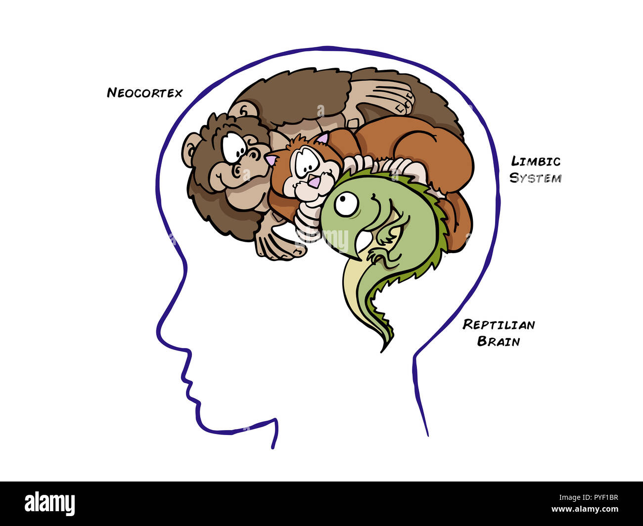 Triune Brain Theory Stock Photohttps://www.alamy.com/image-license-details/?v=1https://www.alamy.com/triune-brain-theory-image223450523.html
RFPYF1BR–Triune Brain Theory
 Isolated colorful vector brains. Medical scientifical logotype. Neurobiology emblem. Intelligence image. Human brain illustration. Graphic cortex Stock Vectorhttps://www.alamy.com/image-license-details/?v=1https://www.alamy.com/isolated-colorful-vector-brains-medical-scientifical-logotype-neurobiology-emblem-intelligence-image-human-brain-illustration-graphic-cortex-image337015438.html
RF2AG8AHJ–Isolated colorful vector brains. Medical scientifical logotype. Neurobiology emblem. Intelligence image. Human brain illustration. Graphic cortex
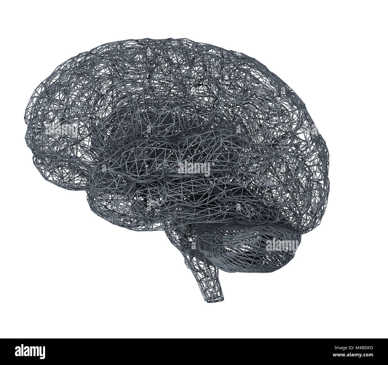 3d brain illustration Stock Photohttps://www.alamy.com/image-license-details/?v=1https://www.alamy.com/stock-photo-3d-brain-illustration-174814513.html
RFM4BDKD–3d brain illustration
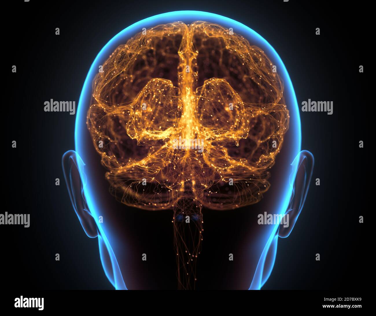 X-ray of the head and human brain in concept of neural connections and electrical pulses. Stock Photohttps://www.alamy.com/image-license-details/?v=1https://www.alamy.com/x-ray-of-the-head-and-human-brain-in-concept-of-neural-connections-and-electrical-pulses-image383193085.html
RF2D7BXK9–X-ray of the head and human brain in concept of neural connections and electrical pulses.
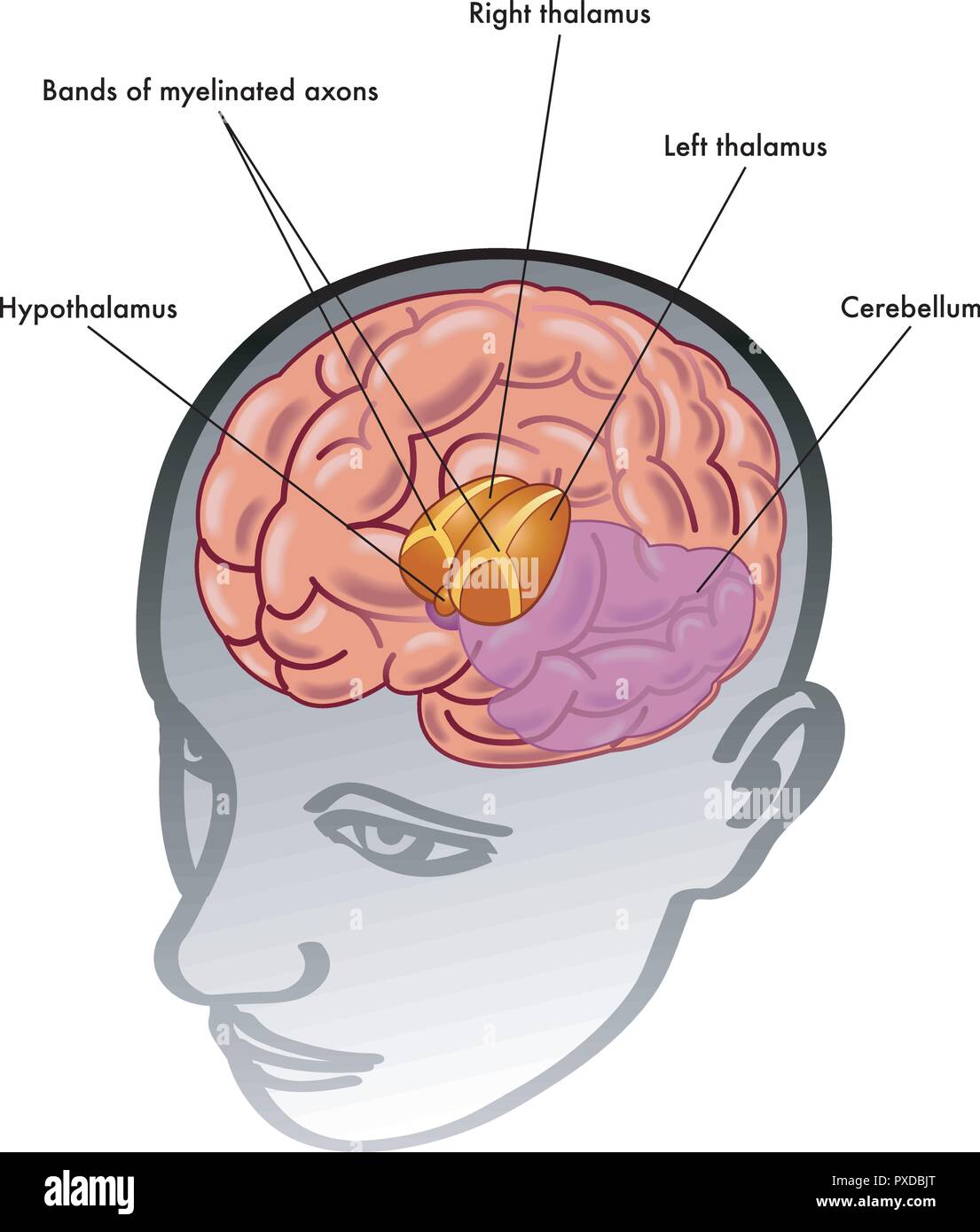 medical illustration of the thalamus and hypothalamus and their position inside the head Stock Vectorhttps://www.alamy.com/image-license-details/?v=1https://www.alamy.com/medical-illustration-of-the-thalamus-and-hypothalamus-and-their-position-inside-the-head-image222800000.html
RFPXDBJT–medical illustration of the thalamus and hypothalamus and their position inside the head
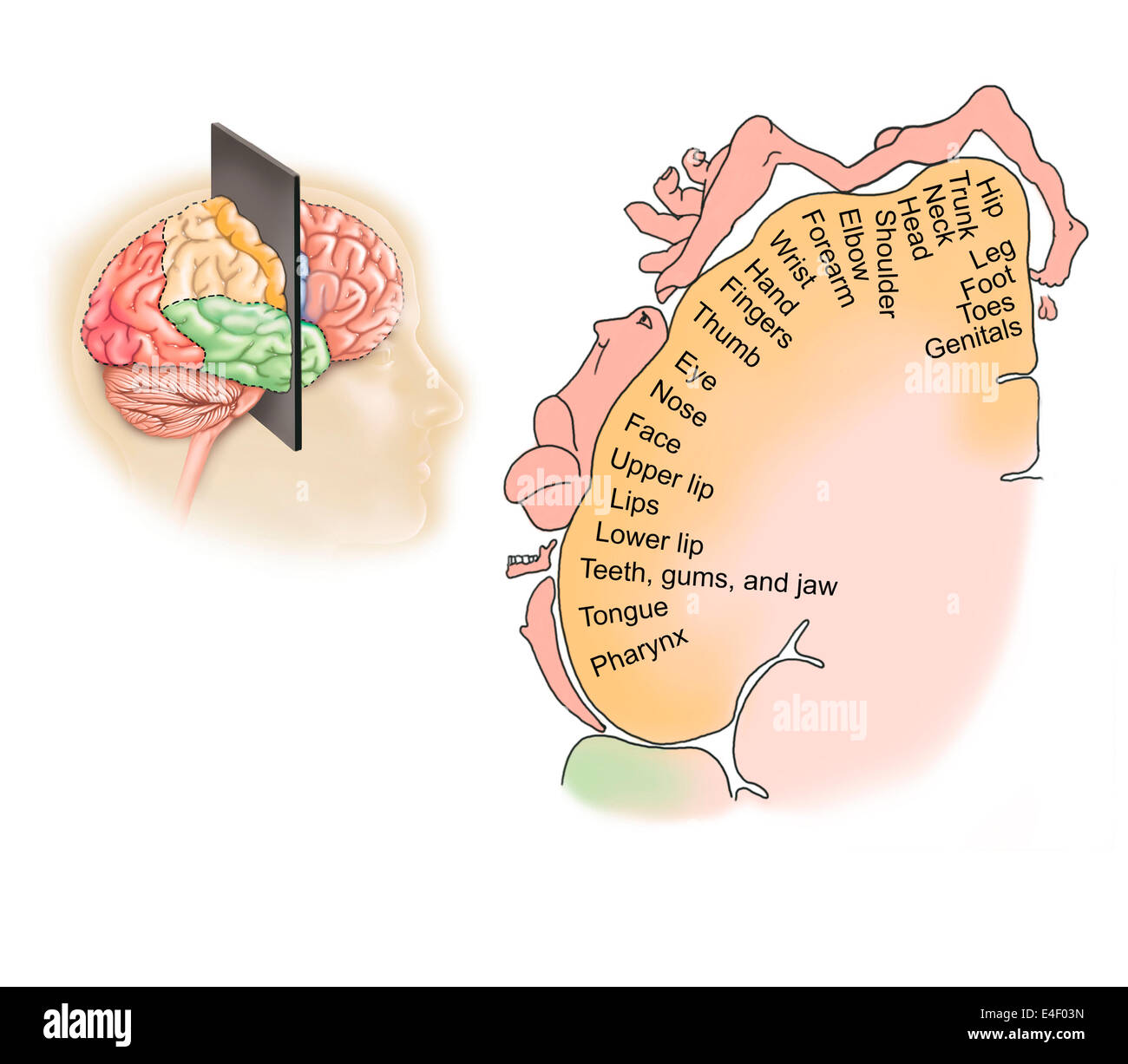 Coronal section through human brain showing the layout of the sensory cortex. Stock Photohttps://www.alamy.com/image-license-details/?v=1https://www.alamy.com/stock-photo-coronal-section-through-human-brain-showing-the-layout-of-the-sensory-71629481.html
RME4F03N–Coronal section through human brain showing the layout of the sensory cortex.
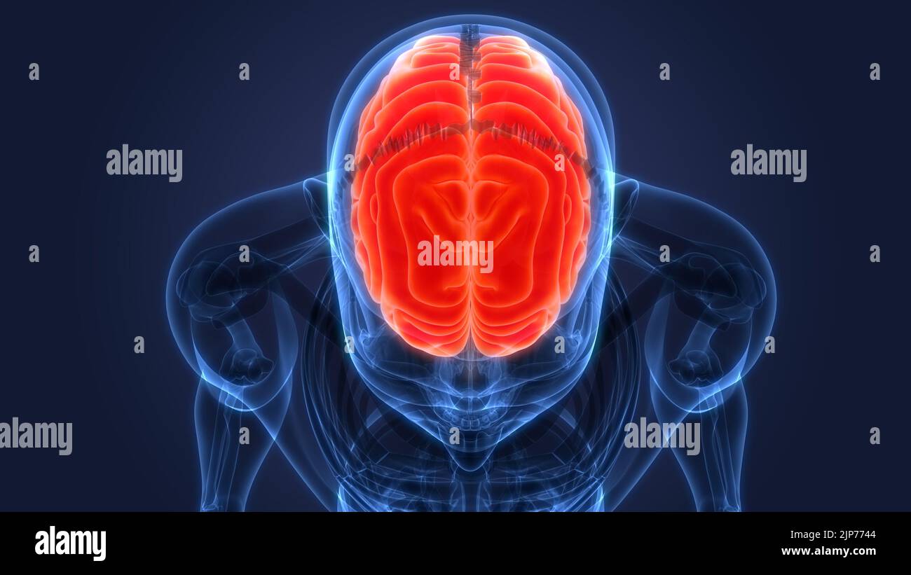 Central Organ of Human Nervous System Brain Anatomy Stock Photohttps://www.alamy.com/image-license-details/?v=1https://www.alamy.com/central-organ-of-human-nervous-system-brain-anatomy-image478361636.html
RF2JP7744–Central Organ of Human Nervous System Brain Anatomy
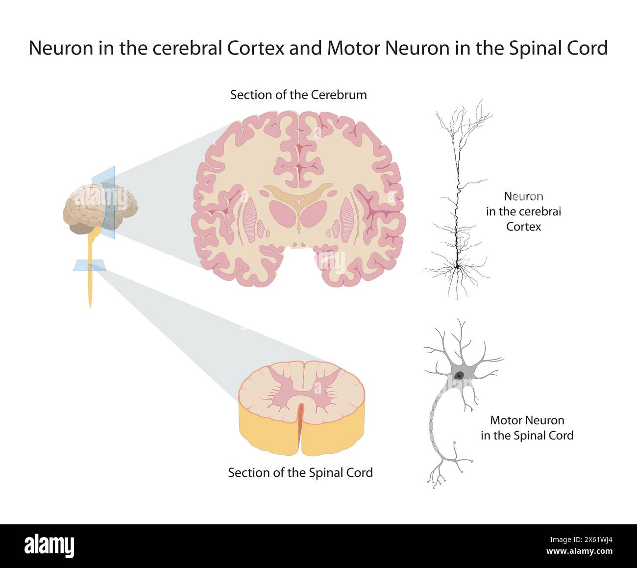 Neuron in the cerebral Cortex and Motor Neuron in the Spinal Cord Stock Photohttps://www.alamy.com/image-license-details/?v=1https://www.alamy.com/neuron-in-the-cerebral-cortex-and-motor-neuron-in-the-spinal-cord-image606092876.html
RF2X61WJ4–Neuron in the cerebral Cortex and Motor Neuron in the Spinal Cord
 Human Internal Organ Brain with Nervous System Anatomy X-ray 3D rendering Stock Photohttps://www.alamy.com/image-license-details/?v=1https://www.alamy.com/human-internal-organ-brain-with-nervous-system-anatomy-x-ray-3d-rendering-image342001502.html
RF2ATBEBA–Human Internal Organ Brain with Nervous System Anatomy X-ray 3D rendering
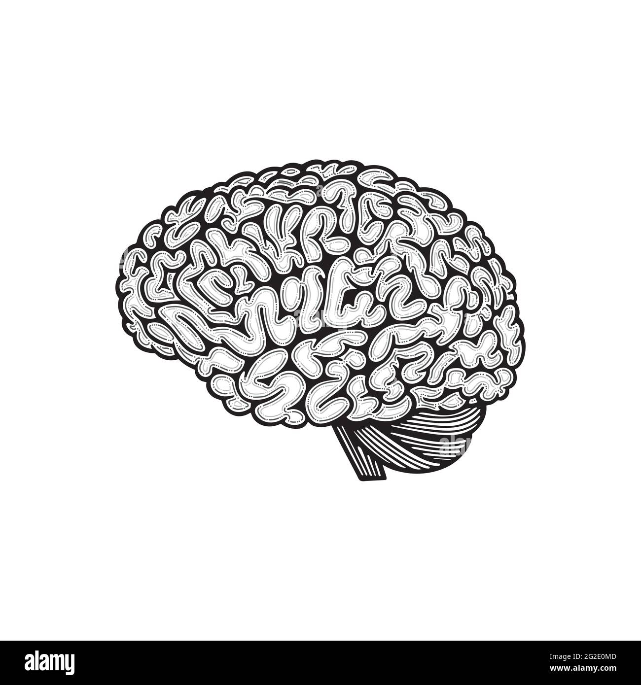 Brain side view. Human brain hand drawn vector illustration. Brain outline drawing. Part of set. Stock Vectorhttps://www.alamy.com/image-license-details/?v=1https://www.alamy.com/brain-side-view-human-brain-hand-drawn-vector-illustration-brain-outline-drawing-part-of-set-image431796413.html
RF2G2E0MD–Brain side view. Human brain hand drawn vector illustration. Brain outline drawing. Part of set.
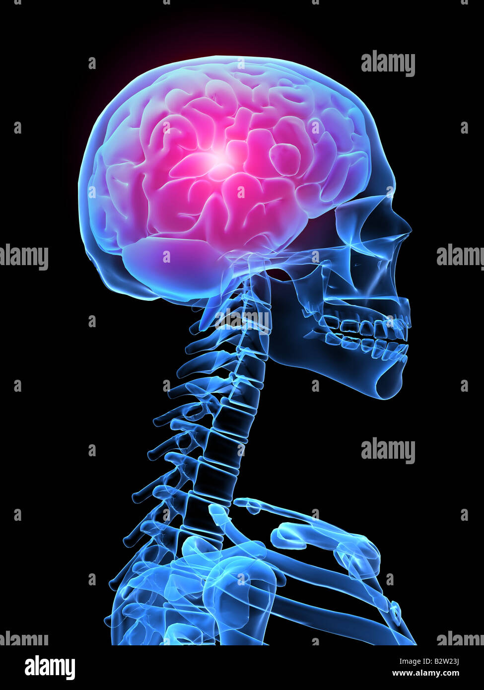 headache Stock Photohttps://www.alamy.com/image-license-details/?v=1https://www.alamy.com/stock-photo-headache-18990150.html
RFB2W23J–headache
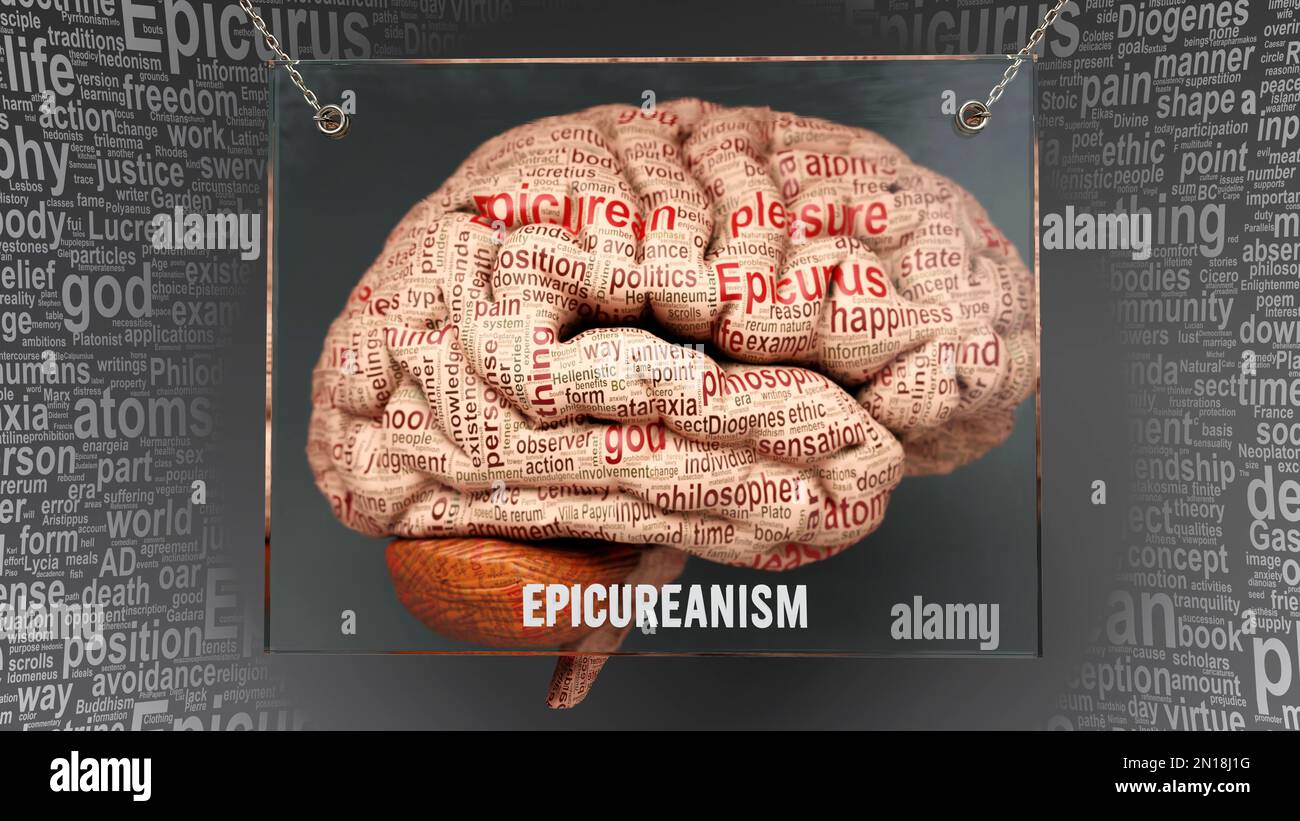 Epicureanism in human brain - dozens of important terms describing Epicureanism properties painted over the brain cortex to symbolize Epicureanism con Stock Photohttps://www.alamy.com/image-license-details/?v=1https://www.alamy.com/epicureanism-in-human-brain-dozens-of-important-terms-describing-epicureanism-properties-painted-over-the-brain-cortex-to-symbolize-epicureanism-con-image517115468.html
RF2N18J1G–Epicureanism in human brain - dozens of important terms describing Epicureanism properties painted over the brain cortex to symbolize Epicureanism con
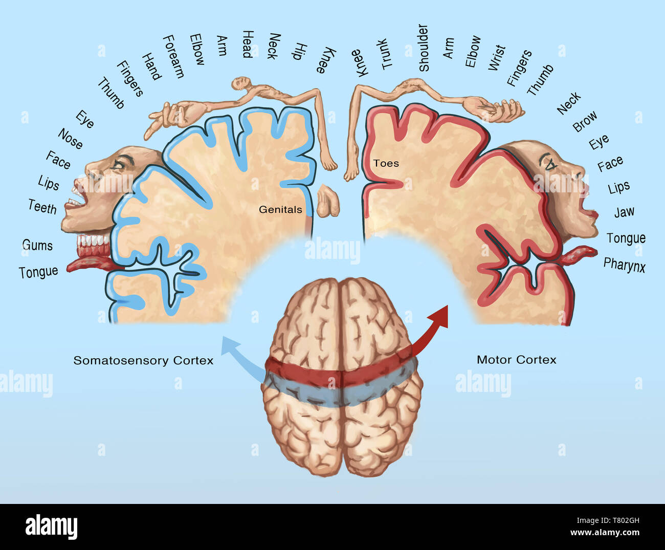 Cortical Homunculus Illustration Stock Photohttps://www.alamy.com/image-license-details/?v=1https://www.alamy.com/cortical-homunculus-illustration-image245864433.html
RMT802GH–Cortical Homunculus Illustration
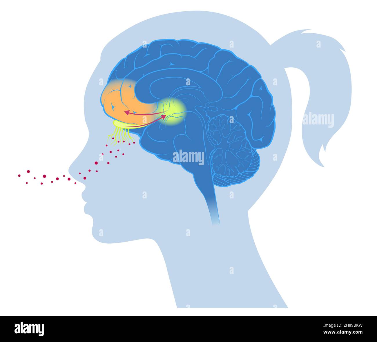 Brain cortex smell emotion Stock Photohttps://www.alamy.com/image-license-details/?v=1https://www.alamy.com/brain-cortex-smell-emotion-image452593565.html
RM2H89BKW–Brain cortex smell emotion
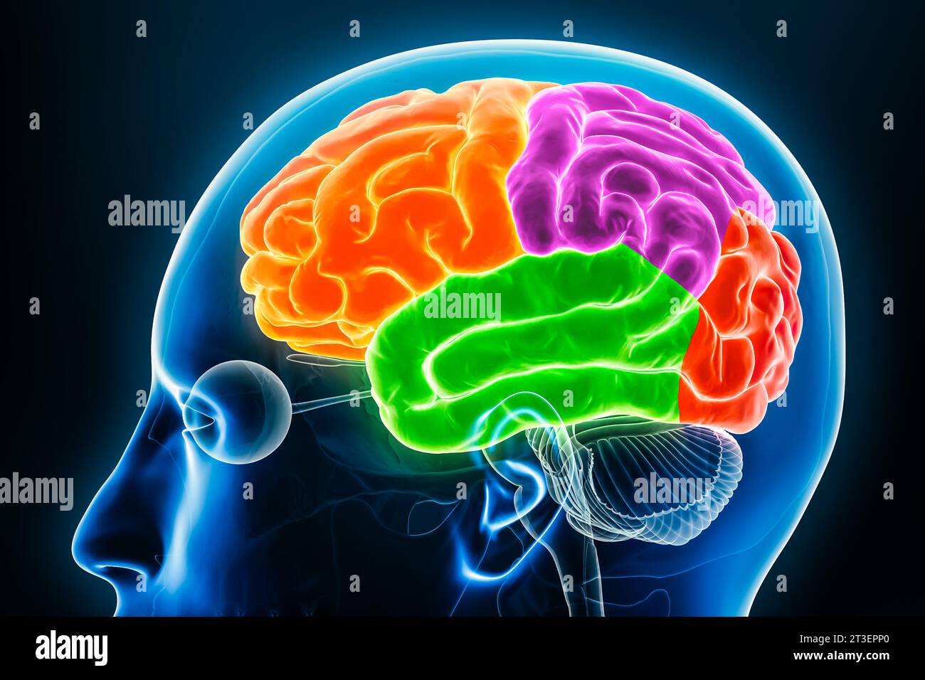 Cerebral cortex lobes in color profile view x-ray 3D rendering illustration. Human brain anatomy, neurology, neuroscience, medical and healthcare, bio Stock Photohttps://www.alamy.com/image-license-details/?v=1https://www.alamy.com/cerebral-cortex-lobes-in-color-profile-view-x-ray-3d-rendering-illustration-human-brain-anatomy-neurology-neuroscience-medical-and-healthcare-bio-image570111304.html
RF2T3EPP0–Cerebral cortex lobes in color profile view x-ray 3D rendering illustration. Human brain anatomy, neurology, neuroscience, medical and healthcare, bio
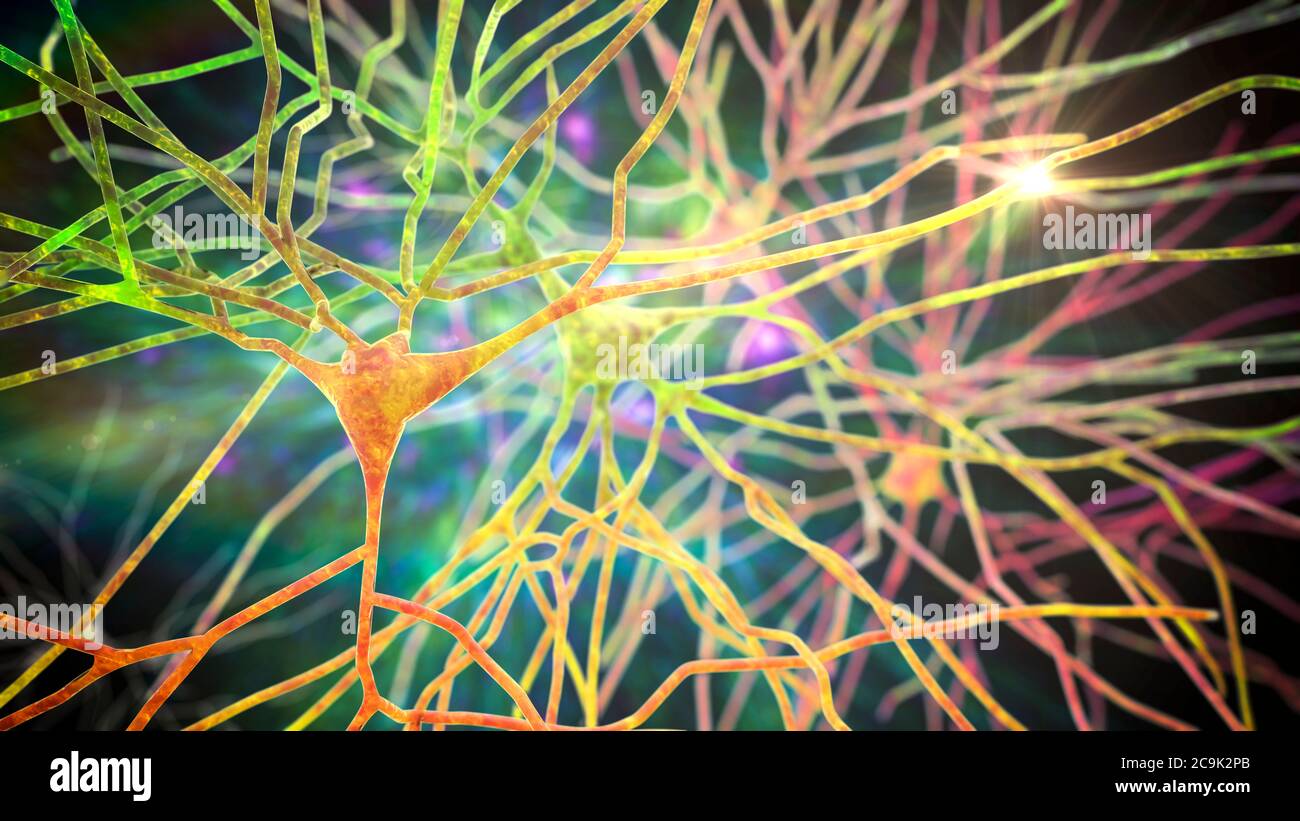 Pyramidal neurons of human brain cortex, computer illustration. Stock Photohttps://www.alamy.com/image-license-details/?v=1https://www.alamy.com/pyramidal-neurons-of-human-brain-cortex-computer-illustration-image367368915.html
RF2C9K2PB–Pyramidal neurons of human brain cortex, computer illustration.
 Human brain nervous system anatomy, medical diagram with parasympathetic and sympathetic nerves. medically accurate, skull cross section, Sebaceous bu Stock Photohttps://www.alamy.com/image-license-details/?v=1https://www.alamy.com/human-brain-nervous-system-anatomy-medical-diagram-with-parasympathetic-and-sympathetic-nerves-medically-accurate-skull-cross-section-sebaceous-bu-image600091869.html
RF2WT8F8D–Human brain nervous system anatomy, medical diagram with parasympathetic and sympathetic nerves. medically accurate, skull cross section, Sebaceous bu
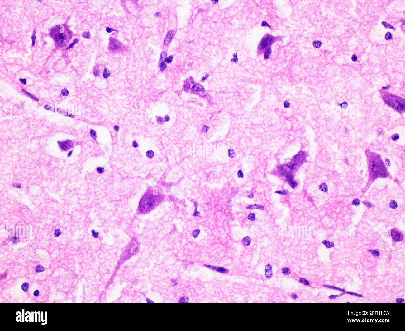 Histology of Human Brain Tissue Viewed at 400x Magnification with Haematoxylin and Eosin Staining. Stock Photohttps://www.alamy.com/image-license-details/?v=1https://www.alamy.com/histology-of-human-brain-tissue-viewed-at-400x-magnification-with-haematoxylin-and-eosin-staining-image560325945.html
RF2RFH1CW–Histology of Human Brain Tissue Viewed at 400x Magnification with Haematoxylin and Eosin Staining.
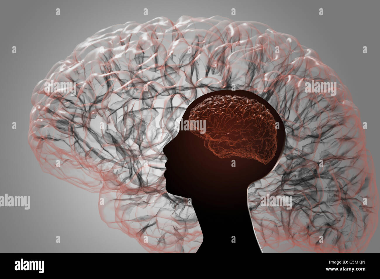 The human brain. Projection of the cerebral cortex Stock Photohttps://www.alamy.com/image-license-details/?v=1https://www.alamy.com/stock-photo-the-human-brain-projection-of-the-cerebral-cortex-106789949.html
RFG5MKJN–The human brain. Projection of the cerebral cortex
 The cerebrum is the most voluminous and most complex part of the brain. It is made up of two hemispheres subdivided into four cerebral lobes, which cover the diencephalon. The most complex functions are completed by the exterior layer of the brain, the cerebral cortex. Stock Photohttps://www.alamy.com/image-license-details/?v=1https://www.alamy.com/the-cerebrum-is-the-most-voluminous-and-most-complex-part-of-the-brain-image156173949.html
RMK229D1–The cerebrum is the most voluminous and most complex part of the brain. It is made up of two hemispheres subdivided into four cerebral lobes, which cover the diencephalon. The most complex functions are completed by the exterior layer of the brain, the cerebral cortex.
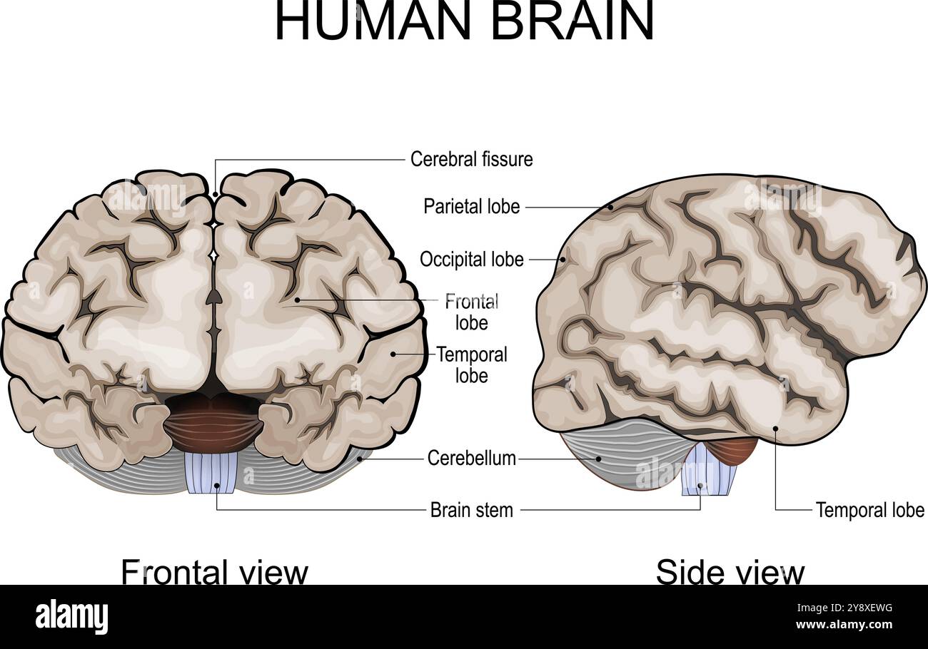 Human brain anatomy. Cerebral hemispheres, Cerebral cortex, Frontal, Parietal, Temporal, Occipital lobes, Cerebellum and Brainstem, Cerebral fissure. Stock Vectorhttps://www.alamy.com/image-license-details/?v=1https://www.alamy.com/human-brain-anatomy-cerebral-hemispheres-cerebral-cortex-frontal-parietal-temporal-occipital-lobes-cerebellum-and-brainstem-cerebral-fissure-image625072940.html
RF2Y8XEWG–Human brain anatomy. Cerebral hemispheres, Cerebral cortex, Frontal, Parietal, Temporal, Occipital lobes, Cerebellum and Brainstem, Cerebral fissure.
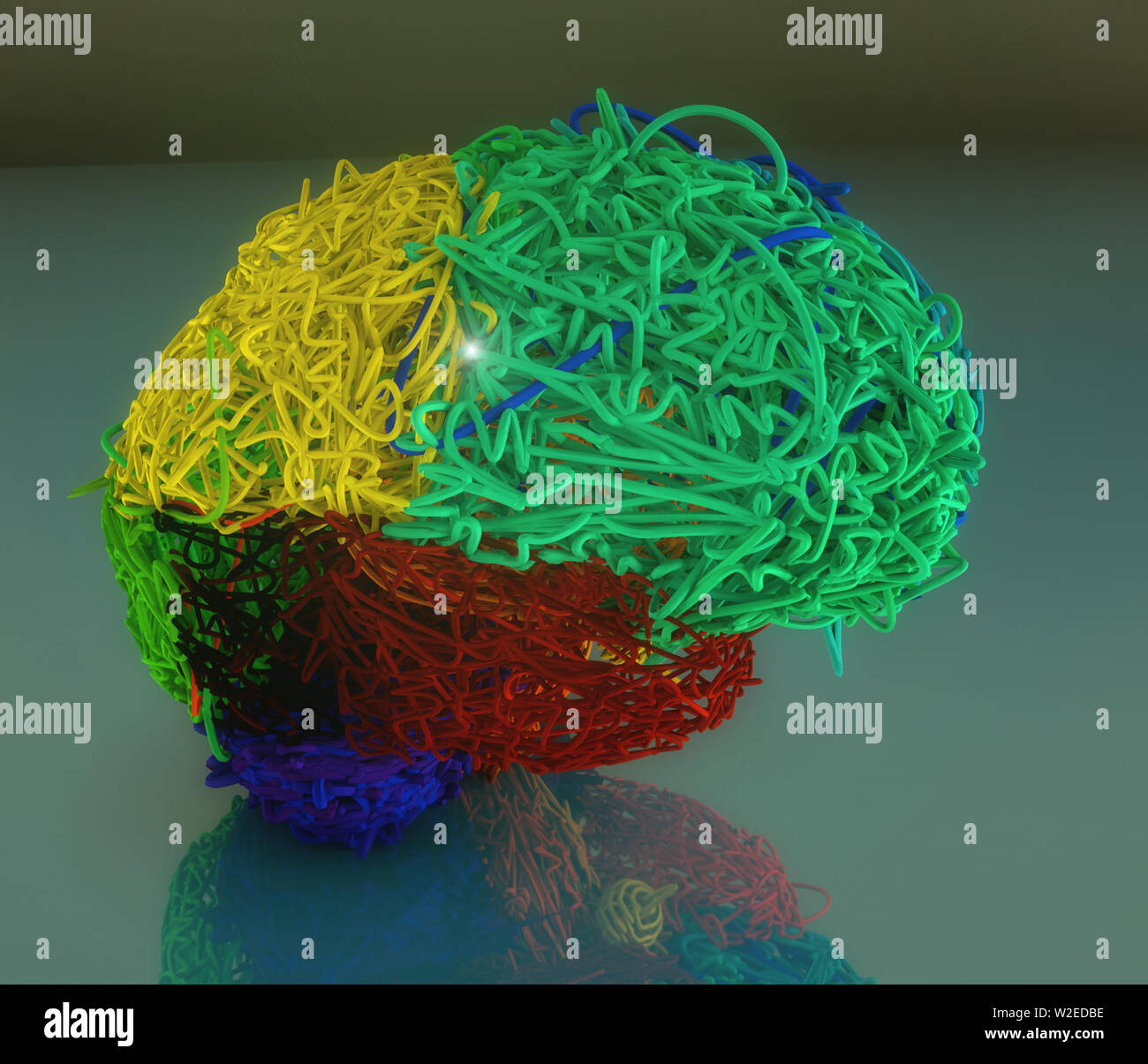 3d colored brain illustration Stock Photohttps://www.alamy.com/image-license-details/?v=1https://www.alamy.com/3d-colored-brain-illustration-image259702674.html
RFW2EDBE–3d colored brain illustration
 3D illustration of artificial intelligence, microchip forming a brain with lines of connections. Stock Photohttps://www.alamy.com/image-license-details/?v=1https://www.alamy.com/3d-illustration-of-artificial-intelligence-microchip-forming-a-brain-with-lines-of-connections-image454202299.html
RF2HAXKJK–3D illustration of artificial intelligence, microchip forming a brain with lines of connections.
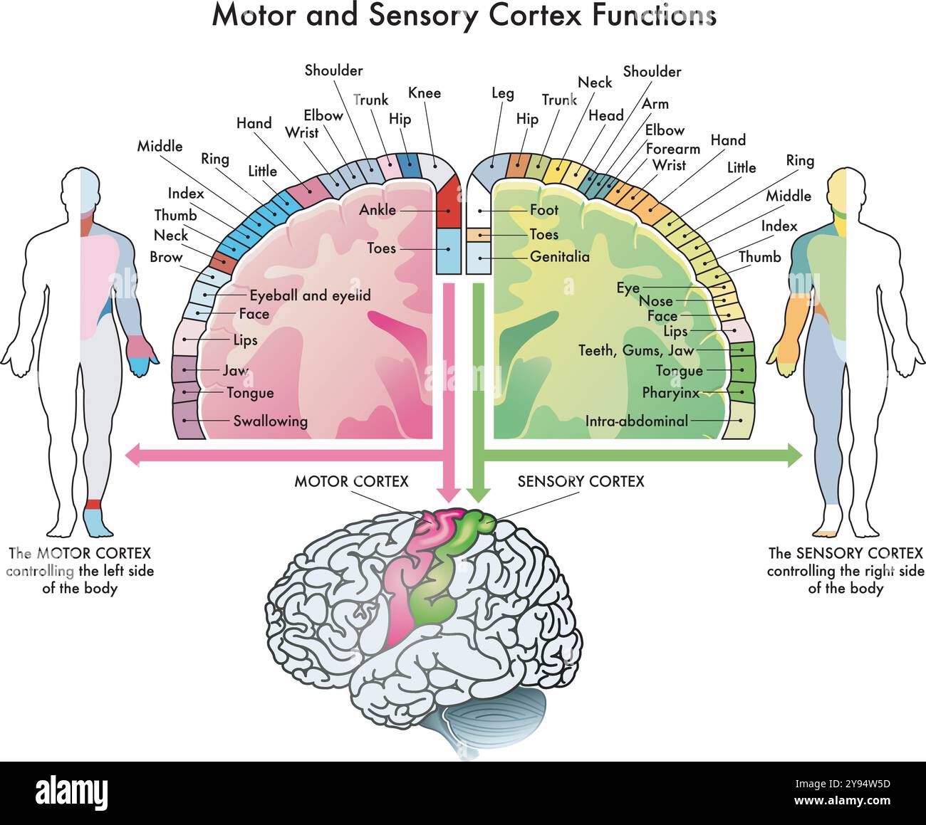 Medical diagram of the functions of the motor cortex and sensory cortex, two parts of the human brain, with annotations. Stock Vectorhttps://www.alamy.com/image-license-details/?v=1https://www.alamy.com/medical-diagram-of-the-functions-of-the-motor-cortex-and-sensory-cortex-two-parts-of-the-human-brain-with-annotations-image625212713.html
RF2Y94W5D–Medical diagram of the functions of the motor cortex and sensory cortex, two parts of the human brain, with annotations.
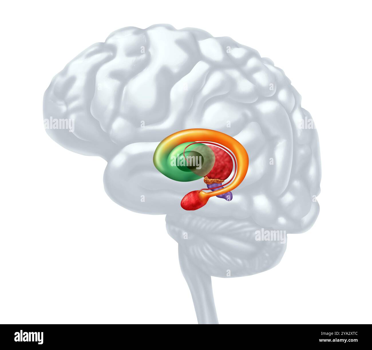 Basal Ganglia as subcortical nuclei inside the human brain controlling motor functions as cognition emotions, and learning and voluntary movements. Stock Photohttps://www.alamy.com/image-license-details/?v=1https://www.alamy.com/basal-ganglia-as-subcortical-nuclei-inside-the-human-brain-controlling-motor-functions-as-cognition-emotions-and-learning-and-voluntary-movements-image625784780.html
RF2YA2XTC–Basal Ganglia as subcortical nuclei inside the human brain controlling motor functions as cognition emotions, and learning and voluntary movements.
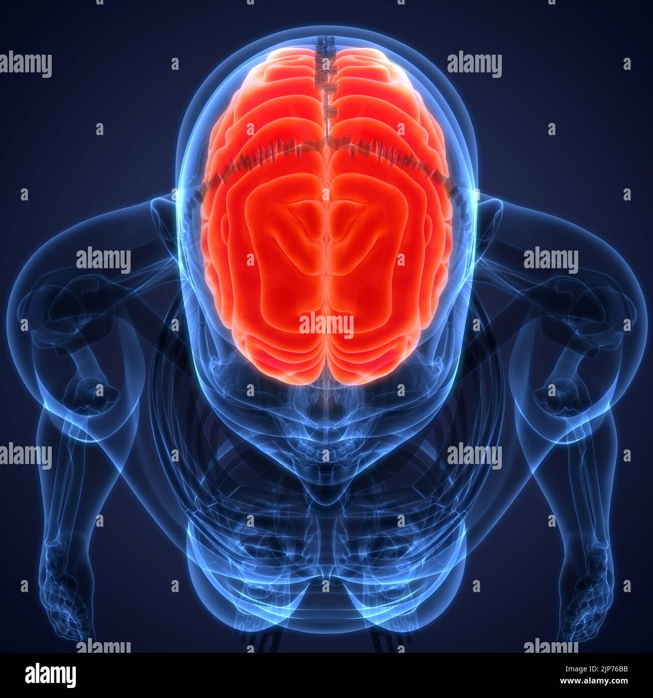 Central Organ of Human Nervous System Brain Anatomy Stock Photohttps://www.alamy.com/image-license-details/?v=1https://www.alamy.com/central-organ-of-human-nervous-system-brain-anatomy-image478361055.html
RF2JP76BB–Central Organ of Human Nervous System Brain Anatomy
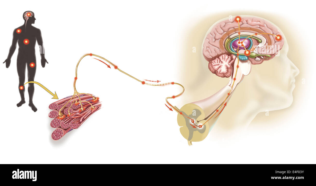 Pathway of a pain message via sensory nerve in injured muscle, to pain gate in spinal cord to limbic system, frontal cortex and Stock Photohttps://www.alamy.com/image-license-details/?v=1https://www.alamy.com/stock-photo-pathway-of-a-pain-message-via-sensory-nerve-in-injured-muscle-to-pain-71629487.html
RME4F03Y–Pathway of a pain message via sensory nerve in injured muscle, to pain gate in spinal cord to limbic system, frontal cortex and
 Human Internal Organ Brain with Nervous System Anatomy X-ray 3D rendering Stock Photohttps://www.alamy.com/image-license-details/?v=1https://www.alamy.com/human-internal-organ-brain-with-nervous-system-anatomy-x-ray-3d-rendering-image342001238.html
RF2ATBE1X–Human Internal Organ Brain with Nervous System Anatomy X-ray 3D rendering
 Unusual vector illustration depicting gyrus and divisions of the human brain. The mind and the mind of mankind. Isolated turuoise logo. Stock Vectorhttps://www.alamy.com/image-license-details/?v=1https://www.alamy.com/unusual-vector-illustration-depicting-gyrus-and-divisions-of-the-human-brain-the-mind-and-the-mind-of-mankind-isolated-turuoise-logo-image337015033.html
RF2AG8A35–Unusual vector illustration depicting gyrus and divisions of the human brain. The mind and the mind of mankind. Isolated turuoise logo.
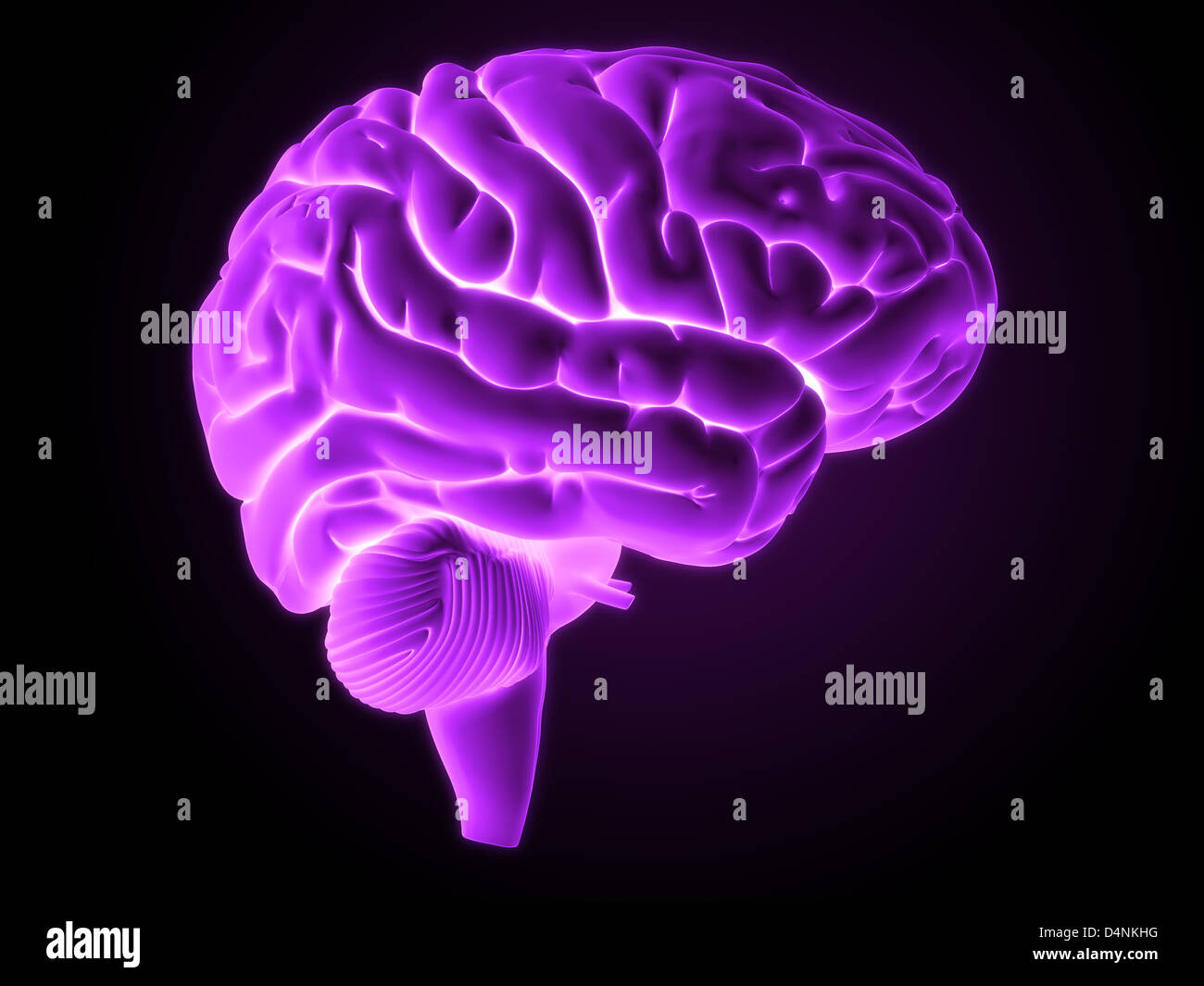 Human brain Stock Photohttps://www.alamy.com/image-license-details/?v=1https://www.alamy.com/stock-photo-human-brain-54566108.html
RFD4NKHG–Human brain
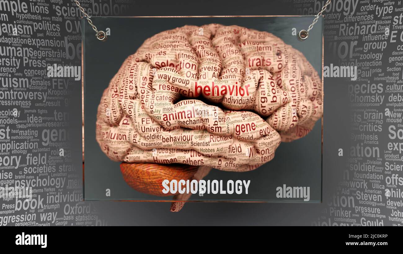 Sociobiology in human brain - dozens of important terms describing Sociobiology properties painted over the brain cortex to symbolize Sociobiology con Stock Photohttps://www.alamy.com/image-license-details/?v=1https://www.alamy.com/sociobiology-in-human-brain-dozens-of-important-terms-describing-sociobiology-properties-painted-over-the-brain-cortex-to-symbolize-sociobiology-con-image472071370.html
RF2JC0KRP–Sociobiology in human brain - dozens of important terms describing Sociobiology properties painted over the brain cortex to symbolize Sociobiology con
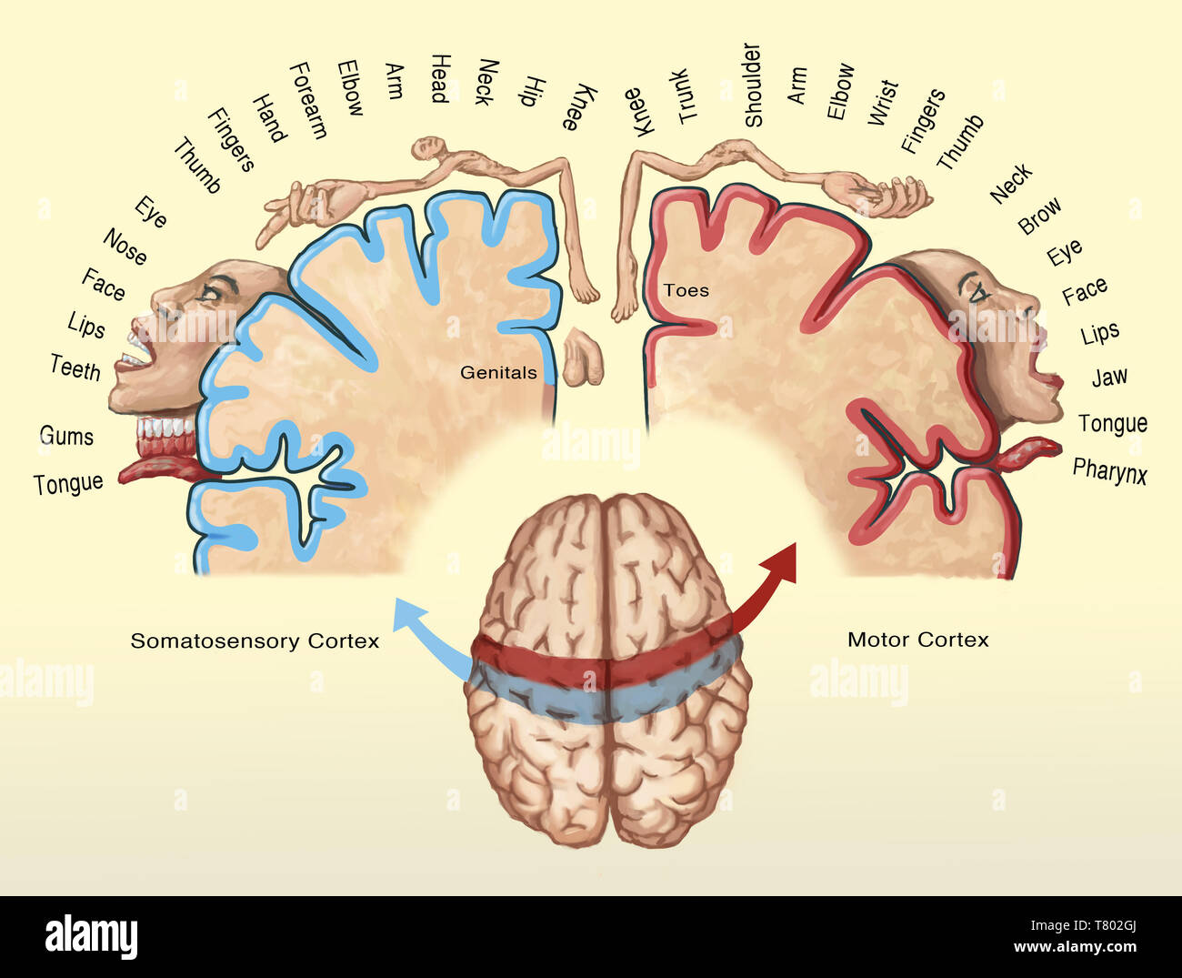 Cortical Homunculus Illustration Stock Photohttps://www.alamy.com/image-license-details/?v=1https://www.alamy.com/cortical-homunculus-illustration-image245864434.html
RMT802GJ–Cortical Homunculus Illustration
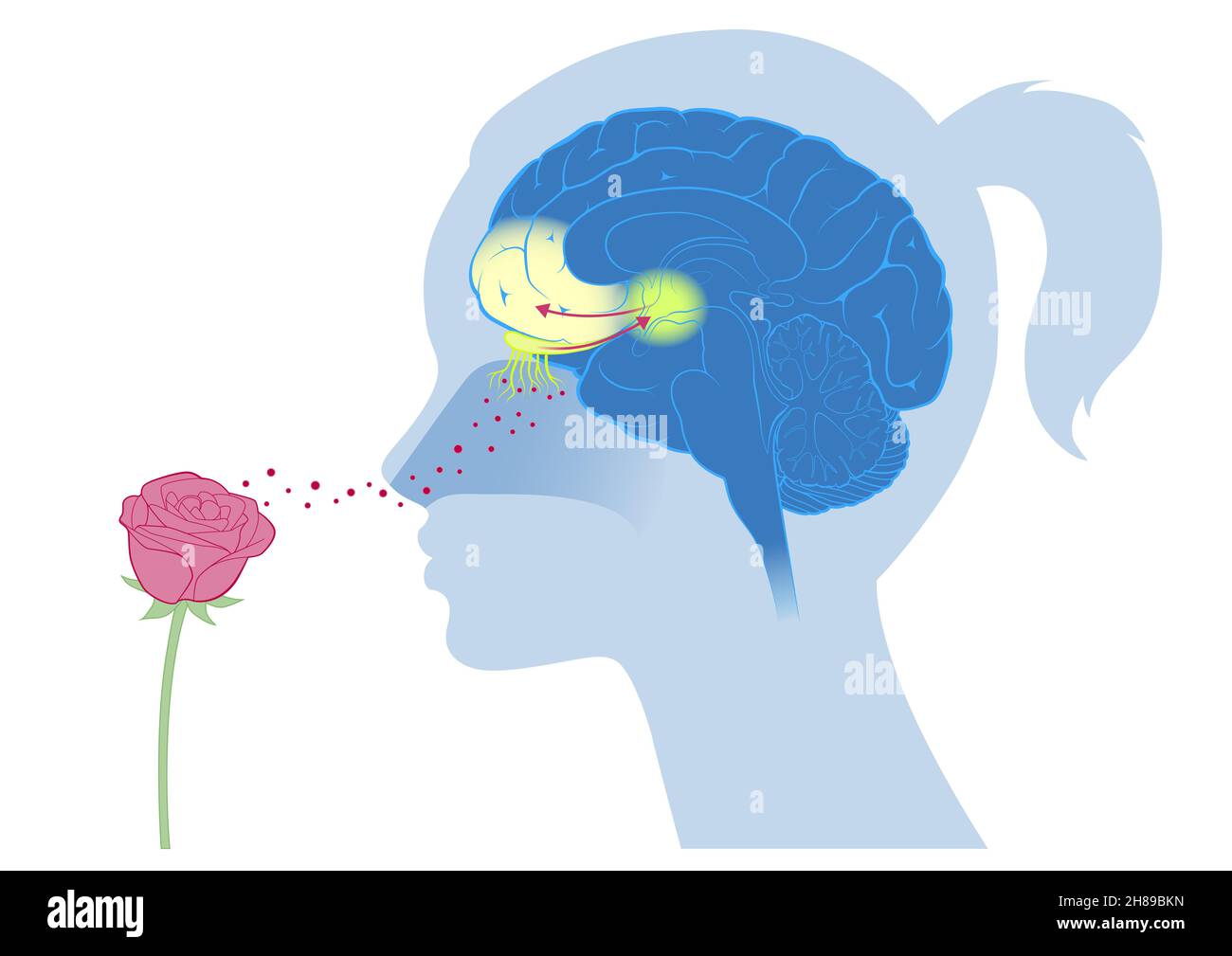 Brain cortex smell emotion Stock Photohttps://www.alamy.com/image-license-details/?v=1https://www.alamy.com/brain-cortex-smell-emotion-image452593561.html
RM2H89BKN–Brain cortex smell emotion
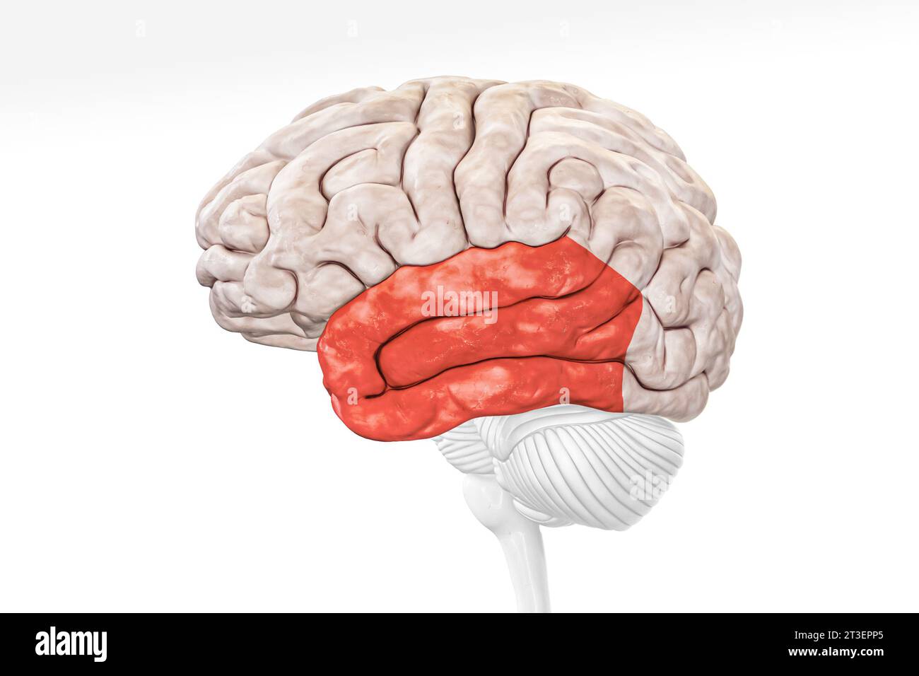 Cerebral cortex temporal lobe in red color profile view isolated on white background 3D rendering illustration. Human brain anatomy, neurology, neuros Stock Photohttps://www.alamy.com/image-license-details/?v=1https://www.alamy.com/cerebral-cortex-temporal-lobe-in-red-color-profile-view-isolated-on-white-background-3d-rendering-illustration-human-brain-anatomy-neurology-neuros-image570111309.html
RF2T3EPP5–Cerebral cortex temporal lobe in red color profile view isolated on white background 3D rendering illustration. Human brain anatomy, neurology, neuros
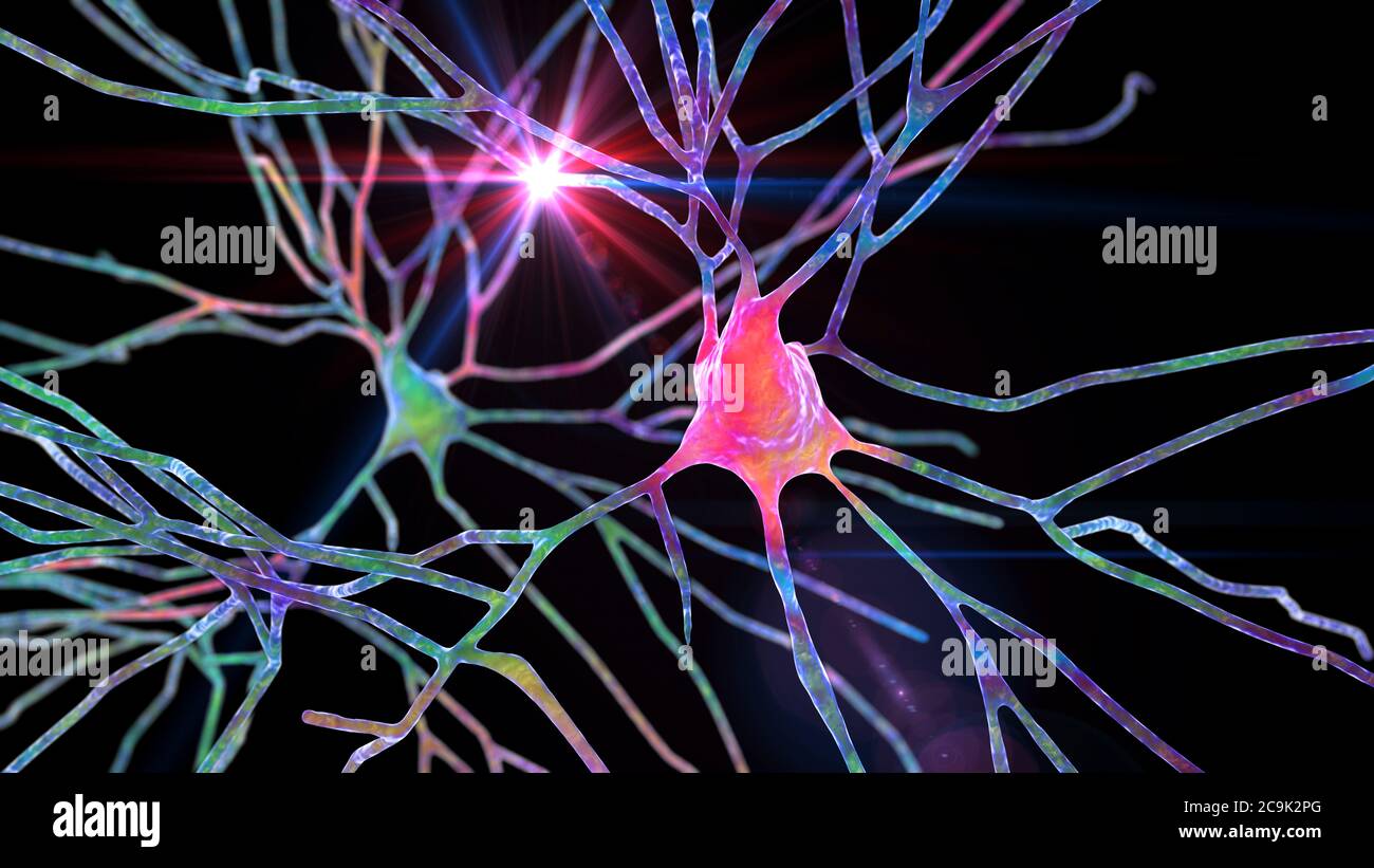 Pyramidal neurons of human brain cortex, computer illustration. Stock Photohttps://www.alamy.com/image-license-details/?v=1https://www.alamy.com/pyramidal-neurons-of-human-brain-cortex-computer-illustration-image367368920.html
RF2C9K2PG–Pyramidal neurons of human brain cortex, computer illustration.
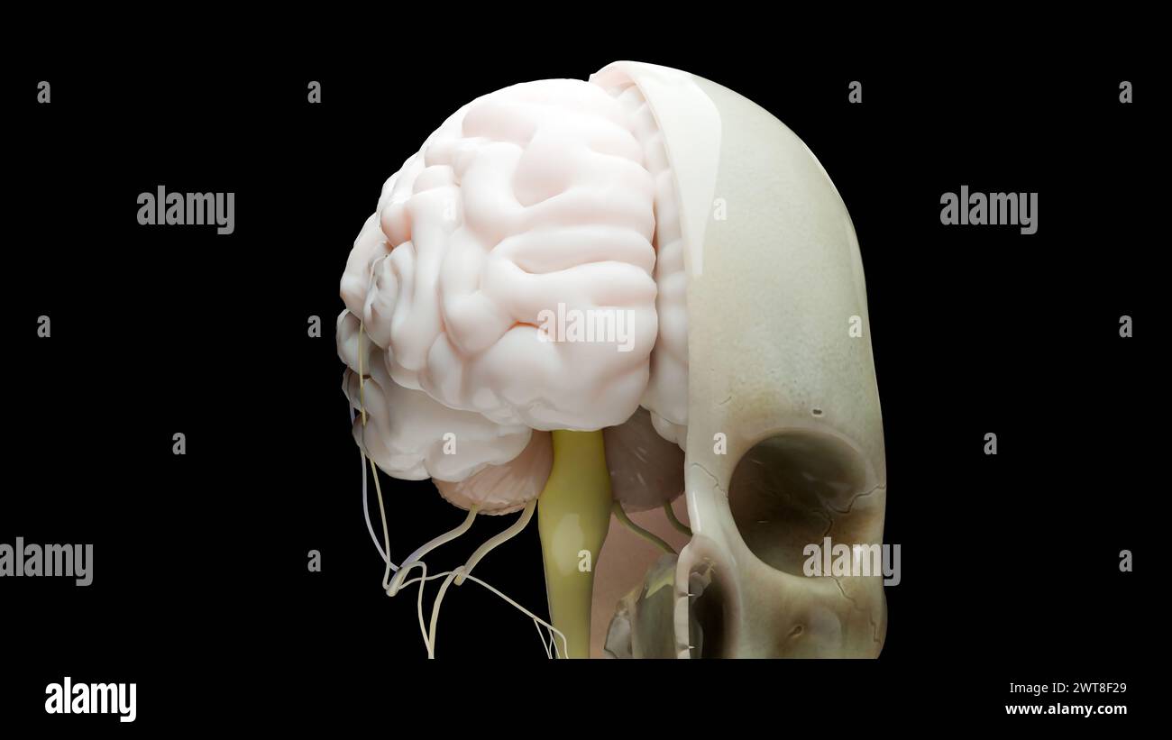 Human brain nervous system anatomy, medical diagram with parasympathetic and sympathetic nerves. medically accurate, skull cross section, Sebaceous bu Stock Photohttps://www.alamy.com/image-license-details/?v=1https://www.alamy.com/human-brain-nervous-system-anatomy-medical-diagram-with-parasympathetic-and-sympathetic-nerves-medically-accurate-skull-cross-section-sebaceous-bu-image600091697.html
RF2WT8F29–Human brain nervous system anatomy, medical diagram with parasympathetic and sympathetic nerves. medically accurate, skull cross section, Sebaceous bu
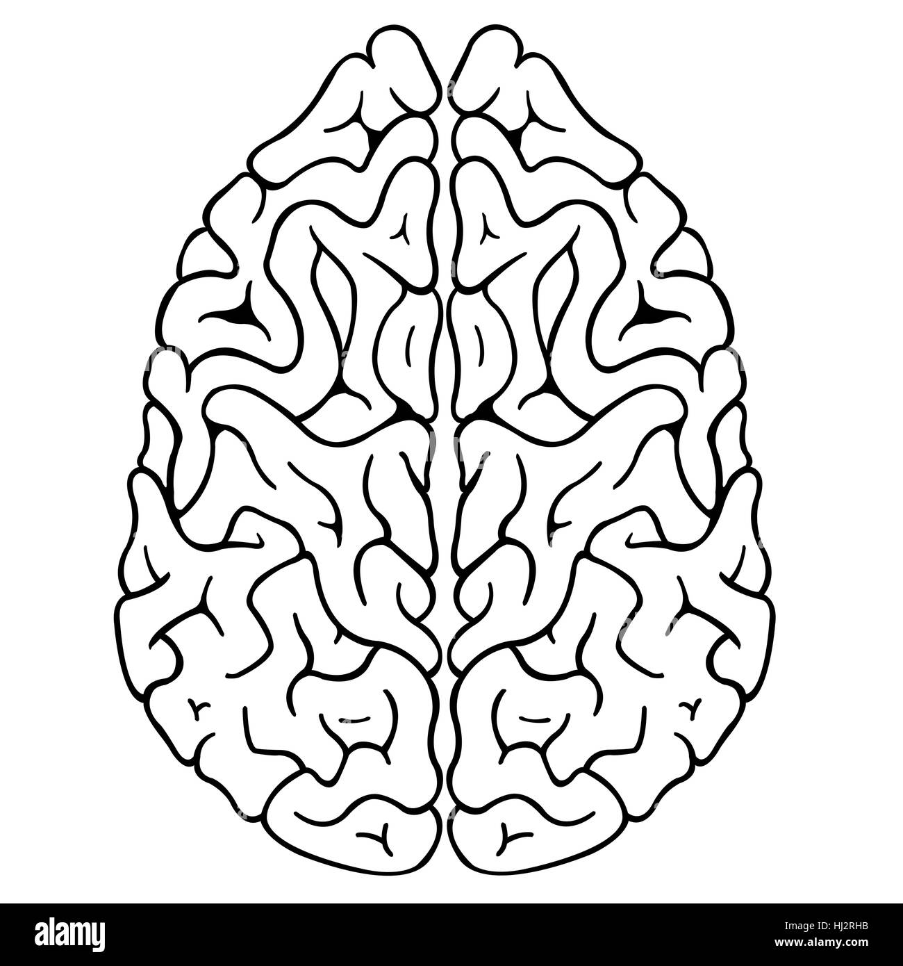 illustration of a brain isolated Stock Photohttps://www.alamy.com/image-license-details/?v=1https://www.alamy.com/stock-photo-illustration-of-a-brain-isolated-131598807.html
RFHJ2RHB–illustration of a brain isolated
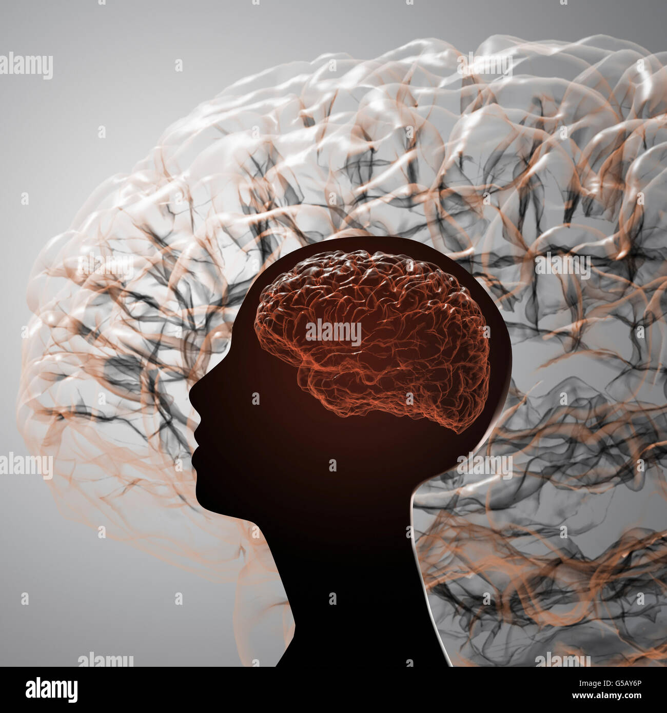 The human brain. Projection of the cerebral cortex Stock Photohttps://www.alamy.com/image-license-details/?v=1https://www.alamy.com/stock-photo-the-human-brain-projection-of-the-cerebral-cortex-106576366.html
RFG5AY6P–The human brain. Projection of the cerebral cortex
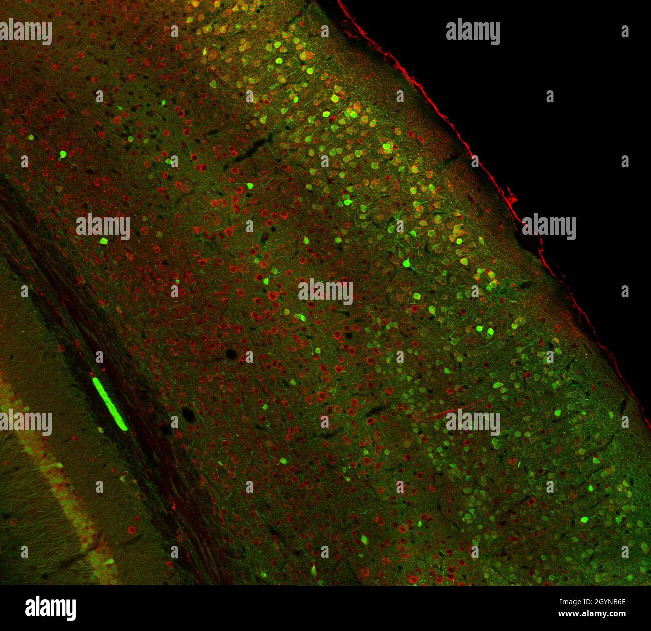 Confocal laser scanning microscopy image of immunofluorescence- labelled cells in the cerebral cortex of the mouse brain Stock Photohttps://www.alamy.com/image-license-details/?v=1https://www.alamy.com/confocal-laser-scanning-microscopy-image-of-immunofluorescence-labelled-cells-in-the-cerebral-cortex-of-the-mouse-brain-image447324710.html
RF2GYNB6E–Confocal laser scanning microscopy image of immunofluorescence- labelled cells in the cerebral cortex of the mouse brain
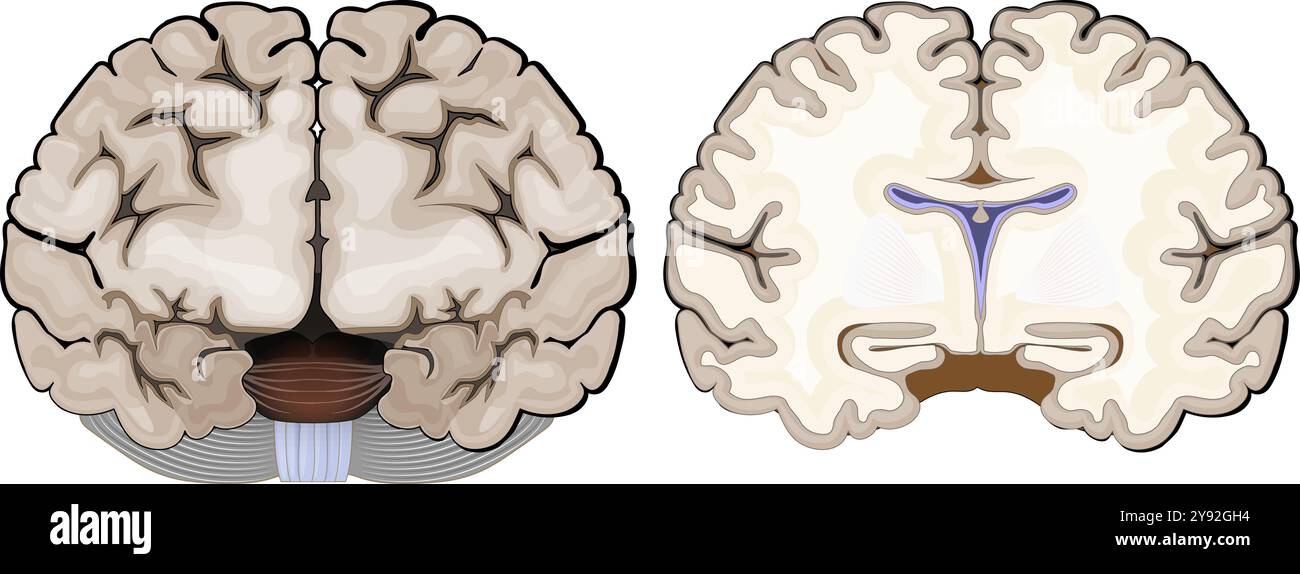 Brain anatomy. Frontal view and cross section of a human brain. Close-up of Hippocampus, and ventricles. Cerebral cortex. Vector illustration Stock Vectorhttps://www.alamy.com/image-license-details/?v=1https://www.alamy.com/brain-anatomy-frontal-view-and-cross-section-of-a-human-brain-close-up-of-hippocampus-and-ventricles-cerebral-cortex-vector-illustration-image625162080.html
RF2Y92GH4–Brain anatomy. Frontal view and cross section of a human brain. Close-up of Hippocampus, and ventricles. Cerebral cortex. Vector illustration
 3d rig line geometric line mesh blend side view art of human organ the brain Stock Vectorhttps://www.alamy.com/image-license-details/?v=1https://www.alamy.com/3d-rig-line-geometric-line-mesh-blend-side-view-art-of-human-organ-the-brain-image470996953.html
RF2JA7NBN–3d rig line geometric line mesh blend side view art of human organ the brain
 Brain shaped maze. Conceptual image of science and medicine. 3D illustration with clipping path included. Stock Photohttps://www.alamy.com/image-license-details/?v=1https://www.alamy.com/brain-shaped-maze-conceptual-image-of-science-and-medicine-3d-illustration-with-clipping-path-included-image454202296.html
RF2HAXKJG–Brain shaped maze. Conceptual image of science and medicine. 3D illustration with clipping path included.
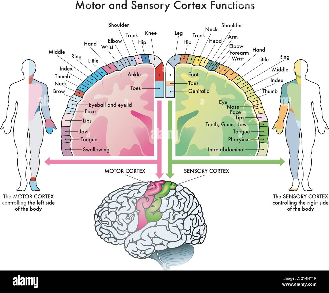 Medical diagram of the functions of the motor cortex and sensory cortex, two parts of the human brain, with annotations. Stock Vectorhttps://www.alamy.com/image-license-details/?v=1https://www.alamy.com/medical-diagram-of-the-functions-of-the-motor-cortex-and-sensory-cortex-two-parts-of-the-human-brain-with-annotations-image622514083.html
RF2Y4NY1R–Medical diagram of the functions of the motor cortex and sensory cortex, two parts of the human brain, with annotations.
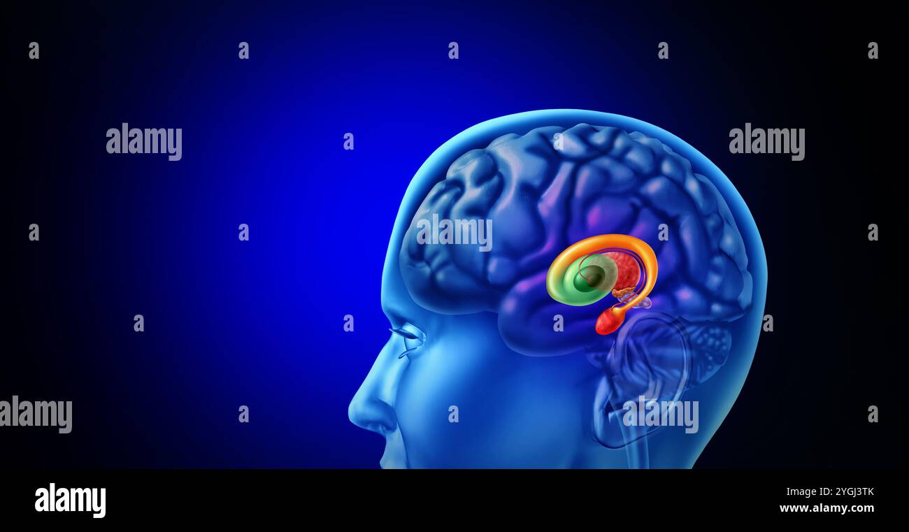 Human Basal Ganglia as subcortical nuclei inside the human brain controlling motor functions as cognition emotions, and learning and voluntary Stock Photohttps://www.alamy.com/image-license-details/?v=1https://www.alamy.com/human-basal-ganglia-as-subcortical-nuclei-inside-the-human-brain-controlling-motor-functions-as-cognition-emotions-and-learning-and-voluntary-image629805923.html
RF2YGJ3TK–Human Basal Ganglia as subcortical nuclei inside the human brain controlling motor functions as cognition emotions, and learning and voluntary
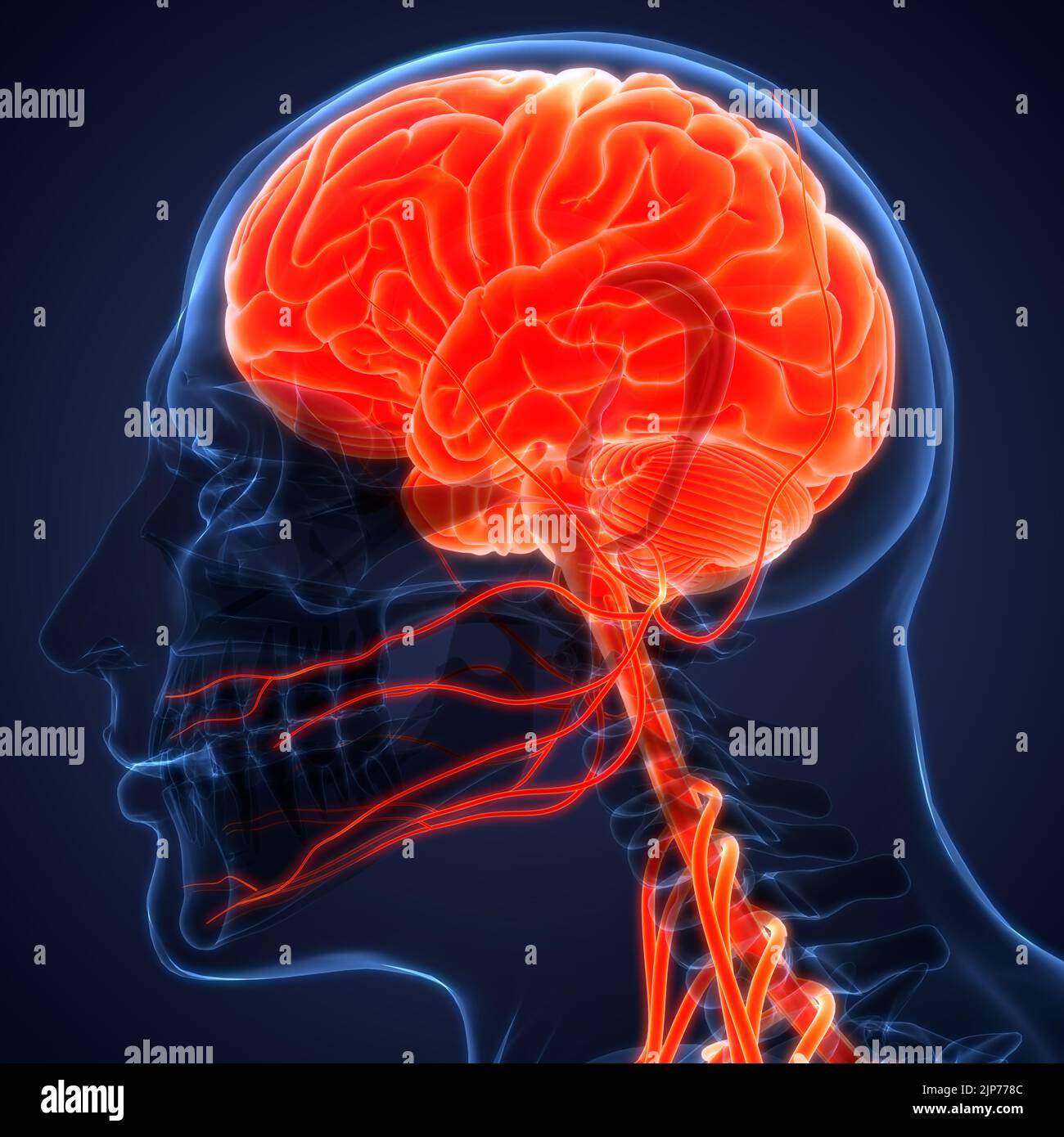 Central Organ of Human Nervous System Brain Anatomy Stock Photohttps://www.alamy.com/image-license-details/?v=1https://www.alamy.com/central-organ-of-human-nervous-system-brain-anatomy-image478361756.html
RF2JP778C–Central Organ of Human Nervous System Brain Anatomy
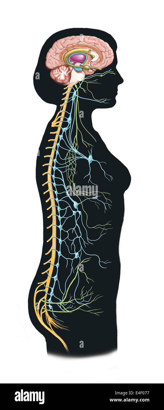 Side view of human body showing autonomic nervous system and limbic system within the brain. Green are parasympathetic nerves. B Stock Photohttps://www.alamy.com/image-license-details/?v=1https://www.alamy.com/stock-photo-side-view-of-human-body-showing-autonomic-nervous-system-and-limbic-71629579.html
RME4F077–Side view of human body showing autonomic nervous system and limbic system within the brain. Green are parasympathetic nerves. B
 Human Internal Organ Brain with Nervous System Anatomy X-ray 3D rendering Stock Photohttps://www.alamy.com/image-license-details/?v=1https://www.alamy.com/human-internal-organ-brain-with-nervous-system-anatomy-x-ray-3d-rendering-image342001363.html
RF2ATBE6B–Human Internal Organ Brain with Nervous System Anatomy X-ray 3D rendering
 Colored areas of the Brain,front view Stock Photohttps://www.alamy.com/image-license-details/?v=1https://www.alamy.com/stock-photo-colored-areas-of-the-brainfront-view-43697506.html
RFCF2GH6–Colored areas of the Brain,front view
 White brain Stock Photohttps://www.alamy.com/image-license-details/?v=1https://www.alamy.com/stock-photo-white-brain-54564978.html
RFD4NJ56–White brain
 Intelligence in human brain - dozens of important terms describing Intelligence properties painted over the brain cortex to symbolize Intelligence con Stock Photohttps://www.alamy.com/image-license-details/?v=1https://www.alamy.com/intelligence-in-human-brain-dozens-of-important-terms-describing-intelligence-properties-painted-over-the-brain-cortex-to-symbolize-intelligence-con-image472074633.html
RF2JC0T09–Intelligence in human brain - dozens of important terms describing Intelligence properties painted over the brain cortex to symbolize Intelligence con
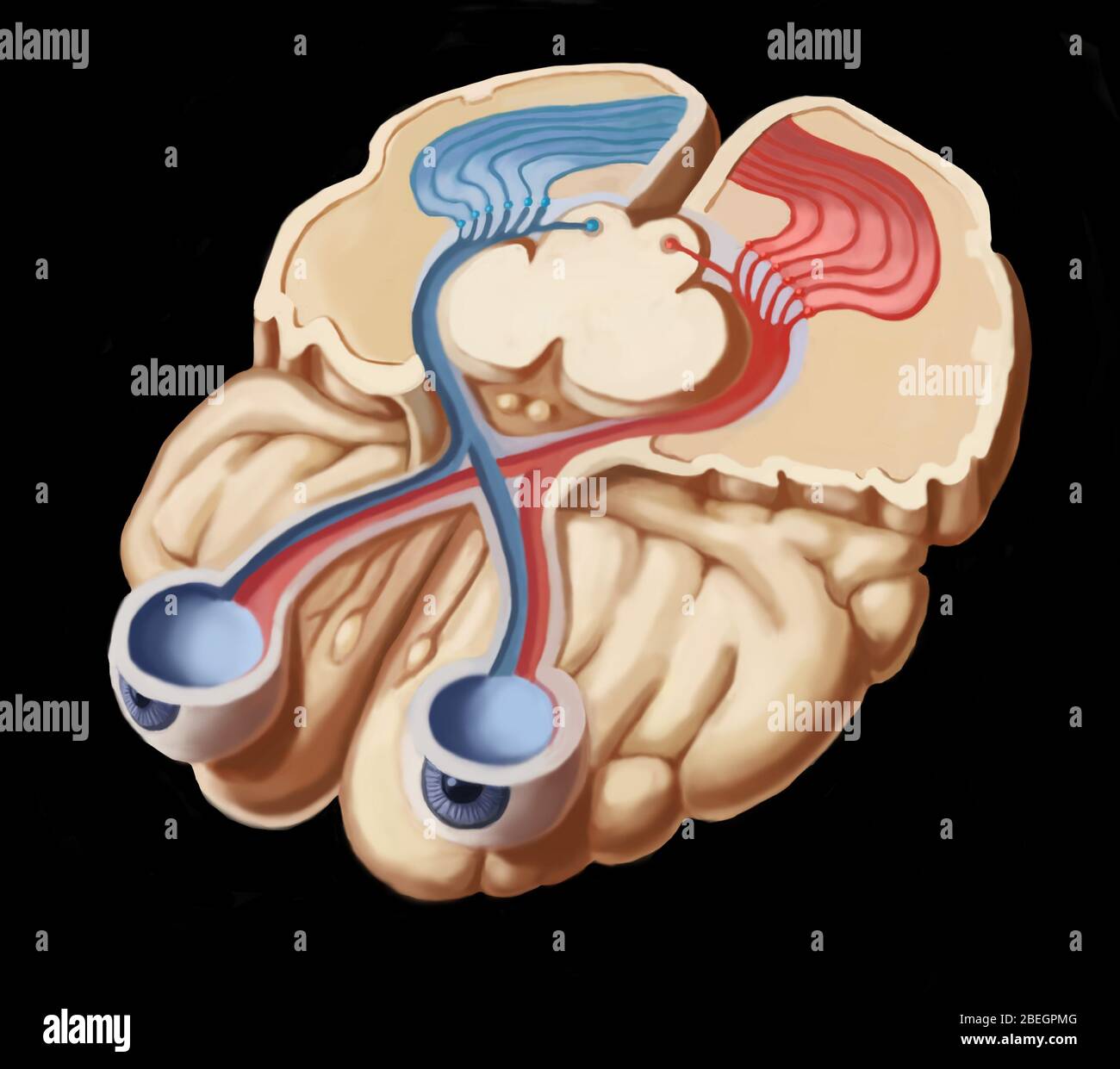 Visual Pathways Stock Photohttps://www.alamy.com/image-license-details/?v=1https://www.alamy.com/visual-pathways-image353181600.html
RM2BEGPMG–Visual Pathways
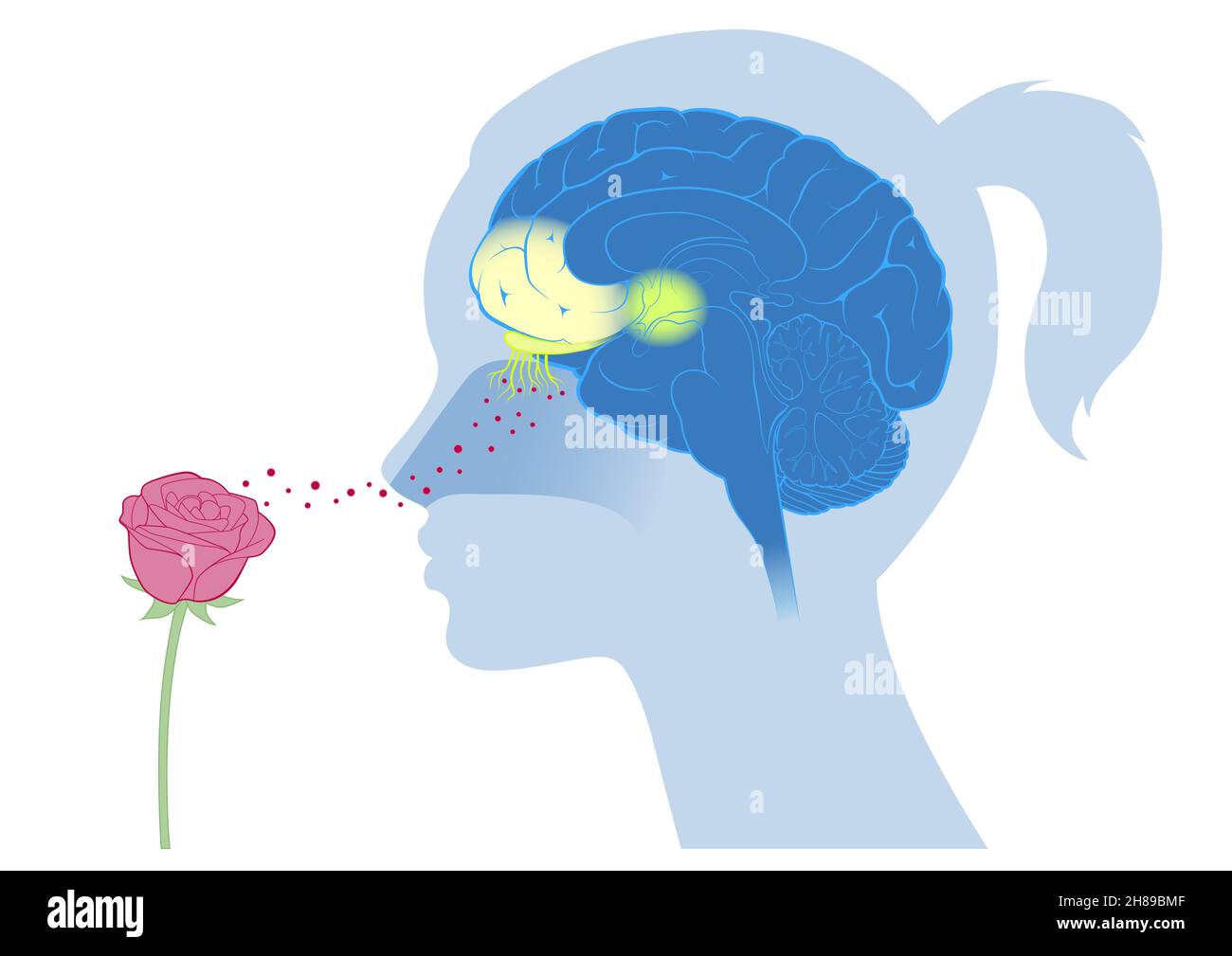 Brain cortex smell emotion Stock Photohttps://www.alamy.com/image-license-details/?v=1https://www.alamy.com/brain-cortex-smell-emotion-image452593583.html
RM2H89BMF–Brain cortex smell emotion
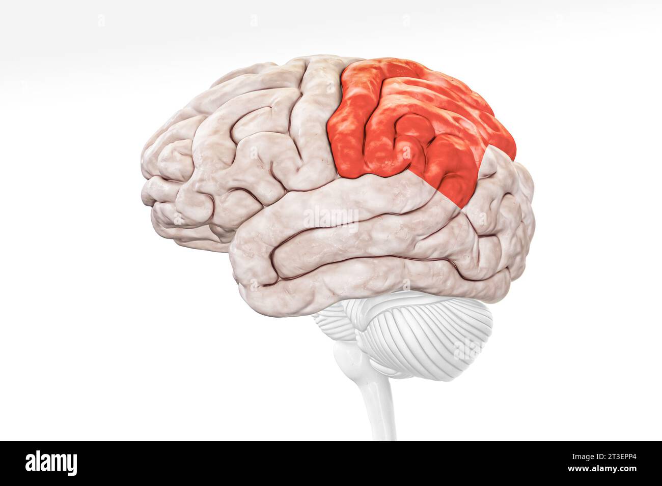 Cerebral cortex parietal lobe in red color profile view isolated on white background 3D rendering illustration. Human brain anatomy, neurology, neuros Stock Photohttps://www.alamy.com/image-license-details/?v=1https://www.alamy.com/cerebral-cortex-parietal-lobe-in-red-color-profile-view-isolated-on-white-background-3d-rendering-illustration-human-brain-anatomy-neurology-neuros-image570111308.html
RF2T3EPP4–Cerebral cortex parietal lobe in red color profile view isolated on white background 3D rendering illustration. Human brain anatomy, neurology, neuros
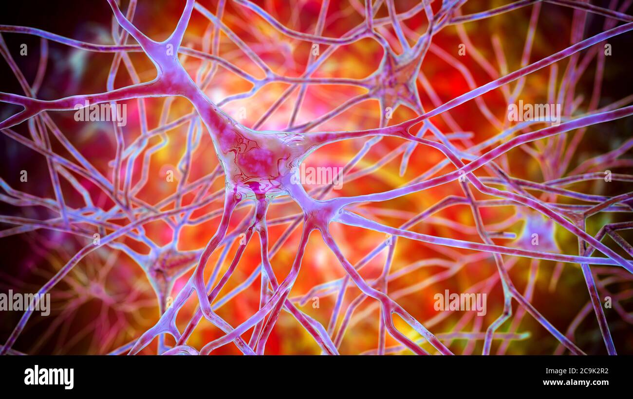 Pyramidal neurons of human brain cortex, computer illustration. Stock Photohttps://www.alamy.com/image-license-details/?v=1https://www.alamy.com/pyramidal-neurons-of-human-brain-cortex-computer-illustration-image367368934.html
RF2C9K2R2–Pyramidal neurons of human brain cortex, computer illustration.
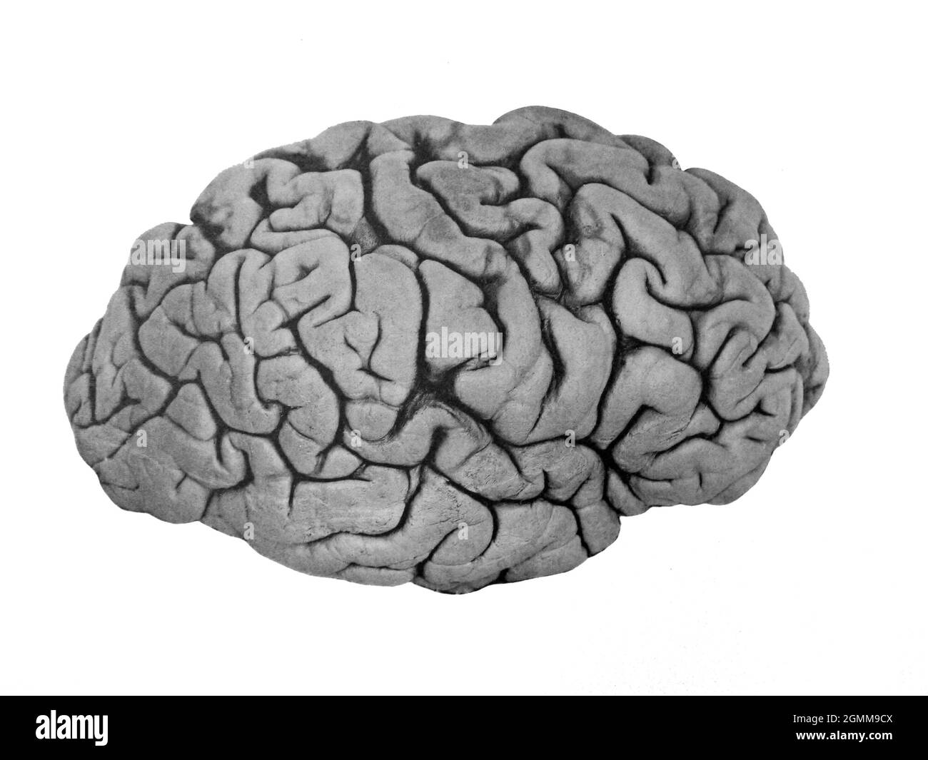 Illustration of the human brain from a medical textbook circa 1900 Stock Photohttps://www.alamy.com/image-license-details/?v=1https://www.alamy.com/illustration-of-the-human-brain-from-a-medical-textbook-circa-1900-image442998778.html
RM2GMM9CX–Illustration of the human brain from a medical textbook circa 1900
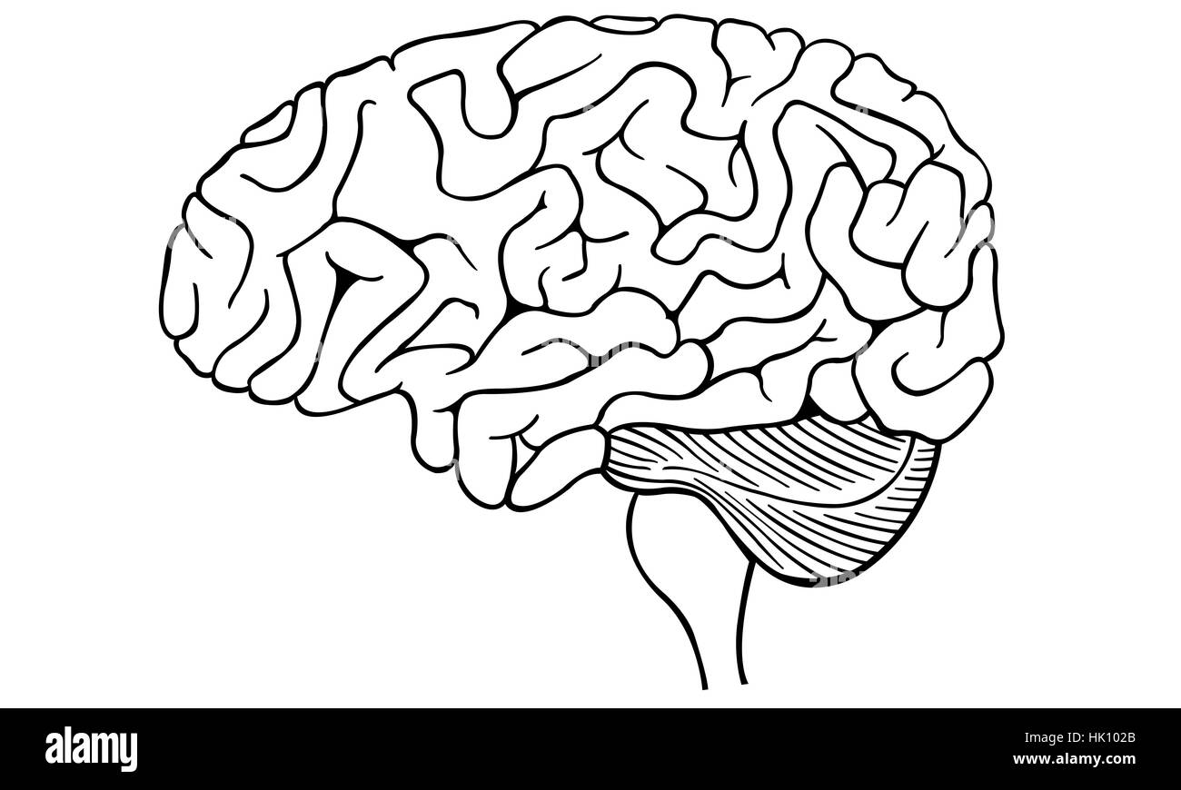 illustration of a human brain isolated Stock Photohttps://www.alamy.com/image-license-details/?v=1https://www.alamy.com/stock-photo-illustration-of-a-human-brain-isolated-132173059.html
RFHK102B–illustration of a human brain isolated
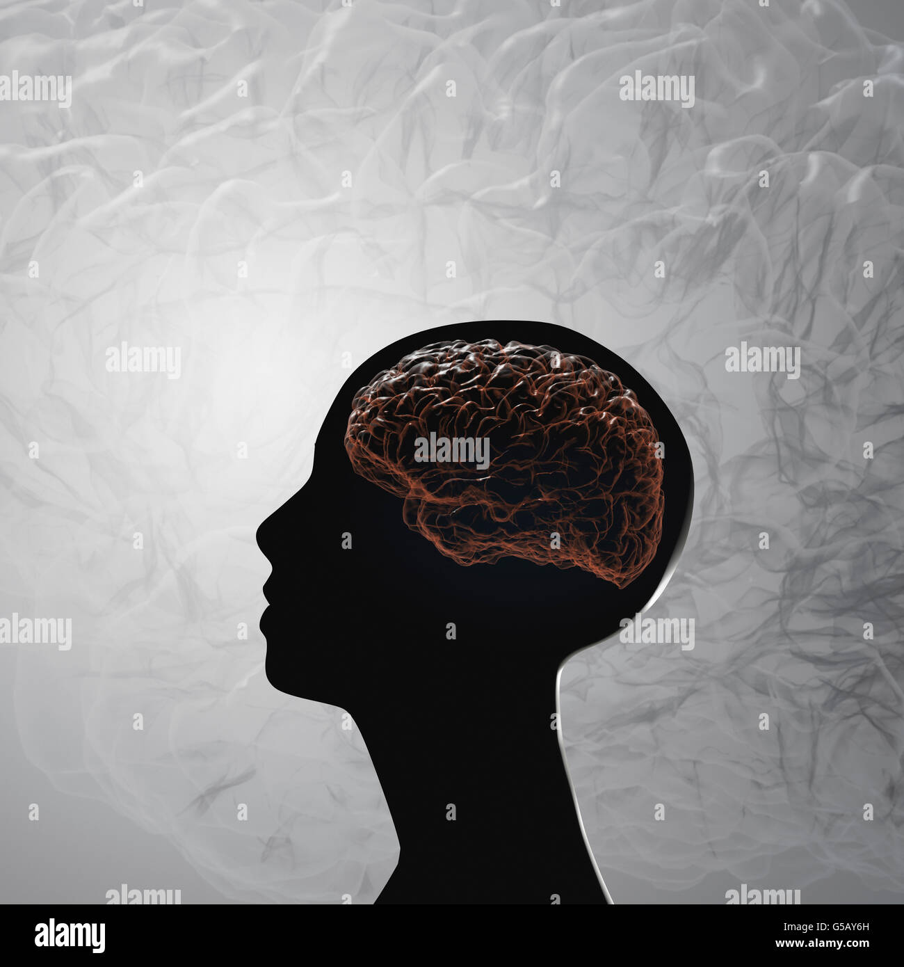 The human brain. Projection of the cerebral cortex Stock Photohttps://www.alamy.com/image-license-details/?v=1https://www.alamy.com/stock-photo-the-human-brain-projection-of-the-cerebral-cortex-106576361.html
RFG5AY6H–The human brain. Projection of the cerebral cortex
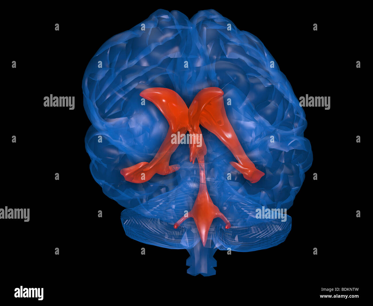 computer graphics model of the human brain showing the location of the cerebral spinal fluid ventricular system within the brain Stock Photohttps://www.alamy.com/image-license-details/?v=1https://www.alamy.com/stock-photo-computer-graphics-model-of-the-human-brain-showing-the-location-of-25635145.html
RMBDKNTW–computer graphics model of the human brain showing the location of the cerebral spinal fluid ventricular system within the brain
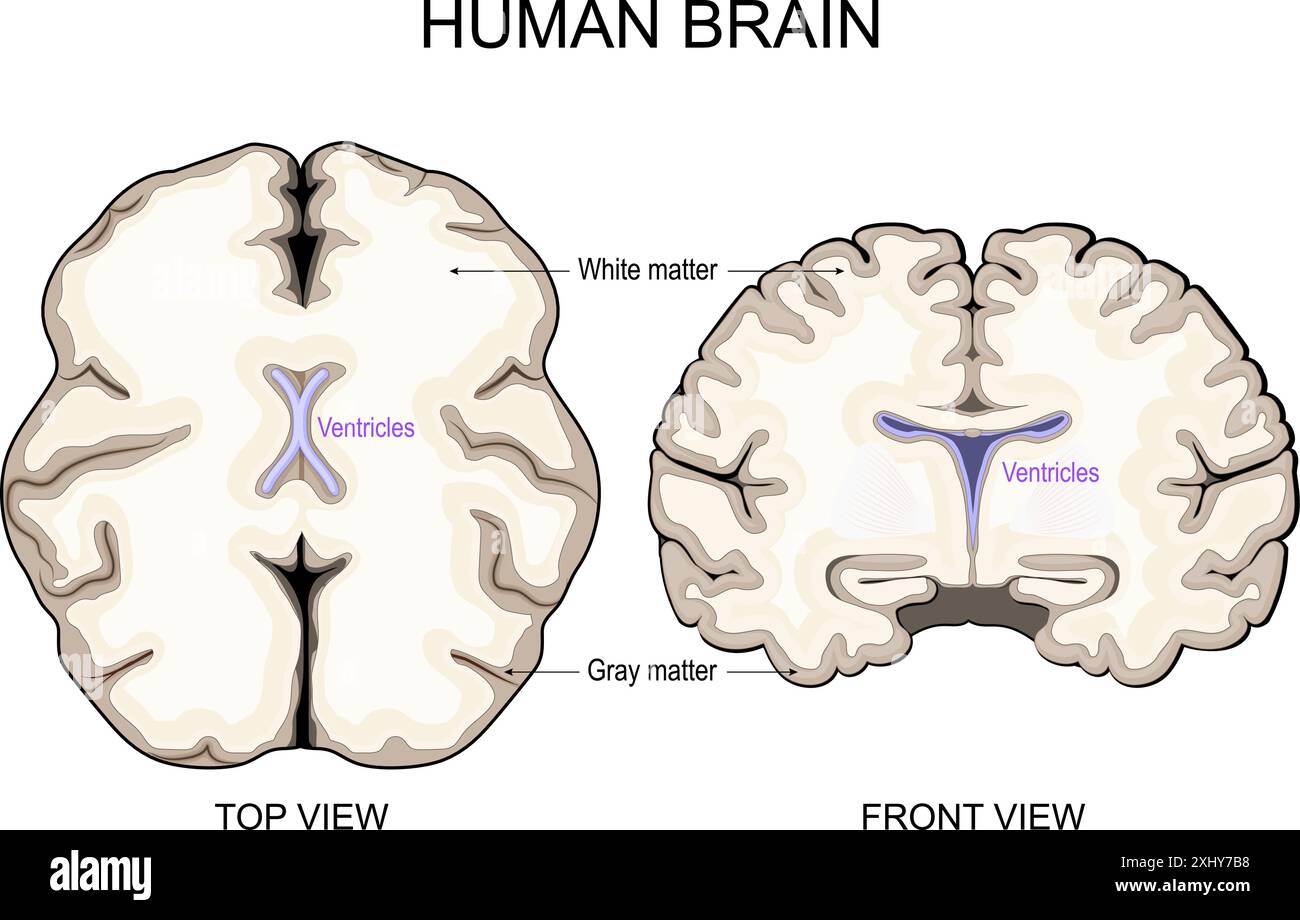 Brain anatomy. White Matter and Gray Matter. Cerebral Cortex and Brain Ventricles with Cerebrospinal Fluid. Cross section of a human brain Front view Stock Vectorhttps://www.alamy.com/image-license-details/?v=1https://www.alamy.com/brain-anatomy-white-matter-and-gray-matter-cerebral-cortex-and-brain-ventricles-with-cerebrospinal-fluid-cross-section-of-a-human-brain-front-view-image613410540.html
RF2XHY7B8–Brain anatomy. White Matter and Gray Matter. Cerebral Cortex and Brain Ventricles with Cerebrospinal Fluid. Cross section of a human brain Front view
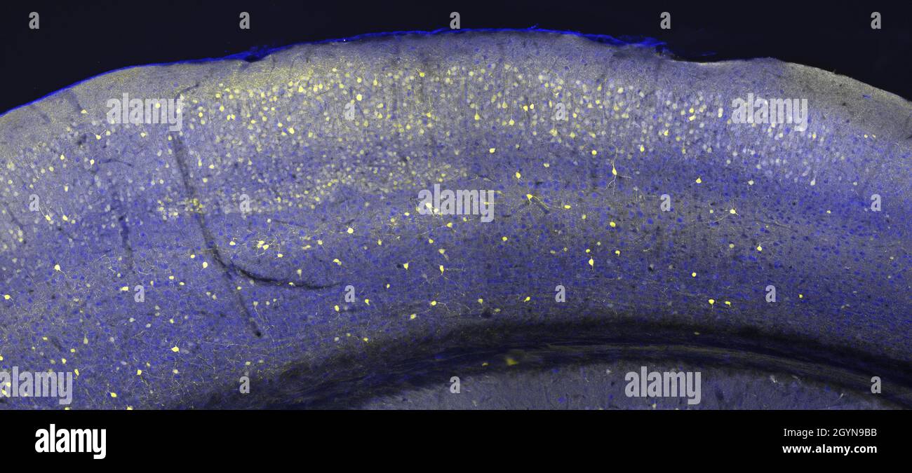 Cerebral cortex in a section of a mouse brain, labelled with immunofluorescence and recorded with confocal laser scanning microscopy Stock Photohttps://www.alamy.com/image-license-details/?v=1https://www.alamy.com/cerebral-cortex-in-a-section-of-a-mouse-brain-labelled-with-immunofluorescence-and-recorded-with-confocal-laser-scanning-microscopy-image447323279.html
RF2GYN9BB–Cerebral cortex in a section of a mouse brain, labelled with immunofluorescence and recorded with confocal laser scanning microscopy
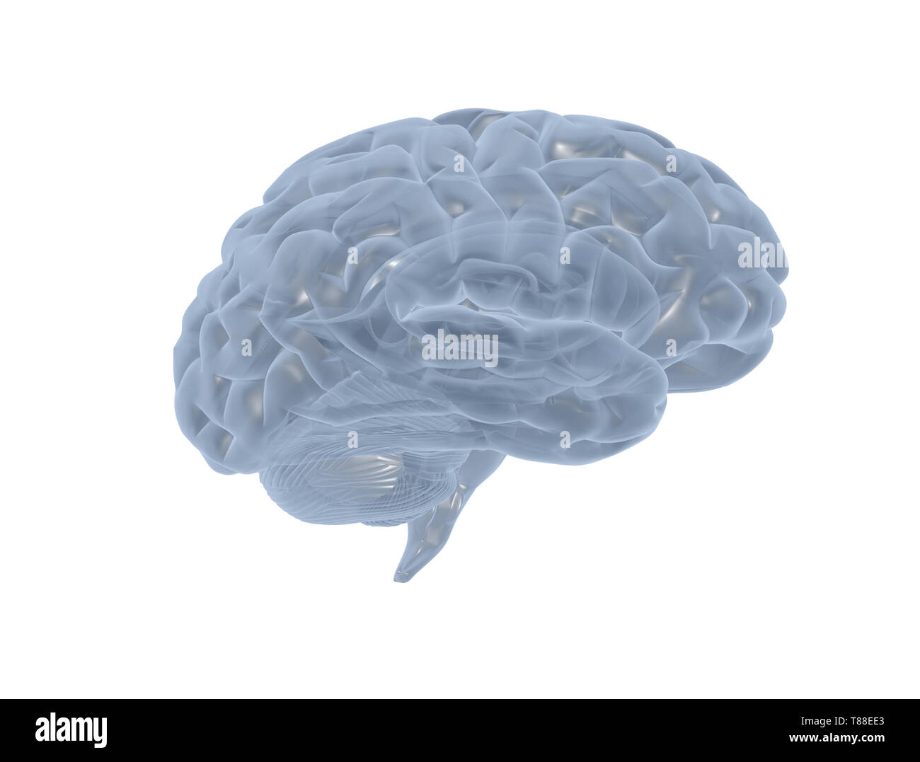 Human Brain Glass Model. Isolated on white background Stock Photohttps://www.alamy.com/image-license-details/?v=1https://www.alamy.com/human-brain-glass-model-isolated-on-white-background-image246049387.html
RFT88EE3–Human Brain Glass Model. Isolated on white background
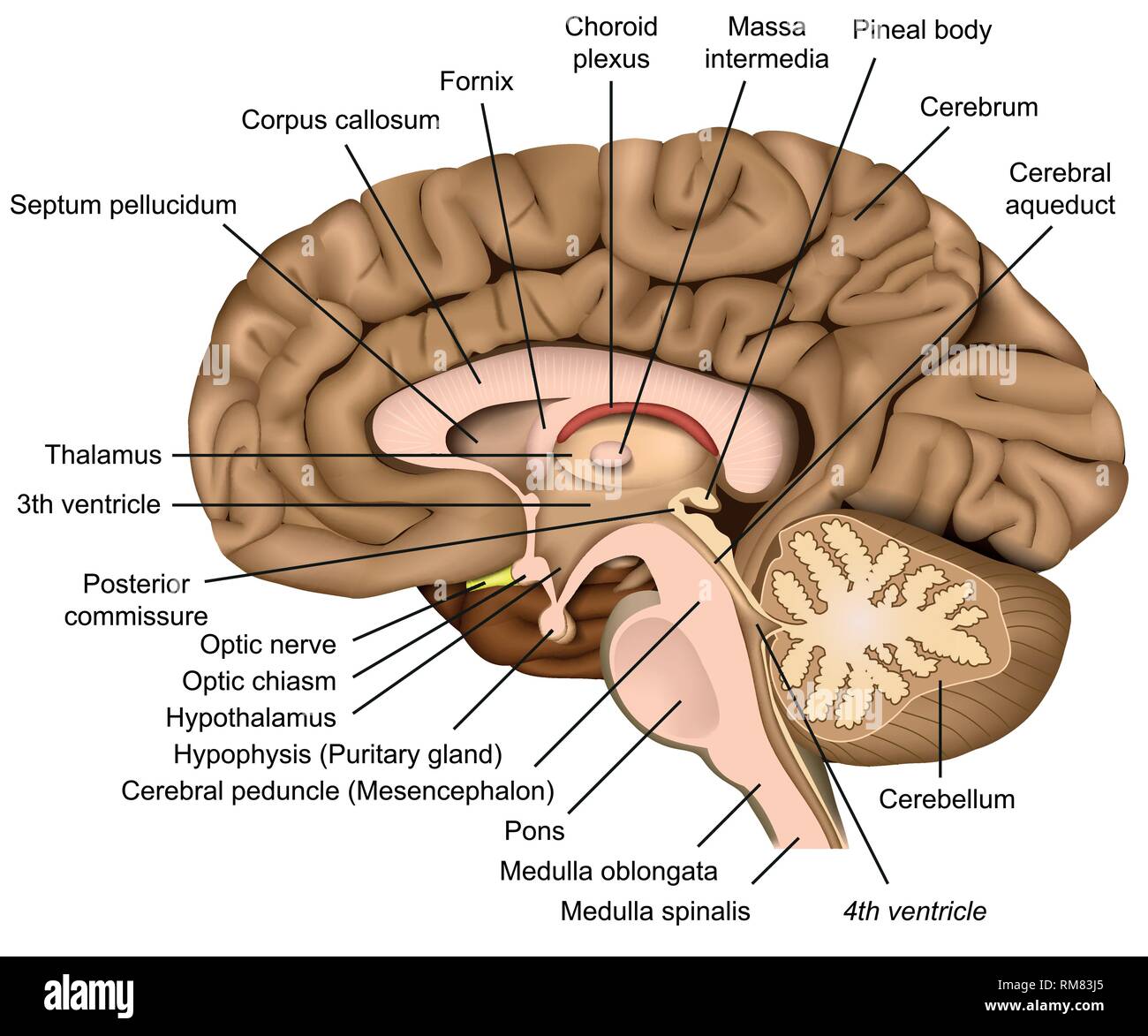 Human brain anatomy 3d vector illustration on white background Stock Vectorhttps://www.alamy.com/image-license-details/?v=1https://www.alamy.com/human-brain-anatomy-3d-vector-illustration-on-white-background-image236206381.html
RFRM83J5–Human brain anatomy 3d vector illustration on white background
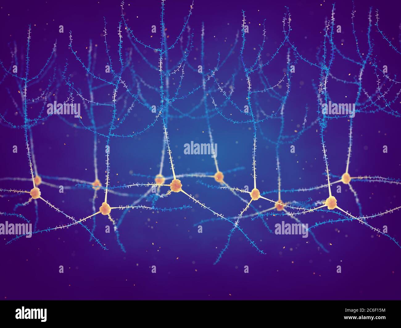 Pyramidal neurons, also known as pyramidal cells, are found in the cerebral cortex, hippocampus and the amygdala, Synaptic plasticity Stock Photohttps://www.alamy.com/image-license-details/?v=1https://www.alamy.com/pyramidal-neurons-also-known-as-pyramidal-cells-are-found-in-the-cerebral-cortex-hippocampus-and-the-amygdala-synaptic-plasticity-image365435888.html
RF2C6F15M–Pyramidal neurons, also known as pyramidal cells, are found in the cerebral cortex, hippocampus and the amygdala, Synaptic plasticity
 Central Organ of Human Nervous System Brain Anatomy Stock Photohttps://www.alamy.com/image-license-details/?v=1https://www.alamy.com/central-organ-of-human-nervous-system-brain-anatomy-image478361060.html
RF2JP76BG–Central Organ of Human Nervous System Brain Anatomy
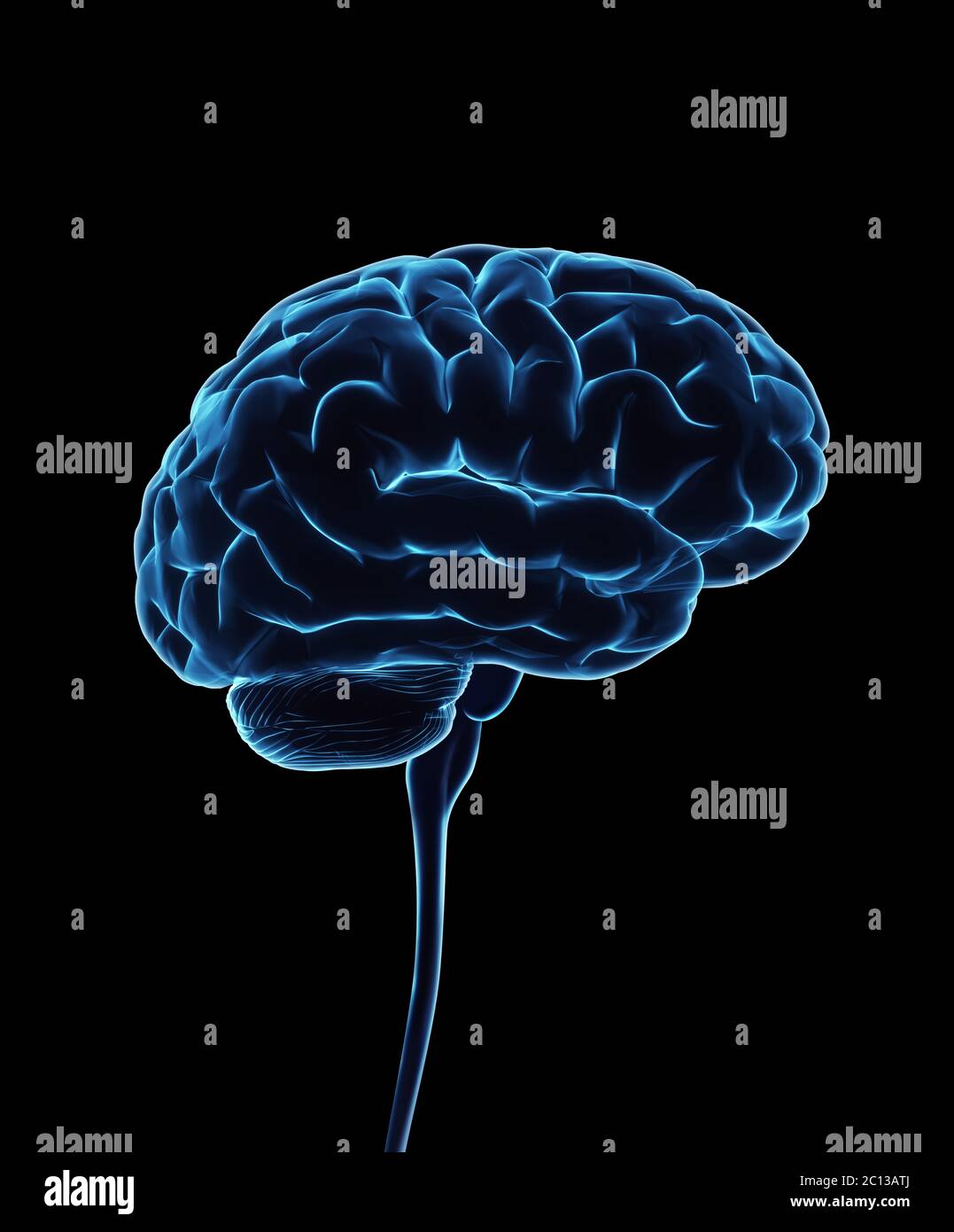 Central nervous system. Brain and spinal cord with clipping path included. Conceptual brain 3D illustration. Stock Photohttps://www.alamy.com/image-license-details/?v=1https://www.alamy.com/central-nervous-system-brain-and-spinal-cord-with-clipping-path-included-conceptual-brain-3d-illustration-image362106770.html
RF2C13ATJ–Central nervous system. Brain and spinal cord with clipping path included. Conceptual brain 3D illustration.
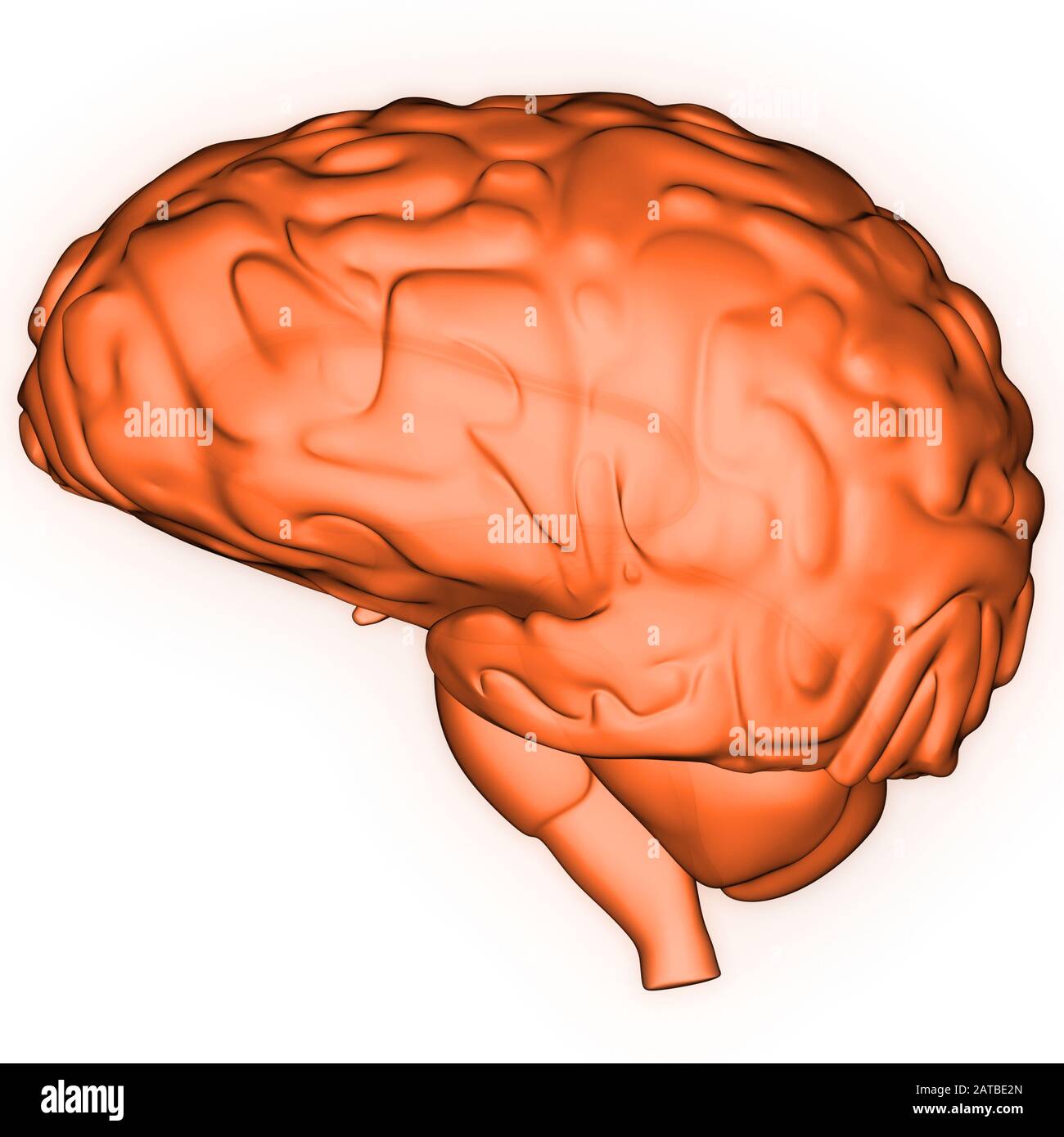 Human Internal Organ Brain with Nervous System Anatomy X-ray 3D rendering Stock Photohttps://www.alamy.com/image-license-details/?v=1https://www.alamy.com/human-internal-organ-brain-with-nervous-system-anatomy-x-ray-3d-rendering-image342001261.html
RF2ATBE2N–Human Internal Organ Brain with Nervous System Anatomy X-ray 3D rendering
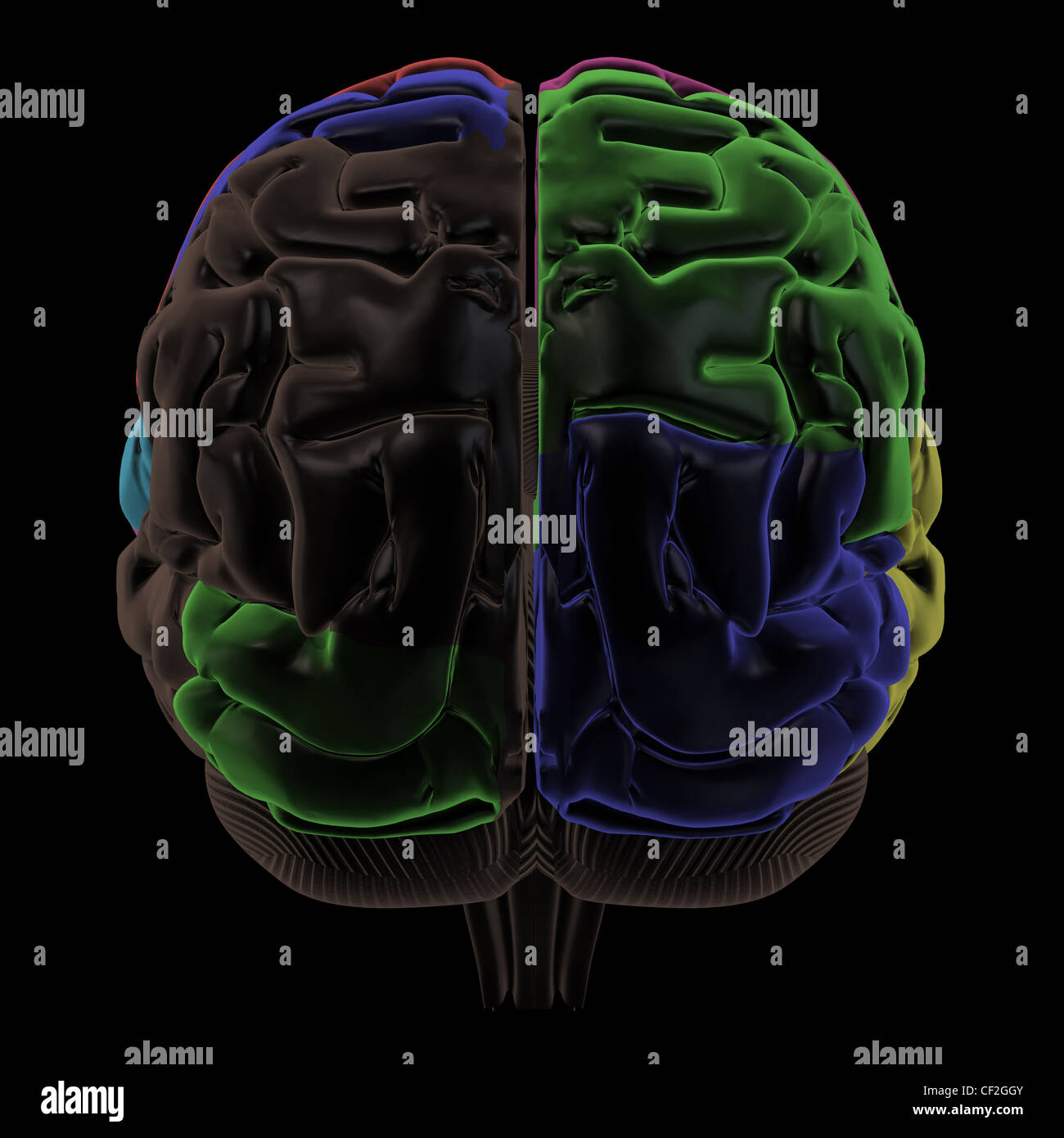 Colored areas of the Brain, back view Stock Photohttps://www.alamy.com/image-license-details/?v=1https://www.alamy.com/stock-photo-colored-areas-of-the-brain-back-view-43697499.html
RFCF2GGY–Colored areas of the Brain, back view
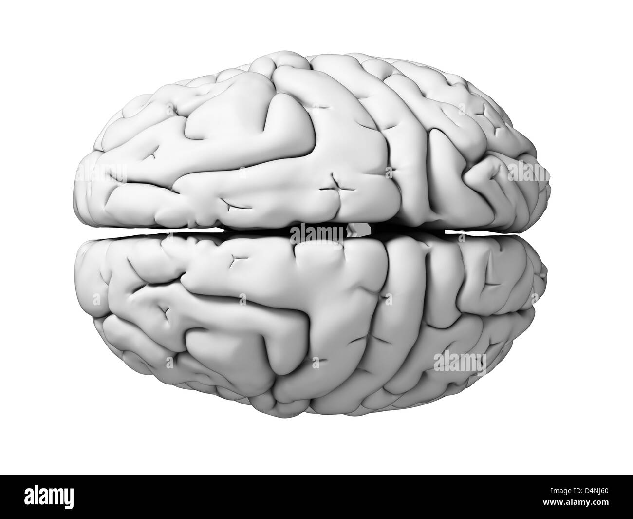 White brain Stock Photohttps://www.alamy.com/image-license-details/?v=1https://www.alamy.com/stock-photo-white-brain-54565000.html
RFD4NJ60–White brain
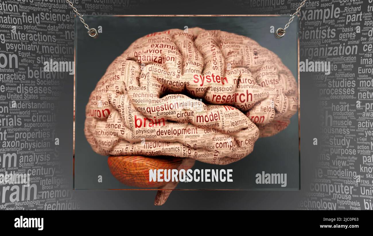 Neuroscience in human brain - dozens of important terms describing Neuroscience properties painted over the brain cortex to symbolize Neuroscience con Stock Photohttps://www.alamy.com/image-license-details/?v=1https://www.alamy.com/neuroscience-in-human-brain-dozens-of-important-terms-describing-neuroscience-properties-painted-over-the-brain-cortex-to-symbolize-neuroscience-con-image472073227.html
RF2JC0P63–Neuroscience in human brain - dozens of important terms describing Neuroscience properties painted over the brain cortex to symbolize Neuroscience con
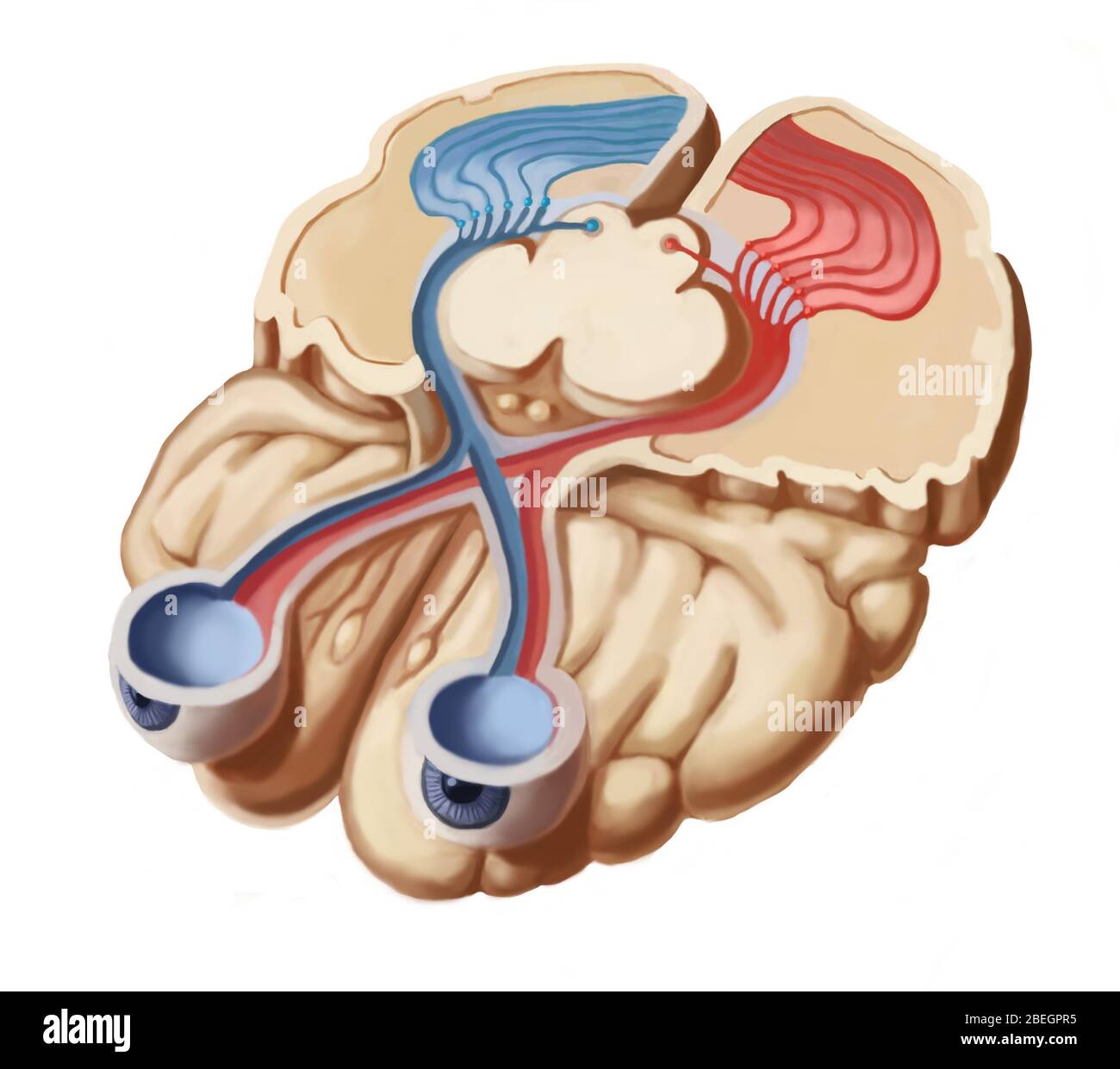 Visual Pathways Stock Photohttps://www.alamy.com/image-license-details/?v=1https://www.alamy.com/visual-pathways-image353181673.html
RM2BEGPR5–Visual Pathways
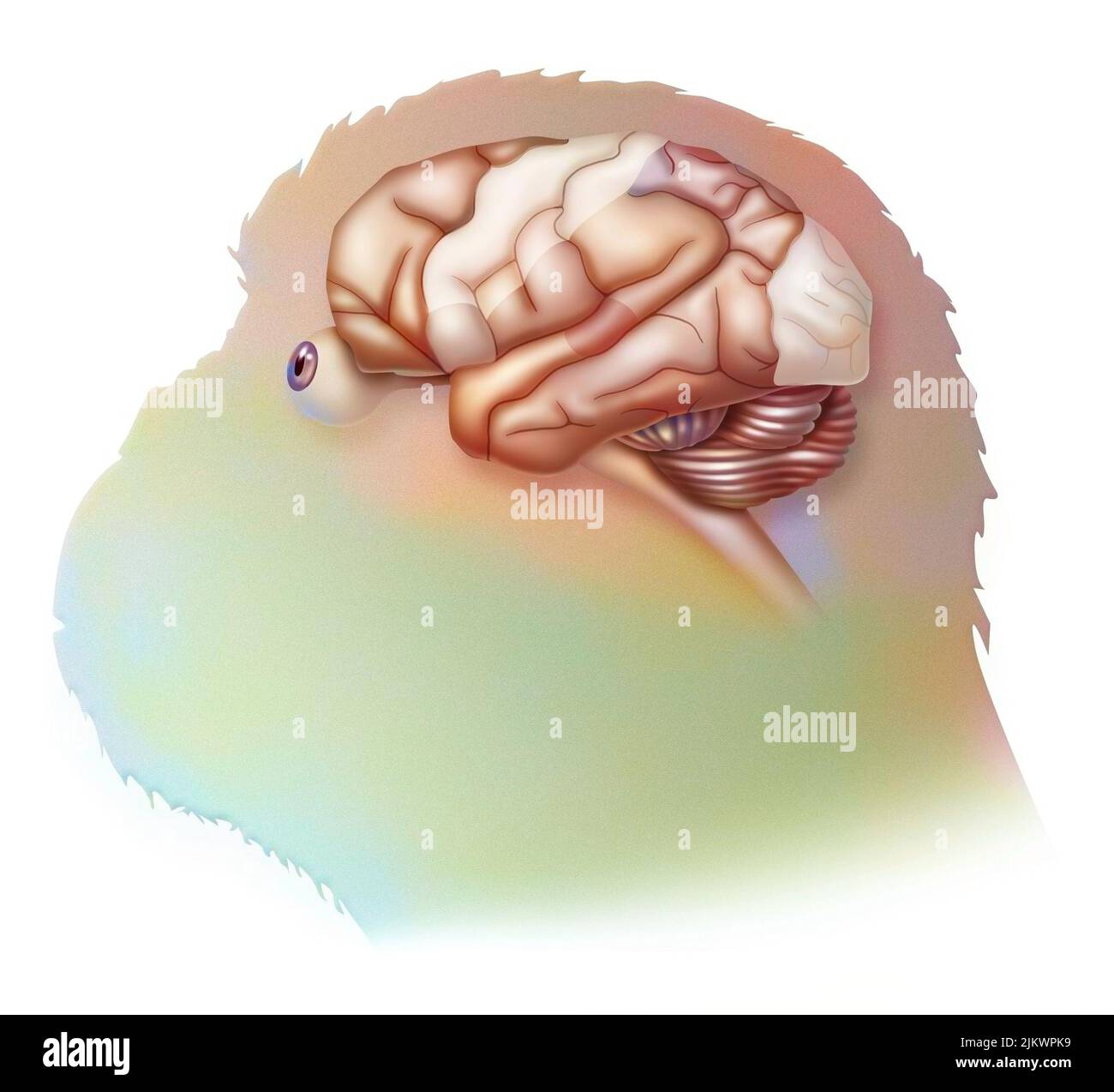 Brain in the chimpanzee with its areas (cognitive, auditory, visual) and cortex (motor, sensory). Stock Photohttps://www.alamy.com/image-license-details/?v=1https://www.alamy.com/brain-in-the-chimpanzee-with-its-areas-cognitive-auditory-visual-and-cortex-motor-sensory-image476924989.html
RF2JKWPK9–Brain in the chimpanzee with its areas (cognitive, auditory, visual) and cortex (motor, sensory).
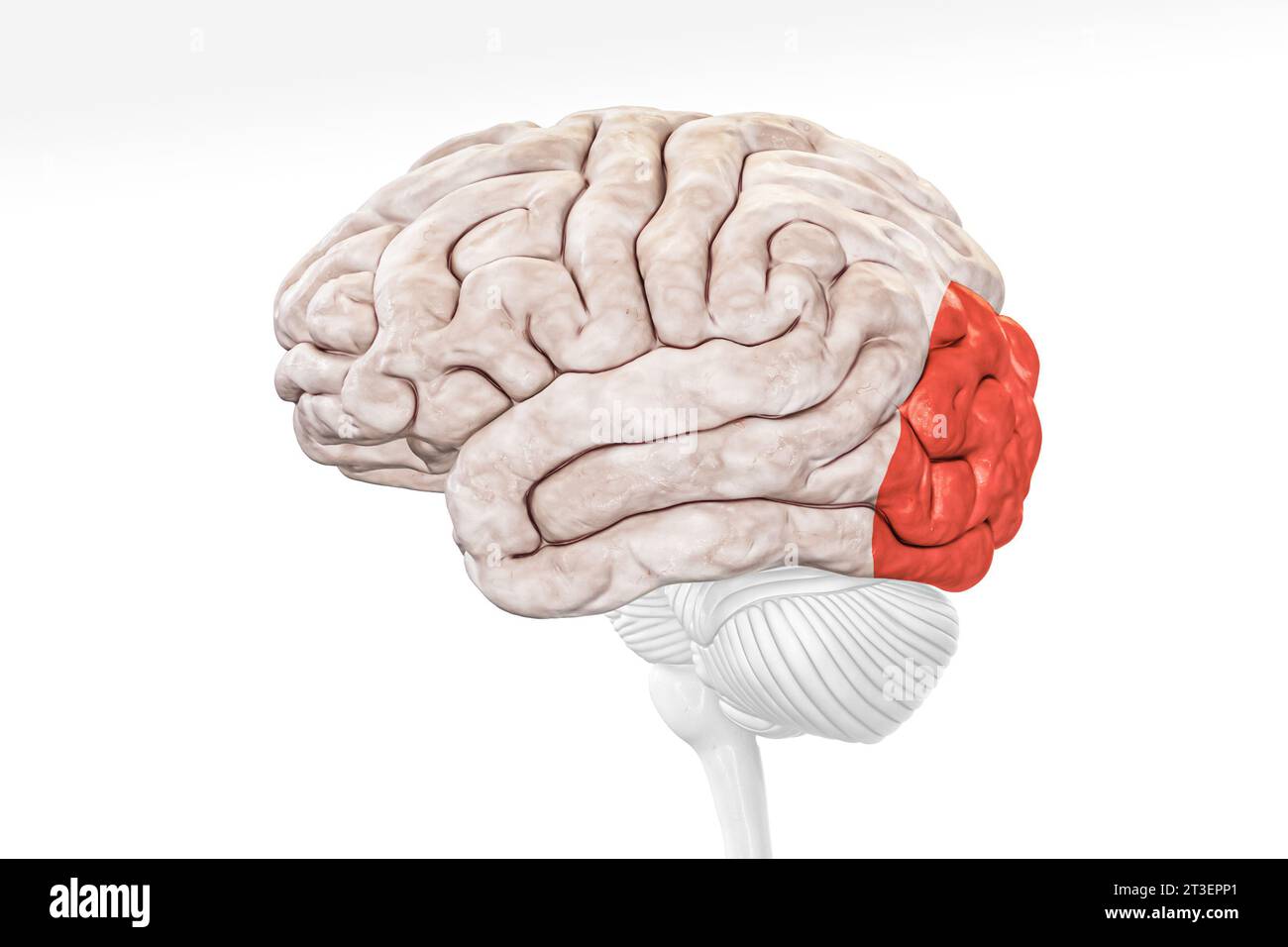 Cerebral cortex occipital lobe in red color profile view isolated on white background 3D rendering illustration. Human brain anatomy, neurology, neuro Stock Photohttps://www.alamy.com/image-license-details/?v=1https://www.alamy.com/cerebral-cortex-occipital-lobe-in-red-color-profile-view-isolated-on-white-background-3d-rendering-illustration-human-brain-anatomy-neurology-neuro-image570111305.html
RF2T3EPP1–Cerebral cortex occipital lobe in red color profile view isolated on white background 3D rendering illustration. Human brain anatomy, neurology, neuro
 Pyramidal neurons of human brain cortex, computer illustration. Stock Photohttps://www.alamy.com/image-license-details/?v=1https://www.alamy.com/pyramidal-neurons-of-human-brain-cortex-computer-illustration-image367368914.html
RF2C9K2PA–Pyramidal neurons of human brain cortex, computer illustration.
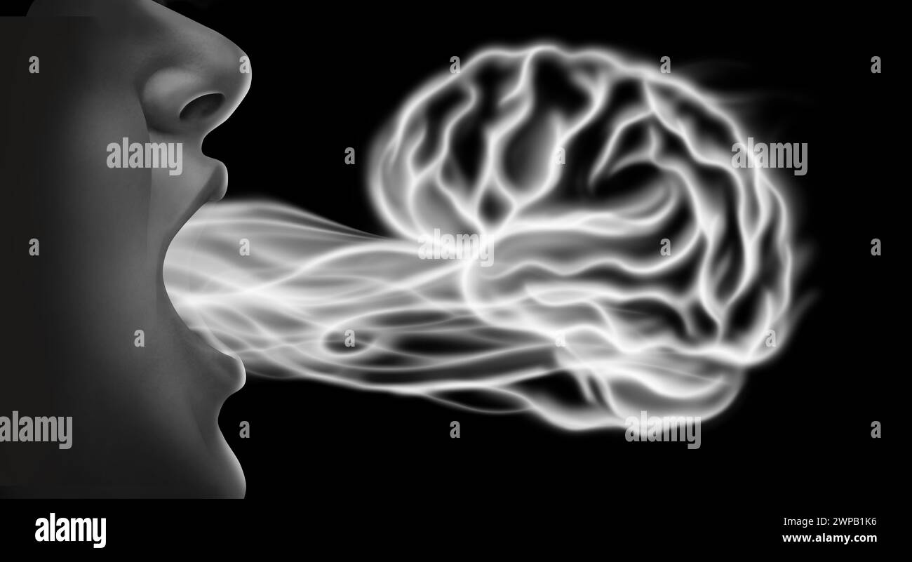 Vaping and brain health and related nicotine addiction disease risk as a person exhaling steam smoke or vapor shaped as a human mind from an electroni Stock Photohttps://www.alamy.com/image-license-details/?v=1https://www.alamy.com/vaping-and-brain-health-and-related-nicotine-addiction-disease-risk-as-a-person-exhaling-steam-smoke-or-vapor-shaped-as-a-human-mind-from-an-electroni-image598917738.html
RF2WPB1K6–Vaping and brain health and related nicotine addiction disease risk as a person exhaling steam smoke or vapor shaped as a human mind from an electroni
 Brain illustration Stock Photohttps://www.alamy.com/image-license-details/?v=1https://www.alamy.com/brain-illustration-image508202185.html
RM2MEPH21–Brain illustration
 Brain. Stock Photohttps://www.alamy.com/image-license-details/?v=1https://www.alamy.com/stock-photo-brain-122268364.html
RFH2WPF8–Brain.
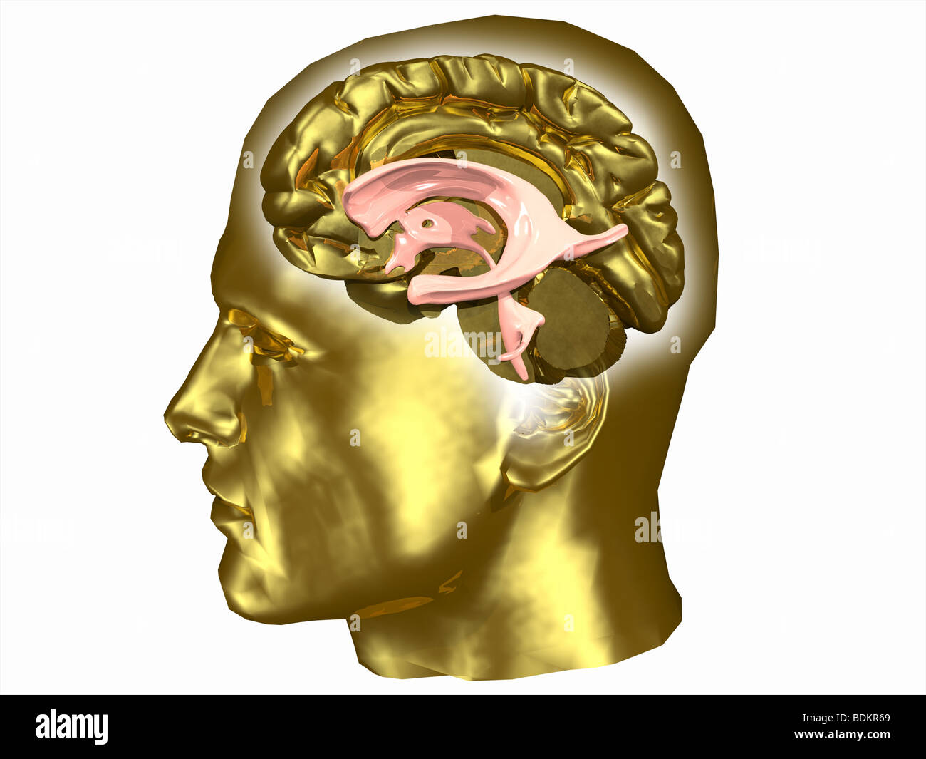 Brain and ventricular system within a human head Stock Photohttps://www.alamy.com/image-license-details/?v=1https://www.alamy.com/stock-photo-brain-and-ventricular-system-within-a-human-head-25636193.html
RMBDKR69–Brain and ventricular system within a human head
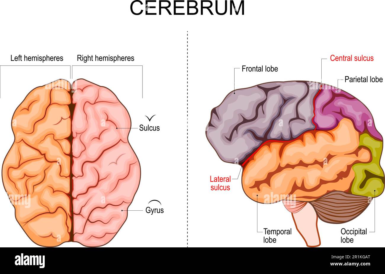 Human brain structure. Hemispheres and lobes of the cerebral cortex. frontal, temporal, occipital, and parietal lobes. lateral and superior view Stock Vectorhttps://www.alamy.com/image-license-details/?v=1https://www.alamy.com/human-brain-structure-hemispheres-and-lobes-of-the-cerebral-cortex-frontal-temporal-occipital-and-parietal-lobes-lateral-and-superior-view-image551776368.html
RF2R1KGAT–Human brain structure. Hemispheres and lobes of the cerebral cortex. frontal, temporal, occipital, and parietal lobes. lateral and superior view
 Cerebral cortex and part of the hippocampus under it in a section of a mouse brain, labelled with immunofluorescence and recorded with confocal laser Stock Photohttps://www.alamy.com/image-license-details/?v=1https://www.alamy.com/cerebral-cortex-and-part-of-the-hippocampus-under-it-in-a-section-of-a-mouse-brain-labelled-with-immunofluorescence-and-recorded-with-confocal-laser-image447323708.html
RF2GYN9XM–Cerebral cortex and part of the hippocampus under it in a section of a mouse brain, labelled with immunofluorescence and recorded with confocal laser
 Vintage anatomy print showing a simplified view of the human brain from above. Stock Photohttps://www.alamy.com/image-license-details/?v=1https://www.alamy.com/vintage-anatomy-print-showing-a-simplified-view-of-the-human-brain-from-above-image327695044.html
RF2A13PAC–Vintage anatomy print showing a simplified view of the human brain from above.
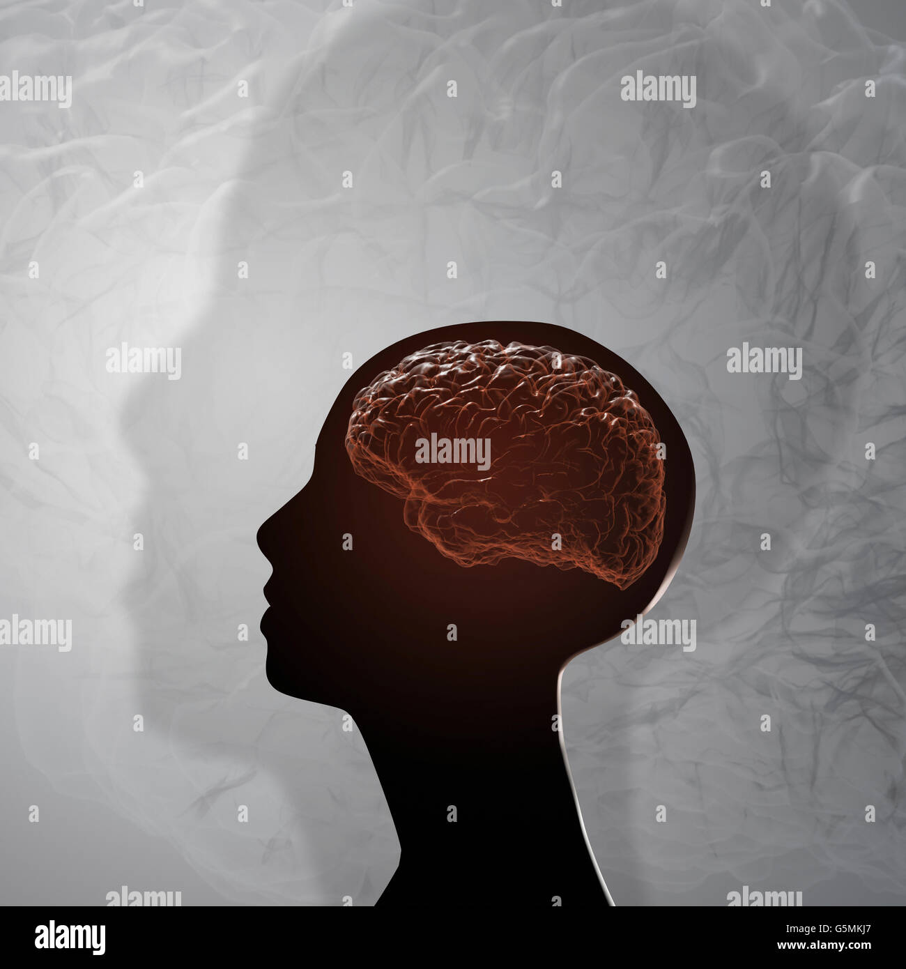 The human brain. Projection of the cerebral cortex Stock Photohttps://www.alamy.com/image-license-details/?v=1https://www.alamy.com/stock-photo-the-human-brain-projection-of-the-cerebral-cortex-106789935.html
RFG5MKJ7–The human brain. Projection of the cerebral cortex
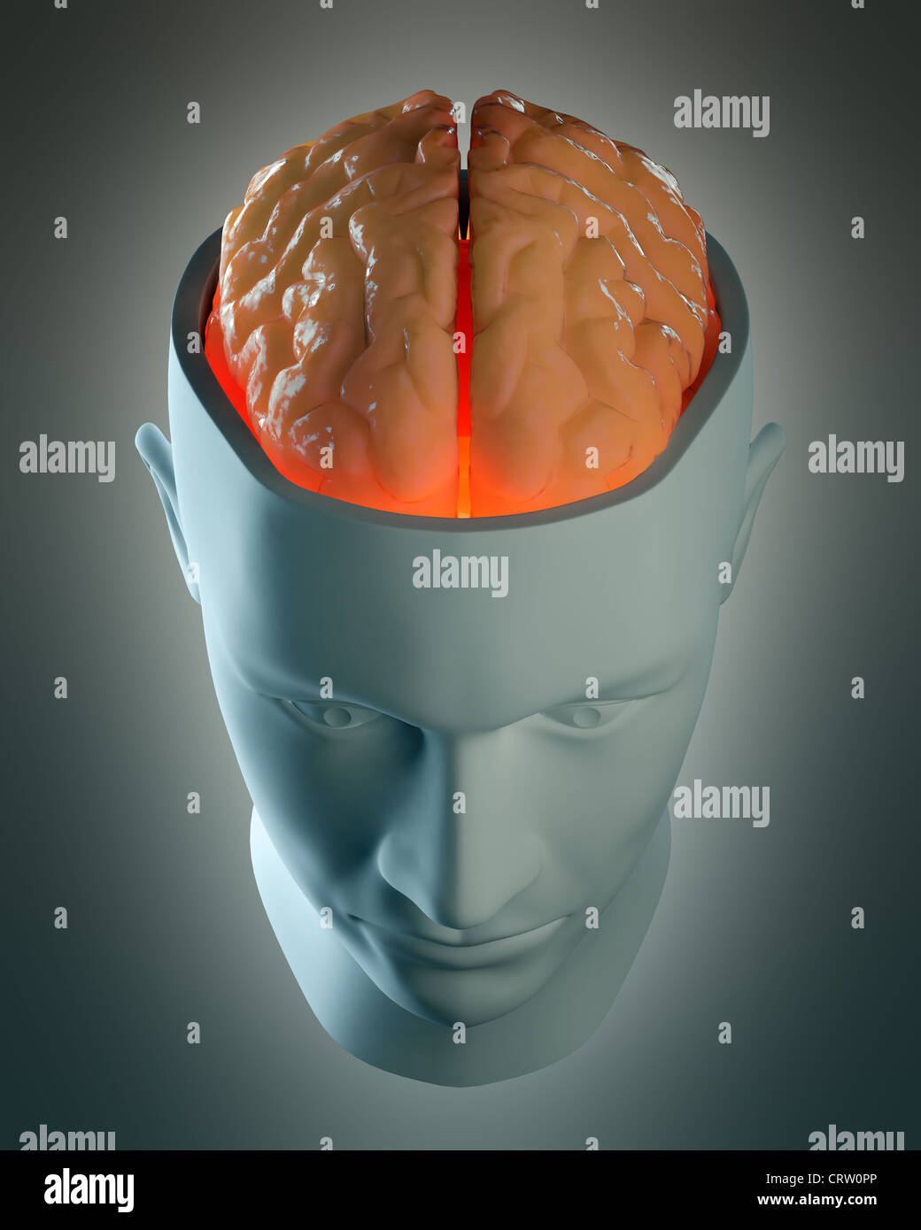 Male head abstract with a visible brain Stock Photohttps://www.alamy.com/image-license-details/?v=1https://www.alamy.com/stock-photo-male-head-abstract-with-a-visible-brain-49107262.html
RFCRW0PP–Male head abstract with a visible brain
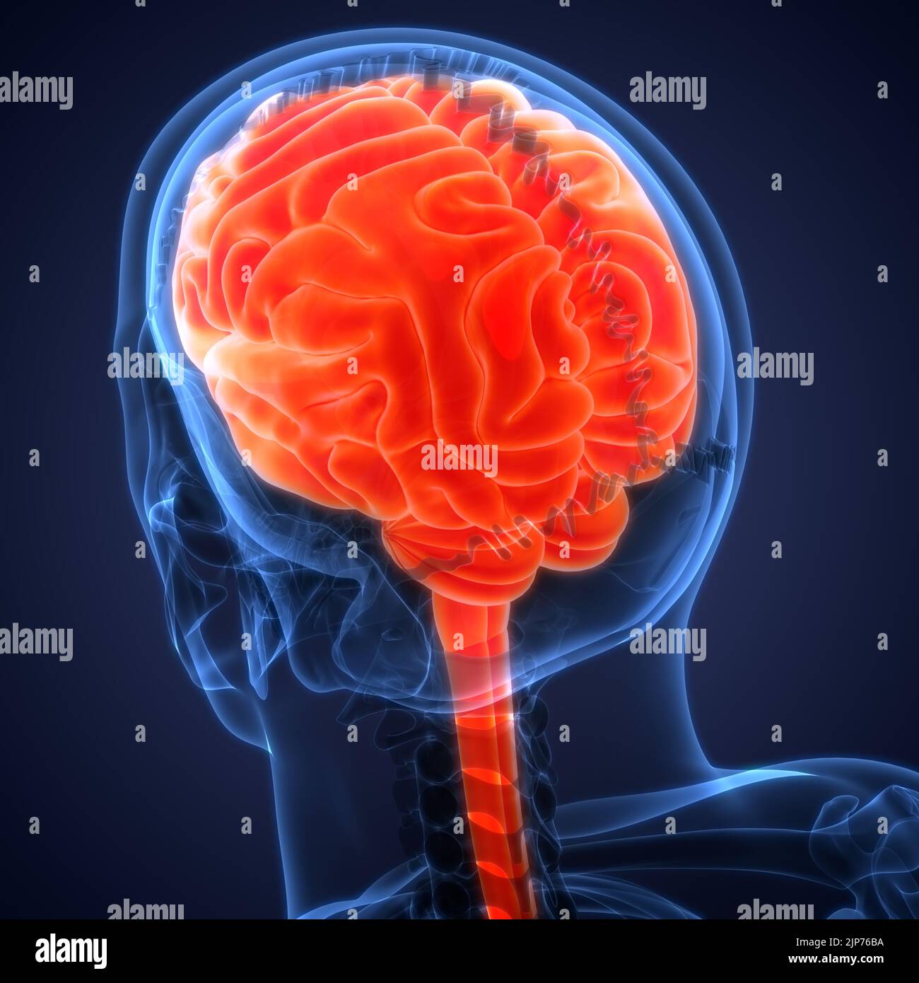 Central Organ of Human Nervous System Brain Anatomy Stock Photohttps://www.alamy.com/image-license-details/?v=1https://www.alamy.com/central-organ-of-human-nervous-system-brain-anatomy-image478361054.html
RF2JP76BA–Central Organ of Human Nervous System Brain Anatomy
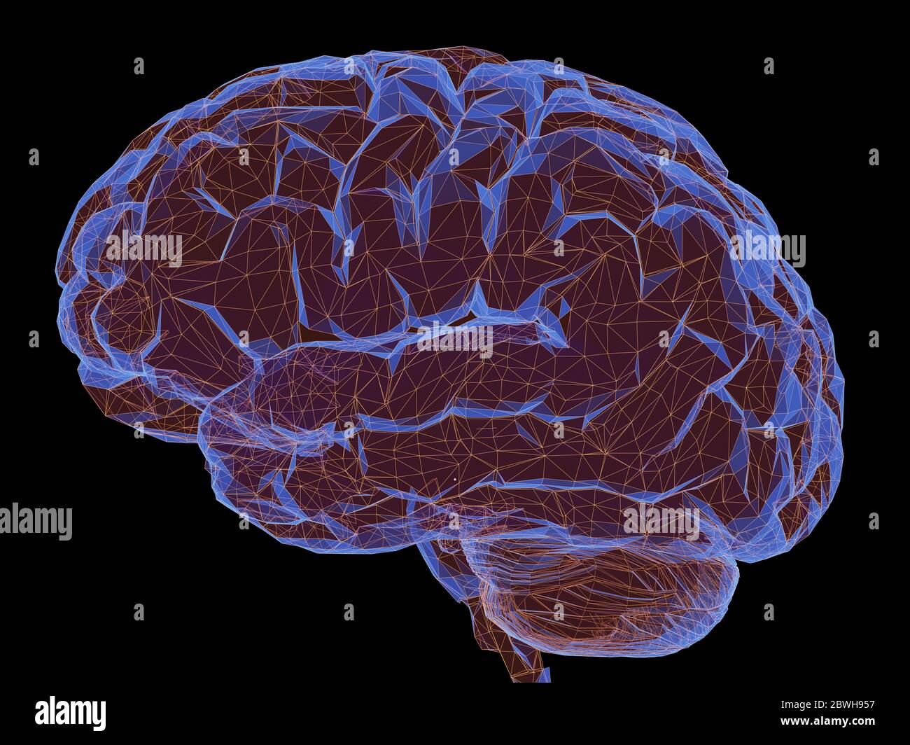 3D illustration. Human brain in a structure of polygonal connections representing the power of the mind. Clipping path included. Stock Photohttps://www.alamy.com/image-license-details/?v=1https://www.alamy.com/3d-illustration-human-brain-in-a-structure-of-polygonal-connections-representing-the-power-of-the-mind-clipping-path-included-image359954147.html
RF2BWH957–3D illustration. Human brain in a structure of polygonal connections representing the power of the mind. Clipping path included.
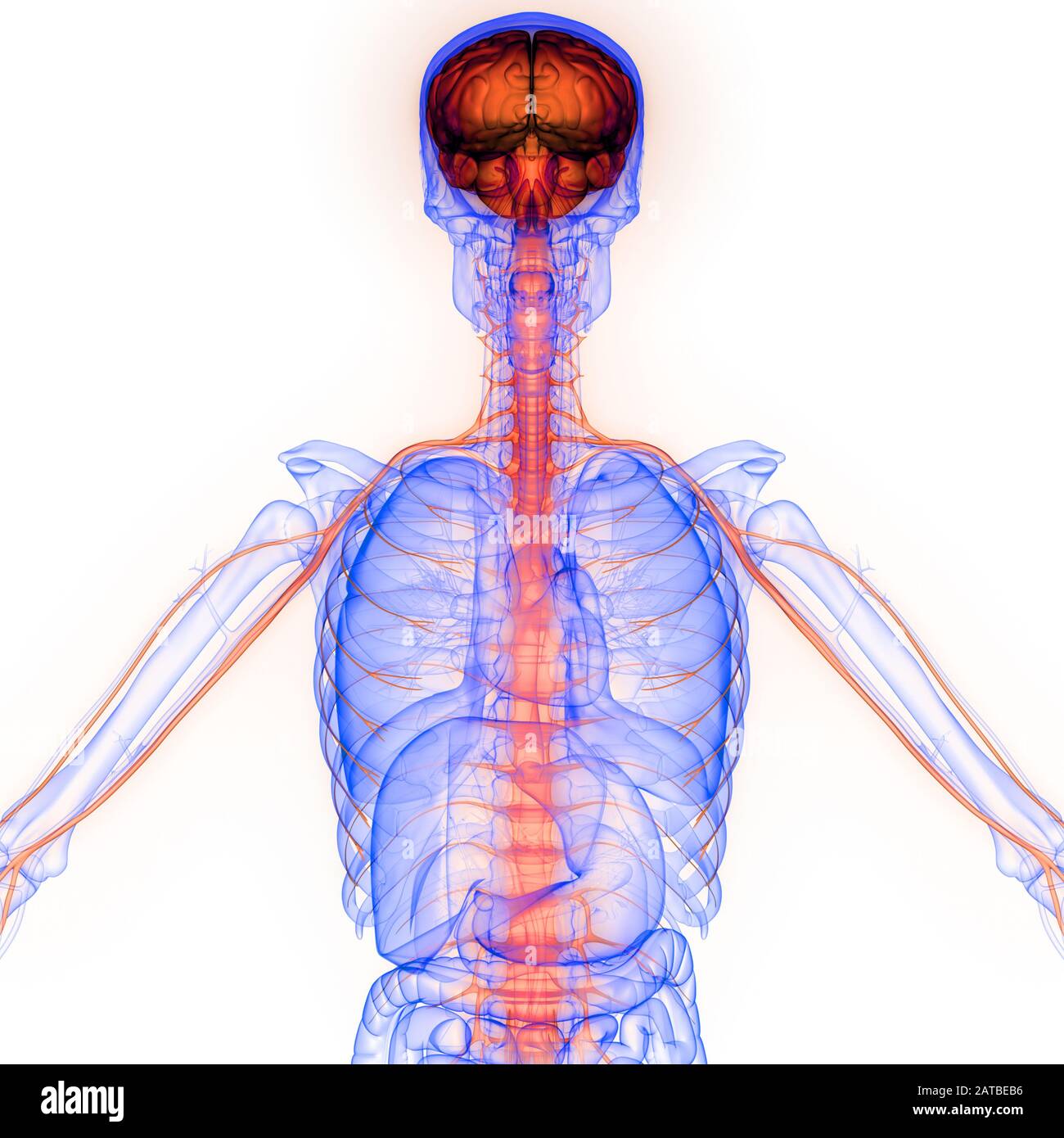 Human Internal Organ Brain with Nervous System Anatomy X-ray 3D rendering Stock Photohttps://www.alamy.com/image-license-details/?v=1https://www.alamy.com/human-internal-organ-brain-with-nervous-system-anatomy-x-ray-3d-rendering-image342001498.html
RF2ATBEB6–Human Internal Organ Brain with Nervous System Anatomy X-ray 3D rendering
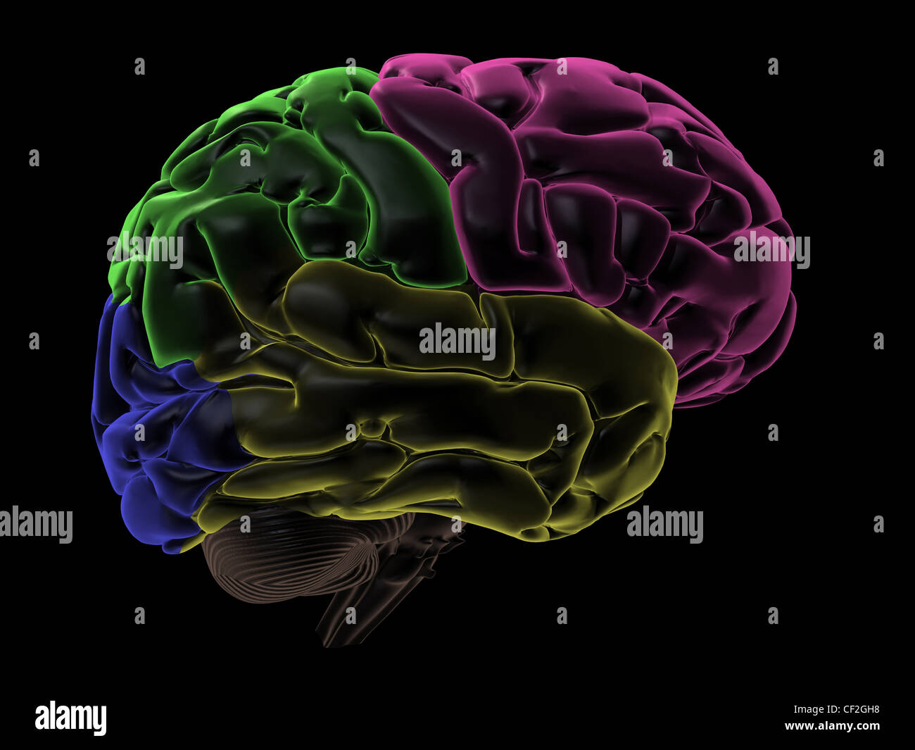 Coloured areas of the Brain, right hemisphere Stock Photohttps://www.alamy.com/image-license-details/?v=1https://www.alamy.com/stock-photo-coloured-areas-of-the-brain-right-hemisphere-43697508.html
RFCF2GH8–Coloured areas of the Brain, right hemisphere
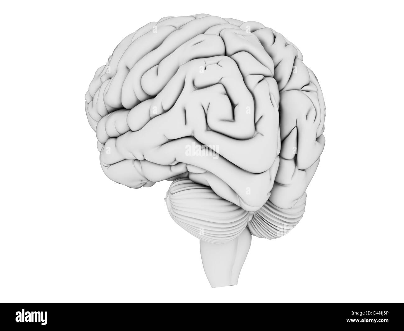 White brain Stock Photohttps://www.alamy.com/image-license-details/?v=1https://www.alamy.com/stock-photo-white-brain-54564994.html
RFD4NJ5P–White brain
 Psychotic disorder in human brain - dozens of terms describing its properties painted over the brain cortex to symbolize its connection to the mind.,3 Stock Photohttps://www.alamy.com/image-license-details/?v=1https://www.alamy.com/psychotic-disorder-in-human-brain-dozens-of-terms-describing-its-properties-painted-over-the-brain-cortex-to-symbolize-its-connection-to-the-mind3-image472069554.html
RF2JC0HEX–Psychotic disorder in human brain - dozens of terms describing its properties painted over the brain cortex to symbolize its connection to the mind.,3
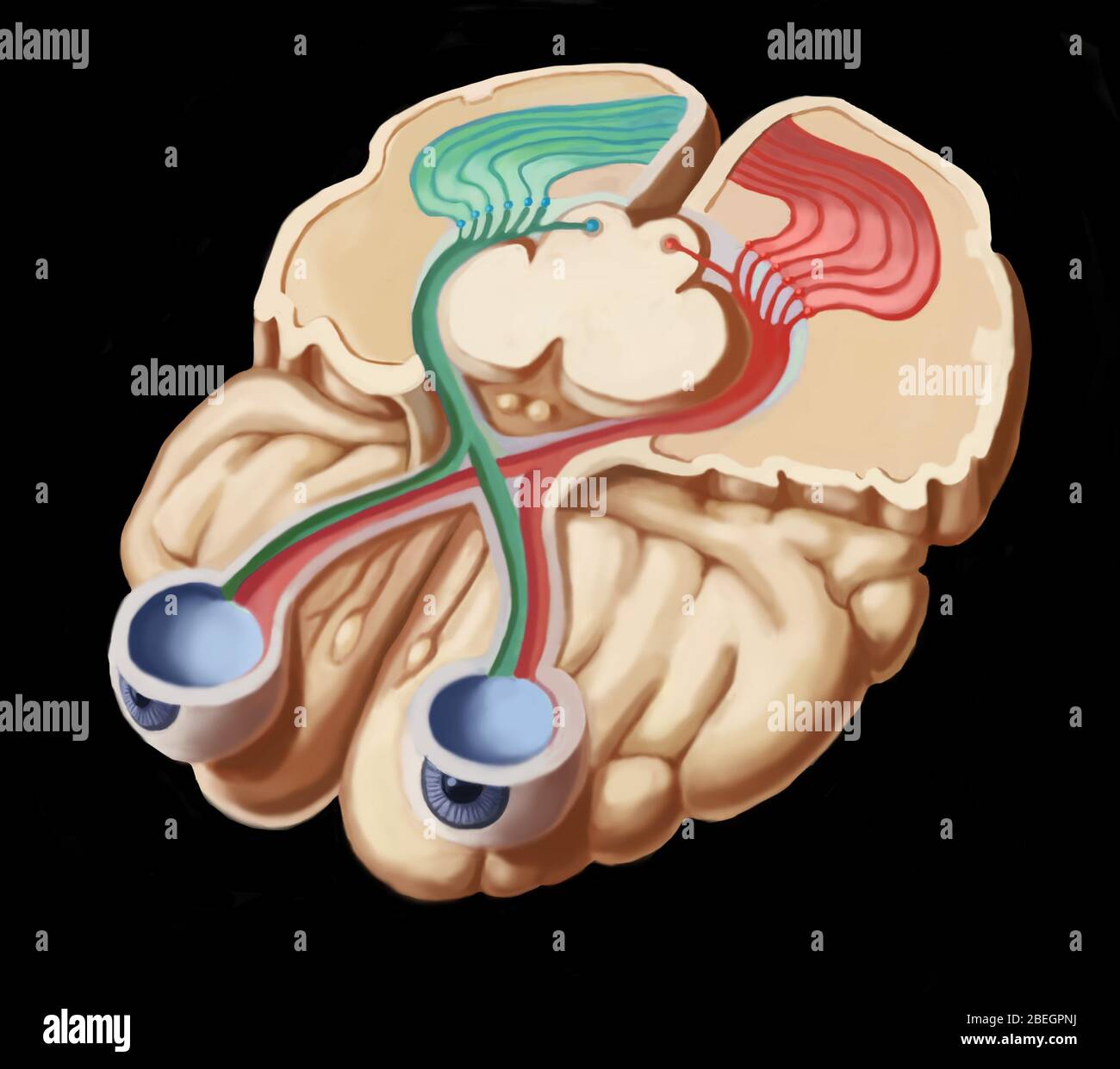 Visual Pathways Stock Photohttps://www.alamy.com/image-license-details/?v=1https://www.alamy.com/visual-pathways-image353181630.html
RM2BEGPNJ–Visual Pathways
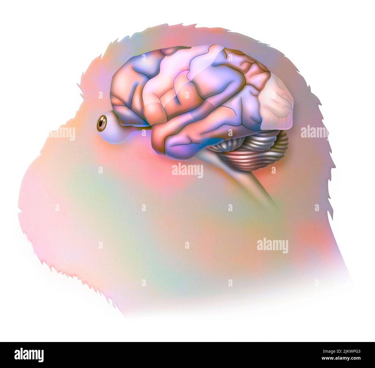 Brain in the chimpanzee with its areas (cognitive, auditory, visual) and cortex (motor, sensory). Stock Photohttps://www.alamy.com/image-license-details/?v=1https://www.alamy.com/brain-in-the-chimpanzee-with-its-areas-cognitive-auditory-visual-and-cortex-motor-sensory-image476924899.html
RF2JKWPG3–Brain in the chimpanzee with its areas (cognitive, auditory, visual) and cortex (motor, sensory).
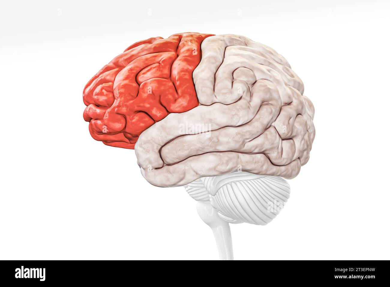 Cerebral cortex frontal lobe in red color profile view isolated on white background 3D rendering illustration. Human brain anatomy, neurology, neurosc Stock Photohttps://www.alamy.com/image-license-details/?v=1https://www.alamy.com/cerebral-cortex-frontal-lobe-in-red-color-profile-view-isolated-on-white-background-3d-rendering-illustration-human-brain-anatomy-neurology-neurosc-image570111301.html
RF2T3EPNW–Cerebral cortex frontal lobe in red color profile view isolated on white background 3D rendering illustration. Human brain anatomy, neurology, neurosc