Quick filters:
Brain structures Stock Photos and Images
 Illustration of human brain structures. Stock Photohttps://www.alamy.com/image-license-details/?v=1https://www.alamy.com/illustration-of-human-brain-structures-image263464169.html
RFW8HR6H–Illustration of human brain structures.
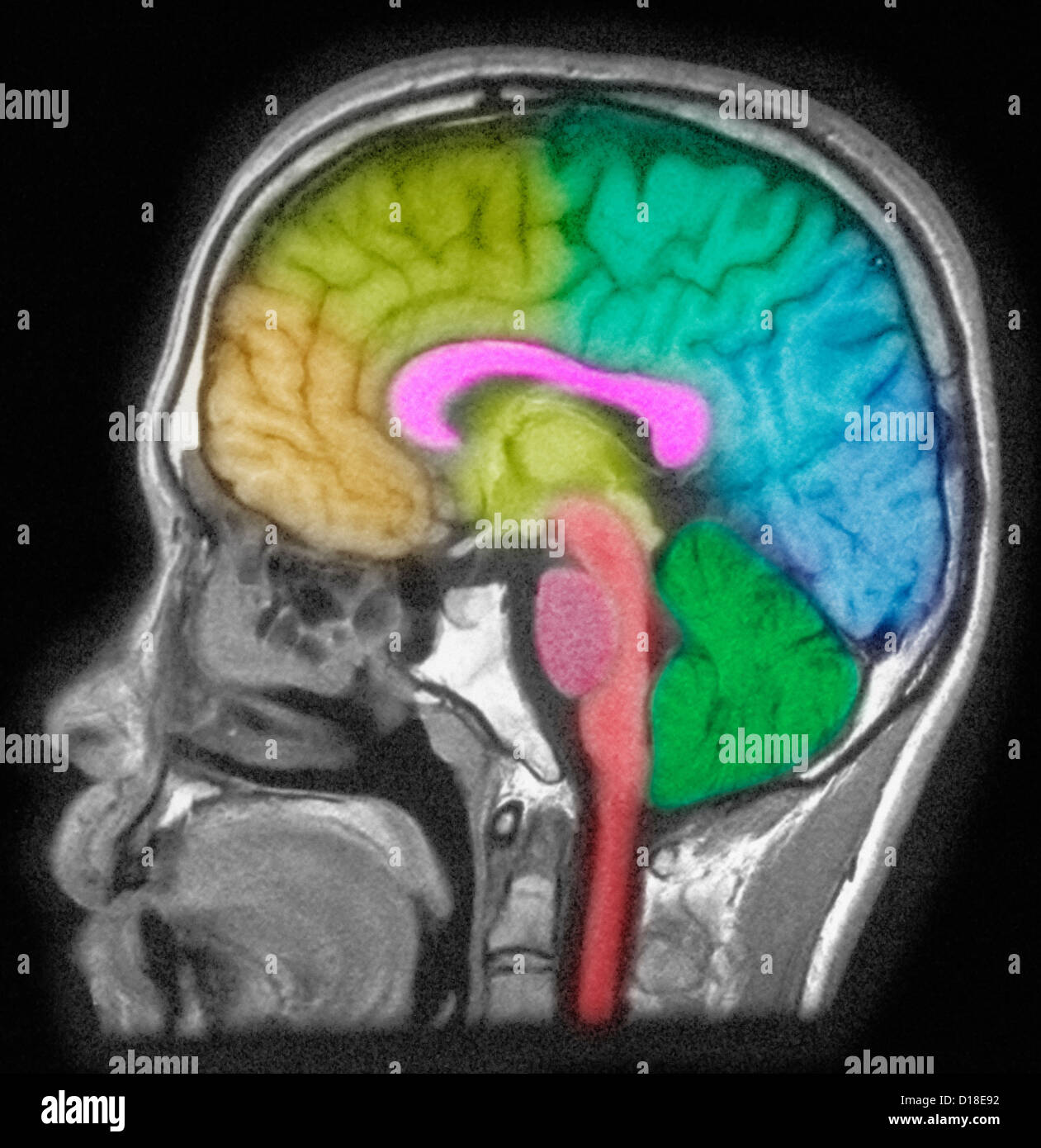 MRI of head showing brain structures Stock Photohttps://www.alamy.com/image-license-details/?v=1https://www.alamy.com/stock-photo-mri-of-head-showing-brain-structures-52432606.html
RFD18E92–MRI of head showing brain structures
 Illustration of brain structures and anatomy Stock Photohttps://www.alamy.com/image-license-details/?v=1https://www.alamy.com/stock-photo-illustration-of-brain-structures-and-anatomy-122194254.html
RFH2PC0E–Illustration of brain structures and anatomy
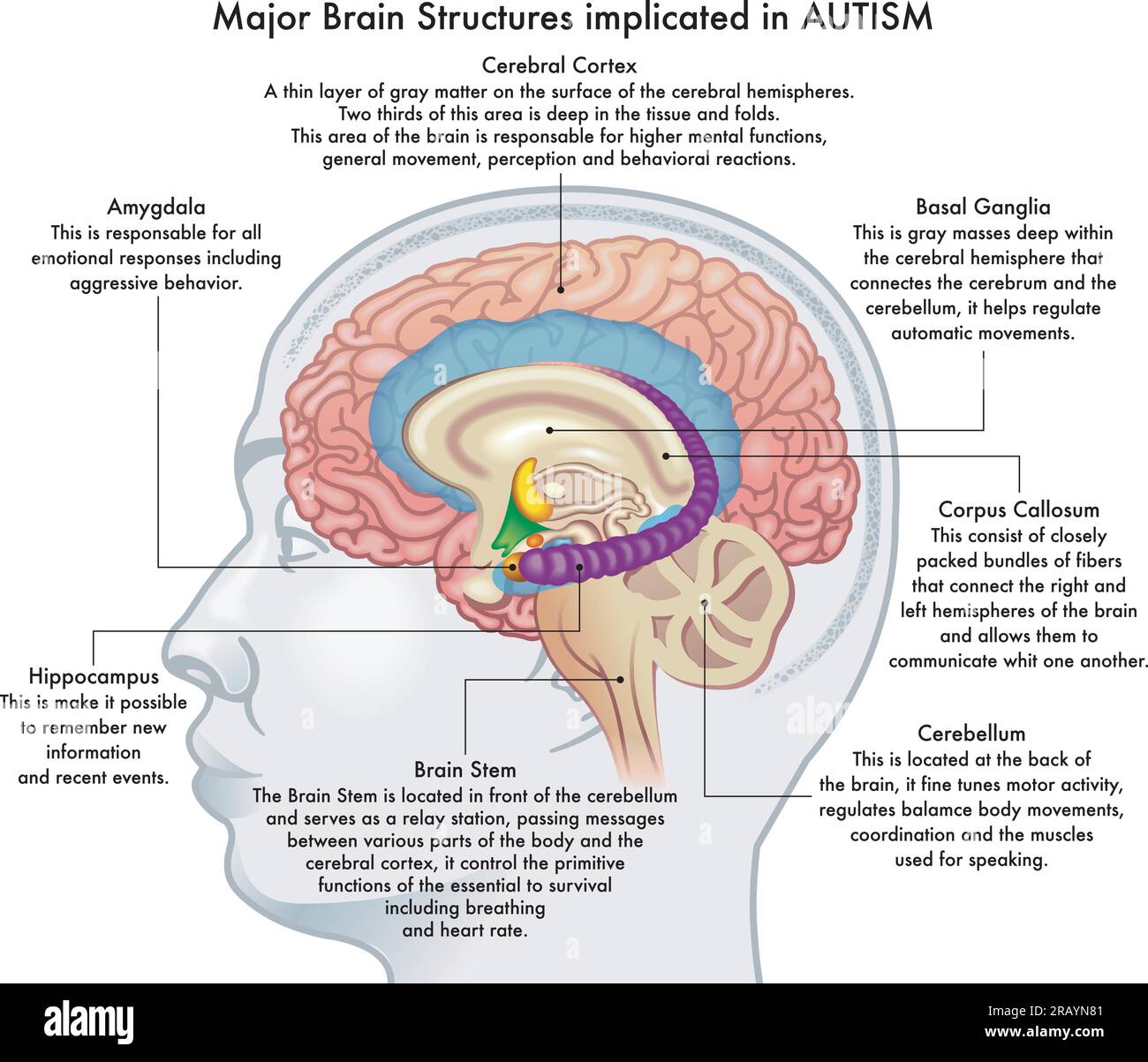 Medical illustration showing major brain structures implicated in autism spectrum disorder, with annotations. Stock Vectorhttps://www.alamy.com/image-license-details/?v=1https://www.alamy.com/medical-illustration-showing-major-brain-structures-implicated-in-autism-spectrum-disorder-with-annotations-image557487729.html
RF2RAYN81–Medical illustration showing major brain structures implicated in autism spectrum disorder, with annotations.
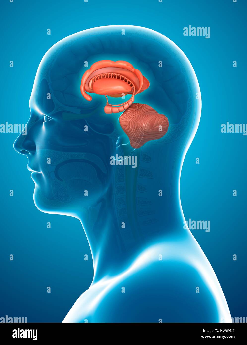 Illustration of human brain structures. Stock Photohttps://www.alamy.com/image-license-details/?v=1https://www.alamy.com/stock-photo-illustration-of-human-brain-structures-135978338.html
RFHW69N6–Illustration of human brain structures.
 Close up of a piece of a coral with brain structures. Stock Photohttps://www.alamy.com/image-license-details/?v=1https://www.alamy.com/stock-photo-close-up-of-a-piece-of-a-coral-with-brain-structures-129576349.html
RMHEPKXN–Close up of a piece of a coral with brain structures.
 Illustration of human brain structures. Stock Photohttps://www.alamy.com/image-license-details/?v=1https://www.alamy.com/stock-photo-illustration-of-human-brain-structures-140556274.html
RFJ4JTXX–Illustration of human brain structures.
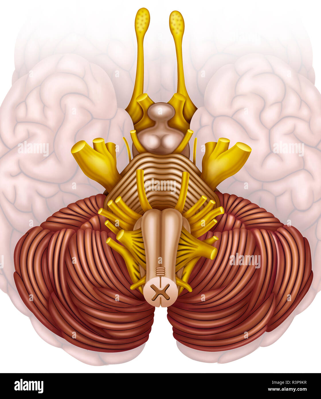 The brain stem, or human brain stem, is the major communication pathway for the brain, spinal cord, and peripheral nerves. Stock Photohttps://www.alamy.com/image-license-details/?v=1https://www.alamy.com/the-brain-stem-or-human-brain-stem-is-the-major-communication-pathway-for-the-brain-spinal-cord-and-peripheral-nerves-image226069307.html
RFR3P9KR–The brain stem, or human brain stem, is the major communication pathway for the brain, spinal cord, and peripheral nerves.
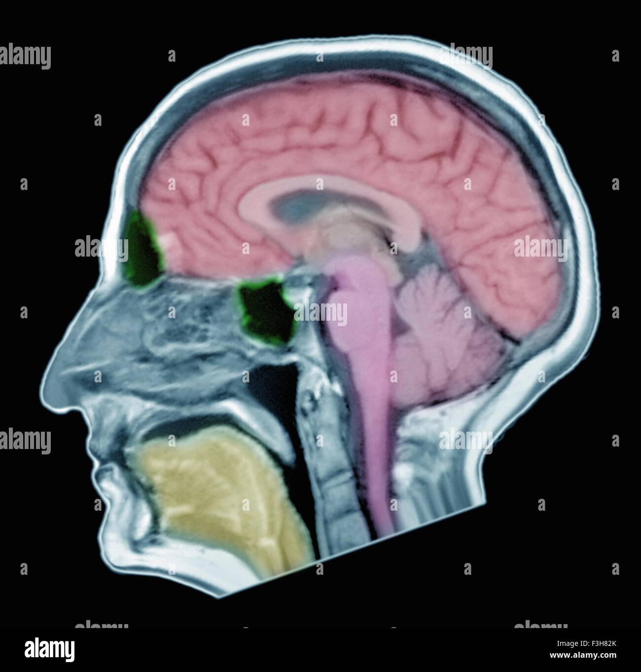 Brain MRI of adult male, normal structures Stock Photohttps://www.alamy.com/image-license-details/?v=1https://www.alamy.com/stock-photo-brain-mri-of-adult-male-normal-structures-88275339.html
RFF3H82K–Brain MRI of adult male, normal structures
 Human brain, chemical structures and data processing against black background Stock Photohttps://www.alamy.com/image-license-details/?v=1https://www.alamy.com/human-brain-chemical-structures-and-data-processing-against-black-background-image608895534.html
RF2XAHGD2–Human brain, chemical structures and data processing against black background
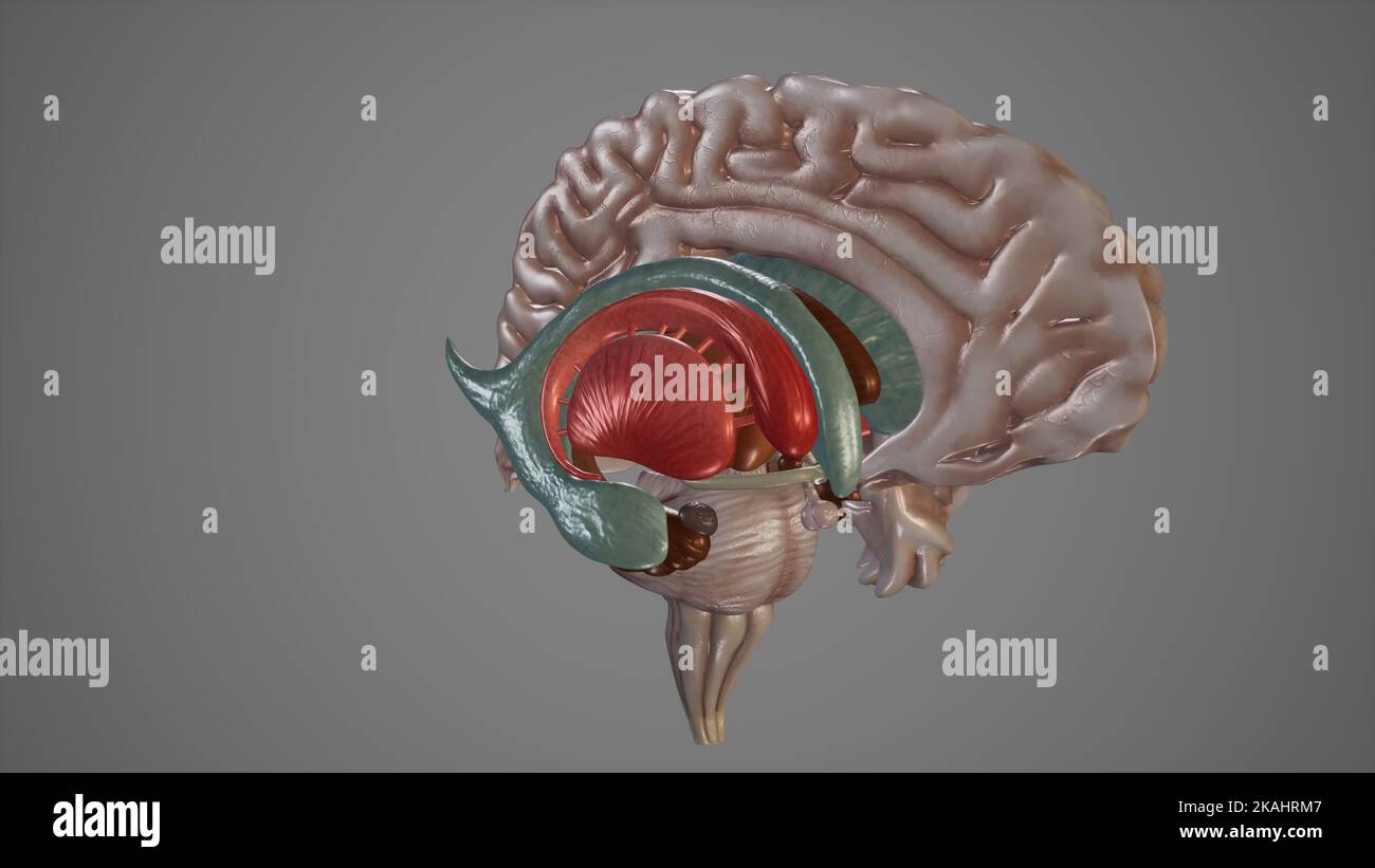 Medical Illustration of Deep Structures of Brain Stock Photohttps://www.alamy.com/image-license-details/?v=1https://www.alamy.com/medical-illustration-of-deep-structures-of-brain-image488428647.html
RF2KAHRM7–Medical Illustration of Deep Structures of Brain
 Image of molecule structures, nucleotides with brain and human representation Stock Photohttps://www.alamy.com/image-license-details/?v=1https://www.alamy.com/image-of-molecule-structures-nucleotides-with-brain-and-human-representation-image611799910.html
RF2XF9W0P–Image of molecule structures, nucleotides with brain and human representation
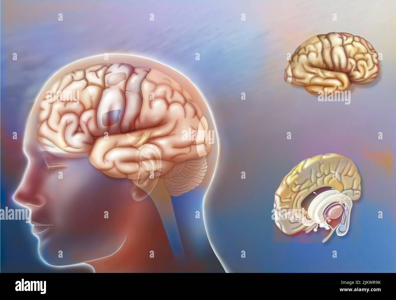 Left and right hemisphere brain areas and midline structures. Stock Photohttps://www.alamy.com/image-license-details/?v=1https://www.alamy.com/left-and-right-hemisphere-brain-areas-and-midline-structures-image476925503.html
RF2JKWR9K–Left and right hemisphere brain areas and midline structures.
 Chemical structures and 3D brain model against data processing Stock Photohttps://www.alamy.com/image-license-details/?v=1https://www.alamy.com/chemical-structures-and-3d-brain-model-against-data-processing-image474544881.html
RF2JG1ARD–Chemical structures and 3D brain model against data processing
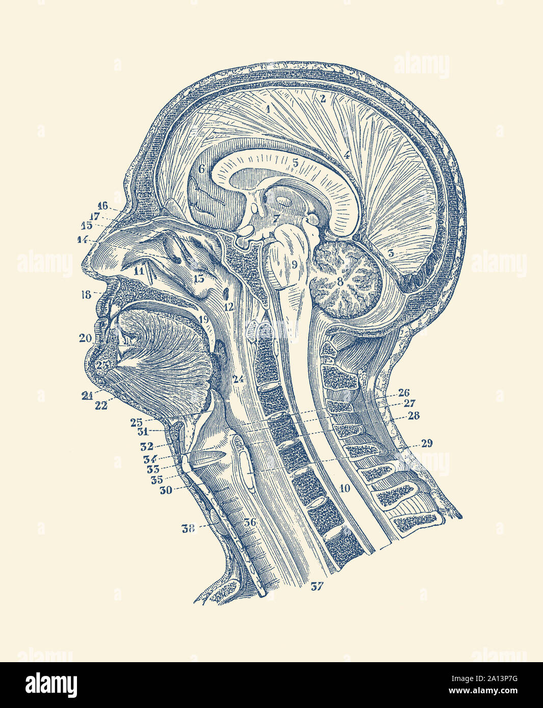 Vintage anatomy print showing a diagram of the structures in and around the human brain. Stock Photohttps://www.alamy.com/image-license-details/?v=1https://www.alamy.com/vintage-anatomy-print-showing-a-diagram-of-the-structures-in-and-around-the-human-brain-image327694964.html
RF2A13P7G–Vintage anatomy print showing a diagram of the structures in and around the human brain.
 Human brain, chemical and molecular structures against data processing and green light trails Stock Photohttps://www.alamy.com/image-license-details/?v=1https://www.alamy.com/human-brain-chemical-and-molecular-structures-against-data-processing-and-green-light-trails-image608895540.html
RF2XAHGD8–Human brain, chemical and molecular structures against data processing and green light trails
 Cells of the Body Stock Photohttps://www.alamy.com/image-license-details/?v=1https://www.alamy.com/cells-of-the-body-image353185372.html
RF2BEGYF8–Cells of the Body
 Composition of human brain and white structures of chemical compounds Stock Photohttps://www.alamy.com/image-license-details/?v=1https://www.alamy.com/composition-of-human-brain-and-white-structures-of-chemical-compounds-image430720189.html
RF2G0MYYW–Composition of human brain and white structures of chemical compounds
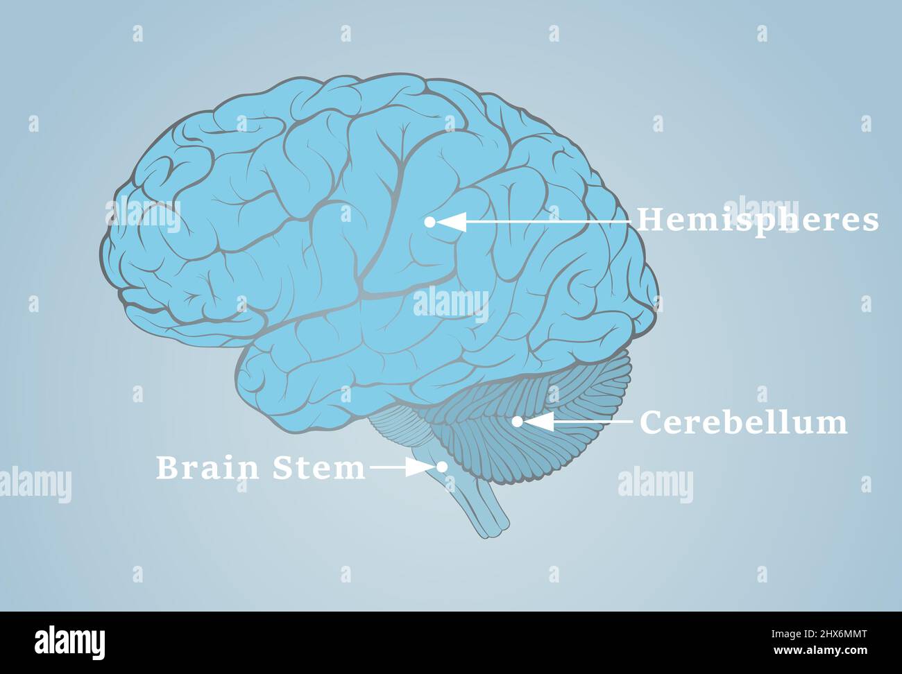 Human brain image with the structures indicated by the arrows Stock Photohttps://www.alamy.com/image-license-details/?v=1https://www.alamy.com/human-brain-image-with-the-structures-indicated-by-the-arrows-image463598600.html
RF2HX6MMT–Human brain image with the structures indicated by the arrows
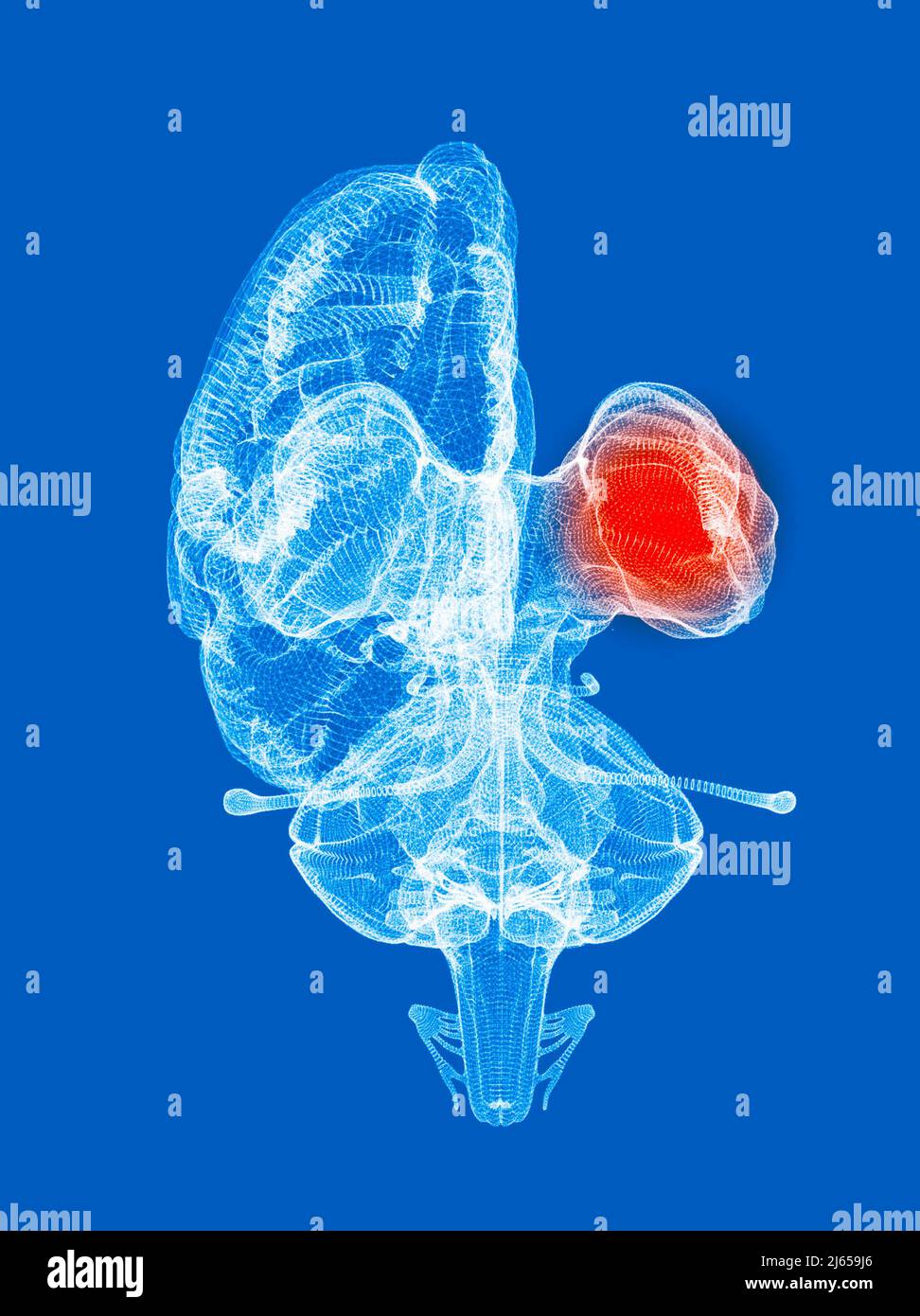 Human brain anatomy front view, internal structures, diencephalon. Thalamus and hypothalamus. 3d rendering Stock Photohttps://www.alamy.com/image-license-details/?v=1https://www.alamy.com/human-brain-anatomy-front-view-internal-structures-diencephalon-thalamus-and-hypothalamus-3d-rendering-image468485198.html
RF2J659J6–Human brain anatomy front view, internal structures, diencephalon. Thalamus and hypothalamus. 3d rendering
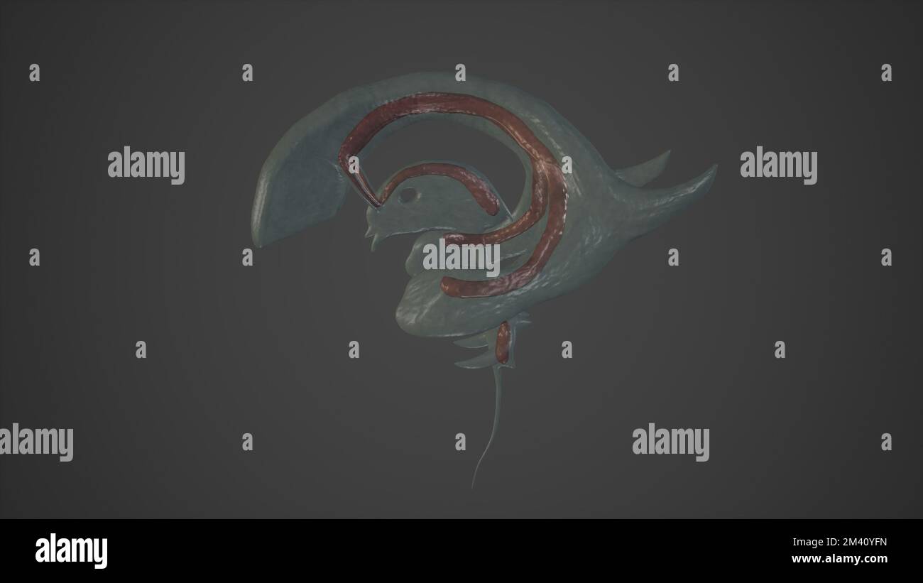 Anatomical Illustration of Choroid Plexuses.3d rendering Stock Photohttps://www.alamy.com/image-license-details/?v=1https://www.alamy.com/anatomical-illustration-of-choroid-plexuses3d-rendering-image501580905.html
RF2M40YFN–Anatomical Illustration of Choroid Plexuses.3d rendering
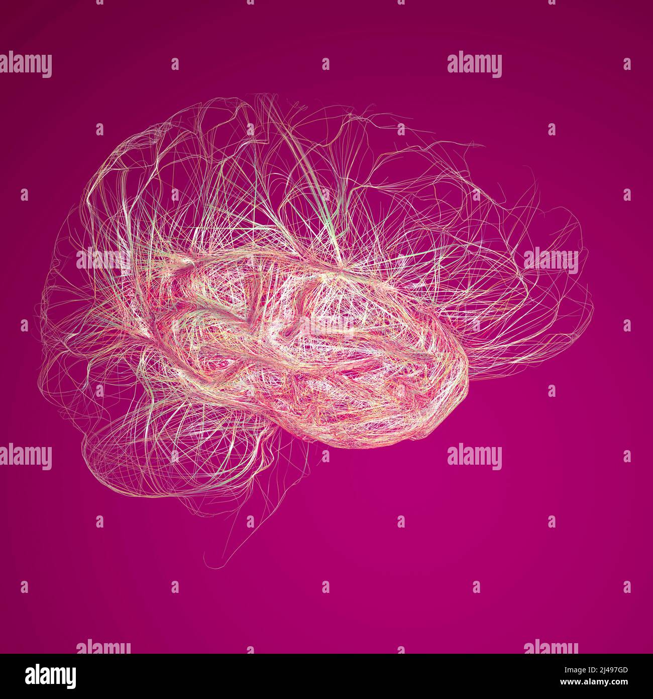 Brain, the temporal lobe is one of the four major lobes of the cerebral cortex, consist of structures that are vital for declarative or long-term memo Stock Photohttps://www.alamy.com/image-license-details/?v=1https://www.alamy.com/brain-the-temporal-lobe-is-one-of-the-four-major-lobes-of-the-cerebral-cortex-consist-of-structures-that-are-vital-for-declarative-or-long-term-memo-image467342077.html
RF2J497GD–Brain, the temporal lobe is one of the four major lobes of the cerebral cortex, consist of structures that are vital for declarative or long-term memo
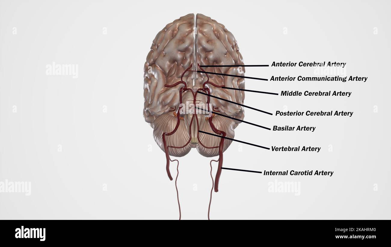 Circle of Willis Anatomy structures. Arterial Supply to the Brain Stock Photohttps://www.alamy.com/image-license-details/?v=1https://www.alamy.com/circle-of-willis-anatomy-structures-arterial-supply-to-the-brain-image488428640.html
RF2KAHRM0–Circle of Willis Anatomy structures. Arterial Supply to the Brain
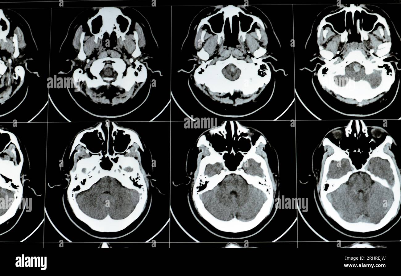 Multi slice CT scan of the brain showing Large brain stem and right centrum semiovale hematoma, normal posterior fossa structures, normal size of vent Stock Photohttps://www.alamy.com/image-license-details/?v=1https://www.alamy.com/multi-slice-ct-scan-of-the-brain-showing-large-brain-stem-and-right-centrum-semiovale-hematoma-normal-posterior-fossa-structures-normal-size-of-vent-image561697329.html
RF2RHREJW–Multi slice CT scan of the brain showing Large brain stem and right centrum semiovale hematoma, normal posterior fossa structures, normal size of vent
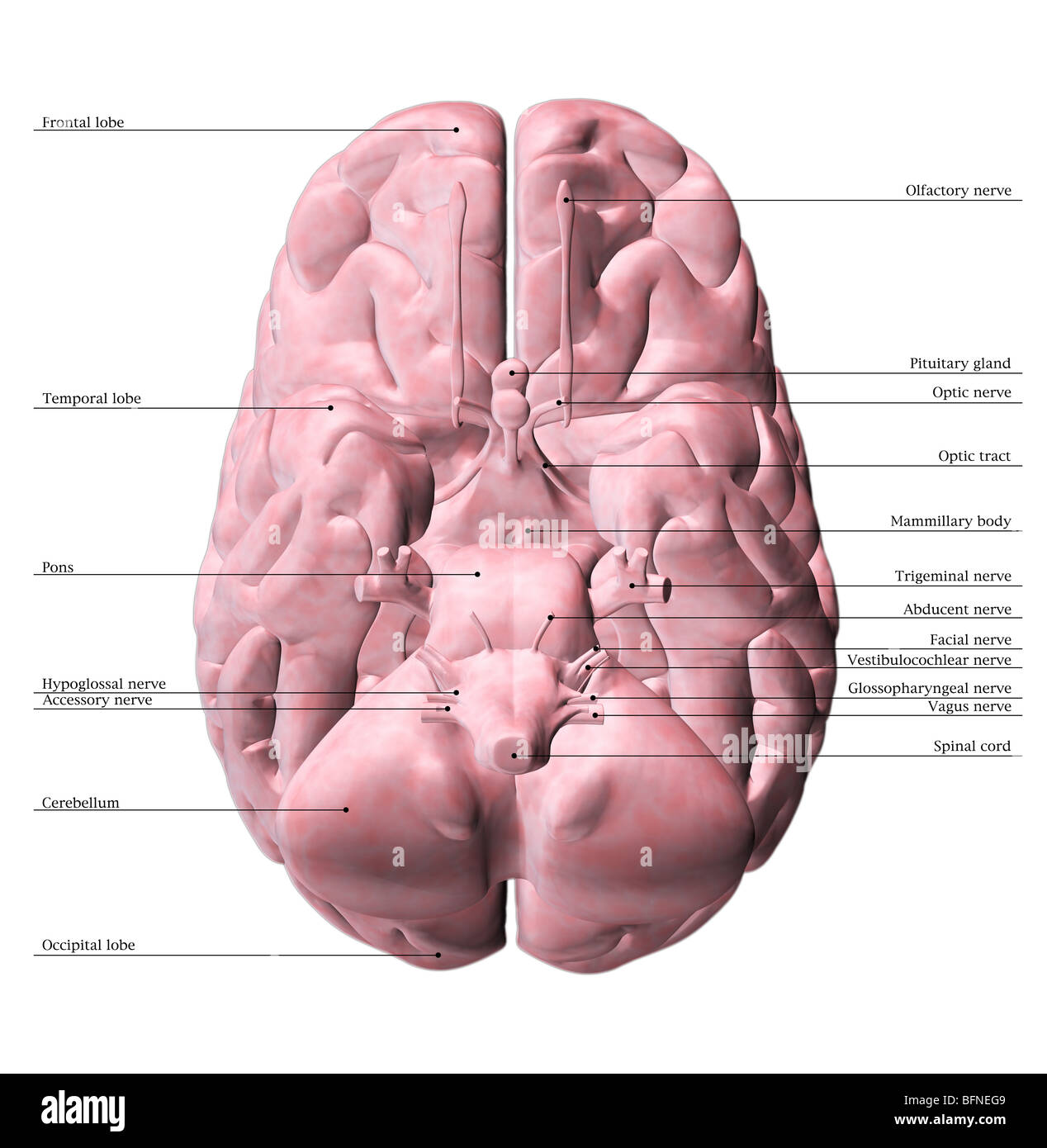 Illustration of the underside of the human brain Stock Photohttps://www.alamy.com/image-license-details/?v=1https://www.alamy.com/stock-photo-illustration-of-the-underside-of-the-human-brain-26902633.html
RMBFNEG9–Illustration of the underside of the human brain
 Illustration of human brain structures. Stock Photohttps://www.alamy.com/image-license-details/?v=1https://www.alamy.com/stock-photo-illustration-of-human-brain-structures-140556402.html
RFJ4JW3E–Illustration of human brain structures.
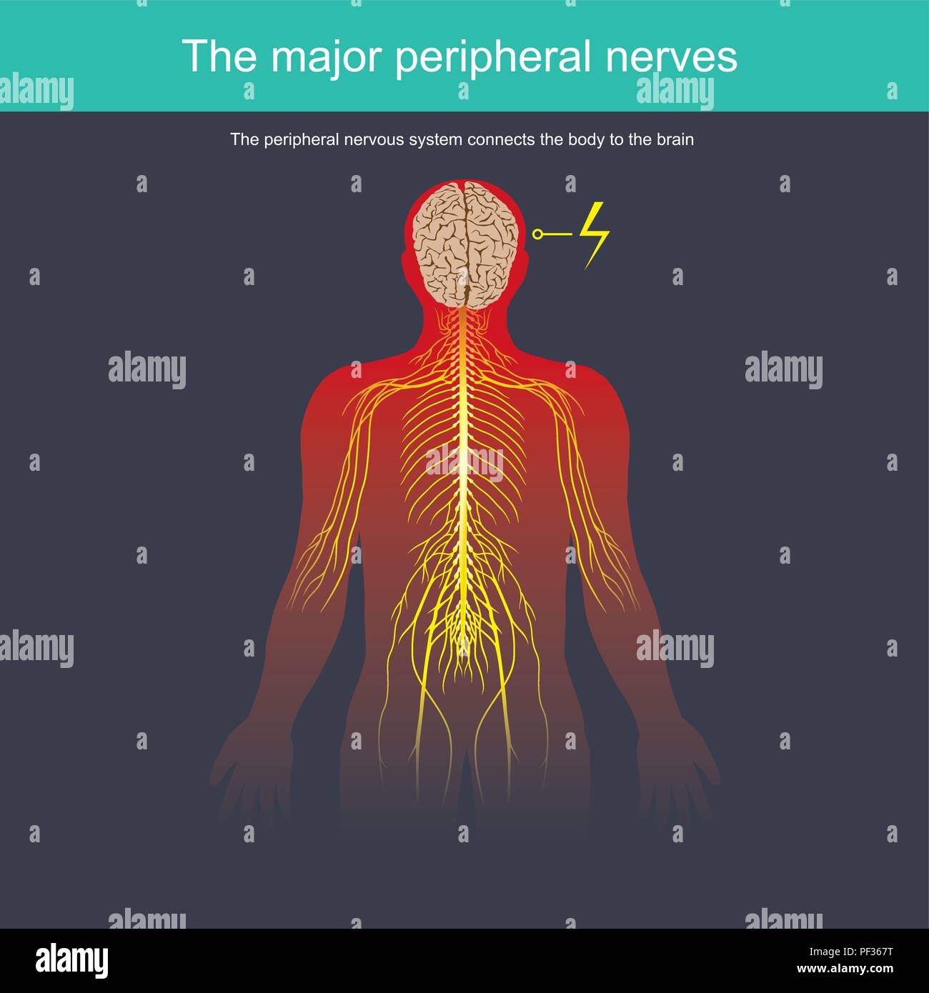 The peripheral nervous system connects the body to the brain Stock Vectorhttps://www.alamy.com/image-license-details/?v=1https://www.alamy.com/the-peripheral-nervous-system-connects-the-body-to-the-brain-image215815036.html
RFPF367T–The peripheral nervous system connects the body to the brain
 Neurology, conceptual image. Anatomical model of human head with vascular structures and nerves in foreground of neurologist's consultation with patie Stock Photohttps://www.alamy.com/image-license-details/?v=1https://www.alamy.com/neurology-conceptual-image-anatomical-model-of-human-head-with-vascular-structures-and-nerves-in-foreground-of-neurologists-consultation-with-patie-image545245072.html
RF2PK21J8–Neurology, conceptual image. Anatomical model of human head with vascular structures and nerves in foreground of neurologist's consultation with patie
 'The Swirl', Brain Rocks of the Coyote Buttes South CBS, Cottonwood Teepees, eroded Navajo sandstone rock formations with Stock Photohttps://www.alamy.com/image-license-details/?v=1https://www.alamy.com/the-swirl-brain-rocks-of-the-coyote-buttes-south-cbs-cottonwood-teepees-image62214568.html
RMDH638T–'The Swirl', Brain Rocks of the Coyote Buttes South CBS, Cottonwood Teepees, eroded Navajo sandstone rock formations with
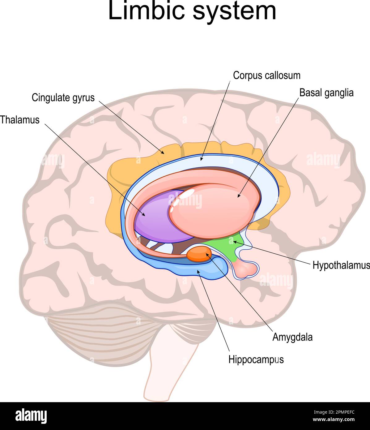 limbic system. Cross section of the human brain. Structure and Anatomical components of limbic system: Hypothalamus, Corpus callosum, Cingulate gyrus, Stock Vectorhttps://www.alamy.com/image-license-details/?v=1https://www.alamy.com/limbic-system-cross-section-of-the-human-brain-structure-and-anatomical-components-of-limbic-system-hypothalamus-corpus-callosum-cingulate-gyrus-image546308880.html
RF2PMPEFC–limbic system. Cross section of the human brain. Structure and Anatomical components of limbic system: Hypothalamus, Corpus callosum, Cingulate gyrus,
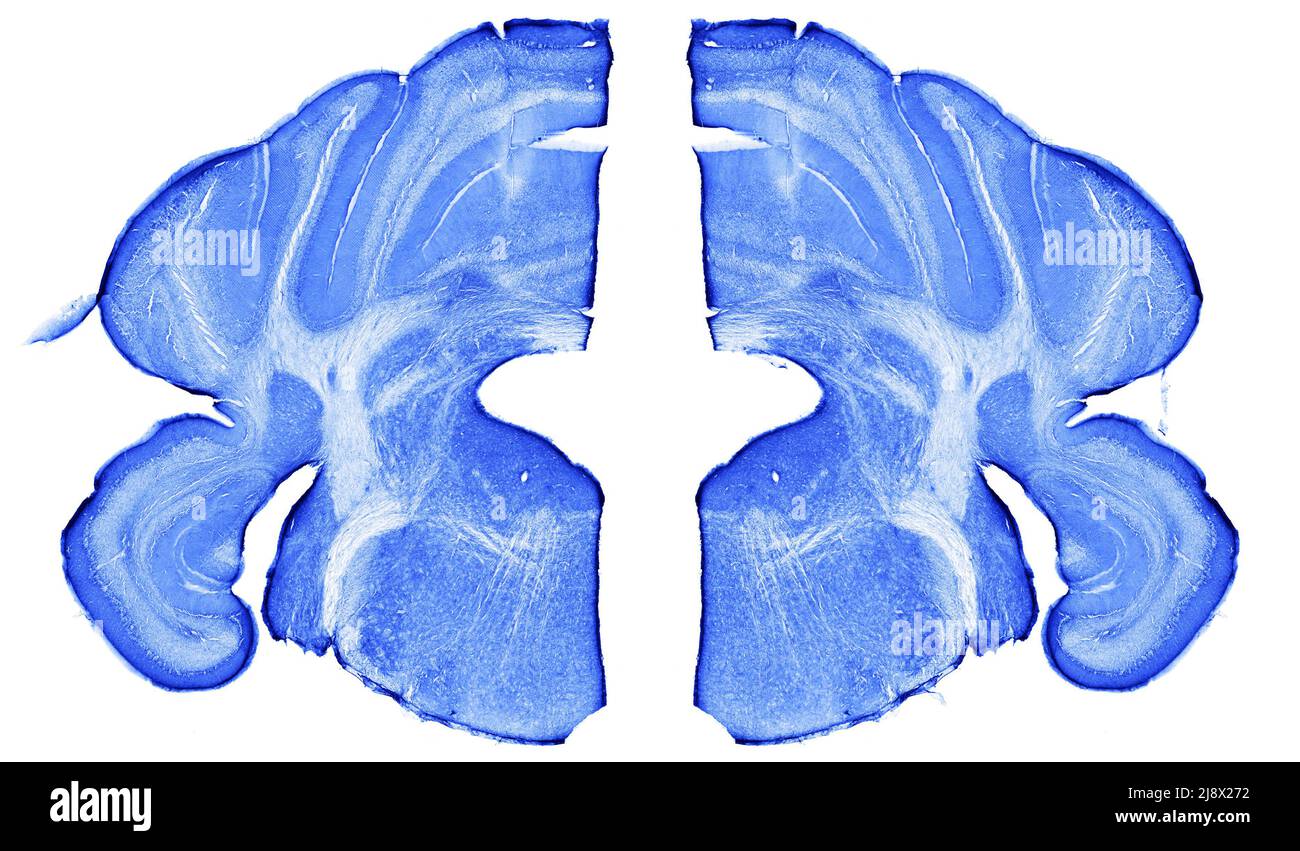 A mouse brain in cross section Stock Photohttps://www.alamy.com/image-license-details/?v=1https://www.alamy.com/a-mouse-brain-in-cross-section-image470169702.html
RM2J8X272–A mouse brain in cross section
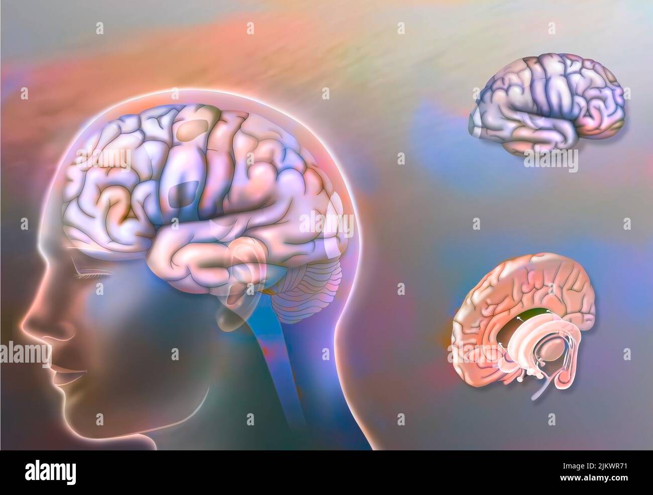 Left and right hemisphere brain areas and midline structures. Stock Photohttps://www.alamy.com/image-license-details/?v=1https://www.alamy.com/left-and-right-hemisphere-brain-areas-and-midline-structures-image476925429.html
RF2JKWR71–Left and right hemisphere brain areas and midline structures.
 Ruvo Brain Health Center in Las Vegas, NV, designed by Frank Gehry Stock Photohttps://www.alamy.com/image-license-details/?v=1https://www.alamy.com/ruvo-brain-health-center-in-las-vegas-nv-designed-by-frank-gehry-image411241723.html
RF2EW1K0B–Ruvo Brain Health Center in Las Vegas, NV, designed by Frank Gehry
 Artificial deep neural networks structures, dark digital background, 3d render illustration Stock Photohttps://www.alamy.com/image-license-details/?v=1https://www.alamy.com/stock-image-artificial-deep-neural-networks-structures-dark-digital-background-167463947.html
RFKMCHY7–Artificial deep neural networks structures, dark digital background, 3d render illustration
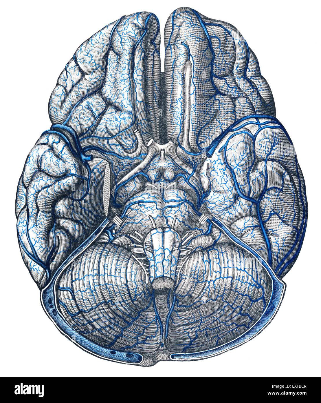 The cervical veins Stock Photohttps://www.alamy.com/image-license-details/?v=1https://www.alamy.com/stock-photo-the-cervical-veins-85160791.html
RMEXFBCR–The cervical veins
 Cells of the Body Stock Photohttps://www.alamy.com/image-license-details/?v=1https://www.alamy.com/cells-of-the-body-image353185442.html
RF2BEGYHP–Cells of the Body
 Composition of human brain and white structures of chemical compounds Stock Photohttps://www.alamy.com/image-license-details/?v=1https://www.alamy.com/composition-of-human-brain-and-white-structures-of-chemical-compounds-image430720215.html
RF2G0N00R–Composition of human brain and white structures of chemical compounds
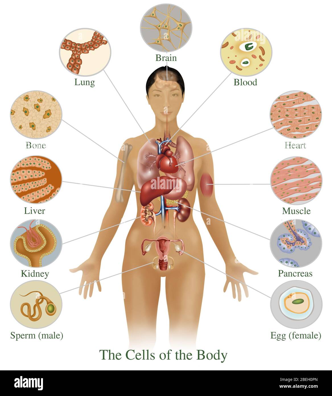 Cells of the Body Stock Photohttps://www.alamy.com/image-license-details/?v=1https://www.alamy.com/cells-of-the-body-image353186365.html
RF2BEH0PN–Cells of the Body
 Image of dna helix rotating over digital screens with medical structures Stock Photohttps://www.alamy.com/image-license-details/?v=1https://www.alamy.com/image-of-dna-helix-rotating-over-digital-screens-with-medical-structures-image611799864.html
RF2XF9TY4–Image of dna helix rotating over digital screens with medical structures
 Artificial intelligence conceptual digital illustration with neural network structures and blue human face, 3d render Stock Photohttps://www.alamy.com/image-license-details/?v=1https://www.alamy.com/stock-image-artificial-intelligence-conceptual-digital-illustration-with-neural-167463942.html
RFKMCHY2–Artificial intelligence conceptual digital illustration with neural network structures and blue human face, 3d render
 Image of equations and atomic structures over happy biracial female student in school corridor Stock Photohttps://www.alamy.com/image-license-details/?v=1https://www.alamy.com/image-of-equations-and-atomic-structures-over-happy-biracial-female-student-in-school-corridor-image623662657.html
RF2Y6J829–Image of equations and atomic structures over happy biracial female student in school corridor
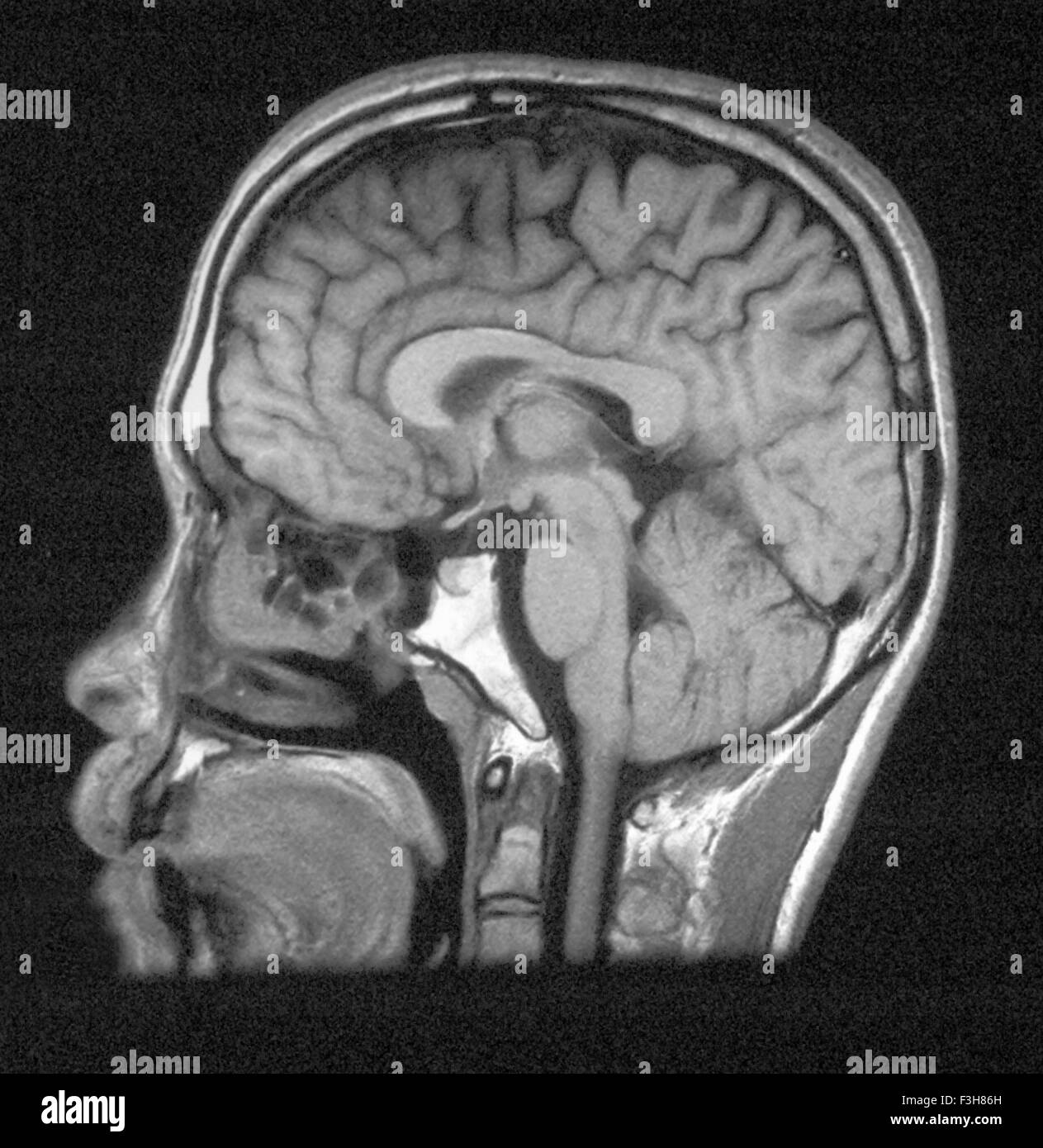 MRI of head showing normal brain structures Stock Photohttps://www.alamy.com/image-license-details/?v=1https://www.alamy.com/stock-photo-mri-of-head-showing-normal-brain-structures-88275449.html
RFF3H86H–MRI of head showing normal brain structures
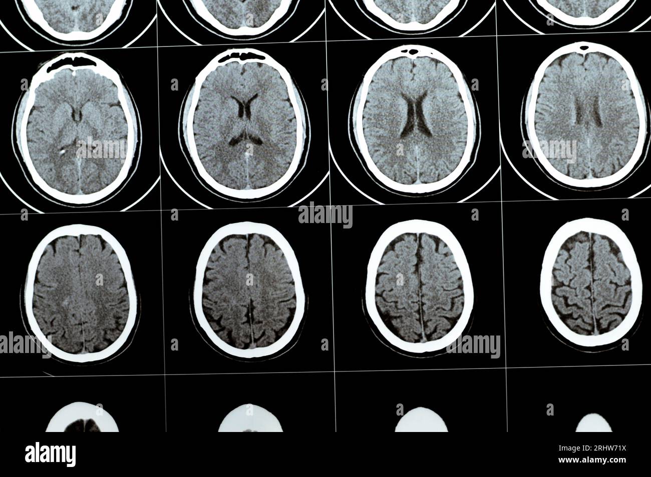 Multi slice CT scan of the brain showing Large brain stem and right centrum semiovale hematoma, normal posterior fossa structures, normal size of vent Stock Photohttps://www.alamy.com/image-license-details/?v=1https://www.alamy.com/multi-slice-ct-scan-of-the-brain-showing-large-brain-stem-and-right-centrum-semiovale-hematoma-normal-posterior-fossa-structures-normal-size-of-vent-image561735270.html
RF2RHW71X–Multi slice CT scan of the brain showing Large brain stem and right centrum semiovale hematoma, normal posterior fossa structures, normal size of vent
 Image of brain, molecular strauctures and layers of data processing over 3d grid on black Stock Photohttps://www.alamy.com/image-license-details/?v=1https://www.alamy.com/image-of-brain-molecular-strauctures-and-layers-of-data-processing-over-3d-grid-on-black-image597414686.html
RF2WKXGEP–Image of brain, molecular strauctures and layers of data processing over 3d grid on black
 Illustration of human brain structures. Stock Photohttps://www.alamy.com/image-license-details/?v=1https://www.alamy.com/stock-photo-illustration-of-human-brain-structures-140556396.html
RFJ4JW38–Illustration of human brain structures.
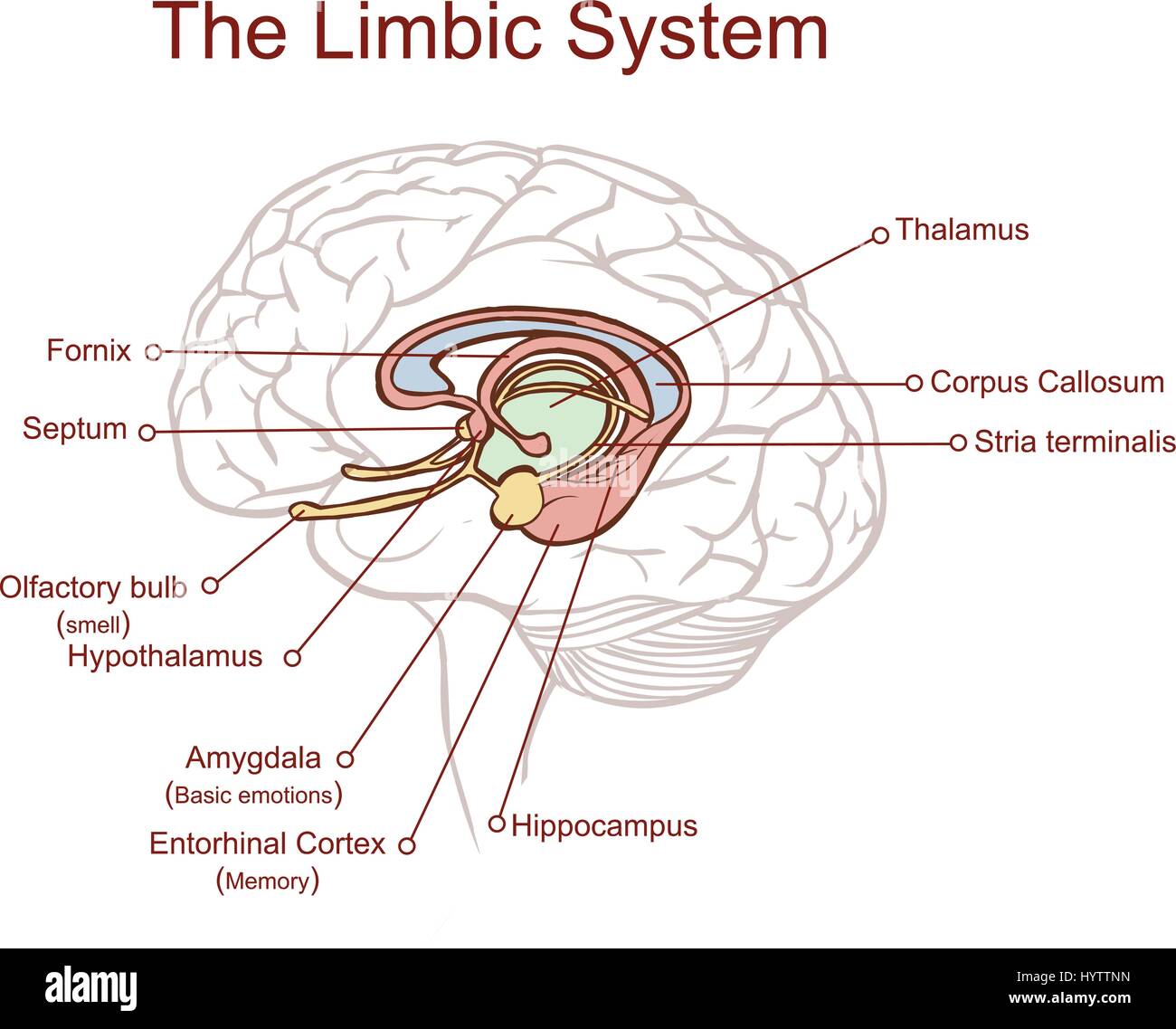 Cross section through the brain showing the limbic system and all related structures Stock Vectorhttps://www.alamy.com/image-license-details/?v=1https://www.alamy.com/stock-photo-cross-section-through-the-brain-showing-the-limbic-system-and-all-137614561.html
RFHYTTNN–Cross section through the brain showing the limbic system and all related structures
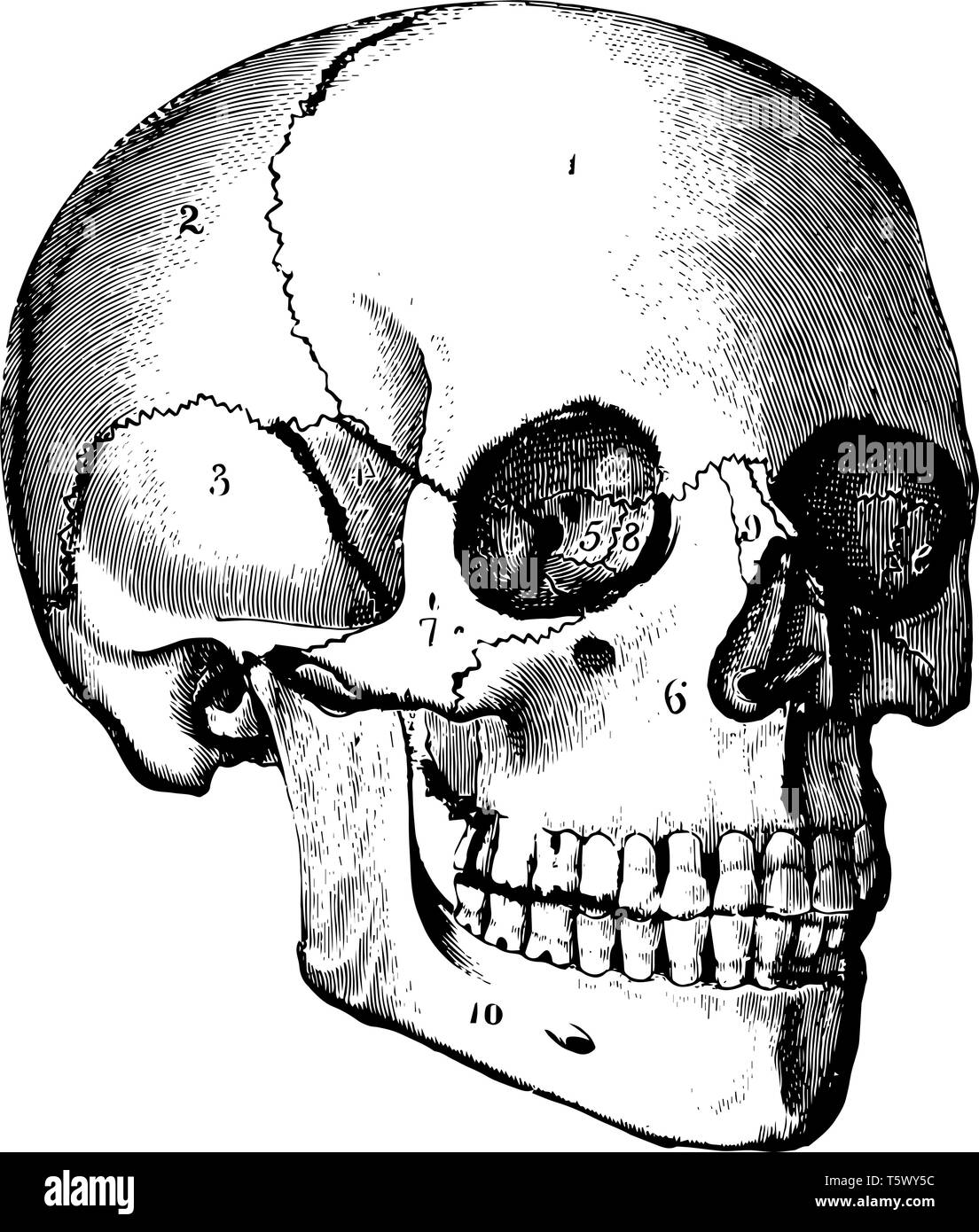 Skull basically supports the structures of the face and provides a protective cavity for the brain vintage line drawing or engraving illustration. Stock Vectorhttps://www.alamy.com/image-license-details/?v=1https://www.alamy.com/skull-basically-supports-the-structures-of-the-face-and-provides-a-protective-cavity-for-the-brain-vintage-line-drawing-or-engraving-illustration-image244588552.html
RFT5WY5C–Skull basically supports the structures of the face and provides a protective cavity for the brain vintage line drawing or engraving illustration.
 'Moonraker', Brain Rocks of the Coyote Buttes South CBS, Cottonwood Teepees, eroded Navajo sandstone rock formations with Stock Photohttps://www.alamy.com/image-license-details/?v=1https://www.alamy.com/moonraker-brain-rocks-of-the-coyote-buttes-south-cbs-cottonwood-teepees-image62214603.html
RMDH63A3–'Moonraker', Brain Rocks of the Coyote Buttes South CBS, Cottonwood Teepees, eroded Navajo sandstone rock formations with
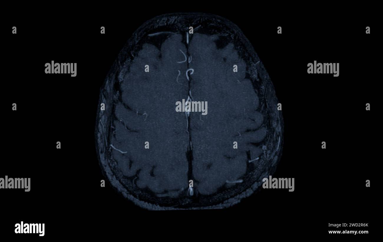 MRA Brain axial view , This imaging technique provides clear visuals of the brain's arterial and venous structures, aiding in the diagnosis of vascula Stock Photohttps://www.alamy.com/image-license-details/?v=1https://www.alamy.com/mra-brain-axial-view-this-imaging-technique-provides-clear-visuals-of-the-brains-arterial-and-venous-structures-aiding-in-the-diagnosis-of-vascula-image593205163.html
RF2WD2R6K–MRA Brain axial view , This imaging technique provides clear visuals of the brain's arterial and venous structures, aiding in the diagnosis of vascula
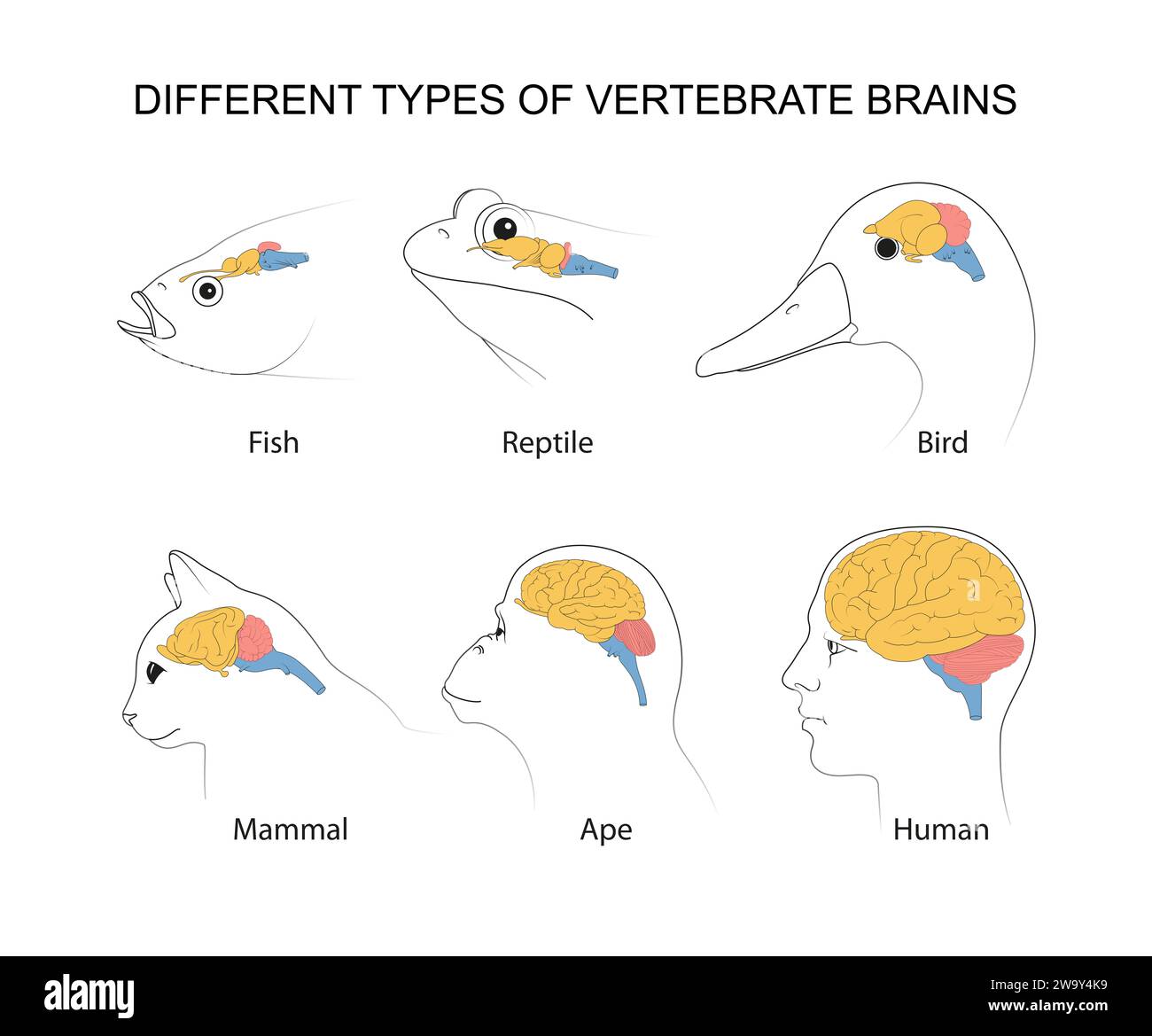 Vertebrate Brains: evolution, structures and functions Stock Photohttps://www.alamy.com/image-license-details/?v=1https://www.alamy.com/vertebrate-brains-evolution-structures-and-functions-image591280797.html
RF2W9Y4K9–Vertebrate Brains: evolution, structures and functions
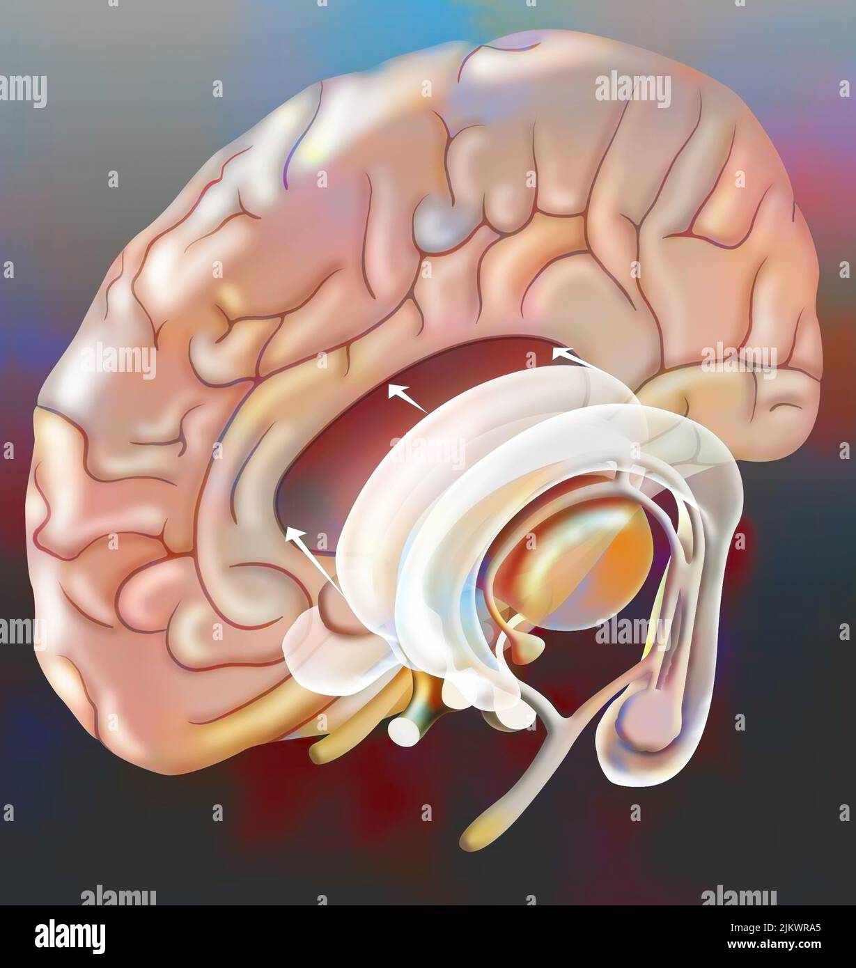 The median structures connecting the cerebral hemispheres (hippocampus, hypothalamus, pituitary..). Stock Photohttps://www.alamy.com/image-license-details/?v=1https://www.alamy.com/the-median-structures-connecting-the-cerebral-hemispheres-hippocampus-hypothalamus-pituitary-image476925517.html
RF2JKWRA5–The median structures connecting the cerebral hemispheres (hippocampus, hypothalamus, pituitary..).
 The courtyard of the Ruvo Brain Health Center in Las Vegas, NV, designed by Frank Gehry Stock Photohttps://www.alamy.com/image-license-details/?v=1https://www.alamy.com/the-courtyard-of-the-ruvo-brain-health-center-in-las-vegas-nv-designed-by-frank-gehry-image411242870.html
RF2EW1MDA–The courtyard of the Ruvo Brain Health Center in Las Vegas, NV, designed by Frank Gehry
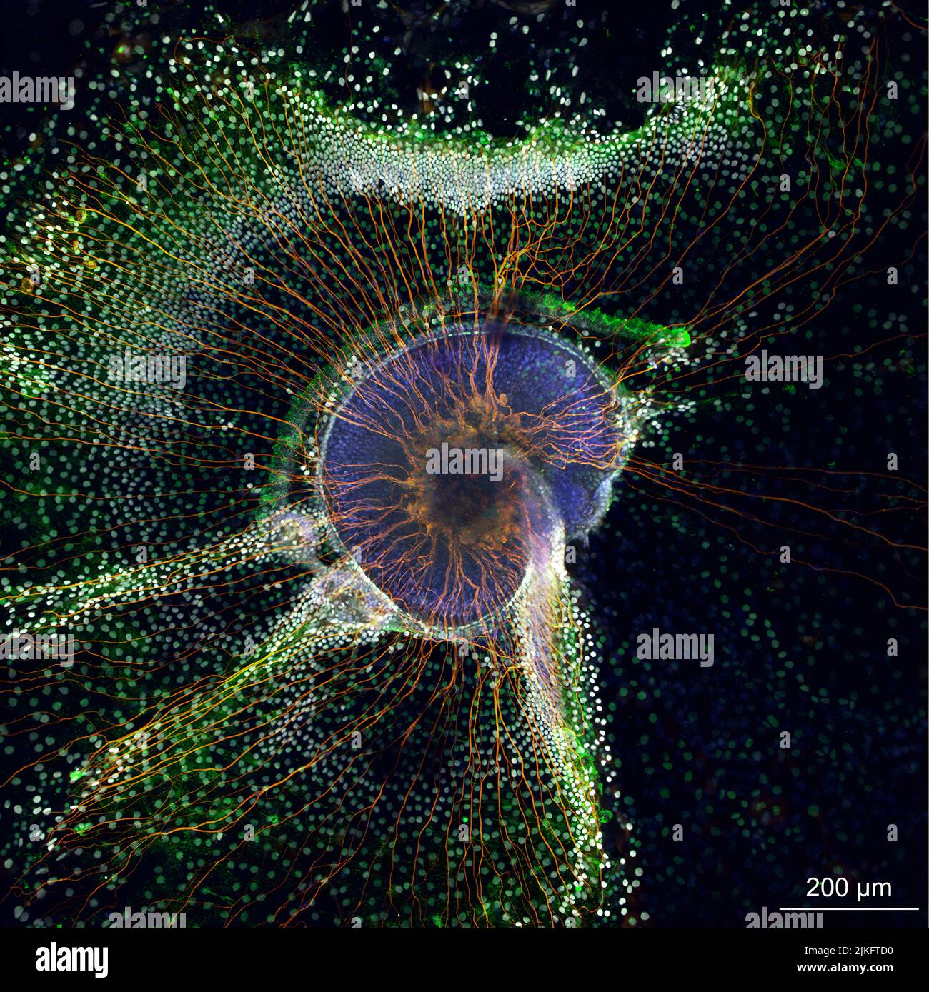 A newborn mouse cochlea (purple) grown in cell culture and neurons (orange) that send information from the cochlea to the brain. The cochlea is the hearing organ of the inner ear. Researchers use these miniature structures to learn how individual cell types work and to test potential therapies for hearing loss. Stock Photohttps://www.alamy.com/image-license-details/?v=1https://www.alamy.com/a-newborn-mouse-cochlea-purple-grown-in-cell-culture-and-neurons-orange-that-send-information-from-the-cochlea-to-the-brain-the-cochlea-is-the-hearing-organ-of-the-inner-ear-researchers-use-these-miniature-structures-to-learn-how-individual-cell-types-work-and-to-test-potential-therapies-for-hearing-loss-image476706860.html
RM2JKFTD0–A newborn mouse cochlea (purple) grown in cell culture and neurons (orange) that send information from the cochlea to the brain. The cochlea is the hearing organ of the inner ear. Researchers use these miniature structures to learn how individual cell types work and to test potential therapies for hearing loss.
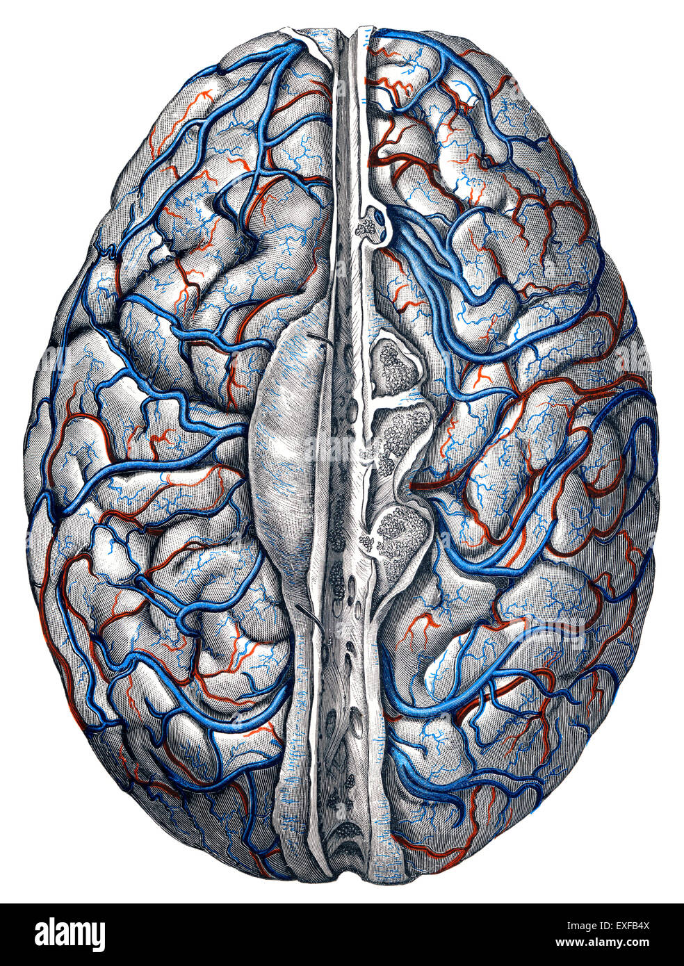 The cervical veins Stock Photohttps://www.alamy.com/image-license-details/?v=1https://www.alamy.com/stock-photo-the-cervical-veins-85160570.html
RMEXFB4X–The cervical veins
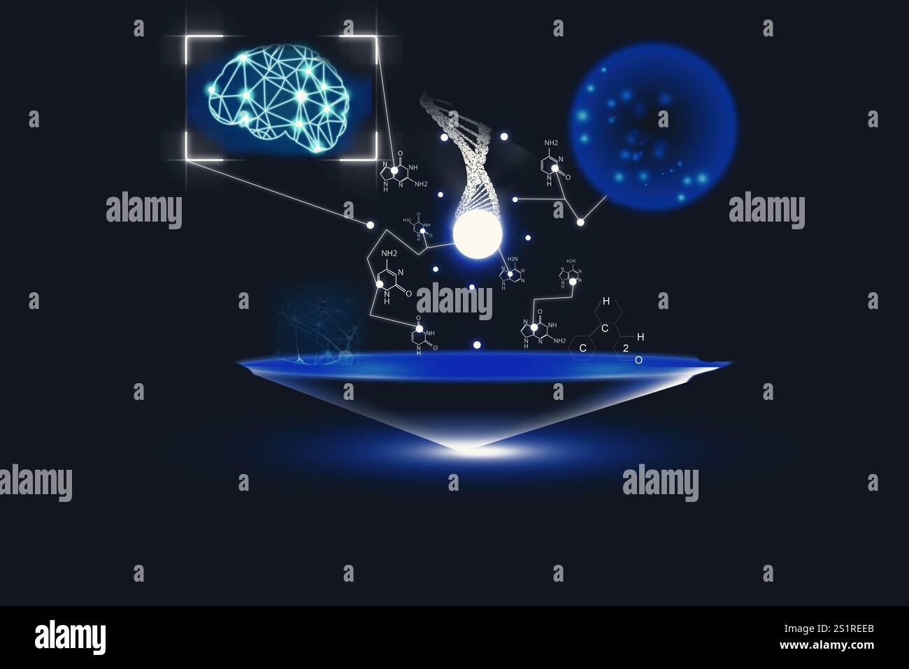 Medical technology. Human brain, DNA, neural network and molecules' structures on dark blue background, illustration Stock Photohttps://www.alamy.com/image-license-details/?v=1https://www.alamy.com/medical-technology-human-brain-dna-neural-network-and-molecules-structures-on-dark-blue-background-illustration-image637914547.html
RF2S1REEB–Medical technology. Human brain, DNA, neural network and molecules' structures on dark blue background, illustration
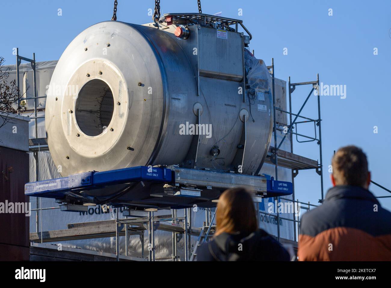 14 November 2022, Saxony-Anhalt, Magdeburg: A crane lifts a magnetic resonance tomograph into its designated place. It is to be the most powerful magnetic resonance tomograph in Europe, according to the Otto von Guericke University Hospital. In the future, the '7 Tesla Connectome' will be able to image and measure brain functions and structures. Another such device is currently said to be located only in Berkeley, California. Photo: Klaus-Dietmar Gabbert/dpa Stock Photohttps://www.alamy.com/image-license-details/?v=1https://www.alamy.com/14-november-2022-saxony-anhalt-magdeburg-a-crane-lifts-a-magnetic-resonance-tomograph-into-its-designated-place-it-is-to-be-the-most-powerful-magnetic-resonance-tomograph-in-europe-according-to-the-otto-von-guericke-university-hospital-in-the-future-the-7-tesla-connectome-will-be-able-to-image-and-measure-brain-functions-and-structures-another-such-device-is-currently-said-to-be-located-only-in-berkeley-california-photo-klaus-dietmar-gabbertdpa-image491032479.html
RM2KETCX7–14 November 2022, Saxony-Anhalt, Magdeburg: A crane lifts a magnetic resonance tomograph into its designated place. It is to be the most powerful magnetic resonance tomograph in Europe, according to the Otto von Guericke University Hospital. In the future, the '7 Tesla Connectome' will be able to image and measure brain functions and structures. Another such device is currently said to be located only in Berkeley, California. Photo: Klaus-Dietmar Gabbert/dpa
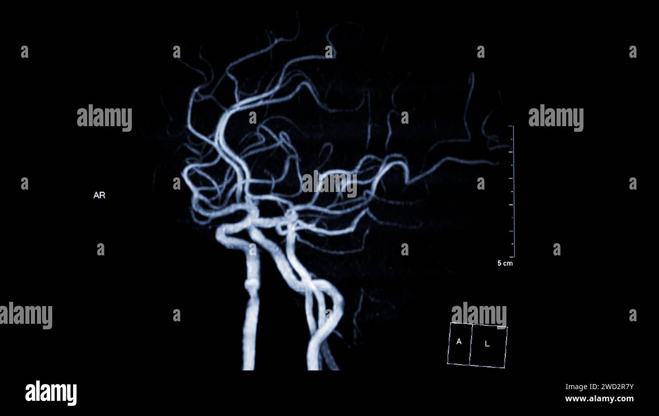 MRA Brain , This imaging technique provides clear visuals of the brain's arterial and venous structures, aiding in the diagnosis of vascular condition Stock Photohttps://www.alamy.com/image-license-details/?v=1https://www.alamy.com/mra-brain-this-imaging-technique-provides-clear-visuals-of-the-brains-arterial-and-venous-structures-aiding-in-the-diagnosis-of-vascular-condition-image593205199.html
RF2WD2R7Y–MRA Brain , This imaging technique provides clear visuals of the brain's arterial and venous structures, aiding in the diagnosis of vascular condition
 Base of the skull separates the brain from the structures of the neck and face, also known as Cranial Base, vintage line drawing or engraving illustra Stock Vectorhttps://www.alamy.com/image-license-details/?v=1https://www.alamy.com/base-of-the-skull-separates-the-brain-from-the-structures-of-the-neck-and-face-also-known-as-cranial-base-vintage-line-drawing-or-engraving-illustra-image367218787.html
RF2C9C78K–Base of the skull separates the brain from the structures of the neck and face, also known as Cranial Base, vintage line drawing or engraving illustra
 Human body examination in HUD style. Modern healthcare with body scan (Anatomy, Dna formula, Ecg monitor, data organs, X-ray, Statistic and Diagrams Stock Vectorhttps://www.alamy.com/image-license-details/?v=1https://www.alamy.com/human-body-examination-in-hud-style-modern-healthcare-with-body-scan-anatomy-dna-formula-ecg-monitor-data-organs-x-ray-statistic-and-diagrams-image425168682.html
RF2FKM2YP–Human body examination in HUD style. Modern healthcare with body scan (Anatomy, Dna formula, Ecg monitor, data organs, X-ray, Statistic and Diagrams
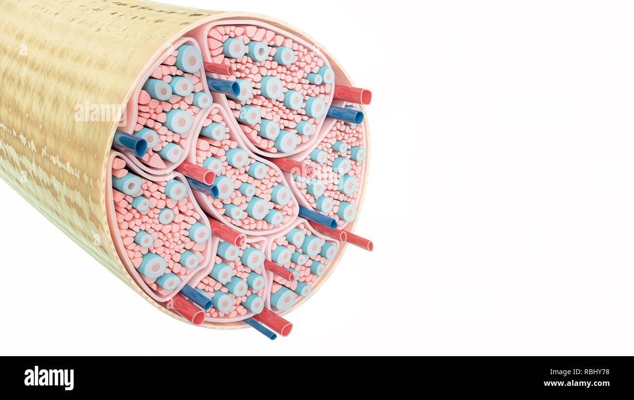 Nerve structure on wihite background- 3D Rendering Stock Photohttps://www.alamy.com/image-license-details/?v=1https://www.alamy.com/nerve-structure-on-wihite-background-3d-rendering-image230890556.html
RFRBHY78–Nerve structure on wihite background- 3D Rendering
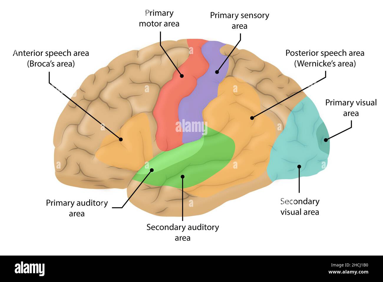 Lateral view of the brain and major structures higilighted Stock Photohttps://www.alamy.com/image-license-details/?v=1https://www.alamy.com/lateral-view-of-the-brain-and-major-structures-higilighted-image455241668.html
RF2HCJ1B0–Lateral view of the brain and major structures higilighted
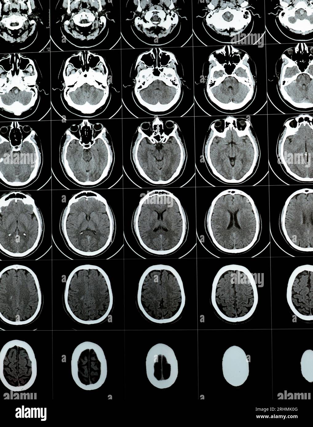 Multi slice CT scan of the brain showing Large brain stem and right centrum semiovale hematoma, normal posterior fossa structures, normal size of vent Stock Photohttps://www.alamy.com/image-license-details/?v=1https://www.alamy.com/multi-slice-ct-scan-of-the-brain-showing-large-brain-stem-and-right-centrum-semiovale-hematoma-normal-posterior-fossa-structures-normal-size-of-vent-image561634880.html
RF2RHMK0G–Multi slice CT scan of the brain showing Large brain stem and right centrum semiovale hematoma, normal posterior fossa structures, normal size of vent
 Image of brain, molecular strauctures and layers of data processing over 3d grid on black Stock Photohttps://www.alamy.com/image-license-details/?v=1https://www.alamy.com/image-of-brain-molecular-strauctures-and-layers-of-data-processing-over-3d-grid-on-black-image597414650.html
RF2WKXGDE–Image of brain, molecular strauctures and layers of data processing over 3d grid on black
 Illustration of human brain structures. Stock Photohttps://www.alamy.com/image-license-details/?v=1https://www.alamy.com/stock-photo-illustration-of-human-brain-structures-140556370.html
RFJ4JW2A–Illustration of human brain structures.
 Image of brains, molecule structures, computer language, african american man using laptop Stock Photohttps://www.alamy.com/image-license-details/?v=1https://www.alamy.com/image-of-brains-molecule-structures-computer-language-african-american-man-using-laptop-image606270039.html
RF2X69YHB–Image of brains, molecule structures, computer language, african american man using laptop
 Detailed close-up of the plastic model of the brain on the South African flag, showing the intricate details and structures. Stock Photohttps://www.alamy.com/image-license-details/?v=1https://www.alamy.com/detailed-close-up-of-the-plastic-model-of-the-brain-on-the-south-african-flag-showing-the-intricate-details-and-structures-image599642587.html
RF2WRG26K–Detailed close-up of the plastic model of the brain on the South African flag, showing the intricate details and structures.
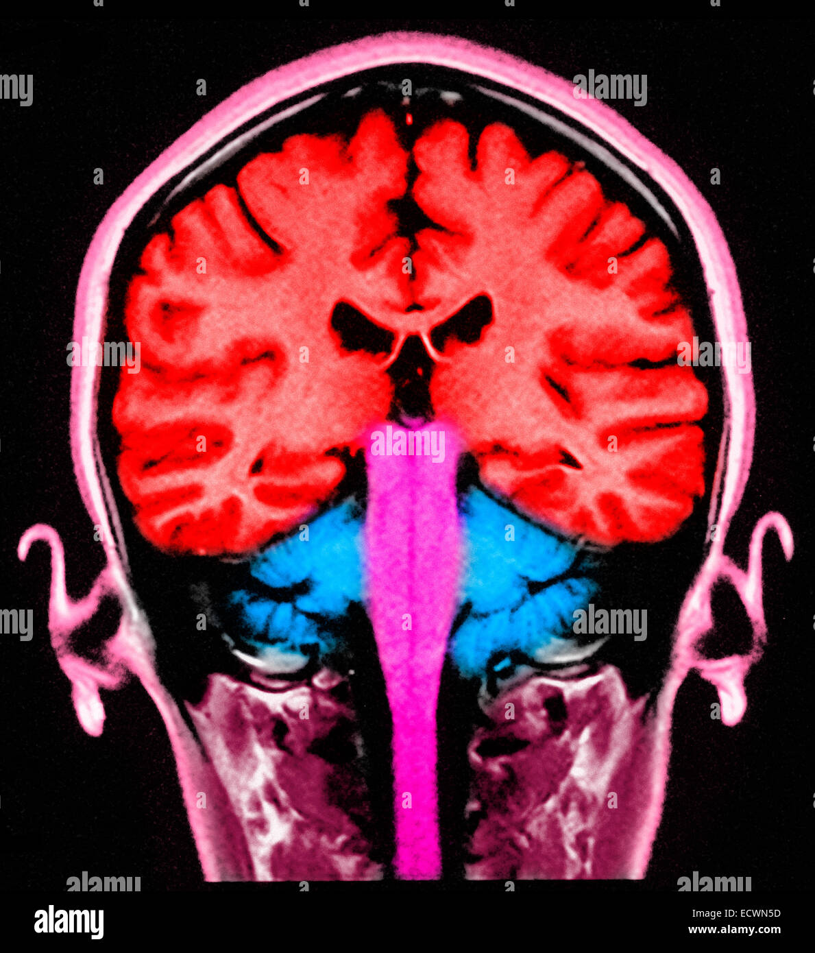 MRI of head showing brain structures. Stock Photohttps://www.alamy.com/image-license-details/?v=1https://www.alamy.com/stock-photo-mri-of-head-showing-brain-structures-76782761.html
RMECWN5D–MRI of head showing brain structures.
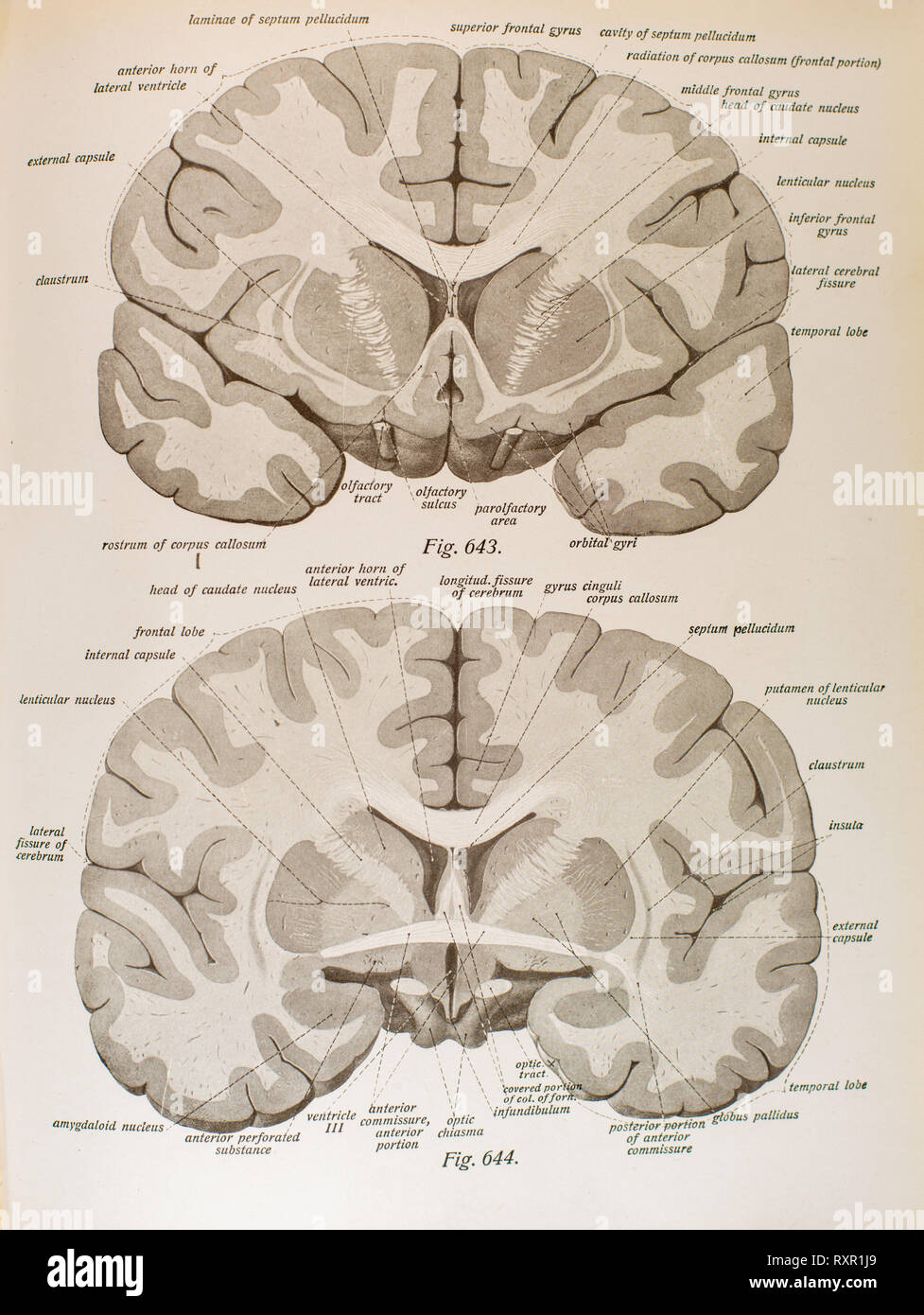 Anatomy of the Human Brain and its related structures Stock Photohttps://www.alamy.com/image-license-details/?v=1https://www.alamy.com/anatomy-of-the-human-brain-and-its-related-structures-image240222033.html
RFRXR1J9–Anatomy of the Human Brain and its related structures
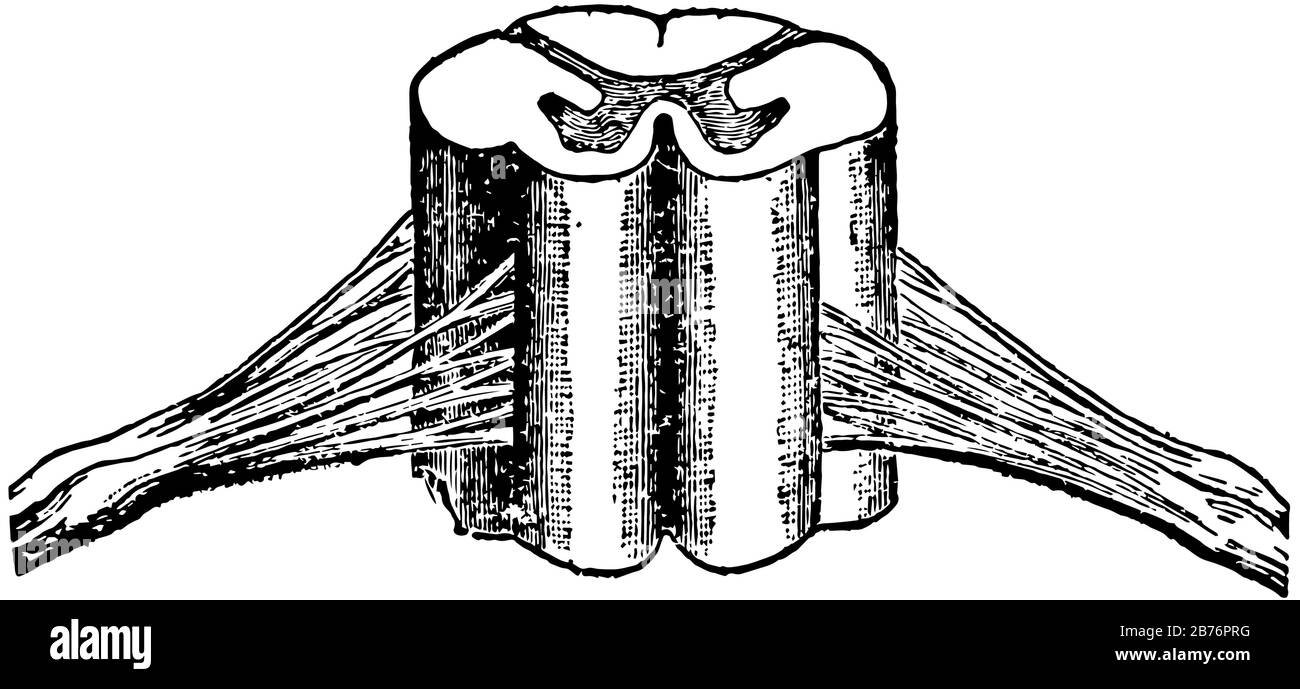 The cord-like structures composed of delicate filaments by which sensation or stimulative impulses are transmitted to and from the brain, vintage line Stock Vectorhttps://www.alamy.com/image-license-details/?v=1https://www.alamy.com/the-cord-like-structures-composed-of-delicate-filaments-by-which-sensation-or-stimulative-impulses-are-transmitted-to-and-from-the-brain-vintage-line-image348659572.html
RF2B76PRG–The cord-like structures composed of delicate filaments by which sensation or stimulative impulses are transmitted to and from the brain, vintage line
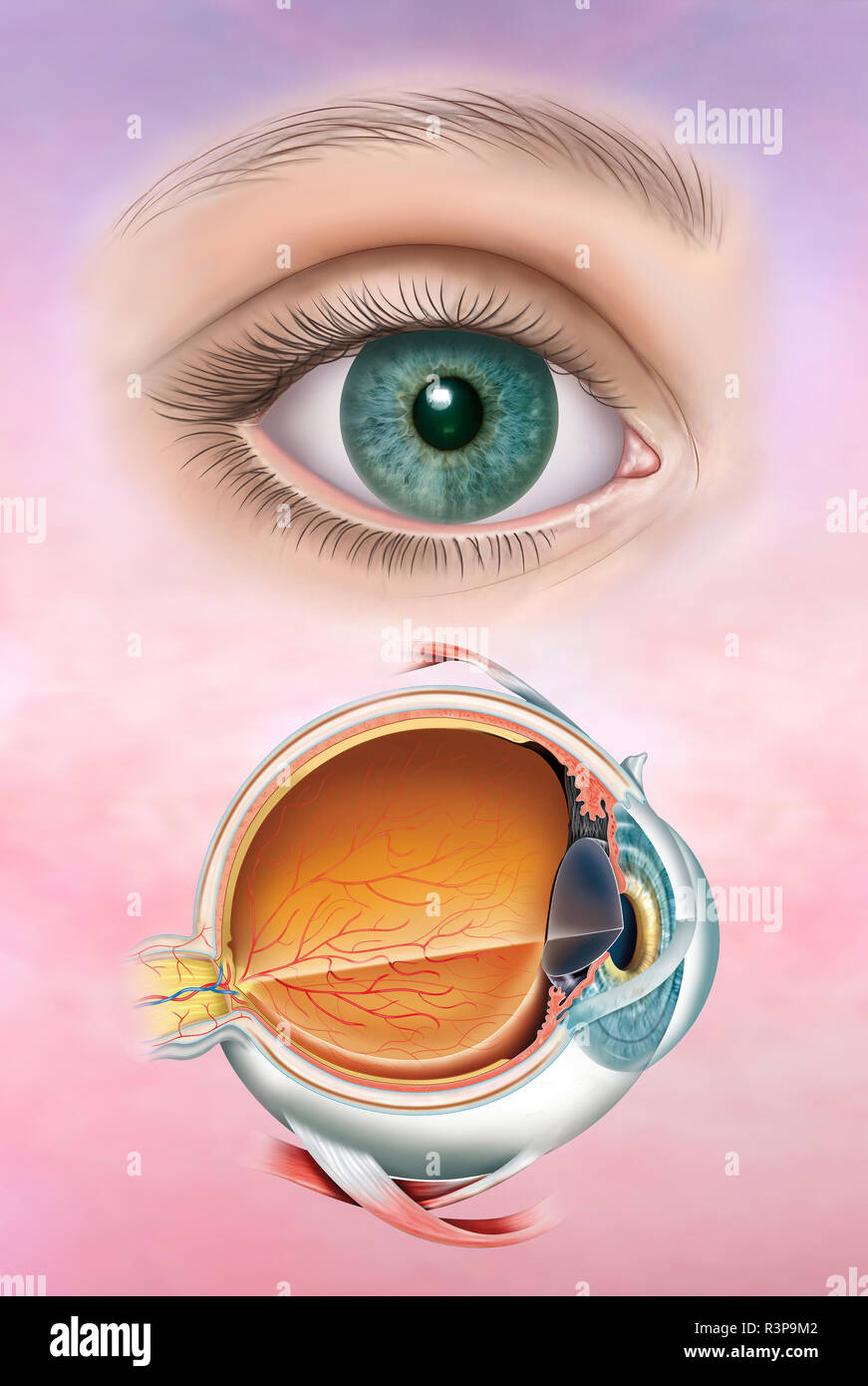 Illustration composed of the human eye in a realistic version and then the anatomy of the eye with its structure and layers that compose it. Stock Photohttps://www.alamy.com/image-license-details/?v=1https://www.alamy.com/illustration-composed-of-the-human-eye-in-a-realistic-version-and-then-the-anatomy-of-the-eye-with-its-structure-and-layers-that-compose-it-image226069314.html
RFR3P9M2–Illustration composed of the human eye in a realistic version and then the anatomy of the eye with its structure and layers that compose it.
 The courtyard of the Ruvo Brain Health Center in Las Vegas, NV, designed by Frank Gehry Stock Photohttps://www.alamy.com/image-license-details/?v=1https://www.alamy.com/the-courtyard-of-the-ruvo-brain-health-center-in-las-vegas-nv-designed-by-frank-gehry-image411750764.html
RF2EWTT8C–The courtyard of the Ruvo Brain Health Center in Las Vegas, NV, designed by Frank Gehry
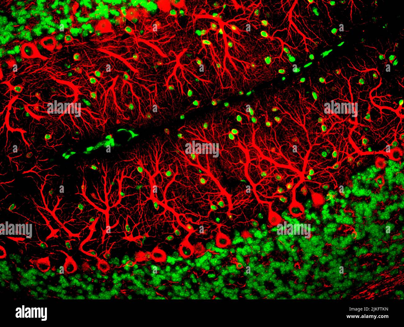 This image captures Purkinje cells, one of the main types of nerve cells contained in the brain. These cells have branching structures created called dendrites which receive signals from other nerve cells. Stock Photohttps://www.alamy.com/image-license-details/?v=1https://www.alamy.com/this-image-captures-purkinje-cells-one-of-the-main-types-of-nerve-cells-contained-in-the-brain-these-cells-have-branching-structures-created-called-dendrites-which-receive-signals-from-other-nerve-cells-image476707049.html
RM2JKFTKN–This image captures Purkinje cells, one of the main types of nerve cells contained in the brain. These cells have branching structures created called dendrites which receive signals from other nerve cells.
 Artificial reef (EcoReef, the brain child of Michael Moore, installed by Dr Mark Erdmann). Ceramic structures placed on reef bomb damaged in the 70s, Stock Photohttps://www.alamy.com/image-license-details/?v=1https://www.alamy.com/artificial-reef-ecoreef-the-brain-child-of-michael-moore-installed-by-dr-mark-erdmann-ceramic-structures-placed-on-reef-bomb-damaged-in-the-70s-image242894195.html
RFT34P0K–Artificial reef (EcoReef, the brain child of Michael Moore, installed by Dr Mark Erdmann). Ceramic structures placed on reef bomb damaged in the 70s,
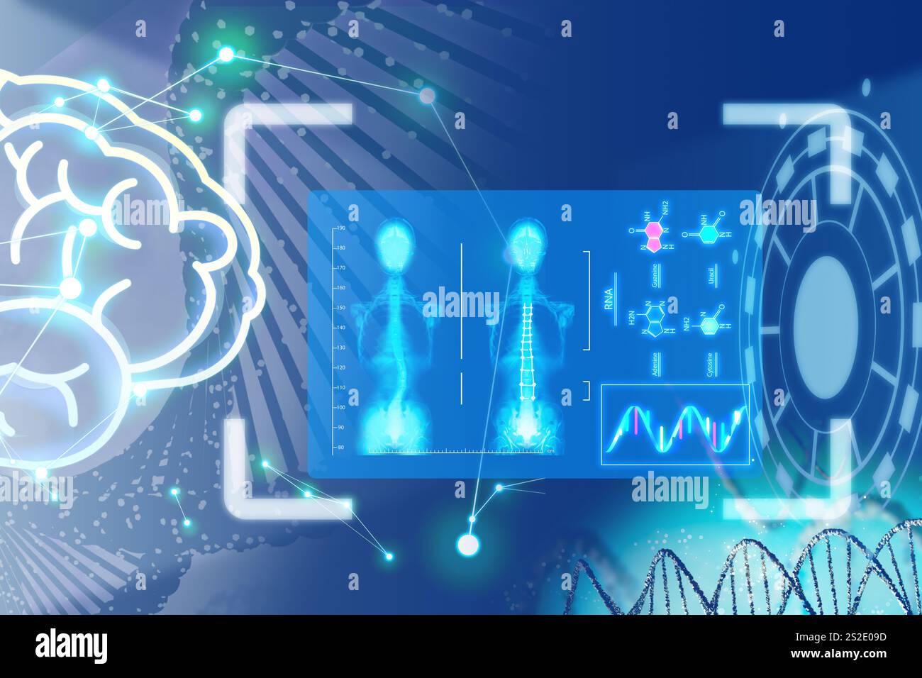 Medical technology. Human skeleton x-ray, molecules' structures, DNA and brain on blue gradient background, illustration Stock Photohttps://www.alamy.com/image-license-details/?v=1https://www.alamy.com/medical-technology-human-skeleton-x-ray-molecules-structures-dna-and-brain-on-blue-gradient-background-illustration-image638320521.html
RF2S2E09D–Medical technology. Human skeleton x-ray, molecules' structures, DNA and brain on blue gradient background, illustration
 14 November 2022, Saxony-Anhalt, Magdeburg: A crane lifts a magnetic resonance tomograph from a low-loader. It will be the most powerful magnetic resonance tomograph in Europe, according to Otto von Guericke University Hospital. The '7 Tesla Connectome' will be able to image and measure brain functions and structures in the future. Another such device is currently said to be located only in Berkeley, California. Photo: Klaus-Dietmar Gabbert/dpa Stock Photohttps://www.alamy.com/image-license-details/?v=1https://www.alamy.com/14-november-2022-saxony-anhalt-magdeburg-a-crane-lifts-a-magnetic-resonance-tomograph-from-a-low-loader-it-will-be-the-most-powerful-magnetic-resonance-tomograph-in-europe-according-to-otto-von-guericke-university-hospital-the-7-tesla-connectome-will-be-able-to-image-and-measure-brain-functions-and-structures-in-the-future-another-such-device-is-currently-said-to-be-located-only-in-berkeley-california-photo-klaus-dietmar-gabbertdpa-image491032470.html
RM2KETCWX–14 November 2022, Saxony-Anhalt, Magdeburg: A crane lifts a magnetic resonance tomograph from a low-loader. It will be the most powerful magnetic resonance tomograph in Europe, according to Otto von Guericke University Hospital. The '7 Tesla Connectome' will be able to image and measure brain functions and structures in the future. Another such device is currently said to be located only in Berkeley, California. Photo: Klaus-Dietmar Gabbert/dpa
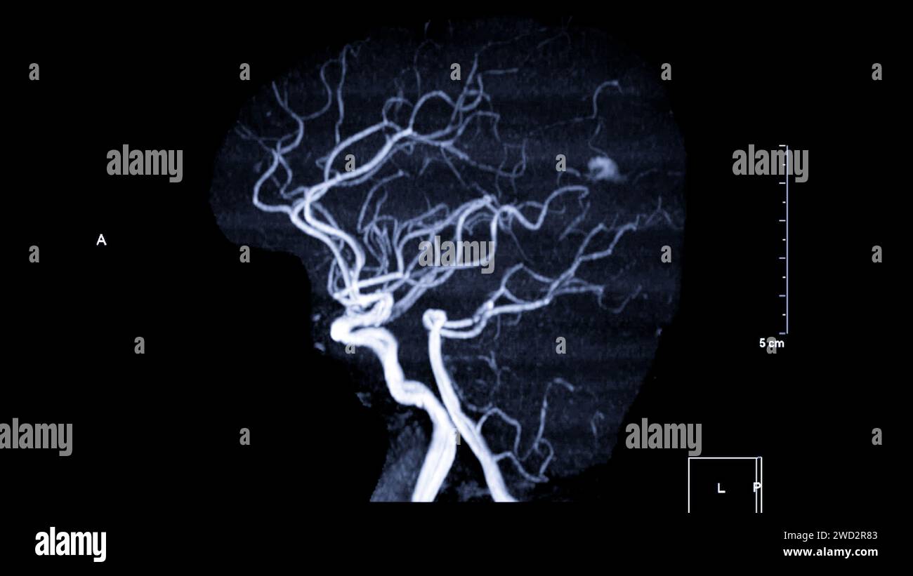 MRA Brain , This imaging technique provides clear visuals of the brain's arterial and venous structures, aiding in the diagnosis of vascular condition Stock Photohttps://www.alamy.com/image-license-details/?v=1https://www.alamy.com/mra-brain-this-imaging-technique-provides-clear-visuals-of-the-brains-arterial-and-venous-structures-aiding-in-the-diagnosis-of-vascular-condition-image593205203.html
RF2WD2R83–MRA Brain , This imaging technique provides clear visuals of the brain's arterial and venous structures, aiding in the diagnosis of vascular condition
RF2S1REEM–Medical technology. Human skeleton x-ray, DNA, brain, neural network, different icons and molecules' structures on blue gradient background, illustrat
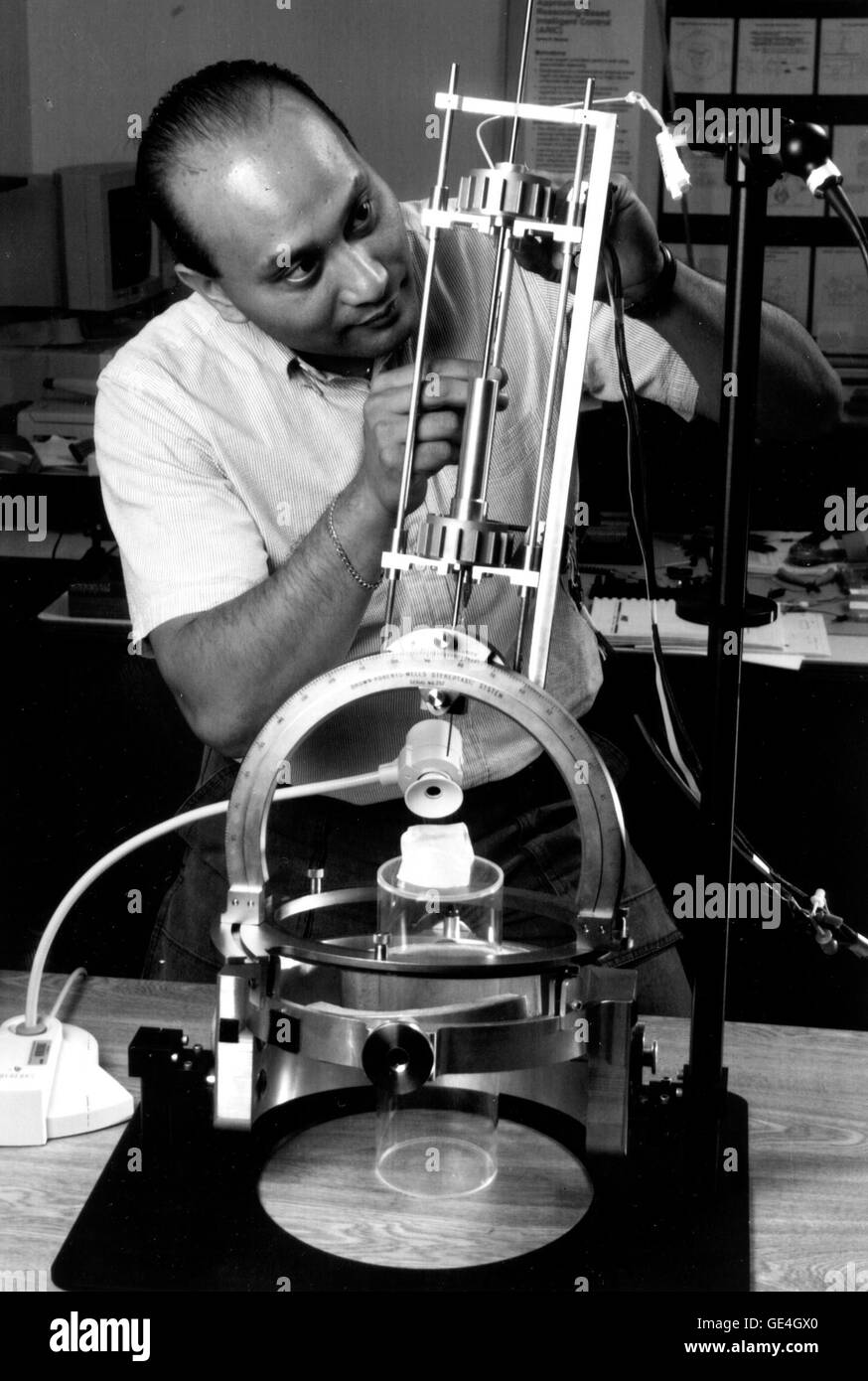 Mechanical Engineer Michael Guerrero works on the Robot Brain Surgeon testbed in the NeuroEngineering Group at the Ames Research Center, Moffett Field, California. Principal investigator Dr. Robert W. Mah states that potentially the simple robot will be able to feel brain structures better than any human surgeon, making slow, very precise movements during an operation. The brain surgery robot that may give surgeons finer control of surgical instruments during delicate brain operations is still under development. Image # : ACD97-0063-2 Date: June 2, 1997 Stock Photohttps://www.alamy.com/image-license-details/?v=1https://www.alamy.com/stock-photo-mechanical-engineer-michael-guerrero-works-on-the-robot-brain-surgeon-111968472.html
RMGE4GX0–Mechanical Engineer Michael Guerrero works on the Robot Brain Surgeon testbed in the NeuroEngineering Group at the Ames Research Center, Moffett Field, California. Principal investigator Dr. Robert W. Mah states that potentially the simple robot will be able to feel brain structures better than any human surgeon, making slow, very precise movements during an operation. The brain surgery robot that may give surgeons finer control of surgical instruments during delicate brain operations is still under development. Image # : ACD97-0063-2 Date: June 2, 1997
 A detailed MRI scan T2-weighted image of the human brain, highlighting the intricate structures and tissues. Stock Photohttps://www.alamy.com/image-license-details/?v=1https://www.alamy.com/a-detailed-mri-scan-t2-weighted-image-of-the-human-brain-highlighting-the-intricate-structures-and-tissues-image618505234.html
RF2XX79MJ–A detailed MRI scan T2-weighted image of the human brain, highlighting the intricate structures and tissues.
 Phlegm art on old electricity substation , Sheffield Stock Photohttps://www.alamy.com/image-license-details/?v=1https://www.alamy.com/stock-photo-phlegm-art-on-old-electricity-substation-sheffield-124730993.html
RMH6WYJ9–Phlegm art on old electricity substation , Sheffield
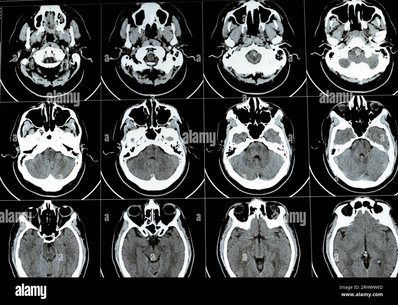 Multi slice CT scan of the brain showing Large brain stem and right centrum semiovale hematoma, normal posterior fossa structures, normal size of vent Stock Photohttps://www.alamy.com/image-license-details/?v=1https://www.alamy.com/multi-slice-ct-scan-of-the-brain-showing-large-brain-stem-and-right-centrum-semiovale-hematoma-normal-posterior-fossa-structures-normal-size-of-vent-image561749509.html
RF2RHWW6D–Multi slice CT scan of the brain showing Large brain stem and right centrum semiovale hematoma, normal posterior fossa structures, normal size of vent
RF2B7059G–Abstract healthcare icon digital medical medicine and science concept innovation technology molecular structures chemical
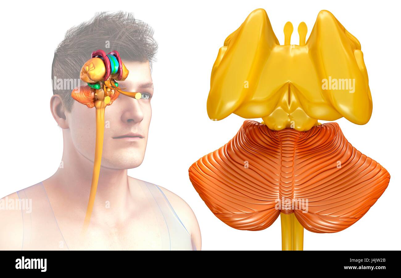 Illustration of human brain structures. Stock Photohttps://www.alamy.com/image-license-details/?v=1https://www.alamy.com/stock-photo-illustration-of-human-brain-structures-140556371.html
RFJ4JW2B–Illustration of human brain structures.
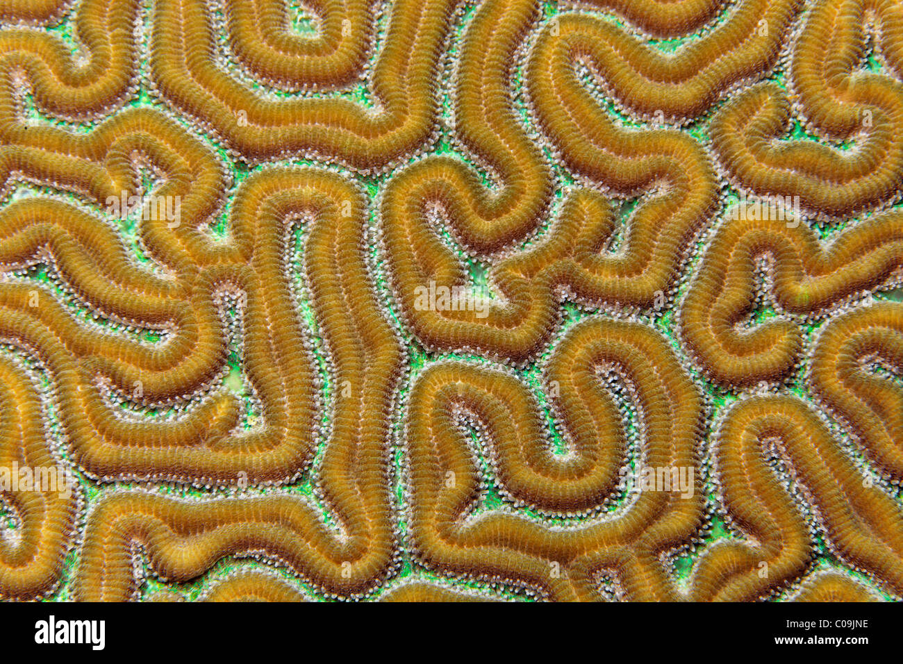 Brain Coral (Favia sp.) Little Tobago, Speyside, Trinidad and Tobago, Lesser Antilles, Caribbean Sea Stock Photohttps://www.alamy.com/image-license-details/?v=1https://www.alamy.com/stock-photo-brain-coral-favia-sp-little-tobago-speyside-trinidad-and-tobago-lesser-34633018.html
RMC09JNE–Brain Coral (Favia sp.) Little Tobago, Speyside, Trinidad and Tobago, Lesser Antilles, Caribbean Sea
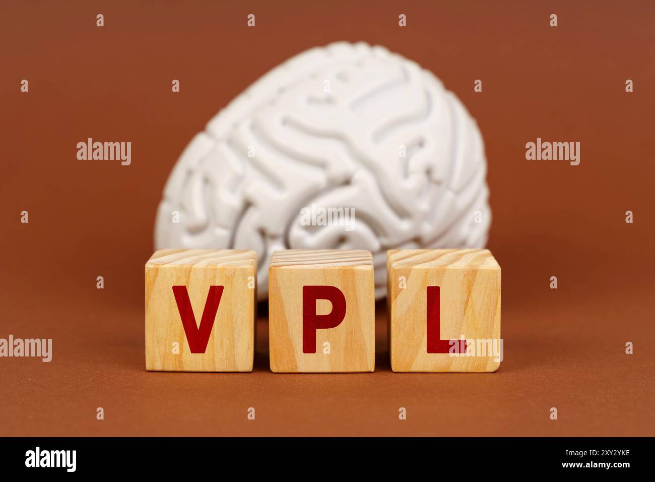 Abstract display of the ventral posterolateral nucleus concept using brain model and letter blocks, inviting exploration of anatomical structures. Stock Photohttps://www.alamy.com/image-license-details/?v=1https://www.alamy.com/abstract-display-of-the-ventral-posterolateral-nucleus-concept-using-brain-model-and-letter-blocks-inviting-exploration-of-anatomical-structures-image619024210.html
RF2XY2YKE–Abstract display of the ventral posterolateral nucleus concept using brain model and letter blocks, inviting exploration of anatomical structures.
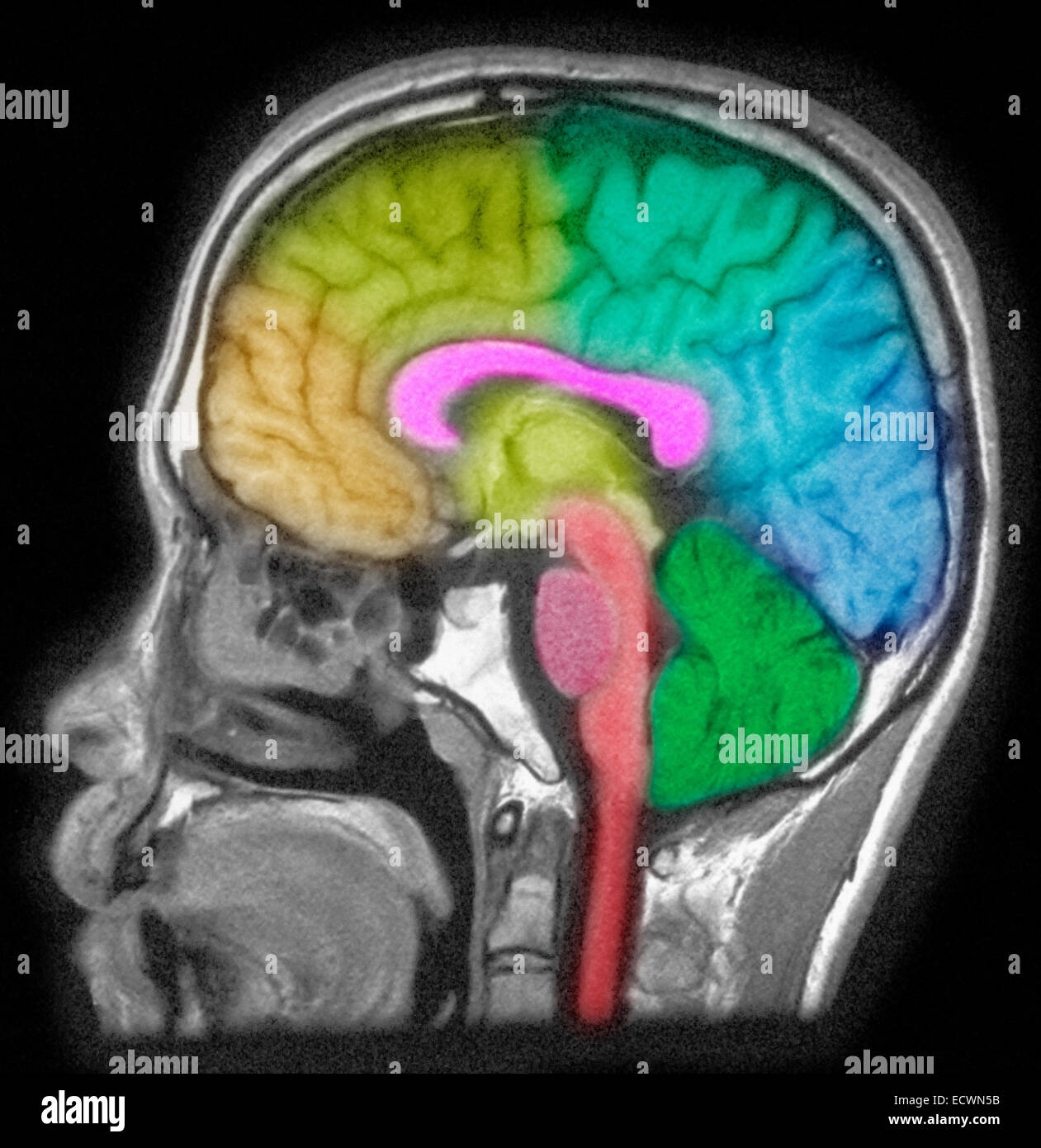 MRI of head showing brain structures. Stock Photohttps://www.alamy.com/image-license-details/?v=1https://www.alamy.com/stock-photo-mri-of-head-showing-brain-structures-76782759.html
RMECWN5B–MRI of head showing brain structures.
 Anatomy of the Human Brain and its related structures Stock Photohttps://www.alamy.com/image-license-details/?v=1https://www.alamy.com/anatomy-of-the-human-brain-and-its-related-structures-image240219892.html
RFRXPXWT–Anatomy of the Human Brain and its related structures
 The dinosaurs of North America, Washington, Govt. Print, 1896, dinosaurs, The illustration showcases a series of detailed brain casts representing various dinosaurs, including specimens like Ceratopsaurus, Cladiosaurus, Stegosaurus, and Triceratops, alongside a recent alligator. Each cast is labeled with corresponding numbers, indicating specific anatomical features of the brain structures. The arrangement allows for a comparative analysis of brain shapes and sizes across these different species, highlighting evolutionary traits and anatomical differences. This visual study serves as an import Stock Photohttps://www.alamy.com/image-license-details/?v=1https://www.alamy.com/the-dinosaurs-of-north-america-washington-govt-print-1896-dinosaurs-the-illustration-showcases-a-series-of-detailed-brain-casts-representing-various-dinosaurs-including-specimens-like-ceratopsaurus-cladiosaurus-stegosaurus-and-triceratops-alongside-a-recent-alligator-each-cast-is-labeled-with-corresponding-numbers-indicating-specific-anatomical-features-of-the-brain-structures-the-arrangement-allows-for-a-comparative-analysis-of-brain-shapes-and-sizes-across-these-different-species-highlighting-evolutionary-traits-and-anatomical-differences-this-visual-study-serves-as-an-import-image636105656.html
RM2YXW374–The dinosaurs of North America, Washington, Govt. Print, 1896, dinosaurs, The illustration showcases a series of detailed brain casts representing various dinosaurs, including specimens like Ceratopsaurus, Cladiosaurus, Stegosaurus, and Triceratops, alongside a recent alligator. Each cast is labeled with corresponding numbers, indicating specific anatomical features of the brain structures. The arrangement allows for a comparative analysis of brain shapes and sizes across these different species, highlighting evolutionary traits and anatomical differences. This visual study serves as an import
 Brain Coral (Platygyra spec.), close-up view Stock Photohttps://www.alamy.com/image-license-details/?v=1https://www.alamy.com/stock-photo-brain-coral-platygyra-spec-close-up-view-76035029.html
RMEBKKCN–Brain Coral (Platygyra spec.), close-up view
 The courtyard of the Ruvo Brain Health Center in Las Vegas, NV, designed by Frank Gehry Stock Photohttps://www.alamy.com/image-license-details/?v=1https://www.alamy.com/the-courtyard-of-the-ruvo-brain-health-center-in-las-vegas-nv-designed-by-frank-gehry-image411750717.html
RF2EWTT6N–The courtyard of the Ruvo Brain Health Center in Las Vegas, NV, designed by Frank Gehry
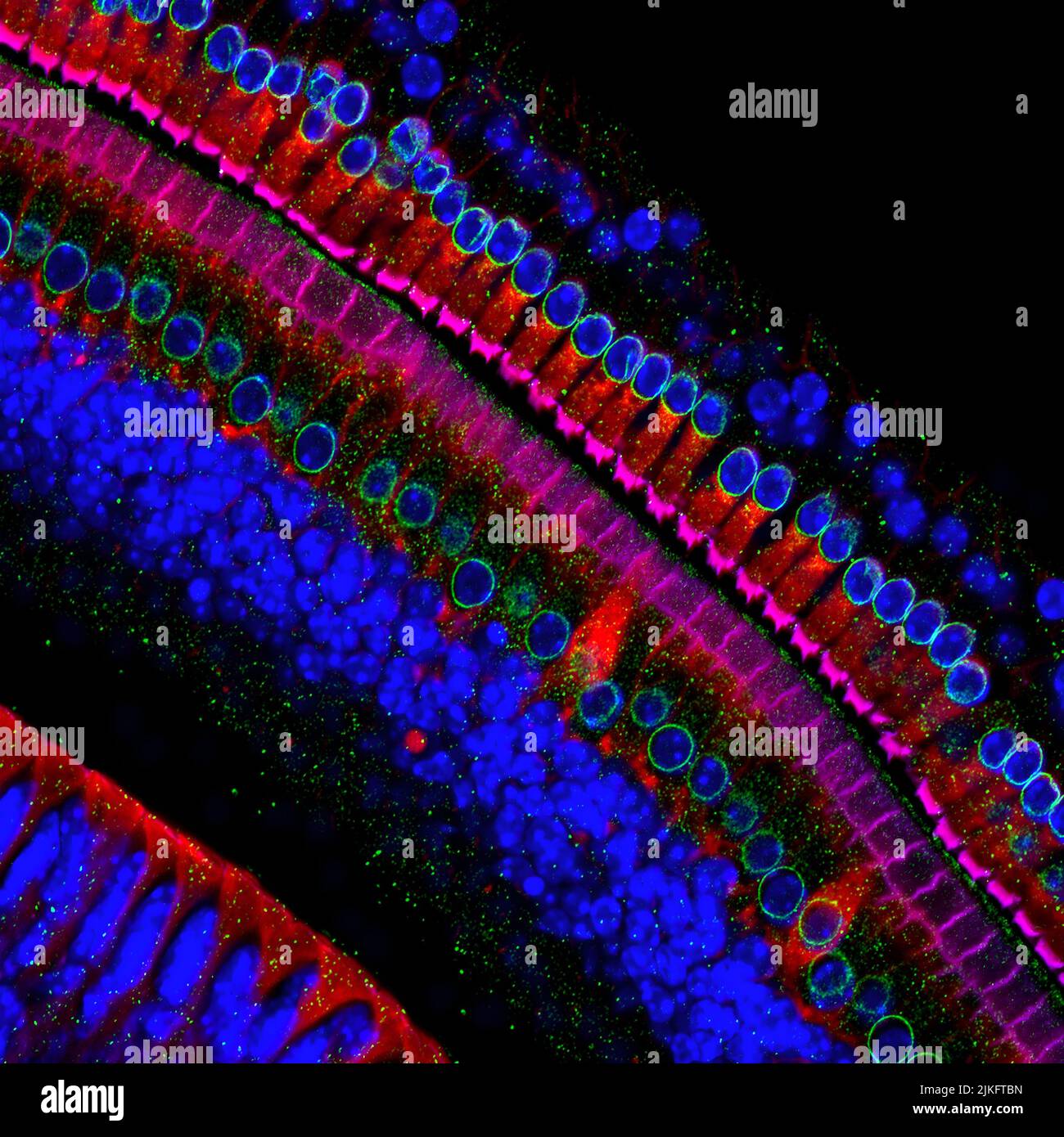 These cells get their name from the hair-like structures that extend from them into the fluid-filled tube of the inner ear. When sound reaches the ear, the hairs bend and the cells convert this movement into signals that are relayed to the brain. When we crank up the music in our cars or join tens of thousands of cheering fans at a football stadium, the noise can cause hair to pinch and break, leading to long-term hearing loss. Stock Photohttps://www.alamy.com/image-license-details/?v=1https://www.alamy.com/these-cells-get-their-name-from-the-hair-like-structures-that-extend-from-them-into-the-fluid-filled-tube-of-the-inner-ear-when-sound-reaches-the-ear-the-hairs-bend-and-the-cells-convert-this-movement-into-signals-that-are-relayed-to-the-brain-when-we-crank-up-the-music-in-our-cars-or-join-tens-of-thousands-of-cheering-fans-at-a-football-stadium-the-noise-can-cause-hair-to-pinch-and-break-leading-to-long-term-hearing-loss-image476706825.html
RM2JKFTBN–These cells get their name from the hair-like structures that extend from them into the fluid-filled tube of the inner ear. When sound reaches the ear, the hairs bend and the cells convert this movement into signals that are relayed to the brain. When we crank up the music in our cars or join tens of thousands of cheering fans at a football stadium, the noise can cause hair to pinch and break, leading to long-term hearing loss.
 Molekuelmodell aus Holz Stock Photohttps://www.alamy.com/image-license-details/?v=1https://www.alamy.com/stock-photo-molekuelmodell-aus-holz-27498700.html
RMBGMJTC–Molekuelmodell aus Holz
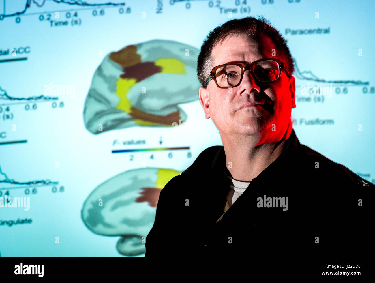 Navy Cmdr. John Hughes, director of the Magnetoencephalography (MEG) Laboratory at the National Intrepid Center of Excellence (NICoE) at Walter Reed National Military Medical Center in Bethesda, Md., stands before an image of magnetic fields generated by the brain, March 16, 2017. Unlike its cousin, the more commonly used electroencephalography (EEG), the MEG brain scan can see changes in structures that reside deep in the brain tissue. By measuring minute changes in magnetic fields generated by the cerebral cortex while resting, while thinking and while engaged in some particular task, defici Stock Photohttps://www.alamy.com/image-license-details/?v=1https://www.alamy.com/stock-photo-navy-cmdr-john-hughes-director-of-the-magnetoencephalography-meg-laboratory-138966716.html
RMJ22DD0–Navy Cmdr. John Hughes, director of the Magnetoencephalography (MEG) Laboratory at the National Intrepid Center of Excellence (NICoE) at Walter Reed National Military Medical Center in Bethesda, Md., stands before an image of magnetic fields generated by the brain, March 16, 2017. Unlike its cousin, the more commonly used electroencephalography (EEG), the MEG brain scan can see changes in structures that reside deep in the brain tissue. By measuring minute changes in magnetic fields generated by the cerebral cortex while resting, while thinking and while engaged in some particular task, defici
 14 November 2022, Saxony-Anhalt, Magdeburg: A worker attaches the chains of a crane to a magnetic resonance scanner to be lifted into place. It is the most powerful magnetic resonance tomograph in Europe, according to Otto von Guericke University Hospital. In the future, the '7 Tesla Connectome' will be able to image and measure brain functions and structures. Another such device is currently said to be located only in Berkeley, California. Photo: Klaus-Dietmar Gabbert/dpa Stock Photohttps://www.alamy.com/image-license-details/?v=1https://www.alamy.com/14-november-2022-saxony-anhalt-magdeburg-a-worker-attaches-the-chains-of-a-crane-to-a-magnetic-resonance-scanner-to-be-lifted-into-place-it-is-the-most-powerful-magnetic-resonance-tomograph-in-europe-according-to-otto-von-guericke-university-hospital-in-the-future-the-7-tesla-connectome-will-be-able-to-image-and-measure-brain-functions-and-structures-another-such-device-is-currently-said-to-be-located-only-in-berkeley-california-photo-klaus-dietmar-gabbertdpa-image491032521.html
RM2KETCYN–14 November 2022, Saxony-Anhalt, Magdeburg: A worker attaches the chains of a crane to a magnetic resonance scanner to be lifted into place. It is the most powerful magnetic resonance tomograph in Europe, according to Otto von Guericke University Hospital. In the future, the '7 Tesla Connectome' will be able to image and measure brain functions and structures. Another such device is currently said to be located only in Berkeley, California. Photo: Klaus-Dietmar Gabbert/dpa
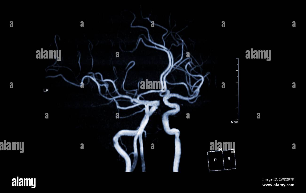 MRA Brain , This imaging technique provides clear visuals of the brain's arterial and venous structures, aiding in the diagnosis of vascular condition Stock Photohttps://www.alamy.com/image-license-details/?v=1https://www.alamy.com/mra-brain-this-imaging-technique-provides-clear-visuals-of-the-brains-arterial-and-venous-structures-aiding-in-the-diagnosis-of-vascular-condition-image593205191.html
RF2WD2R7K–MRA Brain , This imaging technique provides clear visuals of the brain's arterial and venous structures, aiding in the diagnosis of vascular condition
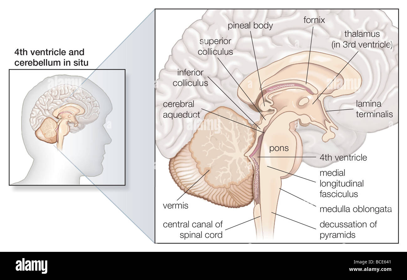 Sagittal section of the human brain, showing structures of the cerebellum, brainstem, and cerebral ventricles. Stock Photohttps://www.alamy.com/image-license-details/?v=1https://www.alamy.com/stock-photo-sagittal-section-of-the-human-brain-showing-structures-of-the-cerebellum-24898385.html
RMBCE641–Sagittal section of the human brain, showing structures of the cerebellum, brainstem, and cerebral ventricles.
 Advances in Genetic Testing concept. Scientists conduct research in a lab setting, analyzing DNA and brain structures. Medical innovation and diagnostics. Vector illustration. Stock Vectorhttps://www.alamy.com/image-license-details/?v=1https://www.alamy.com/advances-in-genetic-testing-concept-scientists-conduct-research-in-a-lab-setting-analyzing-dna-and-brain-structures-medical-innovation-and-diagnostics-vector-illustration-image617484637.html
RF2XTGRXN–Advances in Genetic Testing concept. Scientists conduct research in a lab setting, analyzing DNA and brain structures. Medical innovation and diagnostics. Vector illustration.
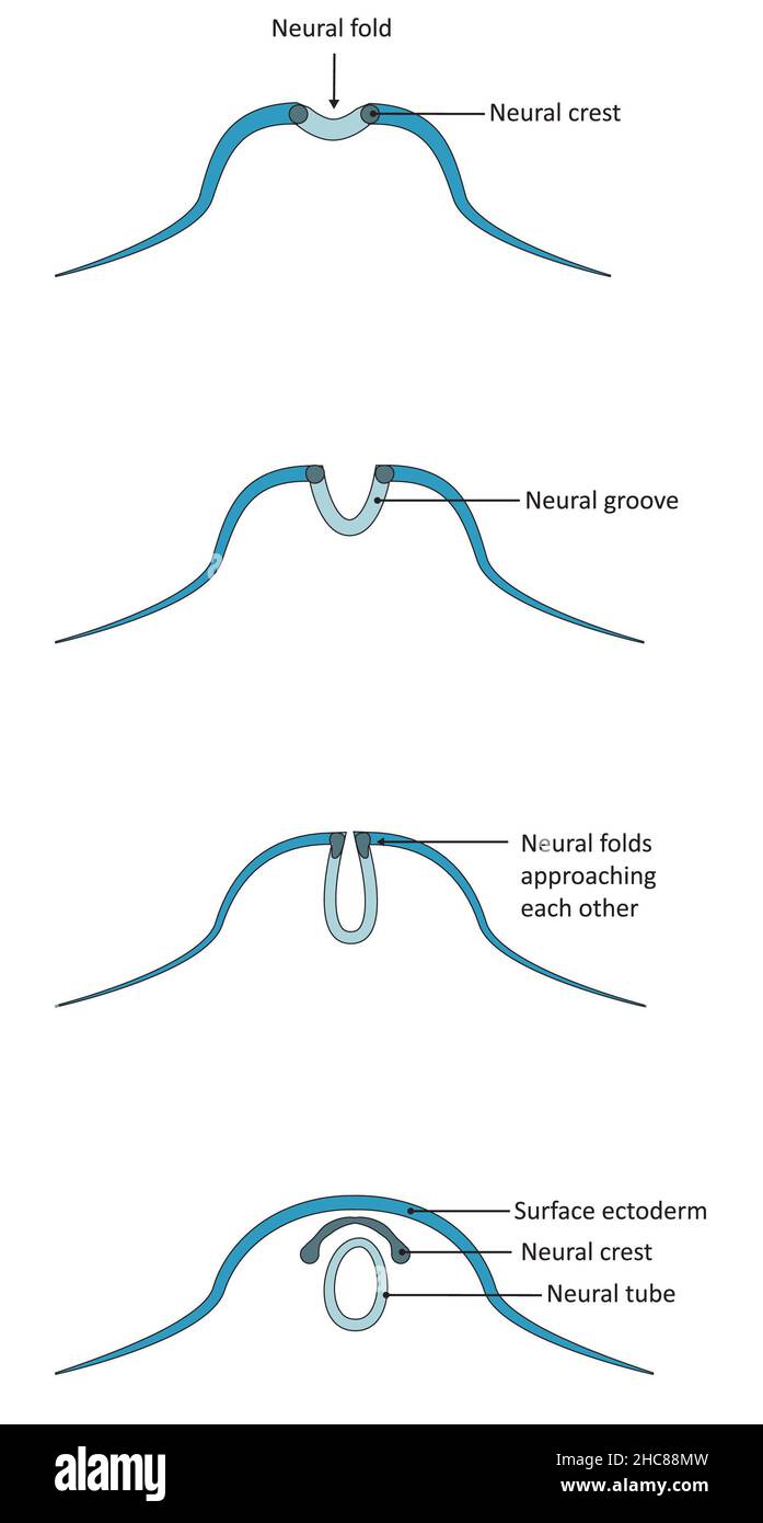 Neurulation, development of the structures derived from the ectoderm. Stock Photohttps://www.alamy.com/image-license-details/?v=1https://www.alamy.com/neurulation-development-of-the-structures-derived-from-the-ectoderm-image455027913.html
RF2HC88MW–Neurulation, development of the structures derived from the ectoderm.
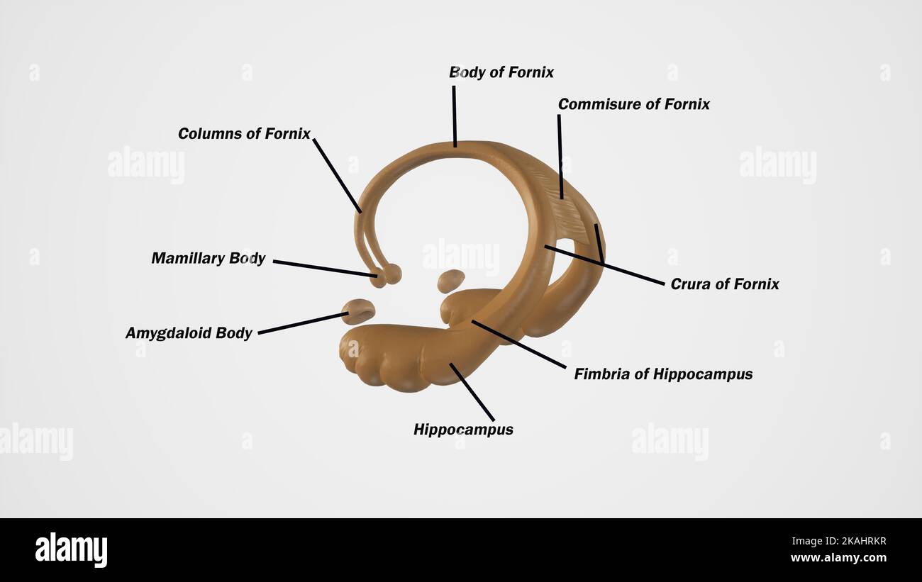 medically accurate illustration of Cerebral Fornix Stock Photohttps://www.alamy.com/image-license-details/?v=1https://www.alamy.com/medically-accurate-illustration-of-cerebral-fornix-image488428635.html
RF2KAHRKR–medically accurate illustration of Cerebral Fornix
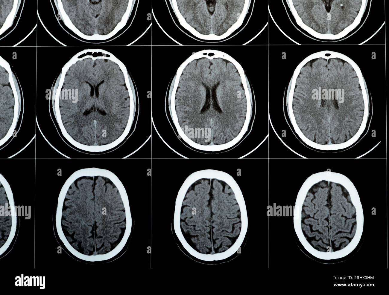 Multi slice CT scan of the brain showing Large brain stem and right centrum semiovale hematoma, normal posterior fossa structures, normal size of vent Stock Photohttps://www.alamy.com/image-license-details/?v=1https://www.alamy.com/multi-slice-ct-scan-of-the-brain-showing-large-brain-stem-and-right-centrum-semiovale-hematoma-normal-posterior-fossa-structures-normal-size-of-vent-image561752176.html
RF2RHX0HM–Multi slice CT scan of the brain showing Large brain stem and right centrum semiovale hematoma, normal posterior fossa structures, normal size of vent