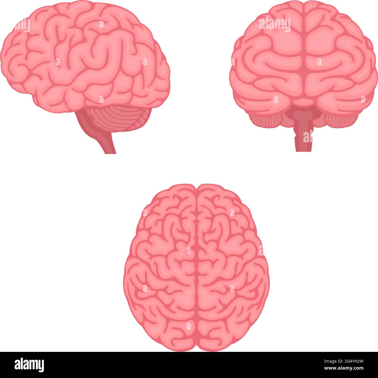
Brainstem Cut Out Stock Images
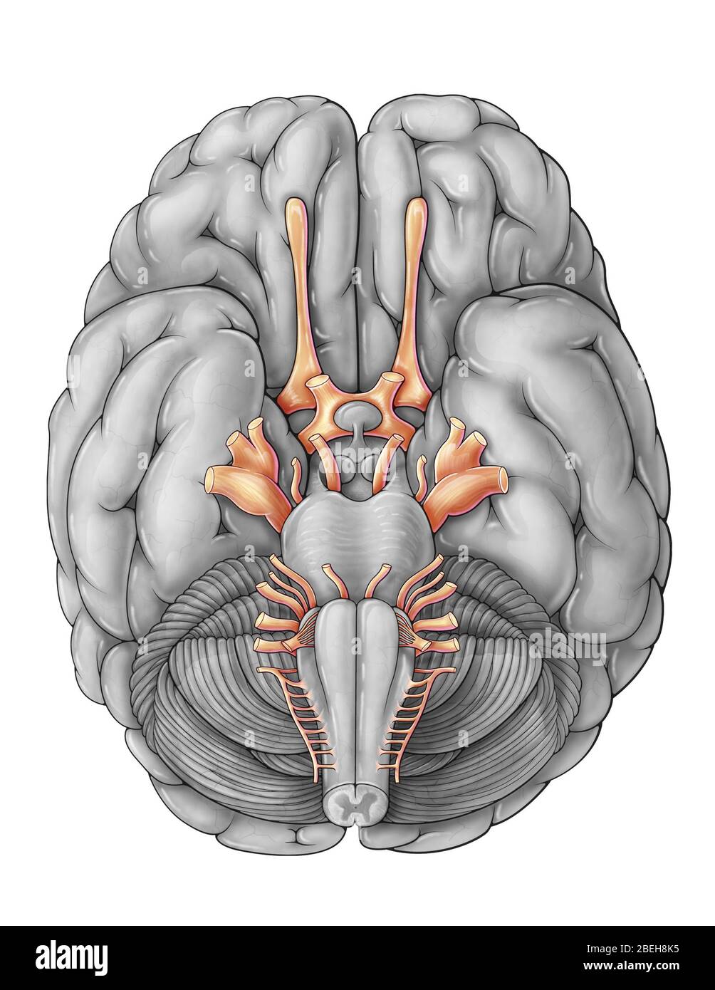 Cranial Nerves, Illustration Stock Photohttps://www.alamy.com/image-license-details/?v=1https://www.alamy.com/cranial-nerves-illustration-image353192537.html
RM2BEH8K5–Cranial Nerves, Illustration
 The brainstem Stock Photohttps://www.alamy.com/image-license-details/?v=1https://www.alamy.com/stock-photo-the-brainstem-13167490.html
RFACJ7HR–The brainstem
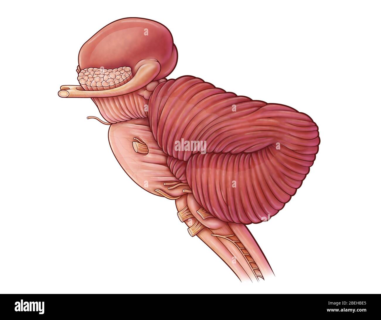 Diencephalon and Brainstem, illustration Stock Photohttps://www.alamy.com/image-license-details/?v=1https://www.alamy.com/diencephalon-and-brainstem-illustration-image353194749.html
RM2BEHBE5–Diencephalon and Brainstem, illustration
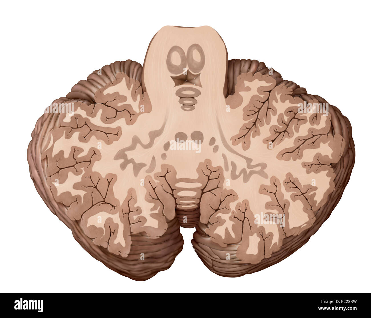 The cerebellum is the part of the brain located under the cerebrum, behind the brainstem. It ensures motor coordination as well as the maintenance of balance and posture. The cerebellum also allows for harmonious voluntary movements, without useless effort, without losing balance and without trembling. Stock Photohttps://www.alamy.com/image-license-details/?v=1https://www.alamy.com/the-cerebellum-is-the-part-of-the-brain-located-under-the-cerebrum-image156173469.html
RMK228RW–The cerebellum is the part of the brain located under the cerebrum, behind the brainstem. It ensures motor coordination as well as the maintenance of balance and posture. The cerebellum also allows for harmonious voluntary movements, without useless effort, without losing balance and without trembling.
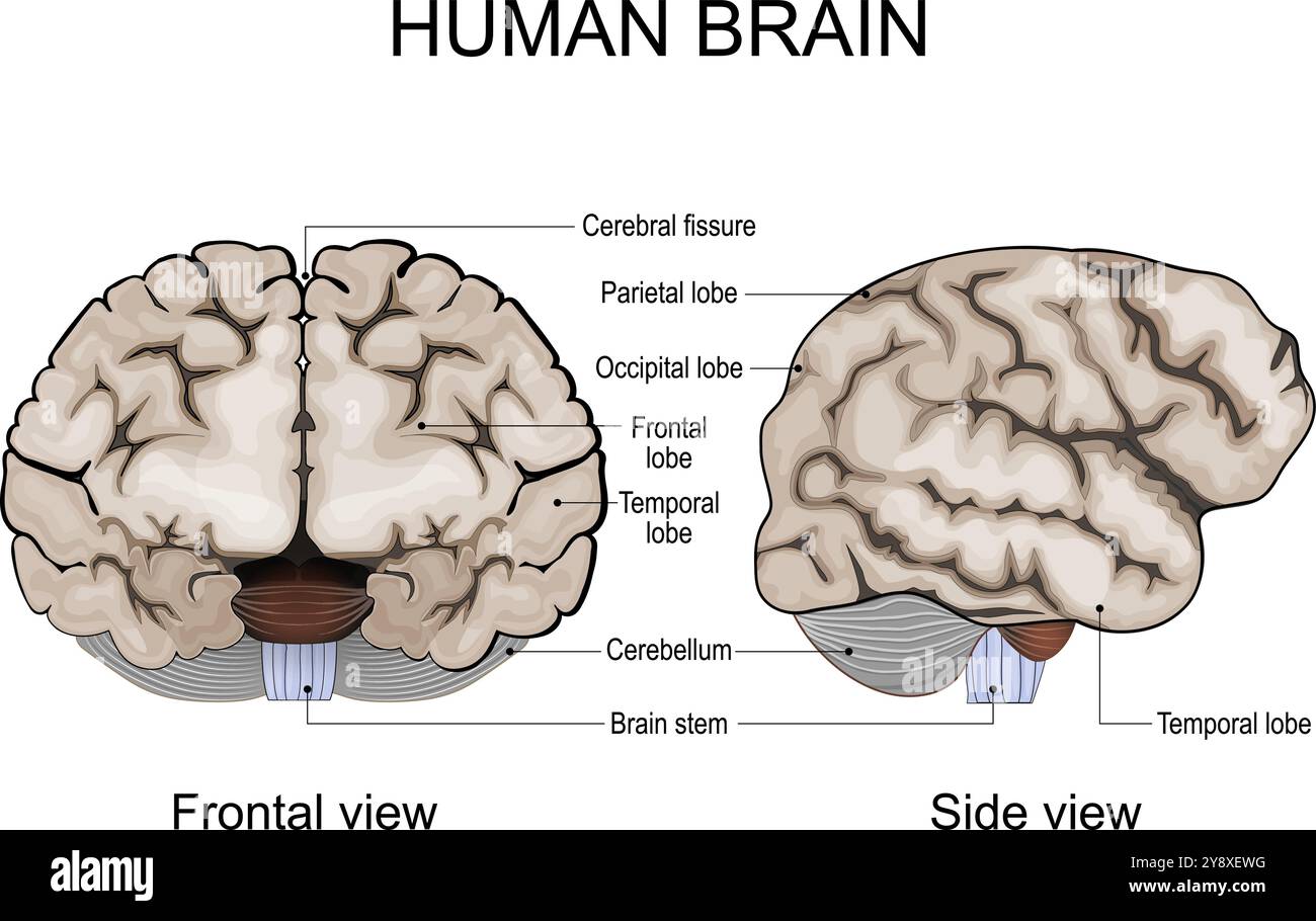 Human brain anatomy. Cerebral hemispheres, Cerebral cortex, Frontal, Parietal, Temporal, Occipital lobes, Cerebellum and Brainstem, Cerebral fissure. Stock Vectorhttps://www.alamy.com/image-license-details/?v=1https://www.alamy.com/human-brain-anatomy-cerebral-hemispheres-cerebral-cortex-frontal-parietal-temporal-occipital-lobes-cerebellum-and-brainstem-cerebral-fissure-image625072940.html
RF2Y8XEWG–Human brain anatomy. Cerebral hemispheres, Cerebral cortex, Frontal, Parietal, Temporal, Occipital lobes, Cerebellum and Brainstem, Cerebral fissure.
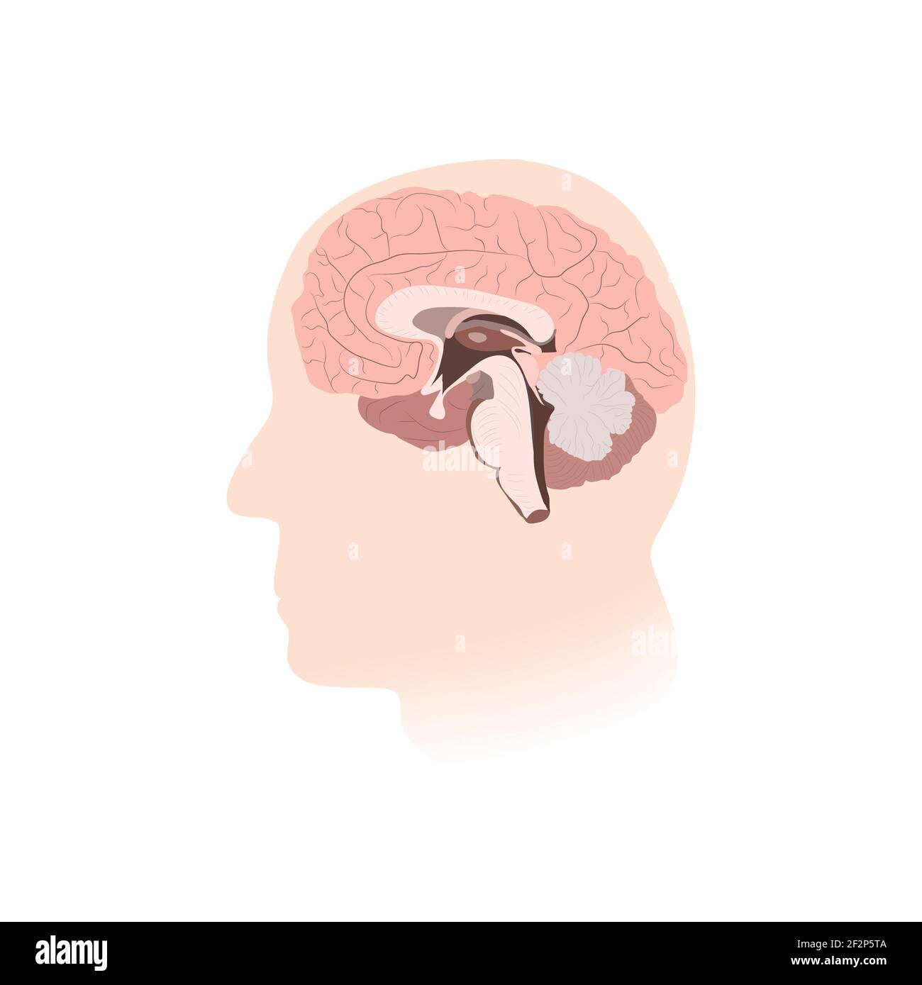 Inner view of brain, illustration Stock Photohttps://www.alamy.com/image-license-details/?v=1https://www.alamy.com/inner-view-of-brain-illustration-image414765690.html
RF2F2P5TA–Inner view of brain, illustration
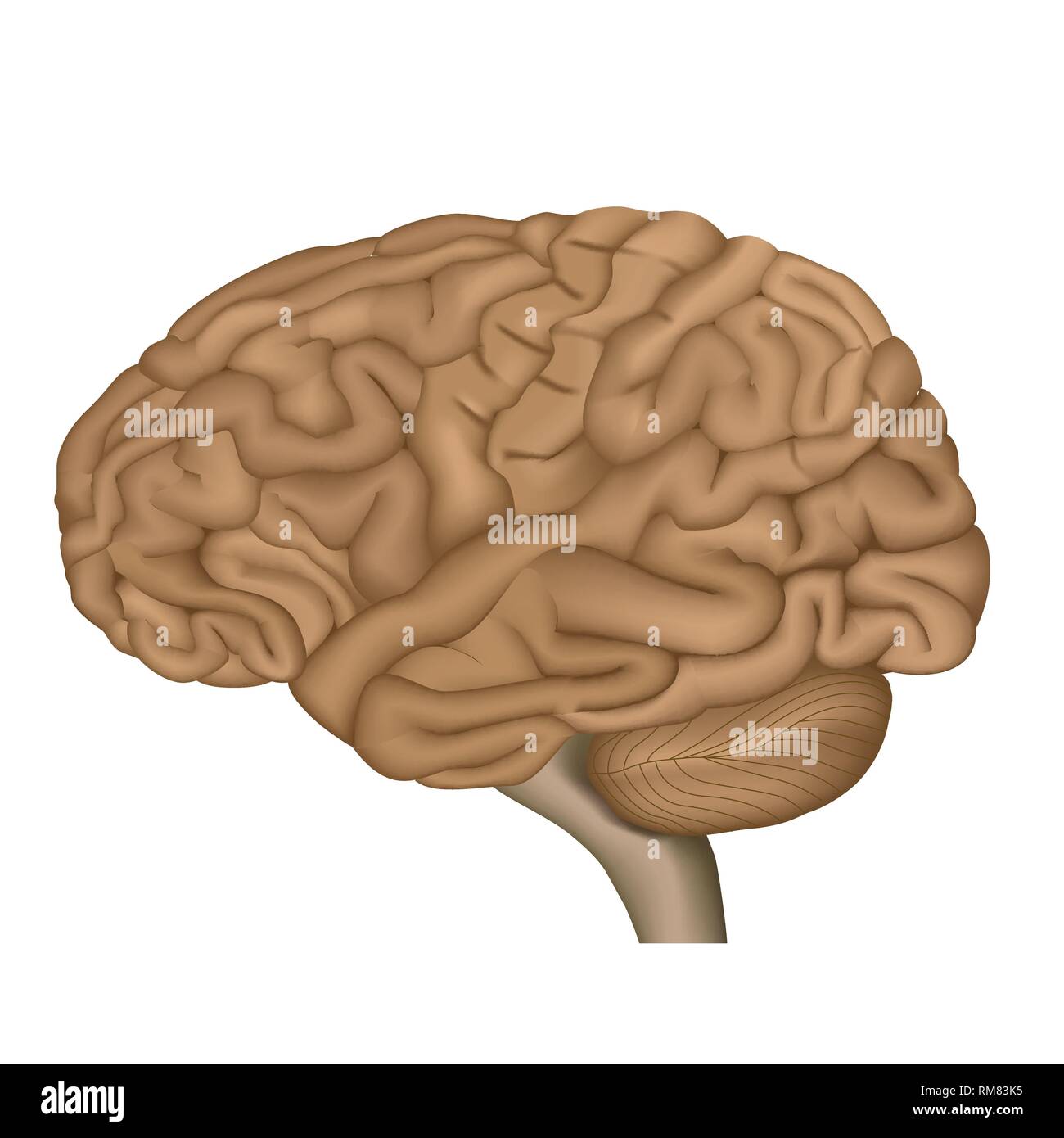 Human brain anatomy 3d vector illustration on white background Stock Vectorhttps://www.alamy.com/image-license-details/?v=1https://www.alamy.com/human-brain-anatomy-3d-vector-illustration-on-white-background-image236206409.html
RFRM83K5–Human brain anatomy 3d vector illustration on white background
 Basal ganglia anatomy, illustration Stock Photohttps://www.alamy.com/image-license-details/?v=1https://www.alamy.com/basal-ganglia-anatomy-illustration-image469205180.html
RF2J7A3YT–Basal ganglia anatomy, illustration
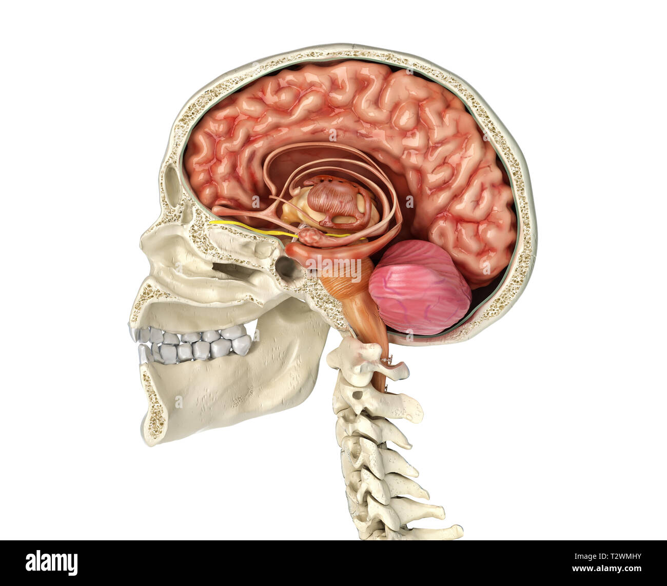 Human skull mid sagittal cross-section with brain. Side view on white background. Stock Photohttps://www.alamy.com/image-license-details/?v=1https://www.alamy.com/human-skull-mid-sagittal-cross-section-with-brain-side-view-on-white-background-image242739447.html
RFT2WMHY–Human skull mid sagittal cross-section with brain. Side view on white background.
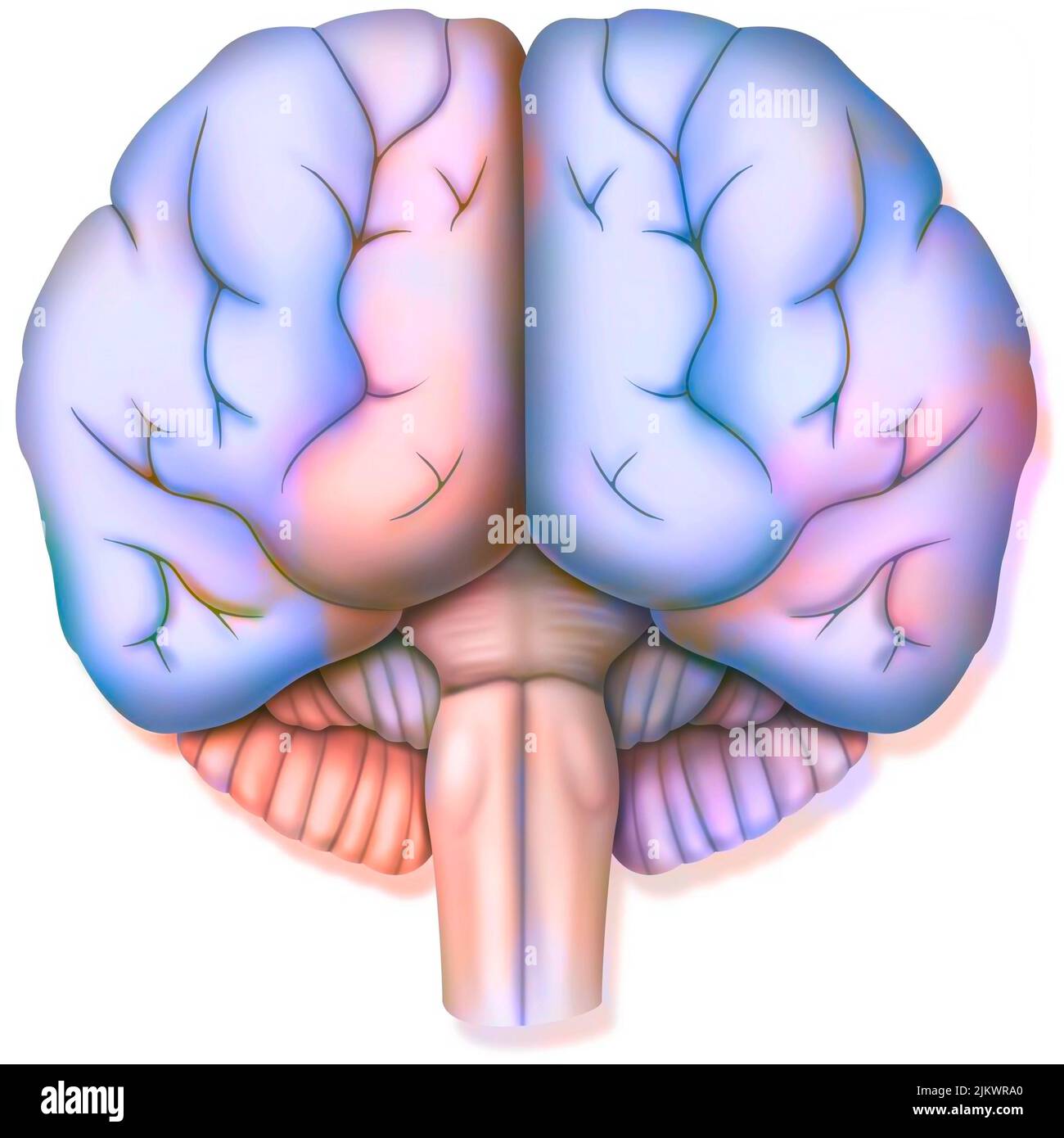 Brain, with the two cerebral hemispheres, the cerebellum and the brainstem. Stock Photohttps://www.alamy.com/image-license-details/?v=1https://www.alamy.com/brain-with-the-two-cerebral-hemispheres-the-cerebellum-and-the-brainstem-image476925512.html
RF2JKWRA0–Brain, with the two cerebral hemispheres, the cerebellum and the brainstem.
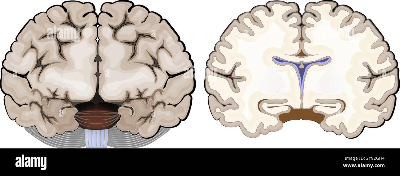 Brain anatomy. Frontal view and cross section of a human brain. Close-up of Hippocampus, and ventricles. Cerebral cortex. Vector illustration Stock Vectorhttps://www.alamy.com/image-license-details/?v=1https://www.alamy.com/brain-anatomy-frontal-view-and-cross-section-of-a-human-brain-close-up-of-hippocampus-and-ventricles-cerebral-cortex-vector-illustration-image625162080.html
RF2Y92GH4–Brain anatomy. Frontal view and cross section of a human brain. Close-up of Hippocampus, and ventricles. Cerebral cortex. Vector illustration
 Model of a human brain isolated on white background Stock Photohttps://www.alamy.com/image-license-details/?v=1https://www.alamy.com/model-of-a-human-brain-isolated-on-white-background-image471194311.html
RF2JAGN47–Model of a human brain isolated on white background
RF2YBGH24–One continuous line drawing of half human brain and love heart shape logo icon. Psychological split affection logotype symbol template concept.
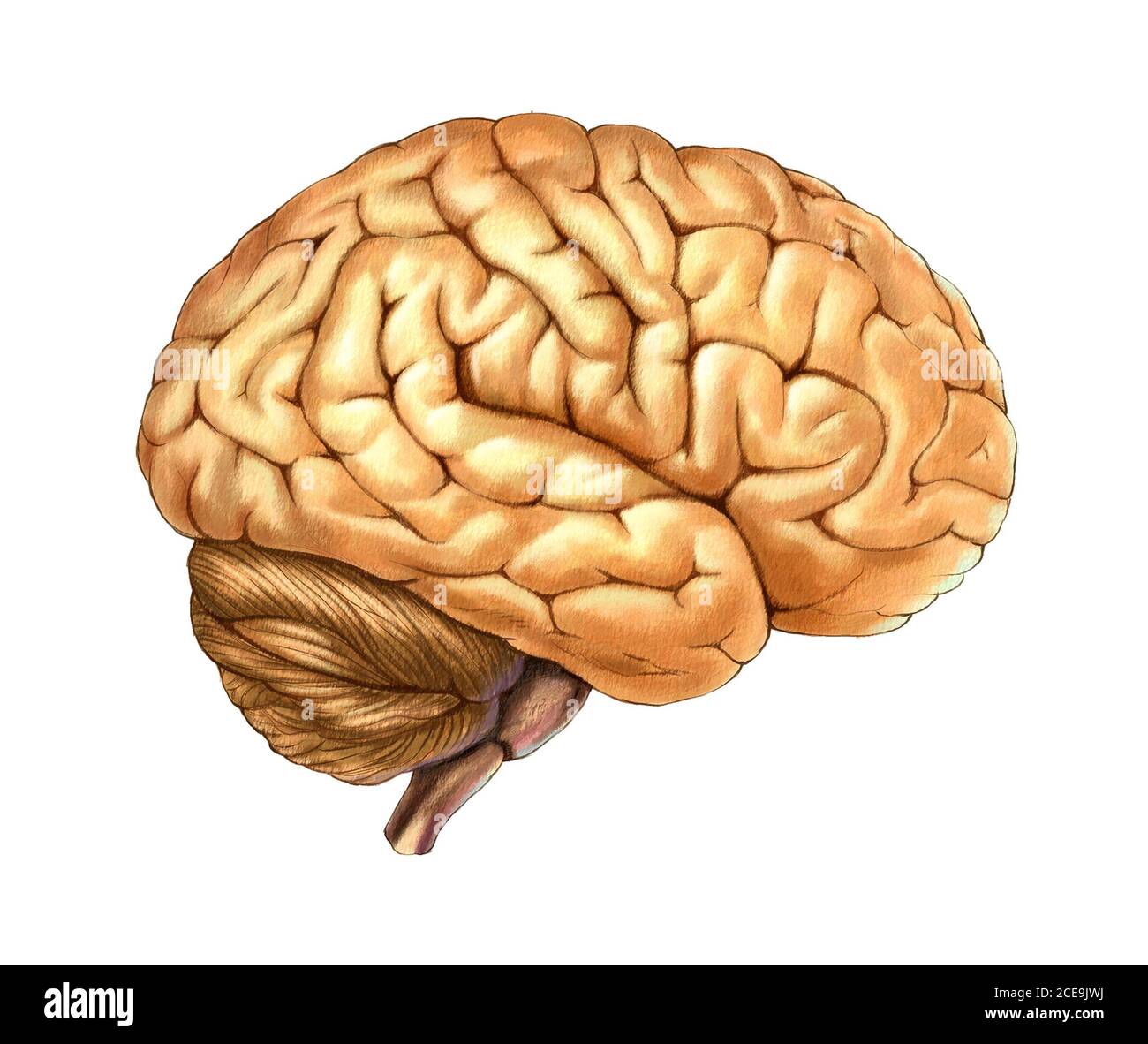 Human brain illustration Stock Photohttps://www.alamy.com/image-license-details/?v=1https://www.alamy.com/human-brain-illustration-image370235310.html
RF2CE9JWJ–Human brain illustration
 human brain with big ears and legs, 3d illustration Stock Photohttps://www.alamy.com/image-license-details/?v=1https://www.alamy.com/stock-photo-human-brain-with-big-ears-and-legs-3d-illustration-90768393.html
RMF7JT09–human brain with big ears and legs, 3d illustration
 Brainstem glioma, illustration Stock Photohttps://www.alamy.com/image-license-details/?v=1https://www.alamy.com/brainstem-glioma-illustration-image414765683.html
RF2F2P5T3–Brainstem glioma, illustration
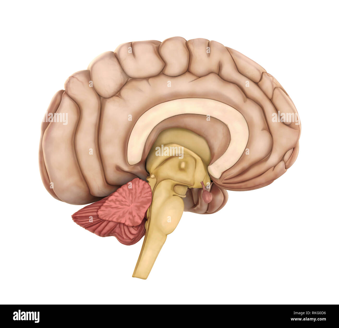 Human Brain Anatomy Isolated Stock Photohttps://www.alamy.com/image-license-details/?v=1https://www.alamy.com/human-brain-anatomy-isolated-image235764850.html
RFRKG0D6–Human Brain Anatomy Isolated
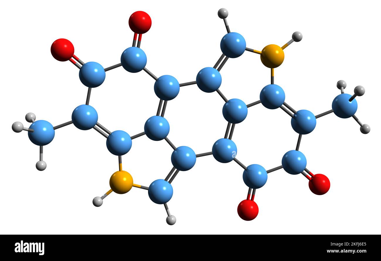 3D image of Melanin skeletal formula - molecular chemical structure of natural pigment isolated on white background Stock Photohttps://www.alamy.com/image-license-details/?v=1https://www.alamy.com/3d-image-of-melanin-skeletal-formula-molecular-chemical-structure-of-natural-pigment-isolated-on-white-background-image491510381.html
RF2KFJ6E5–3D image of Melanin skeletal formula - molecular chemical structure of natural pigment isolated on white background
 The Peduncles of the Cerebellum are the part of the midbrain that link the remainder of the brainstem to the thalami and thereby, the cerebrum, vintag Stock Vectorhttps://www.alamy.com/image-license-details/?v=1https://www.alamy.com/the-peduncles-of-the-cerebellum-are-the-part-of-the-midbrain-that-link-the-remainder-of-the-brainstem-to-the-thalami-and-thereby-the-cerebrum-vintag-image348663589.html
RF2B76YY1–The Peduncles of the Cerebellum are the part of the midbrain that link the remainder of the brainstem to the thalami and thereby, the cerebrum, vintag
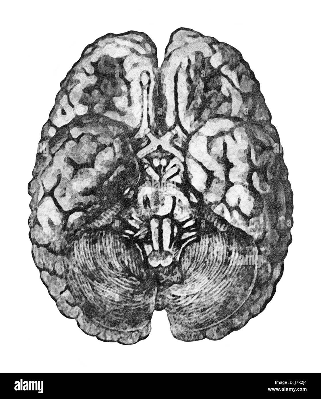 Under Surface of the Brain. Anatomy education concept - View from below of the brain and brainstem. Stock Photohttps://www.alamy.com/image-license-details/?v=1https://www.alamy.com/stock-photo-under-surface-of-the-brain-anatomy-education-concept-view-from-below-142492508.html
RFJ7R2J4–Under Surface of the Brain. Anatomy education concept - View from below of the brain and brainstem.
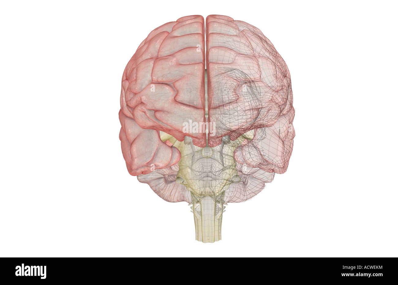 The brainstem Stock Photohttps://www.alamy.com/image-license-details/?v=1https://www.alamy.com/stock-photo-the-brainstem-13235719.html
RFACWEKM–The brainstem
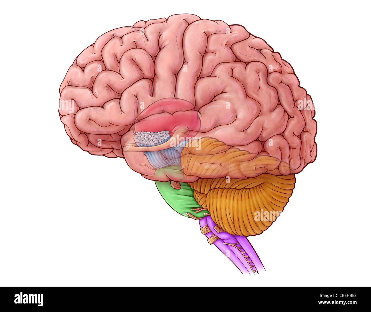 Diencephalon and Brainstem, illustration Stock Photohttps://www.alamy.com/image-license-details/?v=1https://www.alamy.com/diencephalon-and-brainstem-illustration-image353194747.html
RM2BEHBE3–Diencephalon and Brainstem, illustration
 At the Base of the Brain is the brainstem, which connects the cerebrum with the spinal cord, vintage line drawing or engraving illustration. Stock Vectorhttps://www.alamy.com/image-license-details/?v=1https://www.alamy.com/at-the-base-of-the-brain-is-the-brainstem-which-connects-the-cerebrum-with-the-spinal-cord-vintage-line-drawing-or-engraving-illustration-image359325661.html
RF2BTGKF9–At the Base of the Brain is the brainstem, which connects the cerebrum with the spinal cord, vintage line drawing or engraving illustration.
 Page from a 19th-century anatomy textbook showing the deep dissection of the brainstem Stock Photohttps://www.alamy.com/image-license-details/?v=1https://www.alamy.com/page-from-a-19th-century-anatomy-textbook-showing-the-deep-dissection-of-the-brainstem-image398876041.html
RF2E4XACW–Page from a 19th-century anatomy textbook showing the deep dissection of the brainstem
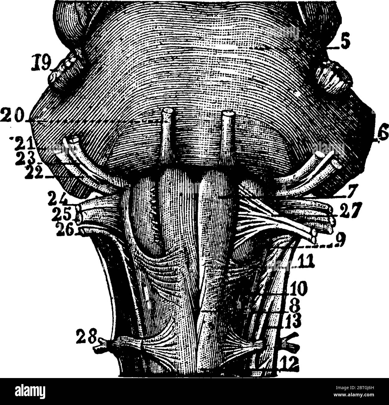 The Medulla Oblongata located at brainstem It's functions are involuntary, or done without thought, vintage line drawing or engraving illustration. Stock Vectorhttps://www.alamy.com/image-license-details/?v=1https://www.alamy.com/the-medulla-oblongata-located-at-brainstem-its-functions-are-involuntary-or-done-without-thought-vintage-line-drawing-or-engraving-illustration-image359324633.html
RF2BTGJ6H–The Medulla Oblongata located at brainstem It's functions are involuntary, or done without thought, vintage line drawing or engraving illustration.
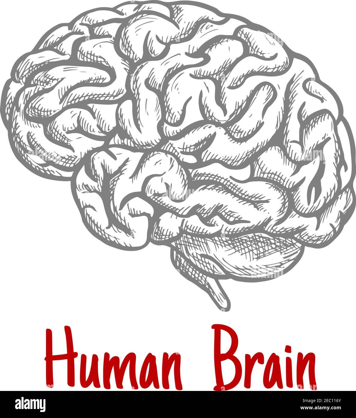 Vintage engraving sketch of human brain with anatomically detailed brainstem and hindbrain. Medicine, science or brainstorm concept Stock Vectorhttps://www.alamy.com/image-license-details/?v=1https://www.alamy.com/vintage-engraving-sketch-of-human-brain-with-anatomically-detailed-brainstem-and-hindbrain-medicine-science-or-brainstorm-concept-image403237267.html
RF2EC116Y–Vintage engraving sketch of human brain with anatomically detailed brainstem and hindbrain. Medicine, science or brainstorm concept
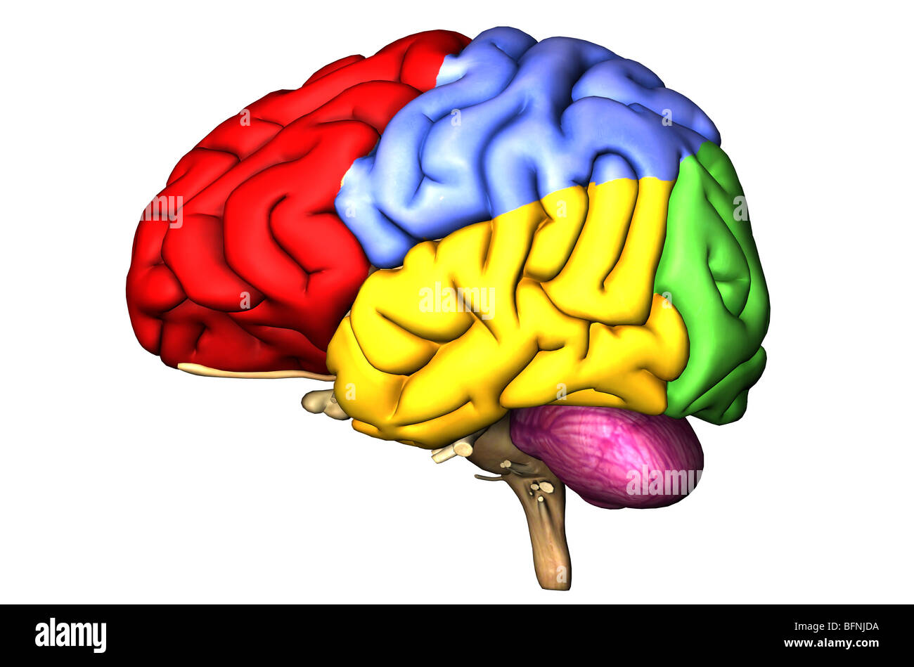 Illustration of the human brain showing the cerebral lobes and cerebellum in colors Stock Photohttps://www.alamy.com/image-license-details/?v=1https://www.alamy.com/stock-photo-illustration-of-the-human-brain-showing-the-cerebral-lobes-and-cerebellum-26905686.html
RMBFNJDA–Illustration of the human brain showing the cerebral lobes and cerebellum in colors
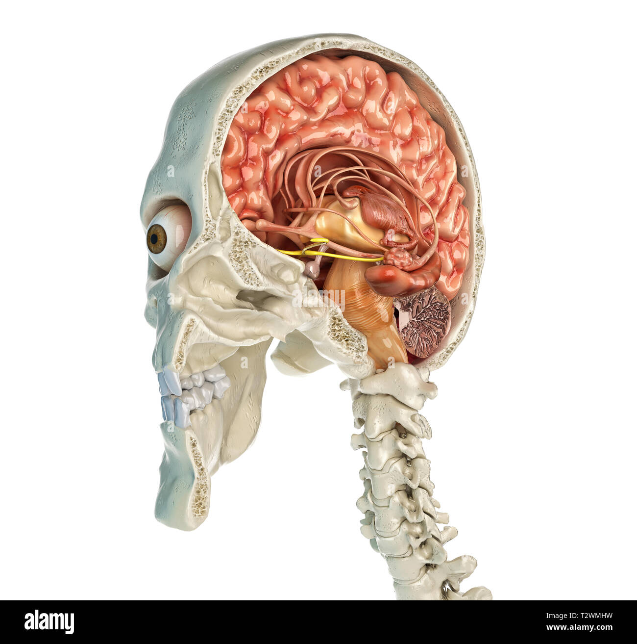 Human skull mid sagittal cross-section with brain. Perspective view on white background. Stock Photohttps://www.alamy.com/image-license-details/?v=1https://www.alamy.com/human-skull-mid-sagittal-cross-section-with-brain-perspective-view-on-white-background-image242739445.html
RFT2WMHW–Human skull mid sagittal cross-section with brain. Perspective view on white background.
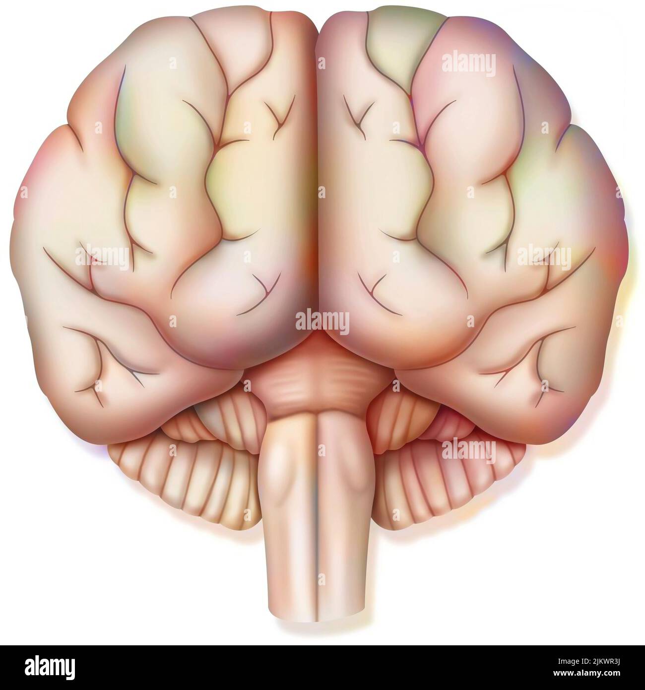 Brain, with the two cerebral hemispheres, the cerebellum and the brainstem. Stock Photohttps://www.alamy.com/image-license-details/?v=1https://www.alamy.com/brain-with-the-two-cerebral-hemispheres-the-cerebellum-and-the-brainstem-image476925334.html
RF2JKWR3J–Brain, with the two cerebral hemispheres, the cerebellum and the brainstem.
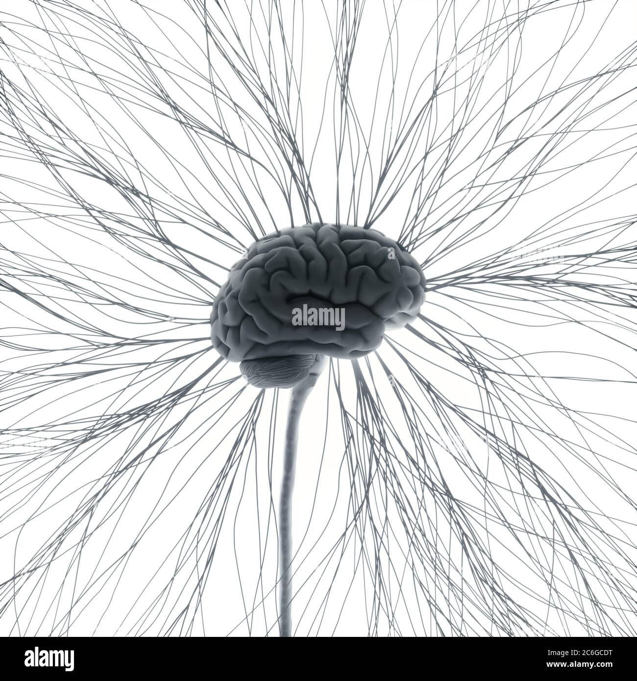 Central nervous system. Brain and spinal cord with branch coming out away. Conceptual brain 3D illustration. Stock Photohttps://www.alamy.com/image-license-details/?v=1https://www.alamy.com/central-nervous-system-brain-and-spinal-cord-with-branch-coming-out-away-conceptual-brain-3d-illustration-image365466692.html
RF2C6GCDT–Central nervous system. Brain and spinal cord with branch coming out away. Conceptual brain 3D illustration.
 Spinal Stenosis as a degenerative illness in the human vertebrae causing compressed spine nerves medical concept as a 3D illustration. Stock Photohttps://www.alamy.com/image-license-details/?v=1https://www.alamy.com/stock-photo-spinal-stenosis-as-a-degenerative-illness-in-the-human-vertebrae-causing-172986529.html
RFM1C629–Spinal Stenosis as a degenerative illness in the human vertebrae causing compressed spine nerves medical concept as a 3D illustration.
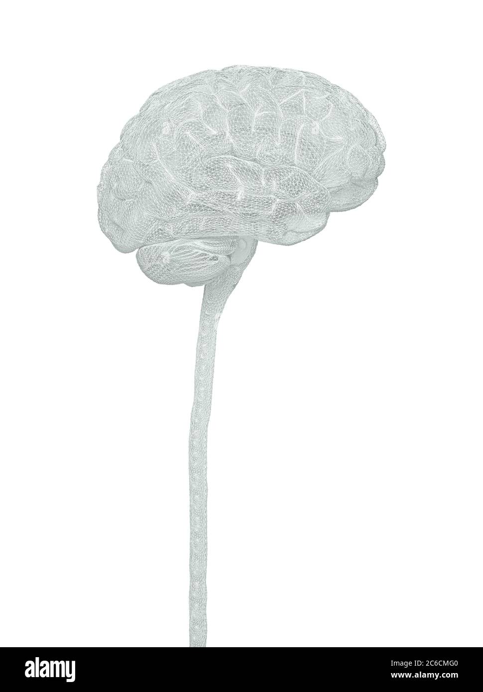 Central nervous system. Brain and spinal cord with clipping path included. Conceptual brain 3D illustration. Stock Photohttps://www.alamy.com/image-license-details/?v=1https://www.alamy.com/central-nervous-system-brain-and-spinal-cord-with-clipping-path-included-conceptual-brain-3d-illustration-image365385216.html
RF2C6CMG0–Central nervous system. Brain and spinal cord with clipping path included. Conceptual brain 3D illustration.
 Anatomy of human nervous system and lymphatic system, front view Stock Photohttps://www.alamy.com/image-license-details/?v=1https://www.alamy.com/anatomy-of-human-nervous-system-and-lymphatic-system-front-view-image619446543.html
RM2XYP6AR–Anatomy of human nervous system and lymphatic system, front view
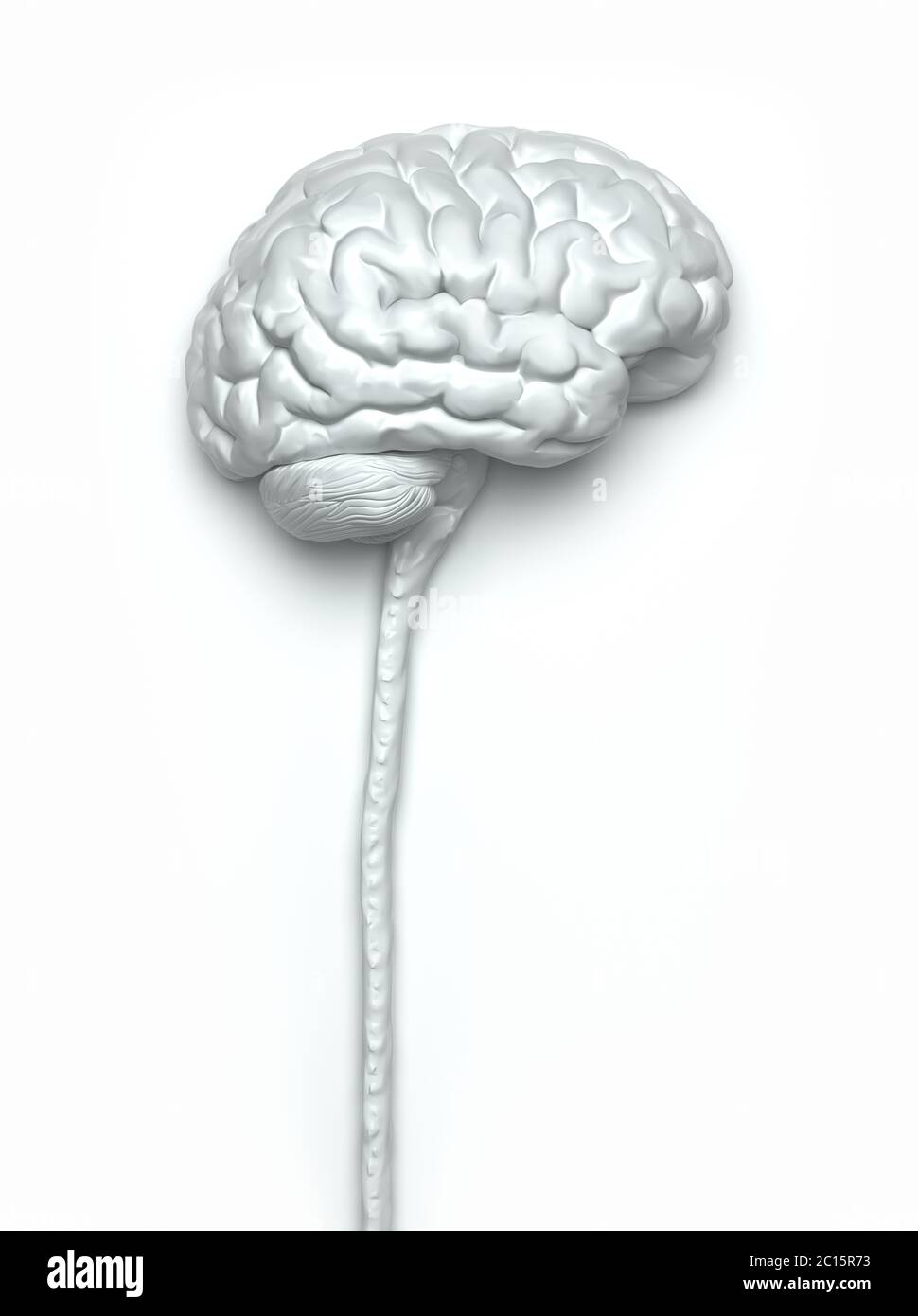 Central nervous system. Brain and spinal cord with clipping path included. Conceptual brain 3D illustration. Stock Photohttps://www.alamy.com/image-license-details/?v=1https://www.alamy.com/central-nervous-system-brain-and-spinal-cord-with-clipping-path-included-conceptual-brain-3d-illustration-image362160375.html
RF2C15R73–Central nervous system. Brain and spinal cord with clipping path included. Conceptual brain 3D illustration.
 Brainstem glioma in childhood, illustration Stock Photohttps://www.alamy.com/image-license-details/?v=1https://www.alamy.com/brainstem-glioma-in-childhood-illustration-image414765642.html
RF2F2P5PJ–Brainstem glioma in childhood, illustration
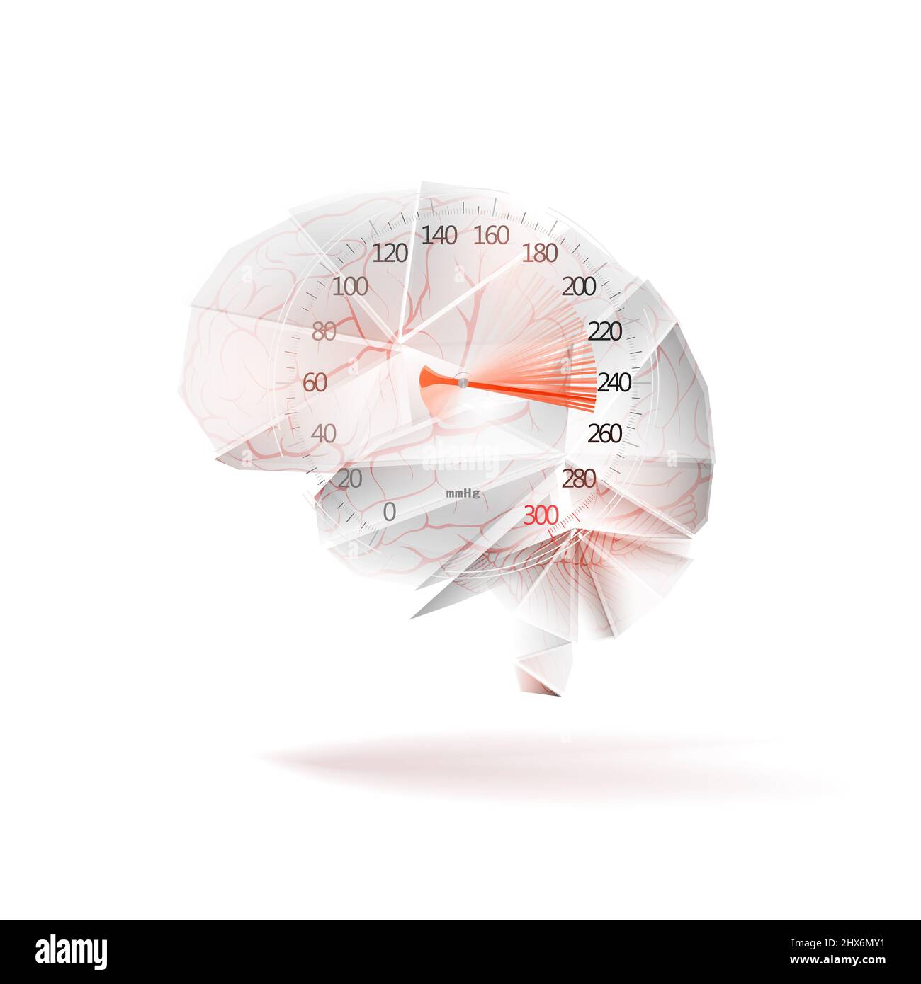 Sphygmomanometer with the rise of blood pressure and the brain as a concept of arterial blood hypertension. Isolated on white background. Illustration Stock Photohttps://www.alamy.com/image-license-details/?v=1https://www.alamy.com/sphygmomanometer-with-the-rise-of-blood-pressure-and-the-brain-as-a-concept-of-arterial-blood-hypertension-isolated-on-white-background-illustration-image463598773.html
RF2HX6MY1–Sphygmomanometer with the rise of blood pressure and the brain as a concept of arterial blood hypertension. Isolated on white background. Illustration
 Illustration of the cranial nerves anatomy. The cranial nerves are a set of 12 paired nerves that arise directly from the brain. The first two nerves (olfactory and optic) arise from the cerebrum, whereas the remaining ten emerge from the brainstem. The names of the cranial nerves relate to their function and they are numerically identified in roman numerals. Stock Photohttps://www.alamy.com/image-license-details/?v=1https://www.alamy.com/illustration-of-the-cranial-nerves-anatomy-the-cranial-nerves-are-a-set-of-12-paired-nerves-that-arise-directly-from-the-brain-the-first-two-nerves-olfactory-and-optic-arise-from-the-cerebrum-whereas-the-remaining-ten-emerge-from-the-brainstem-the-names-of-the-cranial-nerves-relate-to-their-function-and-they-are-numerically-identified-in-roman-numerals-image628777660.html
RF2YEY890–Illustration of the cranial nerves anatomy. The cranial nerves are a set of 12 paired nerves that arise directly from the brain. The first two nerves (olfactory and optic) arise from the cerebrum, whereas the remaining ten emerge from the brainstem. The names of the cranial nerves relate to their function and they are numerically identified in roman numerals.
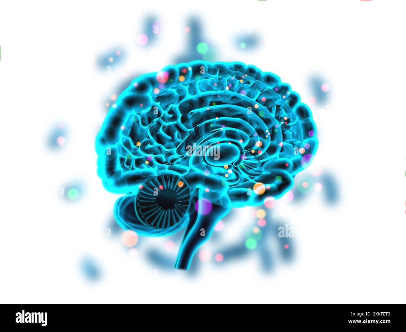 Cross section of human brain on medical background. 3d illusstration Stock Photohttps://www.alamy.com/image-license-details/?v=1https://www.alamy.com/cross-section-of-human-brain-on-medical-background-3d-illusstration-image614382275.html
RF2XKFET3–Cross section of human brain on medical background. 3d illusstration
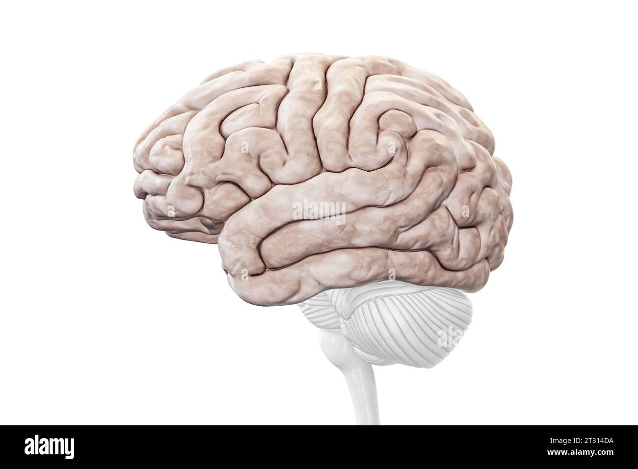 Cerebral cortex or hemisphere profile view isolated on white background accurate 3D rendering illustration. Human brain anatomy, neurology, neuroscien Stock Photohttps://www.alamy.com/image-license-details/?v=1https://www.alamy.com/cerebral-cortex-or-hemisphere-profile-view-isolated-on-white-background-accurate-3d-rendering-illustration-human-brain-anatomy-neurology-neuroscien-image569811574.html
RF2T314DA–Cerebral cortex or hemisphere profile view isolated on white background accurate 3D rendering illustration. Human brain anatomy, neurology, neuroscien
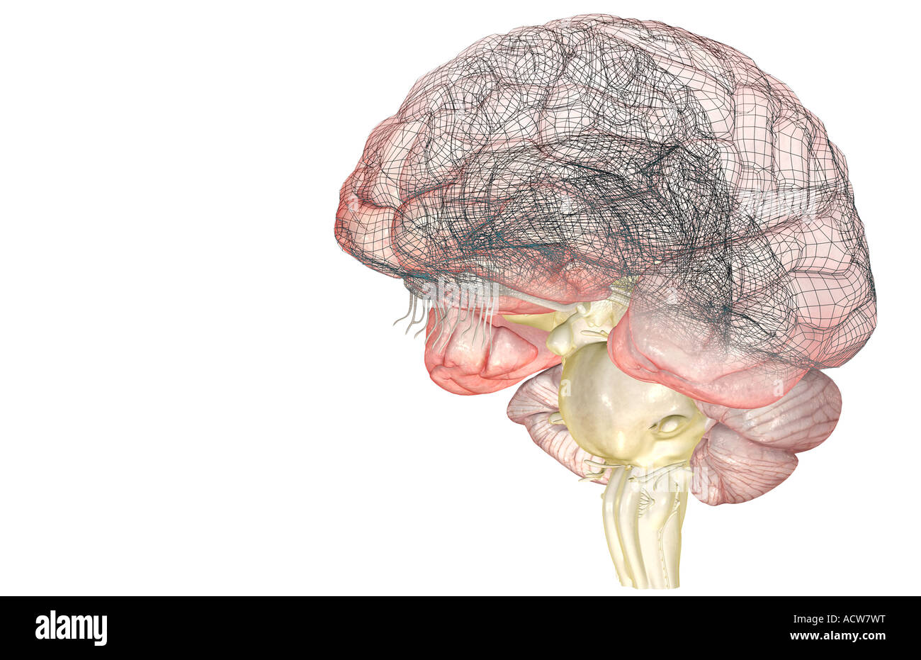 The brainstem Stock Photohttps://www.alamy.com/image-license-details/?v=1https://www.alamy.com/stock-photo-the-brainstem-13233443.html
RFACW7WT–The brainstem
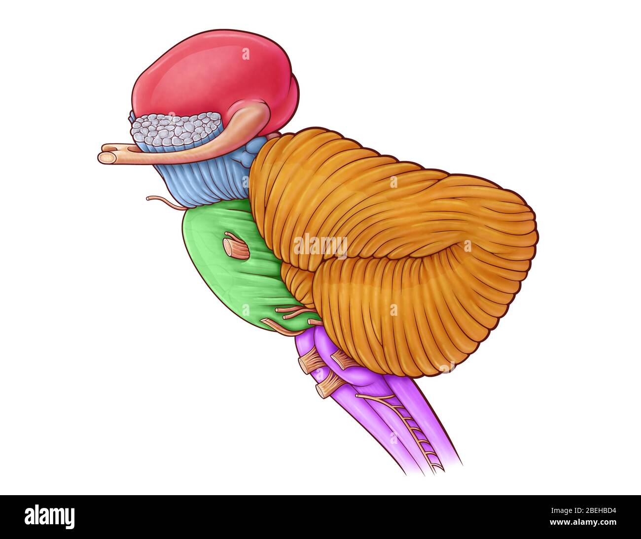 An illustration of the diencephalon, brainstem and cerebellum. Stock Photohttps://www.alamy.com/image-license-details/?v=1https://www.alamy.com/an-illustration-of-the-diencephalon-brainstem-and-cerebellum-image353194720.html
RM2BEHBD4–An illustration of the diencephalon, brainstem and cerebellum.
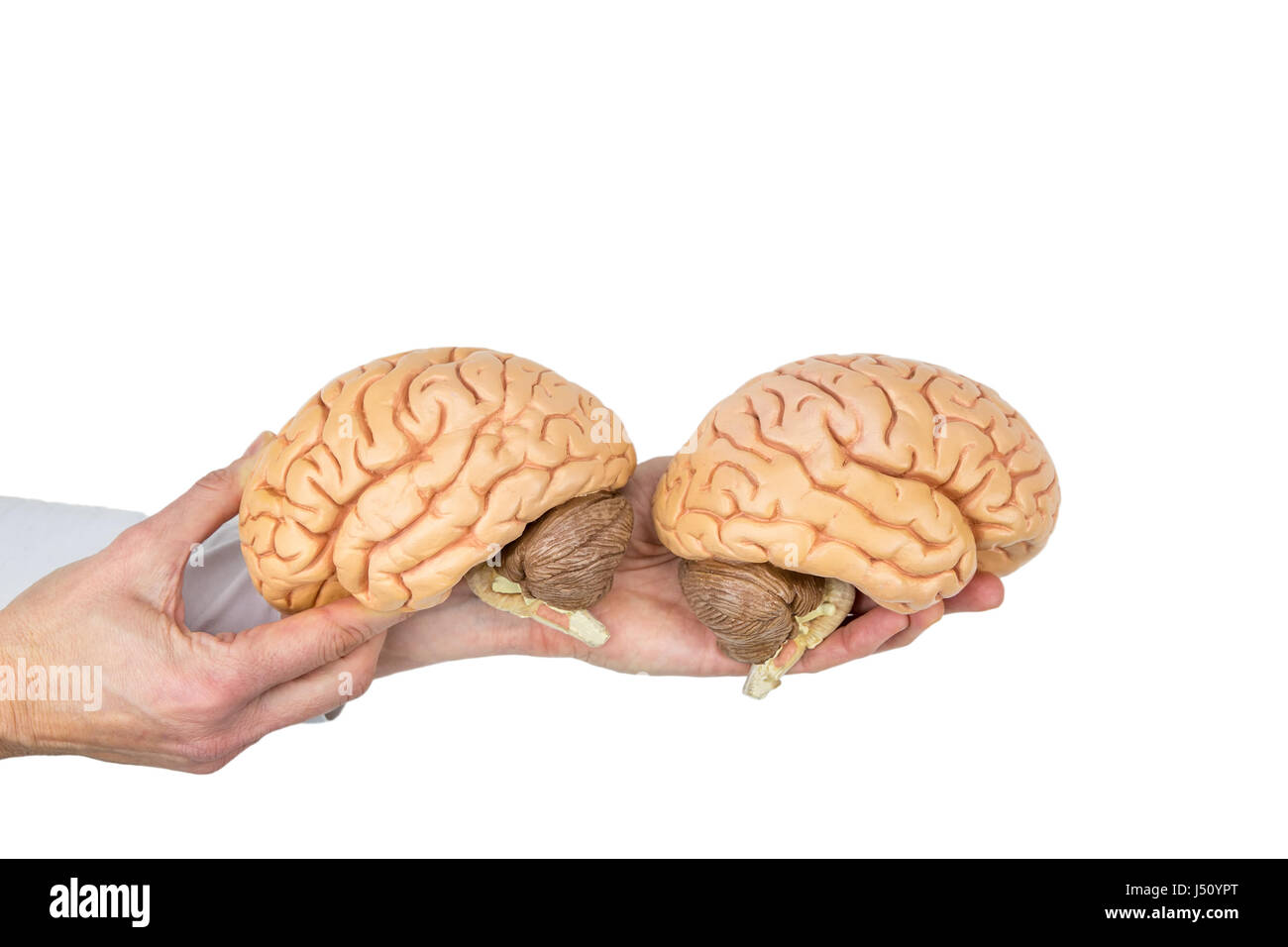 Hands holding models human brain hemispheres isolated on white background Stock Photohttps://www.alamy.com/image-license-details/?v=1https://www.alamy.com/stock-photo-hands-holding-models-human-brain-hemispheres-isolated-on-white-background-140778032.html
RFJ50YPT–Hands holding models human brain hemispheres isolated on white background
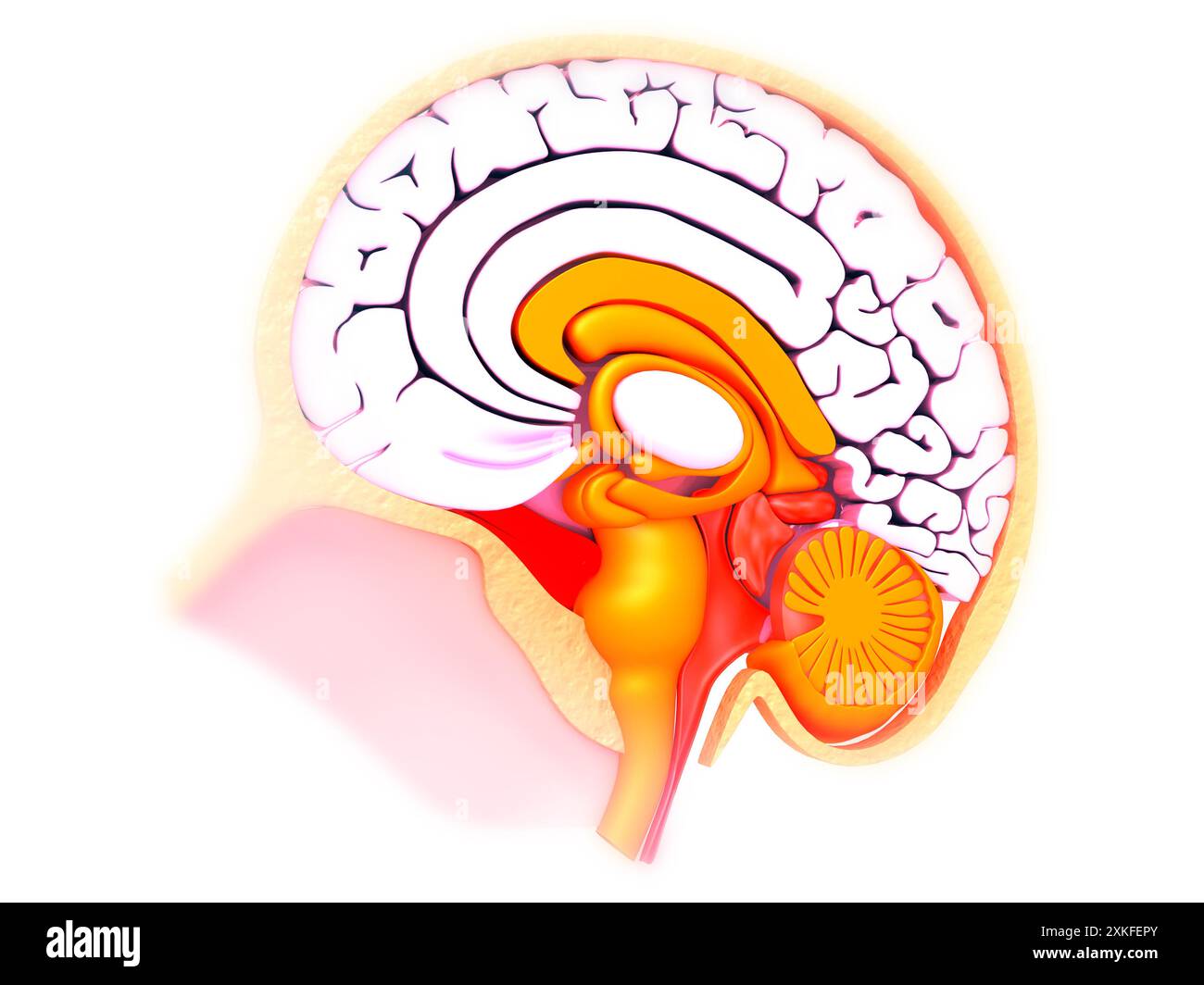 Cross section of human brain on medical background. 3d illusstration Stock Photohttps://www.alamy.com/image-license-details/?v=1https://www.alamy.com/cross-section-of-human-brain-on-medical-background-3d-illusstration-image614382243.html
RF2XKFEPY–Cross section of human brain on medical background. 3d illusstration
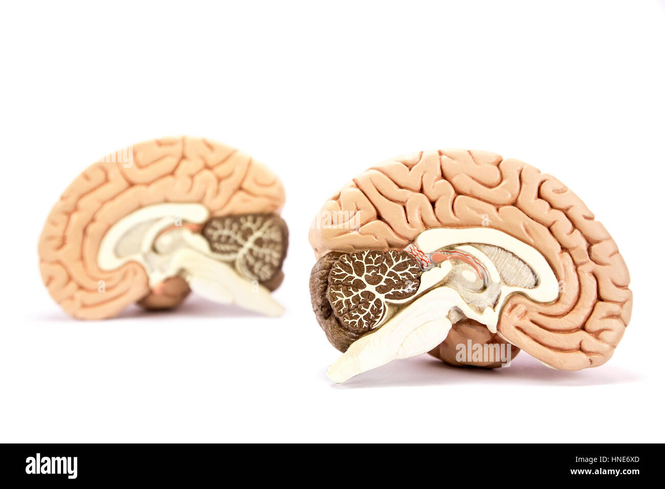 Two human brain hemispheres models isolated on white background Stock Photohttps://www.alamy.com/image-license-details/?v=1https://www.alamy.com/stock-photo-two-human-brain-hemispheres-models-isolated-on-white-background-133693125.html
RFHNE6XD–Two human brain hemispheres models isolated on white background
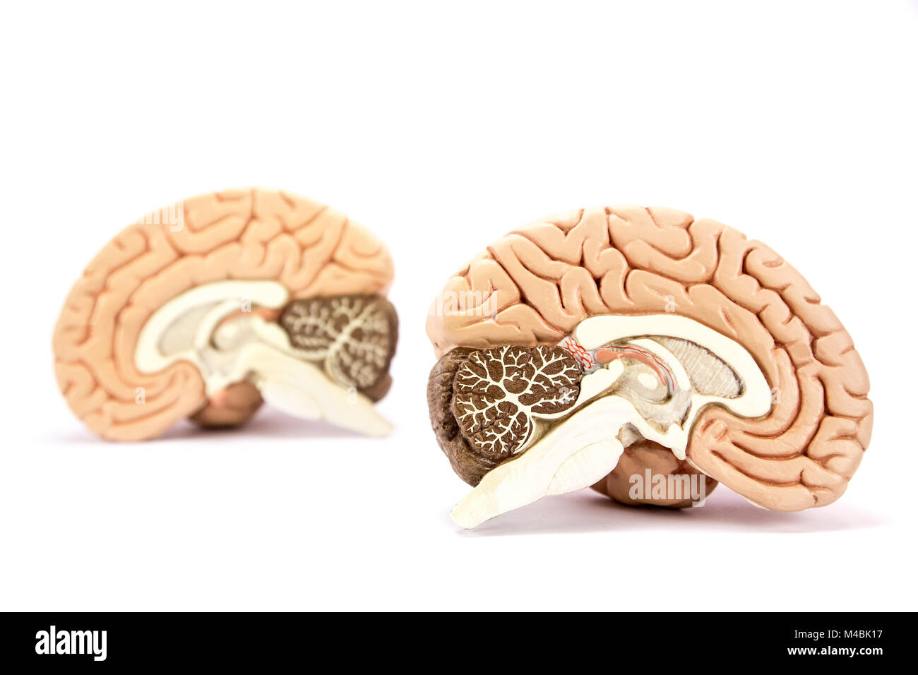 Human brains model isolated on white background Stock Photohttps://www.alamy.com/image-license-details/?v=1https://www.alamy.com/stock-photo-human-brains-model-isolated-on-white-background-174818707.html
RMM4BK17–Human brains model isolated on white background
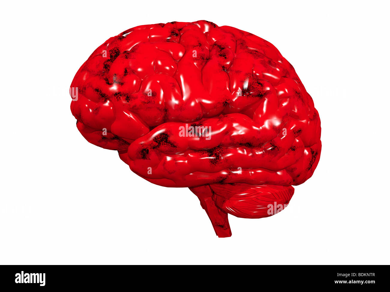 illustration of the human brain Stock Photohttps://www.alamy.com/image-license-details/?v=1https://www.alamy.com/stock-photo-illustration-of-the-human-brain-25635143.html
RMBDKNTR–illustration of the human brain
 Human skull mid sagittal cross-section with brain. Side view on black background. Stock Photohttps://www.alamy.com/image-license-details/?v=1https://www.alamy.com/human-skull-mid-sagittal-cross-section-with-brain-side-view-on-black-background-image242739455.html
RFT2WMJ7–Human skull mid sagittal cross-section with brain. Side view on black background.
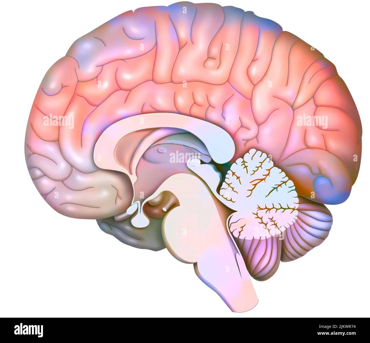 Median sagittal section of the brain with cerebellum and beginning of the brainstem. Stock Photohttps://www.alamy.com/image-license-details/?v=1https://www.alamy.com/median-sagittal-section-of-the-brain-with-cerebellum-and-beginning-of-the-brainstem-image476925432.html
RF2JKWR74–Median sagittal section of the brain with cerebellum and beginning of the brainstem.
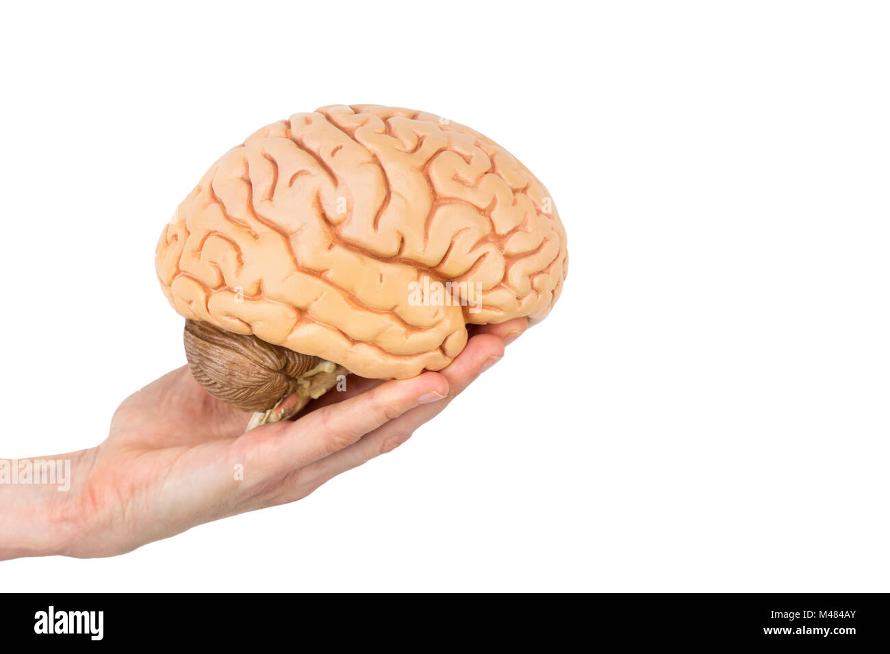 Hand holding model human brains isolated on white background Stock Photohttps://www.alamy.com/image-license-details/?v=1https://www.alamy.com/stock-photo-hand-holding-model-human-brains-isolated-on-white-background-174741363.html
RMM484AY–Hand holding model human brains isolated on white background
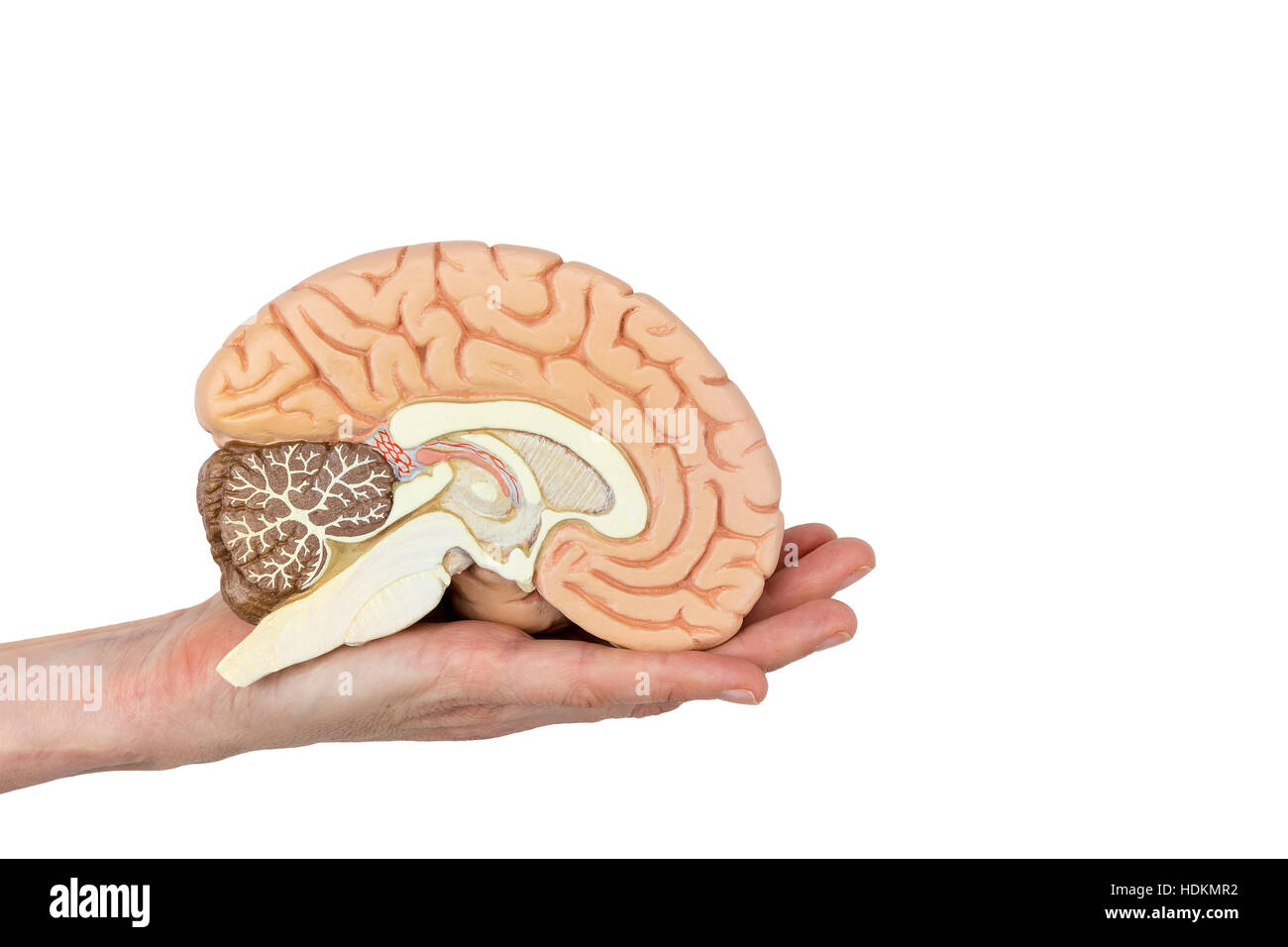 Hand holding left brain hemisphere isolated on white background Stock Photohttps://www.alamy.com/image-license-details/?v=1https://www.alamy.com/stock-photo-hand-holding-left-brain-hemisphere-isolated-on-white-background-128896518.html
RFHDKMR2–Hand holding left brain hemisphere isolated on white background
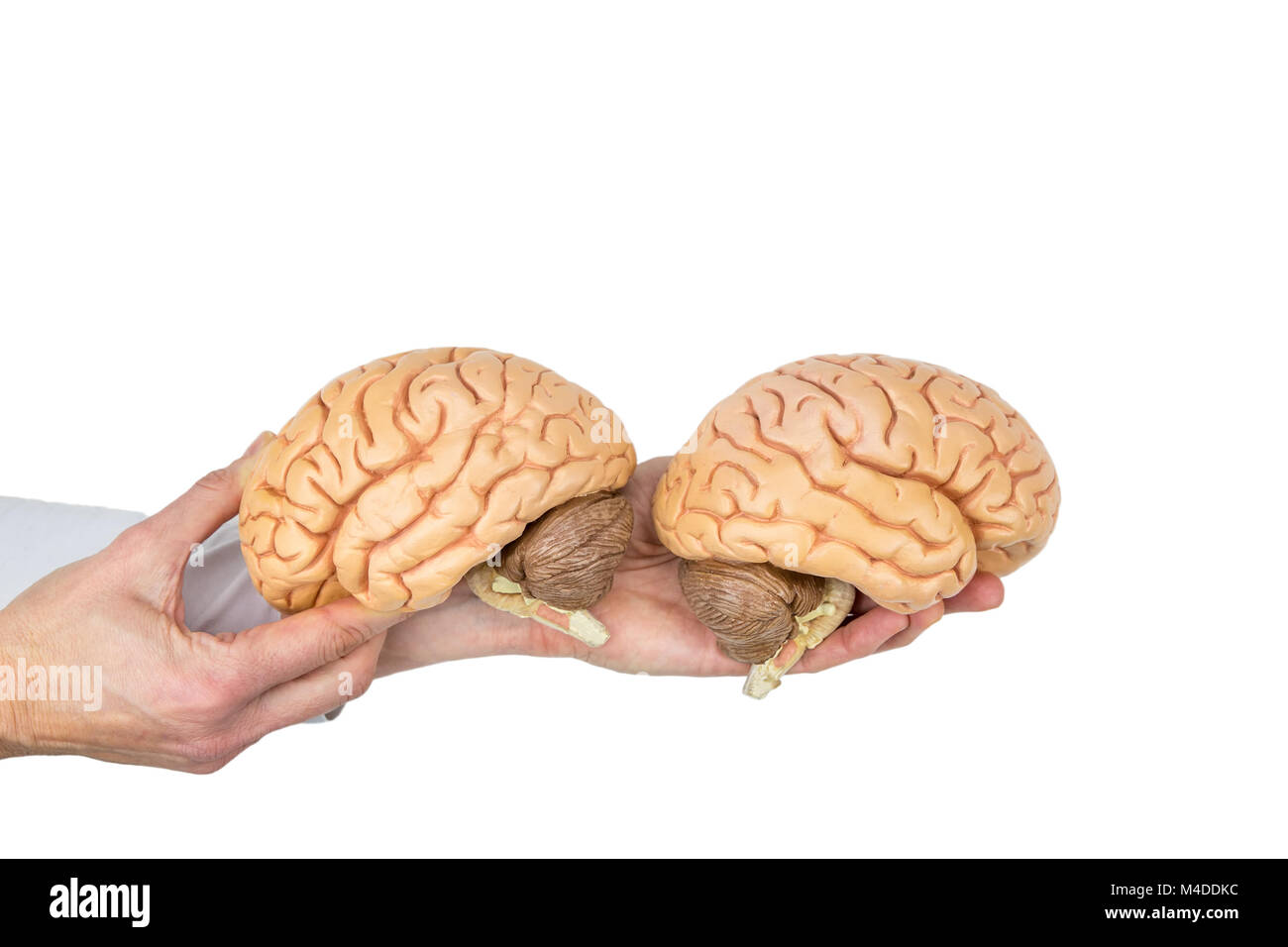 Hands holding model human brain on white background Stock Photohttps://www.alamy.com/image-license-details/?v=1https://www.alamy.com/stock-photo-hands-holding-model-human-brain-on-white-background-174858416.html
RMM4DDKC–Hands holding model human brain on white background
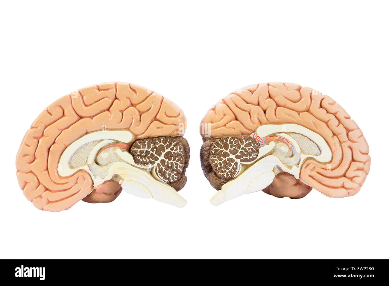 Cross section of two artificial human hemispheres, two halves of brain for education, isolated on white background Stock Photohttps://www.alamy.com/image-license-details/?v=1https://www.alamy.com/stock-photo-cross-section-of-two-artificial-human-hemispheres-two-halves-of-brain-84709956.html
RFEWPTBG–Cross section of two artificial human hemispheres, two halves of brain for education, isolated on white background
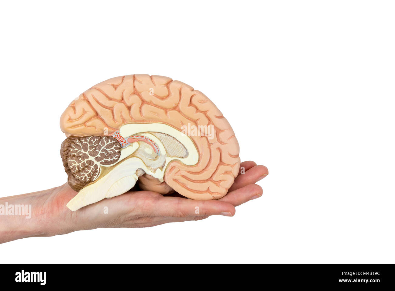 Hand holding brain hemisphere on white background Stock Photohttps://www.alamy.com/image-license-details/?v=1https://www.alamy.com/stock-photo-hand-holding-brain-hemisphere-on-white-background-174822856.html
RMM4BT9C–Hand holding brain hemisphere on white background
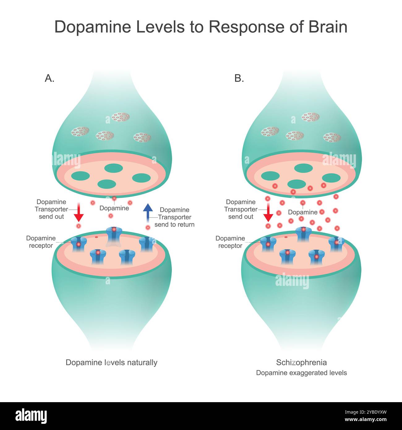 Dopamine Levels to Response of Brain. Dopamine Levels naturally and Dopamine levels in patient. Stock Photohttps://www.alamy.com/image-license-details/?v=1https://www.alamy.com/dopamine-levels-to-response-of-brain-dopamine-levels-naturally-and-dopamine-levels-in-patient-image626641761.html
RF2YBDYXW–Dopamine Levels to Response of Brain. Dopamine Levels naturally and Dopamine levels in patient.
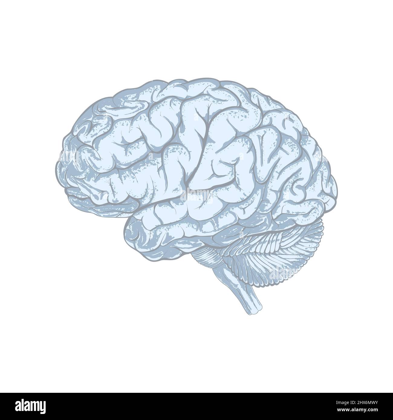 Sketchy style human brain abstract. Isolated on white background. Illustration Stock Photohttps://www.alamy.com/image-license-details/?v=1https://www.alamy.com/sketchy-style-human-brain-abstract-isolated-on-white-background-illustration-image463598743.html
RF2HX6MWY–Sketchy style human brain abstract. Isolated on white background. Illustration
 Illustration of the cranial nerves anatomy. The cranial nerves are a set of 12 paired nerves that arise directly from the brain. The first two nerves (olfactory and optic) arise from the cerebrum, whereas the remaining ten emerge from the brainstem. The names of the cranial nerves relate to their function and they are numerically identified in roman numerals. Stock Photohttps://www.alamy.com/image-license-details/?v=1https://www.alamy.com/illustration-of-the-cranial-nerves-anatomy-the-cranial-nerves-are-a-set-of-12-paired-nerves-that-arise-directly-from-the-brain-the-first-two-nerves-olfactory-and-optic-arise-from-the-cerebrum-whereas-the-remaining-ten-emerge-from-the-brainstem-the-names-of-the-cranial-nerves-relate-to-their-function-and-they-are-numerically-identified-in-roman-numerals-image628777672.html
RF2YEY89C–Illustration of the cranial nerves anatomy. The cranial nerves are a set of 12 paired nerves that arise directly from the brain. The first two nerves (olfactory and optic) arise from the cerebrum, whereas the remaining ten emerge from the brainstem. The names of the cranial nerves relate to their function and they are numerically identified in roman numerals.
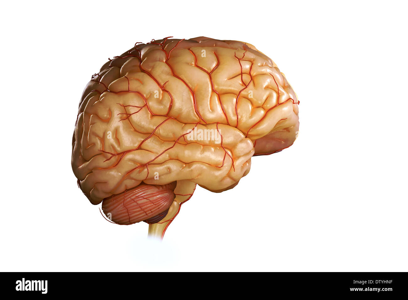 Human Brain Stock Photohttps://www.alamy.com/image-license-details/?v=1https://www.alamy.com/human-brain-image66989483.html
RMDTYHNF–Human Brain
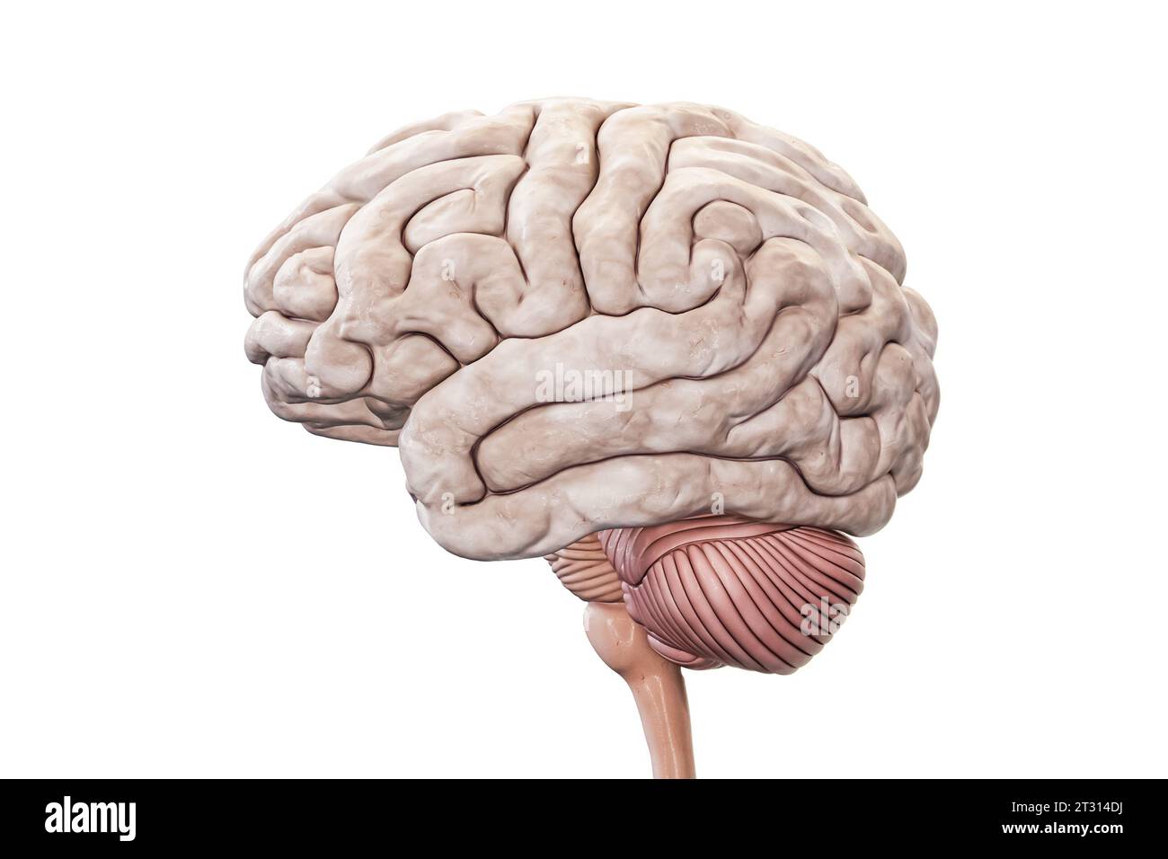 Human brain with cortex, cerebellum and brain stem profile view isolated on white background accurate 3D rendering illustration. Anatomy, neurology, n Stock Photohttps://www.alamy.com/image-license-details/?v=1https://www.alamy.com/human-brain-with-cortex-cerebellum-and-brain-stem-profile-view-isolated-on-white-background-accurate-3d-rendering-illustration-anatomy-neurology-n-image569811582.html
RF2T314DJ–Human brain with cortex, cerebellum and brain stem profile view isolated on white background accurate 3D rendering illustration. Anatomy, neurology, n
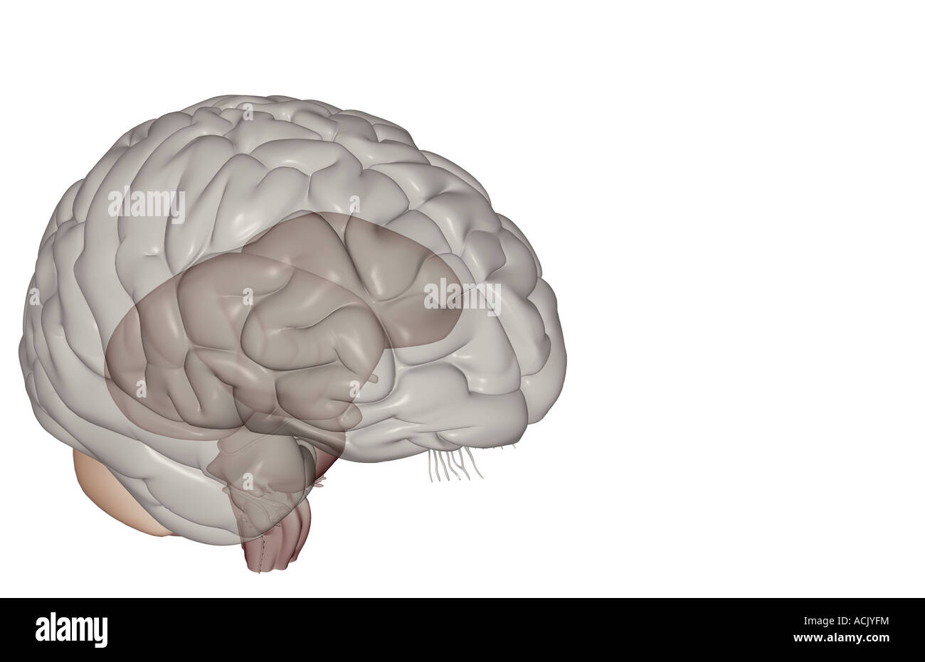 The brainstem Stock Photohttps://www.alamy.com/image-license-details/?v=1https://www.alamy.com/stock-photo-the-brainstem-13174183.html
RFACJYFM–The brainstem
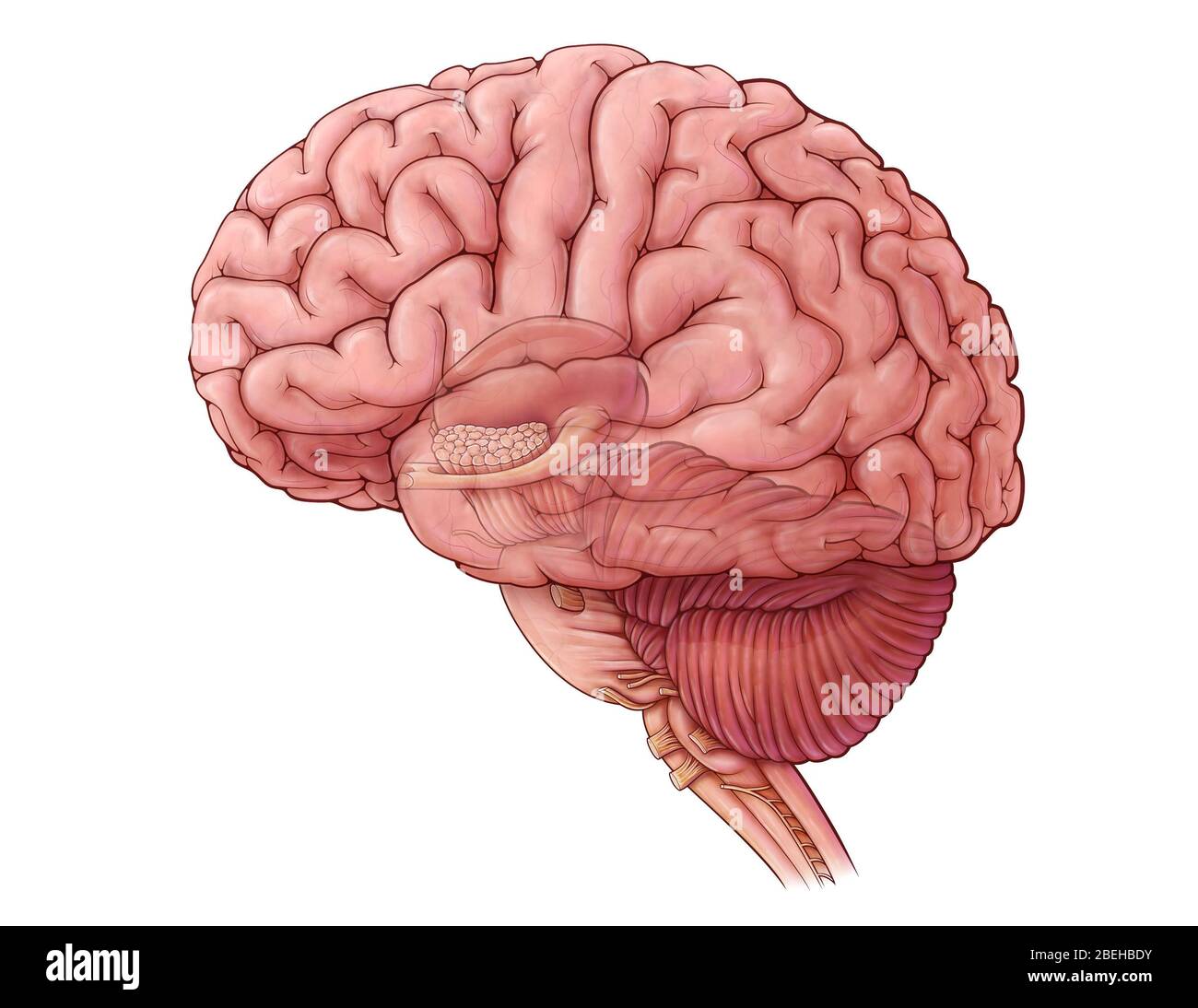 Diencephalon and Brainstem, illustration Stock Photohttps://www.alamy.com/image-license-details/?v=1https://www.alamy.com/diencephalon-and-brainstem-illustration-image353194743.html
RM2BEHBDY–Diencephalon and Brainstem, illustration
 Human brain on laptop isolated on white background Stock Photohttps://www.alamy.com/image-license-details/?v=1https://www.alamy.com/human-brain-on-laptop-isolated-on-white-background-image462355560.html
RF2HT636G–Human brain on laptop isolated on white background
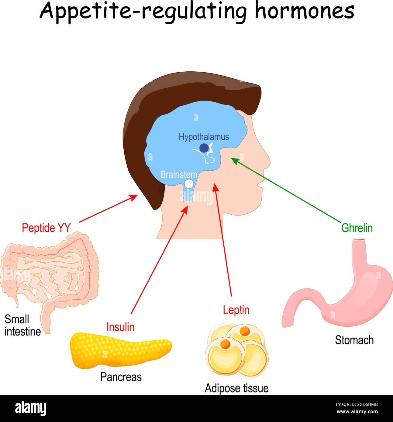 Leptin, ghrelin, insulin and Peptide YY. hormones that regulate metabolism, appetite, satiety and hunger. vector illustration Stock Vectorhttps://www.alamy.com/image-license-details/?v=1https://www.alamy.com/leptin-ghrelin-insulin-and-peptide-yy-hormones-that-regulate-metabolism-appetite-satiety-and-hunger-vector-illustration-image438395339.html
RF2GD6HMB–Leptin, ghrelin, insulin and Peptide YY. hormones that regulate metabolism, appetite, satiety and hunger. vector illustration
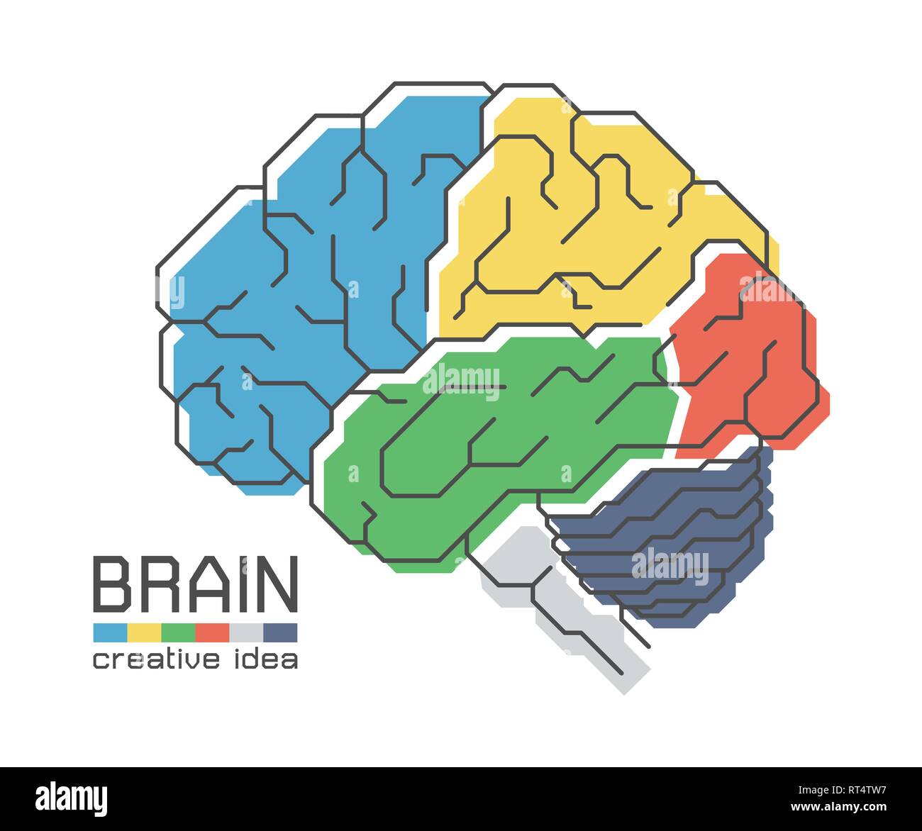 Brain anatomy with flat color design and outline stroke . Frontal Parietal Temporal Occipital lobe Cerebellum and Brainstem . Creative idea concept . Stock Vectorhttps://www.alamy.com/image-license-details/?v=1https://www.alamy.com/brain-anatomy-with-flat-color-design-and-outline-stroke-frontal-parietal-temporal-occipital-lobe-cerebellum-and-brainstem-creative-idea-concept-image238593859.html
RFRT4TW7–Brain anatomy with flat color design and outline stroke . Frontal Parietal Temporal Occipital lobe Cerebellum and Brainstem . Creative idea concept .
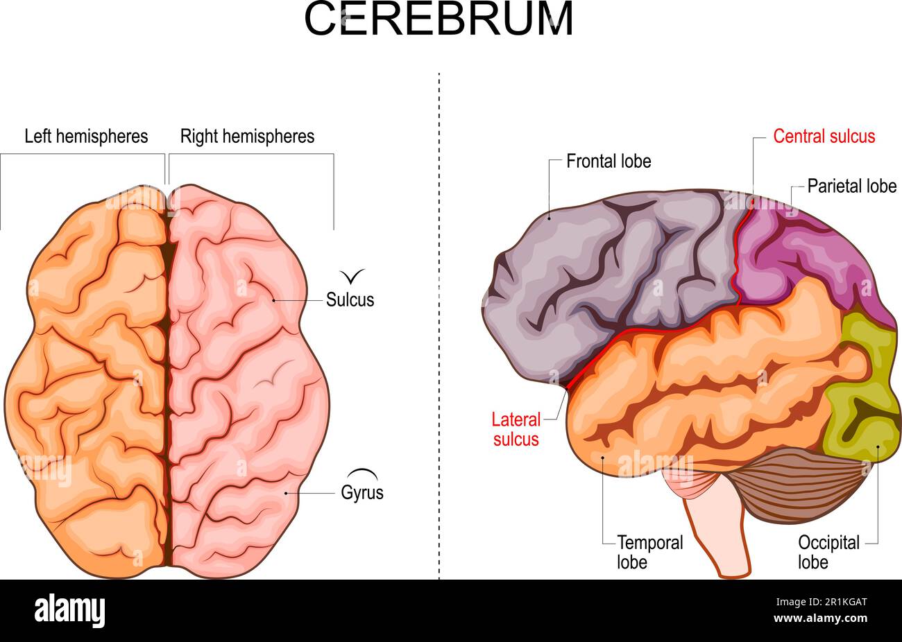 Human brain structure. Hemispheres and lobes of the cerebral cortex. frontal, temporal, occipital, and parietal lobes. lateral and superior view Stock Vectorhttps://www.alamy.com/image-license-details/?v=1https://www.alamy.com/human-brain-structure-hemispheres-and-lobes-of-the-cerebral-cortex-frontal-temporal-occipital-and-parietal-lobes-lateral-and-superior-view-image551776368.html
RF2R1KGAT–Human brain structure. Hemispheres and lobes of the cerebral cortex. frontal, temporal, occipital, and parietal lobes. lateral and superior view
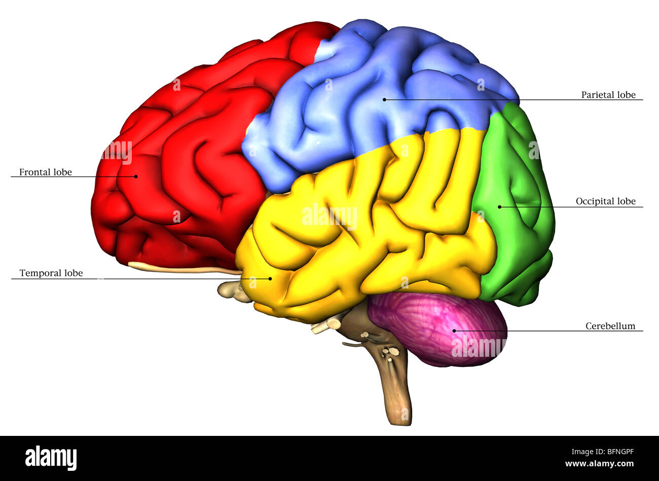 Illustration of the human brain Stock Photohttps://www.alamy.com/image-license-details/?v=1https://www.alamy.com/stock-photo-illustration-of-the-human-brain-26904375.html
RMBFNGPF–Illustration of the human brain
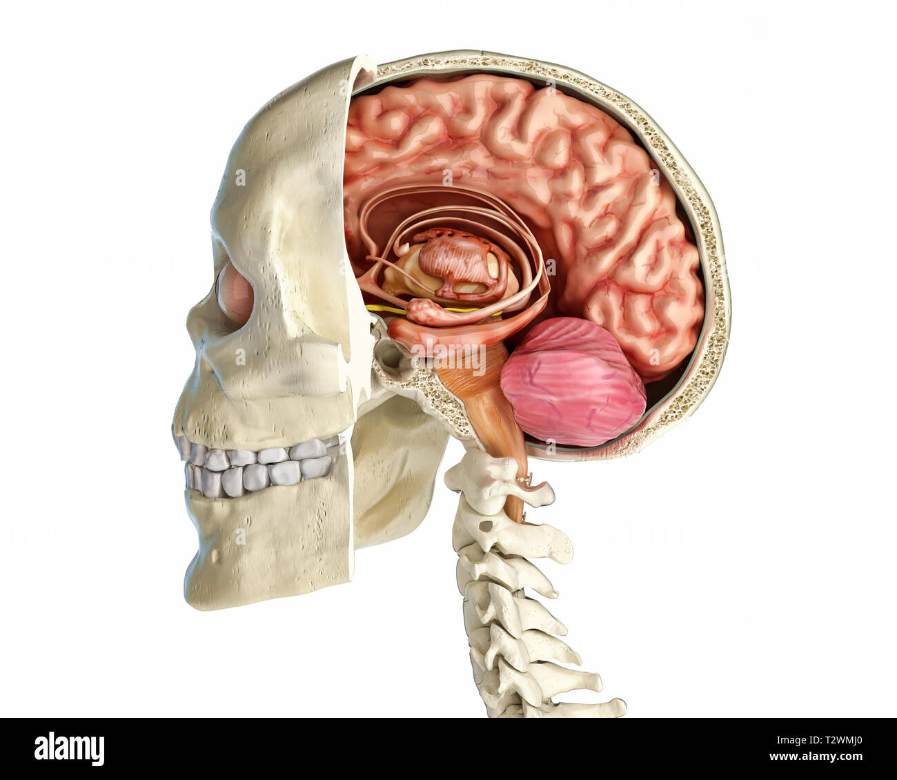 Human skull mid sagittal cross-section with brain. Side view on white background. Stock Photohttps://www.alamy.com/image-license-details/?v=1https://www.alamy.com/human-skull-mid-sagittal-cross-section-with-brain-side-view-on-white-background-image242739448.html
RFT2WMJ0–Human skull mid sagittal cross-section with brain. Side view on white background.
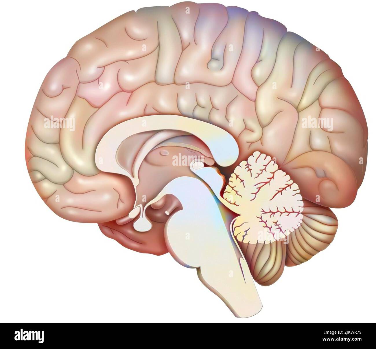 Median sagittal section of the brain with cerebellum and beginning of the brainstem. Stock Photohttps://www.alamy.com/image-license-details/?v=1https://www.alamy.com/median-sagittal-section-of-the-brain-with-cerebellum-and-beginning-of-the-brainstem-image476925437.html
RF2JKWR79–Median sagittal section of the brain with cerebellum and beginning of the brainstem.
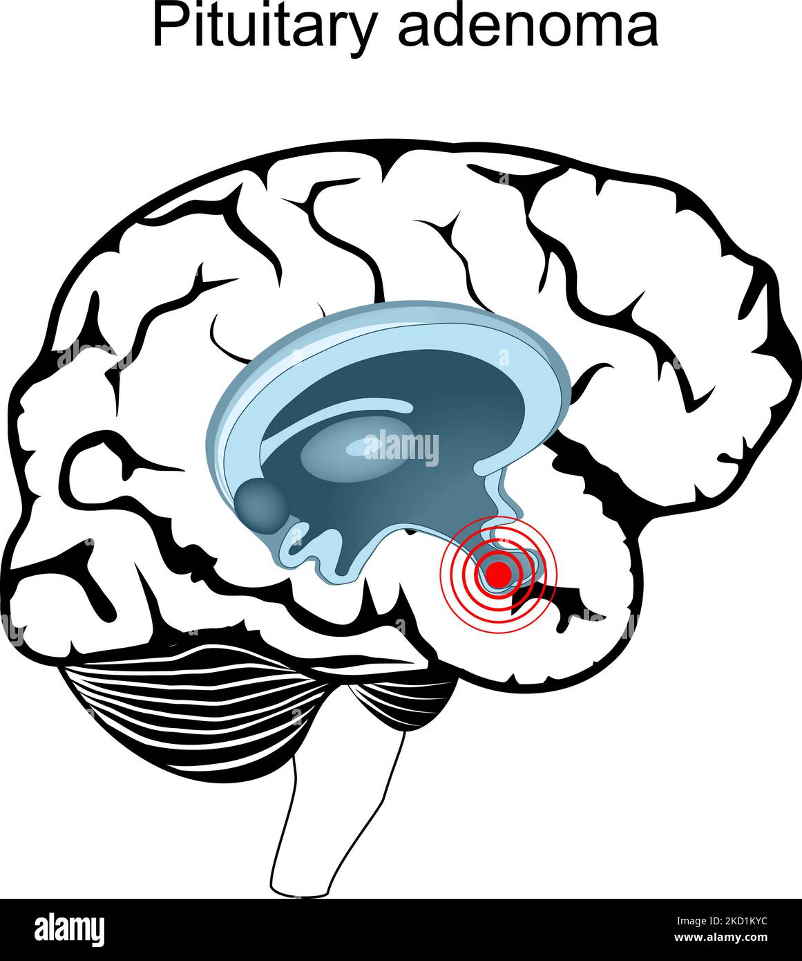 Pituitary adenoma. Cross section of human brain. Close-up of hypophysis. endocrine gland. vector poster Stock Vectorhttps://www.alamy.com/image-license-details/?v=1https://www.alamy.com/pituitary-adenoma-cross-section-of-human-brain-close-up-of-hypophysis-endocrine-gland-vector-poster-image489918448.html
RF2KD1KYC–Pituitary adenoma. Cross section of human brain. Close-up of hypophysis. endocrine gland. vector poster
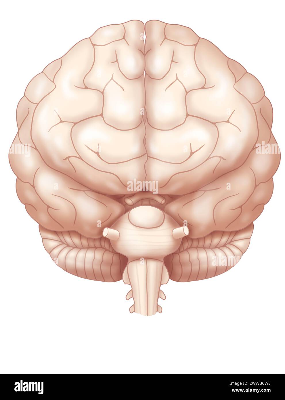 Anterior view of the brain with the two cerebral hemispheres, the cerebellum and the beginning of the brainstem. Stock Photohttps://www.alamy.com/image-license-details/?v=1https://www.alamy.com/anterior-view-of-the-brain-with-the-two-cerebral-hemispheres-the-cerebellum-and-the-beginning-of-the-brainstem-image600770506.html
RM2WWBCWE–Anterior view of the brain with the two cerebral hemispheres, the cerebellum and the beginning of the brainstem.
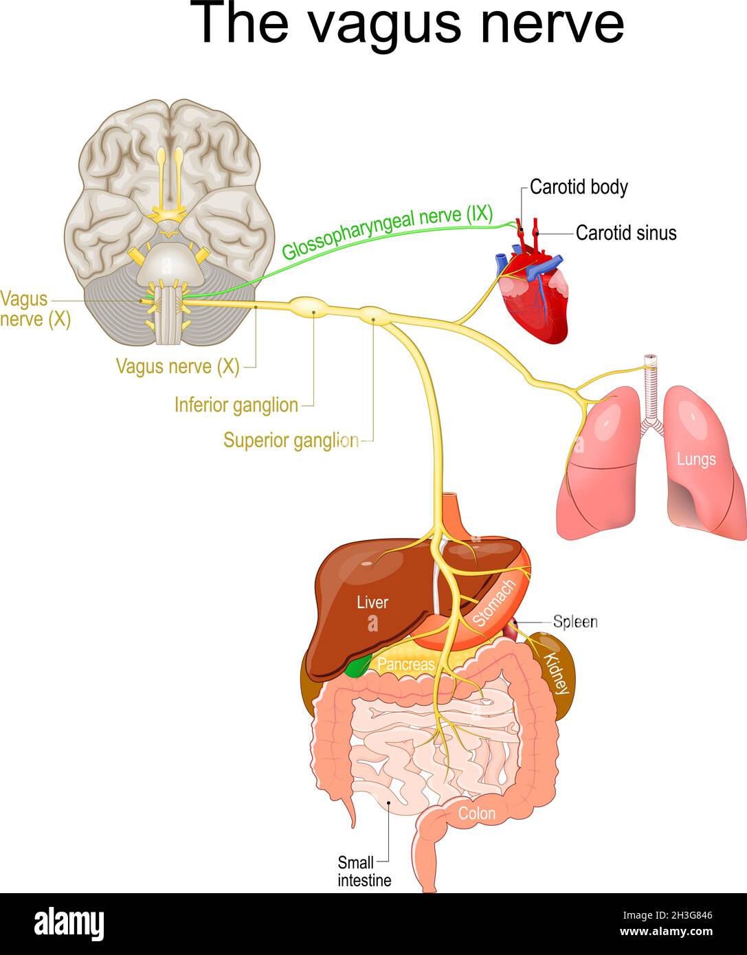 vagus nerve. parasympathetic nervous system. Medical diagram. Vector illustration to explain about human's nerve system. Stock Vectorhttps://www.alamy.com/image-license-details/?v=1https://www.alamy.com/vagus-nerve-parasympathetic-nervous-system-medical-diagram-vector-illustration-to-explain-about-humans-nerve-system-image449671158.html
RF2H3G846–vagus nerve. parasympathetic nervous system. Medical diagram. Vector illustration to explain about human's nerve system.
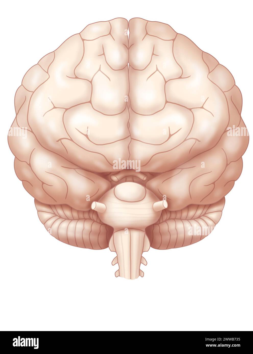 Anterior view of the brain with the two cerebral hemispheres, the cerebellum and the beginning of the brainstem. Stock Photohttps://www.alamy.com/image-license-details/?v=1https://www.alamy.com/anterior-view-of-the-brain-with-the-two-cerebral-hemispheres-the-cerebellum-and-the-beginning-of-the-brainstem-image600765961.html
RM2WWB735–Anterior view of the brain with the two cerebral hemispheres, the cerebellum and the beginning of the brainstem.
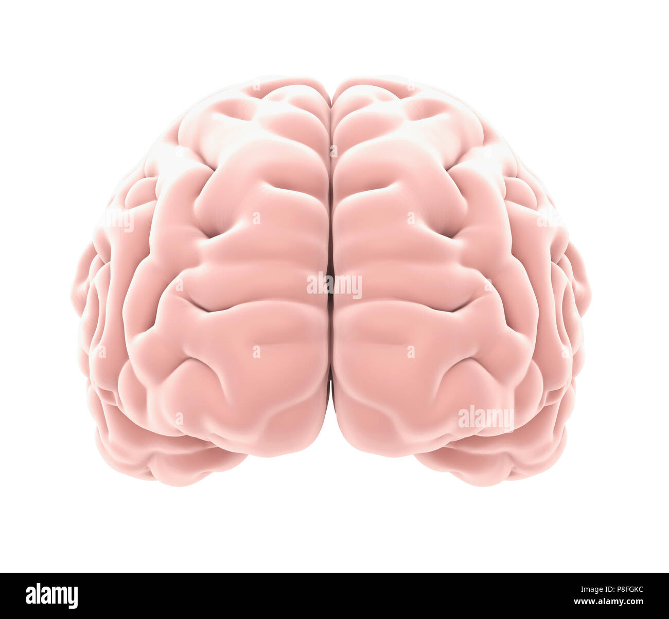 Human Brain Anatomy Isolated Stock Photohttps://www.alamy.com/image-license-details/?v=1https://www.alamy.com/human-brain-anatomy-isolated-image211784032.html
RFP8FGKC–Human Brain Anatomy Isolated
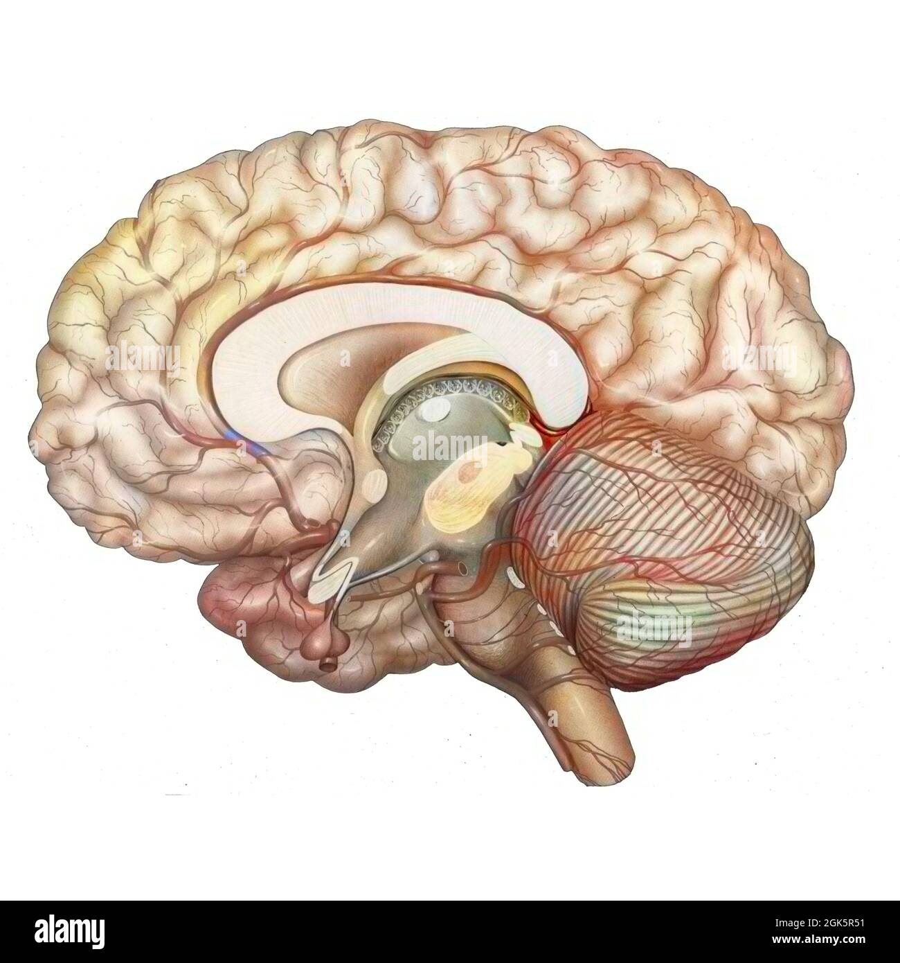 Cerebral vasculature: arteries of the diencephalon, cerebellum and brainstem. Stock Photohttps://www.alamy.com/image-license-details/?v=1https://www.alamy.com/cerebral-vasculature-arteries-of-the-diencephalon-cerebellum-and-brainstem-image442065597.html
RF2GK5R51–Cerebral vasculature: arteries of the diencephalon, cerebellum and brainstem.
 Human brain composed of paper with a shadow. Isolated on white background. Illustration Stock Photohttps://www.alamy.com/image-license-details/?v=1https://www.alamy.com/human-brain-composed-of-paper-with-a-shadow-isolated-on-white-background-illustration-image463598672.html
RF2HX6MRC–Human brain composed of paper with a shadow. Isolated on white background. Illustration
 Illustration of the cranial nerves anatomy. The cranial nerves are a set of 12 paired nerves that arise directly from the brain. The first two nerves (olfactory and optic) arise from the cerebrum, whereas the remaining ten emerge from the brainstem. The names of the cranial nerves relate to their function and they are numerically identified in roman numerals. Stock Photohttps://www.alamy.com/image-license-details/?v=1https://www.alamy.com/illustration-of-the-cranial-nerves-anatomy-the-cranial-nerves-are-a-set-of-12-paired-nerves-that-arise-directly-from-the-brain-the-first-two-nerves-olfactory-and-optic-arise-from-the-cerebrum-whereas-the-remaining-ten-emerge-from-the-brainstem-the-names-of-the-cranial-nerves-relate-to-their-function-and-they-are-numerically-identified-in-roman-numerals-image628777664.html
RF2YEY894–Illustration of the cranial nerves anatomy. The cranial nerves are a set of 12 paired nerves that arise directly from the brain. The first two nerves (olfactory and optic) arise from the cerebrum, whereas the remaining ten emerge from the brainstem. The names of the cranial nerves relate to their function and they are numerically identified in roman numerals.
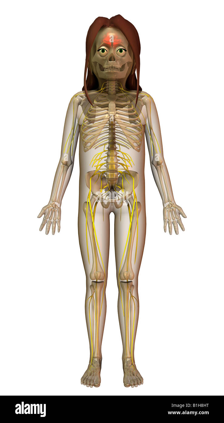 anatomy brain nerves Stock Photohttps://www.alamy.com/image-license-details/?v=1https://www.alamy.com/stock-photo-anatomy-brain-nerves-18204980.html
RMB1H8HT–anatomy brain nerves
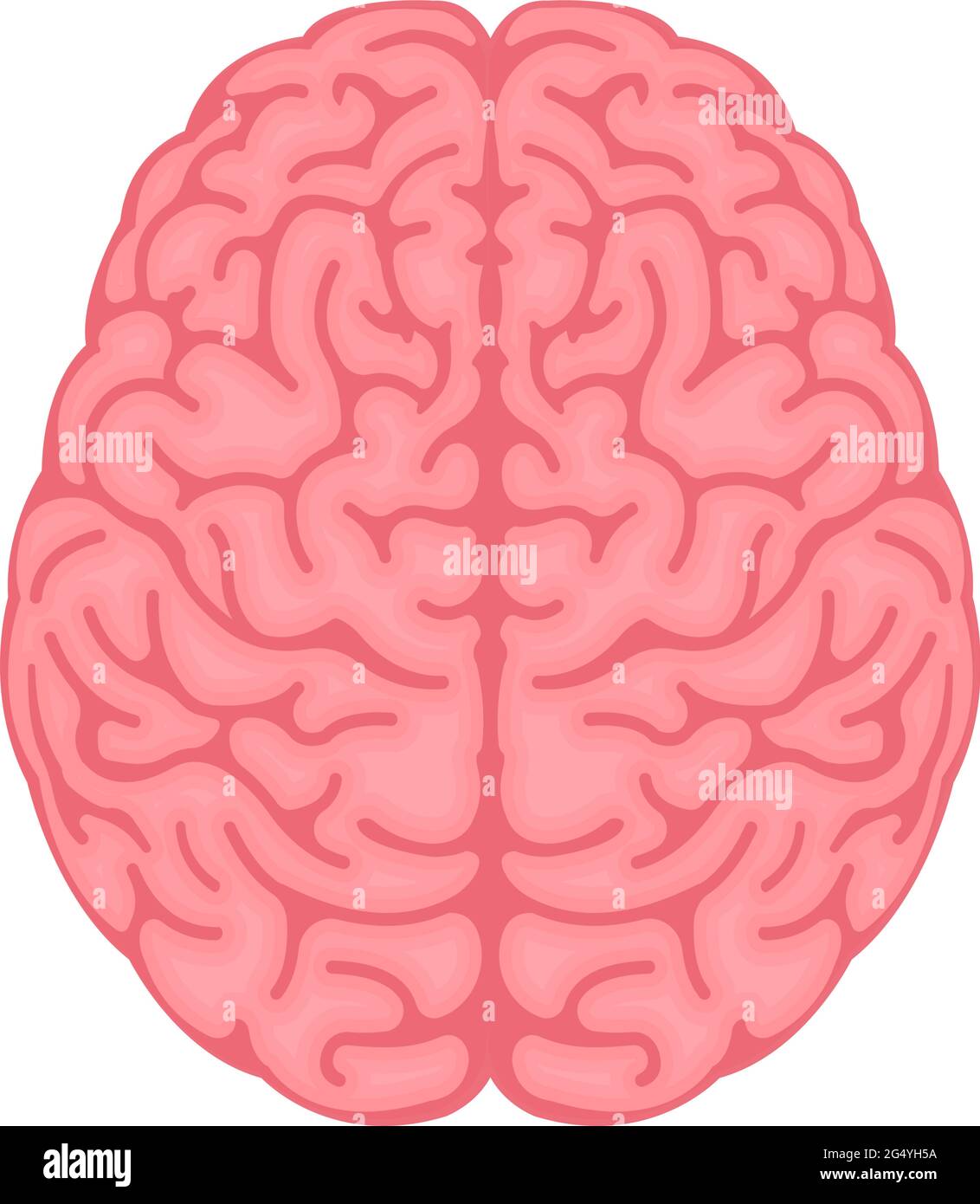 Vector illustration of human brain ( View from above ) Stock Vectorhttps://www.alamy.com/image-license-details/?v=1https://www.alamy.com/vector-illustration-of-human-brain-view-from-above-image433324006.html
RF2G4YH5A–Vector illustration of human brain ( View from above )
 The brainstem Stock Photohttps://www.alamy.com/image-license-details/?v=1https://www.alamy.com/stock-photo-the-brainstem-13165661.html
RFACJ25J–The brainstem
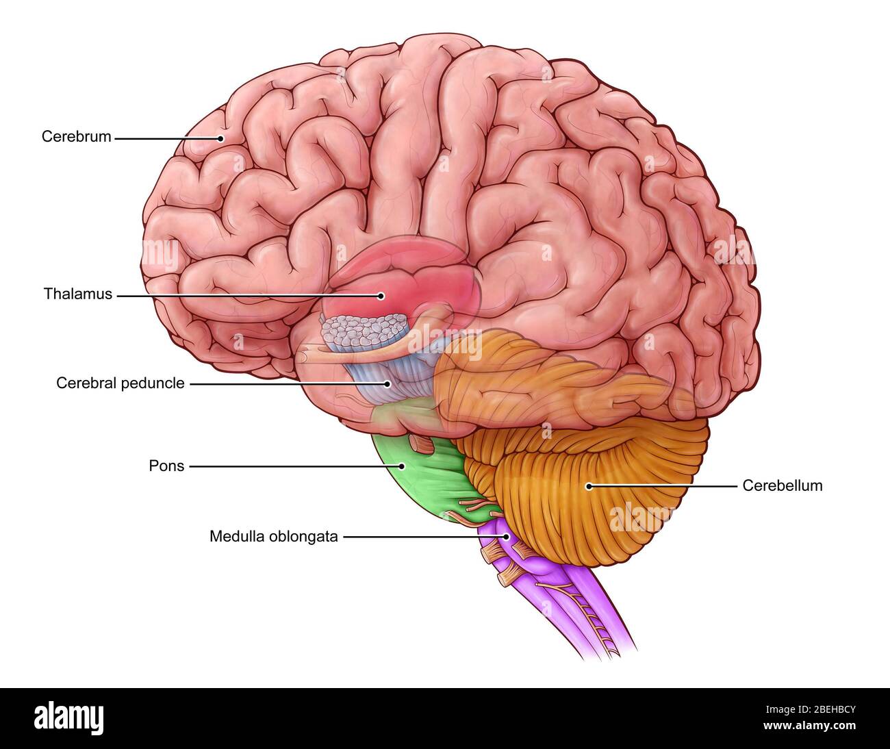 Diencephalon and Brainstem, illustration Stock Photohttps://www.alamy.com/image-license-details/?v=1https://www.alamy.com/diencephalon-and-brainstem-illustration-image353194715.html
RM2BEHBCY–Diencephalon and Brainstem, illustration
 Human brain on hand out of a laptop screen isolated on white Stock Photohttps://www.alamy.com/image-license-details/?v=1https://www.alamy.com/human-brain-on-hand-out-of-a-laptop-screen-isolated-on-white-image462237832.html
RF2HT0N20–Human brain on hand out of a laptop screen isolated on white
 Chordoma word cloud conceptual design isolated on white background. Stock Vectorhttps://www.alamy.com/image-license-details/?v=1https://www.alamy.com/chordoma-word-cloud-conceptual-design-isolated-on-white-background-image636934937.html
RF2S06W09–Chordoma word cloud conceptual design isolated on white background.
 Human brain with coins on white background Stock Photohttps://www.alamy.com/image-license-details/?v=1https://www.alamy.com/human-brain-with-coins-on-white-background-image360140196.html
RF2BWWPDT–Human brain with coins on white background
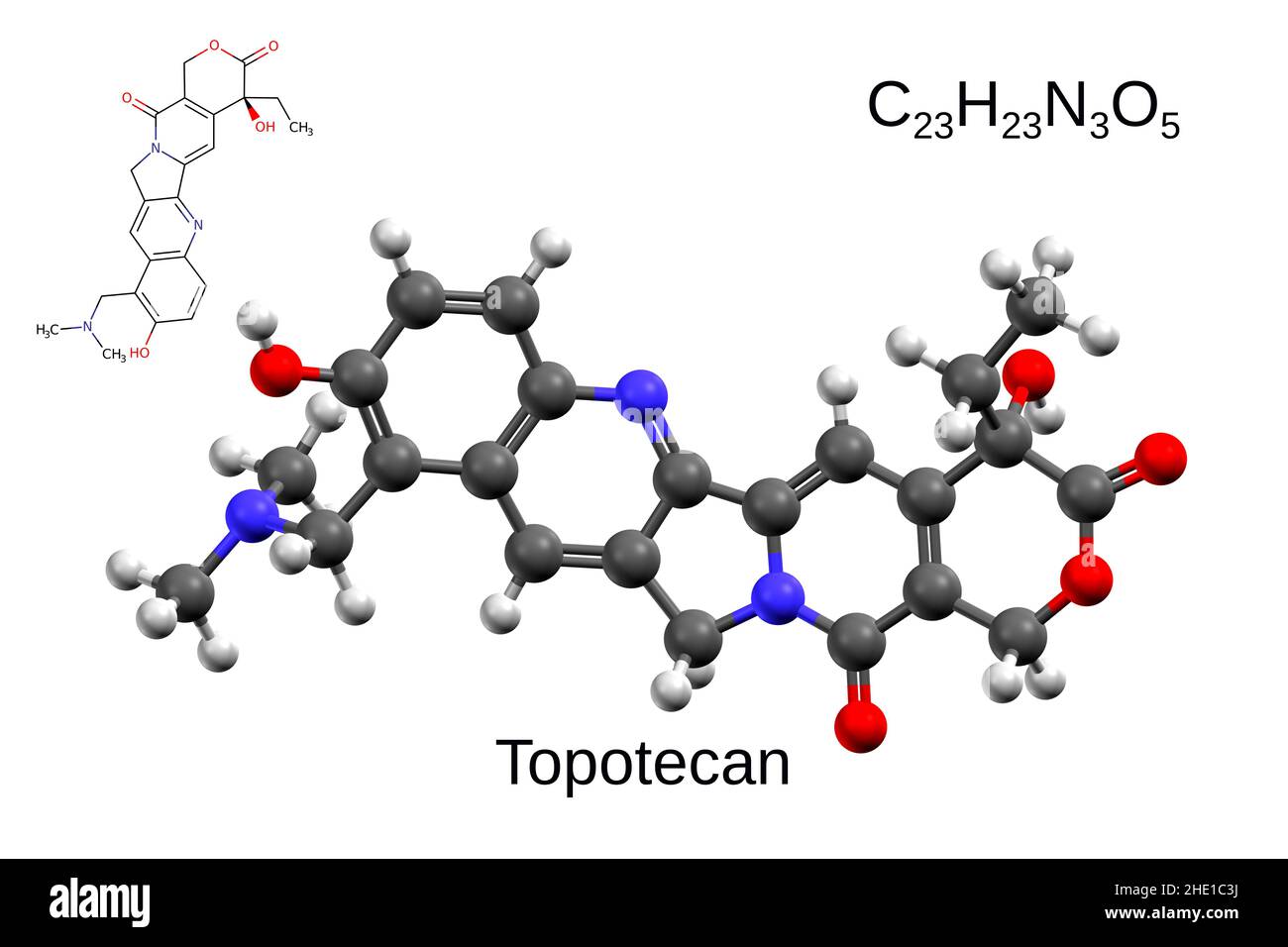 Chemical formula, structural formula and 3D ball-and-stick model of the anticancer drug topotecan, white background Stock Photohttps://www.alamy.com/image-license-details/?v=1https://www.alamy.com/chemical-formula-structural-formula-and-3d-ball-and-stick-model-of-the-anticancer-drug-topotecan-white-background-image456106214.html
RF2HE1C3J–Chemical formula, structural formula and 3D ball-and-stick model of the anticancer drug topotecan, white background
 Human brain on senior hand isolated on white background Stock Photohttps://www.alamy.com/image-license-details/?v=1https://www.alamy.com/human-brain-on-senior-hand-isolated-on-white-background-image624673279.html
RF2Y8893Y–Human brain on senior hand isolated on white background
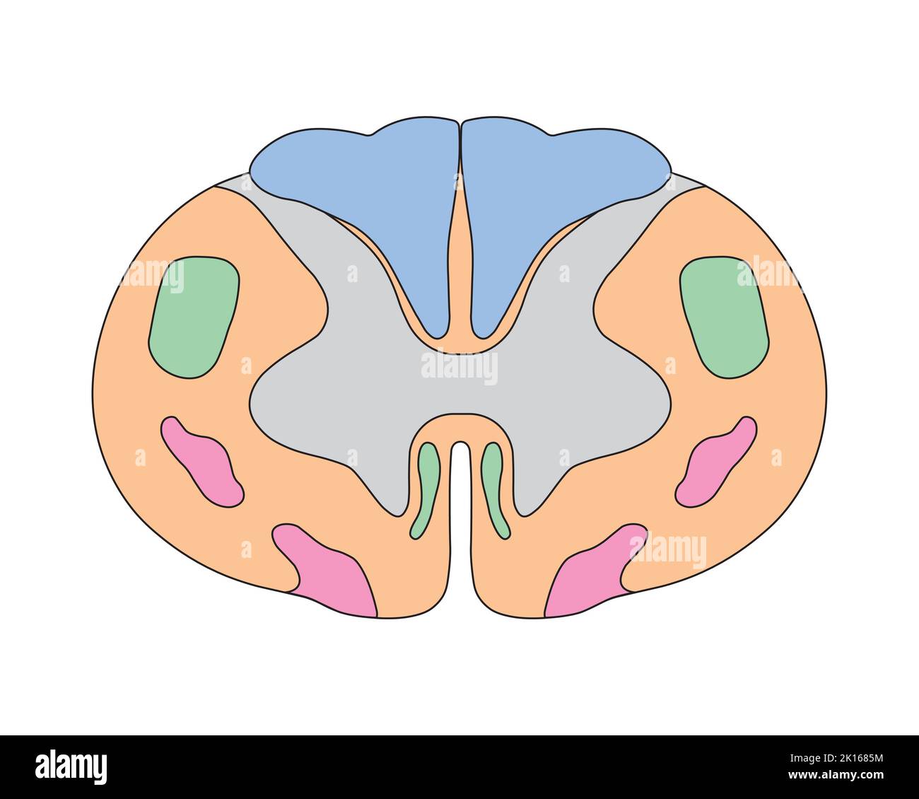 Scientific Designing of Spinal Cord Anatomy. Cervical Spinal Cord Structure. Colorful Symbols. Vector Illustration. Stock Vectorhttps://www.alamy.com/image-license-details/?v=1https://www.alamy.com/scientific-designing-of-spinal-cord-anatomy-cervical-spinal-cord-structure-colorful-symbols-vector-illustration-image482643104.html
RF2K1685M–Scientific Designing of Spinal Cord Anatomy. Cervical Spinal Cord Structure. Colorful Symbols. Vector Illustration.
 Human brain on hand isolated on white background Stock Photohttps://www.alamy.com/image-license-details/?v=1https://www.alamy.com/human-brain-on-hand-isolated-on-white-background-image327599458.html
RF2A0YCCJ–Human brain on hand isolated on white background
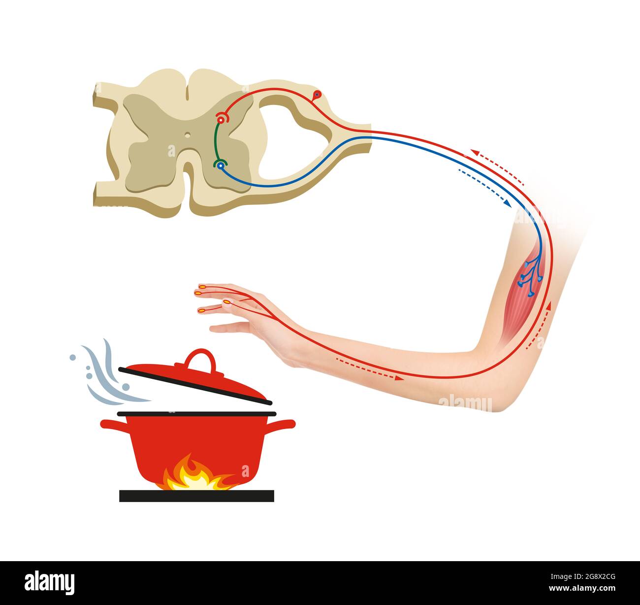 A reflex arc is a neural pathway that controls a reflex Stock Photohttps://www.alamy.com/image-license-details/?v=1https://www.alamy.com/a-reflex-arc-is-a-neural-pathway-that-controls-a-reflex-image435749120.html
RF2G8X2CG–A reflex arc is a neural pathway that controls a reflex
 Top view of the human brain model isolated on white background close up. The concept of neurosurgery, health care Stock Photohttps://www.alamy.com/image-license-details/?v=1https://www.alamy.com/top-view-of-the-human-brain-model-isolated-on-white-background-close-up-the-concept-of-neurosurgery-health-care-image458798087.html
RF2HJC1HY–Top view of the human brain model isolated on white background close up. The concept of neurosurgery, health care
 Human brain on laptop isolated on white background Stock Photohttps://www.alamy.com/image-license-details/?v=1https://www.alamy.com/human-brain-on-laptop-isolated-on-white-background-image483352692.html
RF2K2AH84–Human brain on laptop isolated on white background
 Human brain right hemisphere model isolated on white background close up. The concept of neurosurgery, health care Stock Photohttps://www.alamy.com/image-license-details/?v=1https://www.alamy.com/human-brain-right-hemisphere-model-isolated-on-white-background-close-up-the-concept-of-neurosurgery-health-care-image458798079.html
RF2HJC1HK–Human brain right hemisphere model isolated on white background close up. The concept of neurosurgery, health care
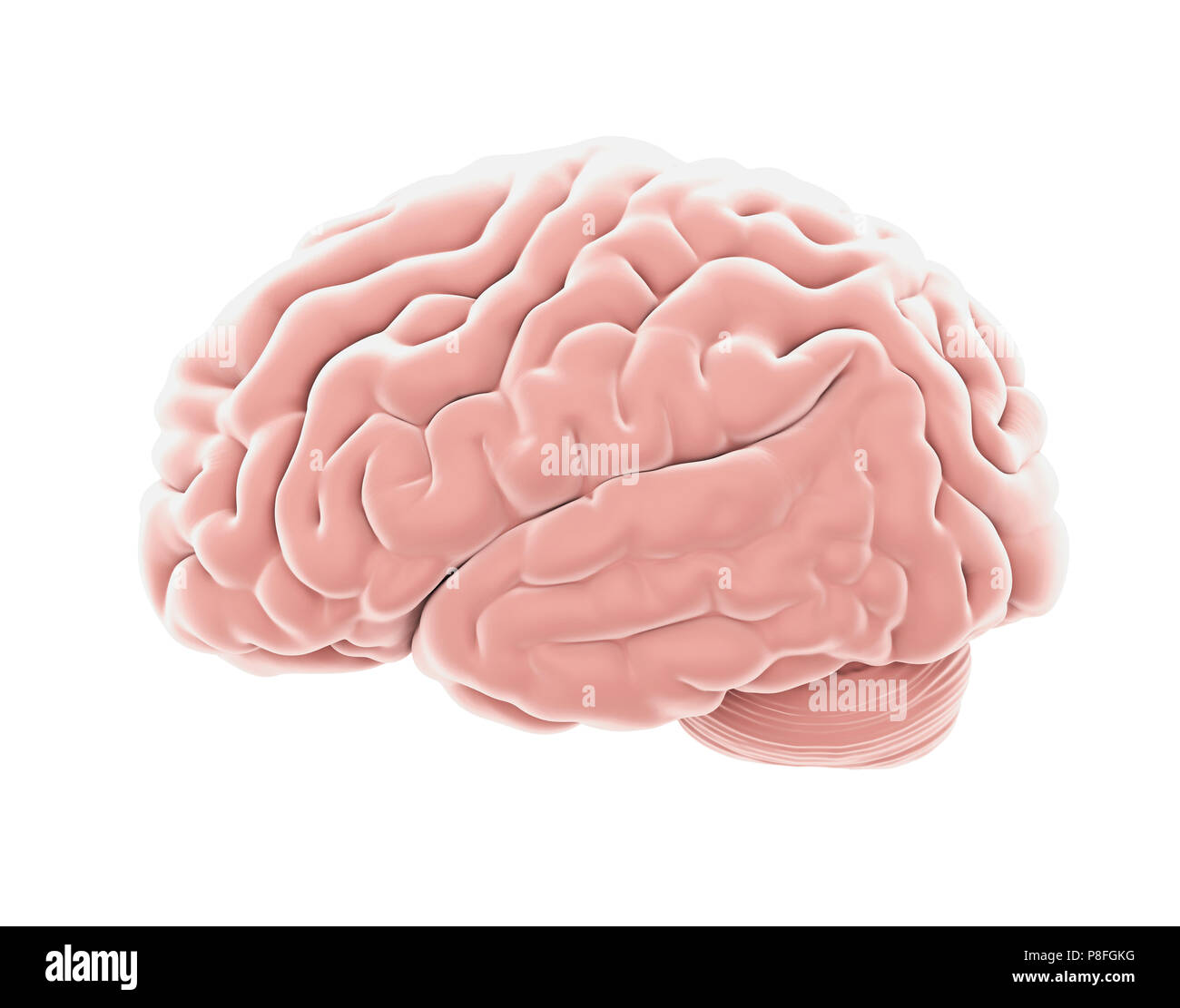 Human Brain Anatomy Isolated Stock Photohttps://www.alamy.com/image-license-details/?v=1https://www.alamy.com/human-brain-anatomy-isolated-image211784036.html
RFP8FGKG–Human Brain Anatomy Isolated
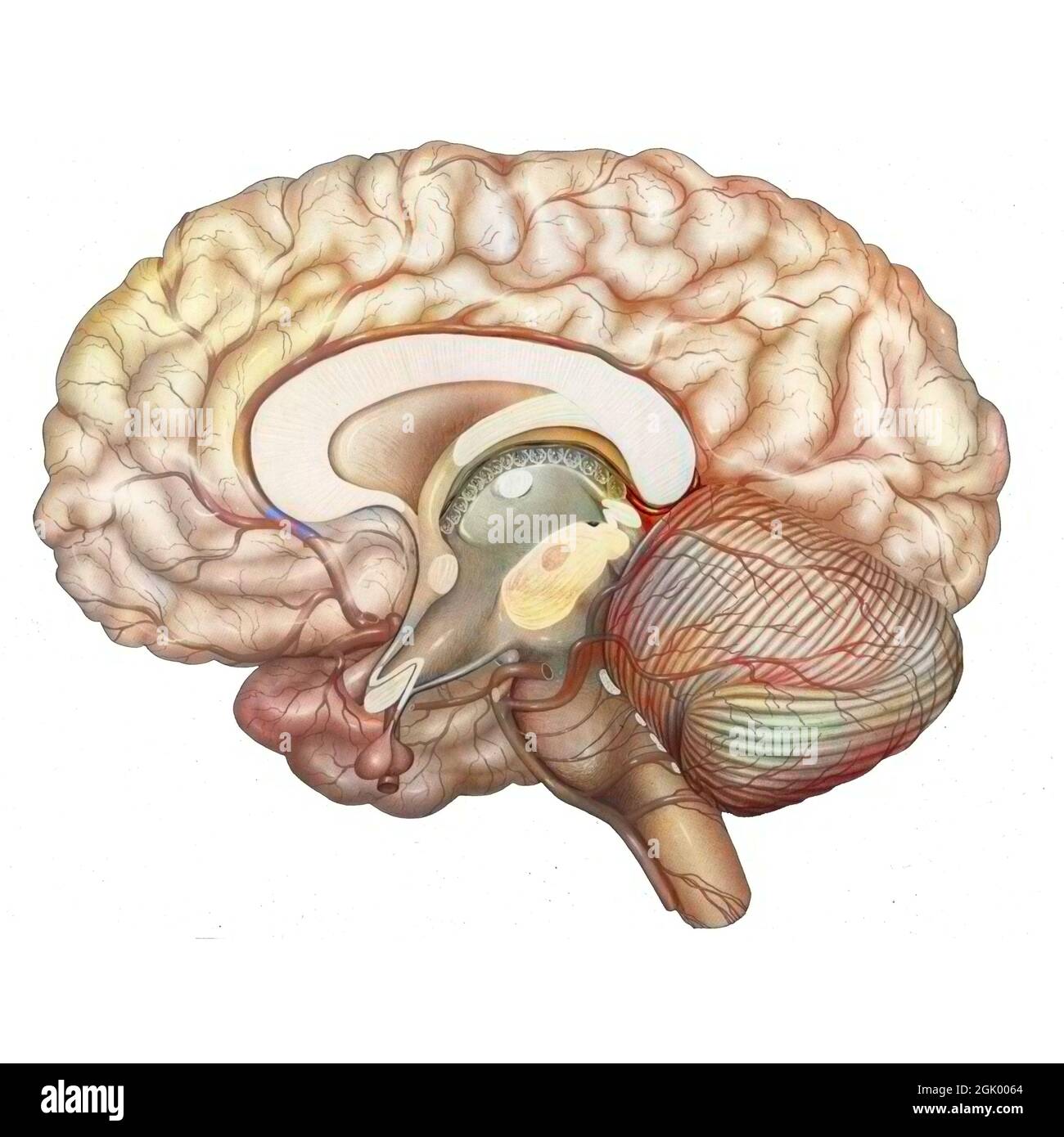 Cerebral vasculature: arteries of the diencephalon, cerebellum and brainstem. Stock Photohttps://www.alamy.com/image-license-details/?v=1https://www.alamy.com/cerebral-vasculature-arteries-of-the-diencephalon-cerebellum-and-brainstem-image441937836.html
RF2GK0064–Cerebral vasculature: arteries of the diencephalon, cerebellum and brainstem.
 Blood vessels (arteries) in abstract human brain as cerebrovascular concept. Isolated on white background. Illustration Stock Photohttps://www.alamy.com/image-license-details/?v=1https://www.alamy.com/blood-vessels-arteries-in-abstract-human-brain-as-cerebrovascular-concept-isolated-on-white-background-illustration-image463598698.html
RF2HX6MTA–Blood vessels (arteries) in abstract human brain as cerebrovascular concept. Isolated on white background. Illustration
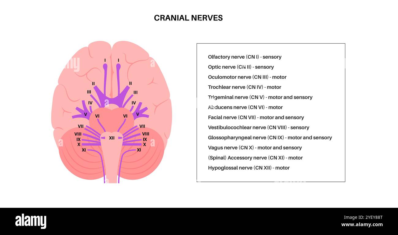 Illustration of the cranial nerves anatomy. The cranial nerves are a set of 12 paired nerves that arise directly from the brain. The first two nerves (olfactory and optic) arise from the cerebrum, whereas the remaining ten emerge from the brainstem. The names of the cranial nerves relate to their function and they are numerically identified in roman numerals. Stock Photohttps://www.alamy.com/image-license-details/?v=1https://www.alamy.com/illustration-of-the-cranial-nerves-anatomy-the-cranial-nerves-are-a-set-of-12-paired-nerves-that-arise-directly-from-the-brain-the-first-two-nerves-olfactory-and-optic-arise-from-the-cerebrum-whereas-the-remaining-ten-emerge-from-the-brainstem-the-names-of-the-cranial-nerves-relate-to-their-function-and-they-are-numerically-identified-in-roman-numerals-image628777656.html
RF2YEY88T–Illustration of the cranial nerves anatomy. The cranial nerves are a set of 12 paired nerves that arise directly from the brain. The first two nerves (olfactory and optic) arise from the cerebrum, whereas the remaining ten emerge from the brainstem. The names of the cranial nerves relate to their function and they are numerically identified in roman numerals.
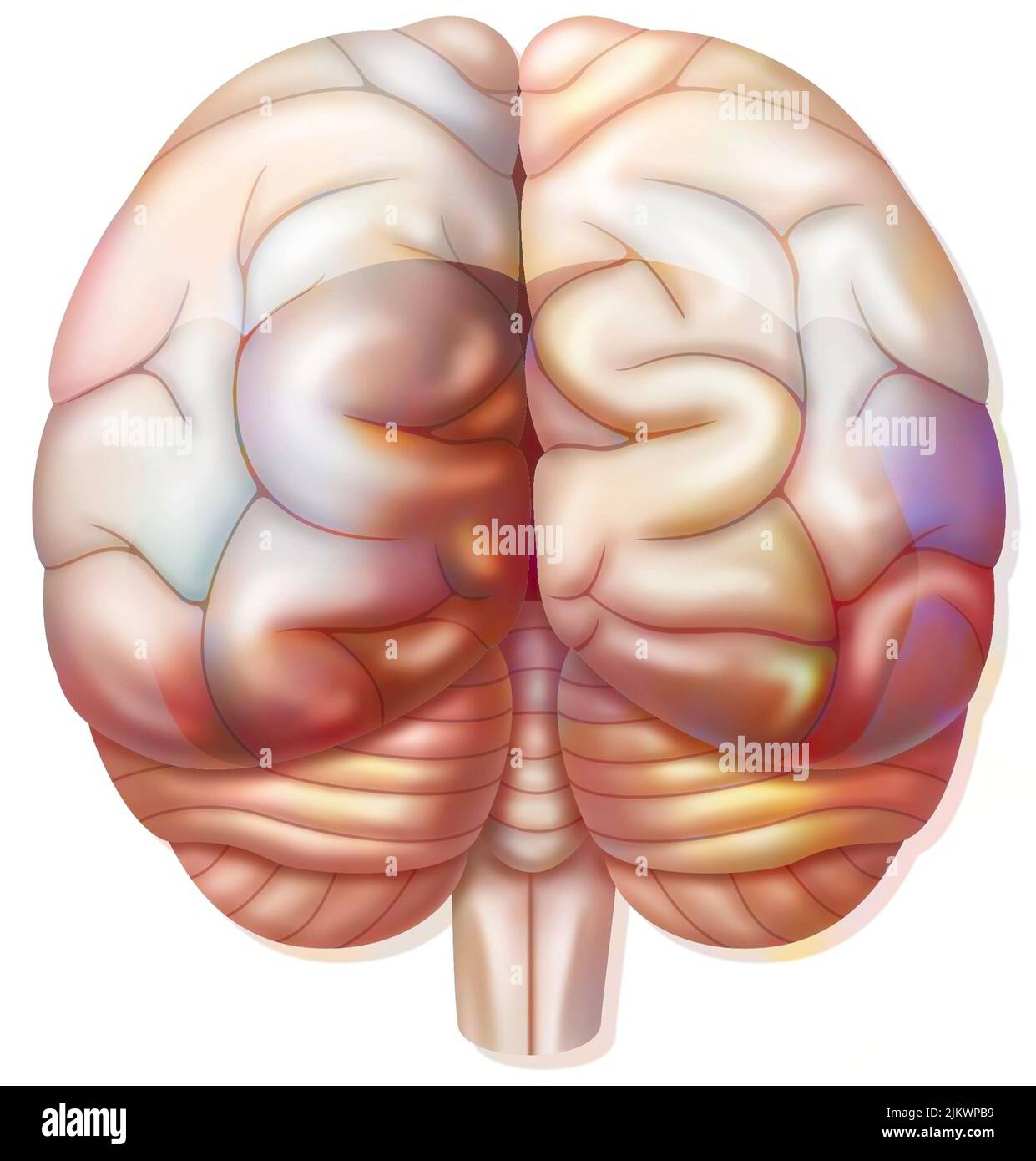 Brain with occipital, parietal, temporal lobes, cerebellum. Stock Photohttps://www.alamy.com/image-license-details/?v=1https://www.alamy.com/brain-with-occipital-parietal-temporal-lobes-cerebellum-image476924765.html
RF2JKWPB9–Brain with occipital, parietal, temporal lobes, cerebellum.
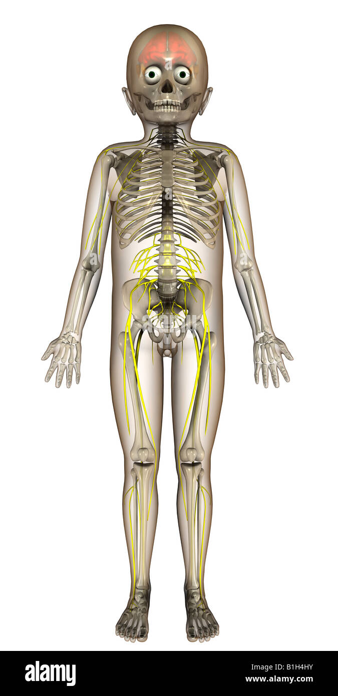 anatomy nerves brain Stock Photohttps://www.alamy.com/image-license-details/?v=1https://www.alamy.com/stock-photo-anatomy-nerves-brain-18201847.html
RMB1H4HY–anatomy nerves brain
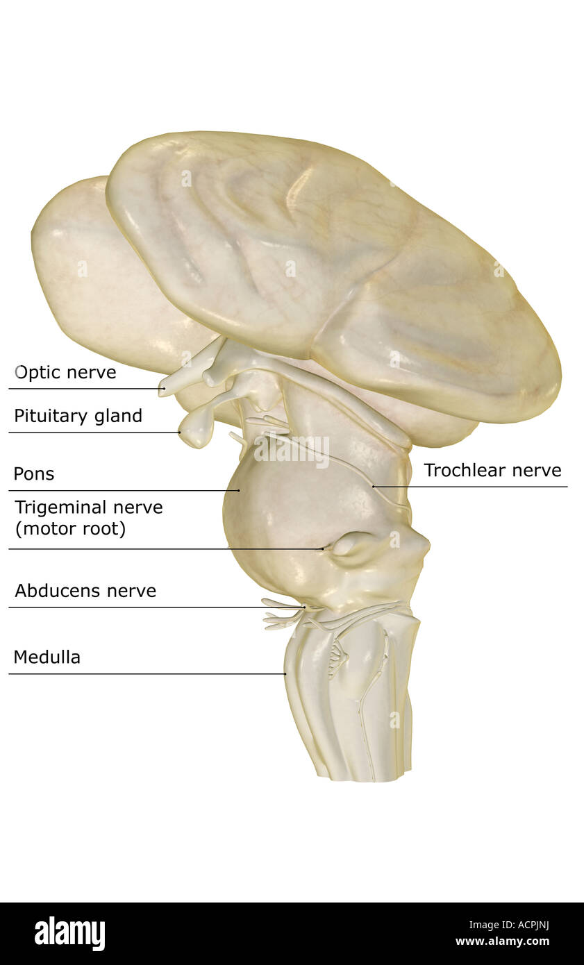 The brainstem Stock Photohttps://www.alamy.com/image-license-details/?v=1https://www.alamy.com/stock-photo-the-brainstem-13208861.html
RFACPJNJ–The brainstem
 Diencephalon and Brainstem, illustration Stock Photohttps://www.alamy.com/image-license-details/?v=1https://www.alamy.com/diencephalon-and-brainstem-illustration-image353194710.html
RM2BEHBCP–Diencephalon and Brainstem, illustration
