Quick filters:
Cancer cells micrograph Stock Photos and Images
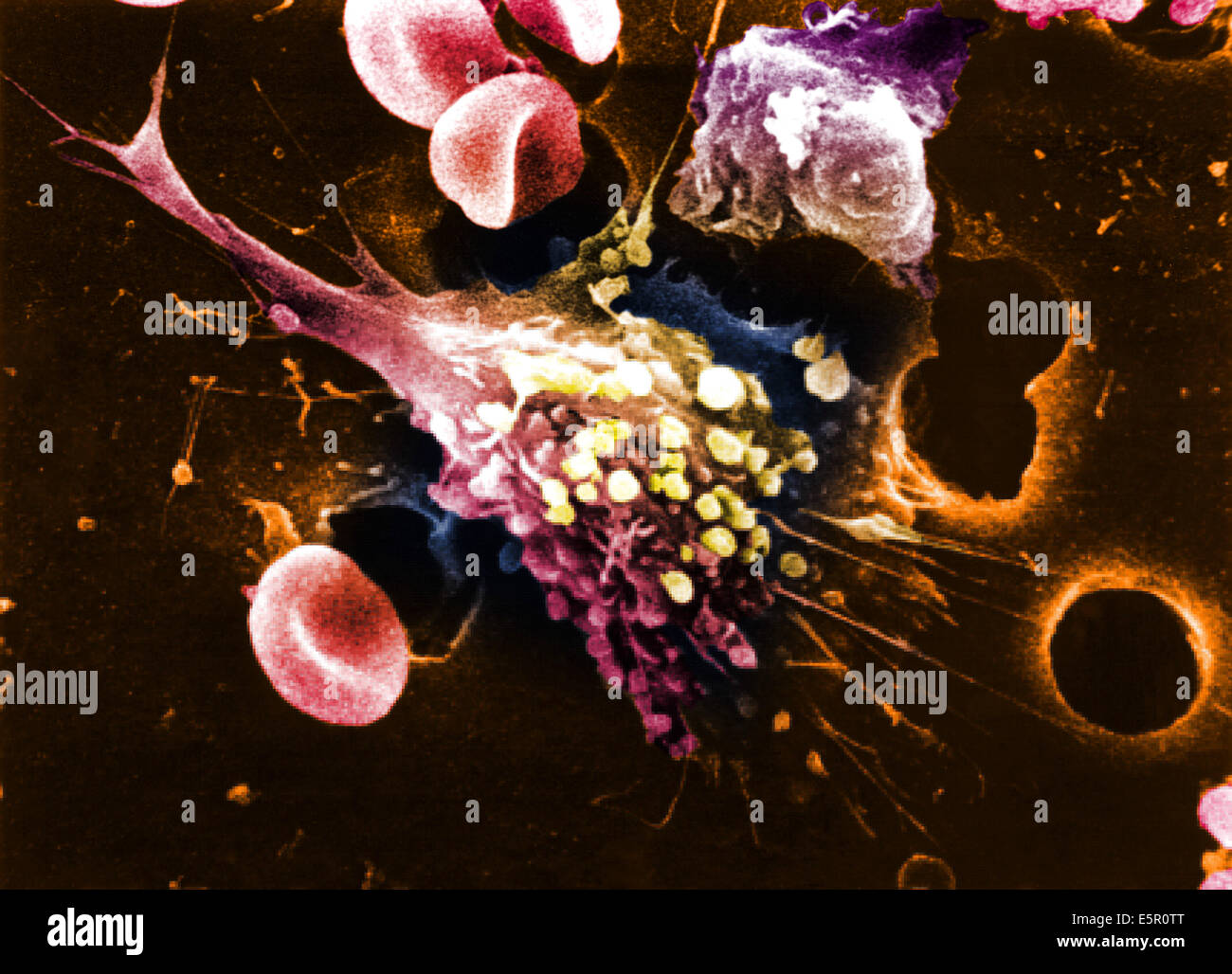 Scanning electron micrograph (SEM) of a cancer cell, Magnification x12000. Stock Photohttps://www.alamy.com/image-license-details/?v=1https://www.alamy.com/stock-photo-scanning-electron-micrograph-sem-of-a-cancer-cell-magnification-x12000-72420344.html
RME5R0TT–Scanning electron micrograph (SEM) of a cancer cell, Magnification x12000.
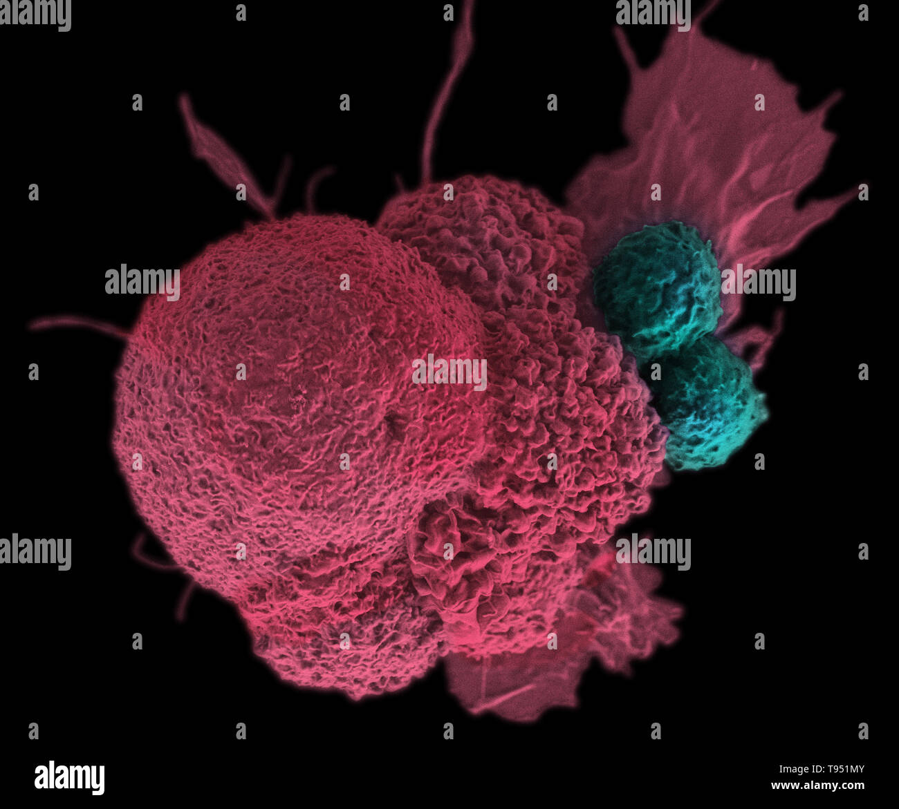 This electron micrograph (SEM) shows an oral squamous cancer cell (red) being attacked by two cytotoxic T cells (blue). The tumor-specific T cells were developed from the patient's own immune system, a personalized cancer vaccine like those that may be developed in the future with the help of NIST's standardized human genomes. This image has been colorized. Stock Photohttps://www.alamy.com/image-license-details/?v=1https://www.alamy.com/this-electron-micrograph-sem-shows-an-oral-squamous-cancer-cell-red-being-attacked-by-two-cytotoxic-t-cells-blue-the-tumor-specific-t-cells-were-developed-from-the-patients-own-immune-system-a-personalized-cancer-vaccine-like-those-that-may-be-developed-in-the-future-with-the-help-of-nists-standardized-human-genomes-this-image-has-been-colorized-image246588187.html
RMT951MY–This electron micrograph (SEM) shows an oral squamous cancer cell (red) being attacked by two cytotoxic T cells (blue). The tumor-specific T cells were developed from the patient's own immune system, a personalized cancer vaccine like those that may be developed in the future with the help of NIST's standardized human genomes. This image has been colorized.
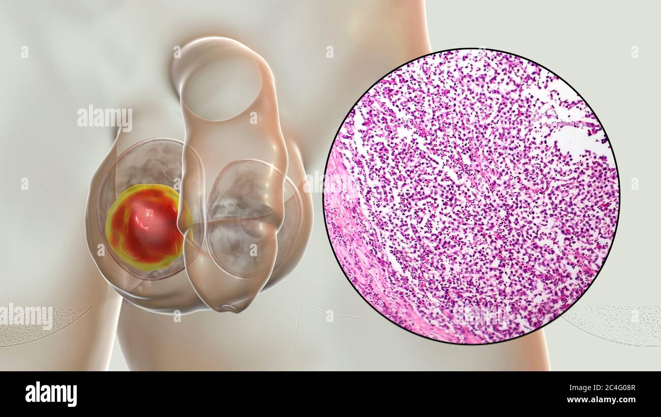 Testicular cancer, a malignant spermatocytic seminoma, computer illustration and light micrograph. A seminoma is a malignant tumour (cancer) of the testis (testicle). Although this is a rare cancer, it is the most common cancer in 15 to 35 year old men. It arises from abnormal germ cells (precursors to sperm cells) in the seminiferous tubules. Surgical removal of the affected testis (orchidectomy) is followed by radiotherapy and chemotherapy if required. Stock Photohttps://www.alamy.com/image-license-details/?v=1https://www.alamy.com/testicular-cancer-a-malignant-spermatocytic-seminoma-computer-illustration-and-light-micrograph-a-seminoma-is-a-malignant-tumour-cancer-of-the-testis-testicle-although-this-is-a-rare-cancer-it-is-the-most-common-cancer-in-15-to-35-year-old-men-it-arises-from-abnormal-germ-cells-precursors-to-sperm-cells-in-the-seminiferous-tubules-surgical-removal-of-the-affected-testis-orchidectomy-is-followed-by-radiotherapy-and-chemotherapy-if-required-image364227831.html
RF2C4G08R–Testicular cancer, a malignant spermatocytic seminoma, computer illustration and light micrograph. A seminoma is a malignant tumour (cancer) of the testis (testicle). Although this is a rare cancer, it is the most common cancer in 15 to 35 year old men. It arises from abnormal germ cells (precursors to sperm cells) in the seminiferous tubules. Surgical removal of the affected testis (orchidectomy) is followed by radiotherapy and chemotherapy if required.
 Shown here is a pseudo-colored scanning electron micrograph of an oral squamous cancer cell (white) attacked by two cytotoxic T cells (red), part of a natural immune response. Nanomedicine researchers create personalized cancer vaccines by loading neo-antigens identified from patient and tumor Stock Photohttps://www.alamy.com/image-license-details/?v=1https://www.alamy.com/shown-here-is-a-pseudo-colored-scanning-electron-micrograph-of-an-oral-squamous-cancer-cell-white-attacked-by-two-cytotoxic-t-cells-red-part-of-a-natural-immune-response-nanomedicine-researchers-create-personalized-cancer-vaccines-by-loading-neo-antigens-identified-from-patient-and-tumor-image476706747.html
RM2JKFT8Y–Shown here is a pseudo-colored scanning electron micrograph of an oral squamous cancer cell (white) attacked by two cytotoxic T cells (red), part of a natural immune response. Nanomedicine researchers create personalized cancer vaccines by loading neo-antigens identified from patient and tumor
 Slices of the tumor under glass. Histological examination of tumor cells for the presence of cancer Stock Photohttps://www.alamy.com/image-license-details/?v=1https://www.alamy.com/slices-of-the-tumor-under-glass-histological-examination-of-tumor-cells-for-the-presence-of-cancer-image369975652.html
RM2CDWRM4–Slices of the tumor under glass. Histological examination of tumor cells for the presence of cancer
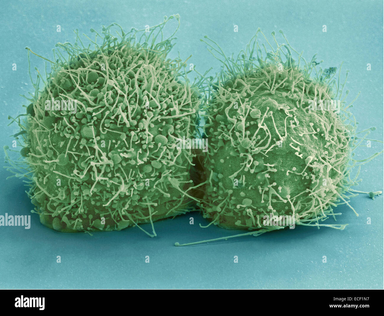 Scanning electron micrograph of just-divided HeLa cells. Zeiss Merlin HR-SEM. Stock Photohttps://www.alamy.com/image-license-details/?v=1https://www.alamy.com/stock-photo-scanning-electron-micrograph-of-just-divided-hela-cells-zeiss-merlin-76548003.html
RFECF1N7–Scanning electron micrograph of just-divided HeLa cells. Zeiss Merlin HR-SEM.
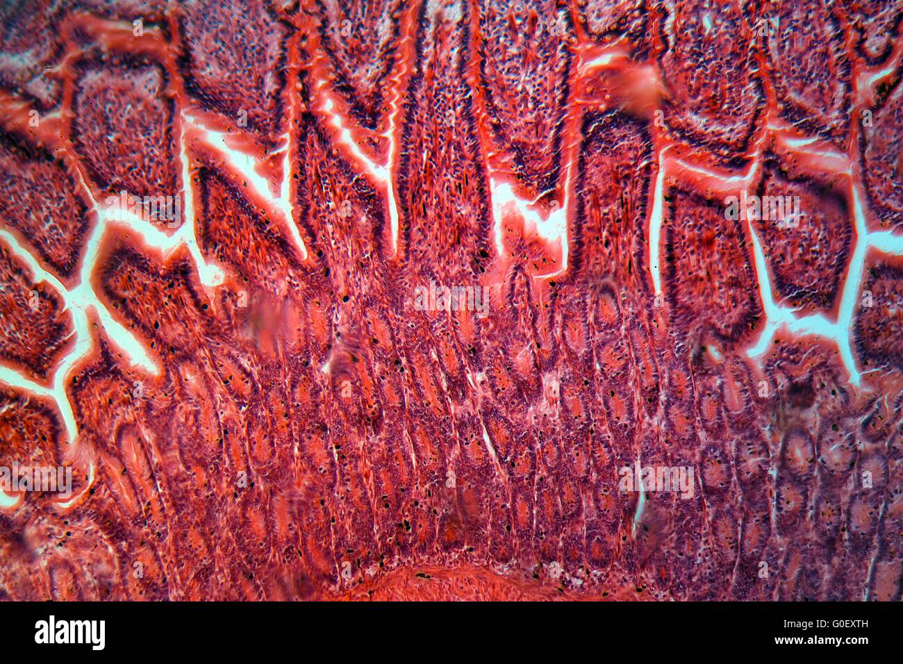 A section trough cells of a small intestine under the microscope. Stock Photohttps://www.alamy.com/image-license-details/?v=1https://www.alamy.com/stock-photo-a-section-trough-cells-of-a-small-intestine-under-the-microscope-103590609.html
RMG0EXTH–A section trough cells of a small intestine under the microscope.
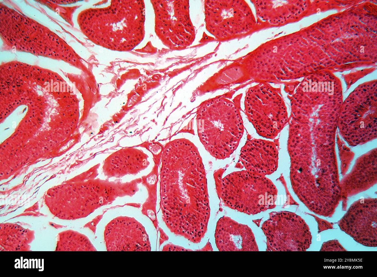 A section trough testicle cells under the microscope Stock Photohttps://www.alamy.com/image-license-details/?v=1https://www.alamy.com/a-section-trough-testicle-cells-under-the-microscope-image624944586.html
RF2Y8MK5E–A section trough testicle cells under the microscope
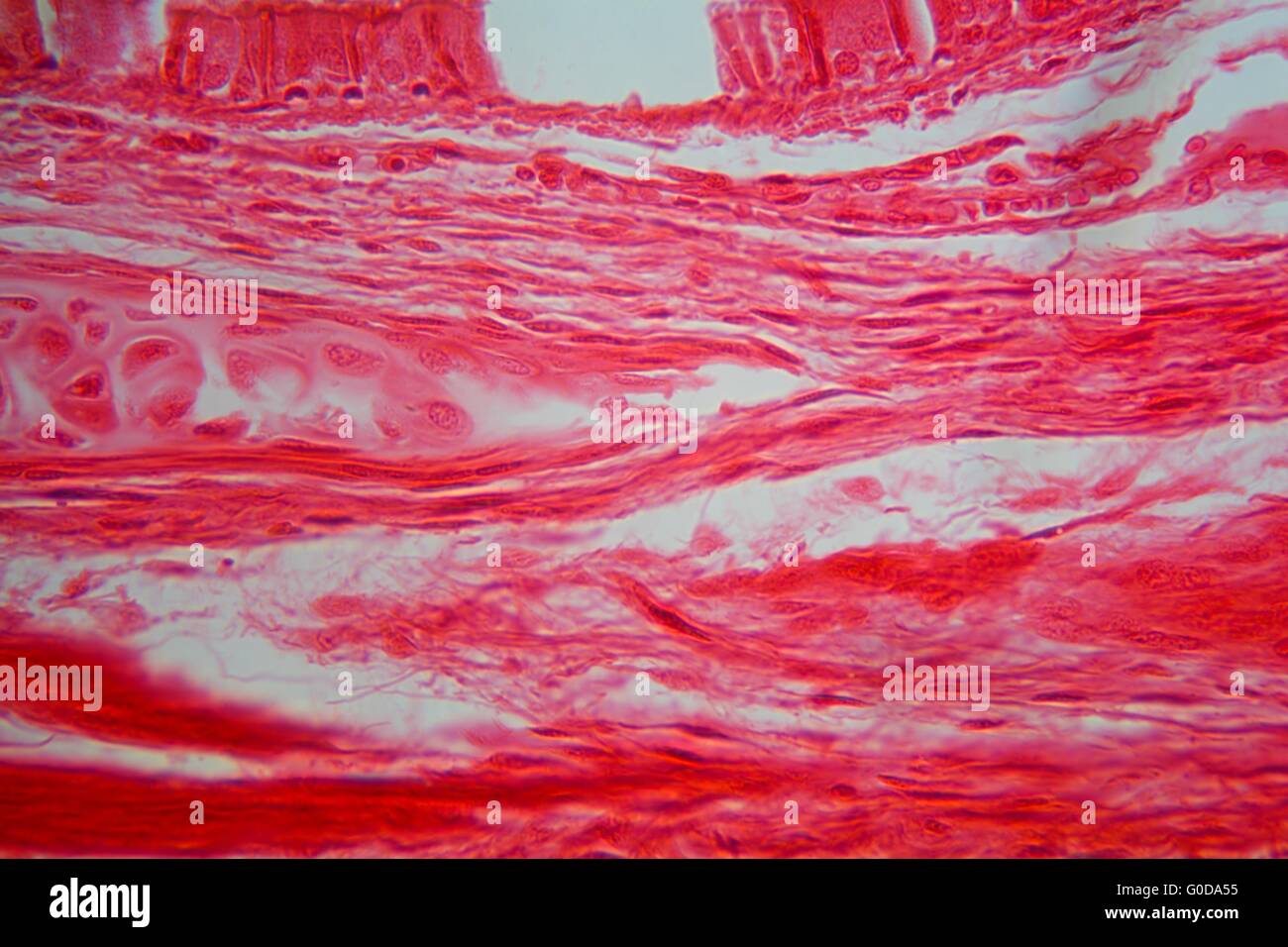 Cells of trachea tissue under the microscope Stock Photohttps://www.alamy.com/image-license-details/?v=1https://www.alamy.com/stock-photo-cells-of-trachea-tissue-under-the-microscope-103555569.html
RMG0DA55–Cells of trachea tissue under the microscope
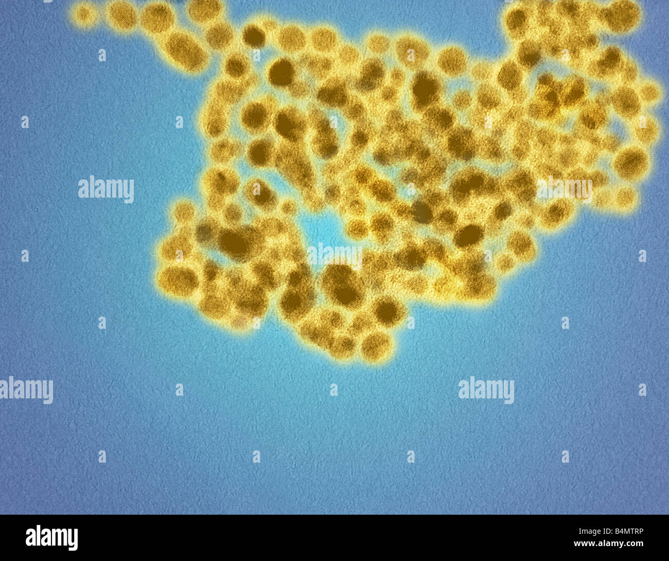 A TEM image of gold nanoparticles coating cells in a glutathione surface corona illustrating the molecular size of nanoparticles Stock Photohttps://www.alamy.com/image-license-details/?v=1https://www.alamy.com/stock-photo-a-tem-image-of-gold-nanoparticles-coating-cells-in-a-glutathione-surface-20127514.html
RMB4MTRP–A TEM image of gold nanoparticles coating cells in a glutathione surface corona illustrating the molecular size of nanoparticles
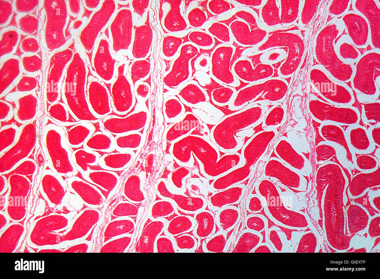 A section trough testicle cells under the microscope. Stock Photohttps://www.alamy.com/image-license-details/?v=1https://www.alamy.com/stock-photo-a-section-trough-testicle-cells-under-the-microscope-103590614.html
RMG0EXTP–A section trough testicle cells under the microscope.
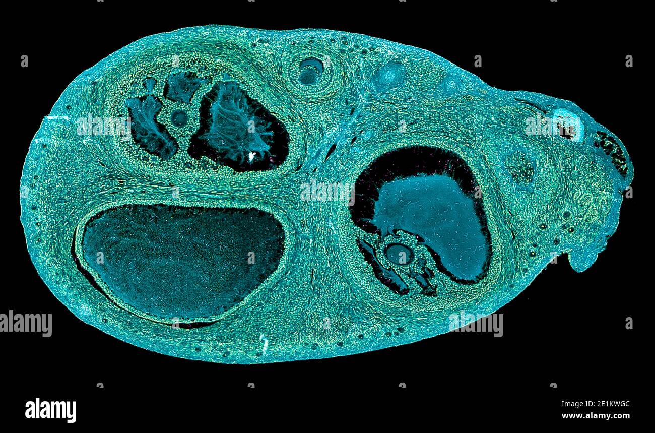 cross section cut of human body cells under a scientific microscope Stock Photohttps://www.alamy.com/image-license-details/?v=1https://www.alamy.com/cross-section-cut-of-human-body-cells-under-a-scientific-microscope-image396890268.html
RF2E1KWGC–cross section cut of human body cells under a scientific microscope
 Close-up of red blood cells Stock Photohttps://www.alamy.com/image-license-details/?v=1https://www.alamy.com/stock-photo-close-up-of-red-blood-cells-91548435.html
RFF8XAXY–Close-up of red blood cells
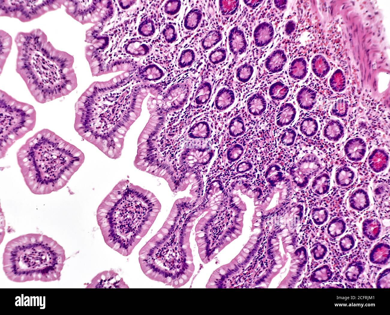 Intestinal cancer cells, brightfield photomicrograph Stock Photohttps://www.alamy.com/image-license-details/?v=1https://www.alamy.com/intestinal-cancer-cells-brightfield-photomicrograph-image371157137.html
RM2CFRJM1–Intestinal cancer cells, brightfield photomicrograph
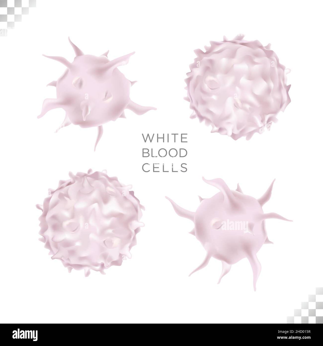 illustration of bioscience of immunity white blood cell circulating the human body Stock Vectorhttps://www.alamy.com/image-license-details/?v=1https://www.alamy.com/illustration-of-bioscience-of-immunity-white-blood-cell-circulating-the-human-body-image455461043.html
RF2HD015R–illustration of bioscience of immunity white blood cell circulating the human body
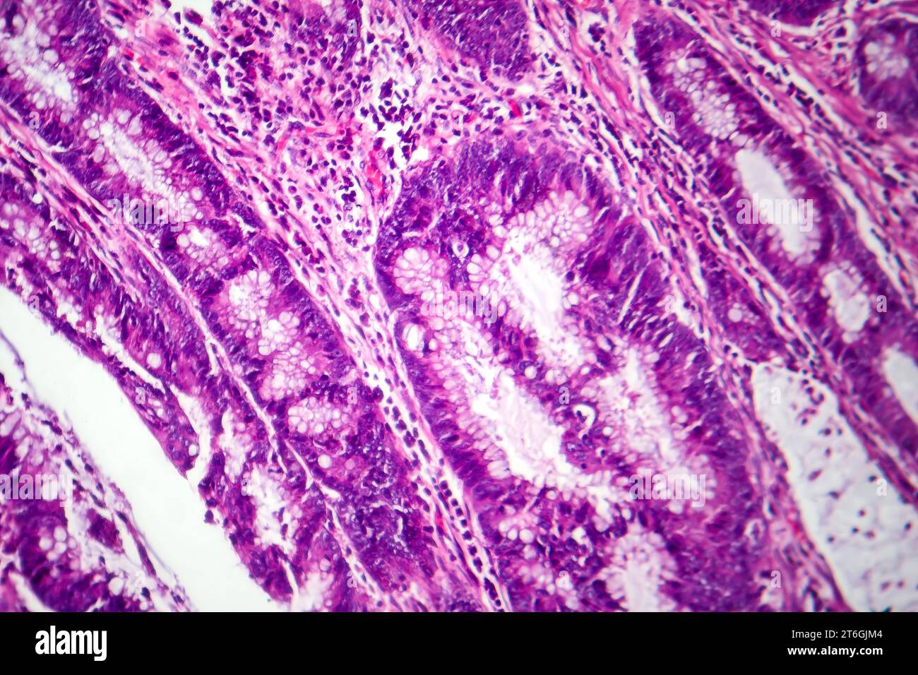 Photomicrograph of colon adenocarcinoma, illustrating malignant glandular cells characteristic of colon cancer. Stock Photohttps://www.alamy.com/image-license-details/?v=1https://www.alamy.com/photomicrograph-of-colon-adenocarcinoma-illustrating-malignant-glandular-cells-characteristic-of-colon-cancer-image571995988.html
RF2T6GJM4–Photomicrograph of colon adenocarcinoma, illustrating malignant glandular cells characteristic of colon cancer.
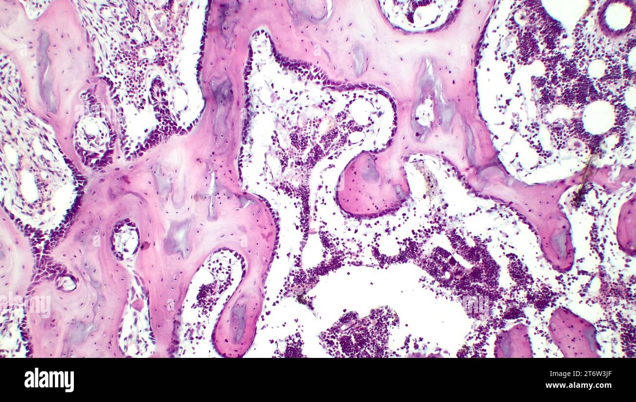 Human red bone marrow, hematopoietic press. The big tsit beetveen erythrocytes are megakaryocytes. Hematoxylin and eosin stain. Stock Photohttps://www.alamy.com/image-license-details/?v=1https://www.alamy.com/human-red-bone-marrow-hematopoietic-press-the-big-tsit-beetveen-erythrocytes-are-megakaryocytes-hematoxylin-and-eosin-stain-image572181751.html
RF2T6W3JF–Human red bone marrow, hematopoietic press. The big tsit beetveen erythrocytes are megakaryocytes. Hematoxylin and eosin stain.
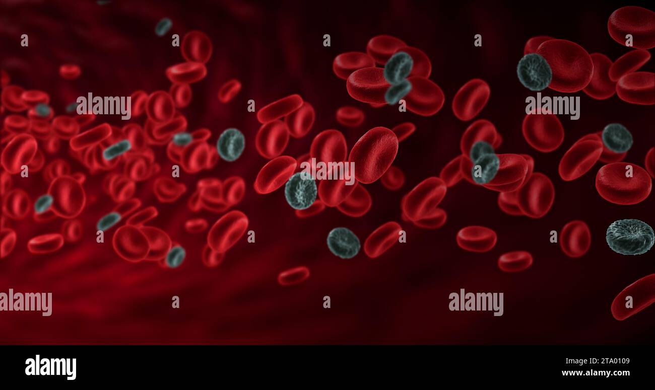 red blood cells in an artery with diseased cancer cells, flow inside body, medical human health-care Stock Photohttps://www.alamy.com/image-license-details/?v=1https://www.alamy.com/red-blood-cells-in-an-artery-with-diseased-cancer-cells-flow-inside-body-medical-human-health-care-image574089497.html
RF2TA0109–red blood cells in an artery with diseased cancer cells, flow inside body, medical human health-care
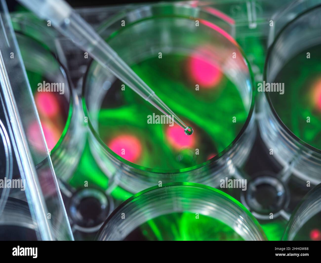 Pipetting sample into multi well plate containing micrograph of cell Stock Photohttps://www.alamy.com/image-license-details/?v=1https://www.alamy.com/pipetting-sample-into-multi-well-plate-containing-micrograph-of-cell-image458289784.html
RF2HHGW88–Pipetting sample into multi well plate containing micrograph of cell
 Scanning electron micrograph (SEM) of a cancer cell attacked by macrophages, Magnification x8000. Stock Photohttps://www.alamy.com/image-license-details/?v=1https://www.alamy.com/stock-photo-scanning-electron-micrograph-sem-of-a-cancer-cell-attacked-by-macrophages-72420343.html
RME5R0TR–Scanning electron micrograph (SEM) of a cancer cell attacked by macrophages, Magnification x8000.
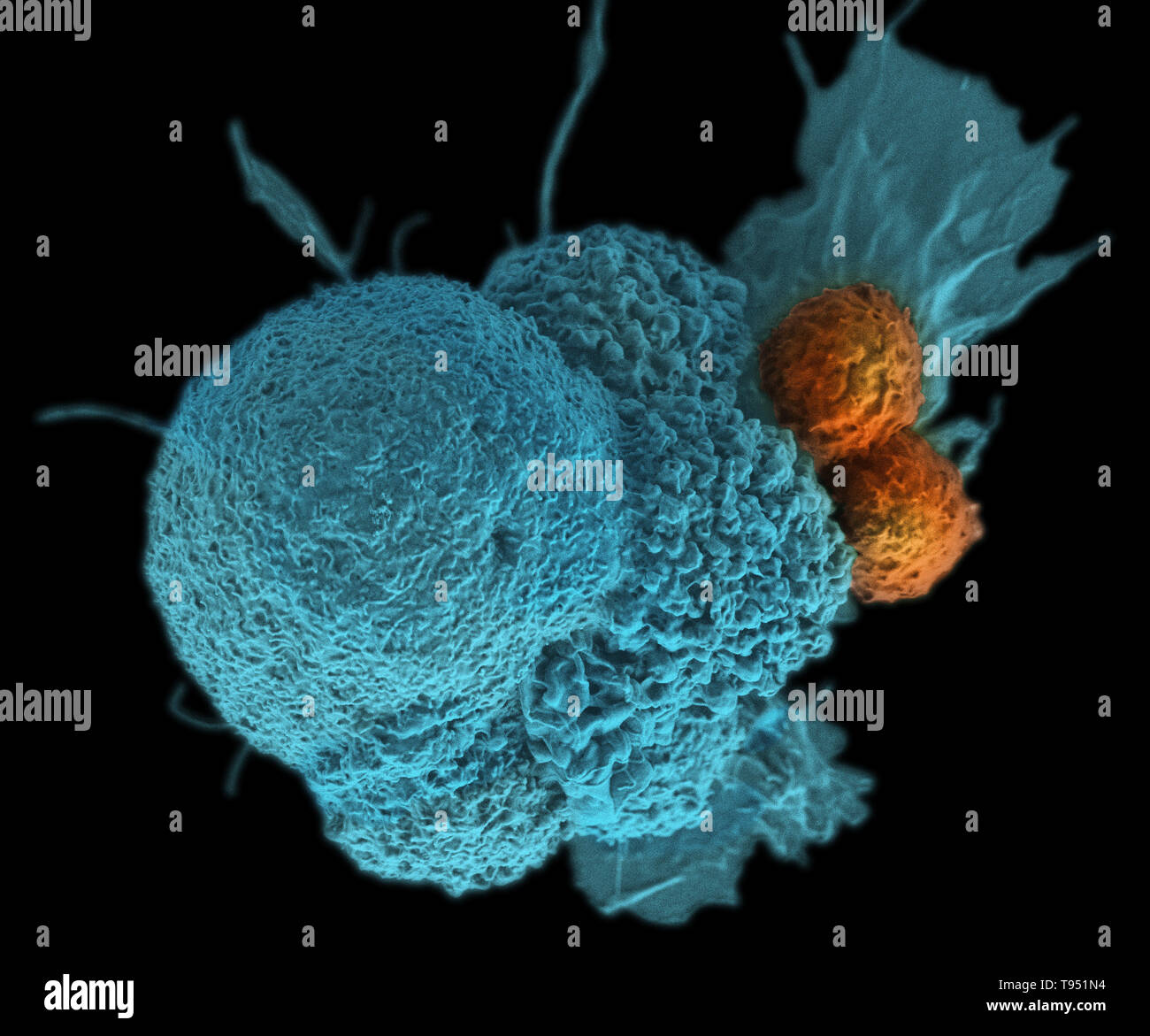 This electron micrograph (SEM) shows an oral squamous cancer cell (blue) being attacked by two cytotoxic T cells (orange). The tumor-specific T cells were developed from the patient's own immune system, a personalized cancer vaccine like those that may be developed in the future with the help of NIST's standardized human genomes. This image has been colorized. Stock Photohttps://www.alamy.com/image-license-details/?v=1https://www.alamy.com/this-electron-micrograph-sem-shows-an-oral-squamous-cancer-cell-blue-being-attacked-by-two-cytotoxic-t-cells-orange-the-tumor-specific-t-cells-were-developed-from-the-patients-own-immune-system-a-personalized-cancer-vaccine-like-those-that-may-be-developed-in-the-future-with-the-help-of-nists-standardized-human-genomes-this-image-has-been-colorized-image246588192.html
RMT951N4–This electron micrograph (SEM) shows an oral squamous cancer cell (blue) being attacked by two cytotoxic T cells (orange). The tumor-specific T cells were developed from the patient's own immune system, a personalized cancer vaccine like those that may be developed in the future with the help of NIST's standardized human genomes. This image has been colorized.
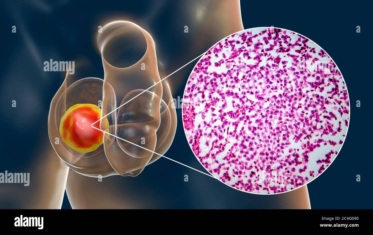 Testicular cancer, a malignant spermatocytic seminoma, computer illustration and light micrograph. A seminoma is a malignant tumour (cancer) of the testis (testicle). Although this is a rare cancer, it is the most common cancer in 15 to 35 year old men. It arises from abnormal germ cells (precursors to sperm cells) in the seminiferous tubules. Surgical removal of the affected testis (orchidectomy) is followed by radiotherapy and chemotherapy if required. Stock Photohttps://www.alamy.com/image-license-details/?v=1https://www.alamy.com/testicular-cancer-a-malignant-spermatocytic-seminoma-computer-illustration-and-light-micrograph-a-seminoma-is-a-malignant-tumour-cancer-of-the-testis-testicle-although-this-is-a-rare-cancer-it-is-the-most-common-cancer-in-15-to-35-year-old-men-it-arises-from-abnormal-germ-cells-precursors-to-sperm-cells-in-the-seminiferous-tubules-surgical-removal-of-the-affected-testis-orchidectomy-is-followed-by-radiotherapy-and-chemotherapy-if-required-image364227836.html
RF2C4G090–Testicular cancer, a malignant spermatocytic seminoma, computer illustration and light micrograph. A seminoma is a malignant tumour (cancer) of the testis (testicle). Although this is a rare cancer, it is the most common cancer in 15 to 35 year old men. It arises from abnormal germ cells (precursors to sperm cells) in the seminiferous tubules. Surgical removal of the affected testis (orchidectomy) is followed by radiotherapy and chemotherapy if required.
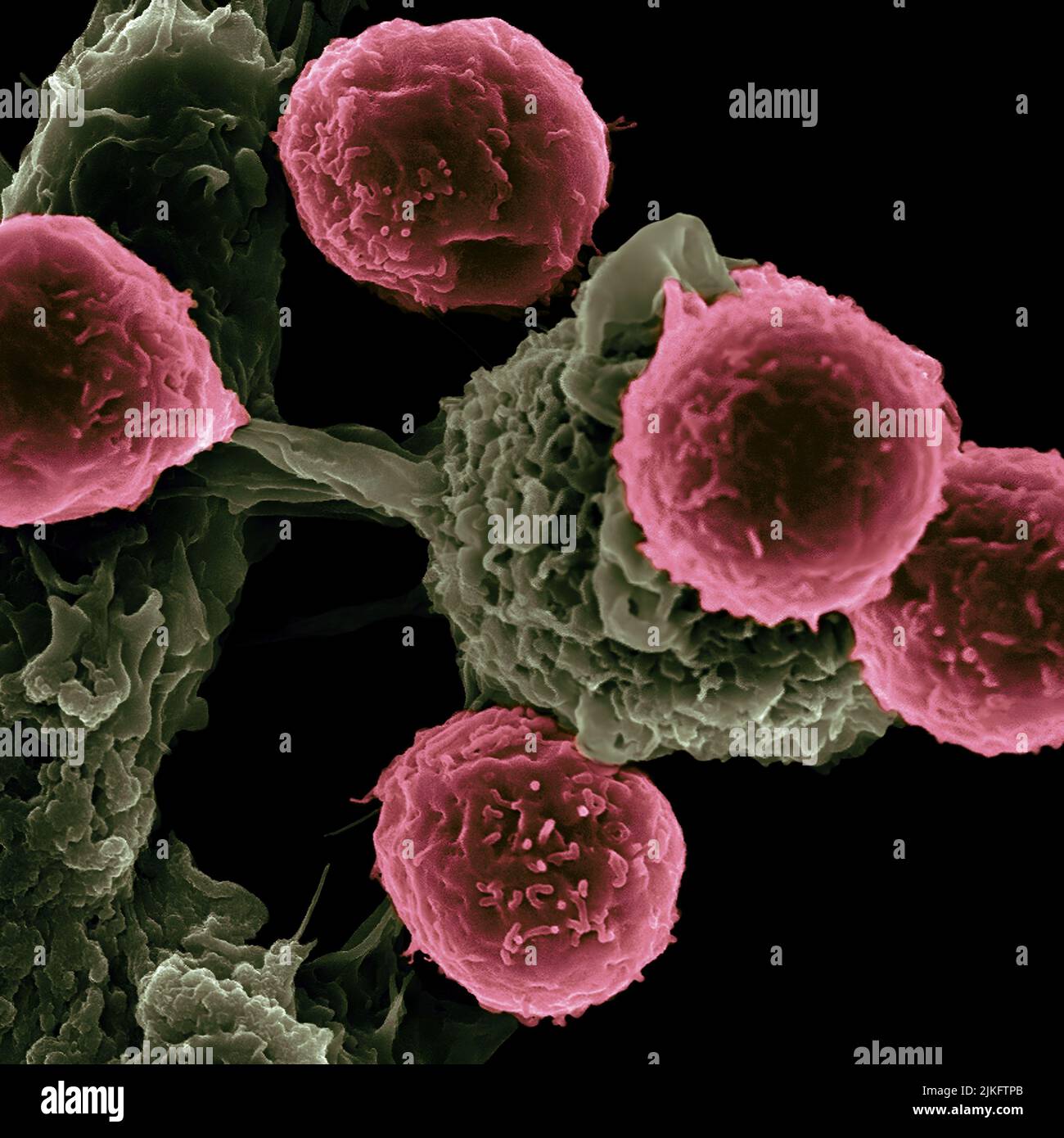 Researchers at the Texas Center for Cancer Nanomedicine (TCCN) have created particle-based vaccines for the treatment of cancer. The particles carry molecules that stimulate immune cells and cancer antigens (proteins) that direct the immune response. This scanning electron microscope image shows dendritic cells, pseudostained green, interacting with T cells, pseudostained pink. Dendritic cells internalize particles, provoke antigens and present peptides to T cells to direct immune responses. Stock Photohttps://www.alamy.com/image-license-details/?v=1https://www.alamy.com/researchers-at-the-texas-center-for-cancer-nanomedicine-tccn-have-created-particle-based-vaccines-for-the-treatment-of-cancer-the-particles-carry-molecules-that-stimulate-immune-cells-and-cancer-antigens-proteins-that-direct-the-immune-response-this-scanning-electron-microscope-image-shows-dendritic-cells-pseudostained-green-interacting-with-t-cells-pseudostained-pink-dendritic-cells-internalize-particles-provoke-antigens-and-present-peptides-to-t-cells-to-direct-immune-responses-image476707123.html
RM2JKFTPB–Researchers at the Texas Center for Cancer Nanomedicine (TCCN) have created particle-based vaccines for the treatment of cancer. The particles carry molecules that stimulate immune cells and cancer antigens (proteins) that direct the immune response. This scanning electron microscope image shows dendritic cells, pseudostained green, interacting with T cells, pseudostained pink. Dendritic cells internalize particles, provoke antigens and present peptides to T cells to direct immune responses.
 Kidney cancer, Colored scanning electron micrograph (SEM) of the cancerous cells of a Wilms' tumor, a type of kidney cancer, Stock Photohttps://www.alamy.com/image-license-details/?v=1https://www.alamy.com/stock-photo-kidney-cancer-colored-scanning-electron-micrograph-sem-of-the-cancerous-72420738.html
RME5R1AX–Kidney cancer, Colored scanning electron micrograph (SEM) of the cancerous cells of a Wilms' tumor, a type of kidney cancer,
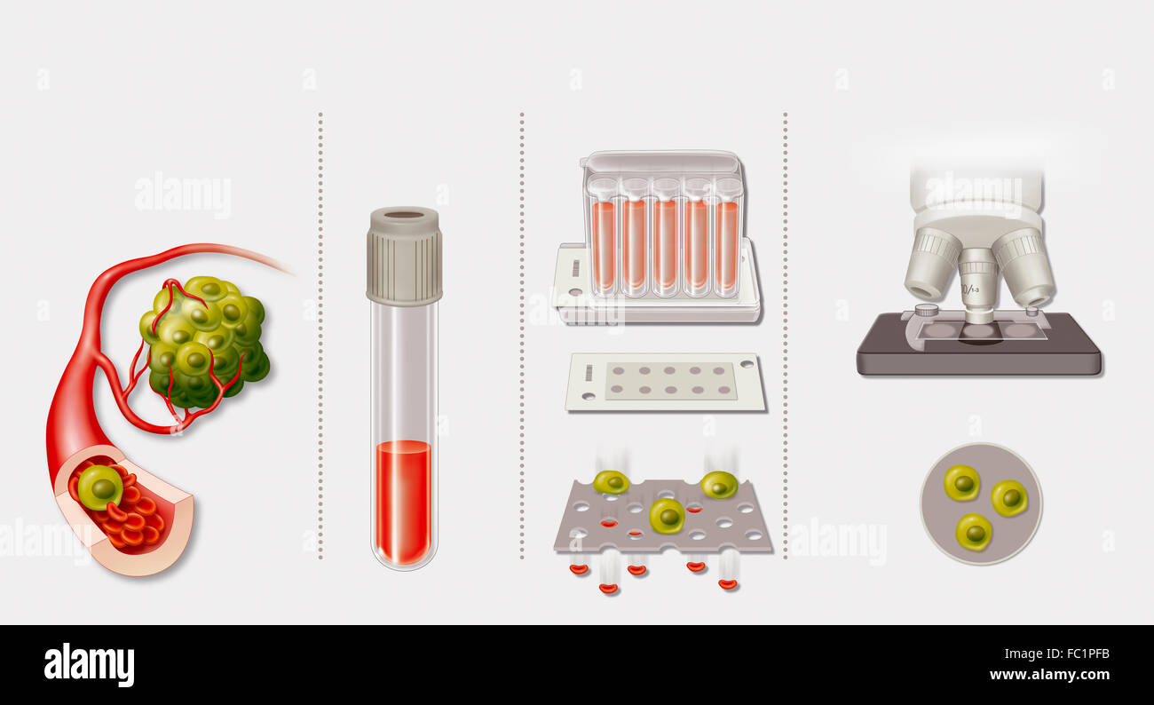 SCREENING FOR CANCER Stock Photohttps://www.alamy.com/image-license-details/?v=1https://www.alamy.com/stock-photo-screening-for-cancer-93467343.html
RMFC1PFB–SCREENING FOR CANCER
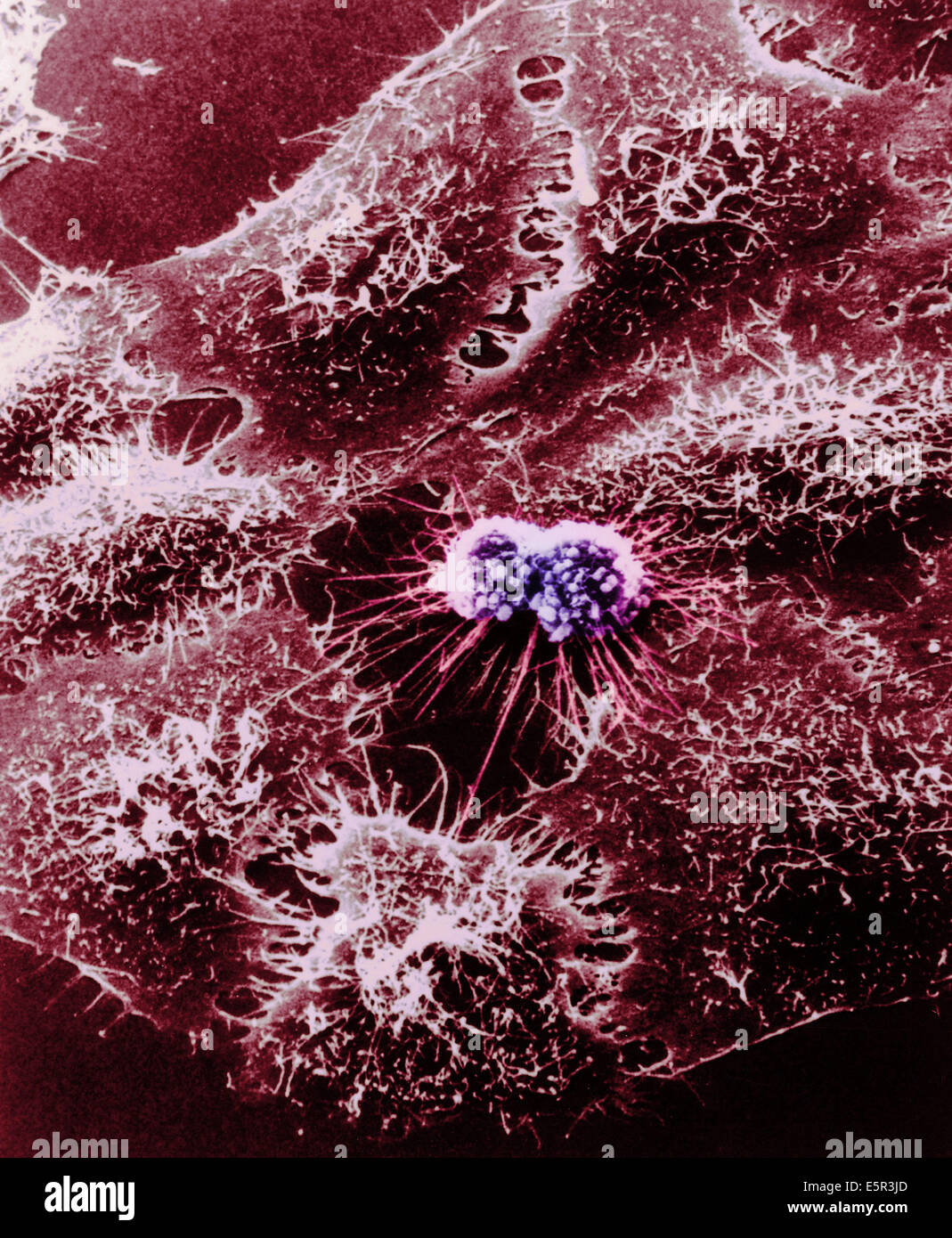 Color enhanced scanning electron micrograph (SEM) of cultured HeLa cells originally derived many years ago from a woman's Stock Photohttps://www.alamy.com/image-license-details/?v=1https://www.alamy.com/stock-photo-color-enhanced-scanning-electron-micrograph-sem-of-cultured-hela-cells-72422517.html
RME5R3JD–Color enhanced scanning electron micrograph (SEM) of cultured HeLa cells originally derived many years ago from a woman's
 A section trough testicle cells under the microscope Stock Photohttps://www.alamy.com/image-license-details/?v=1https://www.alamy.com/a-section-trough-testicle-cells-under-the-microscope-image624943015.html
RF2Y8MH5B–A section trough testicle cells under the microscope
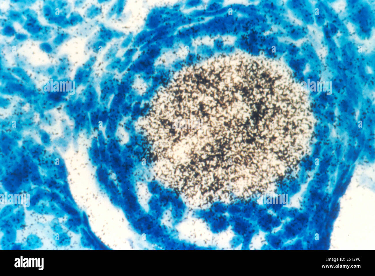 Light micrograph of a mouse oocyte that is expressing an oncogene (a cancer-causing gene). The technique being used to test its Stock Photohttps://www.alamy.com/image-license-details/?v=1https://www.alamy.com/stock-photo-light-micrograph-of-a-mouse-oocyte-that-is-expressing-an-oncogene-72443796.html
RME5T2PC–Light micrograph of a mouse oocyte that is expressing an oncogene (a cancer-causing gene). The technique being used to test its
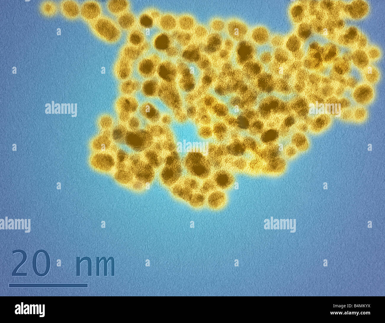 A TEM image of gold nanoparticles coating cells in a glutathione surface corona illustrating the molecular size of nanoparticles Stock Photohttps://www.alamy.com/image-license-details/?v=1https://www.alamy.com/stock-photo-a-tem-image-of-gold-nanoparticles-coating-cells-in-a-glutathione-surface-20123710.html
RMB4MKYX–A TEM image of gold nanoparticles coating cells in a glutathione surface corona illustrating the molecular size of nanoparticles
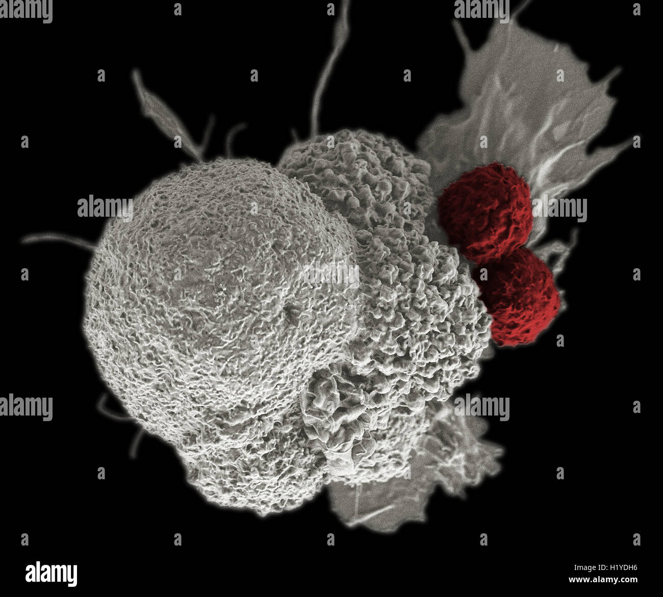 T lymphocytes and cancer cell, Coloured scanning electron micrograph (SEM) . Stock Photohttps://www.alamy.com/image-license-details/?v=1https://www.alamy.com/stock-photo-t-lymphocytes-and-cancer-cell-coloured-scanning-electron-micrograph-121690610.html
RMH1YDH6–T lymphocytes and cancer cell, Coloured scanning electron micrograph (SEM) .
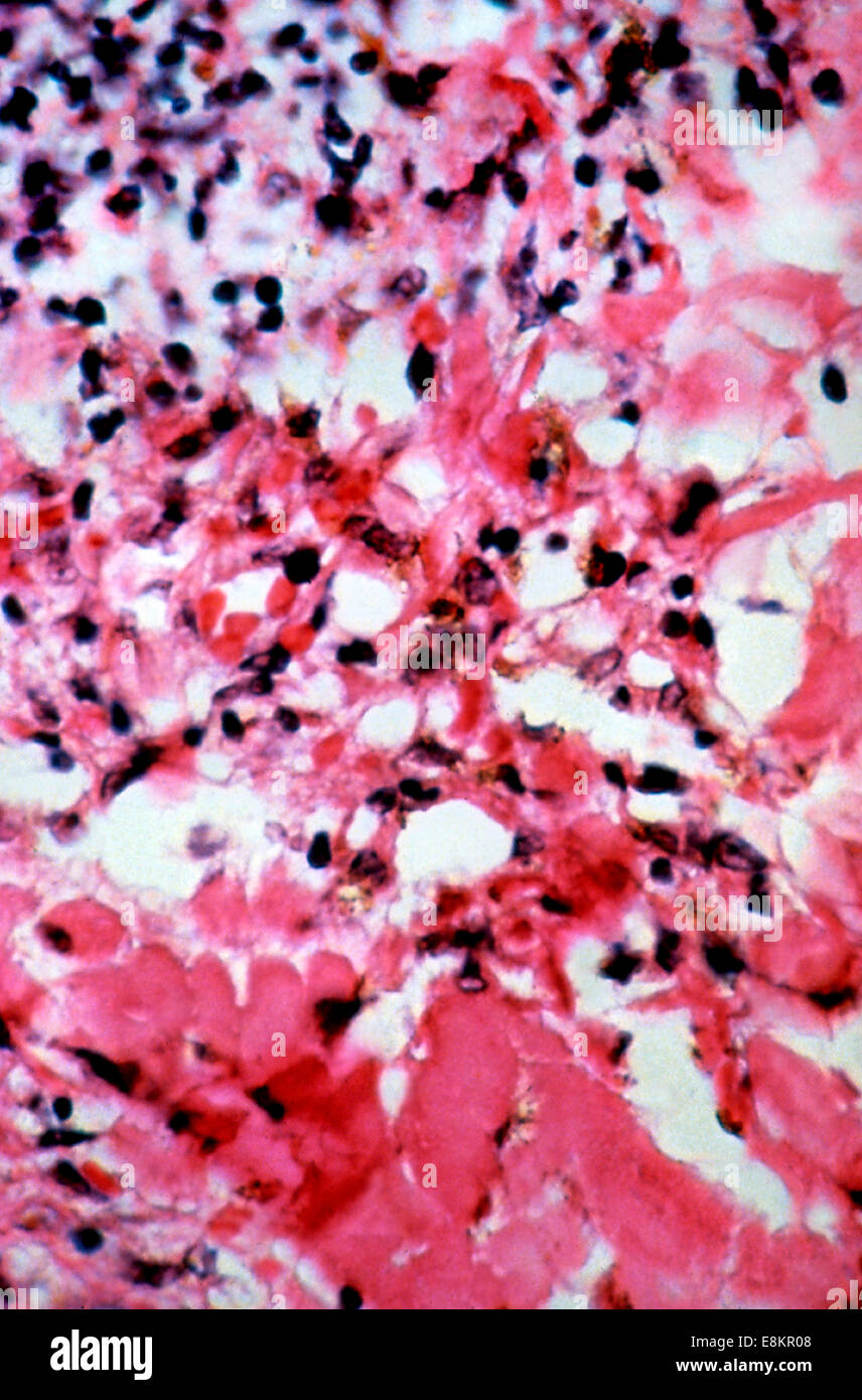 This micrograph depicts histopathologic changes found in case of Kaposis sarcoma lesions This skin biopsy of what turned out Stock Photohttps://www.alamy.com/image-license-details/?v=1https://www.alamy.com/stock-photo-this-micrograph-depicts-histopathologic-changes-found-in-case-of-kaposis-74193848.html
RME8KR08–This micrograph depicts histopathologic changes found in case of Kaposis sarcoma lesions This skin biopsy of what turned out
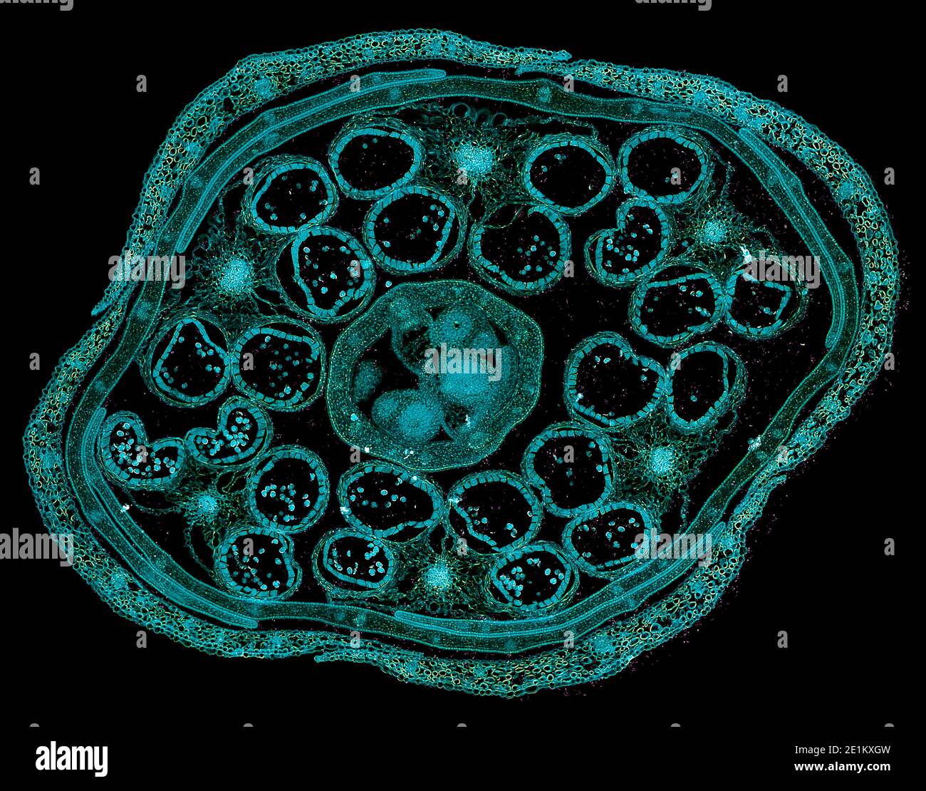 cross section cut of human body cells under a scientific microscope Stock Photohttps://www.alamy.com/image-license-details/?v=1https://www.alamy.com/cross-section-cut-of-human-body-cells-under-a-scientific-microscope-image396891065.html
RF2E1KXGW–cross section cut of human body cells under a scientific microscope
 Immunofluorescent staining of Merkel cell carcinoma tumor tissue illustrating expression of CD200 (green) on the surface of tumor cells. CD200 plays a role in immunosuppression. Endothelial marker CD31 (red) highlights blood vessels. Merkel cell carcinoma is a rare and aggressive skin cancer. Stock Photohttps://www.alamy.com/image-license-details/?v=1https://www.alamy.com/immunofluorescent-staining-of-merkel-cell-carcinoma-tumor-tissue-illustrating-expression-of-cd200-green-on-the-surface-of-tumor-cells-cd200-plays-a-role-in-immunosuppression-endothelial-marker-cd31-red-highlights-blood-vessels-merkel-cell-carcinoma-is-a-rare-and-aggressive-skin-cancer-image476706886.html
RM2JKFTDX–Immunofluorescent staining of Merkel cell carcinoma tumor tissue illustrating expression of CD200 (green) on the surface of tumor cells. CD200 plays a role in immunosuppression. Endothelial marker CD31 (red) highlights blood vessels. Merkel cell carcinoma is a rare and aggressive skin cancer.
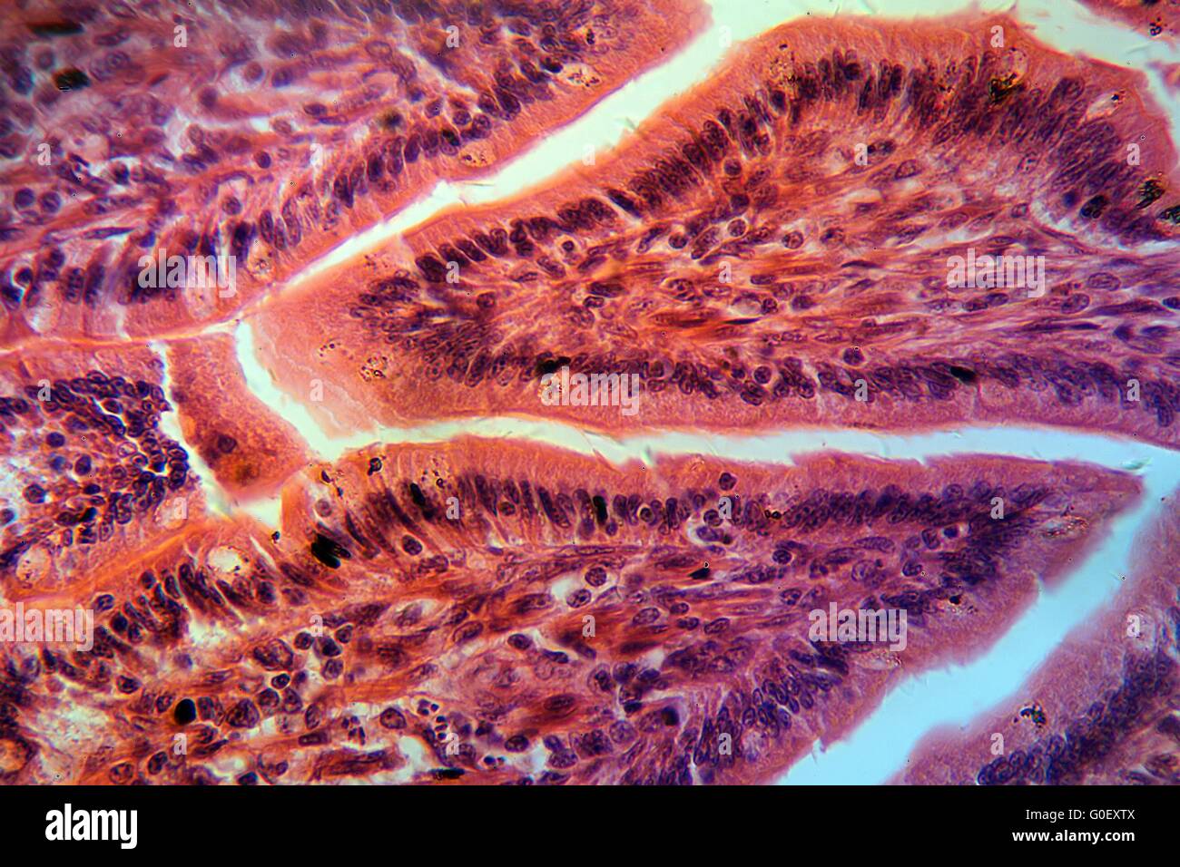 A section trough cells of a small intestine under the microscope. Stock Photohttps://www.alamy.com/image-license-details/?v=1https://www.alamy.com/stock-photo-a-section-trough-cells-of-a-small-intestine-under-the-microscope-103590618.html
RMG0EXTX–A section trough cells of a small intestine under the microscope.
 Photomicrograph of cancerous cells from Ewing's sarcoma that metastasized to the pleural fluid. Stock Photohttps://www.alamy.com/image-license-details/?v=1https://www.alamy.com/stock-photo-photomicrograph-of-cancerous-cells-from-ewings-sarcoma-that-metastasized-72420354.html
RME5R0W6–Photomicrograph of cancerous cells from Ewing's sarcoma that metastasized to the pleural fluid.
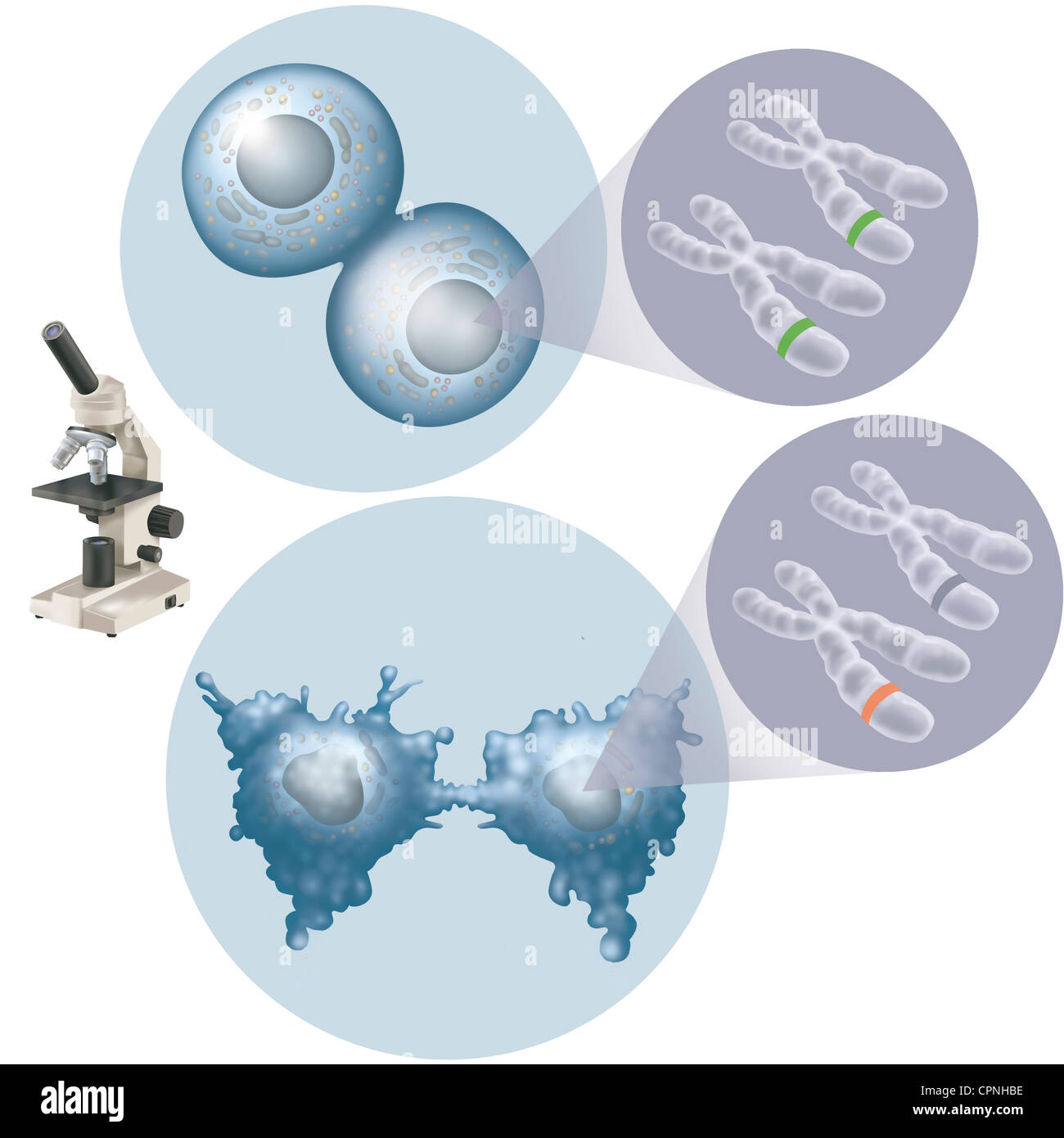 CANCER CELL, ILLUSTRATION Stock Photohttps://www.alamy.com/image-license-details/?v=1https://www.alamy.com/stock-photo-cancer-cell-illustration-48417810.html
RMCPNHBE–CANCER CELL, ILLUSTRATION
 A section trough ovary cells under the microscope Stock Photohttps://www.alamy.com/image-license-details/?v=1https://www.alamy.com/a-section-trough-ovary-cells-under-the-microscope-image624951322.html
RF2Y8MYP2–A section trough ovary cells under the microscope
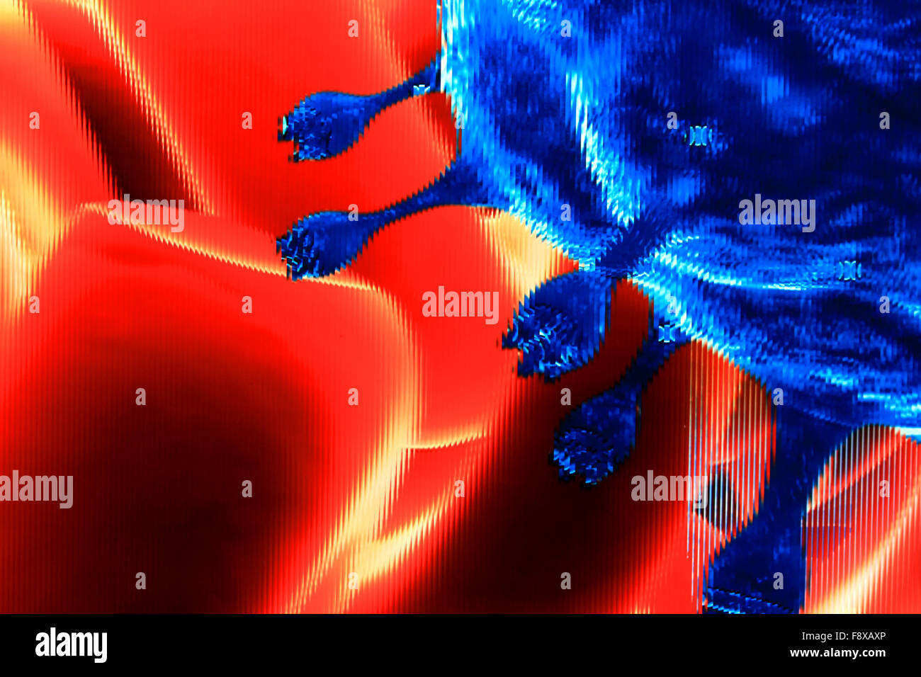 Close-up of red blood cells Stock Photohttps://www.alamy.com/image-license-details/?v=1https://www.alamy.com/stock-photo-close-up-of-red-blood-cells-91548430.html
RFF8XAXP–Close-up of red blood cells
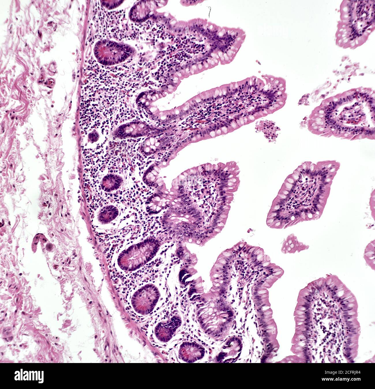 Intestinal cancer cells, brightfield photomicrograph Stock Photohttps://www.alamy.com/image-license-details/?v=1https://www.alamy.com/intestinal-cancer-cells-brightfield-photomicrograph-image371157224.html
RM2CFRJR4–Intestinal cancer cells, brightfield photomicrograph
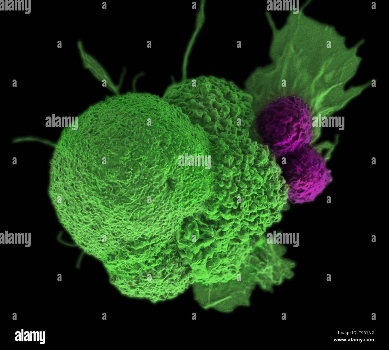 This electron micrograph (SEM) shows an oral squamous cancer cell (green) being attacked by two cytotoxic T cells (purple). The tumor-specific T cells were developed from the patient's own immune system, a personalized cancer vaccine like those that may be developed in the future with the help of NIST's standardized human genomes. This image has been colorized. Stock Photohttps://www.alamy.com/image-license-details/?v=1https://www.alamy.com/this-electron-micrograph-sem-shows-an-oral-squamous-cancer-cell-green-being-attacked-by-two-cytotoxic-t-cells-purple-the-tumor-specific-t-cells-were-developed-from-the-patients-own-immune-system-a-personalized-cancer-vaccine-like-those-that-may-be-developed-in-the-future-with-the-help-of-nists-standardized-human-genomes-this-image-has-been-colorized-image246588190.html
RMT951N2–This electron micrograph (SEM) shows an oral squamous cancer cell (green) being attacked by two cytotoxic T cells (purple). The tumor-specific T cells were developed from the patient's own immune system, a personalized cancer vaccine like those that may be developed in the future with the help of NIST's standardized human genomes. This image has been colorized.
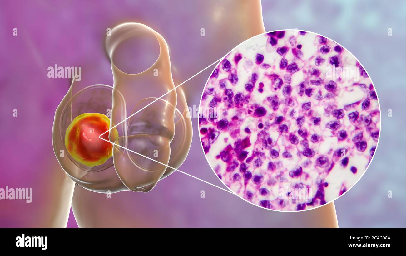 Testicular cancer, a malignant spermatocytic seminoma, computer illustration and light micrograph. A seminoma is a malignant tumour (cancer) of the testis (testicle). Although this is a rare cancer, it is the most common cancer in 15 to 35 year old men. It arises from abnormal germ cells (precursors to sperm cells) in the seminiferous tubules. Surgical removal of the affected testis (orchidectomy) is followed by radiotherapy and chemotherapy if required. Stock Photohttps://www.alamy.com/image-license-details/?v=1https://www.alamy.com/testicular-cancer-a-malignant-spermatocytic-seminoma-computer-illustration-and-light-micrograph-a-seminoma-is-a-malignant-tumour-cancer-of-the-testis-testicle-although-this-is-a-rare-cancer-it-is-the-most-common-cancer-in-15-to-35-year-old-men-it-arises-from-abnormal-germ-cells-precursors-to-sperm-cells-in-the-seminiferous-tubules-surgical-removal-of-the-affected-testis-orchidectomy-is-followed-by-radiotherapy-and-chemotherapy-if-required-image364227818.html
RF2C4G08A–Testicular cancer, a malignant spermatocytic seminoma, computer illustration and light micrograph. A seminoma is a malignant tumour (cancer) of the testis (testicle). Although this is a rare cancer, it is the most common cancer in 15 to 35 year old men. It arises from abnormal germ cells (precursors to sperm cells) in the seminiferous tubules. Surgical removal of the affected testis (orchidectomy) is followed by radiotherapy and chemotherapy if required.
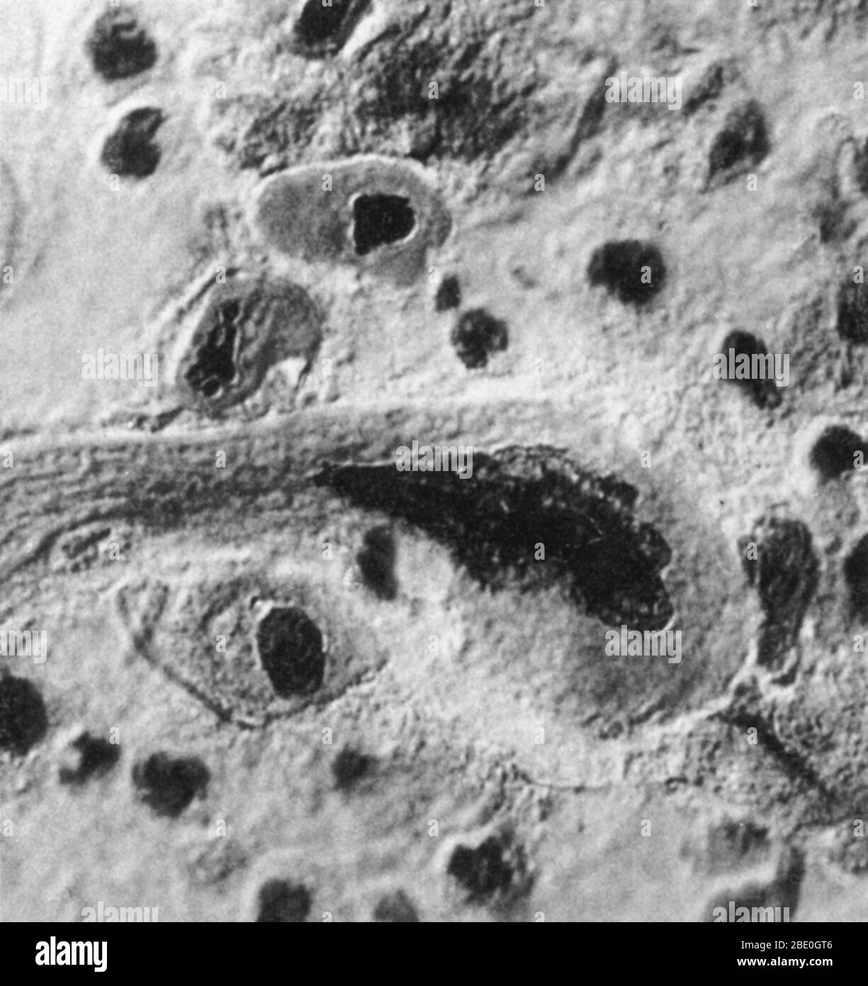 Micrograph showing cells from advanced lung cancer. The diseased cells are deformed and swollen. Stock Photohttps://www.alamy.com/image-license-details/?v=1https://www.alamy.com/micrograph-showing-cells-from-advanced-lung-cancer-the-diseased-cells-are-deformed-and-swollen-image352825766.html
RM2BE0GT6–Micrograph showing cells from advanced lung cancer. The diseased cells are deformed and swollen.
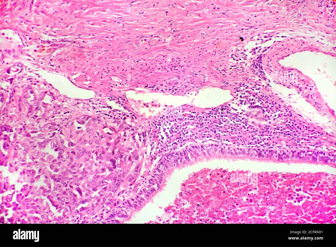 Lung cancer cells, brightfield photomicrograph Stock Photohttps://www.alamy.com/image-license-details/?v=1https://www.alamy.com/lung-cancer-cells-brightfield-photomicrograph-image371157361.html
RM2CFRK01–Lung cancer cells, brightfield photomicrograph
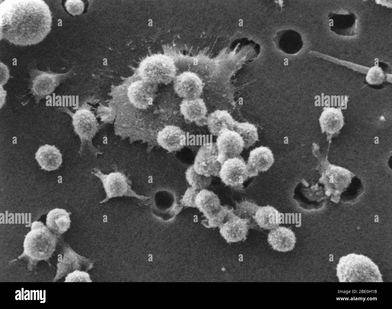 Step two of a six-step sequence of the death of a cancer cell. A buffy coat containing red blood cells, lymphocytes and macrophages is added to the bottom of the membrane. A group of macrophages identify the cancer cell as foreign matter and start to stick to the cancer cell, which still has its spikes. Photo magnification: 4000x Stock Photohttps://www.alamy.com/image-license-details/?v=1https://www.alamy.com/step-two-of-a-six-step-sequence-of-the-death-of-a-cancer-cell-a-buffy-coat-containing-red-blood-cells-lymphocytes-and-macrophages-is-added-to-the-bottom-of-the-membrane-a-group-of-macrophages-identify-the-cancer-cell-as-foreign-matter-and-start-to-stick-to-the-cancer-cell-which-still-has-its-spikes-photo-magnification-4000x-image352825911.html
RM2BE0H1B–Step two of a six-step sequence of the death of a cancer cell. A buffy coat containing red blood cells, lymphocytes and macrophages is added to the bottom of the membrane. A group of macrophages identify the cancer cell as foreign matter and start to stick to the cancer cell, which still has its spikes. Photo magnification: 4000x
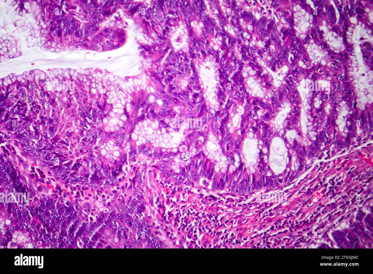 Photomicrograph of colon adenocarcinoma, illustrating malignant glandular cells characteristic of colon cancer. Stock Photohttps://www.alamy.com/image-license-details/?v=1https://www.alamy.com/photomicrograph-of-colon-adenocarcinoma-illustrating-malignant-glandular-cells-characteristic-of-colon-cancer-image571995996.html
RF2T6GJMC–Photomicrograph of colon adenocarcinoma, illustrating malignant glandular cells characteristic of colon cancer.
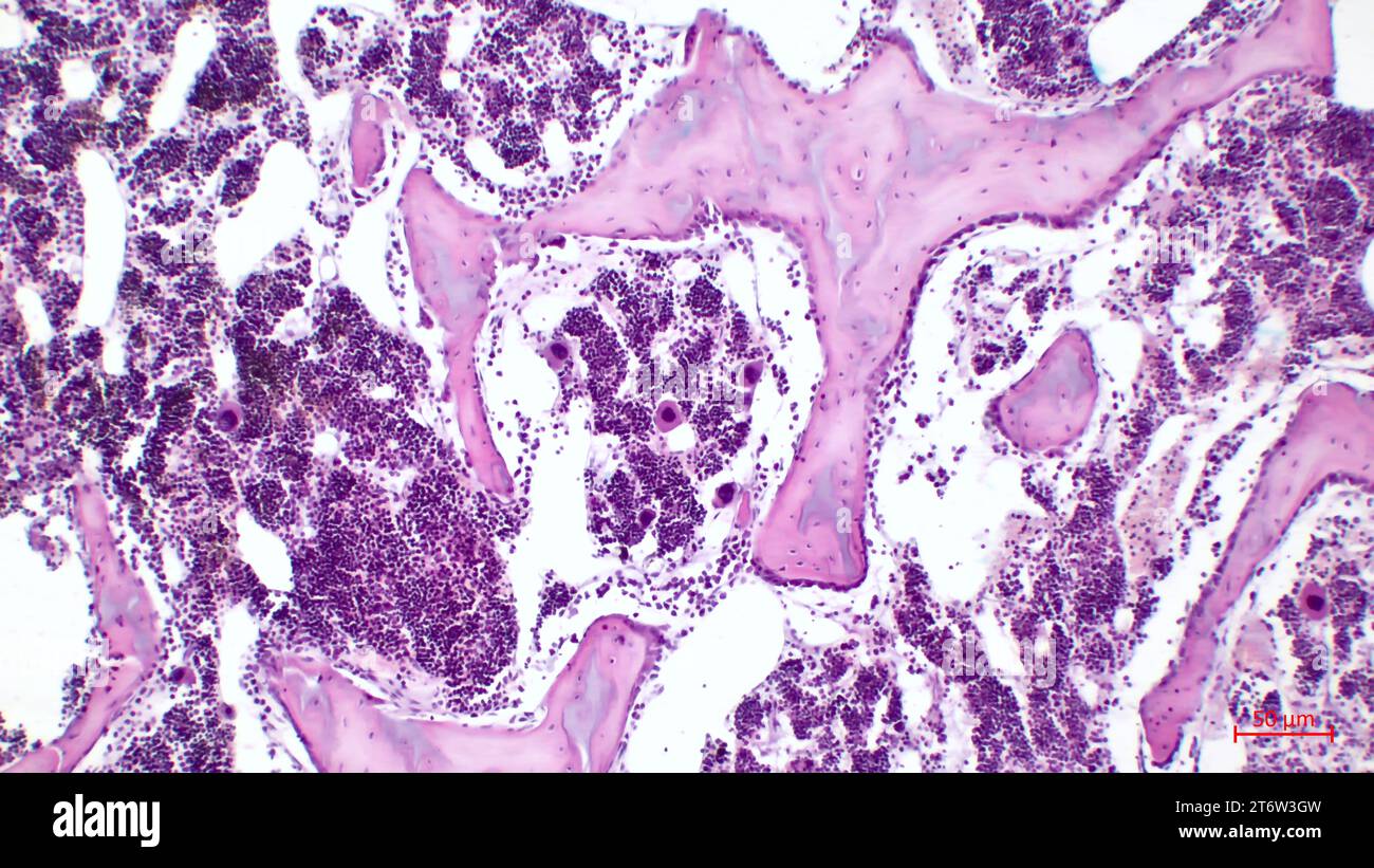 Human red bone marrow, hematopoietic press. The big tsit beetveen erythrocytes are megakaryocytes. Hematoxylin and eosin stain. Stock Photohttps://www.alamy.com/image-license-details/?v=1https://www.alamy.com/human-red-bone-marrow-hematopoietic-press-the-big-tsit-beetveen-erythrocytes-are-megakaryocytes-hematoxylin-and-eosin-stain-image572181705.html
RF2T6W3GW–Human red bone marrow, hematopoietic press. The big tsit beetveen erythrocytes are megakaryocytes. Hematoxylin and eosin stain.
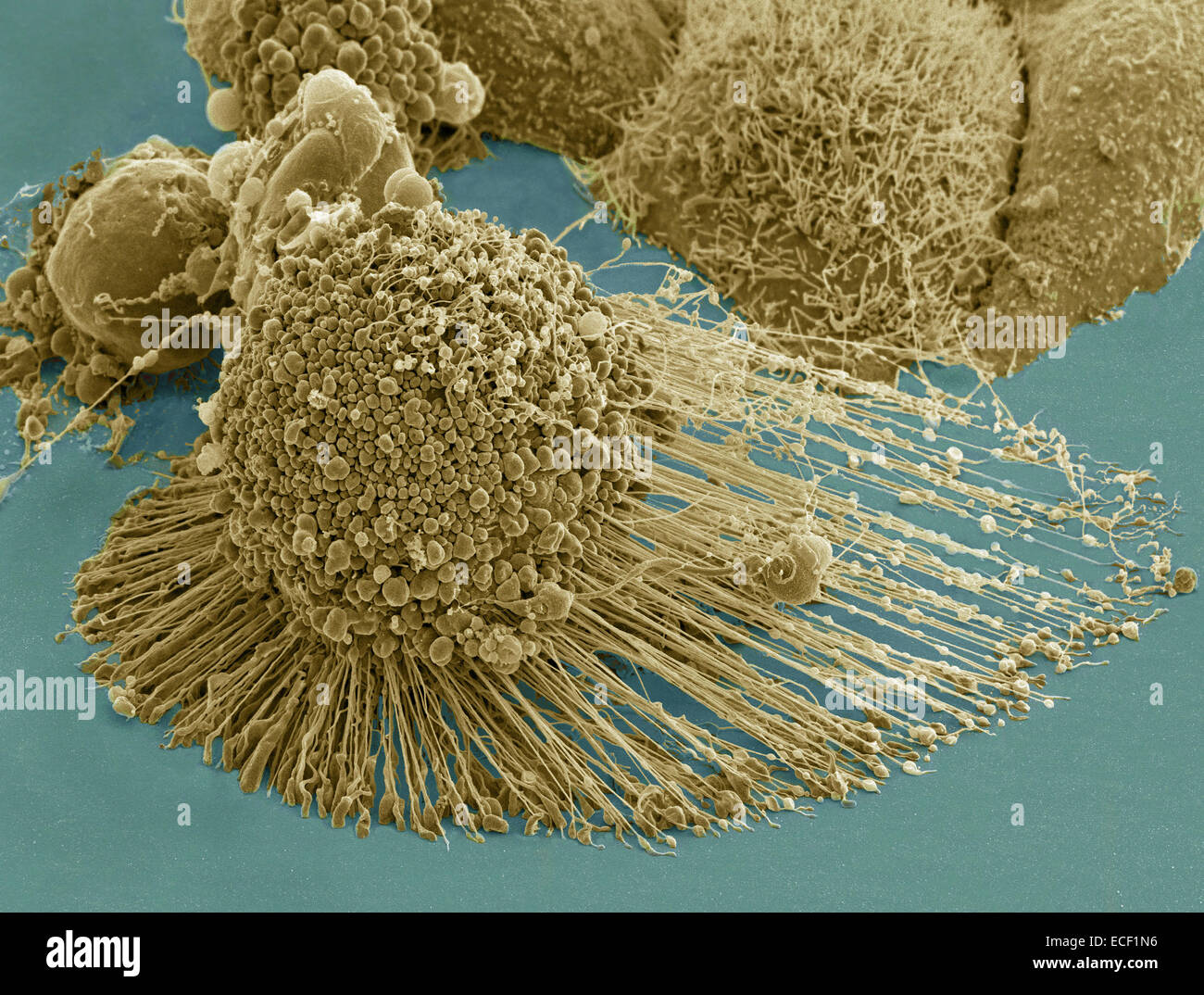 Scanning electron micrograph of an apoptotic HeLa cell. Zeiss Merlin HR-SEM. Stock Photohttps://www.alamy.com/image-license-details/?v=1https://www.alamy.com/stock-photo-scanning-electron-micrograph-of-an-apoptotic-hela-cell-zeiss-merlin-76548002.html
RFECF1N6–Scanning electron micrograph of an apoptotic HeLa cell. Zeiss Merlin HR-SEM.
 False colour EMS image of carbon nanotubes which deliver cancer drugs right to the site of disease during chemotherapy treatment Stock Photohttps://www.alamy.com/image-license-details/?v=1https://www.alamy.com/stock-photo-false-colour-ems-image-of-carbon-nanotubes-which-deliver-cancer-drugs-20123485.html
RMB4MKKW–False colour EMS image of carbon nanotubes which deliver cancer drugs right to the site of disease during chemotherapy treatment
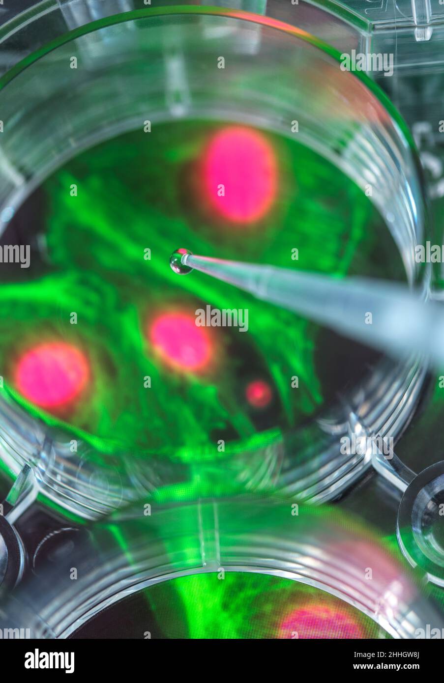 Pipetting sample into multi well plate containing micrograph of cell Stock Photohttps://www.alamy.com/image-license-details/?v=1https://www.alamy.com/pipetting-sample-into-multi-well-plate-containing-micrograph-of-cell-image458289794.html
RF2HHGW8J–Pipetting sample into multi well plate containing micrograph of cell
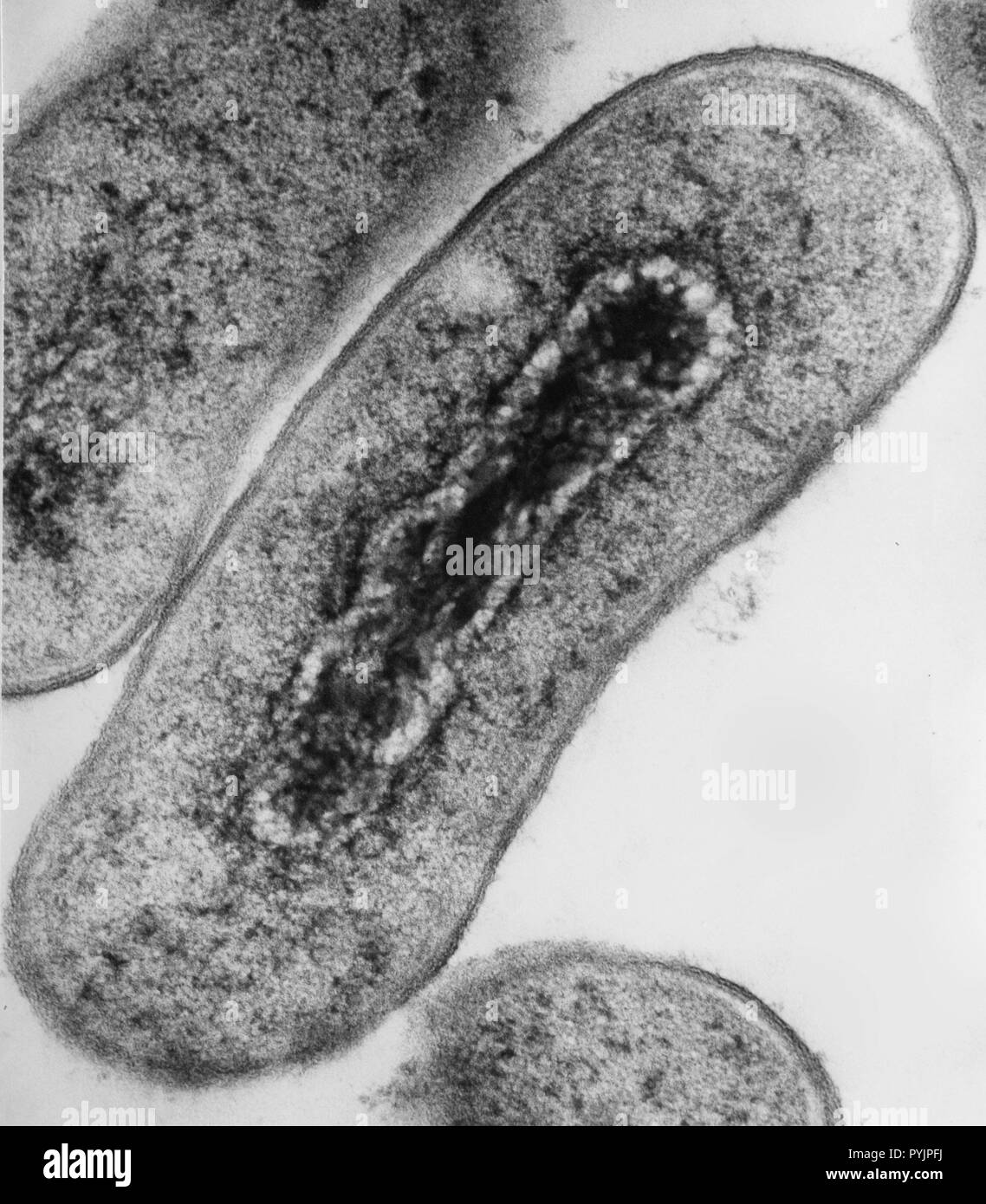 Electron micrograph cross section of Escherichia coli bacteria Stock Photohttps://www.alamy.com/image-license-details/?v=1https://www.alamy.com/electron-micrograph-cross-section-of-escherichia-coli-bacteria-image223532950.html
RMPYJPFJ–Electron micrograph cross section of Escherichia coli bacteria
 cross section cut of human body cells under a scientific microscope Stock Photohttps://www.alamy.com/image-license-details/?v=1https://www.alamy.com/cross-section-cut-of-human-body-cells-under-a-scientific-microscope-image396913543.html
RF2E1MY7K–cross section cut of human body cells under a scientific microscope
 red blood cells in an artery, flow inside body, medical human health-care Stock Photohttps://www.alamy.com/image-license-details/?v=1https://www.alamy.com/red-blood-cells-in-an-artery-flow-inside-body-medical-human-health-care-image574089482.html
RF2TA00YP–red blood cells in an artery, flow inside body, medical human health-care
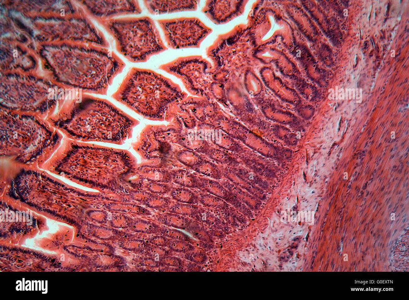 A section trough cells of a small intestine under the microscope. Stock Photohttps://www.alamy.com/image-license-details/?v=1https://www.alamy.com/stock-photo-a-section-trough-cells-of-a-small-intestine-under-the-microscope-103590613.html
RMG0EXTN–A section trough cells of a small intestine under the microscope.
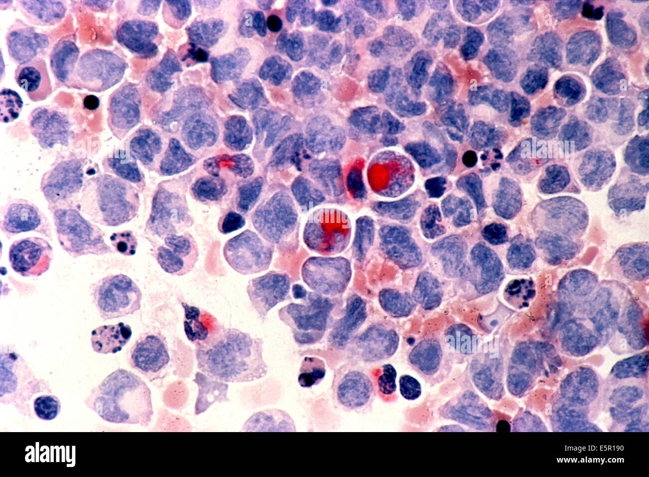 Photomicrograph of human white blood cells with acute myelocytic leukemia ( AML ) in the pericardial fluid, shown with an Stock Photohttps://www.alamy.com/image-license-details/?v=1https://www.alamy.com/stock-photo-photomicrograph-of-human-white-blood-cells-with-acute-myelocytic-leukemia-72420684.html
RME5R190–Photomicrograph of human white blood cells with acute myelocytic leukemia ( AML ) in the pericardial fluid, shown with an
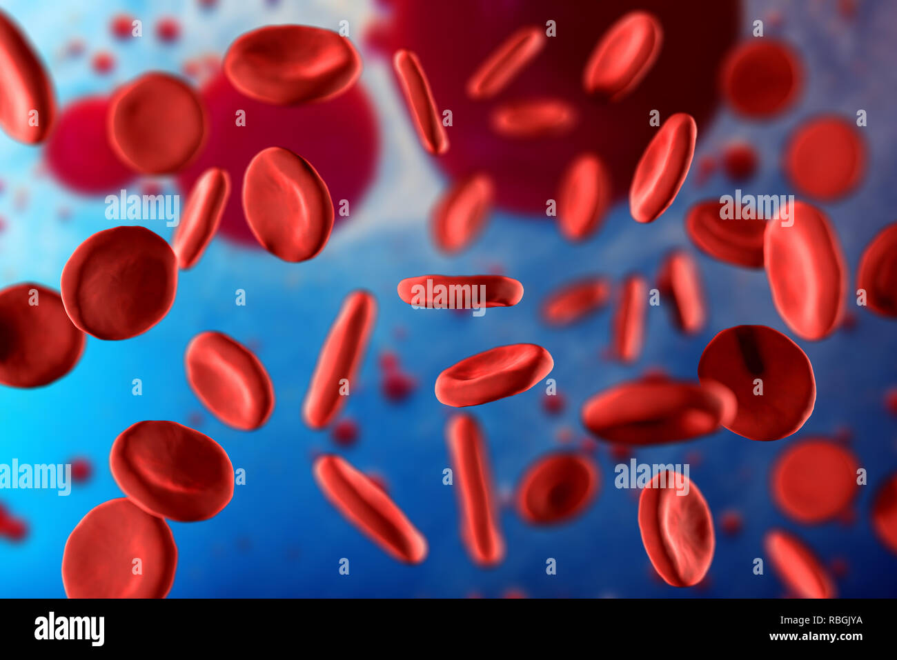 3d illustration of red blood cells erythrocytes close-up under a microscope. Background for scientific medical concept. Stock Photohttps://www.alamy.com/image-license-details/?v=1https://www.alamy.com/3d-illustration-of-red-blood-cells-erythrocytes-close-up-under-a-microscope-background-for-scientific-medical-concept-image230862110.html
RFRBGJYA–3d illustration of red blood cells erythrocytes close-up under a microscope. Background for scientific medical concept.
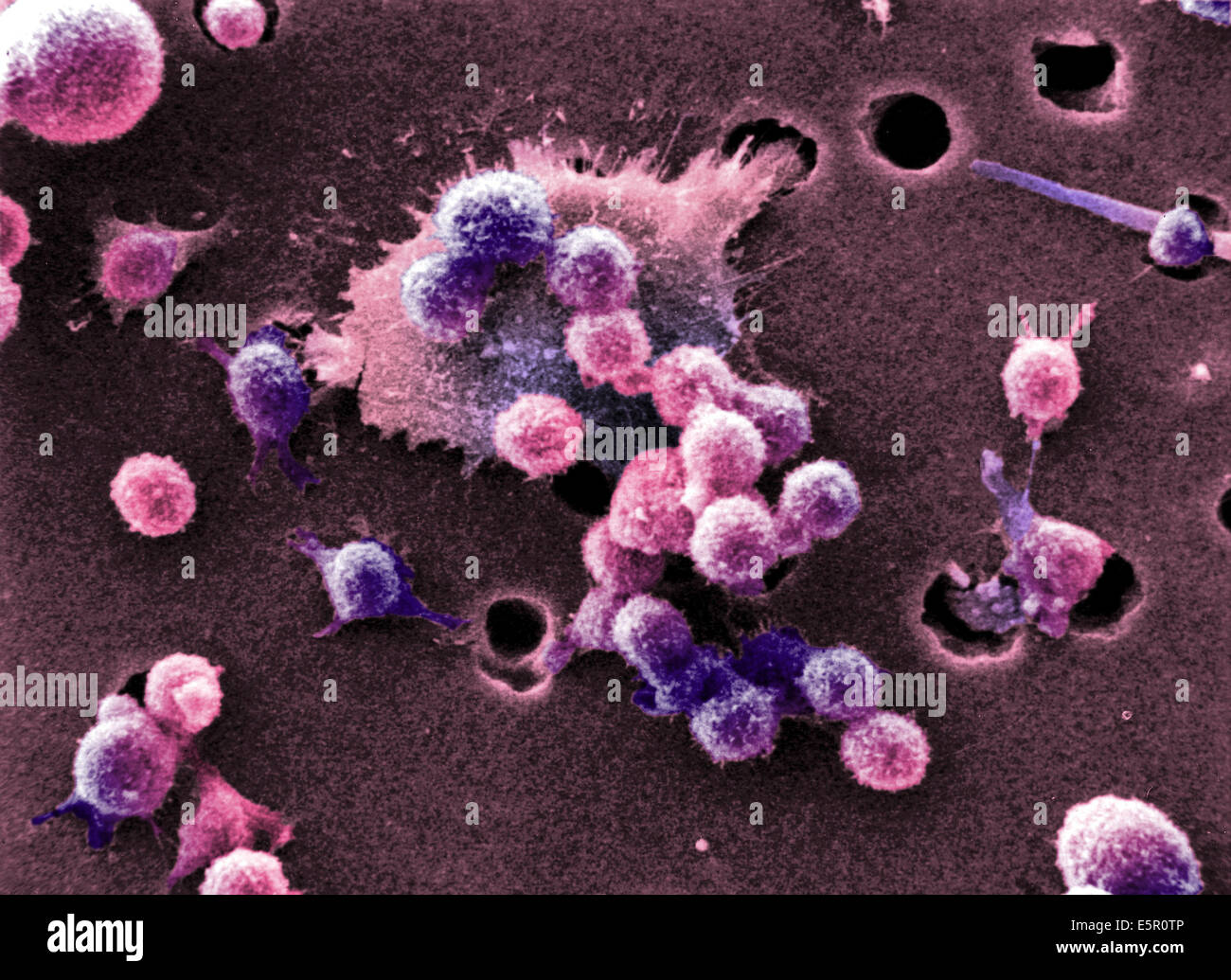 Scanning electron micrograph (SEM) of malignant B-cell lymphocytes seen in Burkitt's lymphoma, Magnification x4000. Stock Photohttps://www.alamy.com/image-license-details/?v=1https://www.alamy.com/stock-photo-scanning-electron-micrograph-sem-of-malignant-b-cell-lymphocytes-seen-72420342.html
RME5R0TP–Scanning electron micrograph (SEM) of malignant B-cell lymphocytes seen in Burkitt's lymphoma, Magnification x4000.
 A section trough ovary cells under the microscope Stock Photohttps://www.alamy.com/image-license-details/?v=1https://www.alamy.com/a-section-trough-ovary-cells-under-the-microscope-image624944825.html
RF2Y8MKE1–A section trough ovary cells under the microscope
 Close-up of red blood cells Stock Photohttps://www.alamy.com/image-license-details/?v=1https://www.alamy.com/stock-photo-close-up-of-red-blood-cells-91548429.html
RFF8XAXN–Close-up of red blood cells
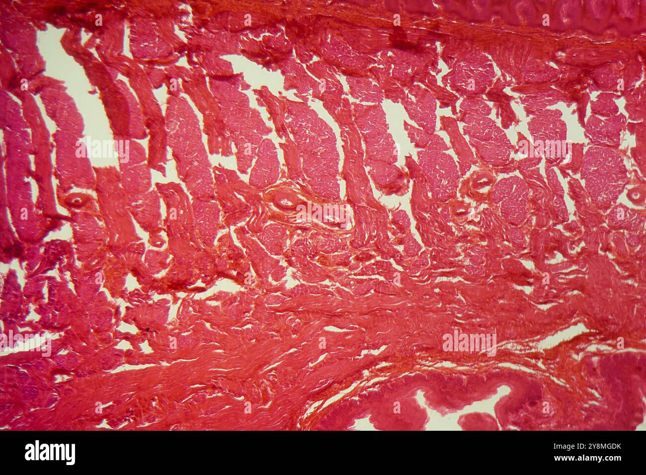 A section trough different tongue cells under the microscope Stock Photohttps://www.alamy.com/image-license-details/?v=1https://www.alamy.com/a-section-trough-different-tongue-cells-under-the-microscope-image624942463.html
RF2Y8MGDK–A section trough different tongue cells under the microscope
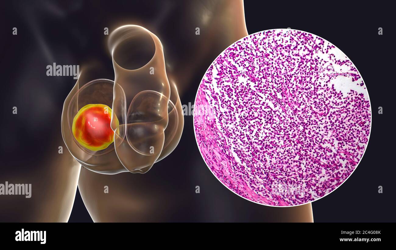 Testicular cancer, a malignant spermatocytic seminoma, computer illustration and light micrograph. A seminoma is a malignant tumour (cancer) of the testis (testicle). Although this is a rare cancer, it is the most common cancer in 15 to 35 year old men. It arises from abnormal germ cells (precursors to sperm cells) in the seminiferous tubules. Surgical removal of the affected testis (orchidectomy) is followed by radiotherapy and chemotherapy if required. Stock Photohttps://www.alamy.com/image-license-details/?v=1https://www.alamy.com/testicular-cancer-a-malignant-spermatocytic-seminoma-computer-illustration-and-light-micrograph-a-seminoma-is-a-malignant-tumour-cancer-of-the-testis-testicle-although-this-is-a-rare-cancer-it-is-the-most-common-cancer-in-15-to-35-year-old-men-it-arises-from-abnormal-germ-cells-precursors-to-sperm-cells-in-the-seminiferous-tubules-surgical-removal-of-the-affected-testis-orchidectomy-is-followed-by-radiotherapy-and-chemotherapy-if-required-image364227827.html
RF2C4G08K–Testicular cancer, a malignant spermatocytic seminoma, computer illustration and light micrograph. A seminoma is a malignant tumour (cancer) of the testis (testicle). Although this is a rare cancer, it is the most common cancer in 15 to 35 year old men. It arises from abnormal germ cells (precursors to sperm cells) in the seminiferous tubules. Surgical removal of the affected testis (orchidectomy) is followed by radiotherapy and chemotherapy if required.
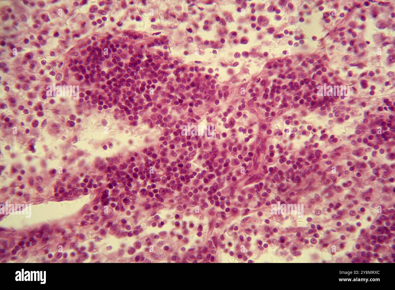 A section trough lymph node cells under the microscope Stock Photohttps://www.alamy.com/image-license-details/?v=1https://www.alamy.com/a-section-trough-lymph-node-cells-under-the-microscope-image624948308.html
RF2Y8MRXC–A section trough lymph node cells under the microscope
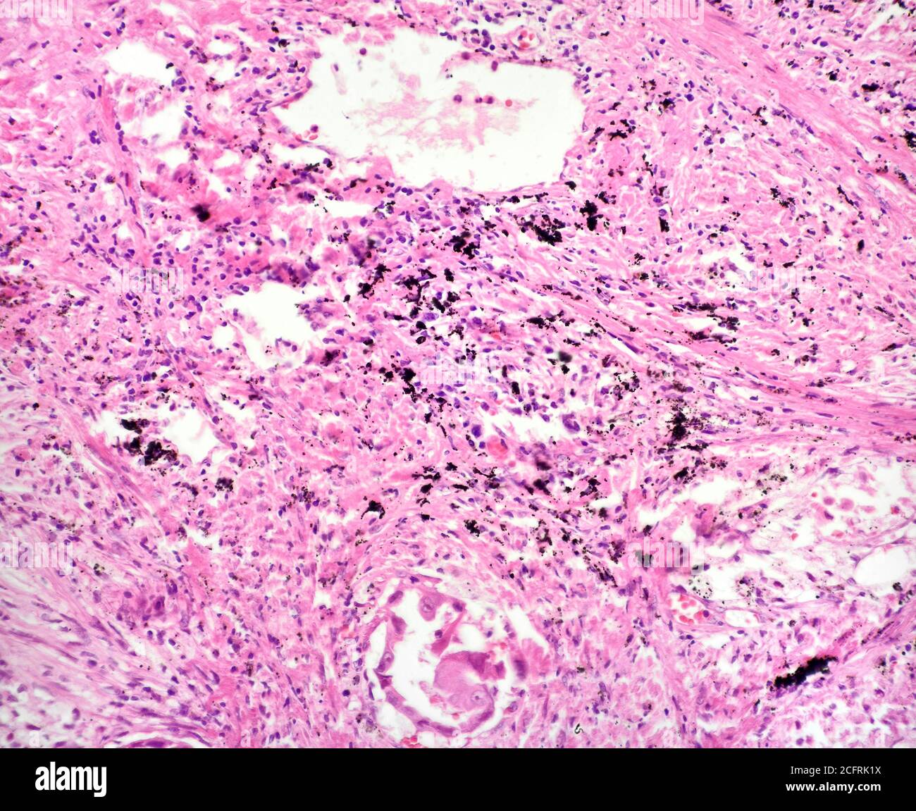 Lung cancer cells, brightfield photomicrograph Stock Photohttps://www.alamy.com/image-license-details/?v=1https://www.alamy.com/lung-cancer-cells-brightfield-photomicrograph-image371157414.html
RM2CFRK1X–Lung cancer cells, brightfield photomicrograph
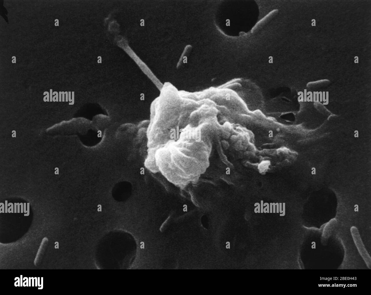 #6 in a six-step sequence of the death of a cancer cell. A cancer cell has migrated through the holes of a matrix coated membrane from the top to the bottom, simulating natural migration of a invading cancer cell between, and sometimes through, the vascular endothelium. Notice the spikes or pseudopodia that are characteristic of an invading cancer cell (1). A buffy coat containing red blood cells, lymphocytes and macrophages is added to the bottom of the membrane. A group of macrophages identify the cancer cell as foreign matter and start to stick to the cancer cell, which still has its spikes Stock Photohttps://www.alamy.com/image-license-details/?v=1https://www.alamy.com/6-in-a-six-step-sequence-of-the-death-of-a-cancer-cell-a-cancer-cell-has-migrated-through-the-holes-of-a-matrix-coated-membrane-from-the-top-to-the-bottom-simulating-natural-migration-of-a-invading-cancer-cell-between-and-sometimes-through-the-vascular-endothelium-notice-the-spikes-or-pseudopodia-that-are-characteristic-of-an-invading-cancer-cell-1-a-buffy-coat-containing-red-blood-cells-lymphocytes-and-macrophages-is-added-to-the-bottom-of-the-membrane-a-group-of-macrophages-identify-the-cancer-cell-as-foreign-matter-and-start-to-stick-to-the-cancer-cell-which-still-has-its-spikes-image352825987.html
RM2BE0H43–#6 in a six-step sequence of the death of a cancer cell. A cancer cell has migrated through the holes of a matrix coated membrane from the top to the bottom, simulating natural migration of a invading cancer cell between, and sometimes through, the vascular endothelium. Notice the spikes or pseudopodia that are characteristic of an invading cancer cell (1). A buffy coat containing red blood cells, lymphocytes and macrophages is added to the bottom of the membrane. A group of macrophages identify the cancer cell as foreign matter and start to stick to the cancer cell, which still has its spikes
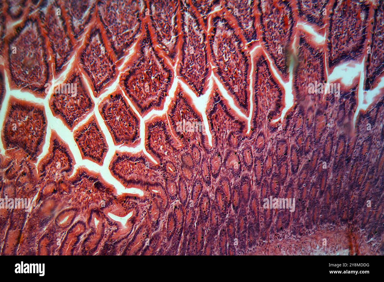 A section trough cells of a small intestine under the microscope Stock Photohttps://www.alamy.com/image-license-details/?v=1https://www.alamy.com/a-section-trough-cells-of-a-small-intestine-under-the-microscope-image624940108.html
RF2Y8MDDG–A section trough cells of a small intestine under the microscope
 Photomicrograph of colon adenocarcinoma, illustrating malignant glandular cells characteristic of colon cancer. Stock Photohttps://www.alamy.com/image-license-details/?v=1https://www.alamy.com/photomicrograph-of-colon-adenocarcinoma-illustrating-malignant-glandular-cells-characteristic-of-colon-cancer-image571995853.html
RF2T6GJF9–Photomicrograph of colon adenocarcinoma, illustrating malignant glandular cells characteristic of colon cancer.
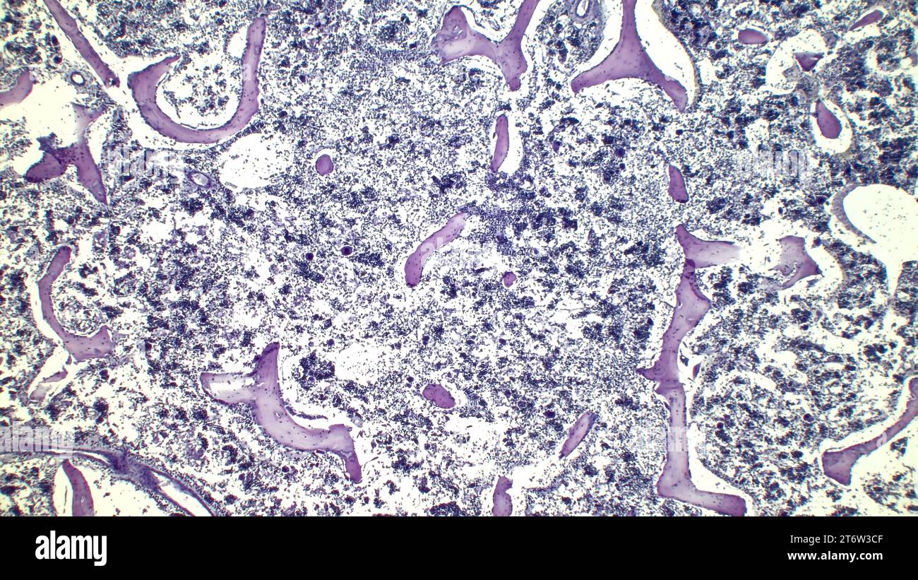 Human red bone marrow, hematopoietic press. The big tsit beetveen erythrocytes are megakaryocytes. Hematoxylin and eosin stain. Stock Photohttps://www.alamy.com/image-license-details/?v=1https://www.alamy.com/human-red-bone-marrow-hematopoietic-press-the-big-tsit-beetveen-erythrocytes-are-megakaryocytes-hematoxylin-and-eosin-stain-image572181583.html
RF2T6W3CF–Human red bone marrow, hematopoietic press. The big tsit beetveen erythrocytes are megakaryocytes. Hematoxylin and eosin stain.
 False colour EMS image of carbon nanotubes which deliver cancer drugs right to the site of disease during chemotherapy treatment Stock Photohttps://www.alamy.com/image-license-details/?v=1https://www.alamy.com/stock-photo-false-colour-ems-image-of-carbon-nanotubes-which-deliver-cancer-drugs-20123993.html
RMB4MMA1–False colour EMS image of carbon nanotubes which deliver cancer drugs right to the site of disease during chemotherapy treatment
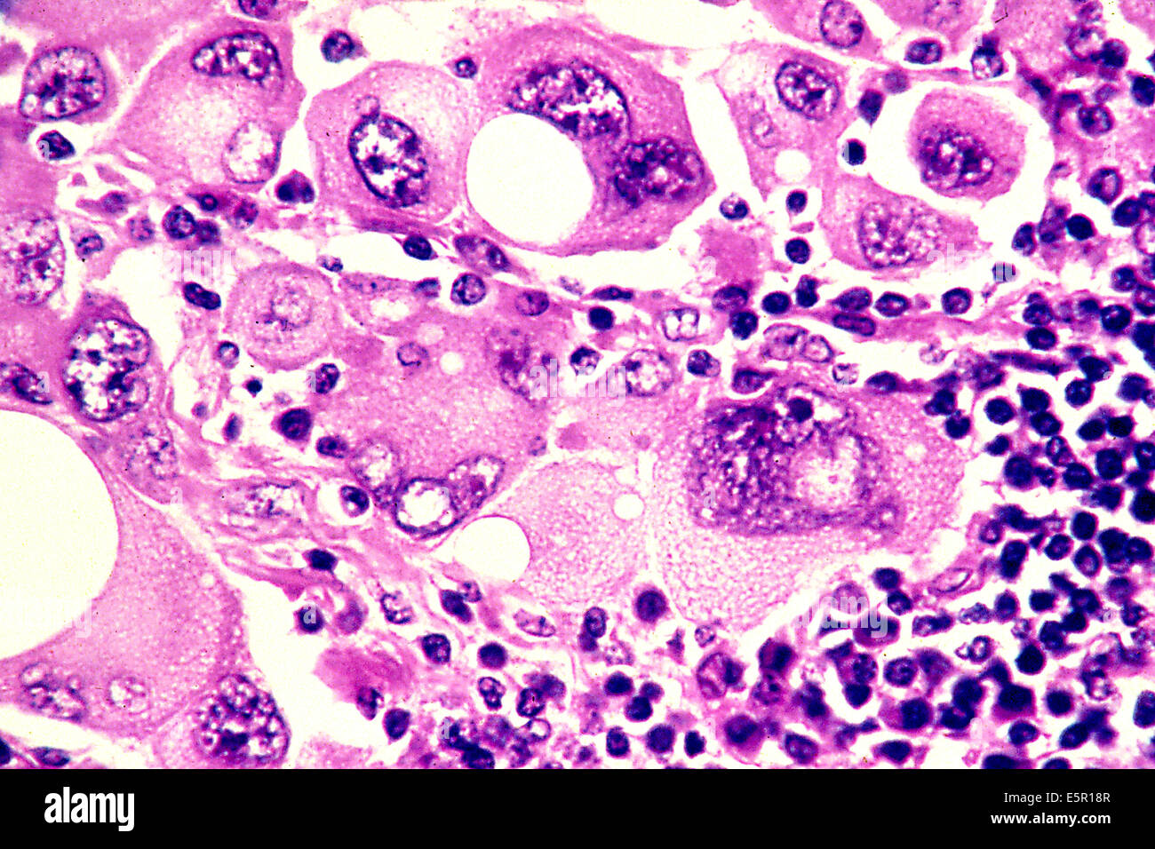 Photomicrograph of human metastatic melanoma cells magnified to 320x. Stock Photohttps://www.alamy.com/image-license-details/?v=1https://www.alamy.com/stock-photo-photomicrograph-of-human-metastatic-melanoma-cells-magnified-to-320x-72420679.html
RME5R18R–Photomicrograph of human metastatic melanoma cells magnified to 320x.
 Cross section of the human tissue in the microscope view. Stock Photohttps://www.alamy.com/image-license-details/?v=1https://www.alamy.com/cross-section-of-the-human-tissue-in-the-microscope-view-image157949805.html
RFK4Y6GD–Cross section of the human tissue in the microscope view.
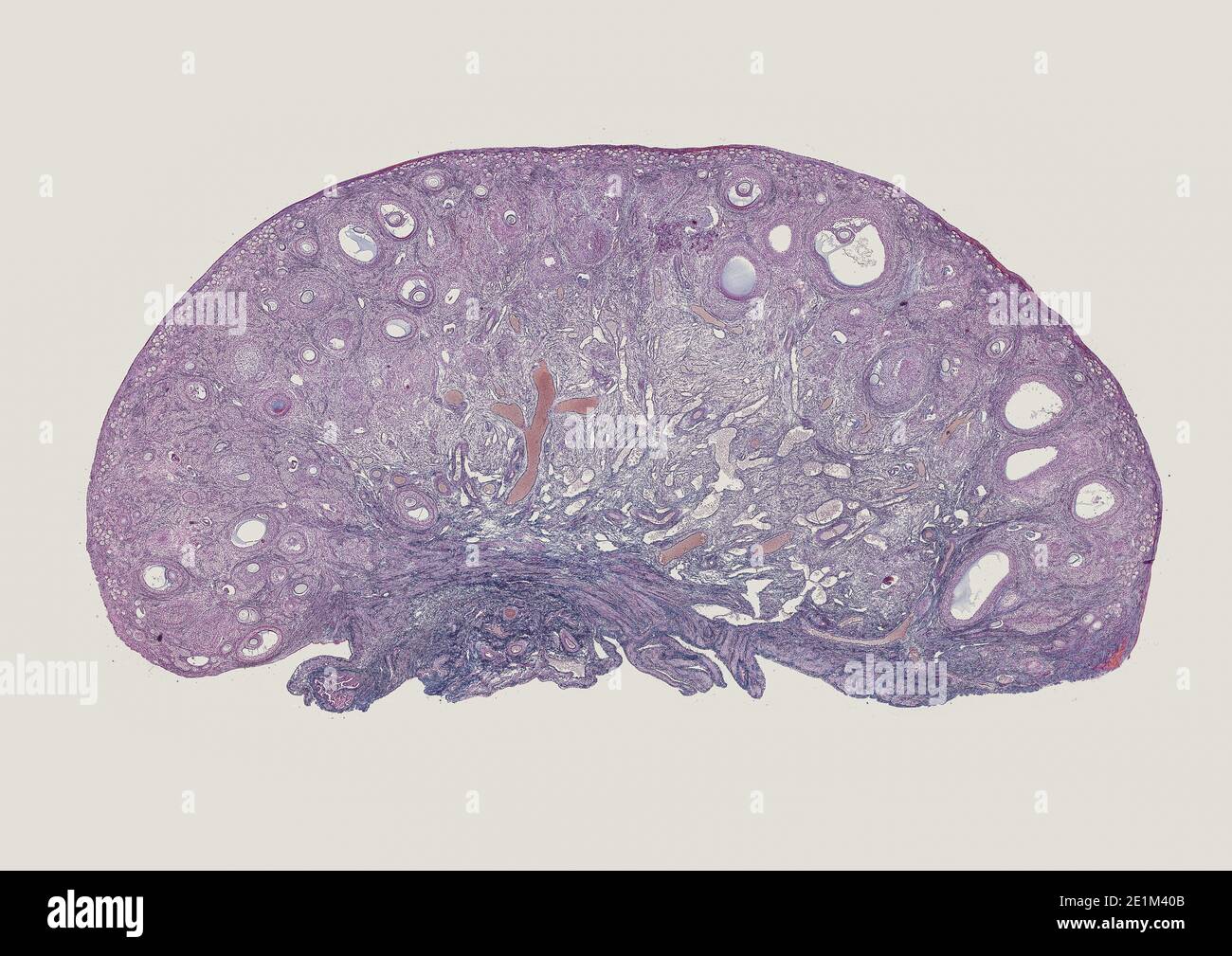 cross section cut of human body cells under a scientific microscope Stock Photohttps://www.alamy.com/image-license-details/?v=1https://www.alamy.com/cross-section-cut-of-human-body-cells-under-a-scientific-microscope-image396895307.html
RF2E1M40B–cross section cut of human body cells under a scientific microscope
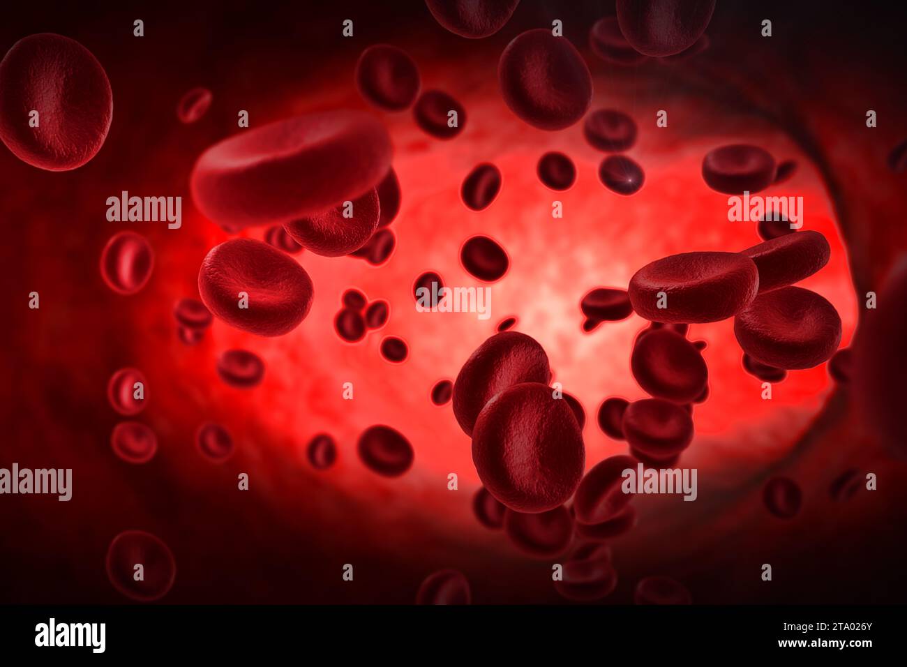 3D rendering red blood cells animation in an artery, flow inside body, medical human health-care concept Stock Photohttps://www.alamy.com/image-license-details/?v=1https://www.alamy.com/3d-rendering-red-blood-cells-animation-in-an-artery-flow-inside-body-medical-human-health-care-concept-image574090467.html
RF2TA026Y–3D rendering red blood cells animation in an artery, flow inside body, medical human health-care concept
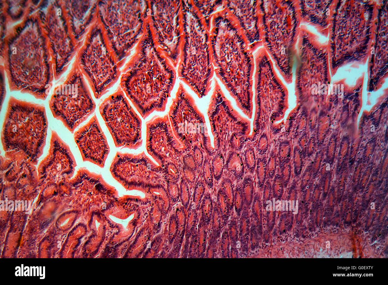 A section trough cells of a small intestine under the microscope. Stock Photohttps://www.alamy.com/image-license-details/?v=1https://www.alamy.com/stock-photo-a-section-trough-cells-of-a-small-intestine-under-the-microscope-103590619.html
RMG0EXTY–A section trough cells of a small intestine under the microscope.
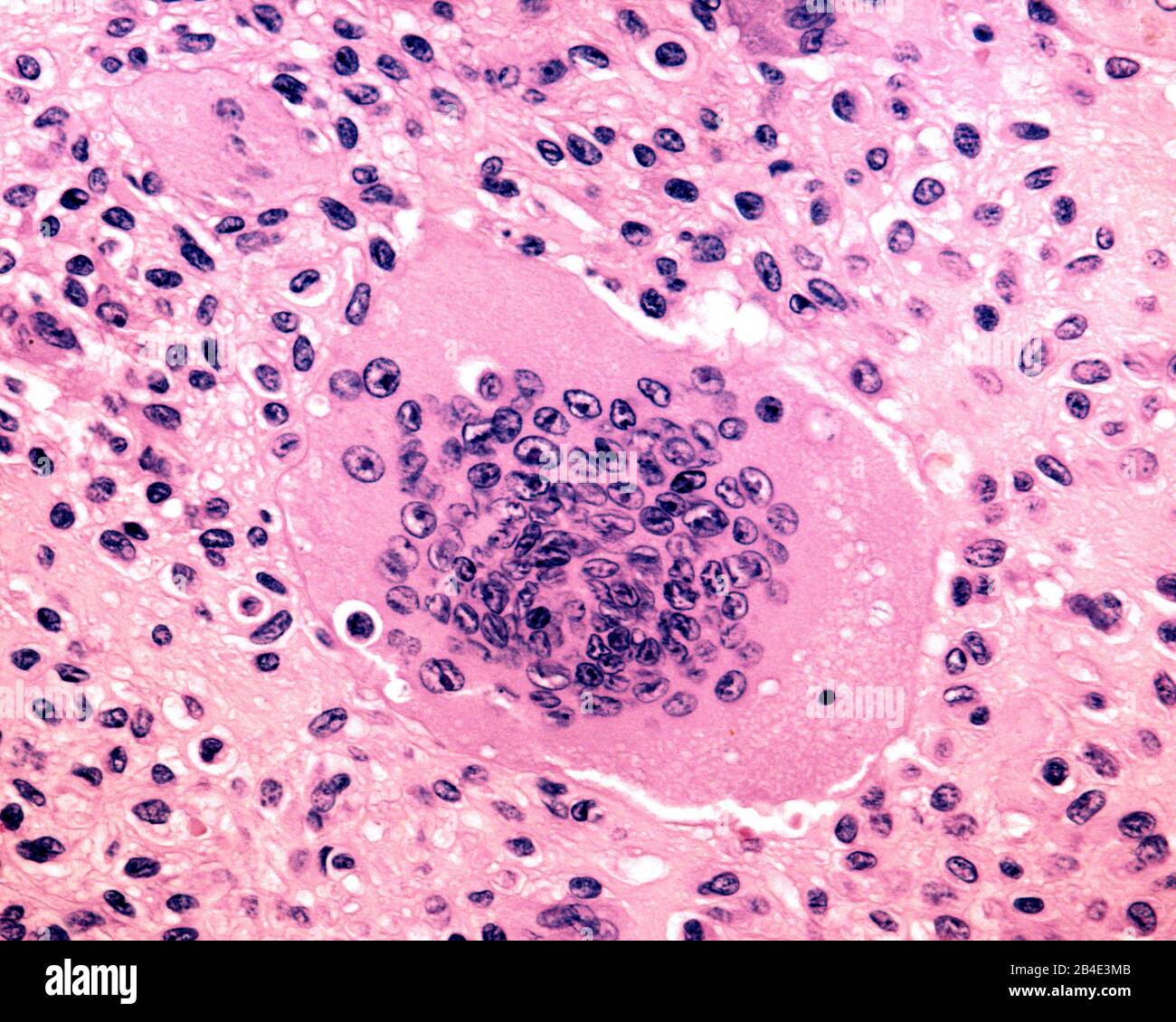 A mutinucleated giant cell derived from osteoclasts. Biopsy of a human giant cell tumour of bone. Light micrograph. Stock Photohttps://www.alamy.com/image-license-details/?v=1https://www.alamy.com/a-mutinucleated-giant-cell-derived-from-osteoclasts-biopsy-of-a-human-giant-cell-tumour-of-bone-light-micrograph-image346976235.html
RF2B4E3MB–A mutinucleated giant cell derived from osteoclasts. Biopsy of a human giant cell tumour of bone. Light micrograph.
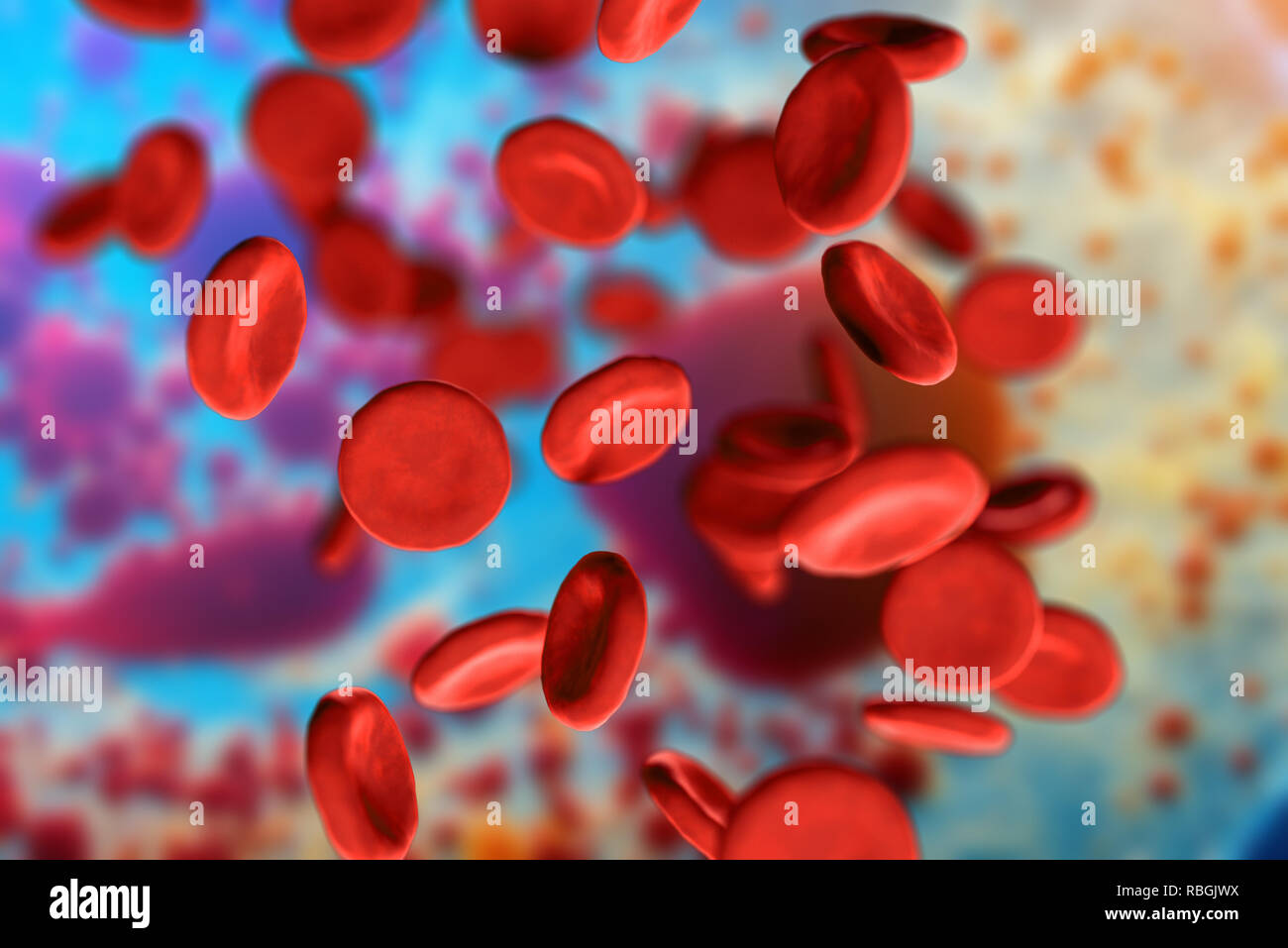 3d illustration of red blood cells erythrocytes close-up under a microscope in the body. Concept for scientific medical background Stock Photohttps://www.alamy.com/image-license-details/?v=1https://www.alamy.com/3d-illustration-of-red-blood-cells-erythrocytes-close-up-under-a-microscope-in-the-body-concept-for-scientific-medical-background-image230862070.html
RFRBGJWX–3d illustration of red blood cells erythrocytes close-up under a microscope in the body. Concept for scientific medical background
 CANCER RESEARCH Stock Photohttps://www.alamy.com/image-license-details/?v=1https://www.alamy.com/stock-photo-cancer-research-139738024.html
RMJ39H7M–CANCER RESEARCH
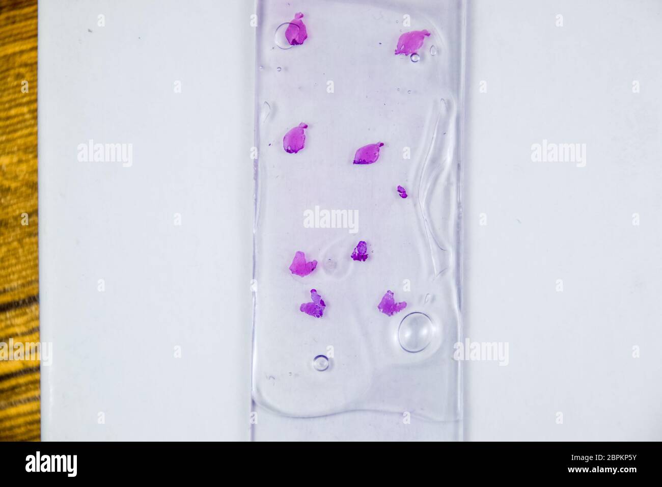 Slices of the tumor under glass. Histological examination of tumor cells for the presence of cancer. Samples of tumor cells under the sleek against th Stock Photohttps://www.alamy.com/image-license-details/?v=1https://www.alamy.com/slices-of-the-tumor-under-glass-histological-examination-of-tumor-cells-for-the-presence-of-cancer-samples-of-tumor-cells-under-the-sleek-against-th-image358164295.html
RF2BPKP5Y–Slices of the tumor under glass. Histological examination of tumor cells for the presence of cancer. Samples of tumor cells under the sleek against th
 Close-up of red blood cells Stock Photohttps://www.alamy.com/image-license-details/?v=1https://www.alamy.com/stock-photo-close-up-of-red-blood-cells-91548418.html
RFF8XAXA–Close-up of red blood cells
 Microscopic View of Cancer Cells Stock Photohttps://www.alamy.com/image-license-details/?v=1https://www.alamy.com/stock-photo-microscopic-view-of-cancer-cells-52076053.html
RMD0M7F1–Microscopic View of Cancer Cells
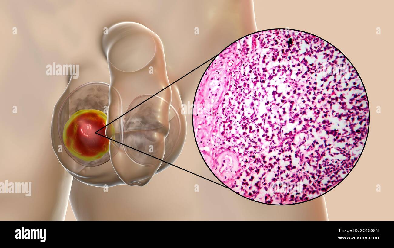 Testicular cancer, a malignant spermatocytic seminoma, computer illustration and light micrograph. A seminoma is a malignant tumour (cancer) of the testis (testicle). Although this is a rare cancer, it is the most common cancer in 15 to 35 year old men. It arises from abnormal germ cells (precursors to sperm cells) in the seminiferous tubules. Surgical removal of the affected testis (orchidectomy) is followed by radiotherapy and chemotherapy if required. Stock Photohttps://www.alamy.com/image-license-details/?v=1https://www.alamy.com/testicular-cancer-a-malignant-spermatocytic-seminoma-computer-illustration-and-light-micrograph-a-seminoma-is-a-malignant-tumour-cancer-of-the-testis-testicle-although-this-is-a-rare-cancer-it-is-the-most-common-cancer-in-15-to-35-year-old-men-it-arises-from-abnormal-germ-cells-precursors-to-sperm-cells-in-the-seminiferous-tubules-surgical-removal-of-the-affected-testis-orchidectomy-is-followed-by-radiotherapy-and-chemotherapy-if-required-image364227829.html
RF2C4G08N–Testicular cancer, a malignant spermatocytic seminoma, computer illustration and light micrograph. A seminoma is a malignant tumour (cancer) of the testis (testicle). Although this is a rare cancer, it is the most common cancer in 15 to 35 year old men. It arises from abnormal germ cells (precursors to sperm cells) in the seminiferous tubules. Surgical removal of the affected testis (orchidectomy) is followed by radiotherapy and chemotherapy if required.
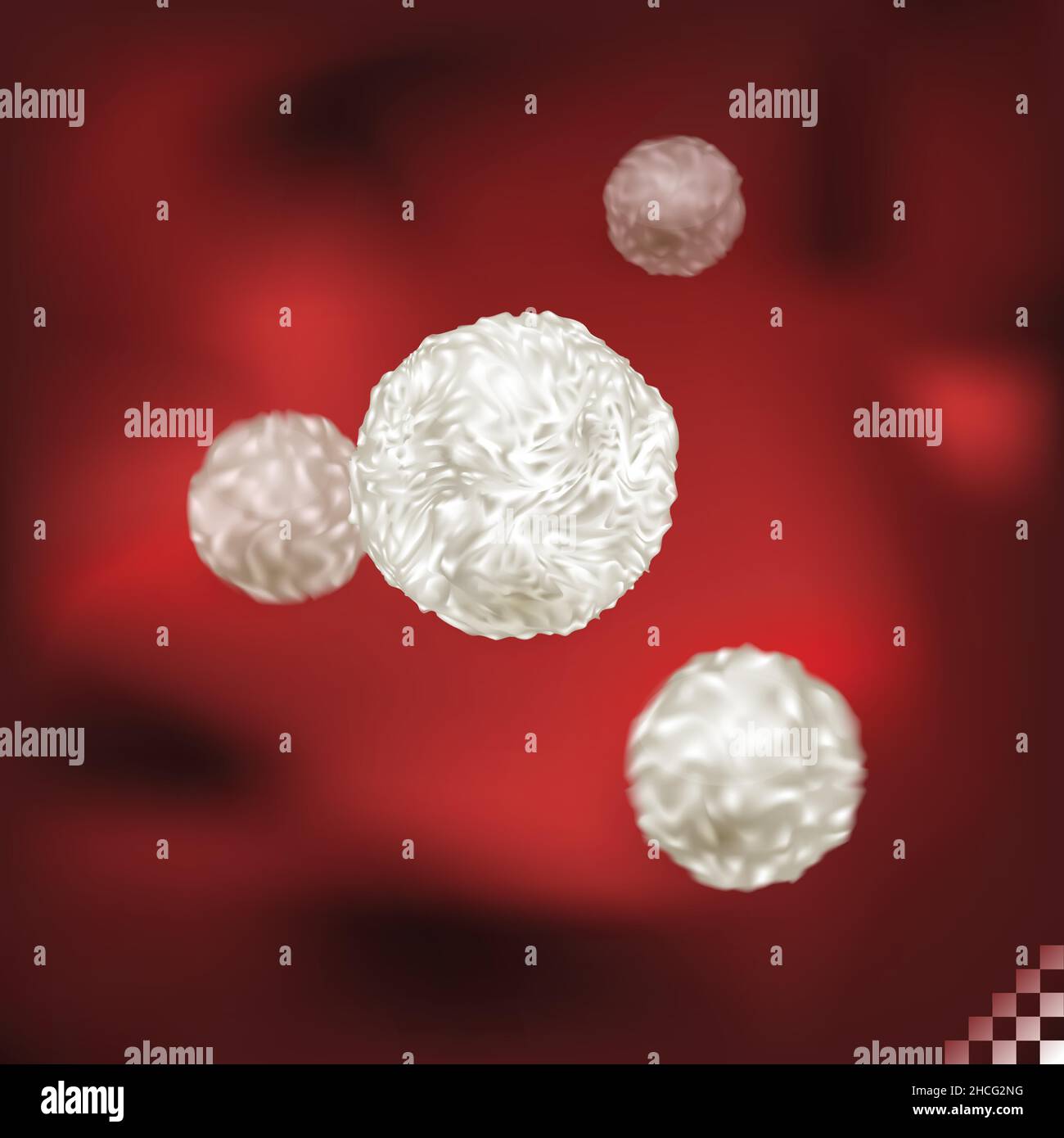 illustration of bioscience of immunity white blood cell circulating the human body Stock Vectorhttps://www.alamy.com/image-license-details/?v=1https://www.alamy.com/illustration-of-bioscience-of-immunity-white-blood-cell-circulating-the-human-body-image455198844.html
RF2HCG2NG–illustration of bioscience of immunity white blood cell circulating the human body
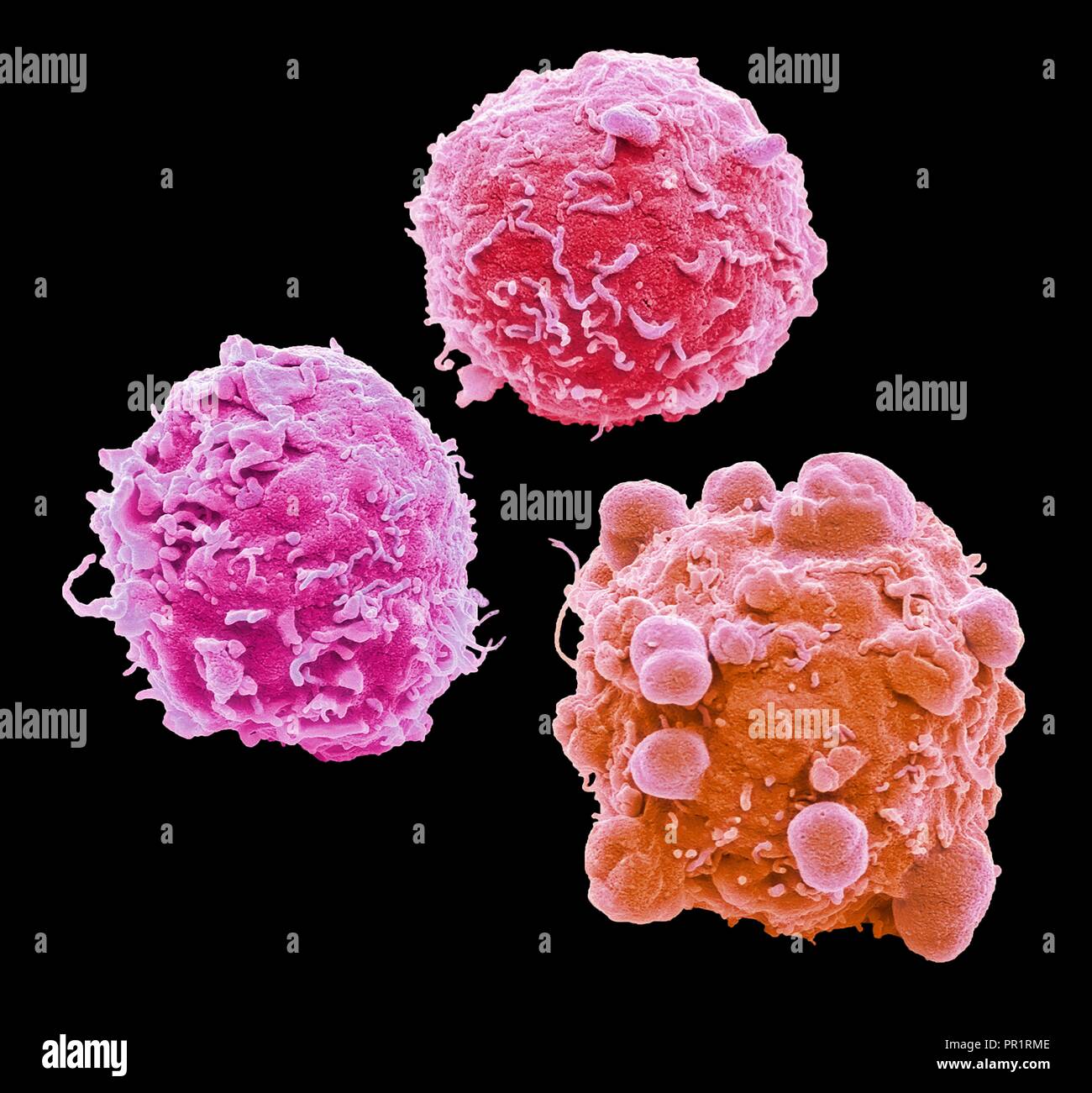 Colorectal cancer cells. Coloured scanning electron micrograph (SEM) of cancer cells from the human colon (large intestine). Cancer of the colon is also known as colorectal cancer. Symptoms include rectal bleeding and abdominal pain. Treatment is with surgery to remove the affected area. Colon cancer is one of the most common cancers in the Western world. Magnification: x 4,000 when printed 10 centimetres wide. Stock Photohttps://www.alamy.com/image-license-details/?v=1https://www.alamy.com/colorectal-cancer-cells-coloured-scanning-electron-micrograph-sem-of-cancer-cells-from-the-human-colon-large-intestine-cancer-of-the-colon-is-also-known-as-colorectal-cancer-symptoms-include-rectal-bleeding-and-abdominal-pain-treatment-is-with-surgery-to-remove-the-affected-area-colon-cancer-is-one-of-the-most-common-cancers-in-the-western-world-magnification-x-4000-when-printed-10-centimetres-wide-image220702062.html
RFPR1RME–Colorectal cancer cells. Coloured scanning electron micrograph (SEM) of cancer cells from the human colon (large intestine). Cancer of the colon is also known as colorectal cancer. Symptoms include rectal bleeding and abdominal pain. Treatment is with surgery to remove the affected area. Colon cancer is one of the most common cancers in the Western world. Magnification: x 4,000 when printed 10 centimetres wide.
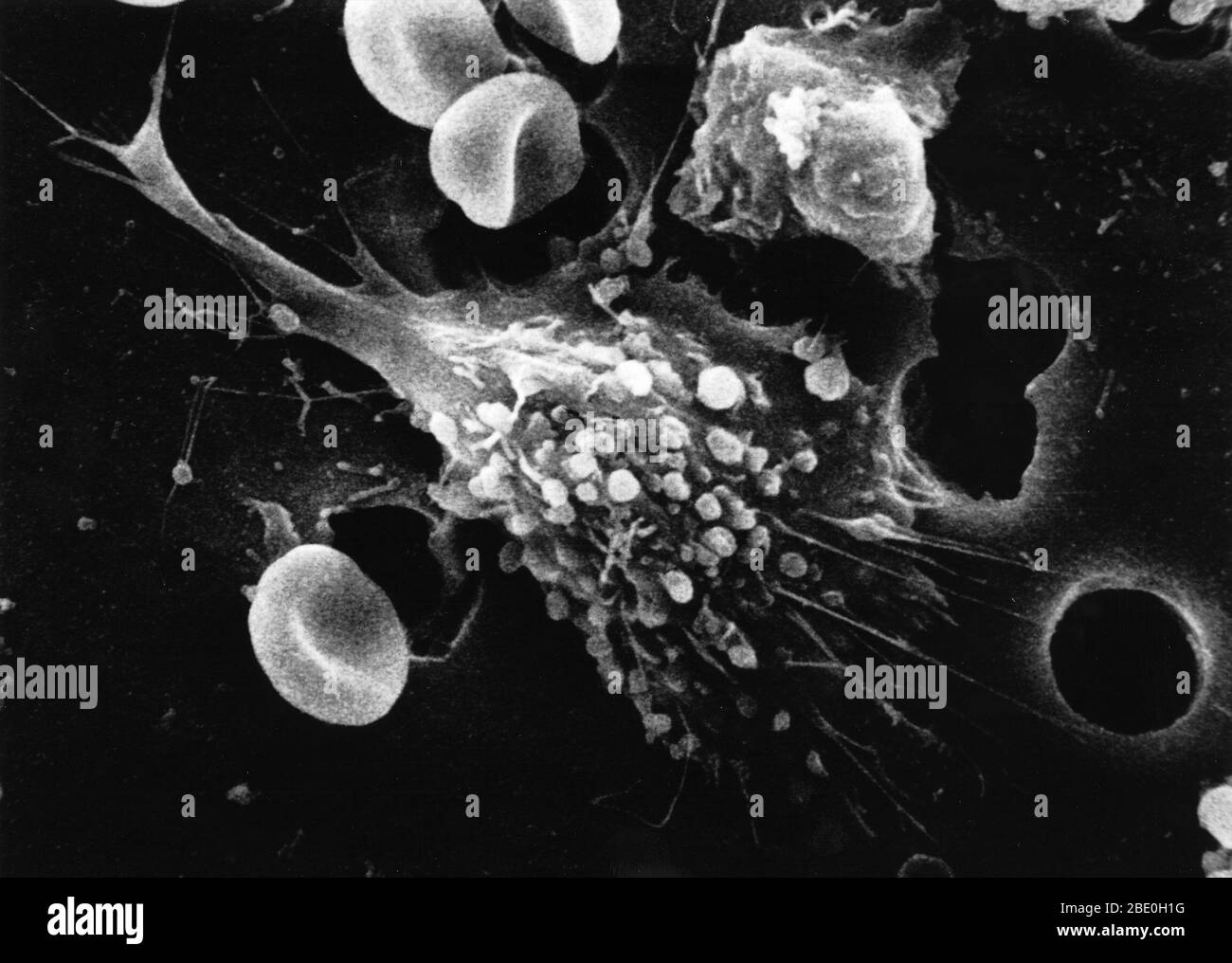 Step one of a six-step sequence of the death of a cancer cell. A cancer cell has migrated through the holes of a matrix coated membrane from the top to the bottom, simulating natural migration of a invading cancer cell between, and sometimes through, the vascular endothelium. Notice the spikes or pseudopodia that are characteristic of an invading cancer cell. Photo magnification: 12,000x Stock Photohttps://www.alamy.com/image-license-details/?v=1https://www.alamy.com/step-one-of-a-six-step-sequence-of-the-death-of-a-cancer-cell-a-cancer-cell-has-migrated-through-the-holes-of-a-matrix-coated-membrane-from-the-top-to-the-bottom-simulating-natural-migration-of-a-invading-cancer-cell-between-and-sometimes-through-the-vascular-endothelium-notice-the-spikes-or-pseudopodia-that-are-characteristic-of-an-invading-cancer-cell-photo-magnification-12000x-image352825916.html
RM2BE0H1G–Step one of a six-step sequence of the death of a cancer cell. A cancer cell has migrated through the holes of a matrix coated membrane from the top to the bottom, simulating natural migration of a invading cancer cell between, and sometimes through, the vascular endothelium. Notice the spikes or pseudopodia that are characteristic of an invading cancer cell. Photo magnification: 12,000x
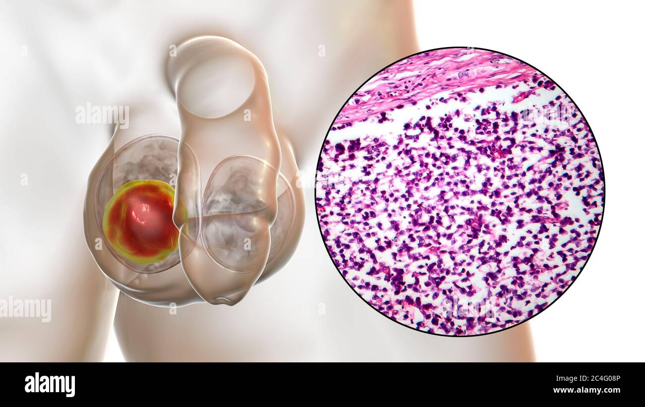 Testicular cancer, a malignant spermatocytic seminoma, computer illustration and light micrograph. A seminoma is a malignant tumour (cancer) of the testis (testicle). Although this is a rare cancer, it is the most common cancer in 15 to 35 year old men. It arises from abnormal germ cells (precursors to sperm cells) in the seminiferous tubules. Surgical removal of the affected testis (orchidectomy) is followed by radiotherapy and chemotherapy if required. Stock Photohttps://www.alamy.com/image-license-details/?v=1https://www.alamy.com/testicular-cancer-a-malignant-spermatocytic-seminoma-computer-illustration-and-light-micrograph-a-seminoma-is-a-malignant-tumour-cancer-of-the-testis-testicle-although-this-is-a-rare-cancer-it-is-the-most-common-cancer-in-15-to-35-year-old-men-it-arises-from-abnormal-germ-cells-precursors-to-sperm-cells-in-the-seminiferous-tubules-surgical-removal-of-the-affected-testis-orchidectomy-is-followed-by-radiotherapy-and-chemotherapy-if-required-image364227830.html
RF2C4G08P–Testicular cancer, a malignant spermatocytic seminoma, computer illustration and light micrograph. A seminoma is a malignant tumour (cancer) of the testis (testicle). Although this is a rare cancer, it is the most common cancer in 15 to 35 year old men. It arises from abnormal germ cells (precursors to sperm cells) in the seminiferous tubules. Surgical removal of the affected testis (orchidectomy) is followed by radiotherapy and chemotherapy if required.
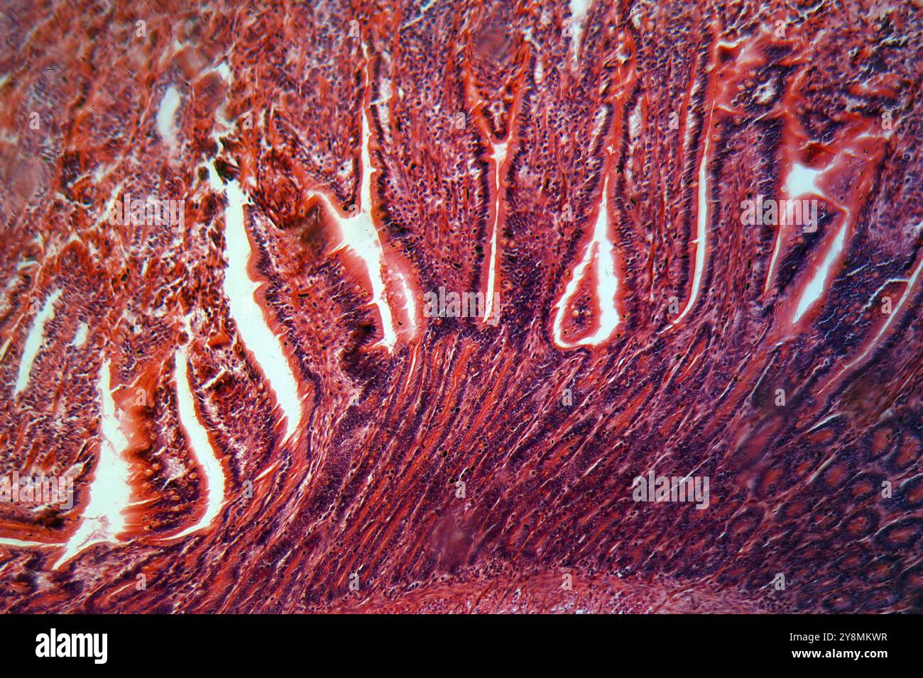 A section trough cells of a small intestine under the microscope Stock Photohttps://www.alamy.com/image-license-details/?v=1https://www.alamy.com/a-section-trough-cells-of-a-small-intestine-under-the-microscope-image624945155.html
RF2Y8MKWR–A section trough cells of a small intestine under the microscope
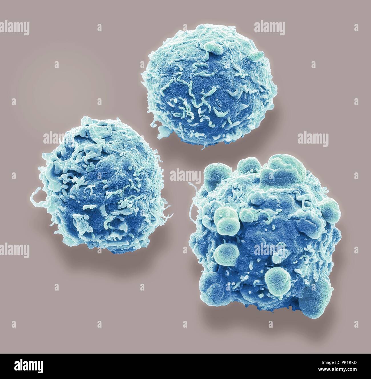 Colorectal cancer cells. Coloured scanning electron micrograph (SEM) of cancer cells from the human colon (large intestine). Cancer of the colon is also known as colorectal cancer. Symptoms include rectal bleeding and abdominal pain. Treatment is with surgery to remove the affected area. Colon cancer is one of the most common cancers in the Western world. Magnification: x 4,000 when printed 10 centimetres wide. Stock Photohttps://www.alamy.com/image-license-details/?v=1https://www.alamy.com/colorectal-cancer-cells-coloured-scanning-electron-micrograph-sem-of-cancer-cells-from-the-human-colon-large-intestine-cancer-of-the-colon-is-also-known-as-colorectal-cancer-symptoms-include-rectal-bleeding-and-abdominal-pain-treatment-is-with-surgery-to-remove-the-affected-area-colon-cancer-is-one-of-the-most-common-cancers-in-the-western-world-magnification-x-4000-when-printed-10-centimetres-wide-image220702033.html
RFPR1RKD–Colorectal cancer cells. Coloured scanning electron micrograph (SEM) of cancer cells from the human colon (large intestine). Cancer of the colon is also known as colorectal cancer. Symptoms include rectal bleeding and abdominal pain. Treatment is with surgery to remove the affected area. Colon cancer is one of the most common cancers in the Western world. Magnification: x 4,000 when printed 10 centimetres wide.
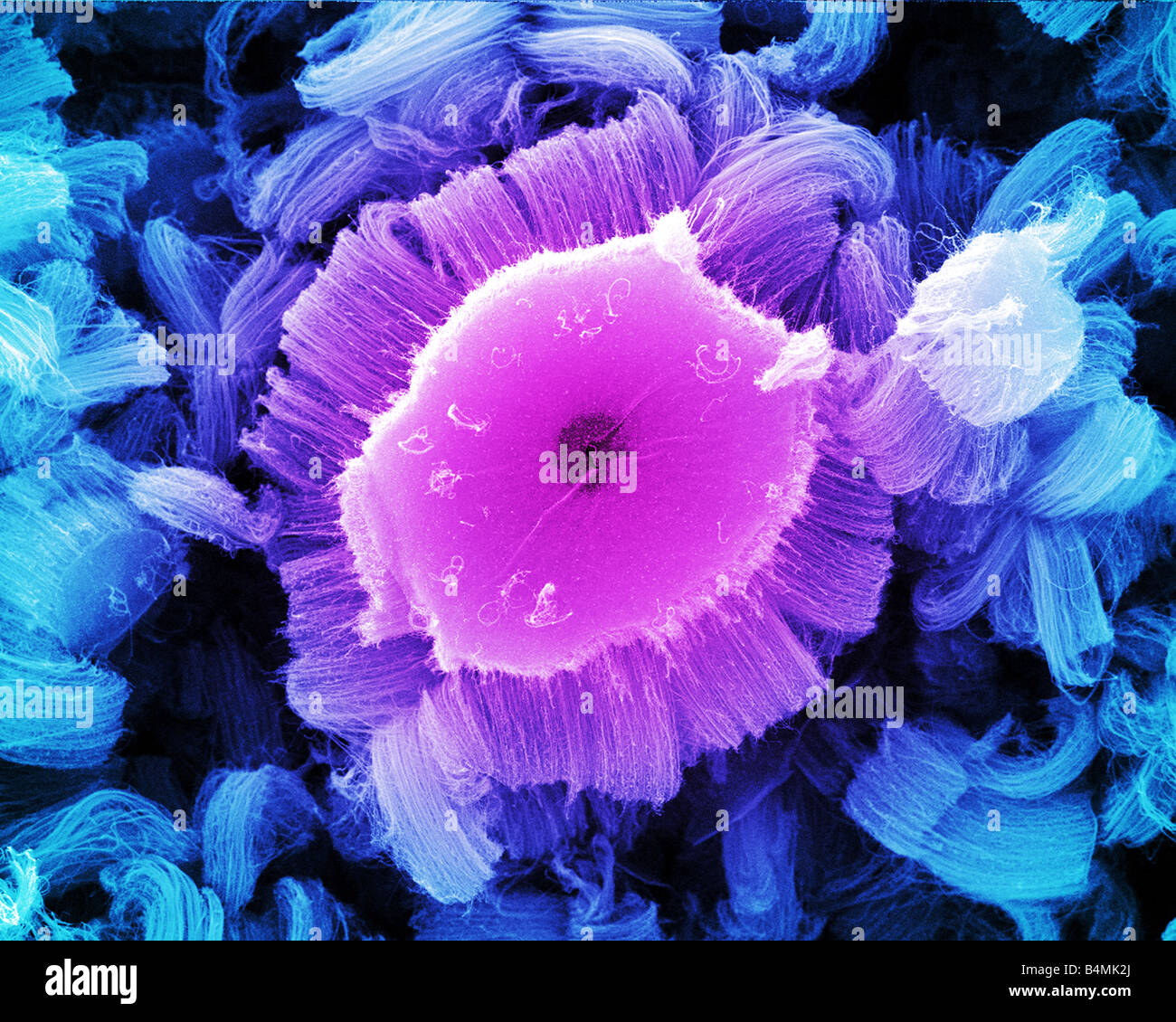 View looking down into carbon nanotube which deliver cancer drugs right to the site of disease during chemotherapy treatment Stock Photohttps://www.alamy.com/image-license-details/?v=1https://www.alamy.com/stock-photo-view-looking-down-into-carbon-nanotube-which-deliver-cancer-drugs-20123002.html
RMB4MK2J–View looking down into carbon nanotube which deliver cancer drugs right to the site of disease during chemotherapy treatment
 A section trough different tongue cells on the tip of a tongue, under the microscope Stock Photohttps://www.alamy.com/image-license-details/?v=1https://www.alamy.com/a-section-trough-different-tongue-cells-on-the-tip-of-a-tongue-under-the-microscope-image624939516.html
RF2Y8MCMC–A section trough different tongue cells on the tip of a tongue, under the microscope
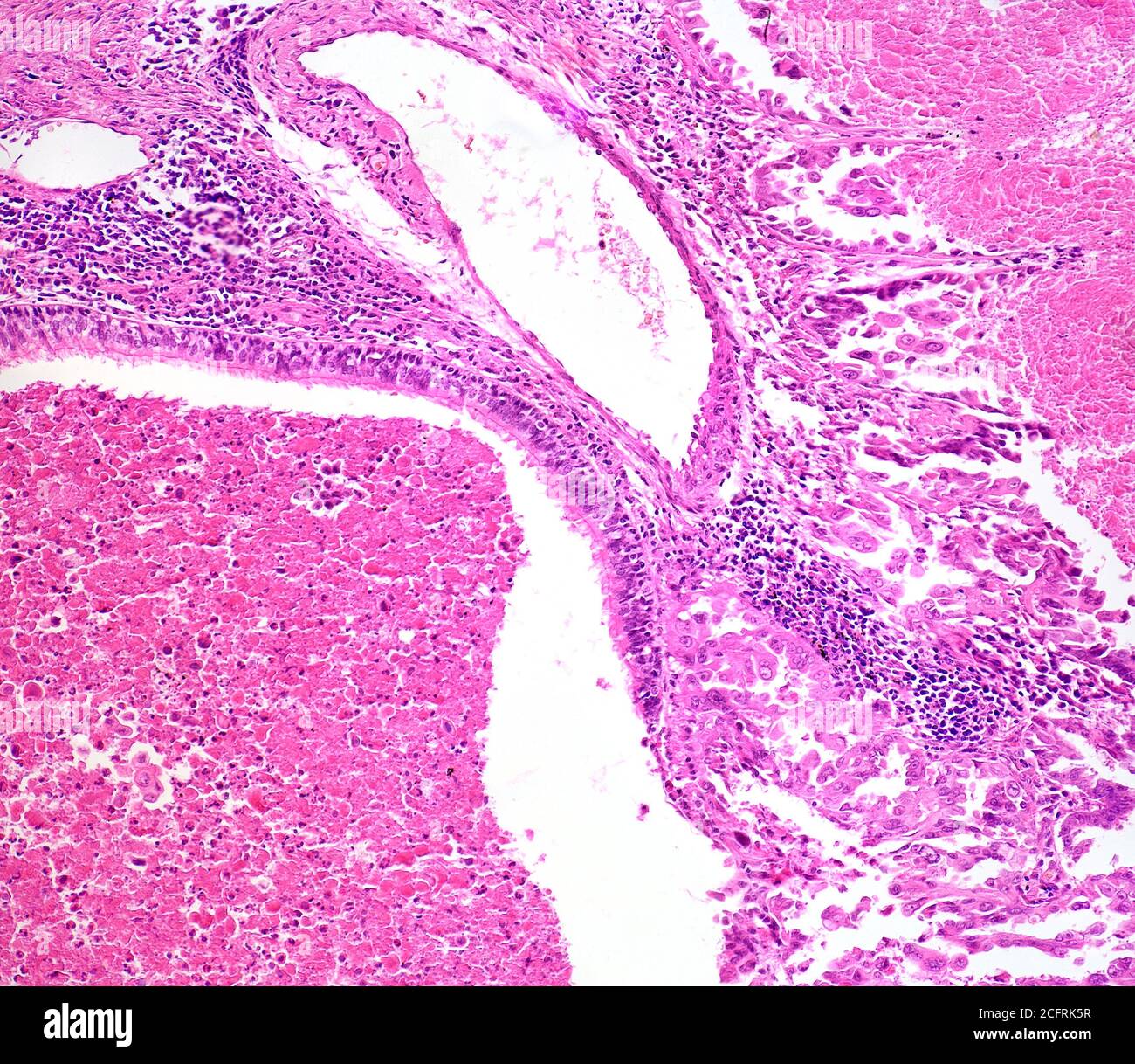 Lung cancer cells, brightfield photomicrograph Stock Photohttps://www.alamy.com/image-license-details/?v=1https://www.alamy.com/lung-cancer-cells-brightfield-photomicrograph-image371157523.html
RM2CFRK5R–Lung cancer cells, brightfield photomicrograph
 cross section cut of human body cells under a scientific microscope Stock Photohttps://www.alamy.com/image-license-details/?v=1https://www.alamy.com/cross-section-cut-of-human-body-cells-under-a-scientific-microscope-image396904112.html
RF2E1MF6T–cross section cut of human body cells under a scientific microscope
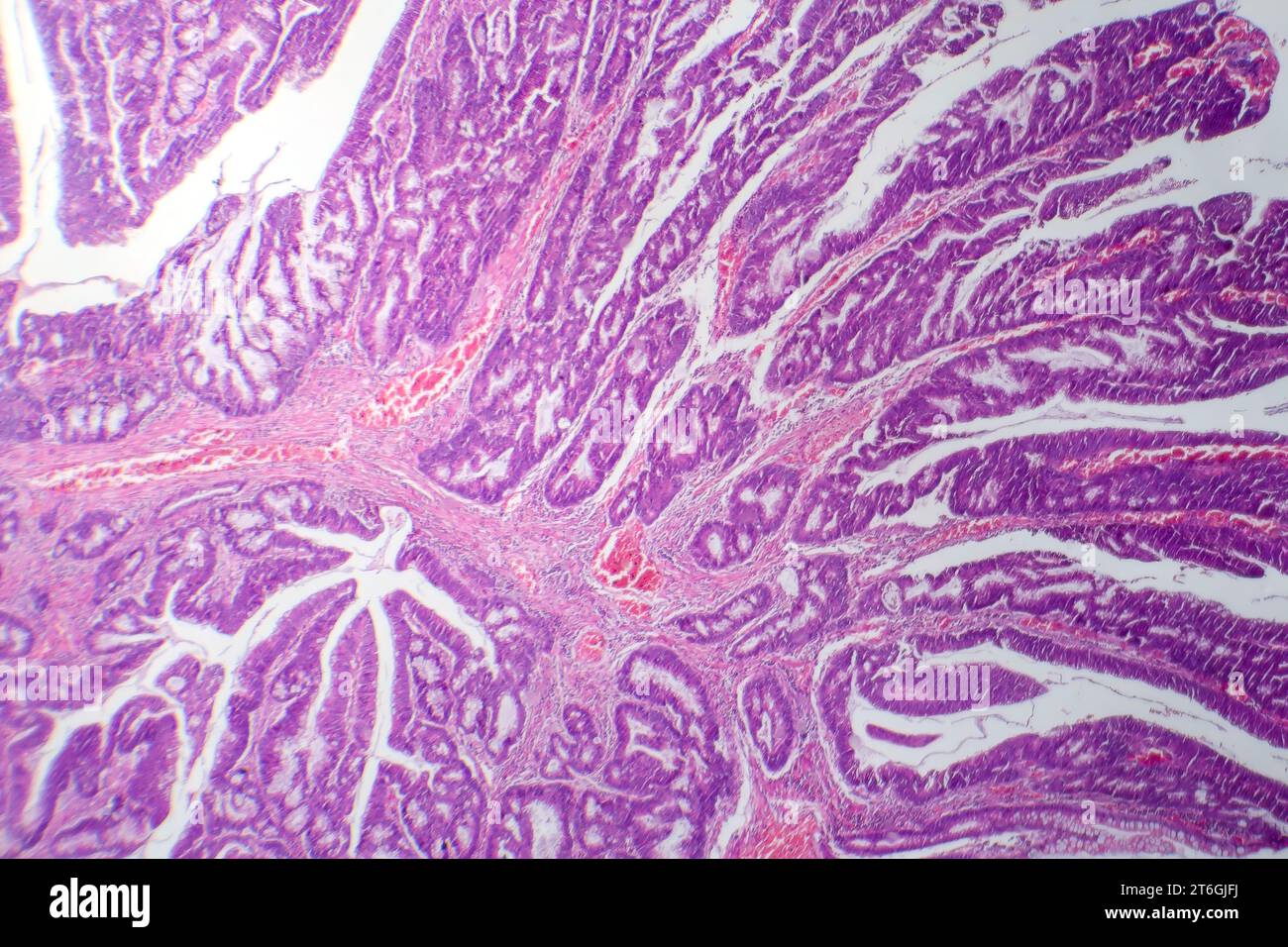 Photomicrograph of colon adenocarcinoma, illustrating malignant glandular cells characteristic of colon cancer. Stock Photohttps://www.alamy.com/image-license-details/?v=1https://www.alamy.com/photomicrograph-of-colon-adenocarcinoma-illustrating-malignant-glandular-cells-characteristic-of-colon-cancer-image571995862.html
RF2T6GJFJ–Photomicrograph of colon adenocarcinoma, illustrating malignant glandular cells characteristic of colon cancer.
 A section trough cells of a small intestine under the microscope. Stock Photohttps://www.alamy.com/image-license-details/?v=1https://www.alamy.com/stock-photo-a-section-trough-cells-of-a-small-intestine-under-the-microscope-103590616.html
RMG0EXTT–A section trough cells of a small intestine under the microscope.
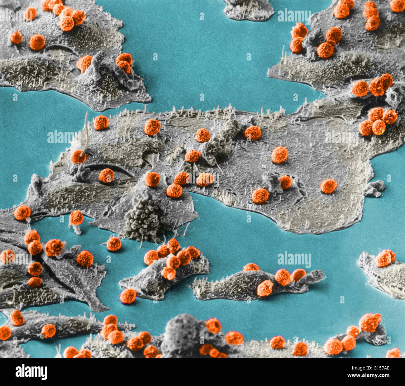 Scanning Electron Micrograph (SEM) showing immune cells (small sphericals, red) attacking cancer cells (larger bodies, grey). Magnification unknown. Stock Photohttps://www.alamy.com/image-license-details/?v=1https://www.alamy.com/stock-photo-scanning-electron-micrograph-sem-showing-immune-cells-small-sphericals-103992406.html
RMG157AE–Scanning Electron Micrograph (SEM) showing immune cells (small sphericals, red) attacking cancer cells (larger bodies, grey). Magnification unknown.
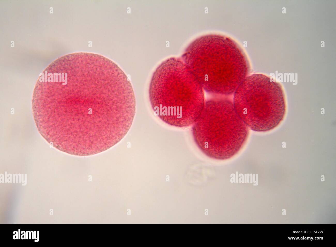 Egg cells under the microscope Stock Photohttps://www.alamy.com/image-license-details/?v=1https://www.alamy.com/stock-photo-egg-cells-under-the-microscope-93549313.html
RFFC5F2W–Egg cells under the microscope
 Cross section of the human tissue in the microscope view. Stock Photohttps://www.alamy.com/image-license-details/?v=1https://www.alamy.com/cross-section-of-the-human-tissue-in-the-microscope-view-image157949781.html
RFK4Y6FH–Cross section of the human tissue in the microscope view.
 red blood cells in an artery, flow inside body, medical human health-care Stock Photohttps://www.alamy.com/image-license-details/?v=1https://www.alamy.com/red-blood-cells-in-an-artery-flow-inside-body-medical-human-health-care-image574089491.html
RF2TA0103–red blood cells in an artery, flow inside body, medical human health-care
 Close-up of red blood cells Stock Photohttps://www.alamy.com/image-license-details/?v=1https://www.alamy.com/stock-photo-close-up-of-red-blood-cells-91548438.html
RFF8XAY2–Close-up of red blood cells
 CANCER RESEARCH Stock Photohttps://www.alamy.com/image-license-details/?v=1https://www.alamy.com/stock-photo-cancer-research-139738025.html
RMJ39H7N–CANCER RESEARCH
 Slices of the tumor under glass. Histological examination of tumor cells for the presence of cancer. Samples of tumor cells under the sleek against th Stock Photohttps://www.alamy.com/image-license-details/?v=1https://www.alamy.com/slices-of-the-tumor-under-glass-histological-examination-of-tumor-cells-for-the-presence-of-cancer-samples-of-tumor-cells-under-the-sleek-against-th-image358164221.html
RF2BPKP39–Slices of the tumor under glass. Histological examination of tumor cells for the presence of cancer. Samples of tumor cells under the sleek against th
 3d illustration of red blood cells erythrocytes close-up under a microscope. Background for scientific medical concept. Stock Photohttps://www.alamy.com/image-license-details/?v=1https://www.alamy.com/3d-illustration-of-red-blood-cells-erythrocytes-close-up-under-a-microscope-background-for-scientific-medical-concept-image230862108.html
RFRBGJY8–3d illustration of red blood cells erythrocytes close-up under a microscope. Background for scientific medical concept.
