Quick filters:
Capillaries diagram Stock Photos and Images
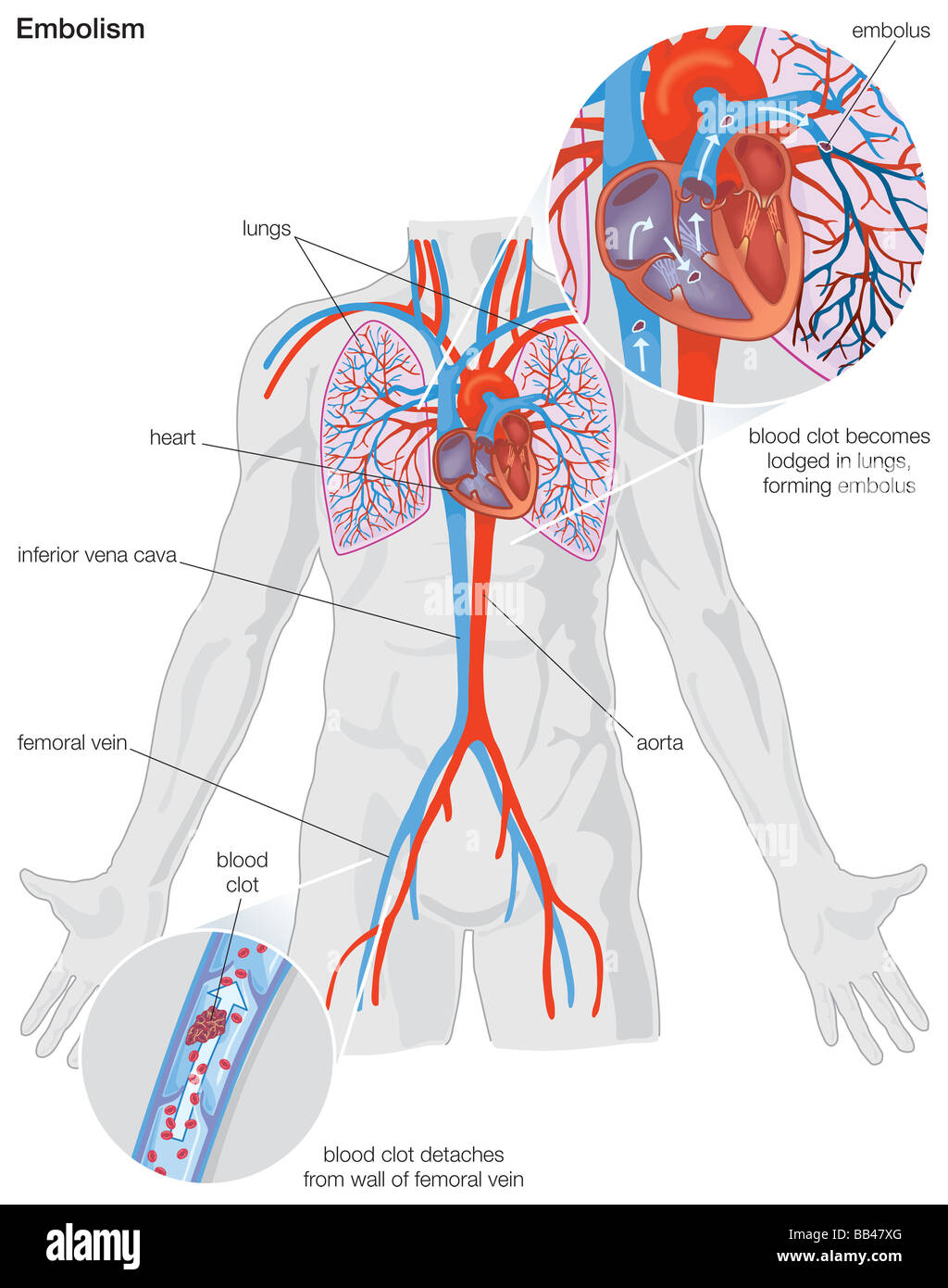 Diagram demonstrating the progression of an arterial embolism Stock Photohttps://www.alamy.com/image-license-details/?v=1https://www.alamy.com/stock-photo-diagram-demonstrating-the-progression-of-an-arterial-embolism-24065624.html
RMBB47XG–Diagram demonstrating the progression of an arterial embolism
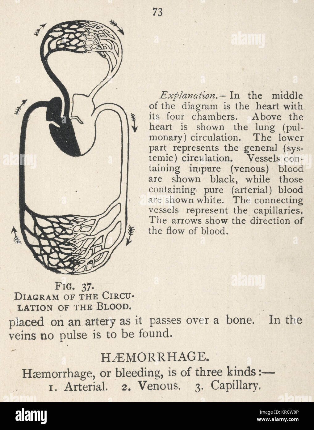 Diagram showing the circulation of the blood Stock Photohttps://www.alamy.com/image-license-details/?v=1https://www.alamy.com/stock-image-diagram-showing-the-circulation-of-the-blood-169313670.html
RMKRCW8P–Diagram showing the circulation of the blood
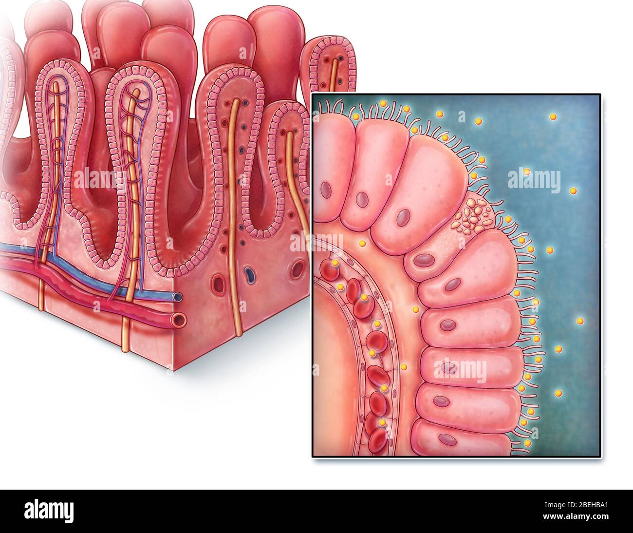 Intestinal Villi, illustration Stock Photohttps://www.alamy.com/image-license-details/?v=1https://www.alamy.com/intestinal-villi-illustration-image353194633.html
RM2BEHBA1–Intestinal Villi, illustration
 Alveolar Carcinoma lung cancer as pulmonary alveoli or alveolus anatomy diagram as a medical concept of a close up of the human anatomy and repiratory Stock Photohttps://www.alamy.com/image-license-details/?v=1https://www.alamy.com/alveolar-carcinoma-lung-cancer-as-pulmonary-alveoli-or-alveolus-anatomy-diagram-as-a-medical-concept-of-a-close-up-of-the-human-anatomy-and-repiratory-image621772126.html
RF2Y3G4KA–Alveolar Carcinoma lung cancer as pulmonary alveoli or alveolus anatomy diagram as a medical concept of a close up of the human anatomy and repiratory
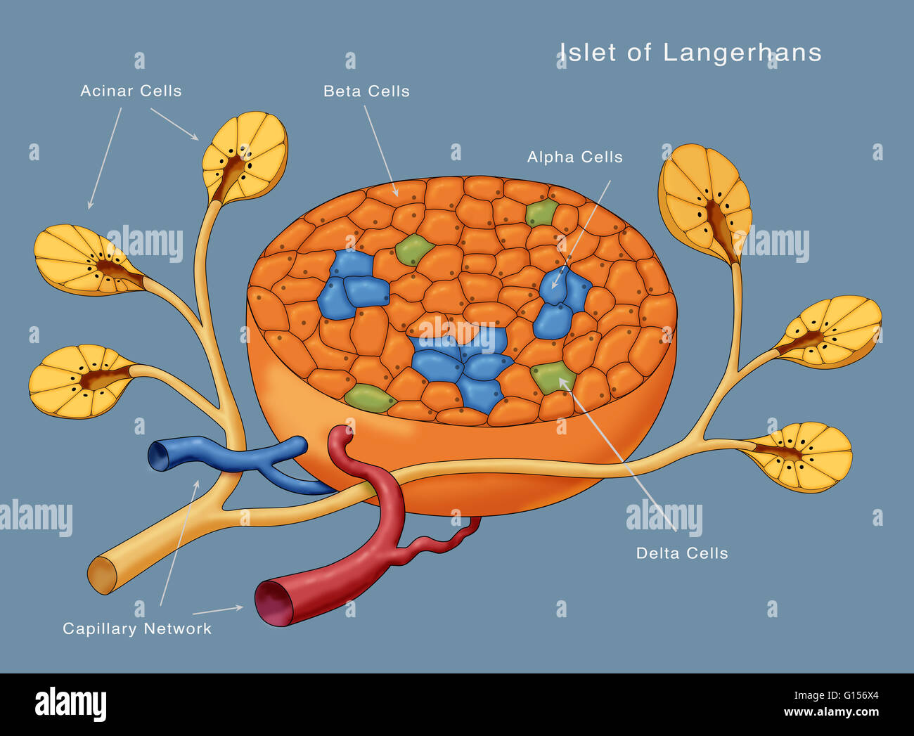 Diagram of the Islets of Langerhans. Shown are the acinar cells, beta cells, alpha cells, delta cells and the capillary network. Stock Photohttps://www.alamy.com/image-license-details/?v=1https://www.alamy.com/stock-photo-diagram-of-the-islets-of-langerhans-shown-are-the-acinar-cells-beta-103992060.html
RMG156X4–Diagram of the Islets of Langerhans. Shown are the acinar cells, beta cells, alpha cells, delta cells and the capillary network.
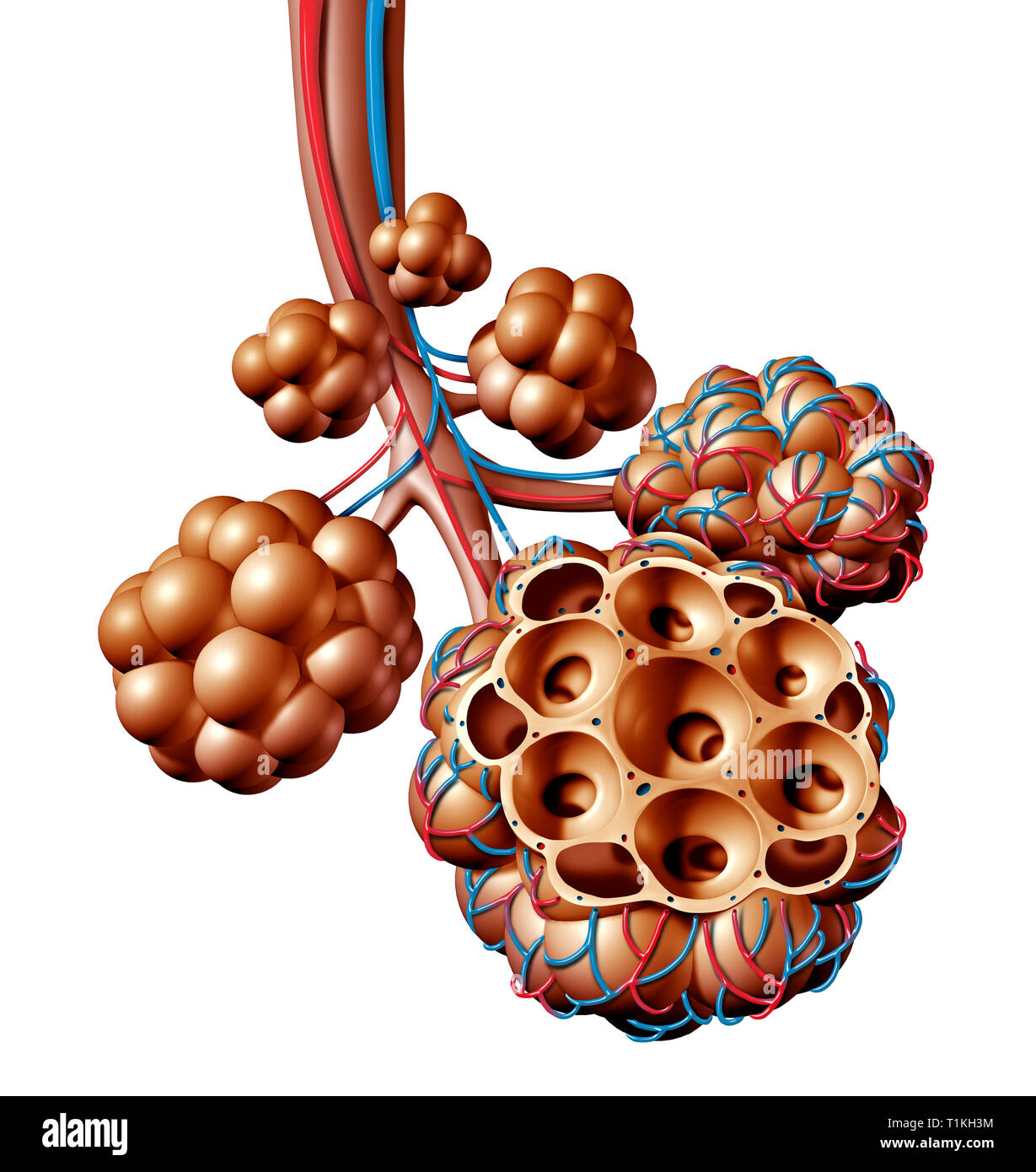 Pulmonary alveoli or alveolus anatomy diagram as a medical concept of a lung anatomy and respiratory and respiration medicine. Stock Photohttps://www.alamy.com/image-license-details/?v=1https://www.alamy.com/pulmonary-alveoli-or-alveolus-anatomy-diagram-as-a-medical-concept-of-a-lung-anatomy-and-respiratory-and-respiration-medicine-image241990328.html
RFT1KH3M–Pulmonary alveoli or alveolus anatomy diagram as a medical concept of a lung anatomy and respiratory and respiration medicine.
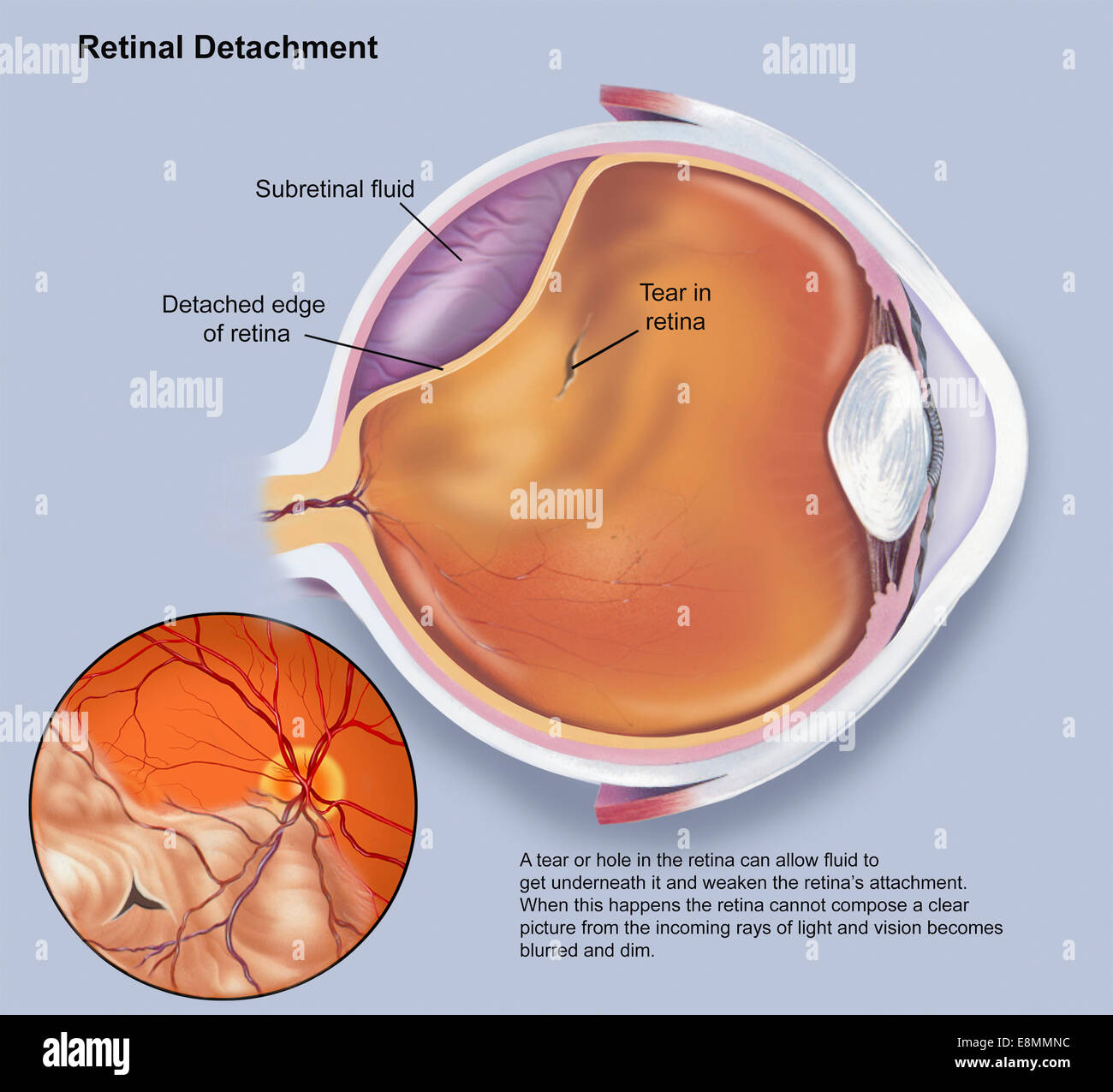 Diagram of a retinal detachment. Stock Photohttps://www.alamy.com/image-license-details/?v=1https://www.alamy.com/stock-photo-diagram-of-a-retinal-detachment-74214040.html
RME8MMNC–Diagram of a retinal detachment.
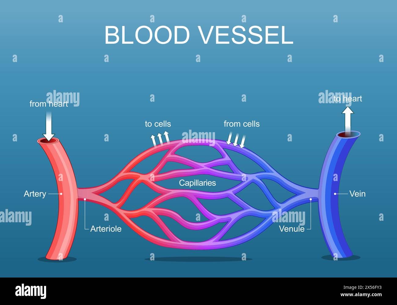 Blood vessels network structure. Arteria is a vessel that carry blood from a heart. Vein is collect blood from organs to the heart. Capillaries conne Stock Vectorhttps://www.alamy.com/image-license-details/?v=1https://www.alamy.com/blood-vessels-network-structure-arteria-is-a-vessel-that-carry-blood-from-a-heart-vein-is-collect-blood-from-organs-to-the-heart-capillaries-conne-image605580391.html
RF2X56FY3–Blood vessels network structure. Arteria is a vessel that carry blood from a heart. Vein is collect blood from organs to the heart. Capillaries conne
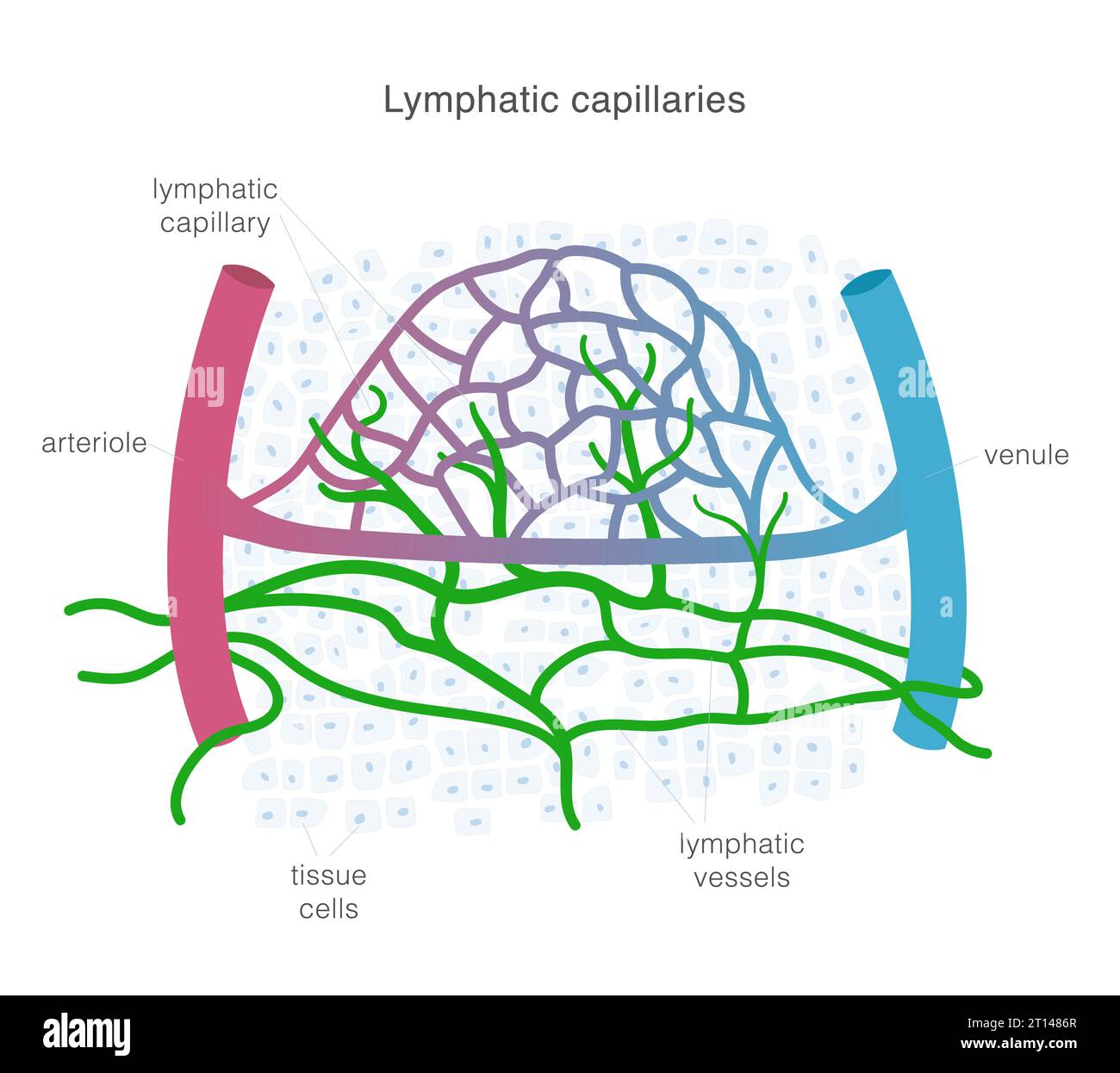 Lymphatic system of capillaries and vessels in complex with blood vessels. Lymph circulation scientific illustration. Stock Vectorhttps://www.alamy.com/image-license-details/?v=1https://www.alamy.com/lymphatic-system-of-capillaries-and-vessels-in-complex-with-blood-vessels-lymph-circulation-scientific-illustration-image568651071.html
RF2T1486R–Lymphatic system of capillaries and vessels in complex with blood vessels. Lymph circulation scientific illustration.
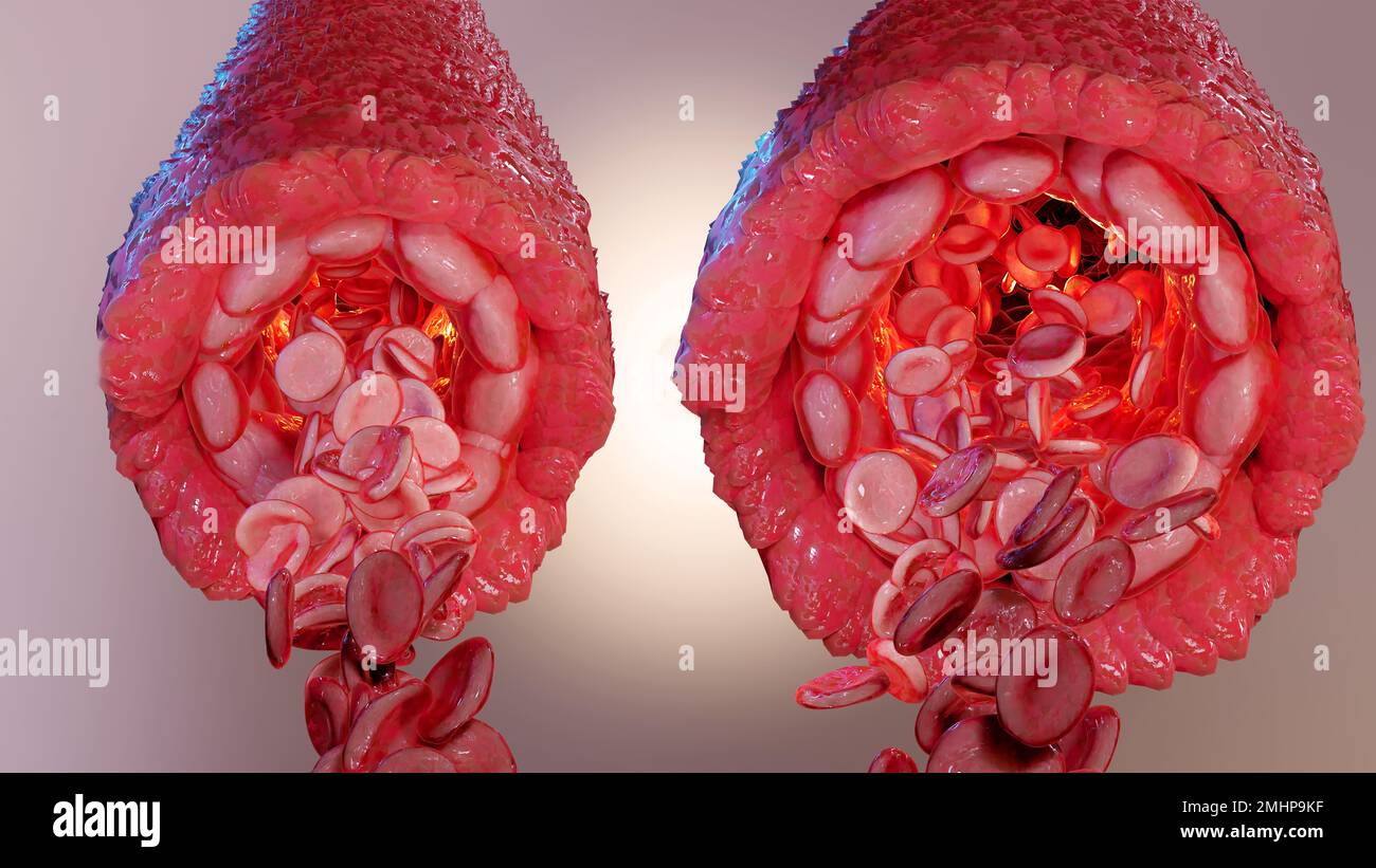 vasoconstriction and vasodilation blood pressure, dilated blood vessels or capillaries or artery, Increase and decrease in blood flow through the blo Stock Photohttps://www.alamy.com/image-license-details/?v=1https://www.alamy.com/vasoconstriction-and-vasodilation-blood-pressure-dilated-blood-vessels-or-capillaries-or-artery-increase-and-decrease-in-blood-flow-through-the-blo-image510040371.html
RM2MHP9KF–vasoconstriction and vasodilation blood pressure, dilated blood vessels or capillaries or artery, Increase and decrease in blood flow through the blo
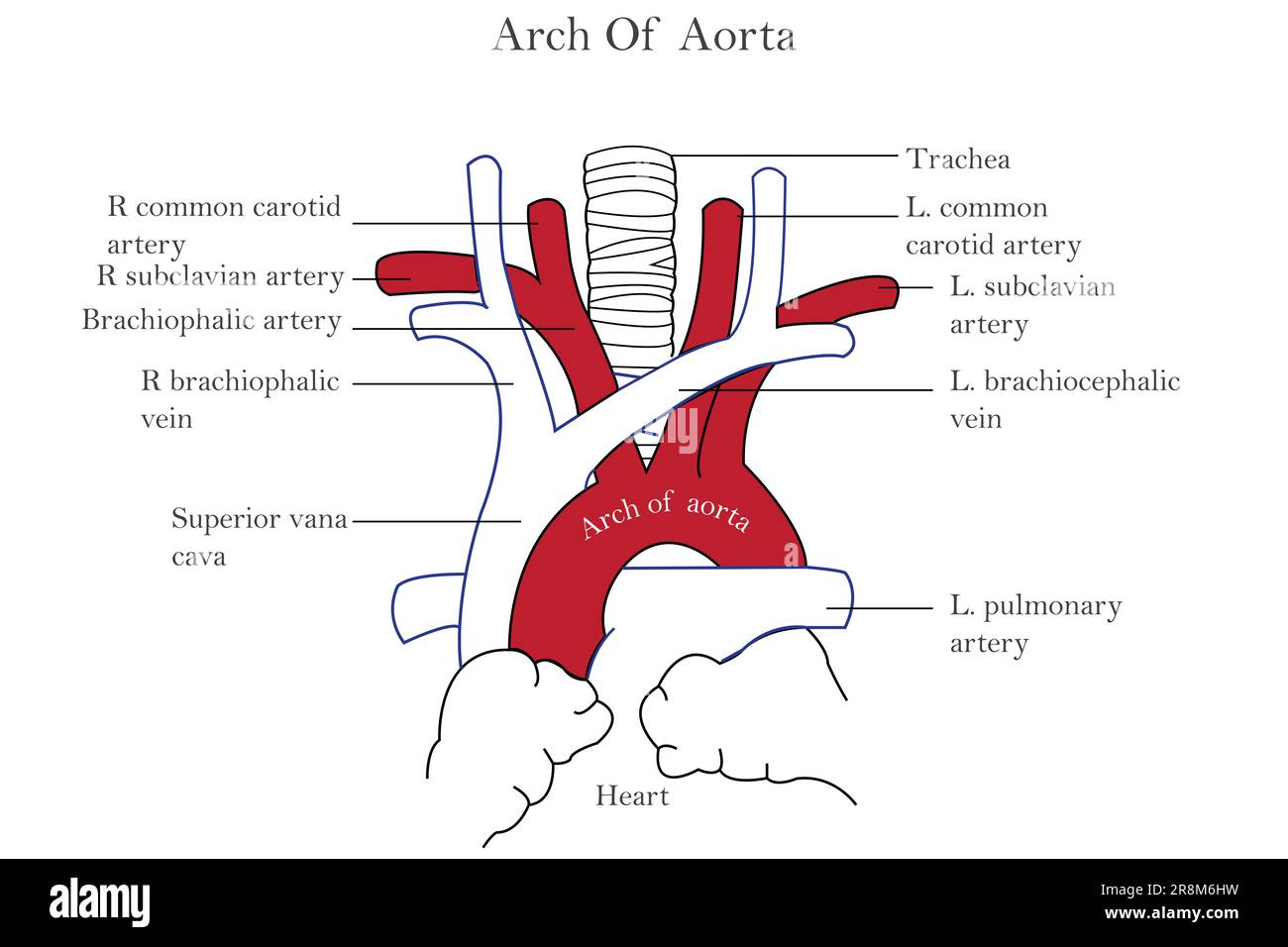 A detailed diagram of the human heart and arch of aorta Stock Photohttps://www.alamy.com/image-license-details/?v=1https://www.alamy.com/a-detailed-diagram-of-the-human-heart-and-arch-of-aorta-image556093269.html
RF2R8M6HW–A detailed diagram of the human heart and arch of aorta
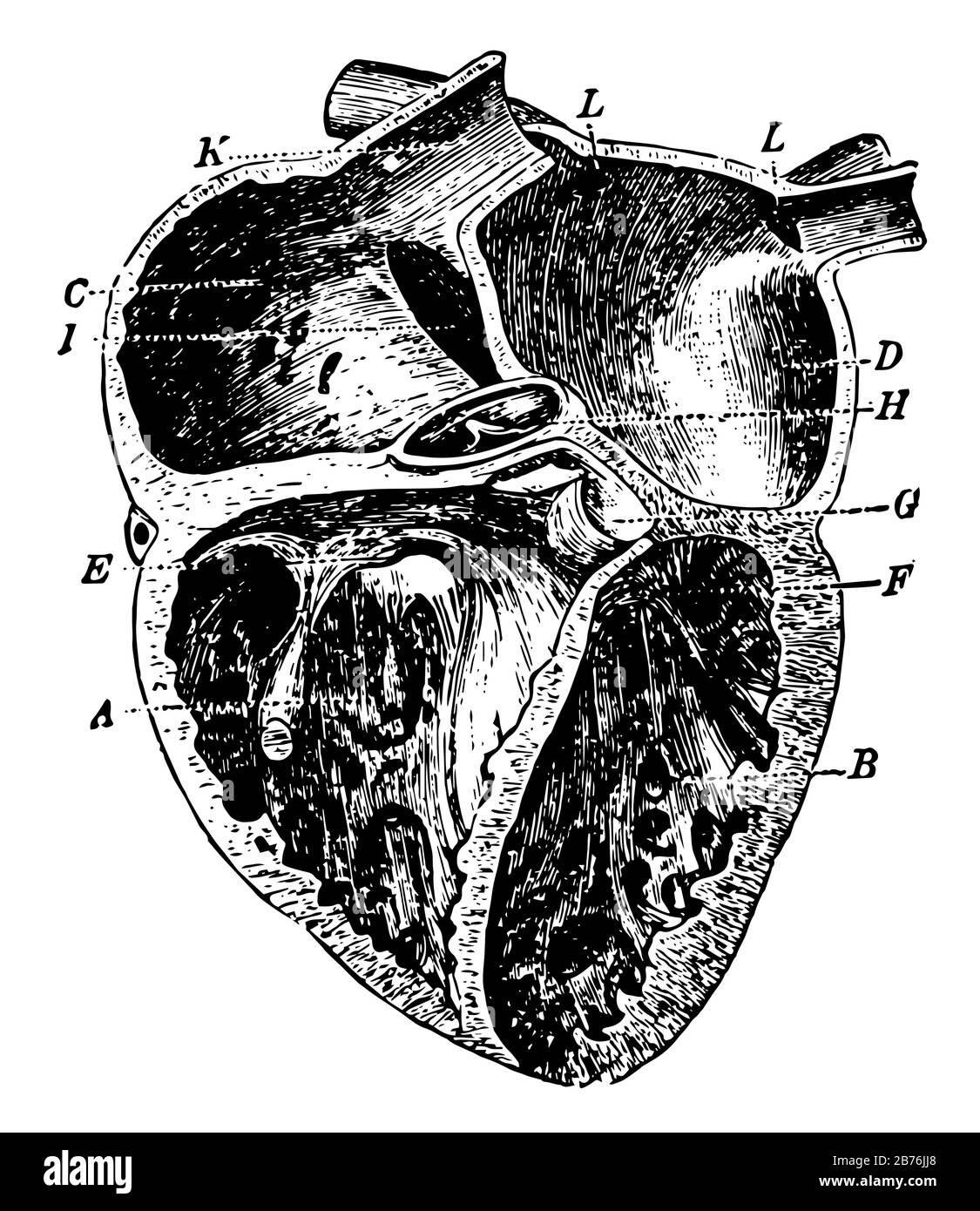 This diagram represents Heart and how the blood circulate through the arteries veins and capillaries, vintage line drawing or engraving illustration Stock Vectorhttps://www.alamy.com/image-license-details/?v=1https://www.alamy.com/this-diagram-represents-heart-and-how-the-blood-circulate-through-the-arteries-veins-and-capillaries-vintage-line-drawing-or-engraving-illustration-image348656288.html
RF2B76JJ8–This diagram represents Heart and how the blood circulate through the arteries veins and capillaries, vintage line drawing or engraving illustration
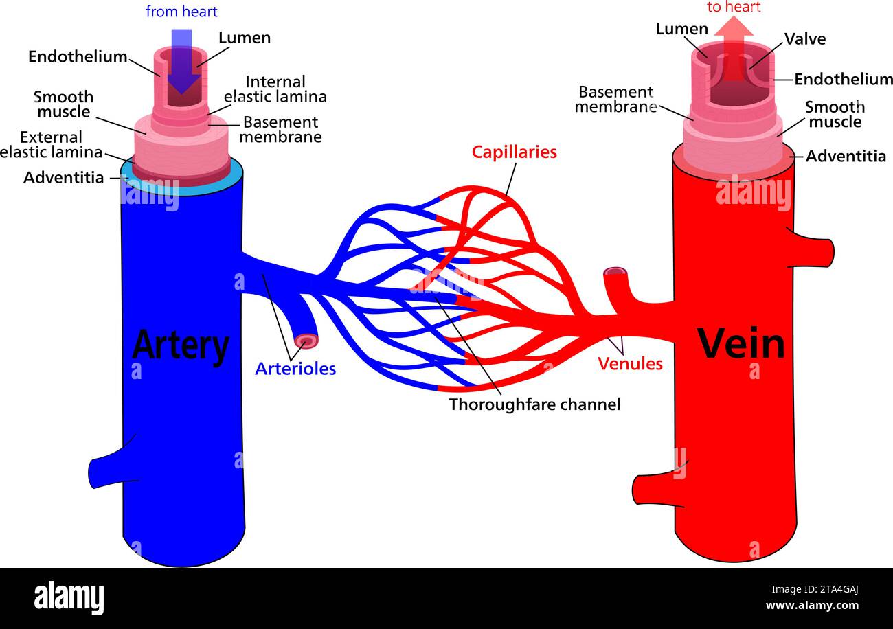 Diagram of blood vessel structures and arteries, veins, and capillaries. Vector illustration. Stock Vectorhttps://www.alamy.com/image-license-details/?v=1https://www.alamy.com/diagram-of-blood-vessel-structures-and-arteries-veins-and-capillaries-vector-illustration-image574189354.html
RF2TA4GAJ–Diagram of blood vessel structures and arteries, veins, and capillaries. Vector illustration.
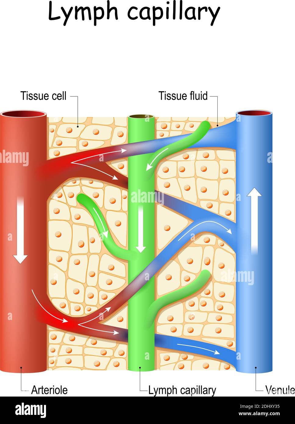 Lymph capillary in human tissue. Blood vessel: Venule and Arteriole. lymphatic system. Fluid bathes the tissues and collecting waste products Stock Vectorhttps://www.alamy.com/image-license-details/?v=1https://www.alamy.com/lymph-capillary-in-human-tissue-blood-vessel-venule-and-arteriole-lymphatic-system-fluid-bathes-the-tissues-and-collecting-waste-products-image389669257.html
RF2DHXY35–Lymph capillary in human tissue. Blood vessel: Venule and Arteriole. lymphatic system. Fluid bathes the tissues and collecting waste products
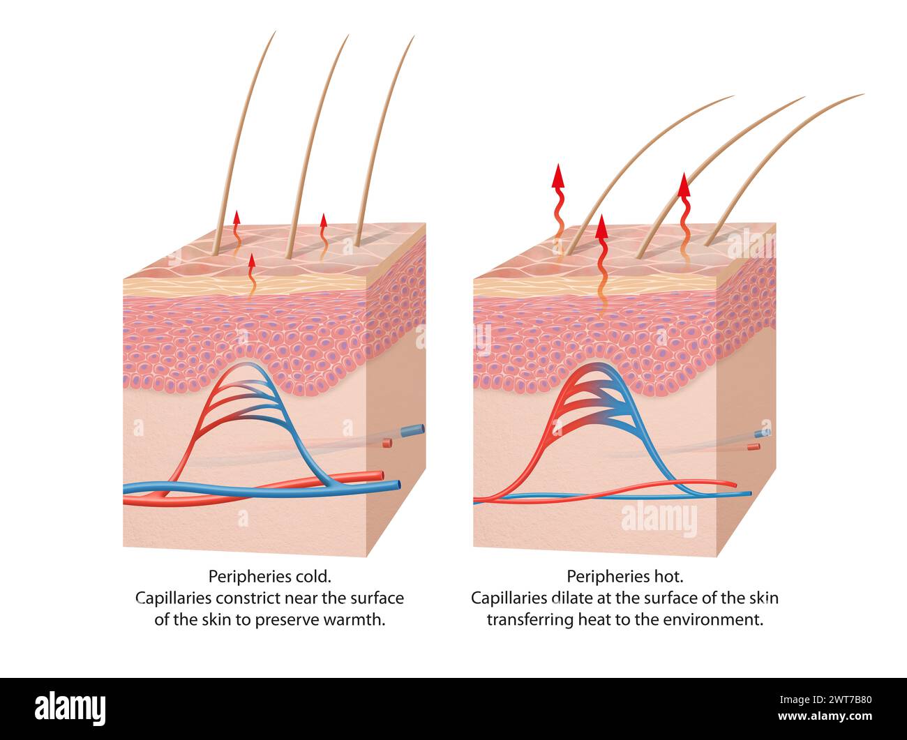 Thermoregulation of the human body Stock Photohttps://www.alamy.com/image-license-details/?v=1https://www.alamy.com/thermoregulation-of-the-human-body-image600066768.html
RF2WT7B80–Thermoregulation of the human body
 Alveoli Supporting Breathing Inside Human Lung Structure Stock Photohttps://www.alamy.com/image-license-details/?v=1https://www.alamy.com/alveoli-supporting-breathing-inside-human-lung-structure-image628160635.html
RF2YDY58B–Alveoli Supporting Breathing Inside Human Lung Structure
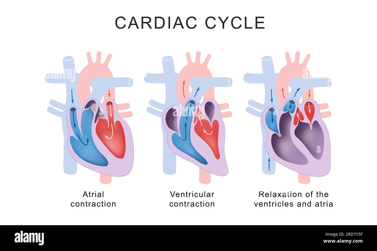 Cardiac Cycle Phases: Systole and Diastole Stock Photohttps://www.alamy.com/image-license-details/?v=1https://www.alamy.com/cardiac-cycle-phases-systole-and-diastole-image558897291.html
RF2RD7Y5F–Cardiac Cycle Phases: Systole and Diastole
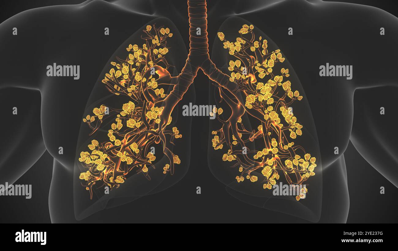 Breathing Process Occurs in Alveoli of Lungs Stock Photohttps://www.alamy.com/image-license-details/?v=1https://www.alamy.com/breathing-process-occurs-in-alveoli-of-lungs-image628224900.html
RF2YE237G–Breathing Process Occurs in Alveoli of Lungs
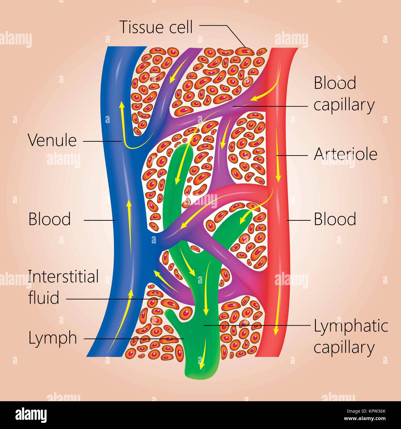 The lymph system, relationship of lymphatic capillaries to tissue cells and blood capillaries, vector medical illustration Stock Vectorhttps://www.alamy.com/image-license-details/?v=1https://www.alamy.com/stock-image-the-lymph-system-relationship-of-lymphatic-capillaries-to-tissue-cells-168967083.html
RFKPW36K–The lymph system, relationship of lymphatic capillaries to tissue cells and blood capillaries, vector medical illustration
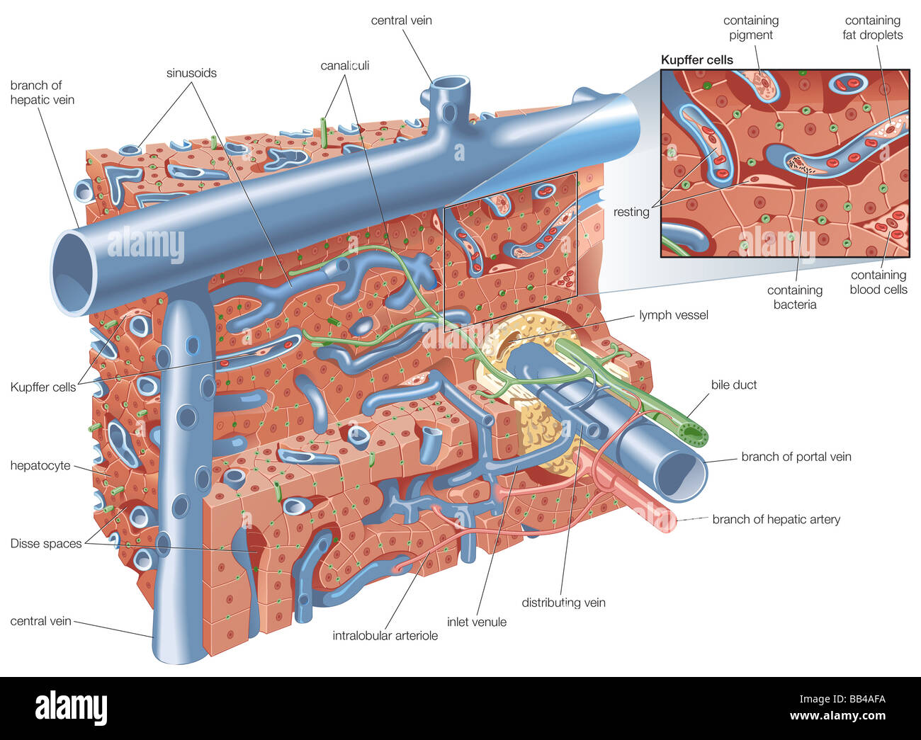 Liver structure schema Stock Photohttps://www.alamy.com/image-license-details/?v=1https://www.alamy.com/stock-photo-liver-structure-schema-24067662.html
RMBB4AFA–Liver structure schema
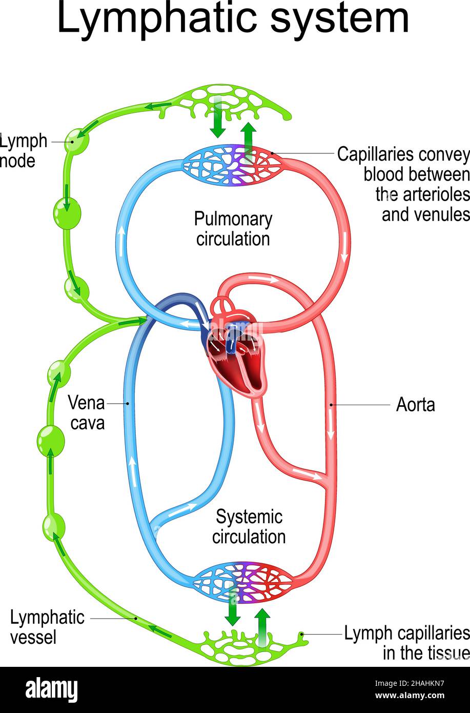 Lymphatic circulation system. parts of immune and Circulatory system. lymph node, blood vessel, Capillaries and heart. vector illustration. human anat Stock Vectorhttps://www.alamy.com/image-license-details/?v=1https://www.alamy.com/lymphatic-circulation-system-parts-of-immune-and-circulatory-system-lymph-node-blood-vessel-capillaries-and-heart-vector-illustration-human-anat-image454004803.html
RF2HAHKN7–Lymphatic circulation system. parts of immune and Circulatory system. lymph node, blood vessel, Capillaries and heart. vector illustration. human anat
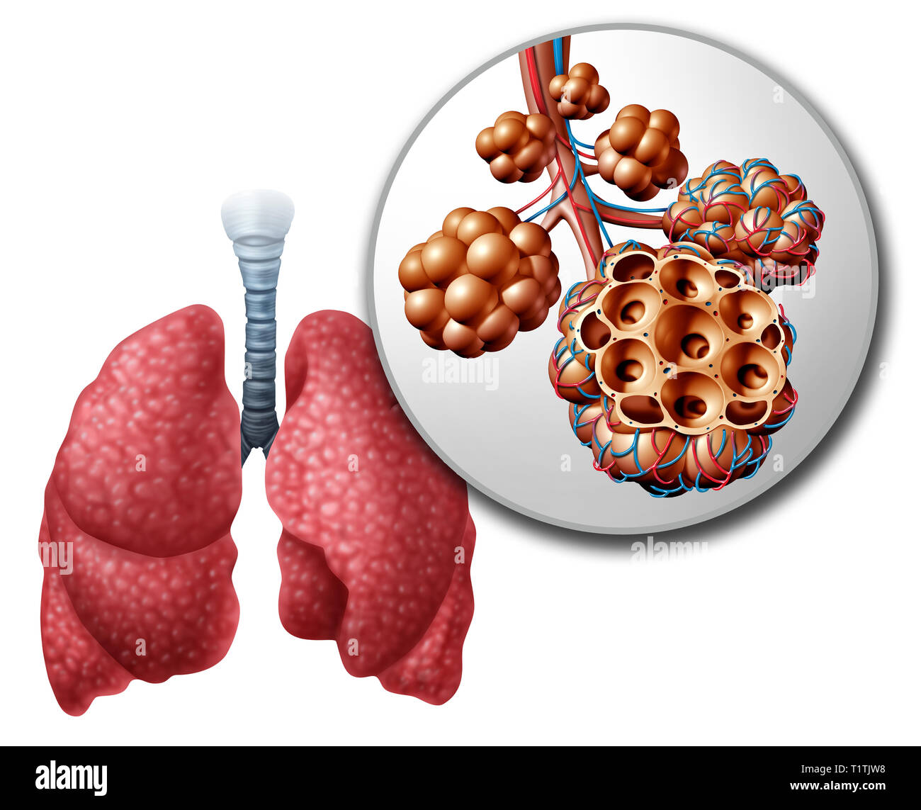 Lung pulmonary alveoli or alveolus anatomy diagram as a medical concept of a close up of the human anatomy and respiratory or respiration medicine. Stock Photohttps://www.alamy.com/image-license-details/?v=1https://www.alamy.com/lung-pulmonary-alveoli-or-alveolus-anatomy-diagram-as-a-medical-concept-of-a-close-up-of-the-human-anatomy-and-respiratory-or-respiration-medicine-image242101476.html
RFT1TJW8–Lung pulmonary alveoli or alveolus anatomy diagram as a medical concept of a close up of the human anatomy and respiratory or respiration medicine.
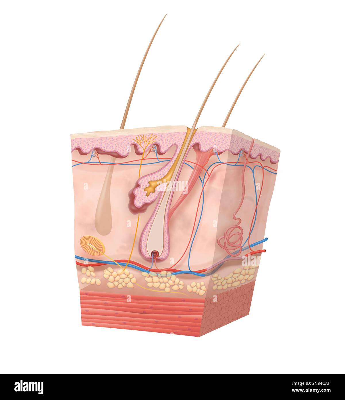 Diagram of human skin structure Stock Photohttps://www.alamy.com/image-license-details/?v=1https://www.alamy.com/diagram-of-human-skin-structure-image521328937.html
RF2N84GAH–Diagram of human skin structure
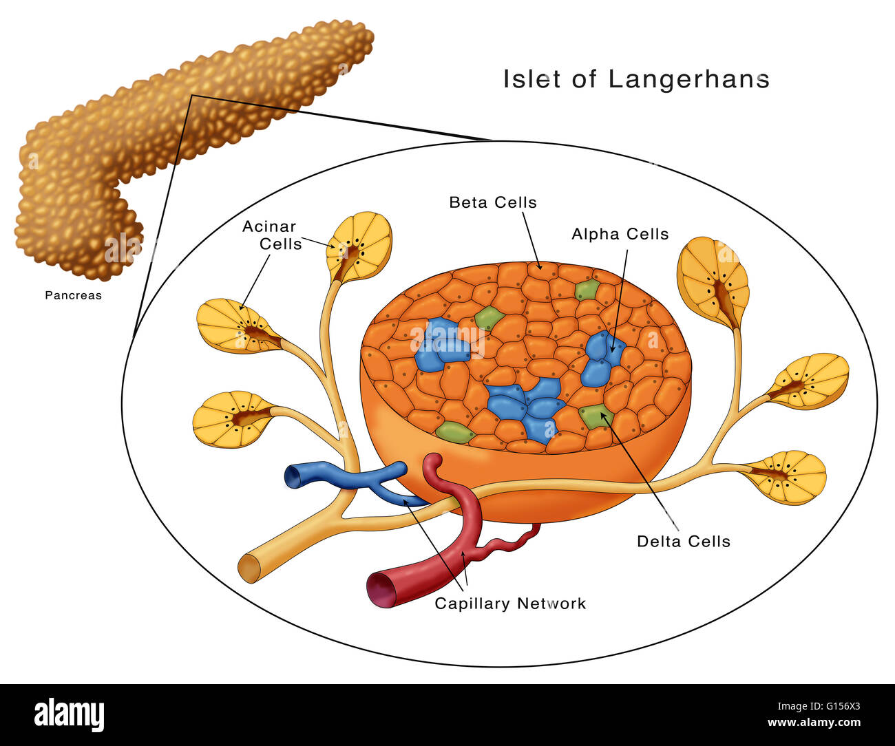 Diagram of the Islets of Langerhans. Shown are the acinar cells, beta cells, alpha cells, delta cells and the capillary network. Stock Photohttps://www.alamy.com/image-license-details/?v=1https://www.alamy.com/stock-photo-diagram-of-the-islets-of-langerhans-shown-are-the-acinar-cells-beta-103992059.html
RMG156X3–Diagram of the Islets of Langerhans. Shown are the acinar cells, beta cells, alpha cells, delta cells and the capillary network.
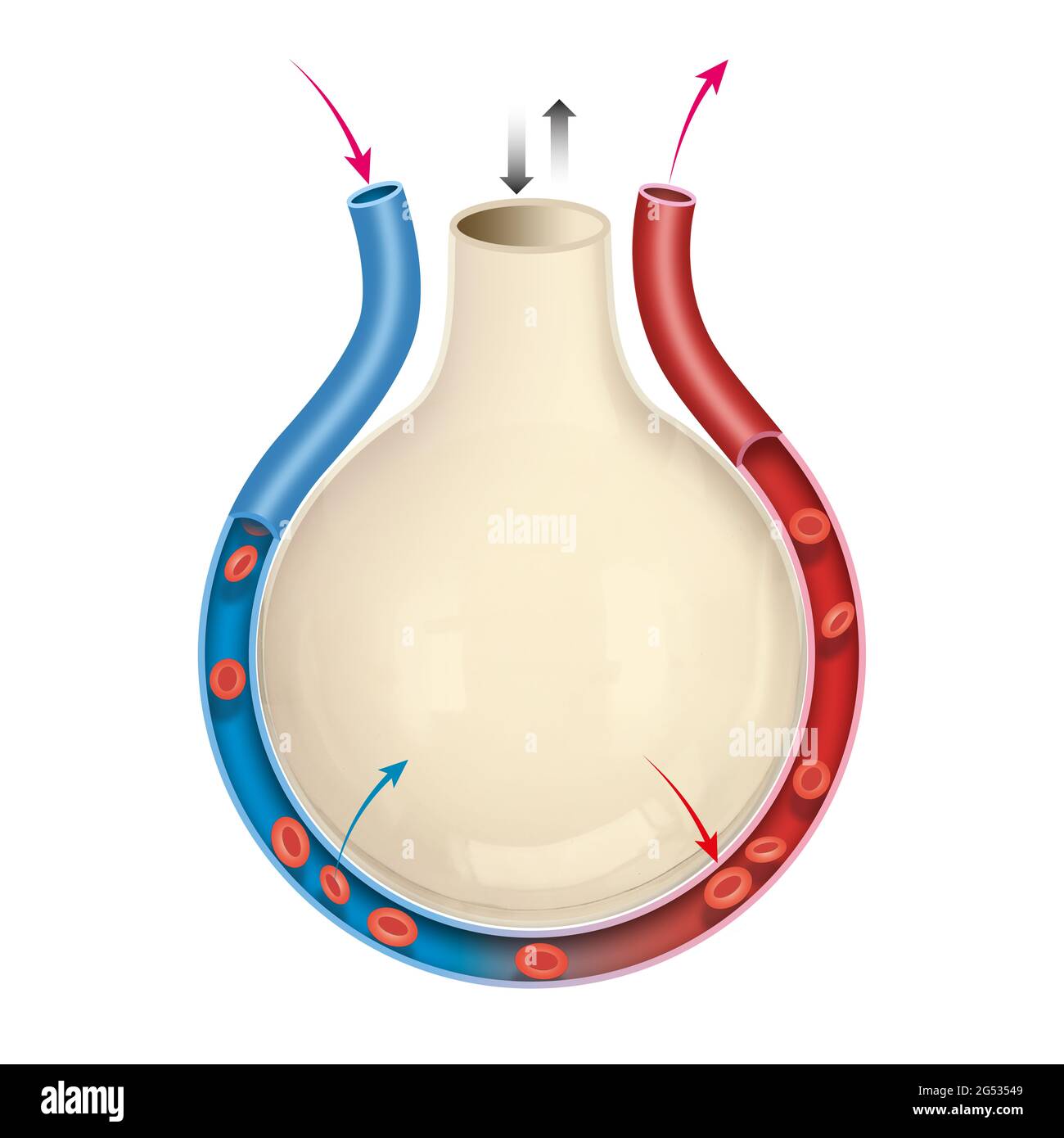 Alveolus gas exchange. Diagram of the alveolus in the lungs showing gaseous exchange Stock Photohttps://www.alamy.com/image-license-details/?v=1https://www.alamy.com/alveolus-gas-exchange-diagram-of-the-alveolus-in-the-lungs-showing-gaseous-exchange-image433402377.html
RF2G53549–Alveolus gas exchange. Diagram of the alveolus in the lungs showing gaseous exchange
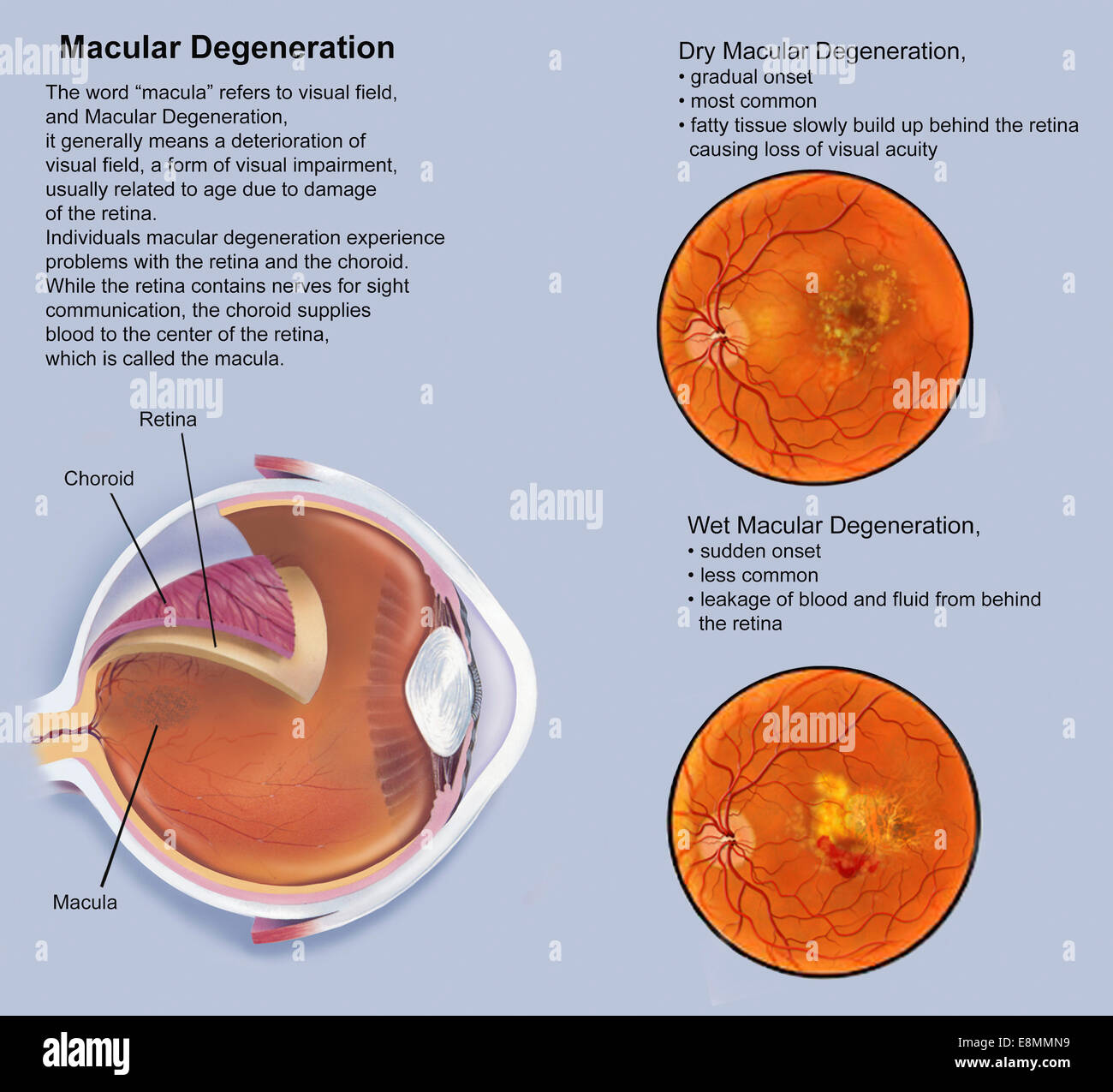 Retina with macular degeneration. Stock Photohttps://www.alamy.com/image-license-details/?v=1https://www.alamy.com/stock-photo-retina-with-macular-degeneration-74214037.html
RME8MMN9–Retina with macular degeneration.
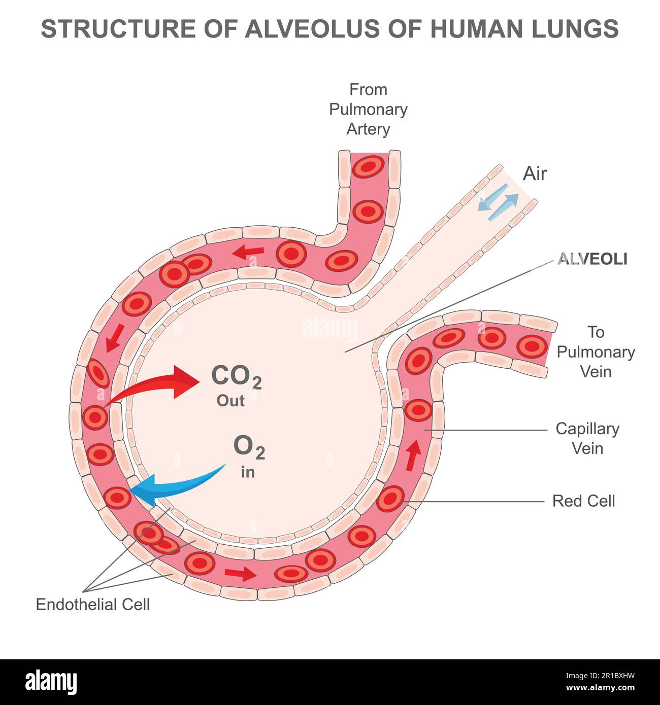 Structure of alveolus of human lungs. Labelled diagram of the alveolus in the lungs showing gaseous exchange. Pulmonary alveolus. alveoli and capillar Stock Vectorhttps://www.alamy.com/image-license-details/?v=1https://www.alamy.com/structure-of-alveolus-of-human-lungs-labelled-diagram-of-the-alveolus-in-the-lungs-showing-gaseous-exchange-pulmonary-alveolus-alveoli-and-capillar-image551608789.html
RF2R1BXHW–Structure of alveolus of human lungs. Labelled diagram of the alveolus in the lungs showing gaseous exchange. Pulmonary alveolus. alveoli and capillar
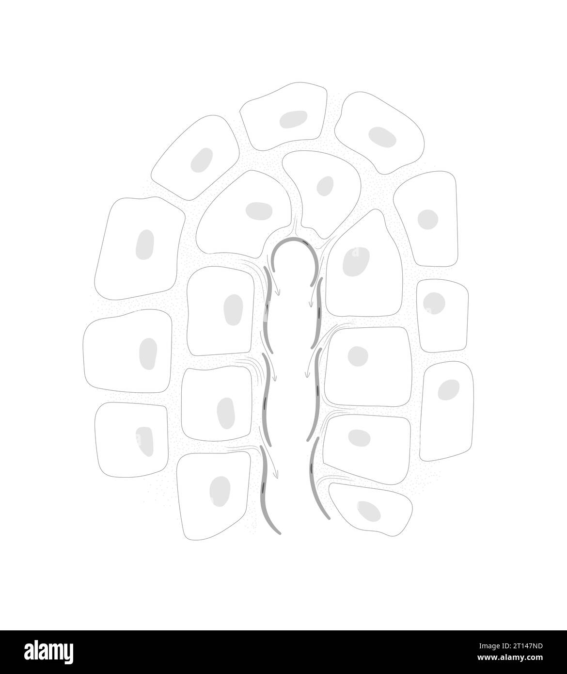 The lymph system line drawing. Structure of the terminal end of lymphatic capillary and surrounding tissue. Stock Vectorhttps://www.alamy.com/image-license-details/?v=1https://www.alamy.com/the-lymph-system-line-drawing-structure-of-the-terminal-end-of-lymphatic-capillary-and-surrounding-tissue-image568650697.html
RF2T147ND–The lymph system line drawing. Structure of the terminal end of lymphatic capillary and surrounding tissue.
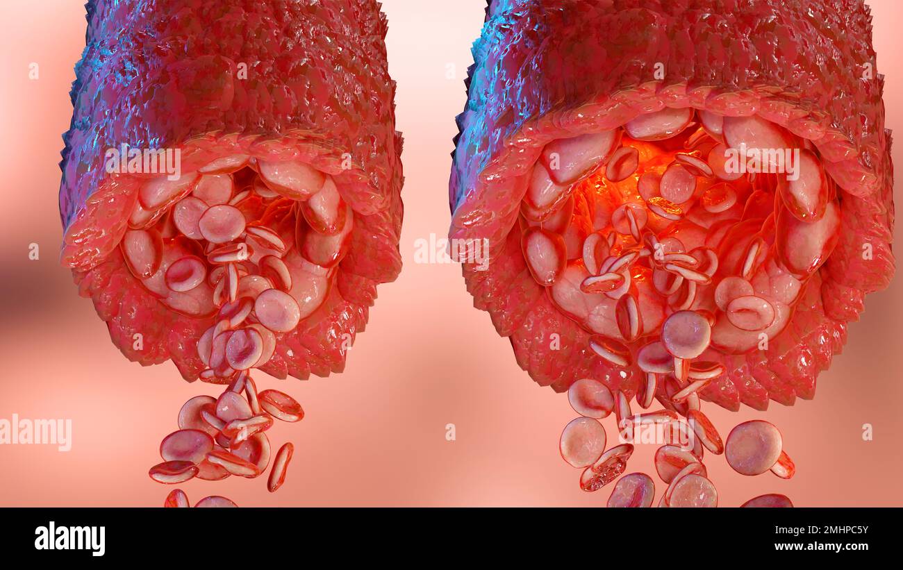 vasoconstriction and vasodilation blood pressure, dilated blood vessels or capillaries or artery, Increase and decrease in blood flow through the blo Stock Photohttps://www.alamy.com/image-license-details/?v=1https://www.alamy.com/vasoconstriction-and-vasodilation-blood-pressure-dilated-blood-vessels-or-capillaries-or-artery-increase-and-decrease-in-blood-flow-through-the-blo-image510042343.html
RM2MHPC5Y–vasoconstriction and vasodilation blood pressure, dilated blood vessels or capillaries or artery, Increase and decrease in blood flow through the blo
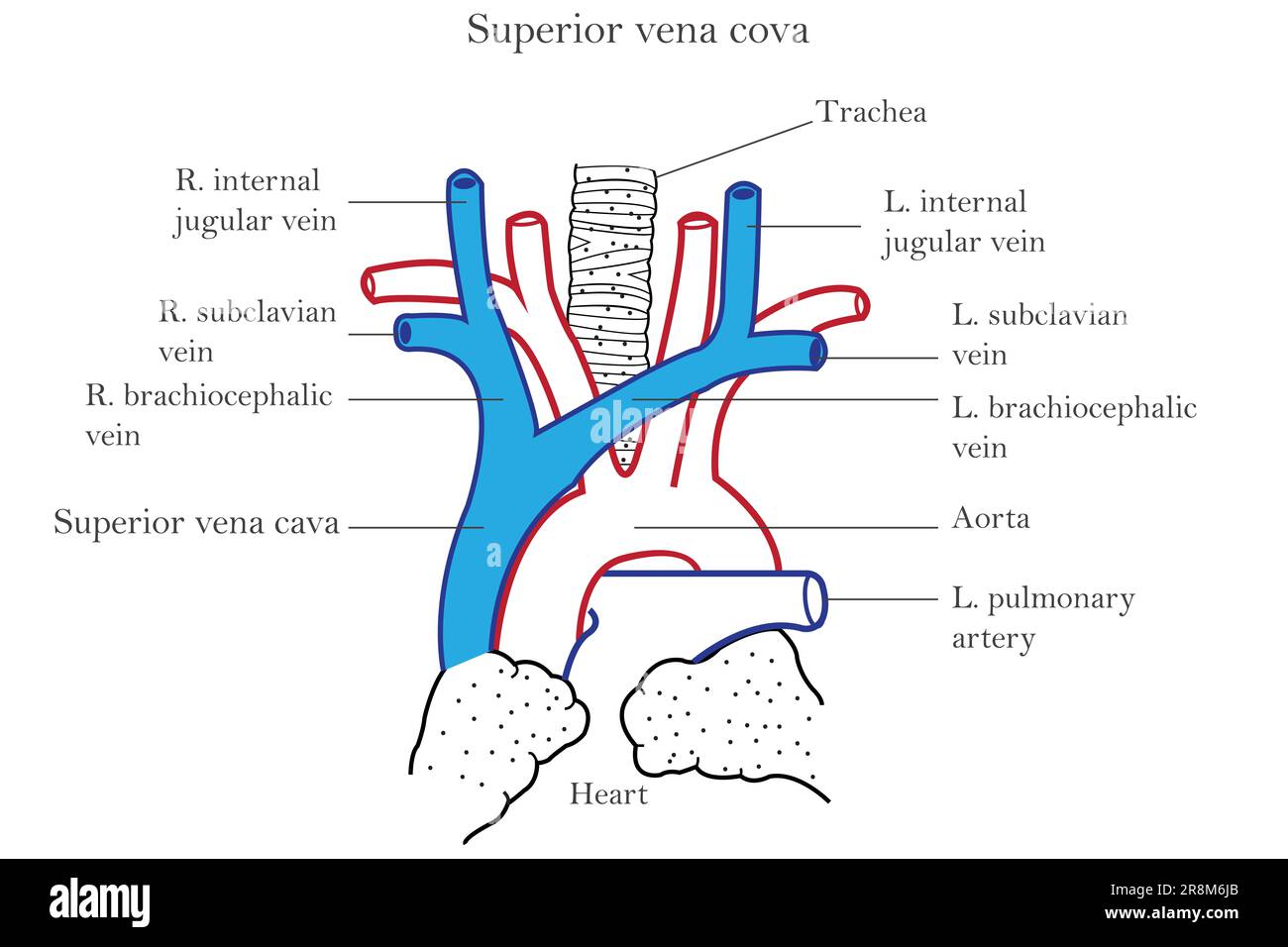 A detailed diagram of the human heart and superior vena cova Stock Photohttps://www.alamy.com/image-license-details/?v=1https://www.alamy.com/a-detailed-diagram-of-the-human-heart-and-superior-vena-cova-image556093283.html
RF2R8M6JB–A detailed diagram of the human heart and superior vena cova
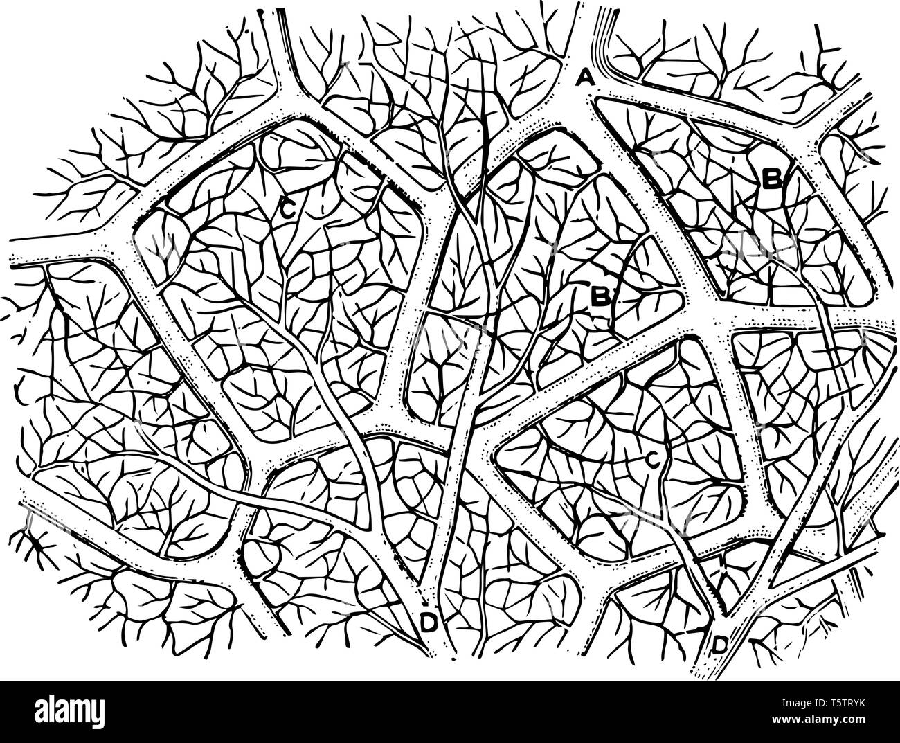 This diagram represents Capillaries of the Air Sac and showing the capillary network of the air sacs vintage line drawing or engraving illustration. Stock Vectorhttps://www.alamy.com/image-license-details/?v=1https://www.alamy.com/this-diagram-represents-capillaries-of-the-air-sac-and-showing-the-capillary-network-of-the-air-sacs-vintage-line-drawing-or-engraving-illustration-image244564087.html
RFT5TRYK–This diagram represents Capillaries of the Air Sac and showing the capillary network of the air sacs vintage line drawing or engraving illustration.
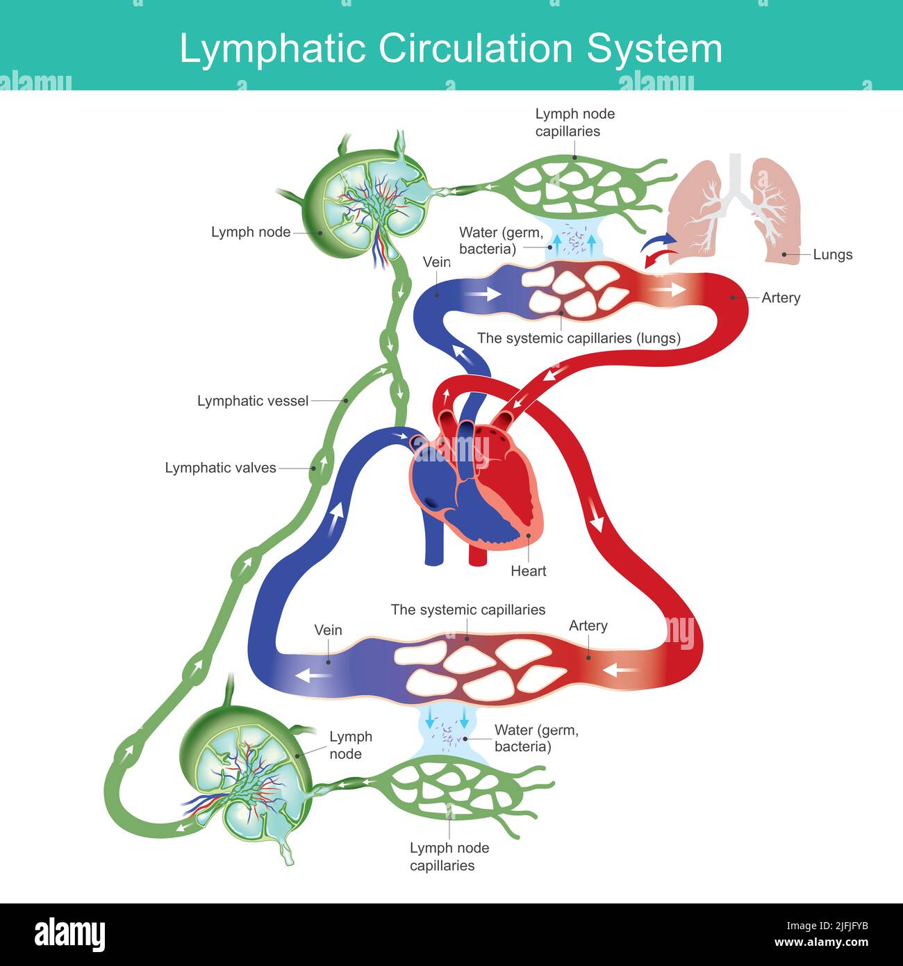 Lymphatic circulation system. diagram the lymphatic circulation system for medical education. Illustration. Stock Vectorhttps://www.alamy.com/image-license-details/?v=1https://www.alamy.com/lymphatic-circulation-system-diagram-the-lymphatic-circulation-system-for-medical-education-illustration-image474307439.html
RF2JFJFYB–Lymphatic circulation system. diagram the lymphatic circulation system for medical education. Illustration.
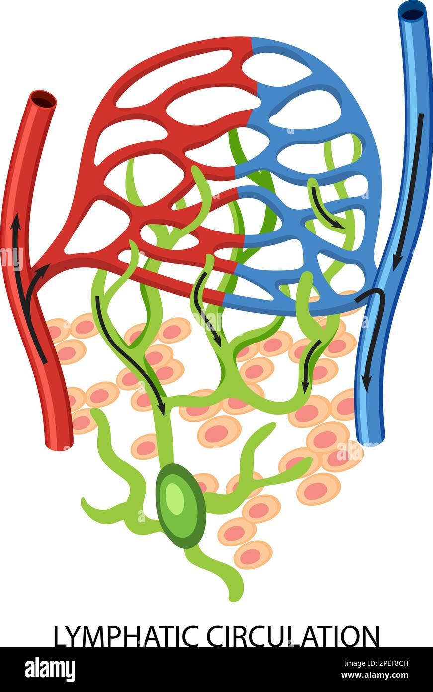 Lymphatic Circulation System Diagram illustration Stock Vectorhttps://www.alamy.com/image-license-details/?v=1https://www.alamy.com/lymphatic-circulation-system-diagram-illustration-image542462497.html
RF2PEF8CH–Lymphatic Circulation System Diagram illustration
 Quain's Elements of Anatomy Col. III published in 1896, circulation. Stock Photohttps://www.alamy.com/image-license-details/?v=1https://www.alamy.com/quains-elements-of-anatomy-col-iii-published-in-1896-circulation-image369926341.html
RF2CDRGR1–Quain's Elements of Anatomy Col. III published in 1896, circulation.
 Diagram of Circulation with parts like liver, Alim, arterial system and many other, labelled, vintage line drawing or engraving illustration. Stock Vectorhttps://www.alamy.com/image-license-details/?v=1https://www.alamy.com/diagram-of-circulation-with-parts-like-liver-alim-arterial-system-and-many-other-labelled-vintage-line-drawing-or-engraving-illustration-image359322182.html
RF2BTGF32–Diagram of Circulation with parts like liver, Alim, arterial system and many other, labelled, vintage line drawing or engraving illustration.
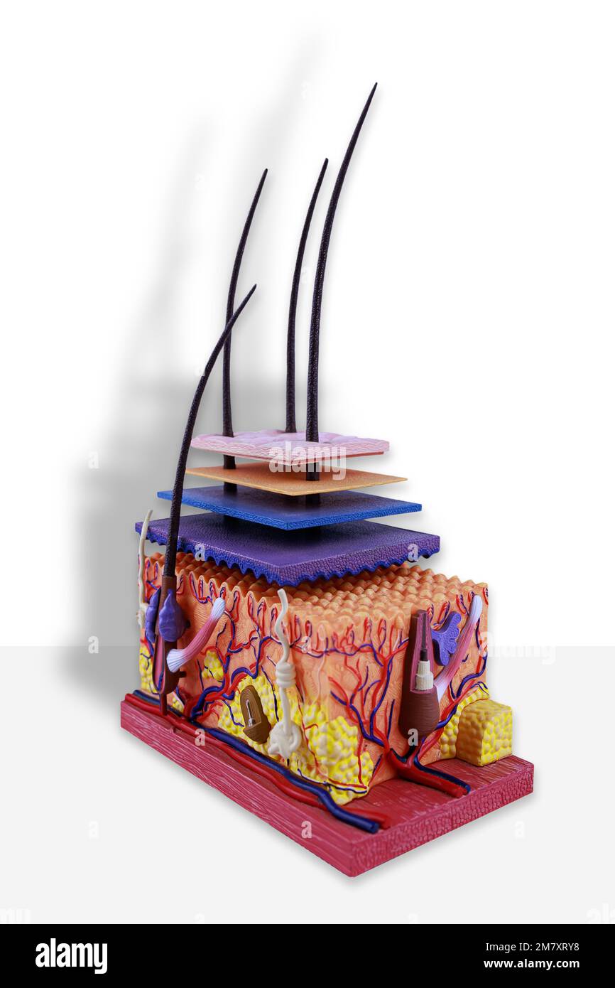 sectional skin model with capillaries and derma layers on white background Stock Photohttps://www.alamy.com/image-license-details/?v=1https://www.alamy.com/sectional-skin-model-with-capillaries-and-derma-layers-on-white-background-image503992812.html
RF2M7XRY8–sectional skin model with capillaries and derma layers on white background
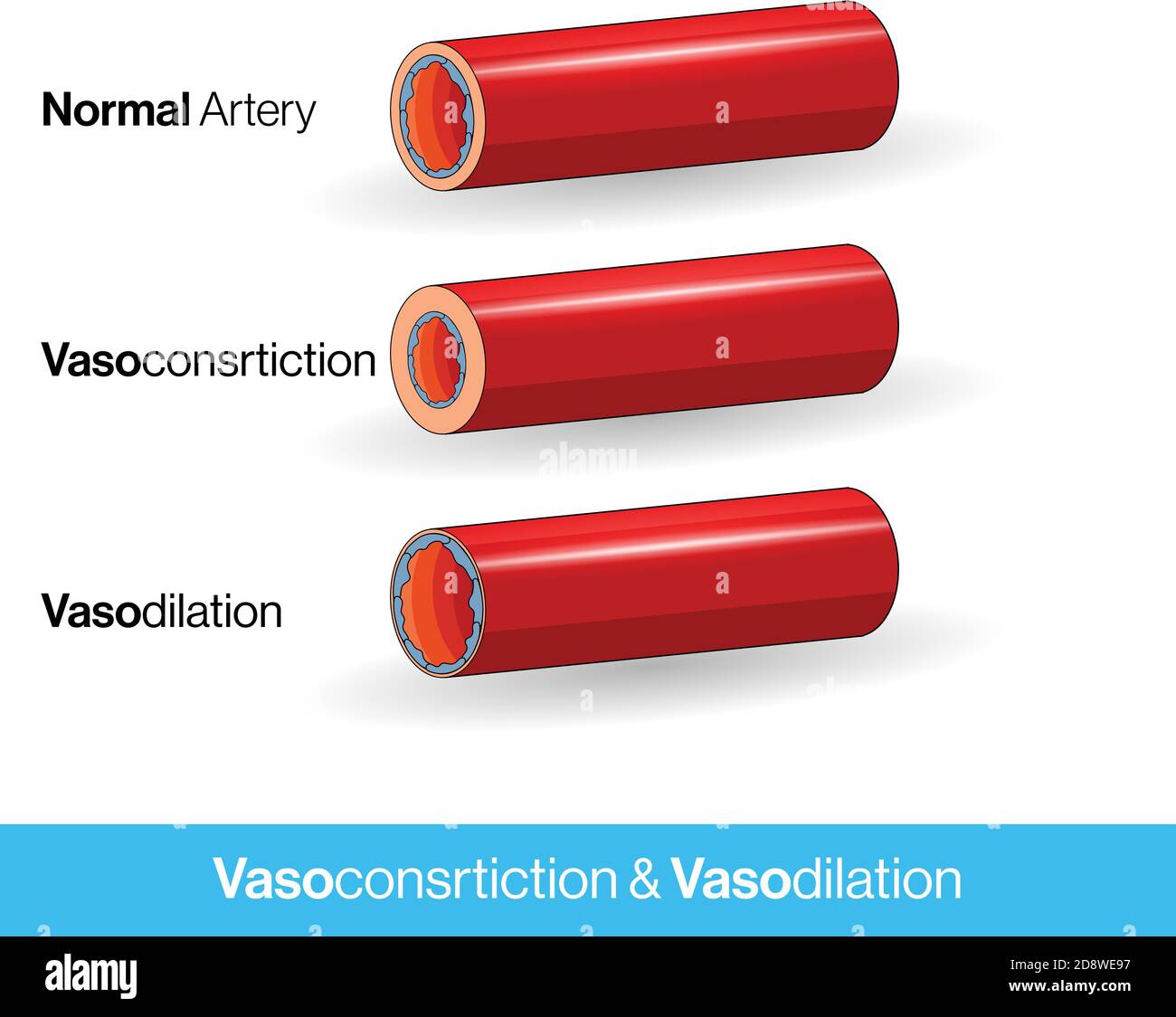 Blood vessels or capillaries or artery showing vasoconstriction and vasodilation blocking the blood flow cross-section and side Stock Vectorhttps://www.alamy.com/image-license-details/?v=1https://www.alamy.com/blood-vessels-or-capillaries-or-artery-showing-vasoconstriction-and-vasodilation-blocking-the-blood-flow-cross-section-and-side-image384105379.html
RF2D8WE97–Blood vessels or capillaries or artery showing vasoconstriction and vasodilation blocking the blood flow cross-section and side
 Interstitial fluid is collected by lymph capillaries from the interstitial space. Lymph then moves through lymphatic vessels to lymph nodes. 3D render Stock Photohttps://www.alamy.com/image-license-details/?v=1https://www.alamy.com/interstitial-fluid-is-collected-by-lymph-capillaries-from-the-interstitial-space-lymph-then-moves-through-lymphatic-vessels-to-lymph-nodes-3d-render-image596596819.html
RF2WJH997–Interstitial fluid is collected by lymph capillaries from the interstitial space. Lymph then moves through lymphatic vessels to lymph nodes. 3D render
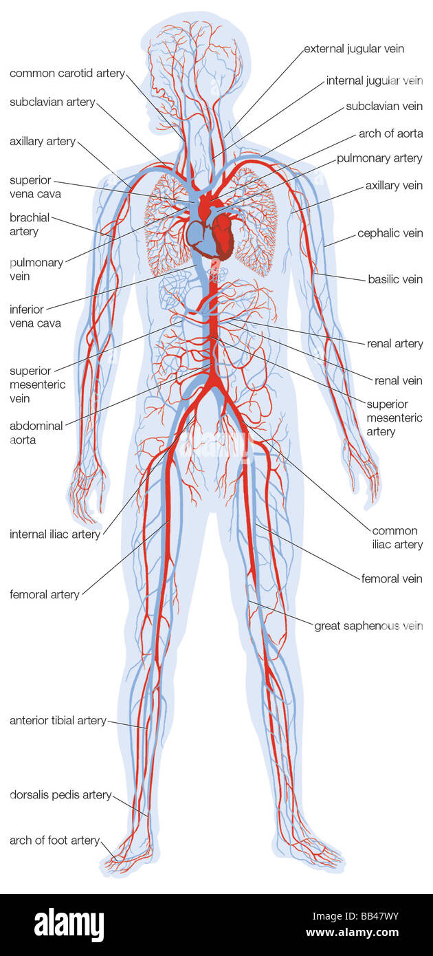 Silhouette of the human body showing the location and extent of the heart and vascular system. Stock Photohttps://www.alamy.com/image-license-details/?v=1https://www.alamy.com/stock-photo-silhouette-of-the-human-body-showing-the-location-and-extent-of-the-24065607.html
RMBB47WY–Silhouette of the human body showing the location and extent of the heart and vascular system.
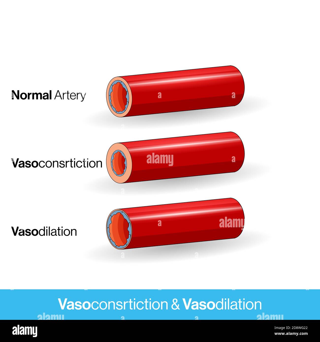 Blood vessels or capillaries or artery showing vasoconstriction and vasodilation blocking the blood flow cross section and side view concept illustrat Stock Photohttps://www.alamy.com/image-license-details/?v=1https://www.alamy.com/blood-vessels-or-capillaries-or-artery-showing-vasoconstriction-and-vasodilation-blocking-the-blood-flow-cross-section-and-side-view-concept-illustrat-image384106746.html
RF2D8WG22–Blood vessels or capillaries or artery showing vasoconstriction and vasodilation blocking the blood flow cross section and side view concept illustrat
 . Bioplasm : an introduction to the study of physiology & medicine. Protoplasm; Histology. PLATE XXI —MECHANISM GOVERNING THE DISTRIBUTION OF BLOOD IN THE CAPILLARIES.. Diagram to show self-regalating mechanism connected -with the minute arteries and camllaries. a, artery with muscular fibre cells ; the dark lines sho-w its diameter when dilated. 4, small vein, c, capillaiy network. Over No. 1 ihe capillaries are dilated, and over No. 2 they are contracted, d is a ganglion cell with at least tiwo sets of nerve fibres connected with it. one of which, e, divides and subdivides, giving off ne Stock Photohttps://www.alamy.com/image-license-details/?v=1https://www.alamy.com/bioplasm-an-introduction-to-the-study-of-physiology-amp-medicine-protoplasm-histology-plate-xxi-mechanism-governing-the-distribution-of-blood-in-the-capillaries-diagram-to-show-self-regalating-mechanism-connected-with-the-minute-arteries-and-camllaries-a-artery-with-muscular-fibre-cells-the-dark-lines-sho-w-its-diameter-when-dilated-4-small-vein-c-capillaiy-network-over-no-1-ihe-capillaries-are-dilated-and-over-no-2-they-are-contracted-d-is-a-ganglion-cell-with-at-least-tiwo-sets-of-nerve-fibres-connected-with-it-one-of-which-e-divides-and-subdivides-giving-off-ne-image234603585.html
RMRHK37D–. Bioplasm : an introduction to the study of physiology & medicine. Protoplasm; Histology. PLATE XXI —MECHANISM GOVERNING THE DISTRIBUTION OF BLOOD IN THE CAPILLARIES.. Diagram to show self-regalating mechanism connected -with the minute arteries and camllaries. a, artery with muscular fibre cells ; the dark lines sho-w its diameter when dilated. 4, small vein, c, capillaiy network. Over No. 1 ihe capillaries are dilated, and over No. 2 they are contracted, d is a ganglion cell with at least tiwo sets of nerve fibres connected with it. one of which, e, divides and subdivides, giving off ne
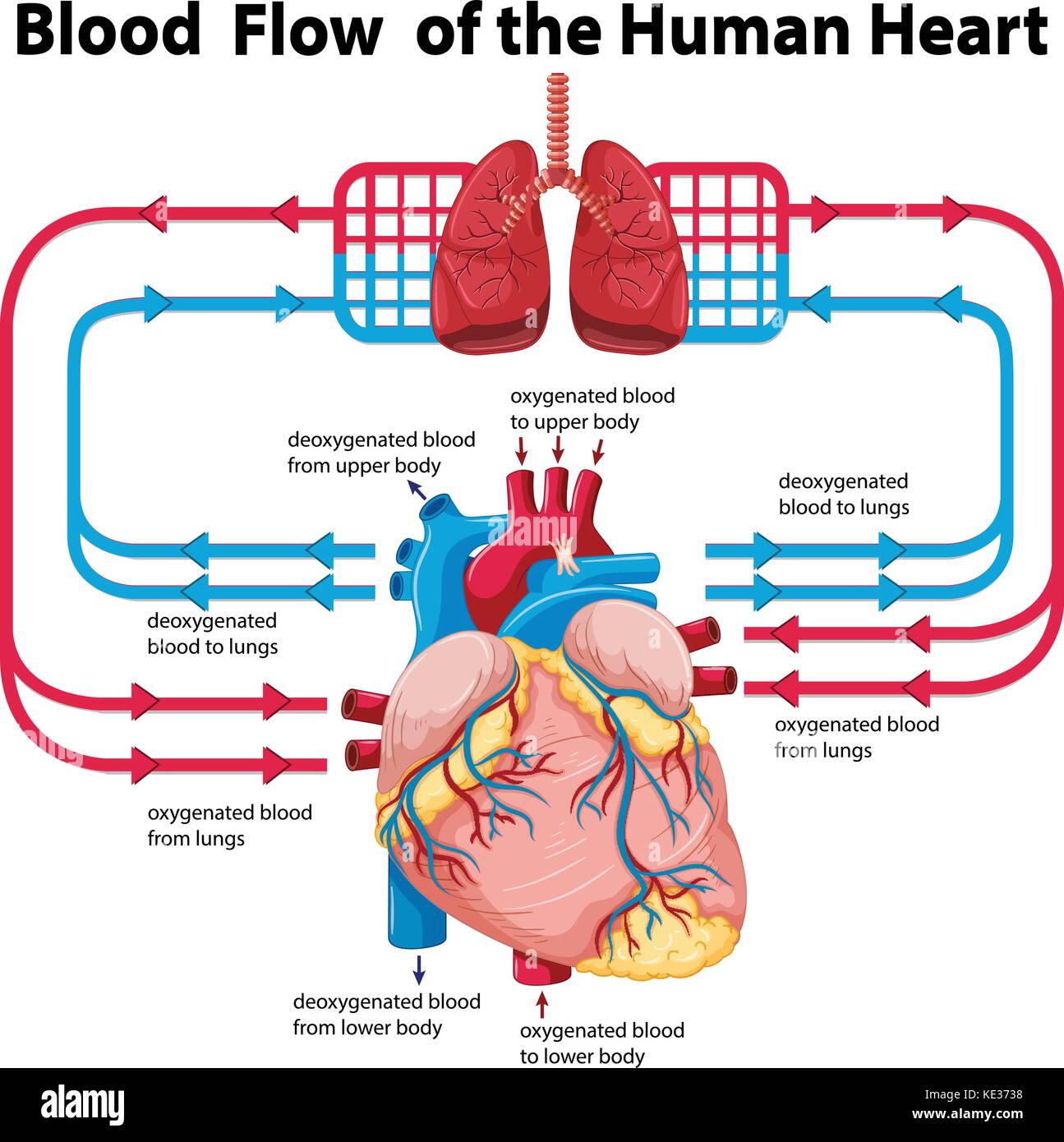 Diagram showing blood flow of human heart illustration Stock Vectorhttps://www.alamy.com/image-license-details/?v=1https://www.alamy.com/stock-image-diagram-showing-blood-flow-of-human-heart-illustration-163569932.html
RFKE3738–Diagram showing blood flow of human heart illustration
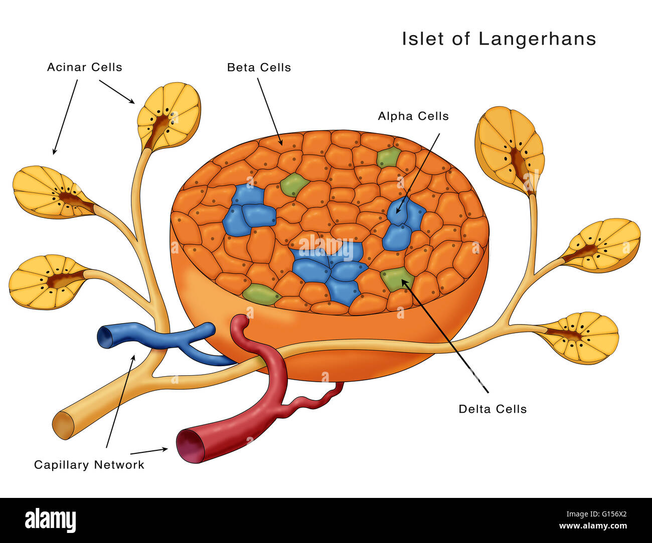 Diagram of the Islets of Langerhans. Shown are the acinar cells, beta cells, alpha cells, delta cells and the capillary network. Stock Photohttps://www.alamy.com/image-license-details/?v=1https://www.alamy.com/stock-photo-diagram-of-the-islets-of-langerhans-shown-are-the-acinar-cells-beta-103992058.html
RMG156X2–Diagram of the Islets of Langerhans. Shown are the acinar cells, beta cells, alpha cells, delta cells and the capillary network.
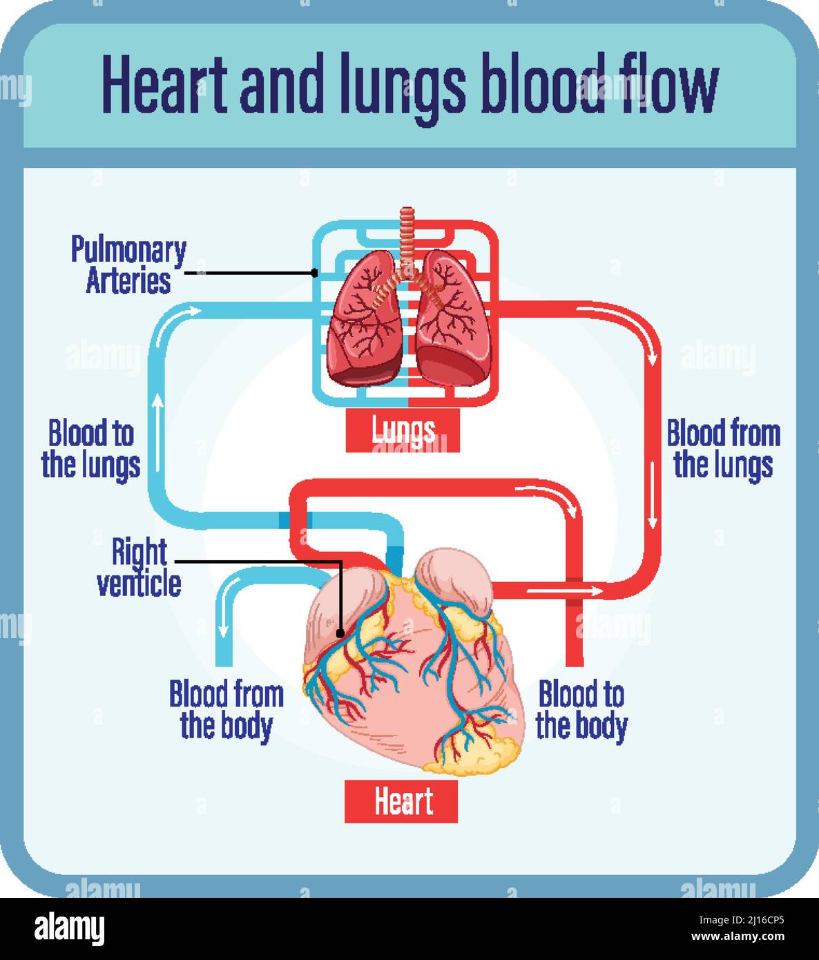 Diagram showing blood flow of human heart illustration Stock Vectorhttps://www.alamy.com/image-license-details/?v=1https://www.alamy.com/diagram-showing-blood-flow-of-human-heart-illustration-image465436333.html
RF2J16CP5–Diagram showing blood flow of human heart illustration
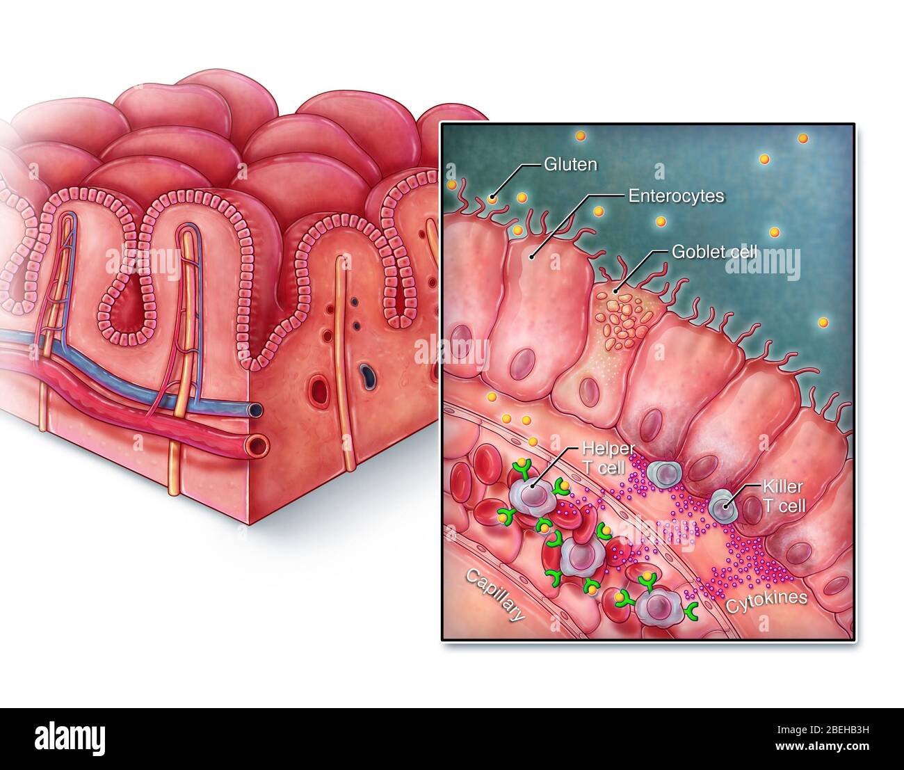 Celiac Disease, Intestinal Villi Stock Photohttps://www.alamy.com/image-license-details/?v=1https://www.alamy.com/celiac-disease-intestinal-villi-image353194453.html
RM2BEHB3H–Celiac Disease, Intestinal Villi
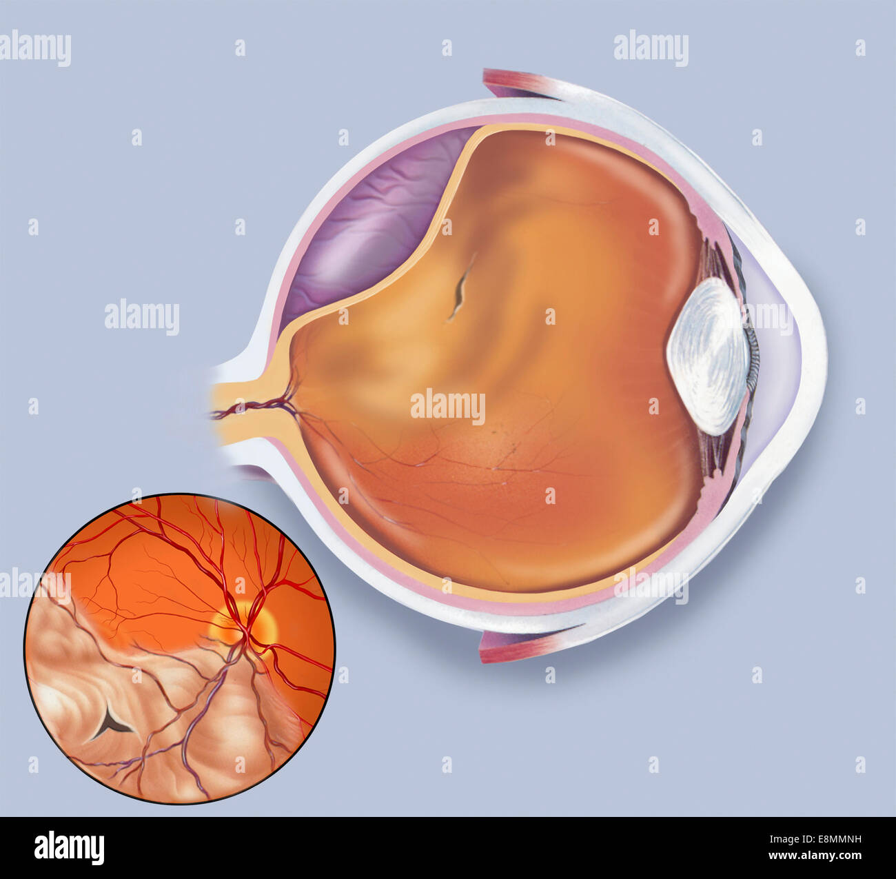 Diagram of a retinal detachment. Stock Photohttps://www.alamy.com/image-license-details/?v=1https://www.alamy.com/stock-photo-diagram-of-a-retinal-detachment-74214045.html
RME8MMNH–Diagram of a retinal detachment.
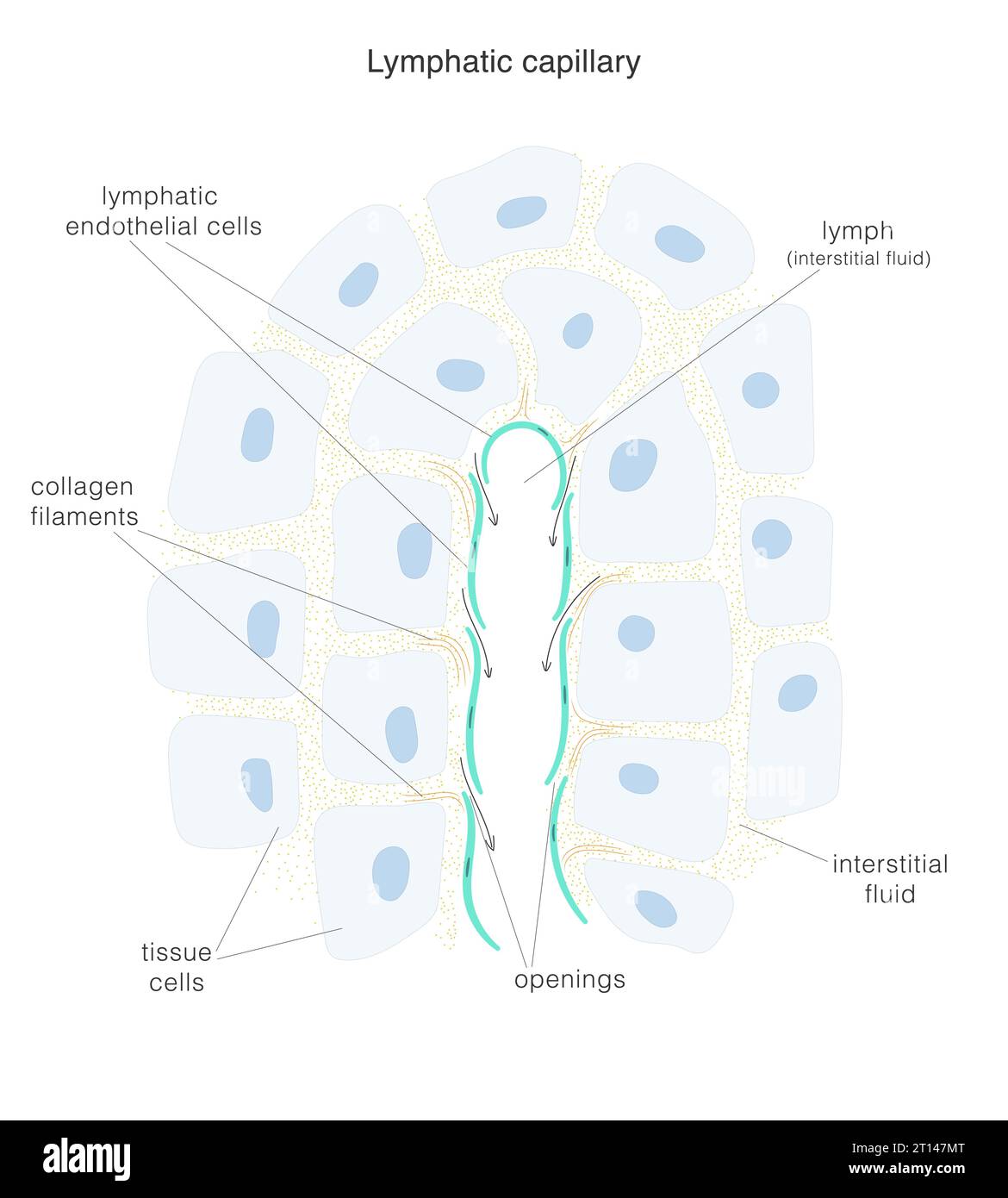 The lymph system. Structure of the terminal end of lymphatic capillary and surrounding tissue. Stock Vectorhttps://www.alamy.com/image-license-details/?v=1https://www.alamy.com/the-lymph-system-structure-of-the-terminal-end-of-lymphatic-capillary-and-surrounding-tissue-image568650680.html
RF2T147MT–The lymph system. Structure of the terminal end of lymphatic capillary and surrounding tissue.
 vasoconstriction and vasodilation blood pressure, dilated blood vessels or capillaries or artery, Increase and decrease in blood flow through the blo Stock Photohttps://www.alamy.com/image-license-details/?v=1https://www.alamy.com/vasoconstriction-and-vasodilation-blood-pressure-dilated-blood-vessels-or-capillaries-or-artery-increase-and-decrease-in-blood-flow-through-the-blo-image510039221.html
RM2MHP86D–vasoconstriction and vasodilation blood pressure, dilated blood vessels or capillaries or artery, Increase and decrease in blood flow through the blo
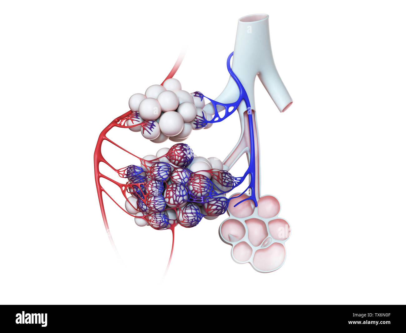 3d rendered illustration of the human alveoli Stock Photohttps://www.alamy.com/image-license-details/?v=1https://www.alamy.com/3d-rendered-illustration-of-the-human-alveoli-image257074399.html
RFTX6N0F–3d rendered illustration of the human alveoli
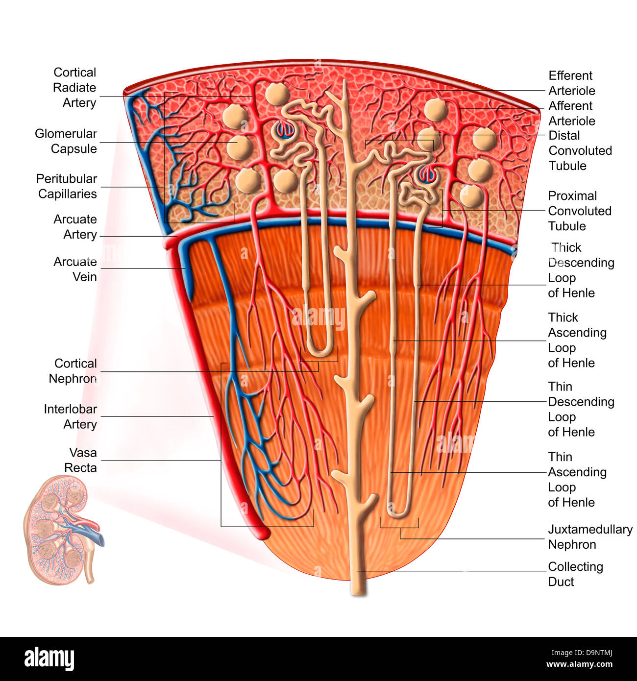 Anatomy of human kidney function. Stock Photohttps://www.alamy.com/image-license-details/?v=1https://www.alamy.com/stock-photo-anatomy-of-human-kidney-function-57643394.html
RFD9NTMJ–Anatomy of human kidney function.
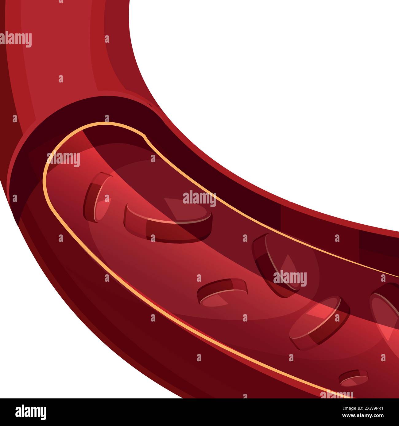 Blood vessel anatomy illustration. Aorta medical structure Stock Vectorhttps://www.alamy.com/image-license-details/?v=1https://www.alamy.com/blood-vessel-anatomy-illustration-aorta-medical-structure-image617944741.html
RF2XW9PR1–Blood vessel anatomy illustration. Aorta medical structure
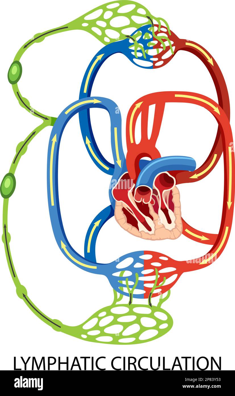 Lymphatic Circulation System Diagram illustration Stock Vectorhttps://www.alamy.com/image-license-details/?v=1https://www.alamy.com/lymphatic-circulation-system-diagram-illustration-image538525823.html
RF2P83Y53–Lymphatic Circulation System Diagram illustration
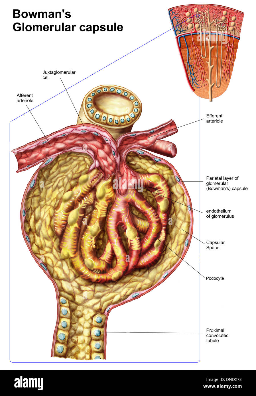 Anatomy of bowman's glomerular capsule. Stock Photohttps://www.alamy.com/image-license-details/?v=1https://www.alamy.com/anatomy-of-bowmans-glomerular-capsule-image64844839.html
RFDNDX73–Anatomy of bowman's glomerular capsule.
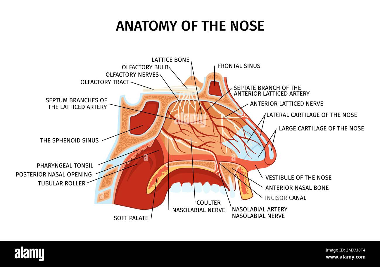 Nose anatomy cross section diagram showing lattice bone arteries nerves cartilage soft palate paranasal sinuses elements flat vector illustration Stock Vectorhttps://www.alamy.com/image-license-details/?v=1https://www.alamy.com/nose-anatomy-cross-section-diagram-showing-lattice-bone-arteries-nerves-cartilage-soft-palate-paranasal-sinuses-elements-flat-vector-illustration-image515521444.html
RF2MXM0T4–Nose anatomy cross section diagram showing lattice bone arteries nerves cartilage soft palate paranasal sinuses elements flat vector illustration
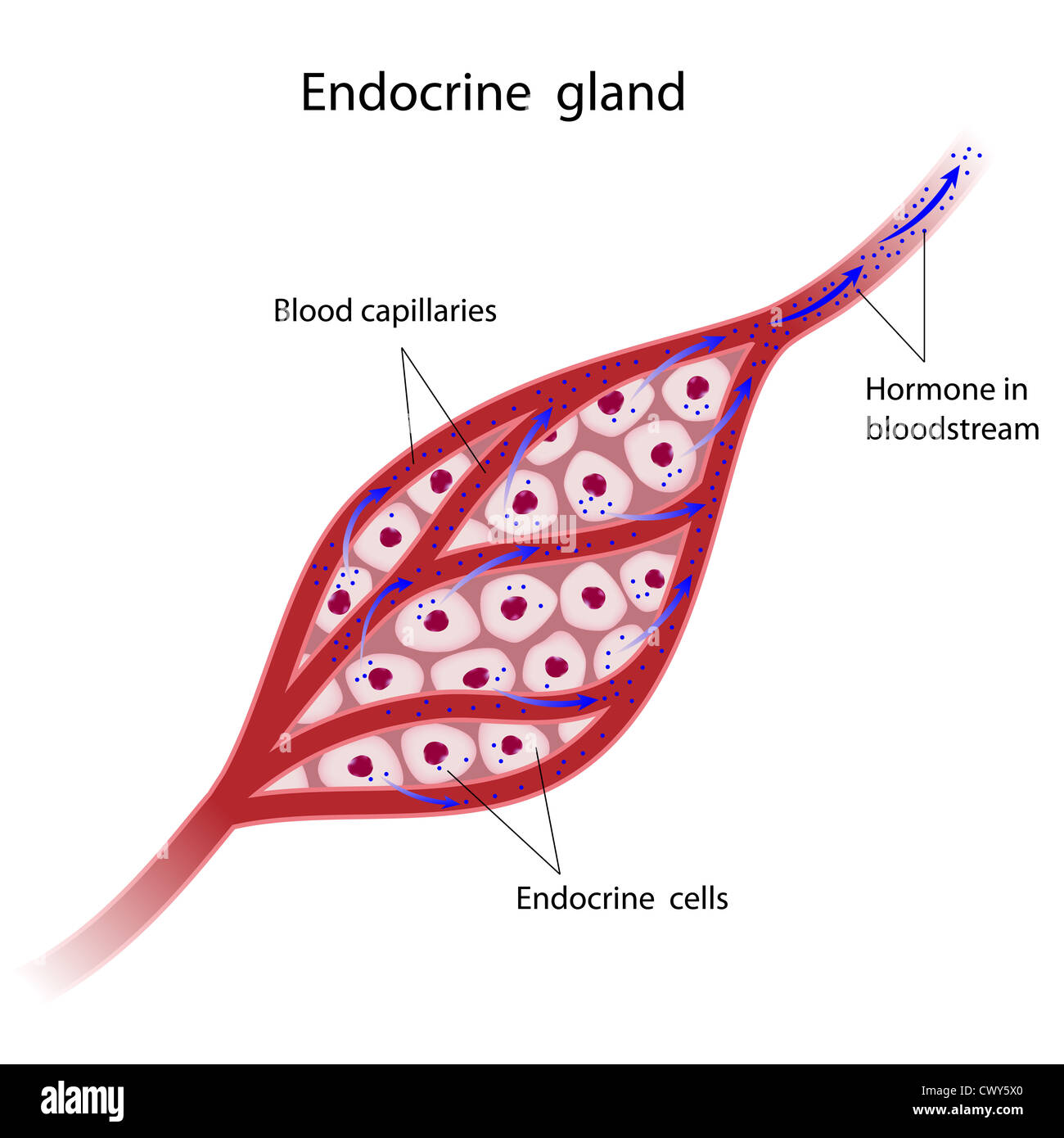 Endocrine glands cells secrete hormones directly to bloodstream Stock Photohttps://www.alamy.com/image-license-details/?v=1https://www.alamy.com/stock-photo-endocrine-glands-cells-secrete-hormones-directly-to-bloodstream-50384488.html
RFCWY5X0–Endocrine glands cells secrete hormones directly to bloodstream
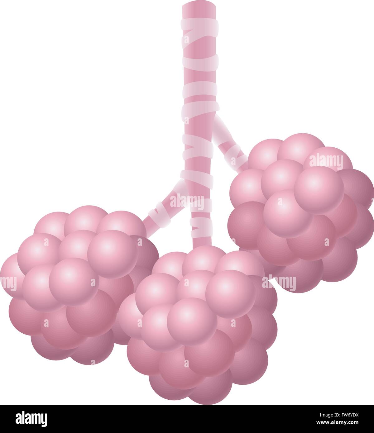 Illustration of human alveoli isolated on white background Stock Vectorhttps://www.alamy.com/image-license-details/?v=1https://www.alamy.com/stock-photo-illustration-of-human-alveoli-isolated-on-white-background-101571510.html
RFFW6YDX–Illustration of human alveoli isolated on white background
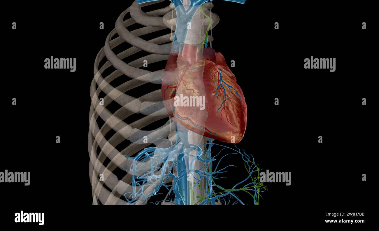 Interstitial fluid is collected by lymph capillaries from the interstitial space. Lymph then moves through lymphatic vessels to lymph nodes. 3D render Stock Photohttps://www.alamy.com/image-license-details/?v=1https://www.alamy.com/interstitial-fluid-is-collected-by-lymph-capillaries-from-the-interstitial-space-lymph-then-moves-through-lymphatic-vessels-to-lymph-nodes-3d-render-image596595311.html
RF2WJH7BB–Interstitial fluid is collected by lymph capillaries from the interstitial space. Lymph then moves through lymphatic vessels to lymph nodes. 3D render
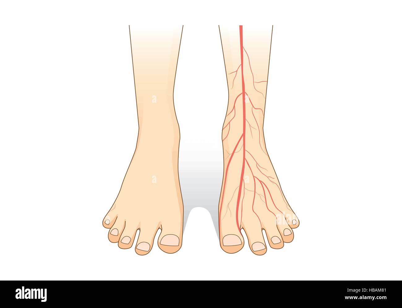 One foot showing a red blood vessel on skin. Stock Vectorhttps://www.alamy.com/image-license-details/?v=1https://www.alamy.com/stock-photo-one-foot-showing-a-red-blood-vessel-on-skin-127469217.html
RFHBAM81–One foot showing a red blood vessel on skin.
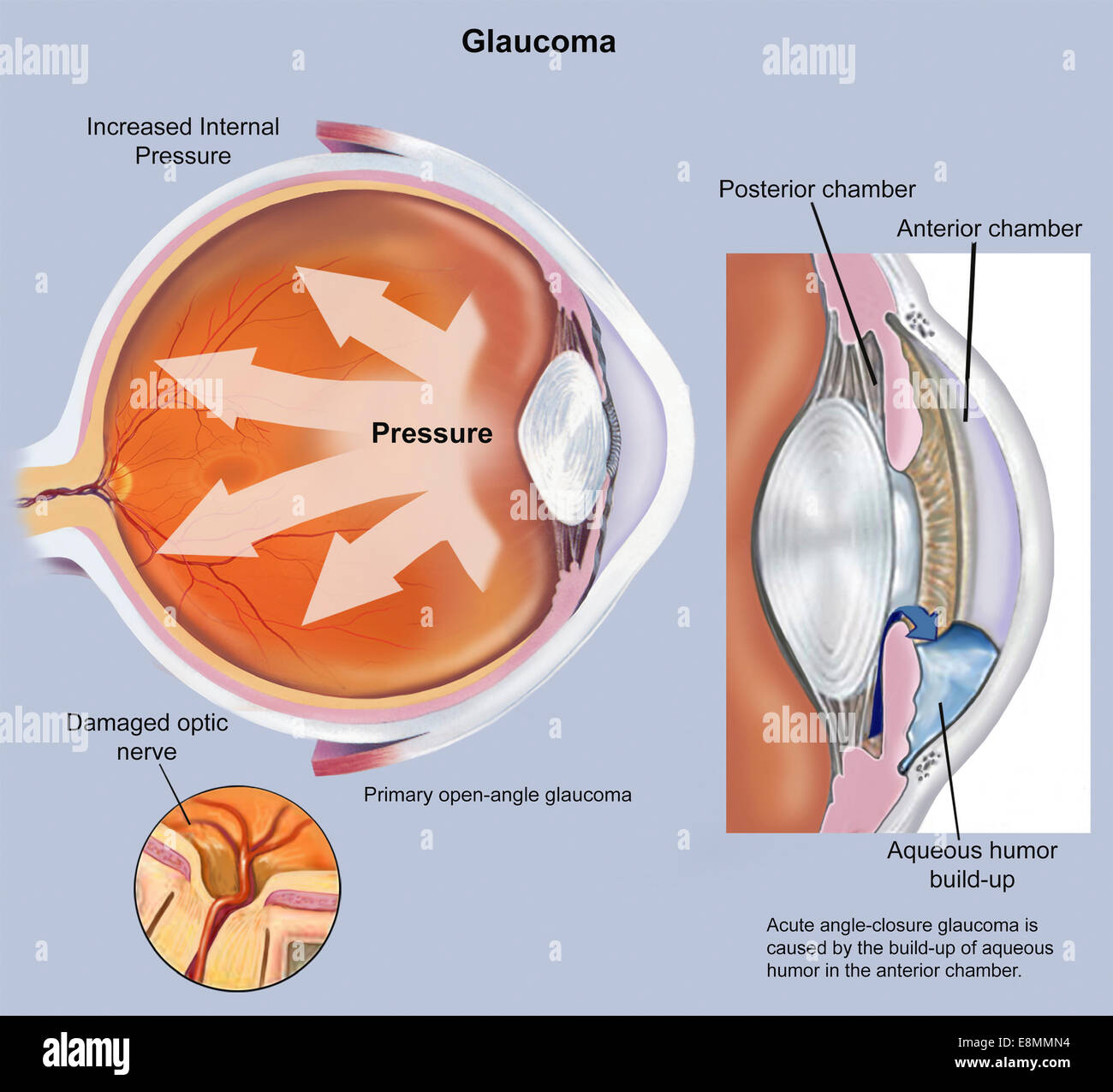 Retina of eye with glaucoma. Stock Photohttps://www.alamy.com/image-license-details/?v=1https://www.alamy.com/stock-photo-retina-of-eye-with-glaucoma-74214032.html
RME8MMN4–Retina of eye with glaucoma.
 A text-book of first aid and emergency treatment . Diagram to Show the Course of the Circulationof the Blood. The pulmonary capillaries are the small vessels of thelungs. The systemic capillaries represent tViose of the skin,head and extremities. The third systeni shown representsthe capillaries of the intestmes and liver. THE NERVOUS SYSTEM 51 THE NERVOUS SYSTEM. All voluntary movements and many of the involuntaryfunctions of the body are under the control of the nervoussystem. It is tluough the medium of the nerves that all theimpulses arising outside of the body become sensory percep- CERE Stock Photohttps://www.alamy.com/image-license-details/?v=1https://www.alamy.com/a-text-book-of-first-aid-and-emergency-treatment-diagram-to-show-the-course-of-the-circulationof-the-blood-the-pulmonary-capillaries-are-the-small-vessels-of-thelungs-the-systemic-capillaries-represent-tviose-of-the-skinhead-and-extremities-the-third-systeni-shown-representsthe-capillaries-of-the-intestmes-and-liver-the-nervous-system-51-the-nervous-system-all-voluntary-movements-and-many-of-the-involuntaryfunctions-of-the-body-are-under-the-control-of-the-nervoussystem-it-is-tluough-the-medium-of-the-nerves-that-all-theimpulses-arising-outside-of-the-body-become-sensory-percep-cere-image340294065.html
RM2ANHMFD–A text-book of first aid and emergency treatment . Diagram to Show the Course of the Circulationof the Blood. The pulmonary capillaries are the small vessels of thelungs. The systemic capillaries represent tViose of the skin,head and extremities. The third systeni shown representsthe capillaries of the intestmes and liver. THE NERVOUS SYSTEM 51 THE NERVOUS SYSTEM. All voluntary movements and many of the involuntaryfunctions of the body are under the control of the nervoussystem. It is tluough the medium of the nerves that all theimpulses arising outside of the body become sensory percep- CERE
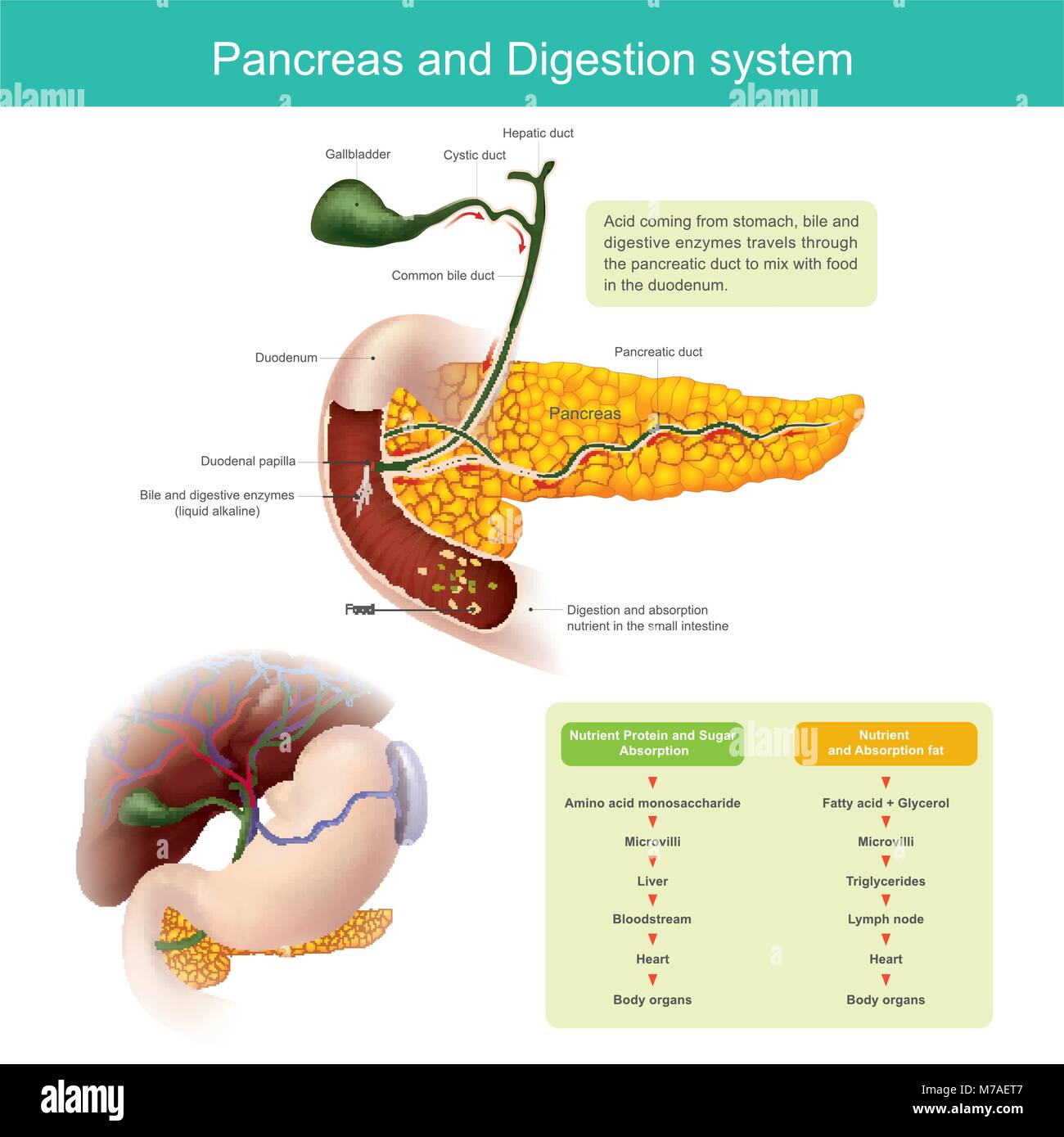 The digestive enzymes travels through the pancreatic duct to mix with food in the duodenum. The liver produce Bile, which is stored in the gall bladde Stock Vectorhttps://www.alamy.com/image-license-details/?v=1https://www.alamy.com/stock-photo-the-digestive-enzymes-travels-through-the-pancreatic-duct-to-mix-with-176637447.html
RFM7AET7–The digestive enzymes travels through the pancreatic duct to mix with food in the duodenum. The liver produce Bile, which is stored in the gall bladde
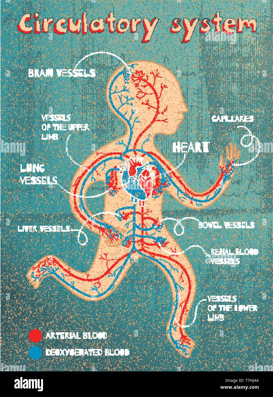 Human circulatory system for kids. Vector color cartoon illustration. Human cardiovascular anatomy scheme. Stock Vectorhttps://www.alamy.com/image-license-details/?v=1https://www.alamy.com/human-circulatory-system-for-kids-vector-color-cartoon-illustration-human-cardiovascular-anatomy-scheme-image245635162.html
RFT7HJ4A–Human circulatory system for kids. Vector color cartoon illustration. Human cardiovascular anatomy scheme.
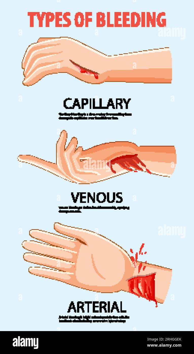 Vector cartoon showing bleeding in capillaries, veins, and arteries Stock Vectorhttps://www.alamy.com/image-license-details/?v=1https://www.alamy.com/vector-cartoon-showing-bleeding-in-capillaries-veins-and-arteries-image561545115.html
RF2RHGGEK–Vector cartoon showing bleeding in capillaries, veins, and arteries
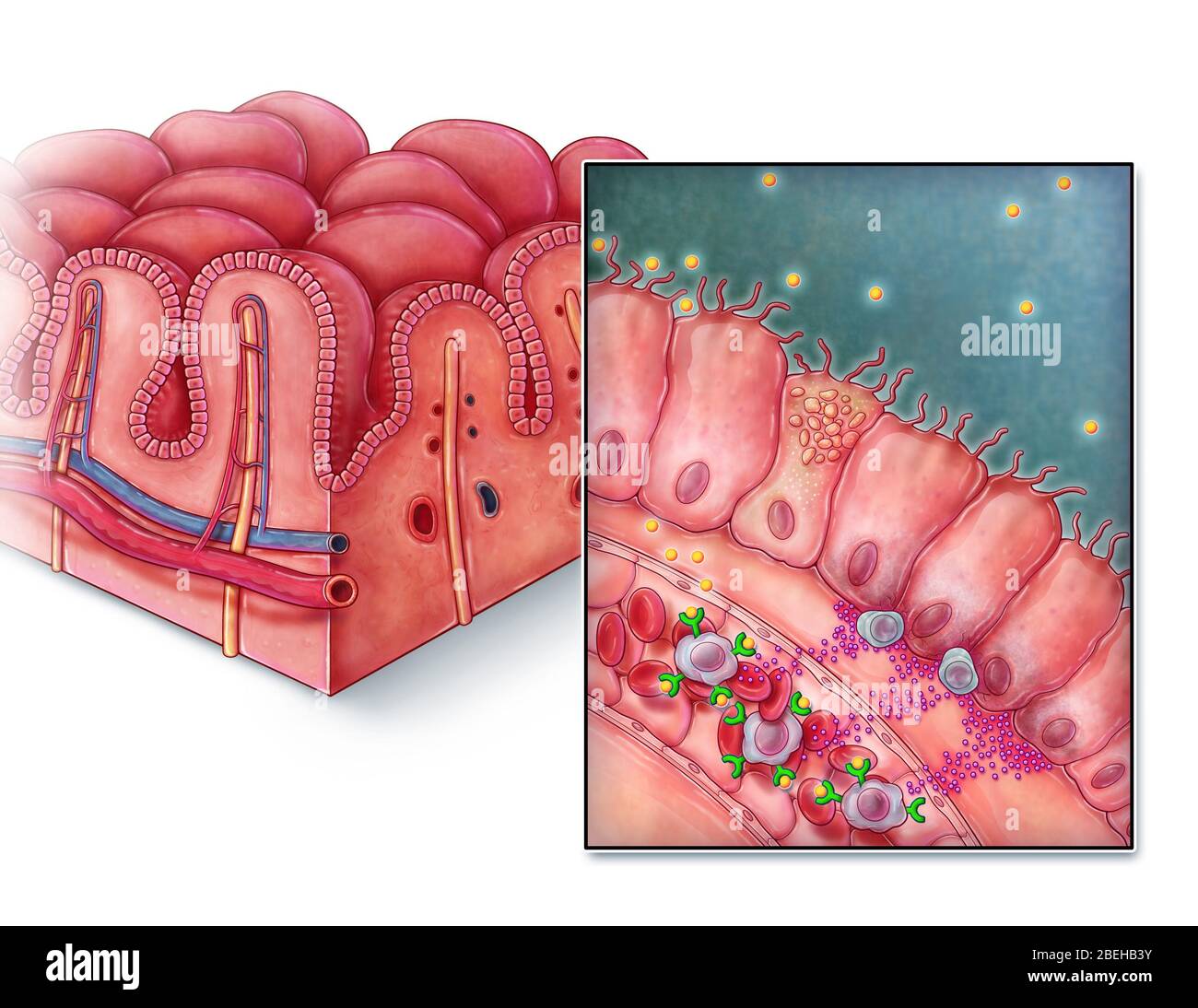 Celiac Disease, Intestinal Villi Stock Photohttps://www.alamy.com/image-license-details/?v=1https://www.alamy.com/celiac-disease-intestinal-villi-image353194463.html
RM2BEHB3Y–Celiac Disease, Intestinal Villi
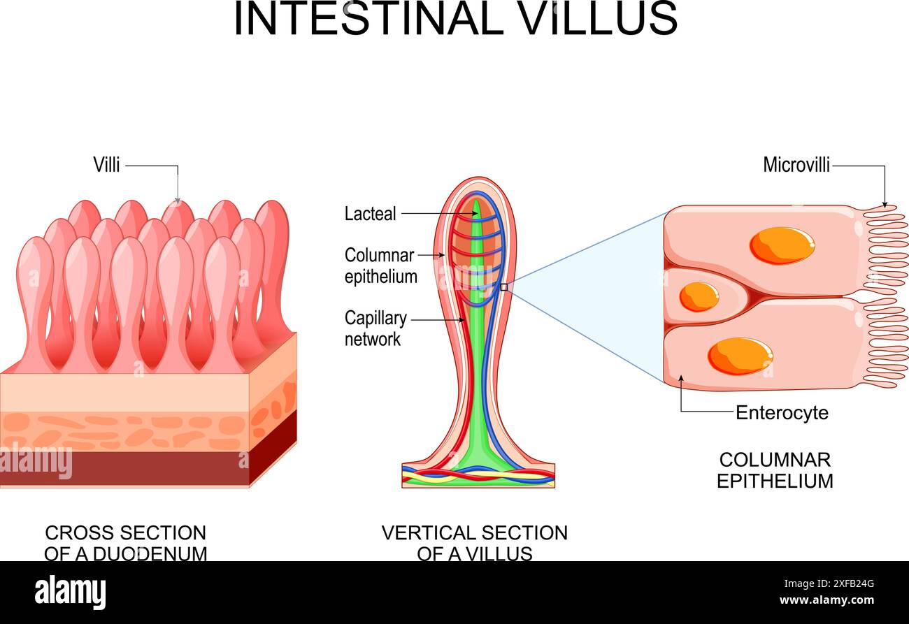 Intestinal villus. Different between Villi and microvilli. Cross section of a duodenum with submucosa, mucosa and muscularis layers. Vertical section Stock Vectorhttps://www.alamy.com/image-license-details/?v=1https://www.alamy.com/intestinal-villus-different-between-villi-and-microvilli-cross-section-of-a-duodenum-with-submucosa-mucosa-and-muscularis-layers-vertical-section-image611825888.html
RF2XFB24G–Intestinal villus. Different between Villi and microvilli. Cross section of a duodenum with submucosa, mucosa and muscularis layers. Vertical section
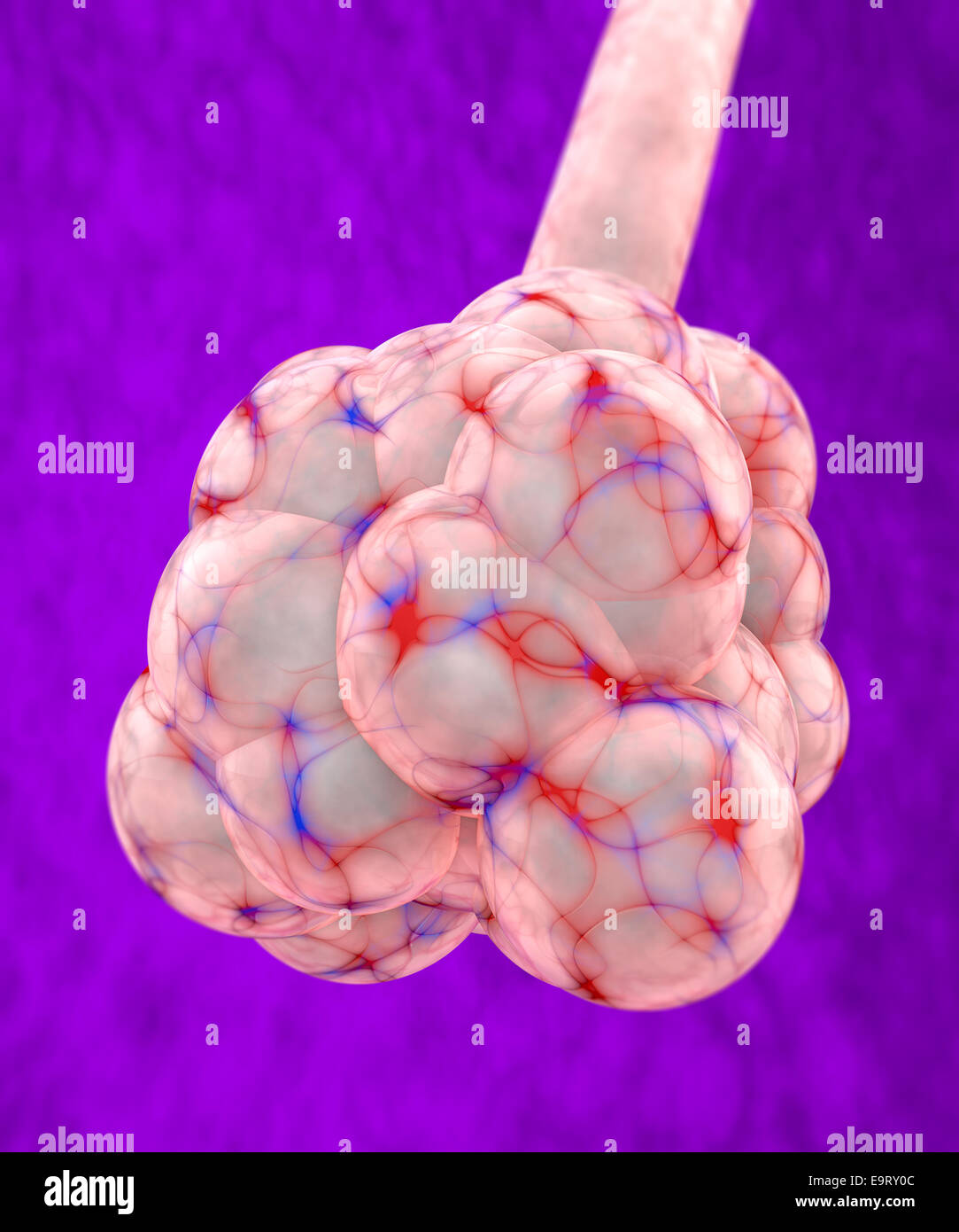 3d pulmonary alveolus on red background Stock Photohttps://www.alamy.com/image-license-details/?v=1https://www.alamy.com/stock-photo-3d-pulmonary-alveolus-on-red-background-74899452.html
RFE9RY0C–3d pulmonary alveolus on red background
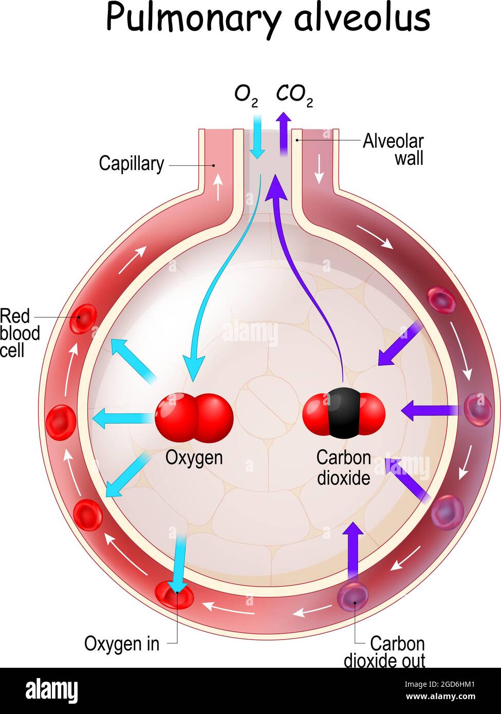 Alveolus Gas Exchange. Anatomy of Pulmonary alveolus. Oxygen And Carbon Dioxide, inhale and exhale Stock Vectorhttps://www.alamy.com/image-license-details/?v=1https://www.alamy.com/alveolus-gas-exchange-anatomy-of-pulmonary-alveolus-oxygen-and-carbon-dioxide-inhale-and-exhale-image438395329.html
RF2GD6HM1–Alveolus Gas Exchange. Anatomy of Pulmonary alveolus. Oxygen And Carbon Dioxide, inhale and exhale
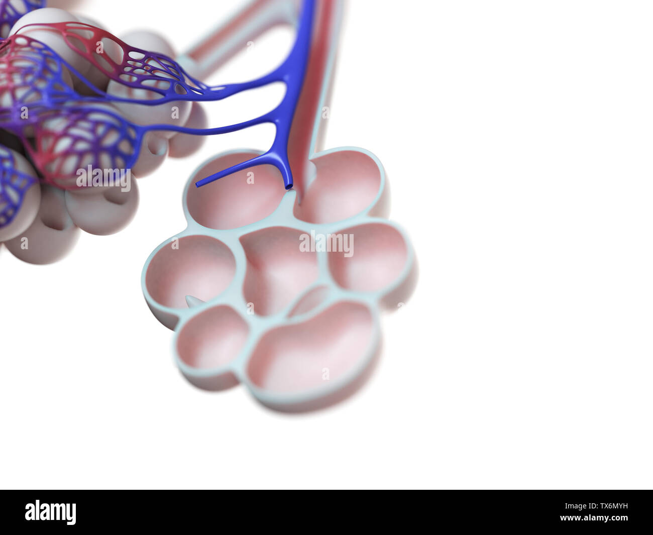 3d rendered illustration of the human alveoli Stock Photohttps://www.alamy.com/image-license-details/?v=1https://www.alamy.com/3d-rendered-illustration-of-the-human-alveoli-image257074373.html
RFTX6MYH–3d rendered illustration of the human alveoli
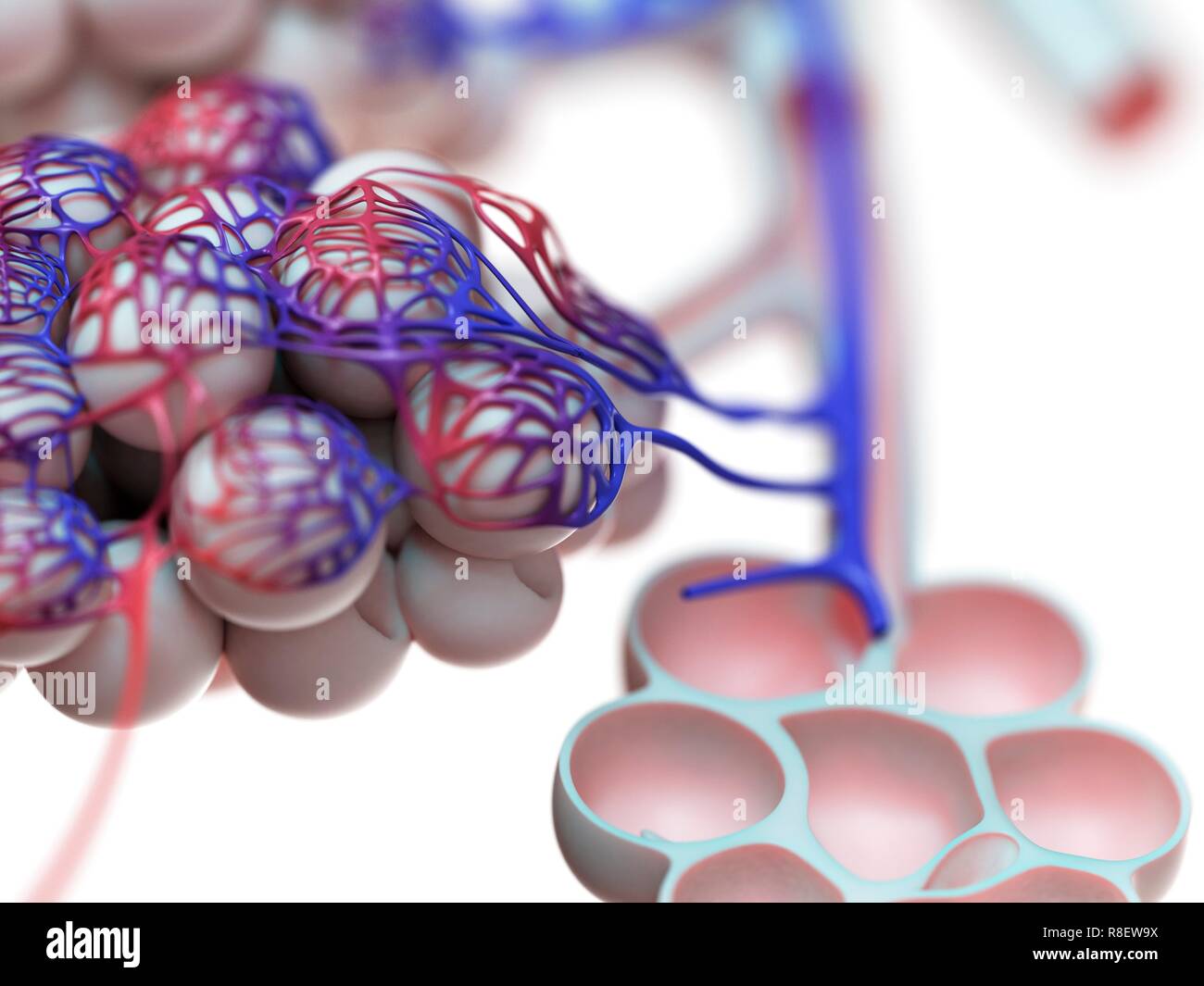 Illustration of the human alveoli. Stock Photohttps://www.alamy.com/image-license-details/?v=1https://www.alamy.com/illustration-of-the-human-alveoli-image228979238.html
RFR8EW9X–Illustration of the human alveoli.
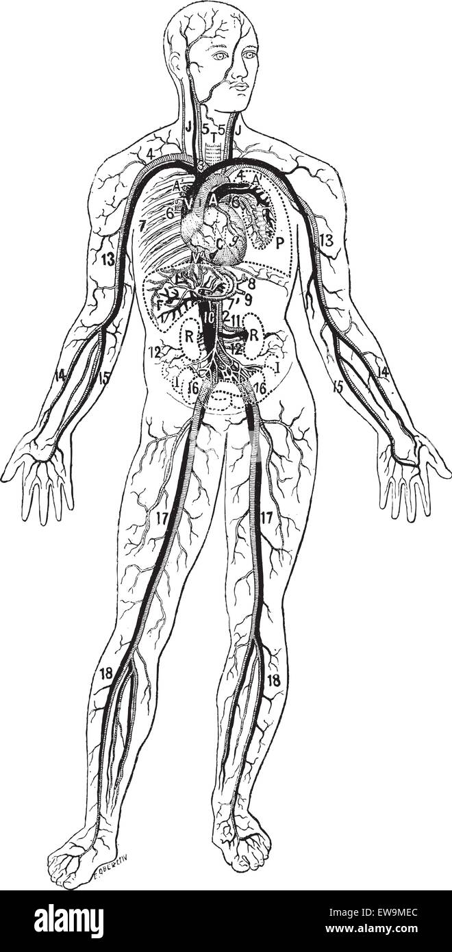 Blood vessels, vintage engraved illustration. Dictionary of words and things - Larive and Fleury - 1895. Stock Vectorhttps://www.alamy.com/image-license-details/?v=1https://www.alamy.com/stock-photo-blood-vessels-vintage-engraved-illustration-dictionary-of-words-and-84421524.html
RFEW9MEC–Blood vessels, vintage engraved illustration. Dictionary of words and things - Larive and Fleury - 1895.
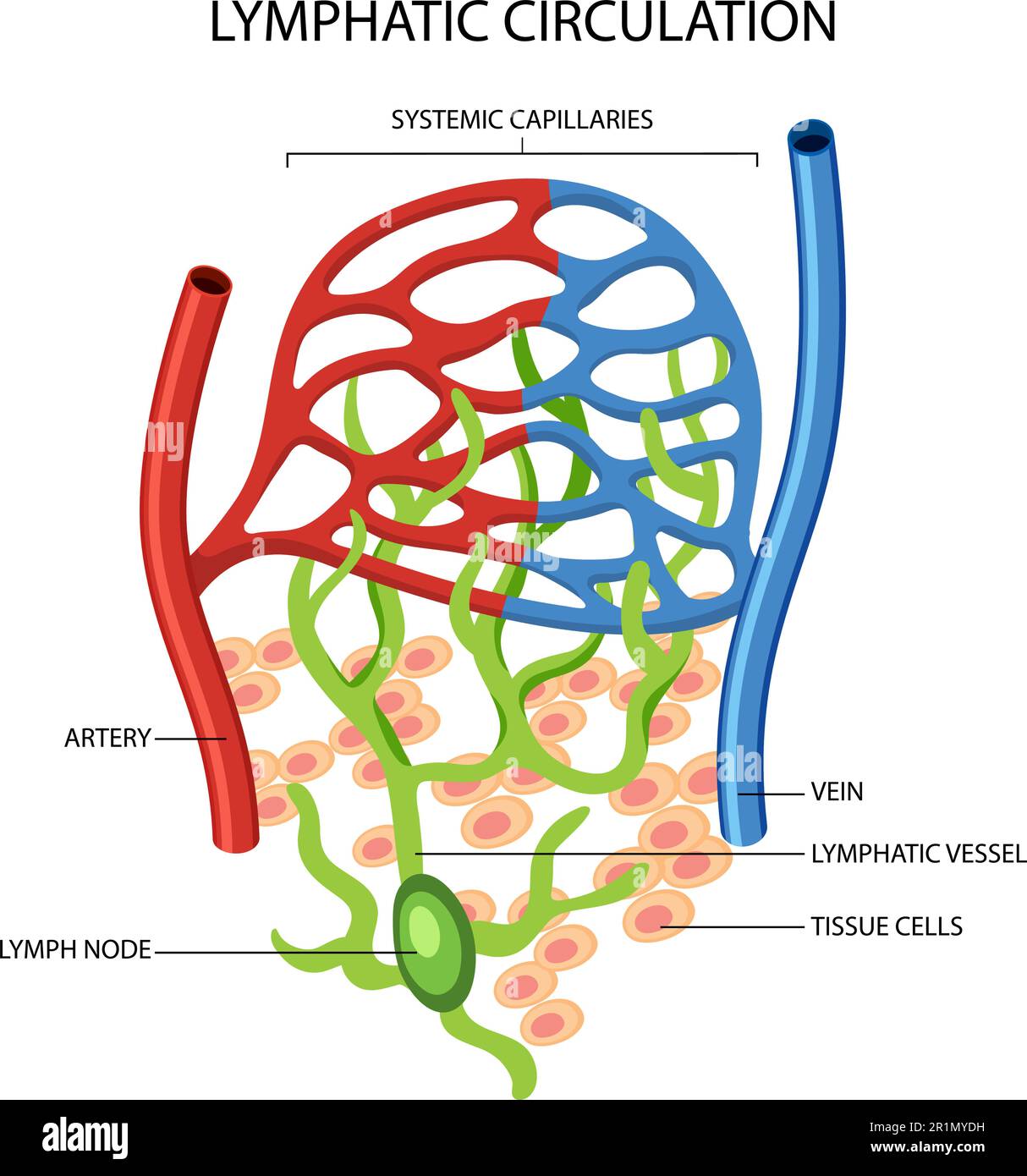 Lymphatic Circulation System Diagram illustration Stock Vectorhttps://www.alamy.com/image-license-details/?v=1https://www.alamy.com/lymphatic-circulation-system-diagram-illustration-image551807021.html
RF2R1MYDH–Lymphatic Circulation System Diagram illustration
 WRIGHT-PATTERSON AIR FORCE BASE, Ohio – Researchers at the Materials and Manufacturing Directorate, Air Force Research Laboratory, have developed a novel, lightweight artificial hair sensor that mimics those used by natural fliers—like bats and crickets—by using carbon nanotube forests grown inside glass fiber capillaries. The hairs are sensitive to air flow changes during flight, enabling quick analysis and response by agile fliers. The diagram pairs an overview of the hair sensor components with an image captured on a Scanning Electron Microscope. Stock Photohttps://www.alamy.com/image-license-details/?v=1https://www.alamy.com/wright-patterson-air-force-base-ohio-researchers-at-the-materials-and-manufacturing-directorate-air-force-research-laboratory-have-developed-a-novel-lightweight-artificial-hair-sensor-that-mimics-those-used-by-natural-flierslike-bats-and-cricketsby-using-carbon-nanotube-forests-grown-inside-glass-fiber-capillaries-the-hairs-are-sensitive-to-air-flow-changes-during-flight-enabling-quick-analysis-and-response-by-agile-fliers-the-diagram-pairs-an-overview-of-the-hair-sensor-components-with-an-image-captured-on-a-scanning-electron-microscope-image207317849.html
RMP1840W–WRIGHT-PATTERSON AIR FORCE BASE, Ohio – Researchers at the Materials and Manufacturing Directorate, Air Force Research Laboratory, have developed a novel, lightweight artificial hair sensor that mimics those used by natural fliers—like bats and crickets—by using carbon nanotube forests grown inside glass fiber capillaries. The hairs are sensitive to air flow changes during flight, enabling quick analysis and response by agile fliers. The diagram pairs an overview of the hair sensor components with an image captured on a Scanning Electron Microscope.
 Blood circulation system in kid body. Education illustration Stock Vectorhttps://www.alamy.com/image-license-details/?v=1https://www.alamy.com/blood-circulation-system-in-kid-body-education-illustration-image600553311.html
RF2WW1FTF–Blood circulation system in kid body. Education illustration
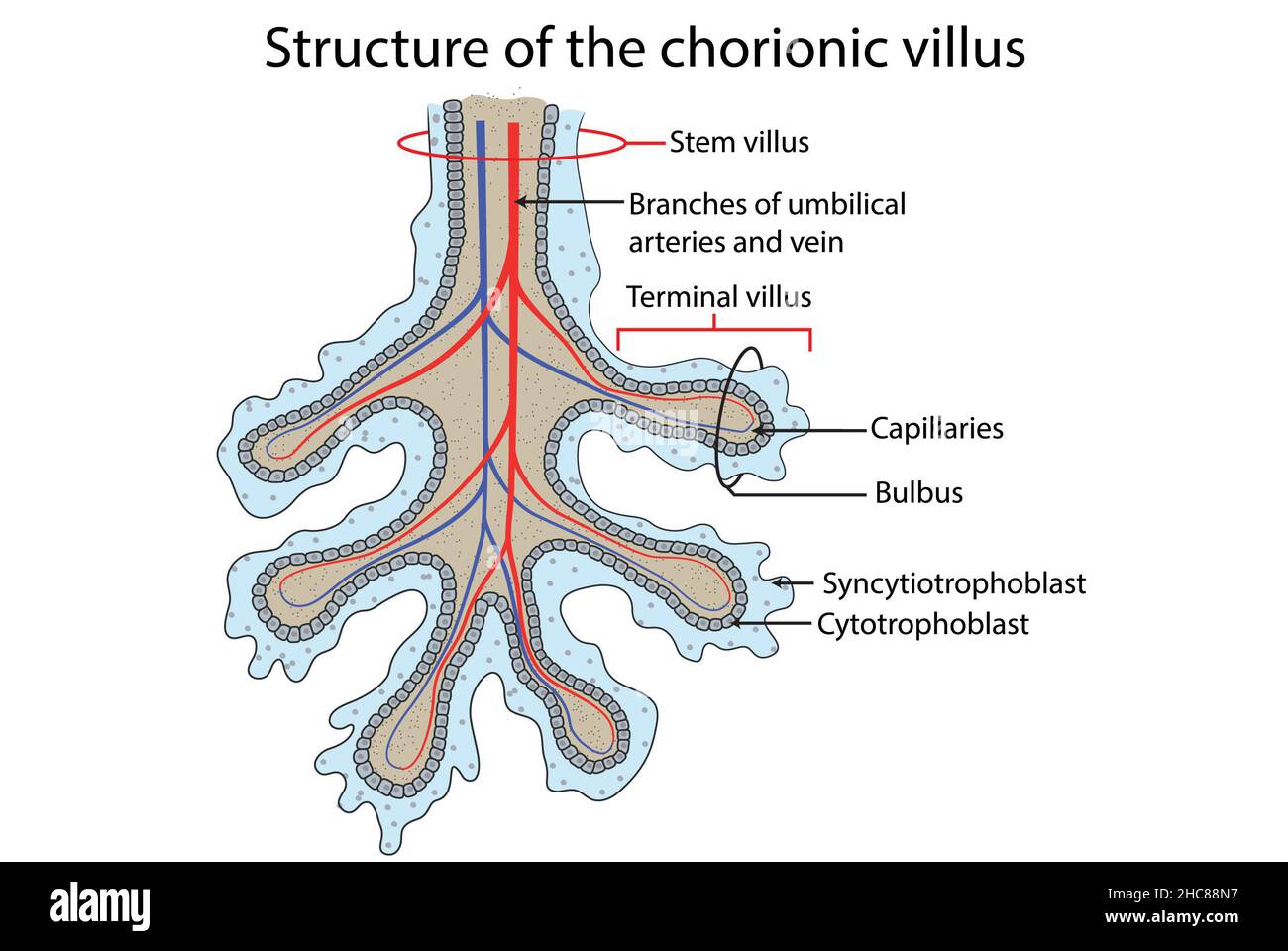 Structure of the chorionic villus, fetal part of the placenta. Stock Photohttps://www.alamy.com/image-license-details/?v=1https://www.alamy.com/structure-of-the-chorionic-villus-fetal-part-of-the-placenta-image455027923.html
RF2HC88N7–Structure of the chorionic villus, fetal part of the placenta.
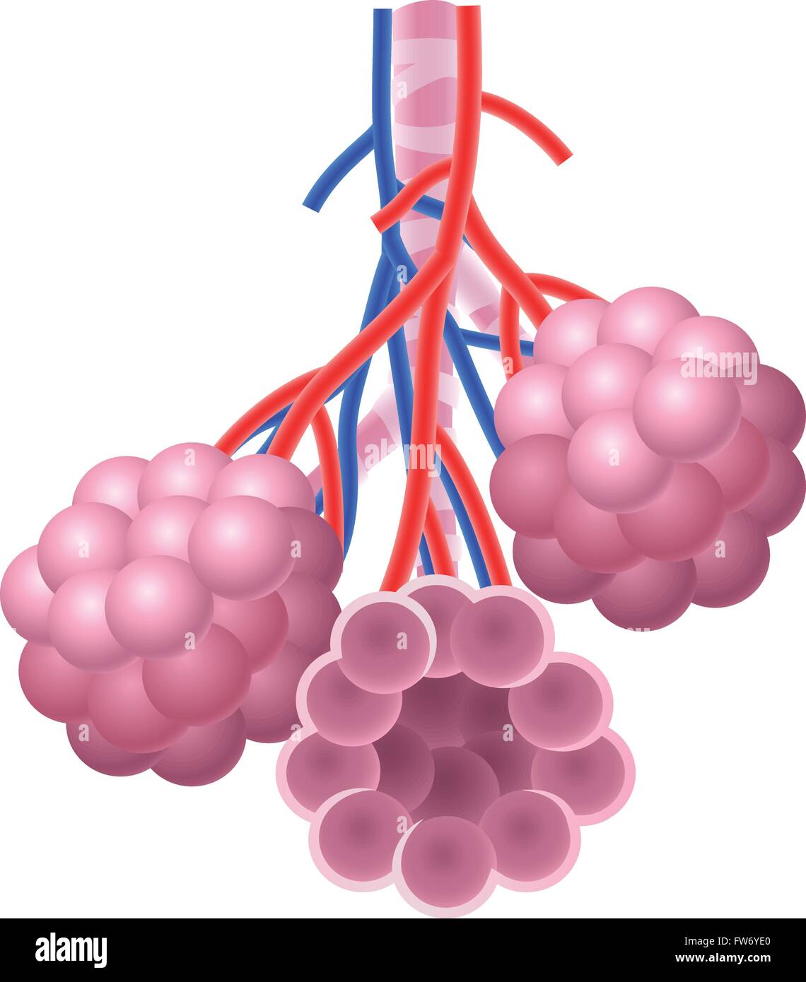 Illustration of Human Alveoli structure Anatomy Stock Vectorhttps://www.alamy.com/image-license-details/?v=1https://www.alamy.com/stock-photo-illustration-of-human-alveoli-structure-anatomy-101571512.html
RFFW6YE0–Illustration of Human Alveoli structure Anatomy
 Interstitial fluid is collected by lymph capillaries from the interstitial space. Lymph then moves through lymphatic vessels to lymph nodes. 3D render Stock Photohttps://www.alamy.com/image-license-details/?v=1https://www.alamy.com/interstitial-fluid-is-collected-by-lymph-capillaries-from-the-interstitial-space-lymph-then-moves-through-lymphatic-vessels-to-lymph-nodes-3d-render-image596593727.html
RF2WJH5AR–Interstitial fluid is collected by lymph capillaries from the interstitial space. Lymph then moves through lymphatic vessels to lymph nodes. 3D render
 Alveoli in Lungs Facilitating Respiration and Oxygenation Stock Photohttps://www.alamy.com/image-license-details/?v=1https://www.alamy.com/alveoli-in-lungs-facilitating-respiration-and-oxygenation-image628160609.html
RF2YDY57D–Alveoli in Lungs Facilitating Respiration and Oxygenation
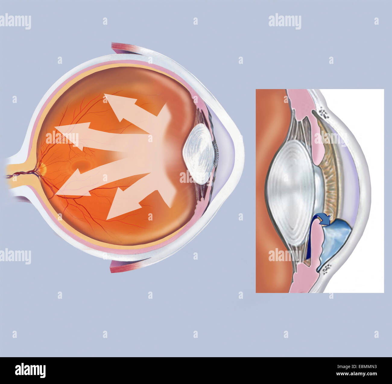 Retina of eye with glaucoma. Stock Photohttps://www.alamy.com/image-license-details/?v=1https://www.alamy.com/stock-photo-retina-of-eye-with-glaucoma-74214031.html
RME8MMN3–Retina of eye with glaucoma.
 Manual of human histology . 5. Vertical sectiou through aportion of a pyramid and the conical sub-stance belonging to it, of an injected Rab-bits kidney. The figure is half-diagram-matic ; X 30 diam. The vessels are represented on the left side, and on the rightthe course of the tubuli uriniferi: a, arterim inierlohulares, with the glomeruliMalpighiani, b, and their vasa efferentia; c, vasa efferentia : d, cortical capillaries;e, vasa efferentia of the outermost corpuscles, proceeding to the superficial capil-laries ; f, vasa efferentia of the innermost glomeruli, continuous with the arteriole Stock Photohttps://www.alamy.com/image-license-details/?v=1https://www.alamy.com/manual-of-human-histology-5-vertical-sectiou-through-aportion-of-a-pyramid-and-the-conical-sub-stance-belonging-to-it-of-an-injected-rab-bits-kidney-the-figure-is-half-diagram-matic-x-30-diam-the-vessels-are-represented-on-the-left-side-and-on-the-rightthe-course-of-the-tubuli-uriniferi-a-arterim-inierlohulares-with-the-glomerulimalpighiani-b-and-their-vasa-efferentia-c-vasa-efferentia-d-cortical-capillariese-vasa-efferentia-of-the-outermost-corpuscles-proceeding-to-the-superficial-capil-laries-f-vasa-efferentia-of-the-innermost-glomeruli-continuous-with-the-arteriole-image338228517.html
RM2AJ7HWW–Manual of human histology . 5. Vertical sectiou through aportion of a pyramid and the conical sub-stance belonging to it, of an injected Rab-bits kidney. The figure is half-diagram-matic ; X 30 diam. The vessels are represented on the left side, and on the rightthe course of the tubuli uriniferi: a, arterim inierlohulares, with the glomeruliMalpighiani, b, and their vasa efferentia; c, vasa efferentia : d, cortical capillaries;e, vasa efferentia of the outermost corpuscles, proceeding to the superficial capil-laries ; f, vasa efferentia of the innermost glomeruli, continuous with the arteriole
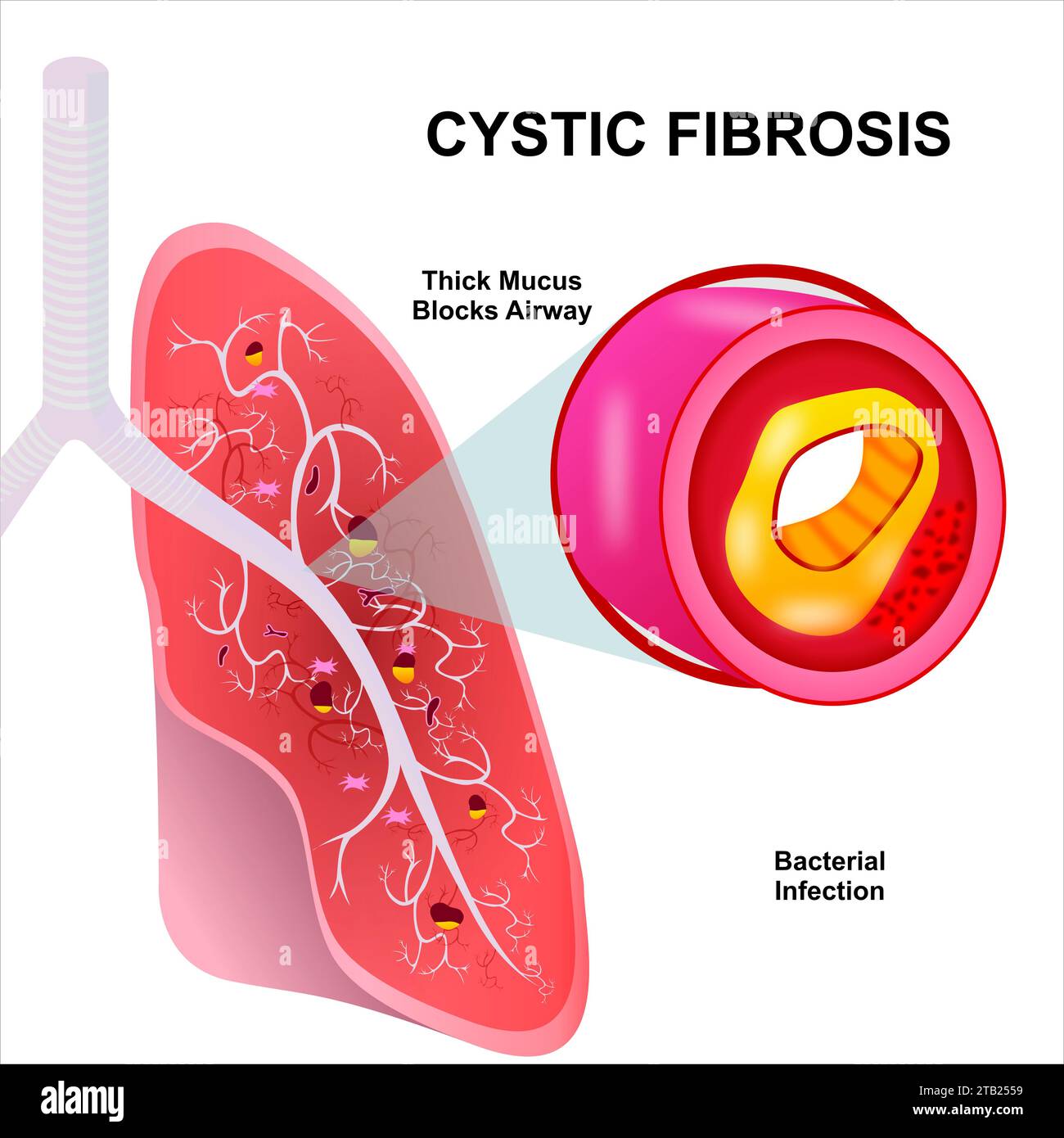 Cystic fibrosis illustration infection on lungs Stock Photohttps://www.alamy.com/image-license-details/?v=1https://www.alamy.com/cystic-fibrosis-illustration-infection-on-lungs-image574751333.html
RF2TB2559–Cystic fibrosis illustration infection on lungs
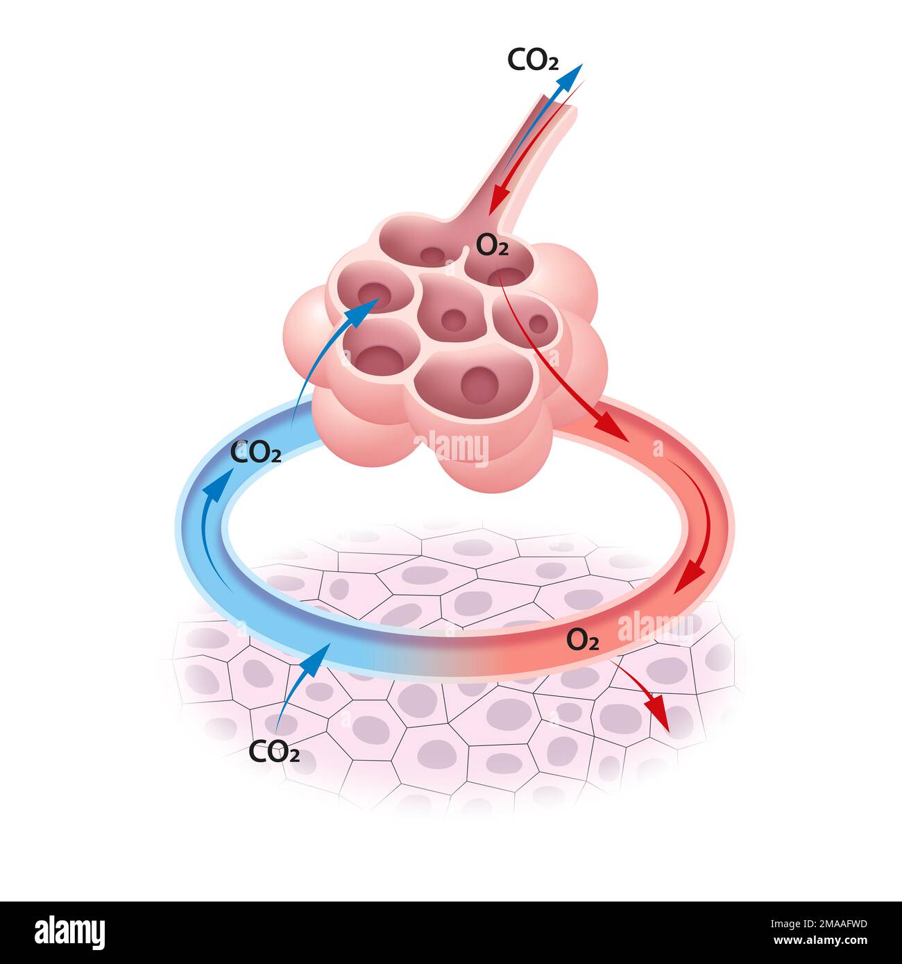 Gas exchange in the alveoli Stock Photohttps://www.alamy.com/image-license-details/?v=1https://www.alamy.com/gas-exchange-in-the-alveoli-image505479225.html
RF2MAAFWD–Gas exchange in the alveoli
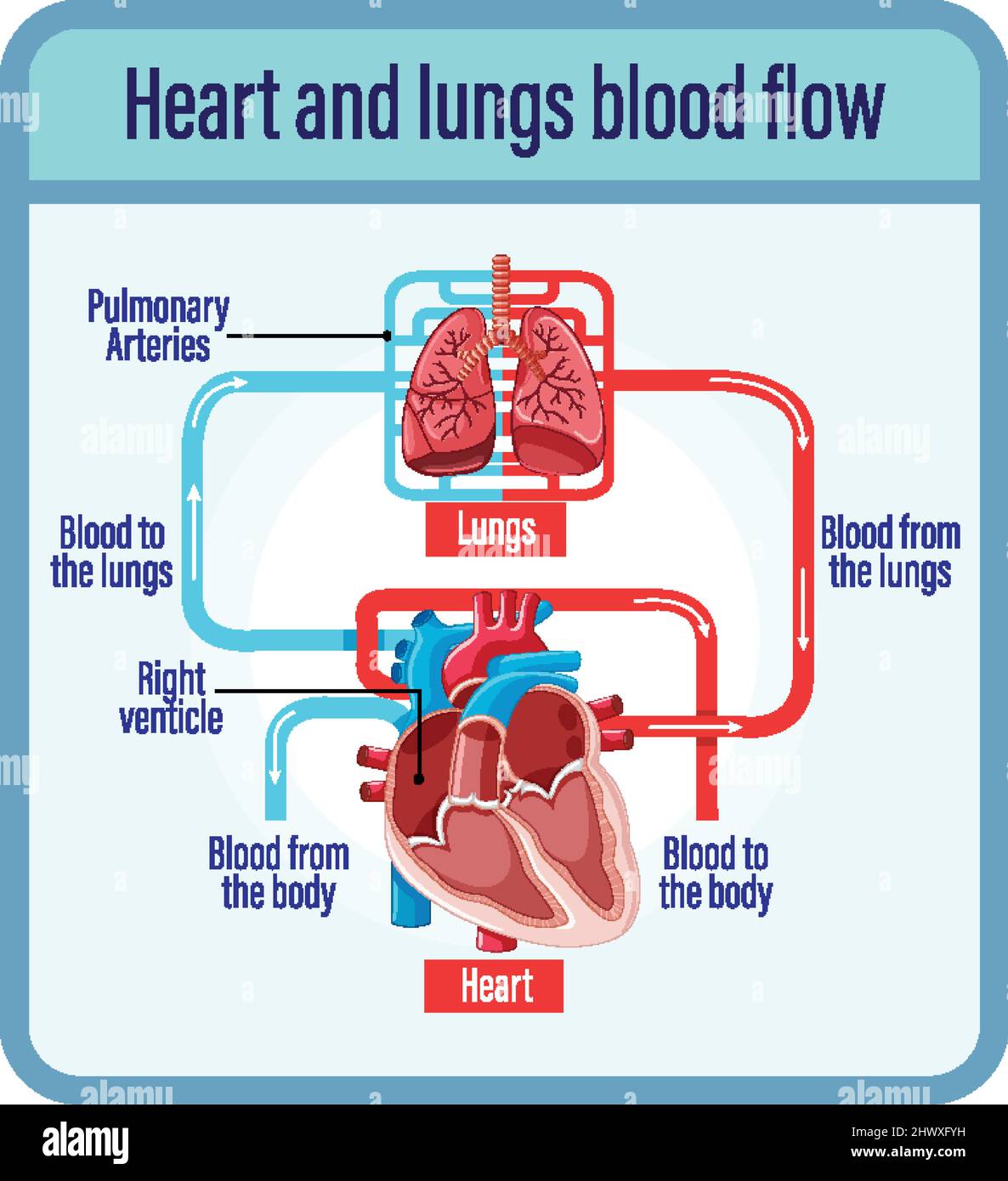 Diagram showing blood flow of human heart illustration Stock Vectorhttps://www.alamy.com/image-license-details/?v=1https://www.alamy.com/diagram-showing-blood-flow-of-human-heart-illustration-image463419253.html
RF2HWXFYH–Diagram showing blood flow of human heart illustration
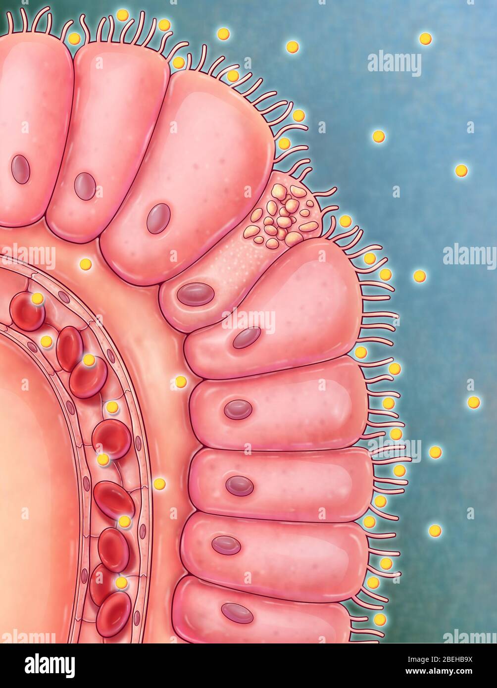 An illustrated close up view of an intestinal villus. Villi are finger-like projections extending into the lumen of the small intestine that absorb digested nutrients. Each villus is lined with columnar epithelium, known as enterocytes, with each cell posessing microvilli to increase surface area for more efficient absorption. Digested nutrients are abosorbed into nearby villus capillaries so that it can then be transported to the rest of the body. Stock Photohttps://www.alamy.com/image-license-details/?v=1https://www.alamy.com/an-illustrated-close-up-view-of-an-intestinal-villus-villi-are-finger-like-projections-extending-into-the-lumen-of-the-small-intestine-that-absorb-digested-nutrients-each-villus-is-lined-with-columnar-epithelium-known-as-enterocytes-with-each-cell-posessing-microvilli-to-increase-surface-area-for-more-efficient-absorption-digested-nutrients-are-abosorbed-into-nearby-villus-capillaries-so-that-it-can-then-be-transported-to-the-rest-of-the-body-image353194630.html
RM2BEHB9X–An illustrated close up view of an intestinal villus. Villi are finger-like projections extending into the lumen of the small intestine that absorb digested nutrients. Each villus is lined with columnar epithelium, known as enterocytes, with each cell posessing microvilli to increase surface area for more efficient absorption. Digested nutrients are abosorbed into nearby villus capillaries so that it can then be transported to the rest of the body.
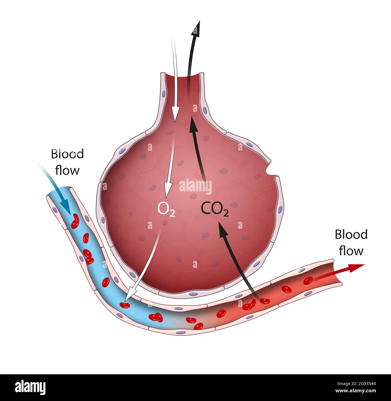 Alveoli are an important part of the respiratory system whose function it is to exchange oxygen and carbon dioxide molecules to and from the bloodstre Stock Photohttps://www.alamy.com/image-license-details/?v=1https://www.alamy.com/alveoli-are-an-important-part-of-the-respiratory-system-whose-function-it-is-to-exchange-oxygen-and-carbon-dioxide-molecules-to-and-from-the-bloodstre-image433402372.html
RF2G53544–Alveoli are an important part of the respiratory system whose function it is to exchange oxygen and carbon dioxide molecules to and from the bloodstre
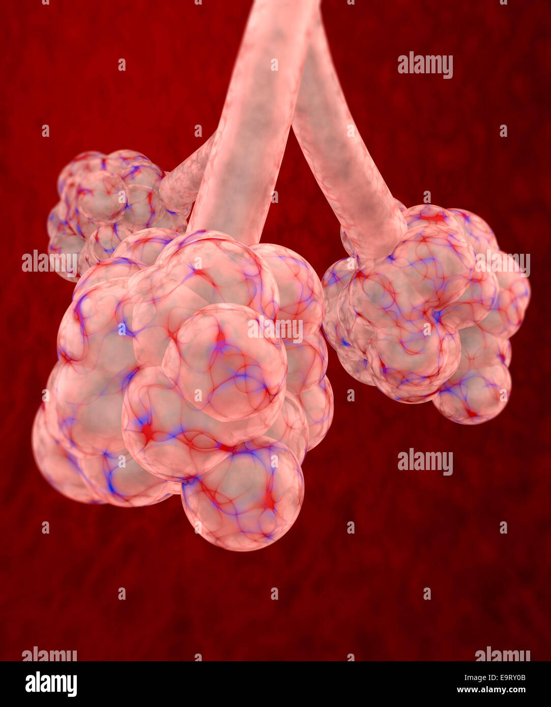 3d pulmonary alveolus on red background Stock Photohttps://www.alamy.com/image-license-details/?v=1https://www.alamy.com/stock-photo-3d-pulmonary-alveolus-on-red-background-74899451.html
RFE9RY0B–3d pulmonary alveolus on red background
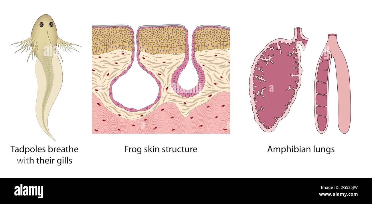 The respiratory system change from tadpoles to adult frogs. Amphibian lungs, Frog skin structure, Tadpoles gills Stock Photohttps://www.alamy.com/image-license-details/?v=1https://www.alamy.com/the-respiratory-system-change-from-tadpoles-to-adult-frogs-amphibian-lungs-frog-skin-structure-tadpoles-gills-image433402785.html
RF2G535JW–The respiratory system change from tadpoles to adult frogs. Amphibian lungs, Frog skin structure, Tadpoles gills
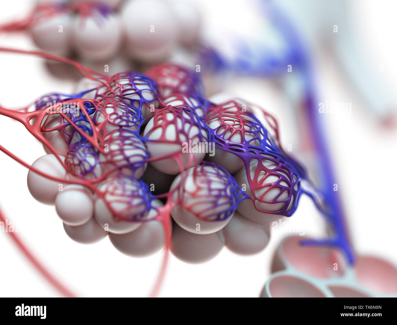 3d rendered illustration of the human alveoli Stock Photohttps://www.alamy.com/image-license-details/?v=1https://www.alamy.com/3d-rendered-illustration-of-the-human-alveoli-image257074405.html
RFTX6N0N–3d rendered illustration of the human alveoli
 Illustration of the human alveoli. Stock Photohttps://www.alamy.com/image-license-details/?v=1https://www.alamy.com/illustration-of-the-human-alveoli-image228979236.html
RFR8EW9T–Illustration of the human alveoli.
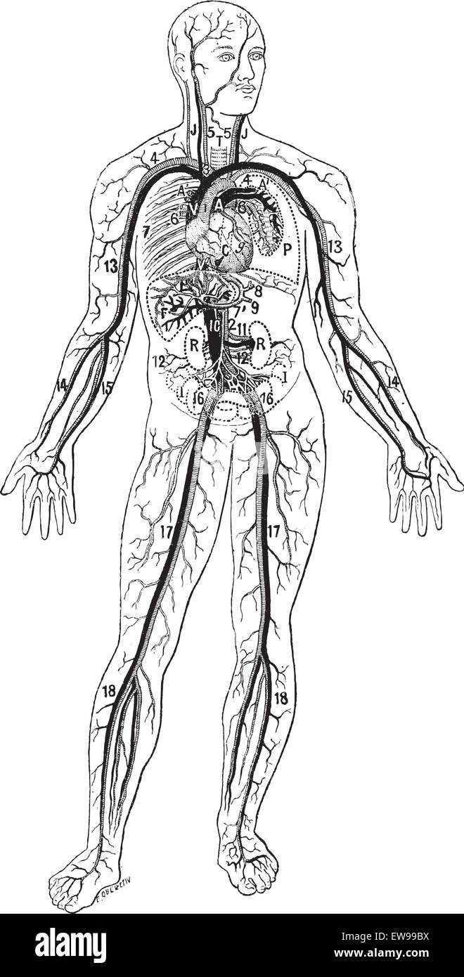 Blood vessels, vintage engraved illustration. Dictionary of words and things - Larive and Fleury - 1895. Stock Vectorhttps://www.alamy.com/image-license-details/?v=1https://www.alamy.com/stock-photo-blood-vessels-vintage-engraved-illustration-dictionary-of-words-and-84412830.html
RFEW99BX–Blood vessels, vintage engraved illustration. Dictionary of words and things - Larive and Fleury - 1895.
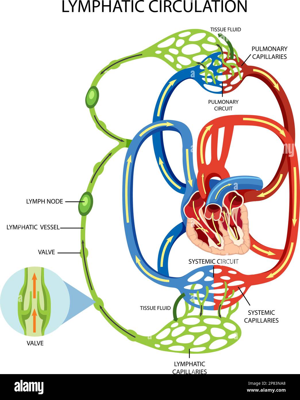 Lymphatic Circulation System Diagram illustration Stock Vectorhttps://www.alamy.com/image-license-details/?v=1https://www.alamy.com/lymphatic-circulation-system-diagram-illustration-image538521264.html
RF2P83NA8–Lymphatic Circulation System Diagram illustration
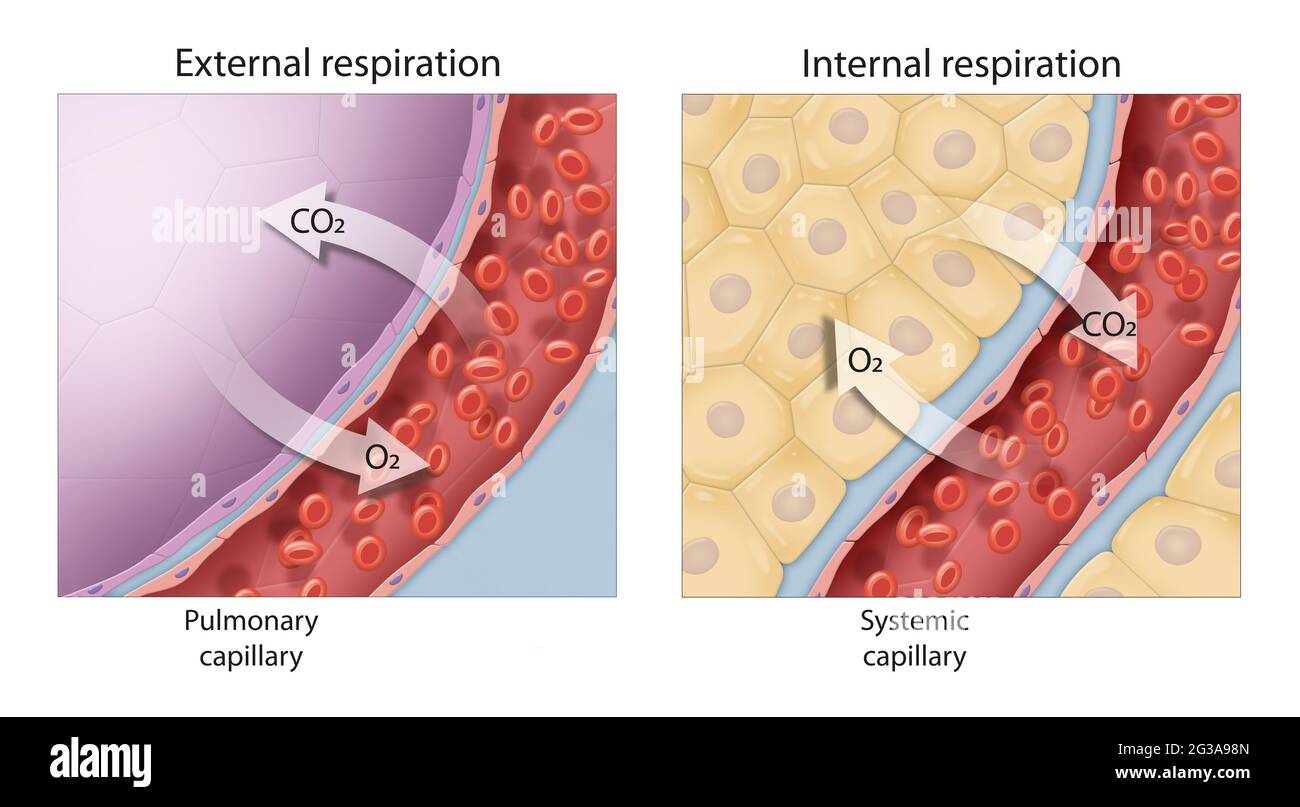 Gas exchange. External and internal respiration. External respiration is the exchange of gases with the external environment, and occurs in the alveol Stock Photohttps://www.alamy.com/image-license-details/?v=1https://www.alamy.com/gas-exchange-external-and-internal-respiration-external-respiration-is-the-exchange-of-gases-with-the-external-environment-and-occurs-in-the-alveol-image432329989.html
RF2G3A98N–Gas exchange. External and internal respiration. External respiration is the exchange of gases with the external environment, and occurs in the alveol
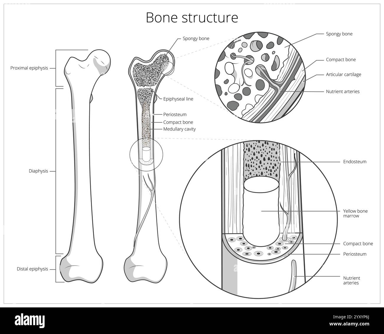 Bone structure medical educational vector Stock Vectorhttps://www.alamy.com/image-license-details/?v=1https://www.alamy.com/bone-structure-medical-educational-vector-image636164442.html
RF2YXYP6J–Bone structure medical educational vector
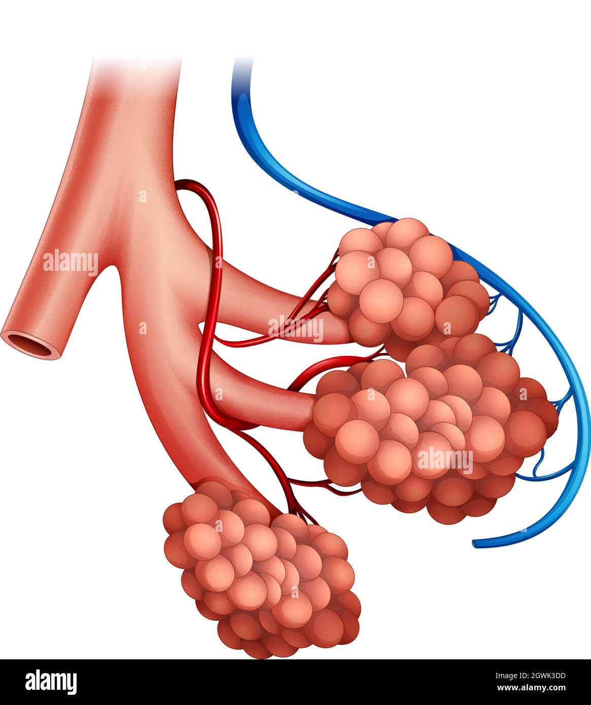 Human alveoli Stock Vectorhttps://www.alamy.com/image-license-details/?v=1https://www.alamy.com/human-alveoli-image446045417.html
RF2GWK3DD–Human alveoli
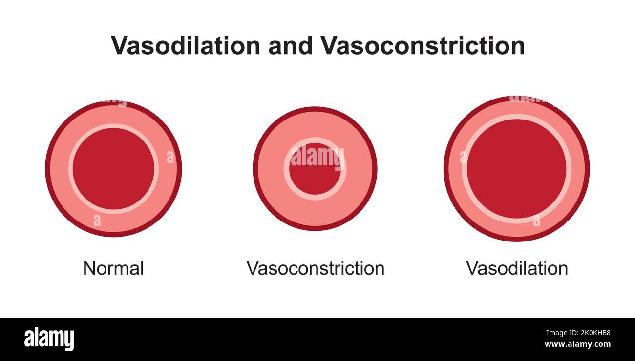 Scientific Designing of Arterial Vasoconstriction and Vasodilation. Comparaison Between Normal, Constricted And Dilated Blood Vessels. Vector. Stock Vectorhttps://www.alamy.com/image-license-details/?v=1https://www.alamy.com/scientific-designing-of-arterial-vasoconstriction-and-vasodilation-comparaison-between-normal-constricted-and-dilated-blood-vessels-vector-image482321036.html
RF2K0KHB8–Scientific Designing of Arterial Vasoconstriction and Vasodilation. Comparaison Between Normal, Constricted And Dilated Blood Vessels. Vector.
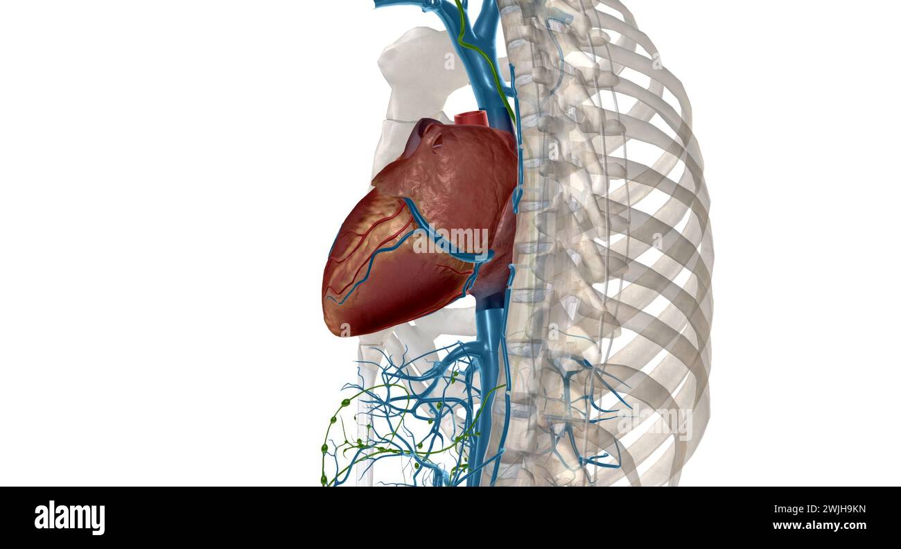 Interstitial fluid is collected by lymph capillaries from the interstitial space. Lymph then moves through lymphatic vessels to lymph nodes. 3D render Stock Photohttps://www.alamy.com/image-license-details/?v=1https://www.alamy.com/interstitial-fluid-is-collected-by-lymph-capillaries-from-the-interstitial-space-lymph-then-moves-through-lymphatic-vessels-to-lymph-nodes-3d-render-image596597113.html
RF2WJH9KN–Interstitial fluid is collected by lymph capillaries from the interstitial space. Lymph then moves through lymphatic vessels to lymph nodes. 3D render
 Alveoli in Lungs Facilitating Respiration and Oxygenation Stock Photohttps://www.alamy.com/image-license-details/?v=1https://www.alamy.com/alveoli-in-lungs-facilitating-respiration-and-oxygenation-image628160614.html
RF2YDY57J–Alveoli in Lungs Facilitating Respiration and Oxygenation
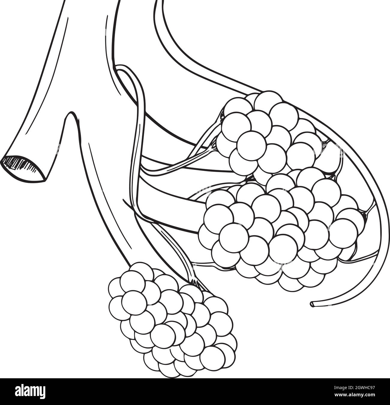 Human alveoli Stock Vectorhttps://www.alamy.com/image-license-details/?v=1https://www.alamy.com/human-alveoli-image446008451.html
RF2GWHC97–Human alveoli
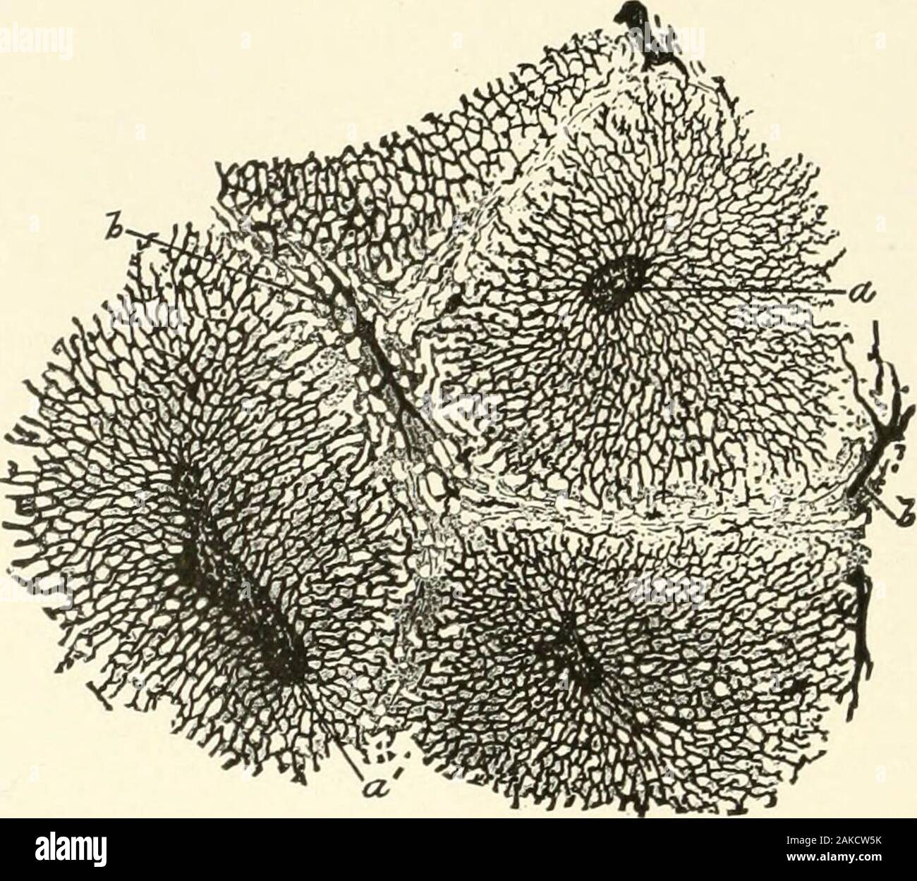 Textbook of normal histology: including an account of the development of the tissues and of the organs . Diagram of the structure of the liver:P. V., the portal or interlobular vein,which breaks up into the capillary net-workof the lobule; H. V., central intralobularvein, a branch of the hepatic; H. A., he-patic artery, supplying nutrition to the in-terlobular structures and terminating in thelobular capillary net-work ; B.D., the inter-lobular bile-duct which takes up the bile-capillaries at the periphery of the lobule. !^8 NORMAL HISTOLOGY. secreting hepatic tissue, comprising the liver-cell Stock Photohttps://www.alamy.com/image-license-details/?v=1https://www.alamy.com/textbook-of-normal-histology-including-an-account-of-the-development-of-the-tissues-and-of-the-organs-diagram-of-the-structure-of-the-liverp-v-the-portal-or-interlobular-veinwhich-breaks-up-into-the-capillary-net-workof-the-lobule-h-v-central-intralobularvein-a-branch-of-the-hepatic-h-a-he-patic-artery-supplying-nutrition-to-the-in-terlobular-structures-and-terminating-in-thelobular-capillary-net-work-bd-the-inter-lobular-bile-duct-which-takes-up-the-bile-capillaries-at-the-periphery-of-the-lobule-!8-normal-histology-secreting-hepatic-tissue-comprising-the-liver-cell-image338958639.html
RM2AKCW5K–Textbook of normal histology: including an account of the development of the tissues and of the organs . Diagram of the structure of the liver:P. V., the portal or interlobular vein,which breaks up into the capillary net-workof the lobule; H. V., central intralobularvein, a branch of the hepatic; H. A., he-patic artery, supplying nutrition to the in-terlobular structures and terminating in thelobular capillary net-work ; B.D., the inter-lobular bile-duct which takes up the bile-capillaries at the periphery of the lobule. !^8 NORMAL HISTOLOGY. secreting hepatic tissue, comprising the liver-cell
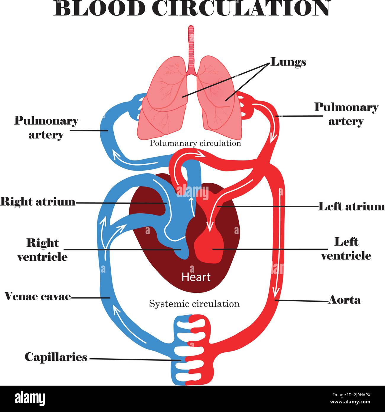 Blood circulation system.Human blood circulation anatomy and digram,heart and lungs.Educational content for biology,medcine and science students. Stock Vectorhttps://www.alamy.com/image-license-details/?v=1https://www.alamy.com/blood-circulation-systemhuman-blood-circulation-anatomy-and-digramheart-and-lungseducational-content-for-biologymedcine-and-science-students-image470593506.html
RF2J9HAPX–Blood circulation system.Human blood circulation anatomy and digram,heart and lungs.Educational content for biology,medcine and science students.
