Quick filters:
Capillaries diagram Stock Photos and Images
 Fish bladders are being prepared for sale. Stock Photohttps://www.alamy.com/image-license-details/?v=1https://www.alamy.com/fish-bladders-are-being-prepared-for-sale-image626735727.html
RF2YBJ7PR–Fish bladders are being prepared for sale.
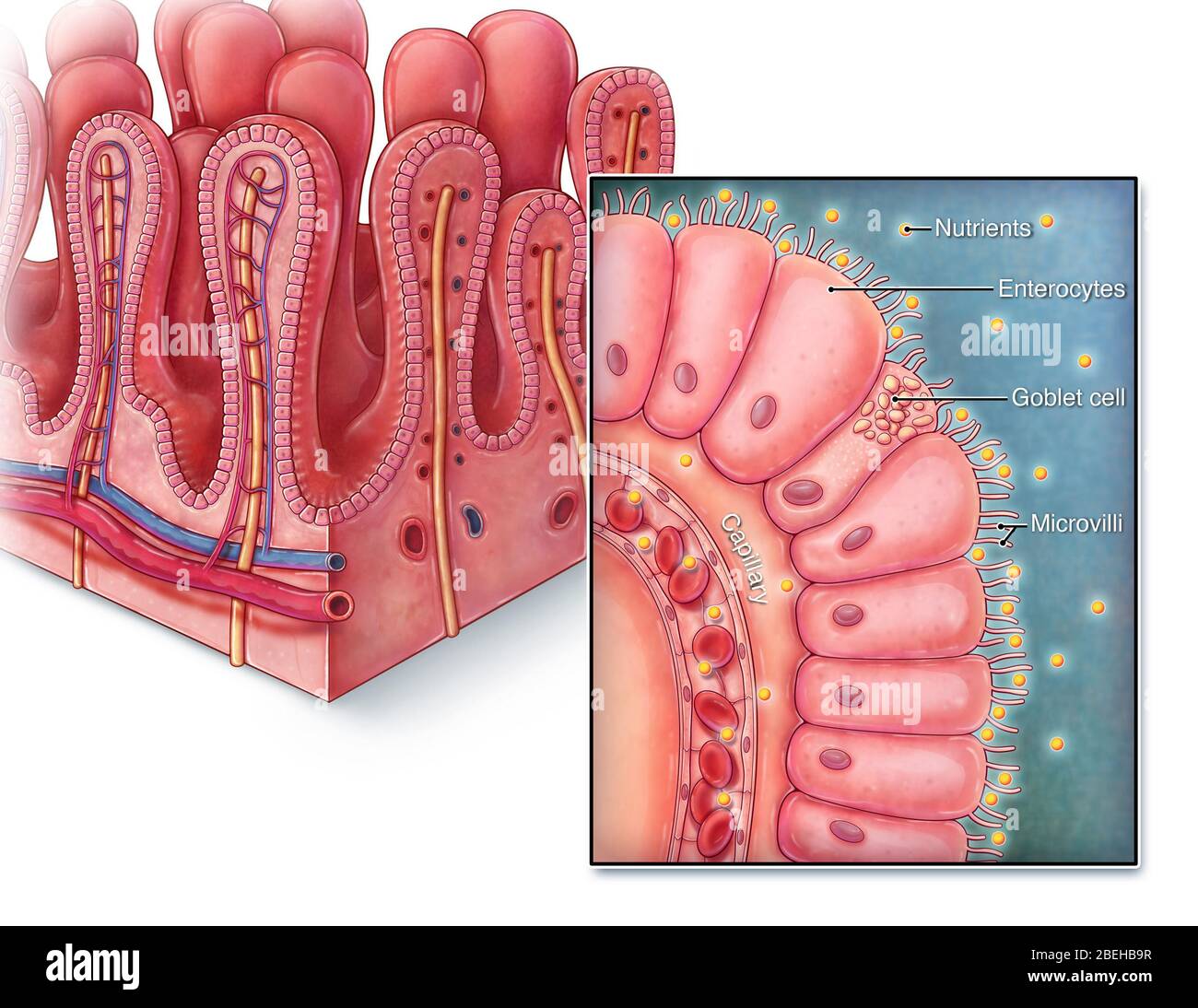 An illustrated section of villi from the small intestine as well as a close up view of a single villus. Villi are finger-like projections that extend into the lumen of the small intestine, increasing surface area for greater nutrient absorption. Each villus is lined with columnar epithelium known as enterocytes, with each cell containing microvilli to further increase surface area. Digested nutrients are absorbed into nearby capillaries so that it can then be transported to the rest of the body. Stock Photohttps://www.alamy.com/image-license-details/?v=1https://www.alamy.com/an-illustrated-section-of-villi-from-the-small-intestine-as-well-as-a-close-up-view-of-a-single-villus-villi-are-finger-like-projections-that-extend-into-the-lumen-of-the-small-intestine-increasing-surface-area-for-greater-nutrient-absorption-each-villus-is-lined-with-columnar-epithelium-known-as-enterocytes-with-each-cell-containing-microvilli-to-further-increase-surface-area-digested-nutrients-are-absorbed-into-nearby-capillaries-so-that-it-can-then-be-transported-to-the-rest-of-the-body-image353194627.html
RM2BEHB9R–An illustrated section of villi from the small intestine as well as a close up view of a single villus. Villi are finger-like projections that extend into the lumen of the small intestine, increasing surface area for greater nutrient absorption. Each villus is lined with columnar epithelium known as enterocytes, with each cell containing microvilli to further increase surface area. Digested nutrients are absorbed into nearby capillaries so that it can then be transported to the rest of the body.
 WRIGHT-PATTERSON AIR FORCE BASE, Ohio – Researchers at the Materials and Manufacturing Directorate, Air Force Research Laboratory, have developed a novel, lightweight artificial hair sensor that mimics those used by natural fliers—like bats and crickets—by using carbon nanotube forests grown inside glass fiber capillaries. The hairs are sensitive to air flow changes during flight, enabling quick analysis and response by agile fliers. The diagram pairs an overview of the hair sensor components with an image captured on a Scanning Electron Microscope. Stock Photohttps://www.alamy.com/image-license-details/?v=1https://www.alamy.com/wright-patterson-air-force-base-ohio-researchers-at-the-materials-and-manufacturing-directorate-air-force-research-laboratory-have-developed-a-novel-lightweight-artificial-hair-sensor-that-mimics-those-used-by-natural-flierslike-bats-and-cricketsby-using-carbon-nanotube-forests-grown-inside-glass-fiber-capillaries-the-hairs-are-sensitive-to-air-flow-changes-during-flight-enabling-quick-analysis-and-response-by-agile-fliers-the-diagram-pairs-an-overview-of-the-hair-sensor-components-with-an-image-captured-on-a-scanning-electron-microscope-image227445180.html
RMR610J4–WRIGHT-PATTERSON AIR FORCE BASE, Ohio – Researchers at the Materials and Manufacturing Directorate, Air Force Research Laboratory, have developed a novel, lightweight artificial hair sensor that mimics those used by natural fliers—like bats and crickets—by using carbon nanotube forests grown inside glass fiber capillaries. The hairs are sensitive to air flow changes during flight, enabling quick analysis and response by agile fliers. The diagram pairs an overview of the hair sensor components with an image captured on a Scanning Electron Microscope.
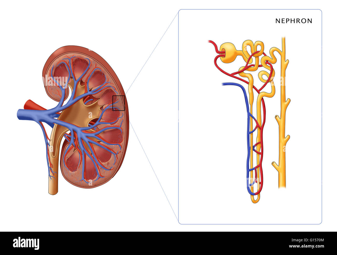 Illustration of the structure of a nephron (the basic structural and functional unit of the kidney) along side a cross section of a kidney. Depicted in the nephron are the glomerulus, bowman's capsule, proximal convoluted tubule, peritubular capillaries, Stock Photohttps://www.alamy.com/image-license-details/?v=1https://www.alamy.com/stock-photo-illustration-of-the-structure-of-a-nephron-the-basic-structural-and-103992132.html
RMG1570M–Illustration of the structure of a nephron (the basic structural and functional unit of the kidney) along side a cross section of a kidney. Depicted in the nephron are the glomerulus, bowman's capsule, proximal convoluted tubule, peritubular capillaries,
 . Elementary biology, animal and human. Biology. 132 ANIMAL BIOLOGY. Fig. 99. - Diagram of the circula- tion of a fish. The blood passes from the capillaries into the veins,* which are thinner-vailed than the arteries. These veins carry the blood back to the heart. The heart (Fig. 100) consists of two principal parts; a thin- walled auricle which receives blood from the veins, and a thick-walled, muscular portion called the ventricle, which forces the blood out into the arteries. 100. Adaptations for breath- ing. — Laboratory study. 1. Raise the gill covers of a preserved fish and find the gil Stock Photohttps://www.alamy.com/image-license-details/?v=1https://www.alamy.com/elementary-biology-animal-and-human-biology-132-animal-biology-fig-99-diagram-of-the-circula-tion-of-a-fish-the-blood-passes-from-the-capillaries-into-the-veins-which-are-thinner-vailed-than-the-arteries-these-veins-carry-the-blood-back-to-the-heart-the-heart-fig-100-consists-of-two-principal-parts-a-thin-walled-auricle-which-receives-blood-from-the-veins-and-a-thick-walled-muscular-portion-called-the-ventricle-which-forces-the-blood-out-into-the-arteries-100-adaptations-for-breath-ing-laboratory-study-1-raise-the-gill-covers-of-a-preserved-fish-and-find-the-gil-image216376905.html
RMPG0PXH–. Elementary biology, animal and human. Biology. 132 ANIMAL BIOLOGY. Fig. 99. - Diagram of the circula- tion of a fish. The blood passes from the capillaries into the veins,* which are thinner-vailed than the arteries. These veins carry the blood back to the heart. The heart (Fig. 100) consists of two principal parts; a thin- walled auricle which receives blood from the veins, and a thick-walled, muscular portion called the ventricle, which forces the blood out into the arteries. 100. Adaptations for breath- ing. — Laboratory study. 1. Raise the gill covers of a preserved fish and find the gil
RFPF9516–Web line icon. Alveolus black on white background
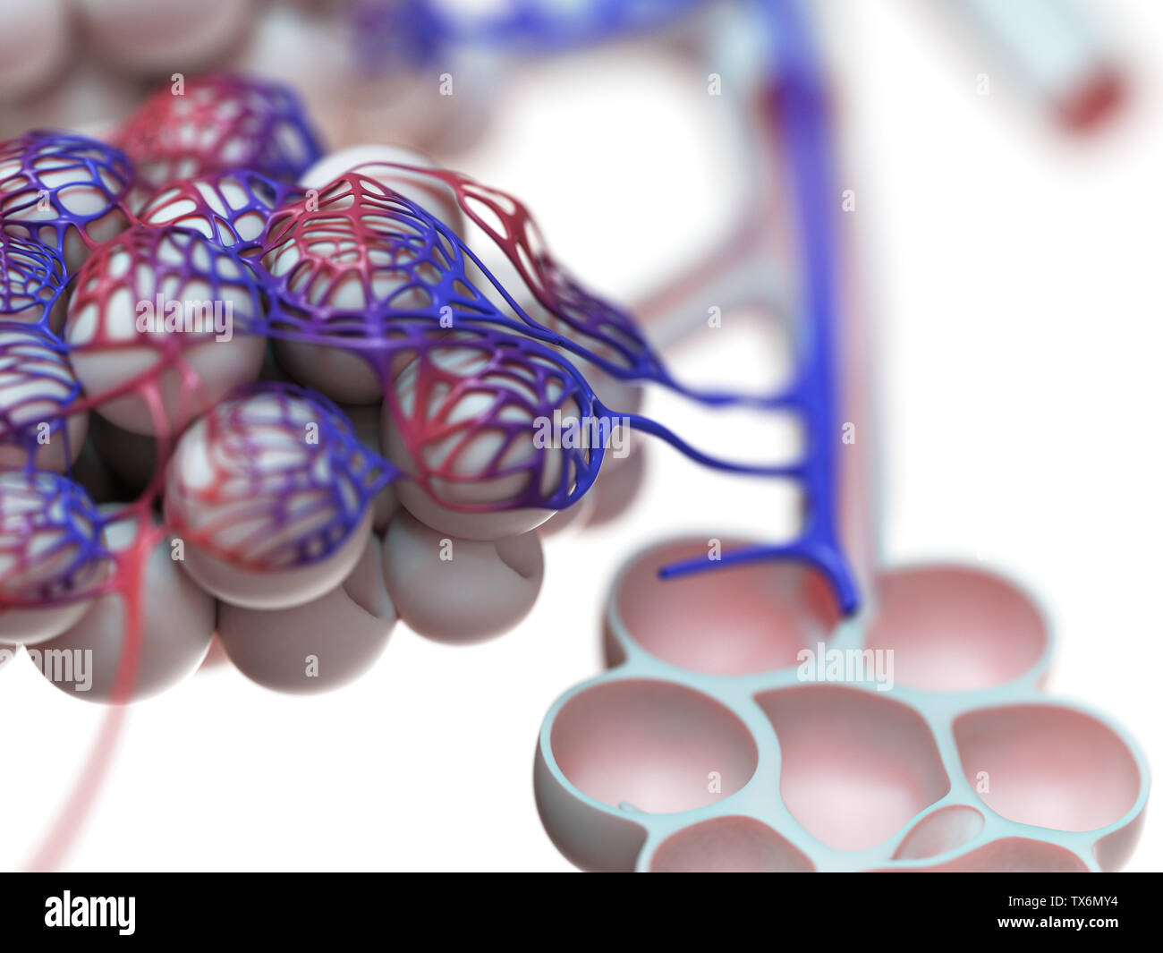 3d rendered illustration of the human alveoli Stock Photohttps://www.alamy.com/image-license-details/?v=1https://www.alamy.com/3d-rendered-illustration-of-the-human-alveoli-image257074360.html
RFTX6MY4–3d rendered illustration of the human alveoli
 Illustration of the human alveoli. Stock Photohttps://www.alamy.com/image-license-details/?v=1https://www.alamy.com/illustration-of-the-human-alveoli-image228979235.html
RFR8EW9R–Illustration of the human alveoli.
RF2S29X64–Virus alveoli icon. Simple illustration of virus alveoli vector icon for web design isolated on white background
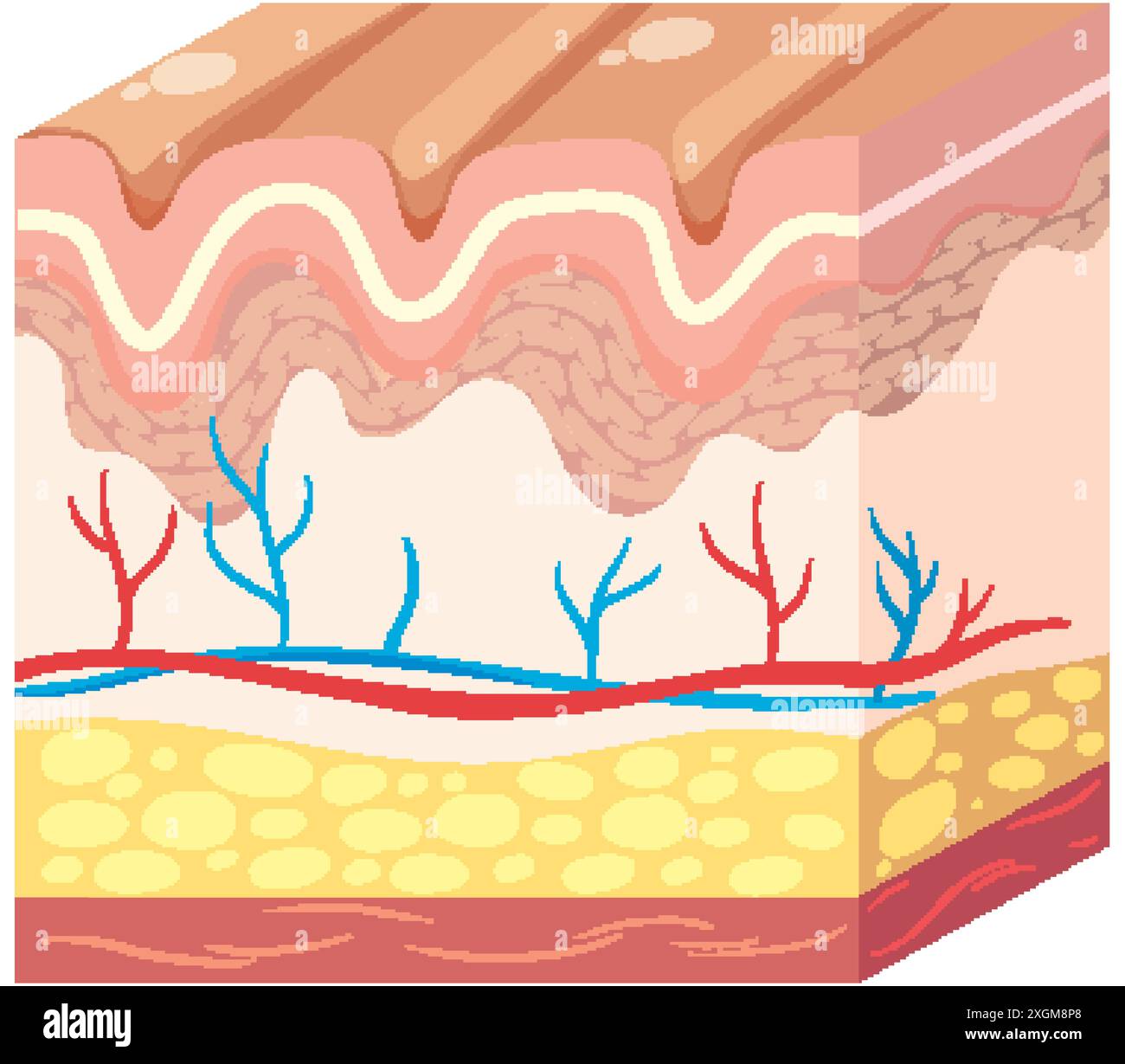 Detailed illustration of skin layers and blood vessels Stock Vectorhttps://www.alamy.com/image-license-details/?v=1https://www.alamy.com/detailed-illustration-of-skin-layers-and-blood-vessels-image612643312.html
RF2XGM8P8–Detailed illustration of skin layers and blood vessels
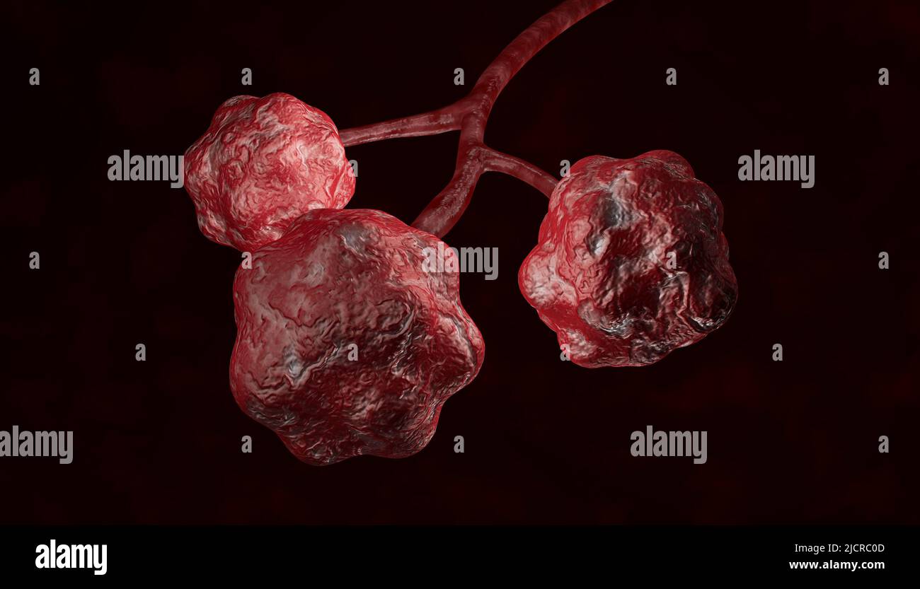 3d Rendering of Alveoli in lungs. Clipping path included Stock Photohttps://www.alamy.com/image-license-details/?v=1https://www.alamy.com/3d-rendering-of-alveoli-in-lungs-clipping-path-included-image472570125.html
RF2JCRC0D–3d Rendering of Alveoli in lungs. Clipping path included
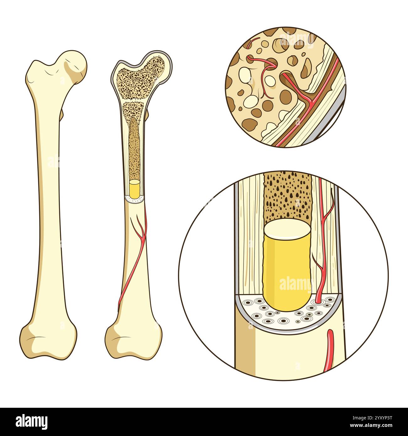 Bone structure medical educational vector Stock Vectorhttps://www.alamy.com/image-license-details/?v=1https://www.alamy.com/bone-structure-medical-educational-vector-image636164364.html
RF2YXYP3T–Bone structure medical educational vector
RF2WA6KHT–Pancreatic cell color line icon. Microorganisms microbes, bacteria. Vector isolated element. Editable stroke.
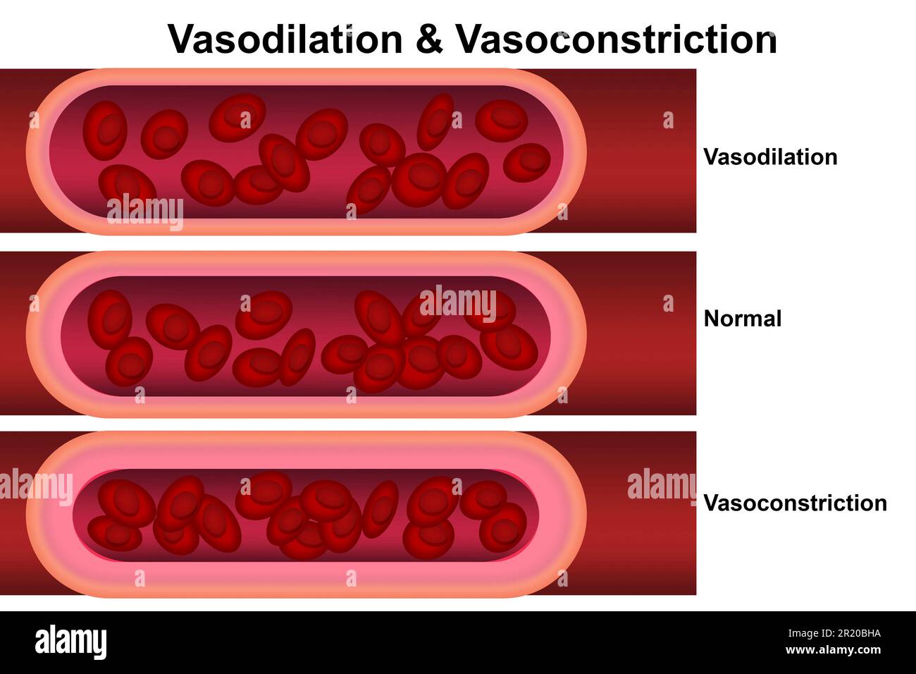 Vasodilation and vasoconstriction. Comparison of Blood vessels, 3d rendering Stock Photohttps://www.alamy.com/image-license-details/?v=1https://www.alamy.com/vasodilation-and-vasoconstriction-comparison-of-blood-vessels-3d-rendering-image551970198.html
RF2R20BHA–Vasodilation and vasoconstriction. Comparison of Blood vessels, 3d rendering
 Alveoli in Lungs Facilitating Respiration and Oxygenation Stock Photohttps://www.alamy.com/image-license-details/?v=1https://www.alamy.com/alveoli-in-lungs-facilitating-respiration-and-oxygenation-image628160618.html
RF2YDY57P–Alveoli in Lungs Facilitating Respiration and Oxygenation
RF2T63F7M–Pancreatic cell color line icon. Microorganisms microbes, bacteria. Vector isolated element. Editable stroke.
 Interstitial fluid is collected by lymph capillaries from the interstitial space. Lymph then moves through lymphatic vessels to lymph nodes. 3D render Stock Photohttps://www.alamy.com/image-license-details/?v=1https://www.alamy.com/interstitial-fluid-is-collected-by-lymph-capillaries-from-the-interstitial-space-lymph-then-moves-through-lymphatic-vessels-to-lymph-nodes-3d-render-image596595676.html
RF2WJH7TC–Interstitial fluid is collected by lymph capillaries from the interstitial space. Lymph then moves through lymphatic vessels to lymph nodes. 3D render
 A text-book of first aid and emergency treatment . Median cephalic External /;cnUmeous uerie. ^/ :-|- FiG. 17.—Front view of the super- Fig. 18.—Superficial veins of the ficial veins of the arm, forearm, and front and inner surface of the lowerhand. extremity. (Gray.) PLATE II Ili/iiKiiiiiri/ < ajji/liiIiis. Diagram to Show the Course of the Circulationof the Blood. The pulmonary capillaries are the small vessels of thelungs. The systemic capillaries represent tViose of the skin,head and extremities. The third systeni shown representsthe capillaries of the intestmes and liver. THE NERVOUS S Stock Photohttps://www.alamy.com/image-license-details/?v=1https://www.alamy.com/a-text-book-of-first-aid-and-emergency-treatment-median-cephalic-external-cnumeous-uerie-fig-17front-view-of-the-super-fig-18superficial-veins-of-the-ficial-veins-of-the-arm-forearm-and-front-and-inner-surface-of-the-lowerhand-extremity-gray-plate-ii-iliiikiiiiiri-lt-ajjiliiiiis-diagram-to-show-the-course-of-the-circulationof-the-blood-the-pulmonary-capillaries-are-the-small-vessels-of-thelungs-the-systemic-capillaries-represent-tviose-of-the-skinhead-and-extremities-the-third-systeni-shown-representsthe-capillaries-of-the-intestmes-and-liver-the-nervous-s-image340295111.html
RM2ANHNTR–A text-book of first aid and emergency treatment . Median cephalic External /;cnUmeous uerie. ^/ :-|- FiG. 17.—Front view of the super- Fig. 18.—Superficial veins of the ficial veins of the arm, forearm, and front and inner surface of the lowerhand. extremity. (Gray.) PLATE II Ili/iiKiiiiiri/ < ajji/liiIiis. Diagram to Show the Course of the Circulationof the Blood. The pulmonary capillaries are the small vessels of thelungs. The systemic capillaries represent tViose of the skin,head and extremities. The third systeni shown representsthe capillaries of the intestmes and liver. THE NERVOUS S
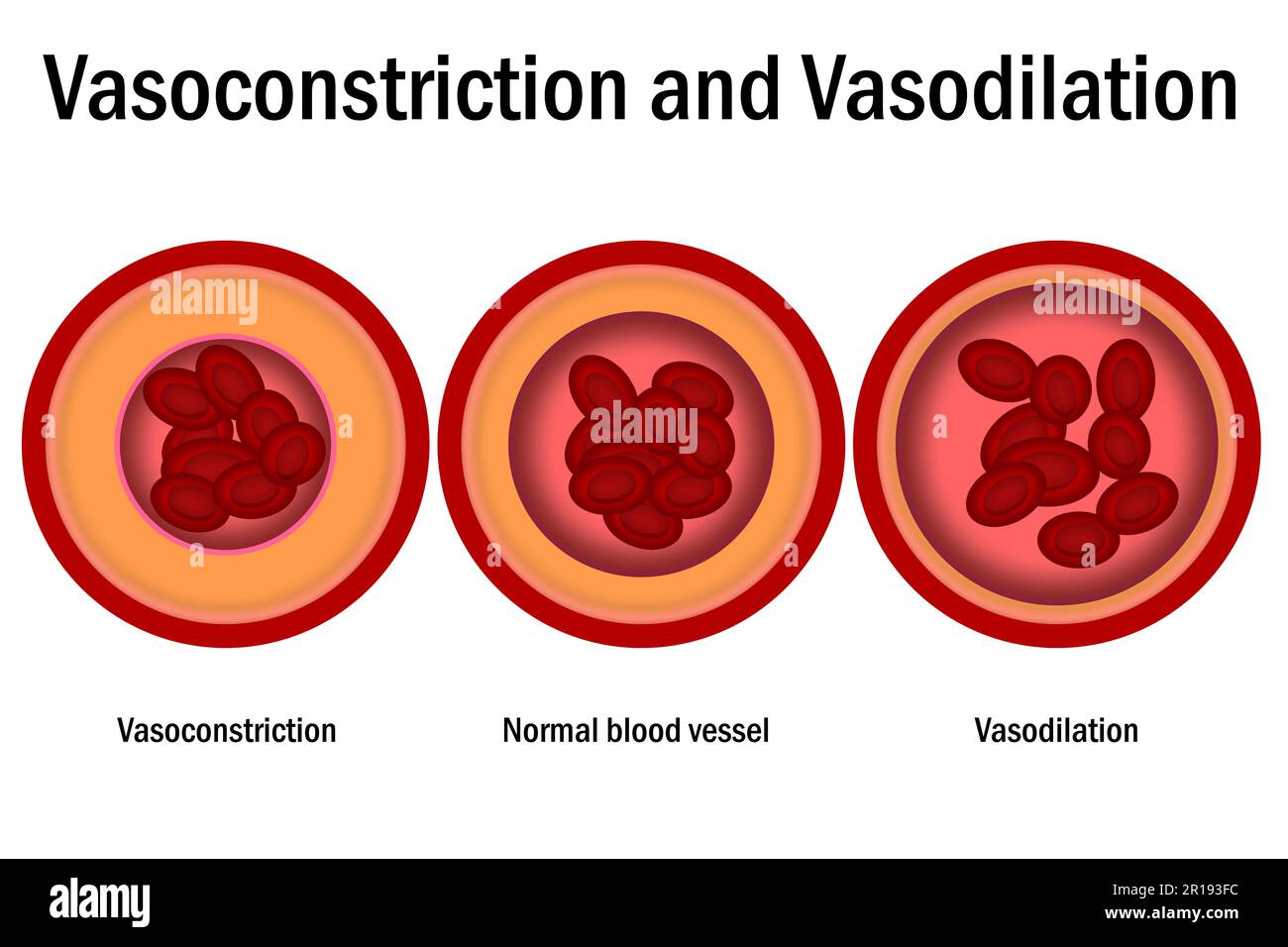 Comparison of normal, vasoconstriction and vasodilation blood vessels with cross section of arteries, 3d rende Stock Photohttps://www.alamy.com/image-license-details/?v=1https://www.alamy.com/comparison-of-normal-vasoconstriction-and-vasodilation-blood-vessels-with-cross-section-of-arteries-3d-rende-image551546784.html
RF2R193FC–Comparison of normal, vasoconstriction and vasodilation blood vessels with cross section of arteries, 3d rende
 Fish bladders are being prepared for sale. Stock Photohttps://www.alamy.com/image-license-details/?v=1https://www.alamy.com/fish-bladders-are-being-prepared-for-sale-image626735640.html
RF2YBJ7KM–Fish bladders are being prepared for sale.
RFR5B1H2–Normal healthy alveoli icon. Realistic illustration of normal healthy alveoli vector icon for web design isolated on white background
 . Capillaries of the Head, etc. Pulmonary Ca- pillaries. Main Arterial Trunk. Capillaries of Splanchnic Area. Capillaries of Trunk and Lower Ex- tremities. Fig. 14.—Diagram of the Circulation. The arrows indicate the course of the blood. Though the pulmonary, the lower and the upper parts of the systemic circulation are represented so as to show the distinctness of each, it will be also apparent that they are not independent. Relative size of different parts of the system is only very generally indicated. it has served its purpose, is in great part removed by another set of vessels, very like Stock Photohttps://www.alamy.com/image-license-details/?v=1https://www.alamy.com/capillaries-of-the-head-etc-pulmonary-ca-pillaries-main-arterial-trunk-capillaries-of-splanchnic-area-capillaries-of-trunk-and-lower-ex-tremities-fig-14diagram-of-the-circulation-the-arrows-indicate-the-course-of-the-blood-though-the-pulmonary-the-lower-and-the-upper-parts-of-the-systemic-circulation-are-represented-so-as-to-show-the-distinctness-of-each-it-will-be-also-apparent-that-they-are-not-independent-relative-size-of-different-parts-of-the-system-is-only-very-generally-indicated-it-has-served-its-purpose-is-in-great-part-removed-by-another-set-of-vessels-very-like-image179906794.html
RMMCKCXJ–. Capillaries of the Head, etc. Pulmonary Ca- pillaries. Main Arterial Trunk. Capillaries of Splanchnic Area. Capillaries of Trunk and Lower Ex- tremities. Fig. 14.—Diagram of the Circulation. The arrows indicate the course of the blood. Though the pulmonary, the lower and the upper parts of the systemic circulation are represented so as to show the distinctness of each, it will be also apparent that they are not independent. Relative size of different parts of the system is only very generally indicated. it has served its purpose, is in great part removed by another set of vessels, very like
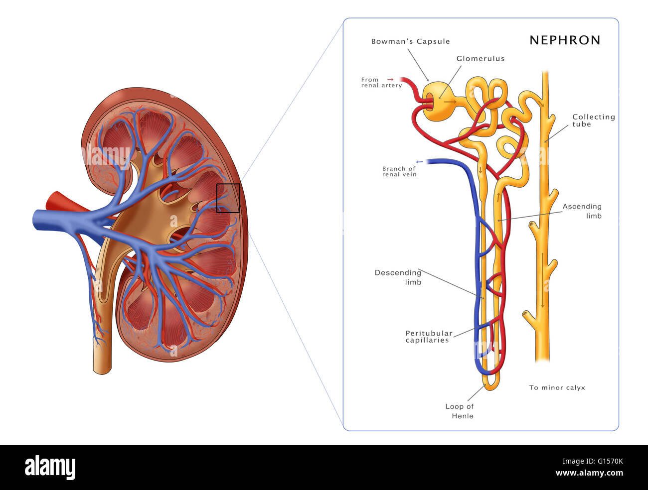 Illustration of the structure of a nephron (the basic structural and functional unit of the kidney) along side a cross section of a kidney. Depicted in the nephron are the glomerulus, bowman's capsule, proximal convoluted tubule, peritubular capillaries, Stock Photohttps://www.alamy.com/image-license-details/?v=1https://www.alamy.com/stock-photo-illustration-of-the-structure-of-a-nephron-the-basic-structural-and-103992131.html
RMG1570K–Illustration of the structure of a nephron (the basic structural and functional unit of the kidney) along side a cross section of a kidney. Depicted in the nephron are the glomerulus, bowman's capsule, proximal convoluted tubule, peritubular capillaries,
 . Elements of the comparative anatomy of vertebrates. Anatomy, Comparative. 324 COMPARATIVE ANATOMY. Fig. 265.—Diagram of Stages in the Development of the Veins in Elasmo- ERANCHS. (I—XI after Rabl, XII after F. Hoohstetter.) Oa, Cp, anterior and posterior cardinal veins ; Cdr, caudal vein ; D,D, vitelline veins ; DC, precaval vein or sinus ; CI, region of the cloaca; H, sinus venosus of heart; /, subintestinal vein ; Jr. V, interrenal vein; Lb, hepatic veins ; **, hepatic sinus ; N'pf, renal portal system ; VP, hepatic portal vein; Vpo, capillaries of the hepatic portal system; t, cardinal si Stock Photohttps://www.alamy.com/image-license-details/?v=1https://www.alamy.com/elements-of-the-comparative-anatomy-of-vertebrates-anatomy-comparative-324-comparative-anatomy-fig-265diagram-of-stages-in-the-development-of-the-veins-in-elasmo-eranchs-ixi-after-rabl-xii-after-f-hoohstetter-oa-cp-anterior-and-posterior-cardinal-veins-cdr-caudal-vein-dd-vitelline-veins-dc-precaval-vein-or-sinus-ci-region-of-the-cloaca-h-sinus-venosus-of-heart-subintestinal-vein-jr-v-interrenal-vein-lb-hepatic-veins-hepatic-sinus-npf-renal-portal-system-vp-hepatic-portal-vein-vpo-capillaries-of-the-hepatic-portal-system-t-cardinal-si-image216418677.html
RMPG2M6D–. Elements of the comparative anatomy of vertebrates. Anatomy, Comparative. 324 COMPARATIVE ANATOMY. Fig. 265.—Diagram of Stages in the Development of the Veins in Elasmo- ERANCHS. (I—XI after Rabl, XII after F. Hoohstetter.) Oa, Cp, anterior and posterior cardinal veins ; Cdr, caudal vein ; D,D, vitelline veins ; DC, precaval vein or sinus ; CI, region of the cloaca; H, sinus venosus of heart; /, subintestinal vein ; Jr. V, interrenal vein; Lb, hepatic veins ; **, hepatic sinus ; N'pf, renal portal system ; VP, hepatic portal vein; Vpo, capillaries of the hepatic portal system; t, cardinal si
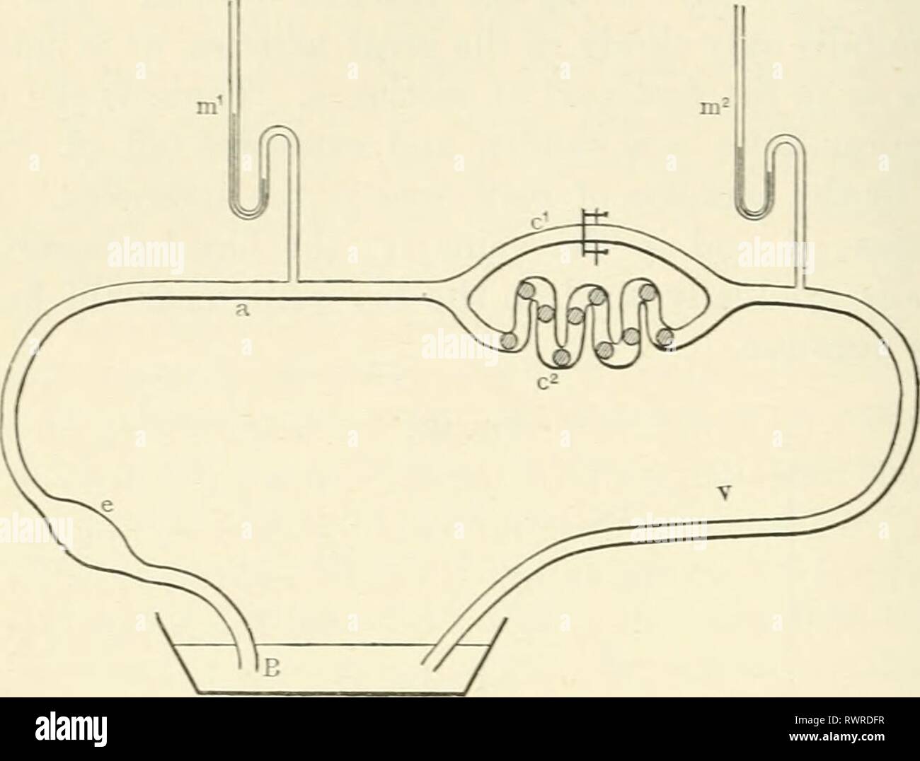 Elements of human physiology (1907) Elements of human physiology elementsofhumanp05star Year: 1907 198 PHYSIOLOGY tube (a) with a tube (c-) ^vhich is packed with sponges to represent the peripheral resistance in the capillaries. From the distal end of (c-) a tube (v) serves to conduct the lluid Fig. 100. Diagram of artificial circulation schema. back to the basin. To side branches of (a) and (v) two mercurial manometers (m' and m-) are connected, and these are arranged to write one below the other on the smoked surface of a kymograph. Another route for the fluid from (a) to (v) is aflbrded Stock Photohttps://www.alamy.com/image-license-details/?v=1https://www.alamy.com/elements-of-human-physiology-1907-elements-of-human-physiology-elementsofhumanp05star-year-1907-198-physiology-tube-a-with-a-tube-c-vhich-is-packed-with-sponges-to-represent-the-peripheral-resistance-in-the-capillaries-from-the-distal-end-of-c-a-tube-v-serves-to-conduct-the-lluid-fig-100-diagram-of-artificial-circulation-schema-back-to-the-basin-to-side-branches-of-a-and-v-two-mercurial-manometers-m-and-m-are-connected-and-these-are-arranged-to-write-one-below-the-other-on-the-smoked-surface-of-a-kymograph-another-route-for-the-fluid-from-a-to-v-is-aflbrded-image239616715.html
RMRWRDFR–Elements of human physiology (1907) Elements of human physiology elementsofhumanp05star Year: 1907 198 PHYSIOLOGY tube (a) with a tube (c-) ^vhich is packed with sponges to represent the peripheral resistance in the capillaries. From the distal end of (c-) a tube (v) serves to conduct the lluid Fig. 100. Diagram of artificial circulation schema. back to the basin. To side branches of (a) and (v) two mercurial manometers (m' and m-) are connected, and these are arranged to write one below the other on the smoked surface of a kymograph. Another route for the fluid from (a) to (v) is aflbrded
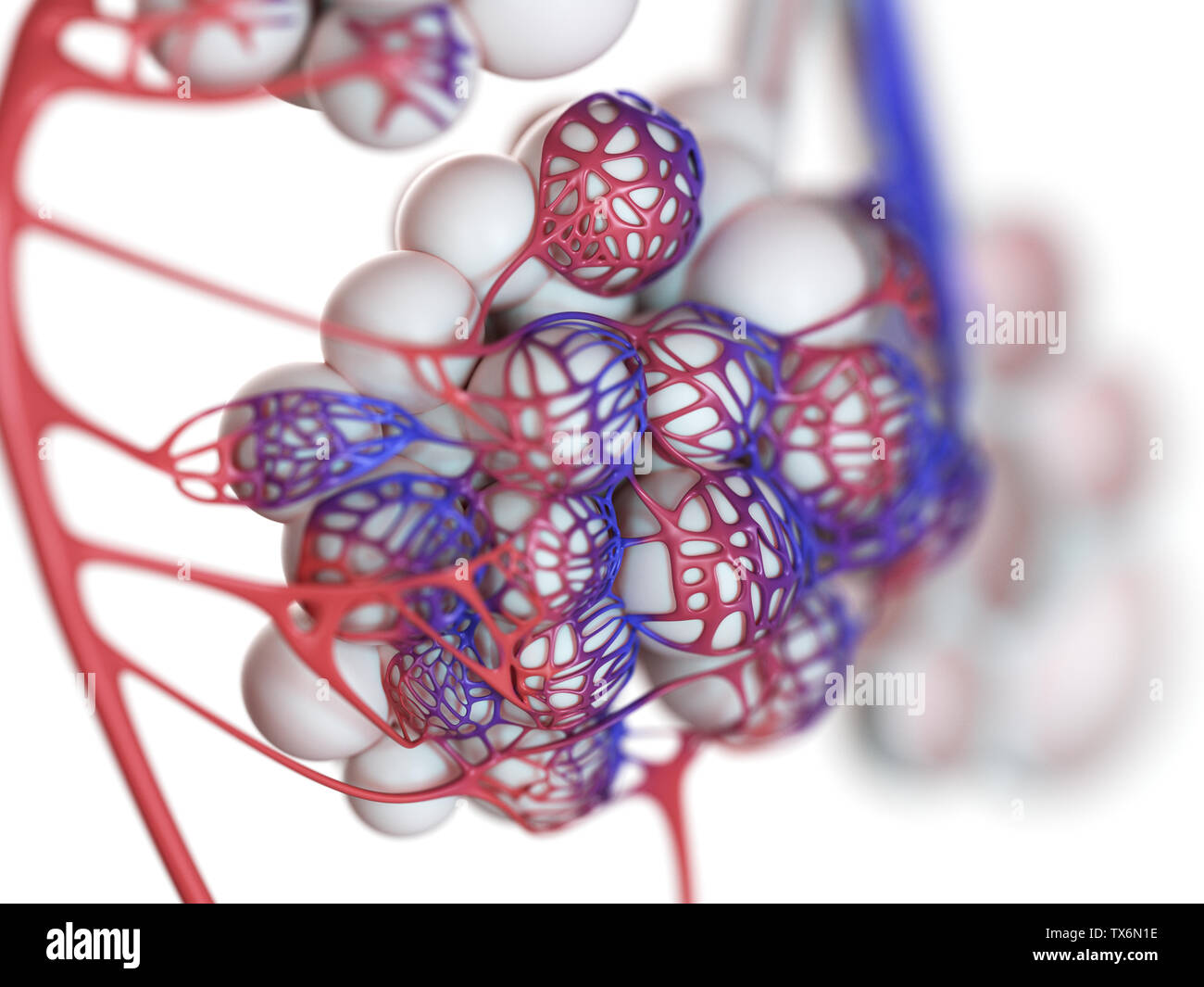 3d rendered illustration of the human alveoli Stock Photohttps://www.alamy.com/image-license-details/?v=1https://www.alamy.com/3d-rendered-illustration-of-the-human-alveoli-image257074426.html
RFTX6N1E–3d rendered illustration of the human alveoli
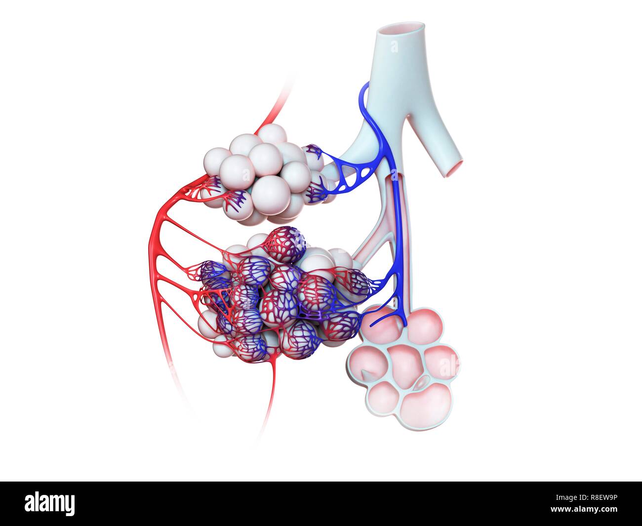 Illustration of the human alveoli. Stock Photohttps://www.alamy.com/image-license-details/?v=1https://www.alamy.com/illustration-of-the-human-alveoli-image228979234.html
RFR8EW9P–Illustration of the human alveoli.
RF2S53C56–Virus alveoli icon. Simple illustration of virus alveoli vector icon for web design isolated on white background
RFTW19RY–Human bone icon. Cartoon of human bone vector icon for web design isolated on white background
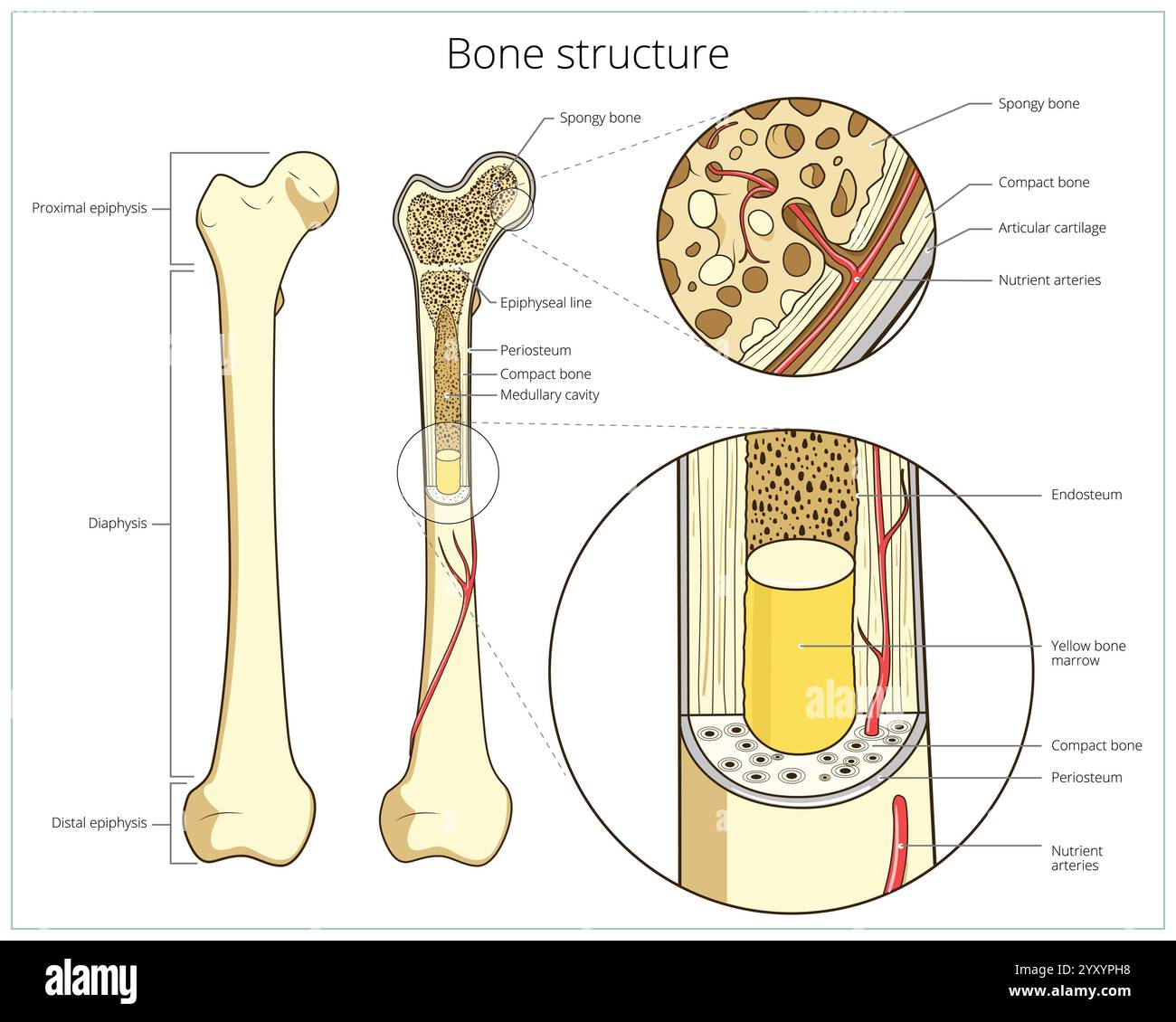 Bone structure medical educational vector Stock Vectorhttps://www.alamy.com/image-license-details/?v=1https://www.alamy.com/bone-structure-medical-educational-vector-image636164740.html
RF2YXYPH8–Bone structure medical educational vector
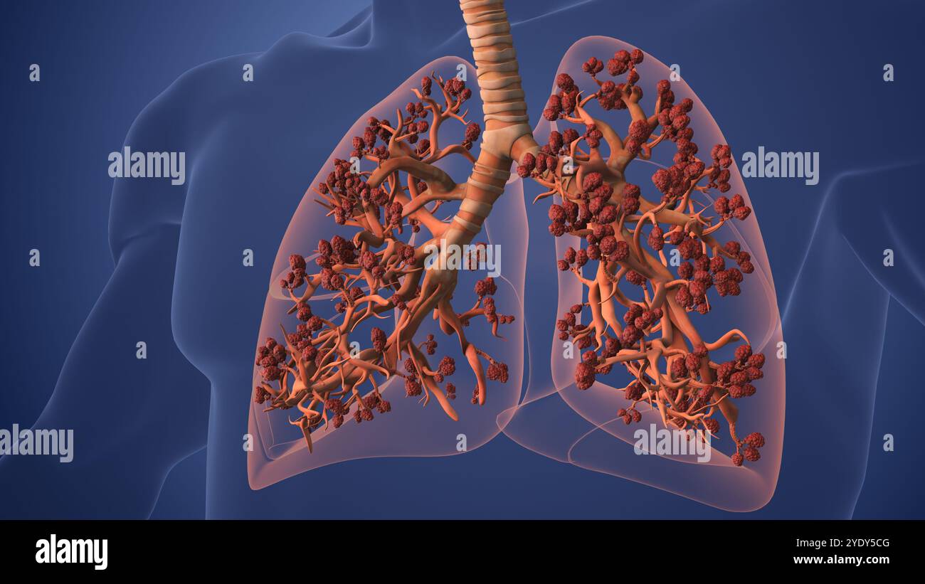 Human Lungs Displaying Alveoli for Oxygen Exchange Stock Photohttps://www.alamy.com/image-license-details/?v=1https://www.alamy.com/human-lungs-displaying-alveoli-for-oxygen-exchange-image628160752.html
RF2YDY5CG–Human Lungs Displaying Alveoli for Oxygen Exchange
 Interstitial fluid is collected by lymph capillaries from the interstitial space. Lymph then moves through lymphatic vessels to lymph nodes. 3D render Stock Photohttps://www.alamy.com/image-license-details/?v=1https://www.alamy.com/interstitial-fluid-is-collected-by-lymph-capillaries-from-the-interstitial-space-lymph-then-moves-through-lymphatic-vessels-to-lymph-nodes-3d-render-image596597712.html
RF2WJHAD4–Interstitial fluid is collected by lymph capillaries from the interstitial space. Lymph then moves through lymphatic vessels to lymph nodes. 3D render
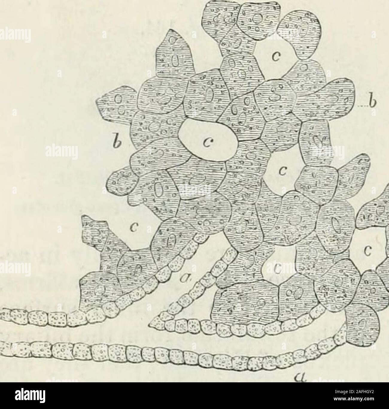 . Human physiology. Minute Portal and Hepatic Veins and Capillaries.a, a. Twigs of the portal vein. d. Twig of the hepatic vein. b. latermediate capillaries. Fig. 166.. Diagram of the arrangement of the cellular parenchyma,5, 6, of the human liver, with reference to tlie radicle.s ofthe interlobular ducts, <i, a, and the vascular .spaces, t, c. do not enter the lobules asaffirmed by Mr. Kiernan, butare confined to the interlo-bular spaces,—the substanceof the lobules being composedof secreting parenchyma andbloodvessels; and that the ac-tion of the liver seems to con-sist in the transmissio Stock Photohttps://www.alamy.com/image-license-details/?v=1https://www.alamy.com/human-physiology-minute-portal-and-hepatic-veins-and-capillariesa-a-twigs-of-the-portal-vein-d-twig-of-the-hepatic-vein-b-latermediate-capillaries-fig-166-diagram-of-the-arrangement-of-the-cellular-parenchyma5-6-of-the-human-liver-with-reference-to-tlie-radicles-ofthe-interlobular-ducts-lti-a-and-the-vascular-spaces-t-c-do-not-enter-the-lobules-asaffirmed-by-mr-kiernan-butare-confined-to-the-interlo-bular-spacesthe-substanceof-the-lobules-being-composedof-secreting-parenchyma-andbloodvessels-and-that-the-ac-tion-of-the-liver-seems-to-con-sist-in-the-transmissio-image336603318.html
RM2AFHGY2–. Human physiology. Minute Portal and Hepatic Veins and Capillaries.a, a. Twigs of the portal vein. d. Twig of the hepatic vein. b. latermediate capillaries. Fig. 166.. Diagram of the arrangement of the cellular parenchyma,5, 6, of the human liver, with reference to tlie radicle.s ofthe interlobular ducts, <i, a, and the vascular .spaces, t, c. do not enter the lobules asaffirmed by Mr. Kiernan, butare confined to the interlo-bular spaces,—the substanceof the lobules being composedof secreting parenchyma andbloodvessels; and that the ac-tion of the liver seems to con-sist in the transmissio
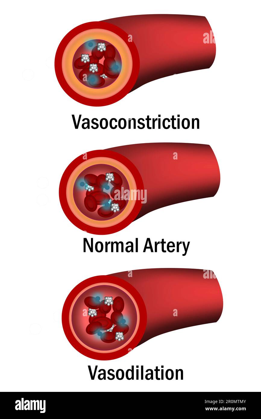 Comparison of normal, vasoconstriction and vasodilation blood vessels with cross section of arteries, 3d rende Stock Photohttps://www.alamy.com/image-license-details/?v=1https://www.alamy.com/comparison-of-normal-vasoconstriction-and-vasodilation-blood-vessels-with-cross-section-of-arteries-3d-rende-image551190219.html
RF2R0MTMY–Comparison of normal, vasoconstriction and vasodilation blood vessels with cross section of arteries, 3d rende
 Fish bladders are being prepared for sale. Stock Photohttps://www.alamy.com/image-license-details/?v=1https://www.alamy.com/fish-bladders-are-being-prepared-for-sale-image626735648.html
RF2YBJ7M0–Fish bladders are being prepared for sale.
 . The elasmobranch fishes . C D Fig. 195. Diagram of development of hepatic portal system in Elasmobranchs, ventral view. (From Eabl, modified.) a.c.v., anterior cardinal vein; cd.v., caudal vein; 7;.^;., hepatic vein; Iv., liver capillaries; om., omphalomesenteric vein; p.c, postcardinal; p.i.v., posterior intestinal vein; r.p., renal portal vein; s.i., subintestinal (intraintestinal) vein; s.v., sinus venosus; v.v., vitelline vein. mesenteric. The right vein remains rudimentary, but the left omphalomes- enteric becomes an important vessel (om..). When the developing liver comes in contact wi Stock Photohttps://www.alamy.com/image-license-details/?v=1https://www.alamy.com/the-elasmobranch-fishes-c-d-fig-195-diagram-of-development-of-hepatic-portal-system-in-elasmobranchs-ventral-view-from-eabl-modified-acv-anterior-cardinal-vein-cdv-caudal-vein-7-hepatic-vein-iv-liver-capillaries-om-omphalomesenteric-vein-pc-postcardinal-piv-posterior-intestinal-vein-rp-renal-portal-vein-si-subintestinal-intraintestinal-vein-sv-sinus-venosus-vv-vitelline-vein-mesenteric-the-right-vein-remains-rudimentary-but-the-left-omphalomes-enteric-becomes-an-important-vessel-om-when-the-developing-liver-comes-in-contact-wi-image178413463.html
RMMA7C5B–. The elasmobranch fishes . C D Fig. 195. Diagram of development of hepatic portal system in Elasmobranchs, ventral view. (From Eabl, modified.) a.c.v., anterior cardinal vein; cd.v., caudal vein; 7;.^;., hepatic vein; Iv., liver capillaries; om., omphalomesenteric vein; p.c, postcardinal; p.i.v., posterior intestinal vein; r.p., renal portal vein; s.i., subintestinal (intraintestinal) vein; s.v., sinus venosus; v.v., vitelline vein. mesenteric. The right vein remains rudimentary, but the left omphalomes- enteric becomes an important vessel (om..). When the developing liver comes in contact wi
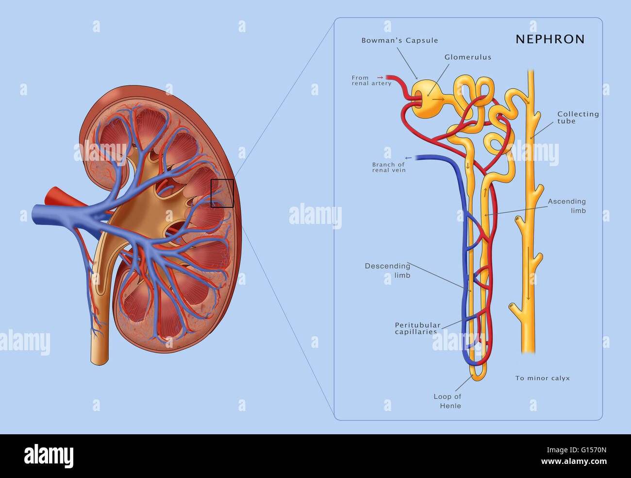 Illustration of the structure of a nephron (the basic structural and functional unit of the kidney) along side a cross section of a kidney. Depicted in the nephron are the glomerulus, bowman's capsule, proximal convoluted tubule, peritubular capillaries, Stock Photohttps://www.alamy.com/image-license-details/?v=1https://www.alamy.com/stock-photo-illustration-of-the-structure-of-a-nephron-the-basic-structural-and-103992133.html
RMG1570N–Illustration of the structure of a nephron (the basic structural and functional unit of the kidney) along side a cross section of a kidney. Depicted in the nephron are the glomerulus, bowman's capsule, proximal convoluted tubule, peritubular capillaries,
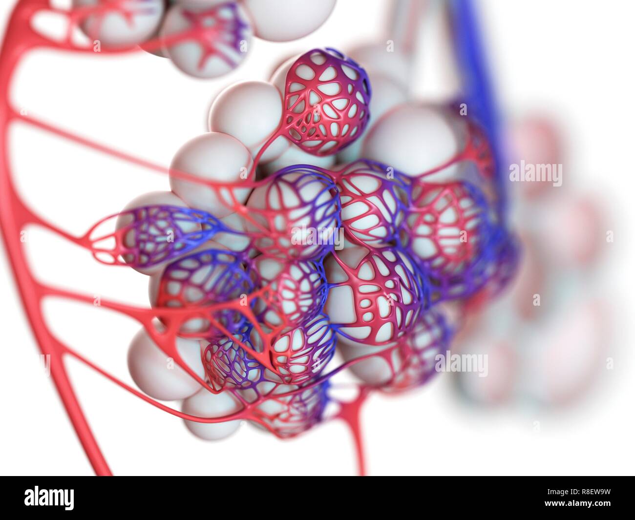 Illustration of the human alveoli. Stock Photohttps://www.alamy.com/image-license-details/?v=1https://www.alamy.com/illustration-of-the-human-alveoli-image228979237.html
RFR8EW9W–Illustration of the human alveoli.
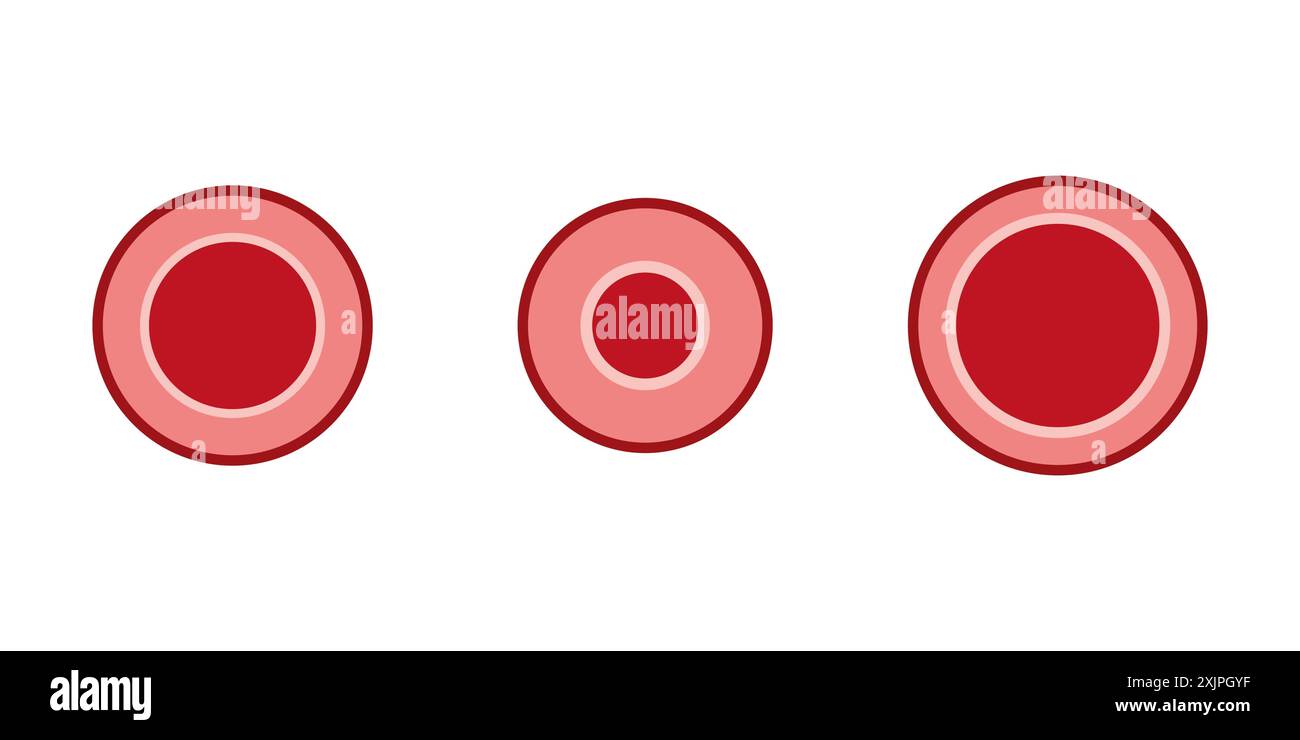 Vasodilation and vasoconstriction, illustration. Stock Photohttps://www.alamy.com/image-license-details/?v=1https://www.alamy.com/vasodilation-and-vasoconstriction-illustration-image613922947.html
RF2XJPGYF–Vasodilation and vasoconstriction, illustration.
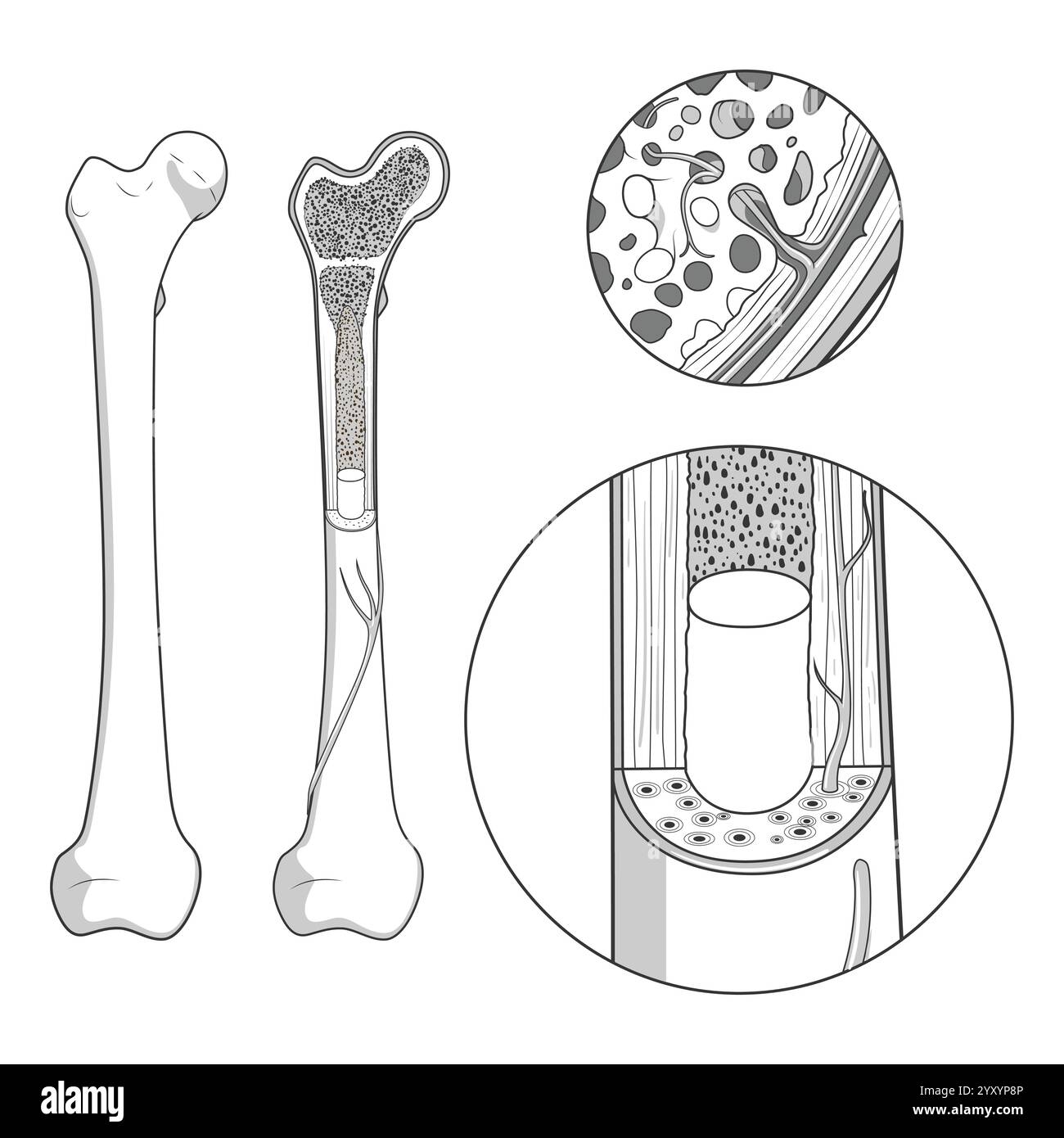 Bone structure medical educational vector Stock Vectorhttps://www.alamy.com/image-license-details/?v=1https://www.alamy.com/bone-structure-medical-educational-vector-image636164502.html
RF2YXYP8P–Bone structure medical educational vector
 Breathing Alveoli Within Structure of Human Lungs Stock Photohttps://www.alamy.com/image-license-details/?v=1https://www.alamy.com/breathing-alveoli-within-structure-of-human-lungs-image628160665.html
RF2YDY59D–Breathing Alveoli Within Structure of Human Lungs
 Interstitial fluid is collected by lymph capillaries from the interstitial space. Lymph then moves through lymphatic vessels to lymph nodes. 3D render Stock Photohttps://www.alamy.com/image-license-details/?v=1https://www.alamy.com/interstitial-fluid-is-collected-by-lymph-capillaries-from-the-interstitial-space-lymph-then-moves-through-lymphatic-vessels-to-lymph-nodes-3d-render-image596595558.html
RF2WJH7M6–Interstitial fluid is collected by lymph capillaries from the interstitial space. Lymph then moves through lymphatic vessels to lymph nodes. 3D render
 A text-book of physiology, for medical students and physicians . AB: An, or ab = According to this method,Vierordt calculated that thevelocity of the blood in thehuman capillaries is equal toabout 0.6 to 0.9 mm. persecond. In the arteries, more- , , . Fig. 185.—Diagram of the eve to show the con- OVer, It may be Observed struction used to determine the size of the retinal 4-V.^i- 4-U„ i j. image when the size of the external object is known: that the average Velocity n, The nodal point of the eye. See text. diminishes the farther one goes from the heart,—that is, the smaller the artery,—an Stock Photohttps://www.alamy.com/image-license-details/?v=1https://www.alamy.com/a-text-book-of-physiology-for-medical-students-and-physicians-ab-an-or-ab-=-according-to-this-methodvierordt-calculated-that-thevelocity-of-the-blood-in-thehuman-capillaries-is-equal-toabout-06-to-09-mm-persecond-in-the-arteries-more-fig-185diagram-of-the-eve-to-show-the-con-over-it-may-be-observed-struction-used-to-determine-the-size-of-the-retinal-4-vi-4-u-i-j-image-when-the-size-of-the-external-object-is-known-that-the-average-velocity-n-the-nodal-point-of-the-eye-see-text-diminishes-the-farther-one-goes-from-the-heartthat-is-the-smaller-the-arteryan-image342942671.html
RM2AWXATF–A text-book of physiology, for medical students and physicians . AB: An, or ab = According to this method,Vierordt calculated that thevelocity of the blood in thehuman capillaries is equal toabout 0.6 to 0.9 mm. persecond. In the arteries, more- , , . Fig. 185.—Diagram of the eve to show the con- OVer, It may be Observed struction used to determine the size of the retinal 4-V.^i- 4-U„ i j. image when the size of the external object is known: that the average Velocity n, The nodal point of the eye. See text. diminishes the farther one goes from the heart,—that is, the smaller the artery,—an
 Fish bladders are being prepared for sale. Stock Photohttps://www.alamy.com/image-license-details/?v=1https://www.alamy.com/fish-bladders-are-being-prepared-for-sale-image626735690.html
RF2YBJ7NE–Fish bladders are being prepared for sale.
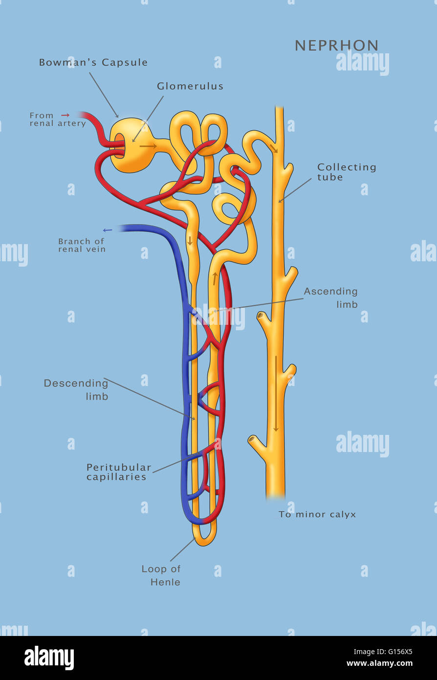 An Illustration of the Nephron of the Kidney. Stock Photohttps://www.alamy.com/image-license-details/?v=1https://www.alamy.com/stock-photo-an-illustration-of-the-nephron-of-the-kidney-103992061.html
RMG156X5–An Illustration of the Nephron of the Kidney.
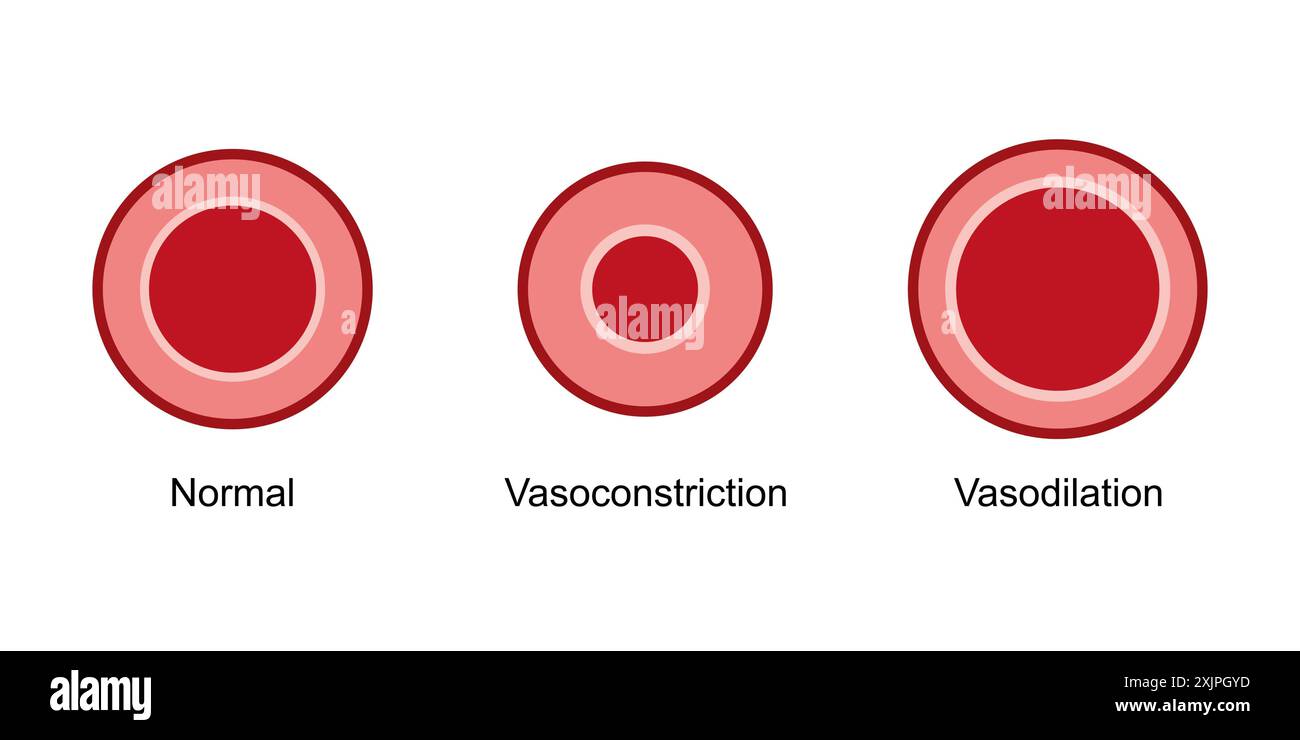 Vasodilation and vasoconstriction, illustration. Stock Photohttps://www.alamy.com/image-license-details/?v=1https://www.alamy.com/vasodilation-and-vasoconstriction-illustration-image613922945.html
RF2XJPGYD–Vasodilation and vasoconstriction, illustration.
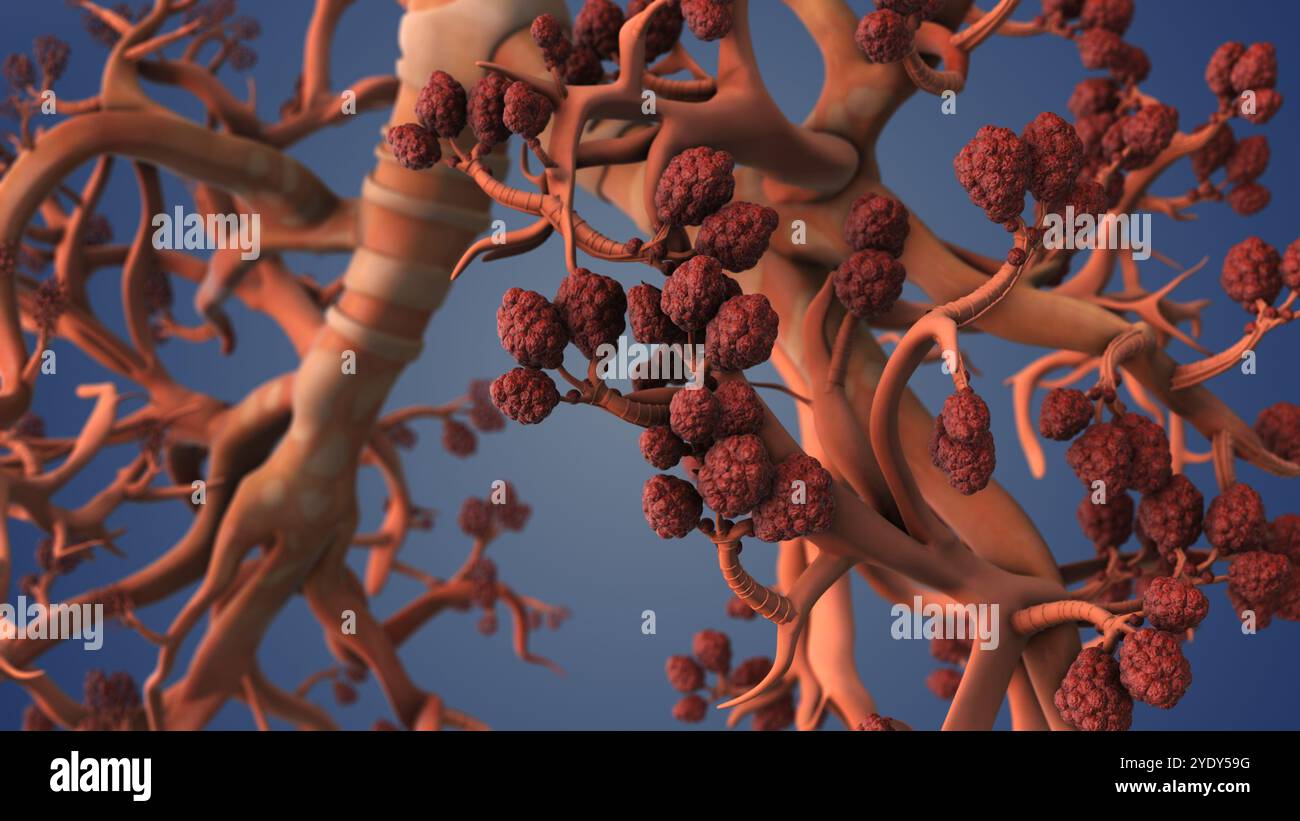 Breathing Alveoli Within Structure of Human Lungs Stock Photohttps://www.alamy.com/image-license-details/?v=1https://www.alamy.com/breathing-alveoli-within-structure-of-human-lungs-image628160668.html
RF2YDY59G–Breathing Alveoli Within Structure of Human Lungs
 Interstitial fluid is collected by lymph capillaries from the interstitial space. Lymph then moves through lymphatic vessels to lymph nodes. 3D render Stock Photohttps://www.alamy.com/image-license-details/?v=1https://www.alamy.com/interstitial-fluid-is-collected-by-lymph-capillaries-from-the-interstitial-space-lymph-then-moves-through-lymphatic-vessels-to-lymph-nodes-3d-render-image596596493.html
RF2WJH8WH–Interstitial fluid is collected by lymph capillaries from the interstitial space. Lymph then moves through lymphatic vessels to lymph nodes. 3D render
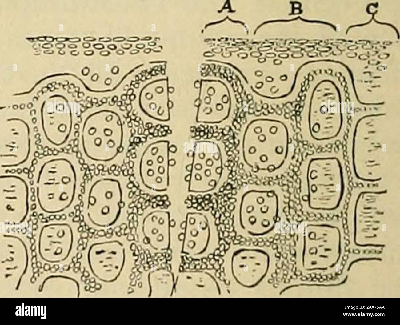 Surgery; its theory and practice . ?la* Wir?- mr^ Diagram representing a simpleincifed wound, immediately af-ter the incision has been made.. Diagram representing an incised wounda few hours after the incision. A.Area of thrombosis—leucocytes mak-ing their way to tlie cut surface. B.Area of dilated capillaries—leticocytesescaping from the vessels into the tis-sues. C. Normal tissues. in the layer of tissue bounding the incision (Fig. 22 and Fig. 23).As a consec|uence, stasis and coagulation of the blood is inducedin the divided smaller vessels and capillaries, and thus the hemor-rhage from the Stock Photohttps://www.alamy.com/image-license-details/?v=1https://www.alamy.com/surgery-its-theory-and-practice-la-wir-mr-diagram-representing-a-simpleincifed-wound-immediately-af-ter-the-incision-has-been-made-diagram-representing-an-incised-wounda-few-hours-after-the-incision-aarea-of-thrombosisleucocytes-mak-ing-their-way-to-tlie-cut-surface-barea-of-dilated-capillariesleticocytesescaping-from-the-vessels-into-the-tis-sues-c-normal-tissues-in-the-layer-of-tissue-bounding-the-incision-fig-22-and-fig-23as-a-consecuence-stasis-and-coagulation-of-the-blood-is-inducedin-the-divided-smaller-vessels-and-capillaries-and-thus-the-hemor-rhage-from-the-image343135922.html
RM2AX75AA–Surgery; its theory and practice . ?la* Wir?- mr^ Diagram representing a simpleincifed wound, immediately af-ter the incision has been made.. Diagram representing an incised wounda few hours after the incision. A.Area of thrombosis—leucocytes mak-ing their way to tlie cut surface. B.Area of dilated capillaries—leticocytesescaping from the vessels into the tis-sues. C. Normal tissues. in the layer of tissue bounding the incision (Fig. 22 and Fig. 23).As a consec|uence, stasis and coagulation of the blood is inducedin the divided smaller vessels and capillaries, and thus the hemor-rhage from the
 Lung Alveoli Supporting Vital Respiration and Oxygen Flow Stock Photohttps://www.alamy.com/image-license-details/?v=1https://www.alamy.com/lung-alveoli-supporting-vital-respiration-and-oxygen-flow-image628224952.html
RF2YE239C–Lung Alveoli Supporting Vital Respiration and Oxygen Flow
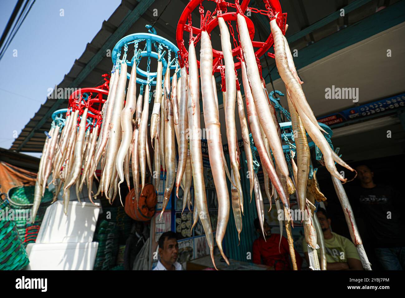 Fish bladders are being prepared for sale. Stock Photohttps://www.alamy.com/image-license-details/?v=1https://www.alamy.com/fish-bladders-are-being-prepared-for-sale-image626735724.html
RF2YBJ7PM–Fish bladders are being prepared for sale.
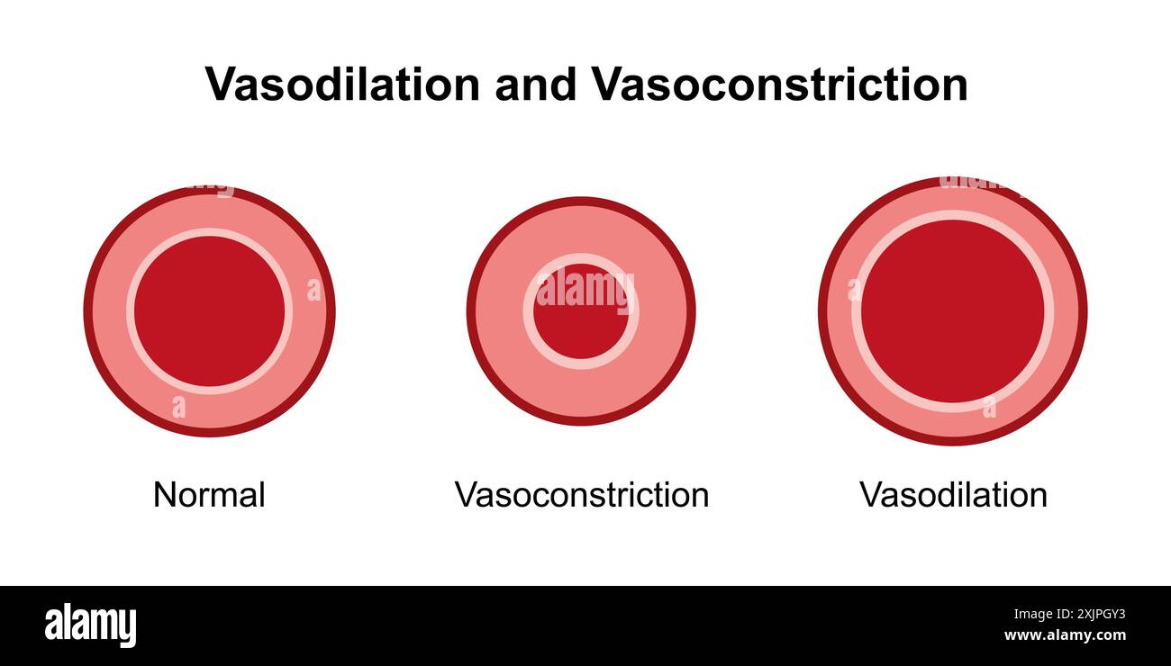 Vasodilation and vasoconstriction, illustration. Stock Photohttps://www.alamy.com/image-license-details/?v=1https://www.alamy.com/vasodilation-and-vasoconstriction-illustration-image613922935.html
RF2XJPGY3–Vasodilation and vasoconstriction, illustration.
 Interstitial fluid is collected by lymph capillaries from the interstitial space. Lymph then moves through lymphatic vessels to lymph nodes. 3D render Stock Photohttps://www.alamy.com/image-license-details/?v=1https://www.alamy.com/interstitial-fluid-is-collected-by-lymph-capillaries-from-the-interstitial-space-lymph-then-moves-through-lymphatic-vessels-to-lymph-nodes-3d-render-image596593377.html
RF2WJH4X9–Interstitial fluid is collected by lymph capillaries from the interstitial space. Lymph then moves through lymphatic vessels to lymph nodes. 3D render
 Textbook of normal histology: including an account of the development of the tissues and of the organs . Section of human kidney partia ly injected : a, interlobu-lar artery giving off afferent twig (6); c, efferent vesselpassing into intertubular capillaries {d); e, convolutedcapillaries of glomeruus ; /, ouier layer of Bowmans cap-sule, the nuclei of whose cells sh nv at g; h, uriniferoustubule in transverse section, i, ii oblique section. 194 NORMAL HISTOLOGY.Fig. 2.37.. Pelvis Diagram of the kidney, showing the course of the uriniferous tubules and of the blood vessels; forconvenience the Stock Photohttps://www.alamy.com/image-license-details/?v=1https://www.alamy.com/textbook-of-normal-histology-including-an-account-of-the-development-of-the-tissues-and-of-the-organs-section-of-human-kidney-partia-ly-injected-a-interlobu-lar-artery-giving-off-afferent-twig-6-c-efferent-vesselpassing-into-intertubular-capillaries-d-e-convolutedcapillaries-of-glomeruus-ouier-layer-of-bowmans-cap-sule-the-nuclei-of-whose-cells-sh-nv-at-g-h-uriniferoustubule-in-transverse-section-i-ii-oblique-section-194-normal-histologyfig-237-pelvis-diagram-of-the-kidney-showing-the-course-of-the-uriniferous-tubules-and-of-the-blood-vessels-forconvenience-the-image338953238.html
RM2AKCJ8P–Textbook of normal histology: including an account of the development of the tissues and of the organs . Section of human kidney partia ly injected : a, interlobu-lar artery giving off afferent twig (6); c, efferent vesselpassing into intertubular capillaries {d); e, convolutedcapillaries of glomeruus ; /, ouier layer of Bowmans cap-sule, the nuclei of whose cells sh nv at g; h, uriniferoustubule in transverse section, i, ii oblique section. 194 NORMAL HISTOLOGY.Fig. 2.37.. Pelvis Diagram of the kidney, showing the course of the uriniferous tubules and of the blood vessels; forconvenience the
 Lung Alveoli Supporting Vital Respiration and Oxygen Flow Stock Photohttps://www.alamy.com/image-license-details/?v=1https://www.alamy.com/lung-alveoli-supporting-vital-respiration-and-oxygen-flow-image628224953.html
RF2YE239D–Lung Alveoli Supporting Vital Respiration and Oxygen Flow
 Fish bladders are being prepared for sale. Stock Photohttps://www.alamy.com/image-license-details/?v=1https://www.alamy.com/fish-bladders-are-being-prepared-for-sale-image626735621.html
RM2YBJ7K1–Fish bladders are being prepared for sale.
 Interstitial fluid is collected by lymph capillaries from the interstitial space. Lymph then moves through lymphatic vessels to lymph nodes. 3D render Stock Photohttps://www.alamy.com/image-license-details/?v=1https://www.alamy.com/interstitial-fluid-is-collected-by-lymph-capillaries-from-the-interstitial-space-lymph-then-moves-through-lymphatic-vessels-to-lymph-nodes-3d-render-image596593618.html
RF2WJH56X–Interstitial fluid is collected by lymph capillaries from the interstitial space. Lymph then moves through lymphatic vessels to lymph nodes. 3D render
 . The American journal of anatomy. ith a normal embryo of the same age, whichwas 13 mm. in length. The area vasculosa of the operated chicks did not always ex-pand uniformly in each direction, as in the normal, but oftengrew out at certain points, usually posteriorly from the embryo,in advance of other portions. Figure 2 is a diagram of one of 184 W. B. CHAPMAN these specimens. The effect of this irregular growth of the areais apparently shown in th6 shape of the capillary plexus^thus, infigure 13 the peripheral capillaries, formed by the breaking up ofthe border vein, have grown out so rapidl Stock Photohttps://www.alamy.com/image-license-details/?v=1https://www.alamy.com/the-american-journal-of-anatomy-ith-a-normal-embryo-of-the-same-age-whichwas-13-mm-in-length-the-area-vasculosa-of-the-operated-chicks-did-not-always-ex-pand-uniformly-in-each-direction-as-in-the-normal-but-oftengrew-out-at-certain-points-usually-posteriorly-from-the-embryoin-advance-of-other-portions-figure-2-is-a-diagram-of-one-of-184-w-b-chapman-these-specimens-the-effect-of-this-irregular-growth-of-the-areais-apparently-shown-in-th6-shape-of-the-capillary-plexusthus-infigure-13-the-peripheral-capillaries-formed-by-the-breaking-up-ofthe-border-vein-have-grown-out-so-rapidl-image336809384.html
RM2AFXYPG–. The American journal of anatomy. ith a normal embryo of the same age, whichwas 13 mm. in length. The area vasculosa of the operated chicks did not always ex-pand uniformly in each direction, as in the normal, but oftengrew out at certain points, usually posteriorly from the embryo,in advance of other portions. Figure 2 is a diagram of one of 184 W. B. CHAPMAN these specimens. The effect of this irregular growth of the areais apparently shown in th6 shape of the capillary plexus^thus, infigure 13 the peripheral capillaries, formed by the breaking up ofthe border vein, have grown out so rapidl
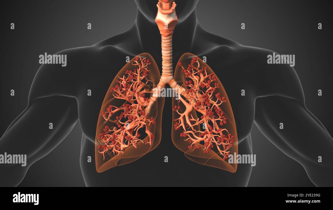 Lung Alveoli Supporting Vital Respiration and Oxygen Flow Stock Photohttps://www.alamy.com/image-license-details/?v=1https://www.alamy.com/lung-alveoli-supporting-vital-respiration-and-oxygen-flow-image628224956.html
RF2YE239G–Lung Alveoli Supporting Vital Respiration and Oxygen Flow
 Fish bladders are being prepared for sale. Stock Photohttps://www.alamy.com/image-license-details/?v=1https://www.alamy.com/fish-bladders-are-being-prepared-for-sale-image626735718.html
RM2YBJ7PE–Fish bladders are being prepared for sale.
 Respiratory Alveoli in Lungs for Oxygen Transfer Stock Photohttps://www.alamy.com/image-license-details/?v=1https://www.alamy.com/respiratory-alveoli-in-lungs-for-oxygen-transfer-image628224978.html
RF2YE23AA–Respiratory Alveoli in Lungs for Oxygen Transfer
 Interstitial fluid is collected by lymph capillaries from the interstitial space. Lymph then moves through lymphatic vessels to lymph nodes. 3D render Stock Photohttps://www.alamy.com/image-license-details/?v=1https://www.alamy.com/interstitial-fluid-is-collected-by-lymph-capillaries-from-the-interstitial-space-lymph-then-moves-through-lymphatic-vessels-to-lymph-nodes-3d-render-image596595790.html
RF2WJH80E–Interstitial fluid is collected by lymph capillaries from the interstitial space. Lymph then moves through lymphatic vessels to lymph nodes. 3D render
 . Elements of human physiology. Physiology. Diagram of capillaries in frog's web. Fig. 5 shows the course taken by the blood in its circula- tion through the body, as it is impelled by the contracting heart.. Diagram of circulation, l.v. Left ventricle, l.a. Left auricle. R.v. Eight ventricle, k.a. Eight auricle, ao. Aorta, s.c. Sys- temic cai^illaries. al. Alimentary canal, p. Portal vein. l. Liver. H.v. Hepatic vein. p.c. Capillaries of lungs. Starting from the left ventricle, the blood is propelled through the aorta into the systemic arteries, and thence into. Please note that these images Stock Photohttps://www.alamy.com/image-license-details/?v=1https://www.alamy.com/elements-of-human-physiology-physiology-diagram-of-capillaries-in-frogs-web-fig-5-shows-the-course-taken-by-the-blood-in-its-circula-tion-through-the-body-as-it-is-impelled-by-the-contracting-heart-diagram-of-circulation-lv-left-ventricle-la-left-auricle-rv-eight-ventricle-ka-eight-auricle-ao-aorta-sc-sys-temic-caiillaries-al-alimentary-canal-p-portal-vein-l-liver-hv-hepatic-vein-pc-capillaries-of-lungs-starting-from-the-left-ventricle-the-blood-is-propelled-through-the-aorta-into-the-systemic-arteries-and-thence-into-please-note-that-these-images-image231492071.html
RMRCHADY–. Elements of human physiology. Physiology. Diagram of capillaries in frog's web. Fig. 5 shows the course taken by the blood in its circula- tion through the body, as it is impelled by the contracting heart.. Diagram of circulation, l.v. Left ventricle, l.a. Left auricle. R.v. Eight ventricle, k.a. Eight auricle, ao. Aorta, s.c. Sys- temic cai^illaries. al. Alimentary canal, p. Portal vein. l. Liver. H.v. Hepatic vein. p.c. Capillaries of lungs. Starting from the left ventricle, the blood is propelled through the aorta into the systemic arteries, and thence into. Please note that these images
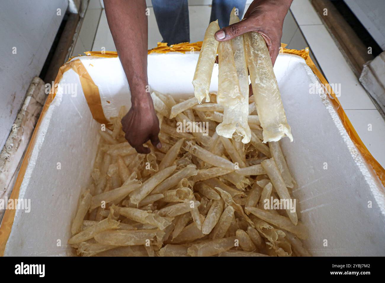 Fish bladders are being prepared for sale. Stock Photohttps://www.alamy.com/image-license-details/?v=1https://www.alamy.com/fish-bladders-are-being-prepared-for-sale-image626735650.html
RF2YBJ7M2–Fish bladders are being prepared for sale.
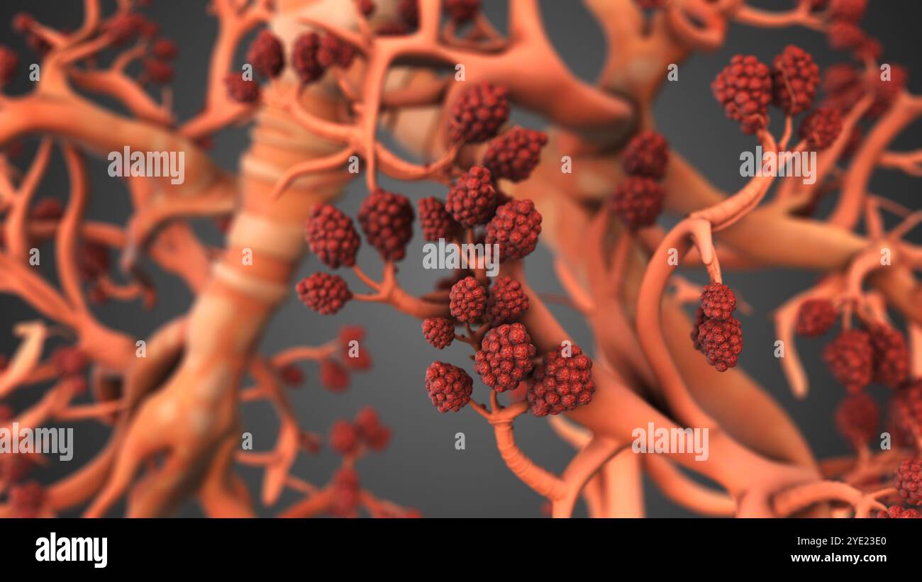 Respiratory Alveoli in Lungs for Oxygen Transfer Stock Photohttps://www.alamy.com/image-license-details/?v=1https://www.alamy.com/respiratory-alveoli-in-lungs-for-oxygen-transfer-image628225080.html
RF2YE23E0–Respiratory Alveoli in Lungs for Oxygen Transfer
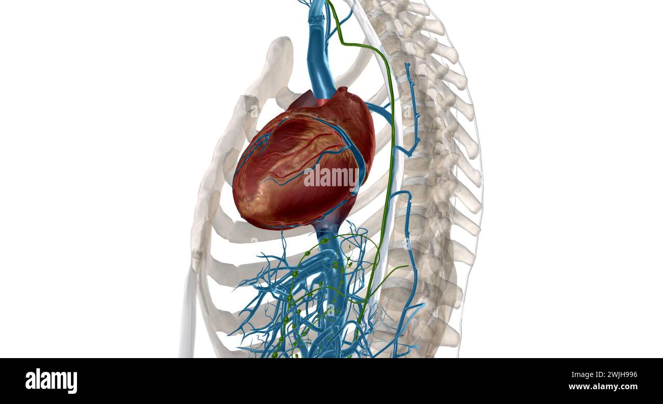 Interstitial fluid is collected by lymph capillaries from the interstitial space. Lymph then moves through lymphatic vessels to lymph nodes. 3D render Stock Photohttps://www.alamy.com/image-license-details/?v=1https://www.alamy.com/interstitial-fluid-is-collected-by-lymph-capillaries-from-the-interstitial-space-lymph-then-moves-through-lymphatic-vessels-to-lymph-nodes-3d-render-image596596818.html
RF2WJH996–Interstitial fluid is collected by lymph capillaries from the interstitial space. Lymph then moves through lymphatic vessels to lymph nodes. 3D render
 The American journal of anatomy . no Fig. 11. Fig. 10.The American Journal op Anatomy.—Vol. 10, No. 1, Jan., 1910. 106 Walter E. Dandy. Plate III. Fig. 12. Diagram of a sagittal view of embryo to show the vascular system.Ht, Heart; A I-II-III, First, second and third aortic arches; Ao, Dorsal aorta;C.Ao, Caudal aorta; U, Umbilical artery (unite in the bauchstiel) ; V, Um-bilical vein (unite in the Bauchstiel) ; Vit, Branch of umbilical vein, is onlysuggestion of a possible vitelline vein; V. am, Venous branch to amnion; Ca,Capillaries; do not contain blood and do not connect with veins; B.I., Stock Photohttps://www.alamy.com/image-license-details/?v=1https://www.alamy.com/the-american-journal-of-anatomy-no-fig-11-fig-10the-american-journal-op-anatomyvol-10-no-1-jan-1910-106-walter-e-dandy-plate-iii-fig-12-diagram-of-a-sagittal-view-of-embryo-to-show-the-vascular-systemht-heart-a-i-ii-iii-first-second-and-third-aortic-arches-ao-dorsal-aortacao-caudal-aorta-u-umbilical-artery-unite-in-the-bauchstiel-v-um-bilical-vein-unite-in-the-bauchstiel-vit-branch-of-umbilical-vein-is-onlysuggestion-of-a-possible-vitelline-vein-v-am-venous-branch-to-amnion-cacapillaries-do-not-contain-blood-and-do-not-connect-with-veins-bi-image340132791.html
RM2ANAARK–The American journal of anatomy . no Fig. 11. Fig. 10.The American Journal op Anatomy.—Vol. 10, No. 1, Jan., 1910. 106 Walter E. Dandy. Plate III. Fig. 12. Diagram of a sagittal view of embryo to show the vascular system.Ht, Heart; A I-II-III, First, second and third aortic arches; Ao, Dorsal aorta;C.Ao, Caudal aorta; U, Umbilical artery (unite in the bauchstiel) ; V, Um-bilical vein (unite in the Bauchstiel) ; Vit, Branch of umbilical vein, is onlysuggestion of a possible vitelline vein; V. am, Venous branch to amnion; Ca,Capillaries; do not contain blood and do not connect with veins; B.I.,
 Fish bladders are being prepared for sale. Stock Photohttps://www.alamy.com/image-license-details/?v=1https://www.alamy.com/fish-bladders-are-being-prepared-for-sale-image626735646.html
RF2YBJ7KX–Fish bladders are being prepared for sale.
 Interstitial fluid is collected by lymph capillaries from the interstitial space. Lymph then moves through lymphatic vessels to lymph nodes. 3D render Stock Photohttps://www.alamy.com/image-license-details/?v=1https://www.alamy.com/interstitial-fluid-is-collected-by-lymph-capillaries-from-the-interstitial-space-lymph-then-moves-through-lymphatic-vessels-to-lymph-nodes-3d-render-image596596264.html
RF2WJH8HC–Interstitial fluid is collected by lymph capillaries from the interstitial space. Lymph then moves through lymphatic vessels to lymph nodes. 3D render
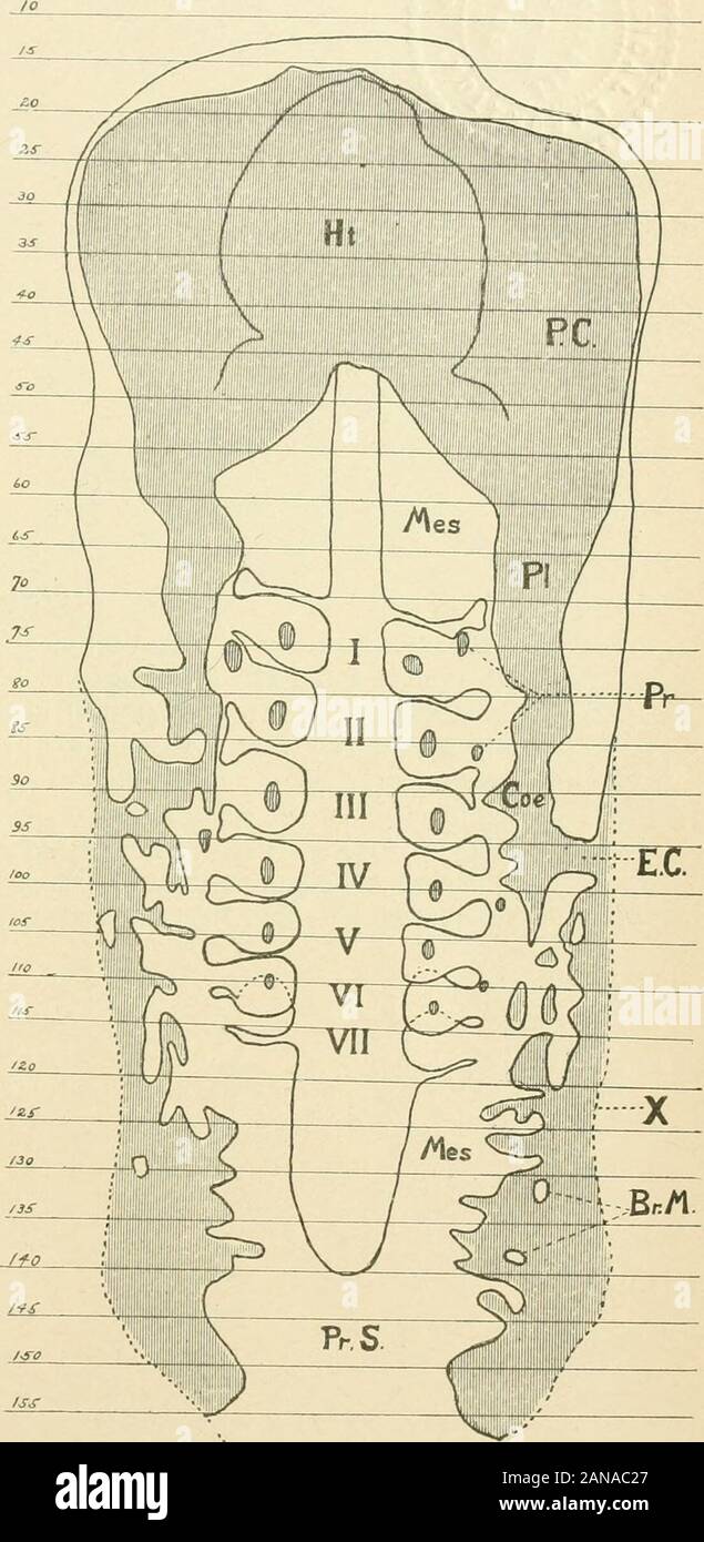 The American journal of anatomy . no Fig. 11. Fig. 10.The American Journal op Anatomy.—Vol. 10, No. 1, Jan., 1910. 106 Walter E. Dandy. Plate III. Fig. 12. Diagram of a sagittal view of embryo to show the vascular system.Ht, Heart; A I-II-III, First, second and third aortic arches; Ao, Dorsal aorta;C.Ao, Caudal aorta; U, Umbilical artery (unite in the bauchstiel) ; V, Um-bilical vein (unite in the Bauchstiel) ; Vit, Branch of umbilical vein, is onlysuggestion of a possible vitelline vein; V. am, Venous branch to amnion; Ca,Capillaries; do not contain blood and do not connect with veins; B.I., Stock Photohttps://www.alamy.com/image-license-details/?v=1https://www.alamy.com/the-american-journal-of-anatomy-no-fig-11-fig-10the-american-journal-op-anatomyvol-10-no-1-jan-1910-106-walter-e-dandy-plate-iii-fig-12-diagram-of-a-sagittal-view-of-embryo-to-show-the-vascular-systemht-heart-a-i-ii-iii-first-second-and-third-aortic-arches-ao-dorsal-aortacao-caudal-aorta-u-umbilical-artery-unite-in-the-bauchstiel-v-um-bilical-vein-unite-in-the-bauchstiel-vit-branch-of-umbilical-vein-is-onlysuggestion-of-a-possible-vitelline-vein-v-am-venous-branch-to-amnion-cacapillaries-do-not-contain-blood-and-do-not-connect-with-veins-bi-image340133759.html
RM2ANAC27–The American journal of anatomy . no Fig. 11. Fig. 10.The American Journal op Anatomy.—Vol. 10, No. 1, Jan., 1910. 106 Walter E. Dandy. Plate III. Fig. 12. Diagram of a sagittal view of embryo to show the vascular system.Ht, Heart; A I-II-III, First, second and third aortic arches; Ao, Dorsal aorta;C.Ao, Caudal aorta; U, Umbilical artery (unite in the bauchstiel) ; V, Um-bilical vein (unite in the Bauchstiel) ; Vit, Branch of umbilical vein, is onlysuggestion of a possible vitelline vein; V. am, Venous branch to amnion; Ca,Capillaries; do not contain blood and do not connect with veins; B.I.,
 Fish bladders are being prepared for sale. Stock Photohttps://www.alamy.com/image-license-details/?v=1https://www.alamy.com/fish-bladders-are-being-prepared-for-sale-image626735716.html
RM2YBJ7PC–Fish bladders are being prepared for sale.
 Interstitial fluid is collected by lymph capillaries from the interstitial space. Lymph then moves through lymphatic vessels to lymph nodes. 3D render Stock Photohttps://www.alamy.com/image-license-details/?v=1https://www.alamy.com/interstitial-fluid-is-collected-by-lymph-capillaries-from-the-interstitial-space-lymph-then-moves-through-lymphatic-vessels-to-lymph-nodes-3d-render-image596593261.html
RF2WJH4P5–Interstitial fluid is collected by lymph capillaries from the interstitial space. Lymph then moves through lymphatic vessels to lymph nodes. 3D render
 . The human body and health : an elementary text-book of essential anatomy, applied physiology and practical hygiene for schools . ^ aid the villi in absorbing certain food from the intestine;they have some partin causing the clot-ting of the blood;and they help pro-tect the body fromharmful germs bydevouring them. Birth and Deathof Corpuscles. —Since the coloringmatter of the bileis derived from thedead red cells, andsince millions ofwhite corpuscles arebeing killed wher-ever pus is formed,there must be some. Fig, 72. — Diagram of the capillaries uniting anartery and vein. The plasma is passi Stock Photohttps://www.alamy.com/image-license-details/?v=1https://www.alamy.com/the-human-body-and-health-an-elementary-text-book-of-essential-anatomy-applied-physiology-and-practical-hygiene-for-schools-aid-the-villi-in-absorbing-certain-food-from-the-intestinethey-have-some-partin-causing-the-clot-ting-of-the-bloodand-they-help-pro-tect-the-body-fromharmful-germs-bydevouring-them-birth-and-deathof-corpuscles-since-the-coloringmatter-of-the-bileis-derived-from-thedead-red-cells-andsince-millions-ofwhite-corpuscles-arebeing-killed-wher-ever-pus-is-formedthere-must-be-some-fig-72-diagram-of-the-capillaries-uniting-anartery-and-vein-the-plasma-is-passi-image370186629.html
RM2CE7CR1–. The human body and health : an elementary text-book of essential anatomy, applied physiology and practical hygiene for schools . ^ aid the villi in absorbing certain food from the intestine;they have some partin causing the clot-ting of the blood;and they help pro-tect the body fromharmful germs bydevouring them. Birth and Deathof Corpuscles. —Since the coloringmatter of the bileis derived from thedead red cells, andsince millions ofwhite corpuscles arebeing killed wher-ever pus is formed,there must be some. Fig, 72. — Diagram of the capillaries uniting anartery and vein. The plasma is passi
 Fish bladders are being prepared for sale. Stock Photohttps://www.alamy.com/image-license-details/?v=1https://www.alamy.com/fish-bladders-are-being-prepared-for-sale-image626735688.html
RF2YBJ7NC–Fish bladders are being prepared for sale.
 . The human body and health : an elementary text-book of essential anatomy, applied physiology and practical hygiene for schools . Fig. 70. — White hlood corpuscles, photographed to show their form when crawling. 108 THE BLOOD. ^^/^ Wherever there is inflamed or diseased tissue, the whitecells are attracted in great numbers. They creep out of the capillaries, andoften large quanti-ties are destroyed,and then they formmost of the pus^ orwhite matter, pres-ent in a boil or other FiCx. 71. —Diagram of a capillary network, show- SUppuration. Theing the white corpuscles crawling through the white C Stock Photohttps://www.alamy.com/image-license-details/?v=1https://www.alamy.com/the-human-body-and-health-an-elementary-text-book-of-essential-anatomy-applied-physiology-and-practical-hygiene-for-schools-fig-70-white-hlood-corpuscles-photographed-to-show-their-form-when-crawling-108-the-blood-wherever-there-is-inflamed-or-diseased-tissue-the-whitecells-are-attracted-in-great-numbers-they-creep-out-of-the-capillaries-andoften-large-quanti-ties-are-destroyedand-then-they-formmost-of-the-pus-orwhite-matter-pres-ent-in-a-boil-or-other-ficx-71-diagram-of-a-capillary-network-show-suppuration-theing-the-white-corpuscles-crawling-through-the-white-c-image370189482.html
RM2CE7GCX–. The human body and health : an elementary text-book of essential anatomy, applied physiology and practical hygiene for schools . Fig. 70. — White hlood corpuscles, photographed to show their form when crawling. 108 THE BLOOD. ^^/^ Wherever there is inflamed or diseased tissue, the whitecells are attracted in great numbers. They creep out of the capillaries, andoften large quanti-ties are destroyed,and then they formmost of the pus^ orwhite matter, pres-ent in a boil or other FiCx. 71. —Diagram of a capillary network, show- SUppuration. Theing the white corpuscles crawling through the white C
 Fish bladders are being prepared for sale. Stock Photohttps://www.alamy.com/image-license-details/?v=1https://www.alamy.com/fish-bladders-are-being-prepared-for-sale-image626735682.html
RF2YBJ7N6–Fish bladders are being prepared for sale.
 . A text-book of comparative physiology for students and practitioners of comparative (veterinary) medicine . Pig. 193—Diagram to illustrate the relative proportions of the ^egate sectional we.; of the different parts of the vascular system (after eo). A, aorta, C, capillars.V, veins. 222 COMPARATIVE PHYSIOLOGY, THE ACTION OF THE MAMMALIAN HEART. What takes place may be thus very briefly stated : Theright auricle contracting- squeezes the blood through the au-ricular-ventricular opening into the right ventricle, never quite Superior VenaCava. Inferior VenaCava. Capillaries of theHead, etc. Pu Stock Photohttps://www.alamy.com/image-license-details/?v=1https://www.alamy.com/a-text-book-of-comparative-physiology-for-students-and-practitioners-of-comparative-veterinary-medicine-pig-193diagram-to-illustrate-the-relative-proportions-of-the-egate-sectional-we-of-the-different-parts-of-the-vascular-system-after-eo-a-aorta-c-capillarsv-veins-222-comparative-physiology-the-action-of-the-mammalian-heart-what-takes-place-may-be-thus-very-briefly-stated-theright-auricle-contracting-squeezes-the-blood-through-the-au-ricular-ventricular-opening-into-the-right-ventricle-never-quite-superior-venacava-inferior-venacava-capillaries-of-thehead-etc-pu-image372528612.html
RM2CJ2418–. A text-book of comparative physiology for students and practitioners of comparative (veterinary) medicine . Pig. 193—Diagram to illustrate the relative proportions of the ^egate sectional we.; of the different parts of the vascular system (after eo). A, aorta, C, capillars.V, veins. 222 COMPARATIVE PHYSIOLOGY, THE ACTION OF THE MAMMALIAN HEART. What takes place may be thus very briefly stated : Theright auricle contracting- squeezes the blood through the au-ricular-ventricular opening into the right ventricle, never quite Superior VenaCava. Inferior VenaCava. Capillaries of theHead, etc. Pu
 Fish bladders are being prepared for sale. Stock Photohttps://www.alamy.com/image-license-details/?v=1https://www.alamy.com/fish-bladders-are-being-prepared-for-sale-image626735730.html
RF2YBJ7PX–Fish bladders are being prepared for sale.
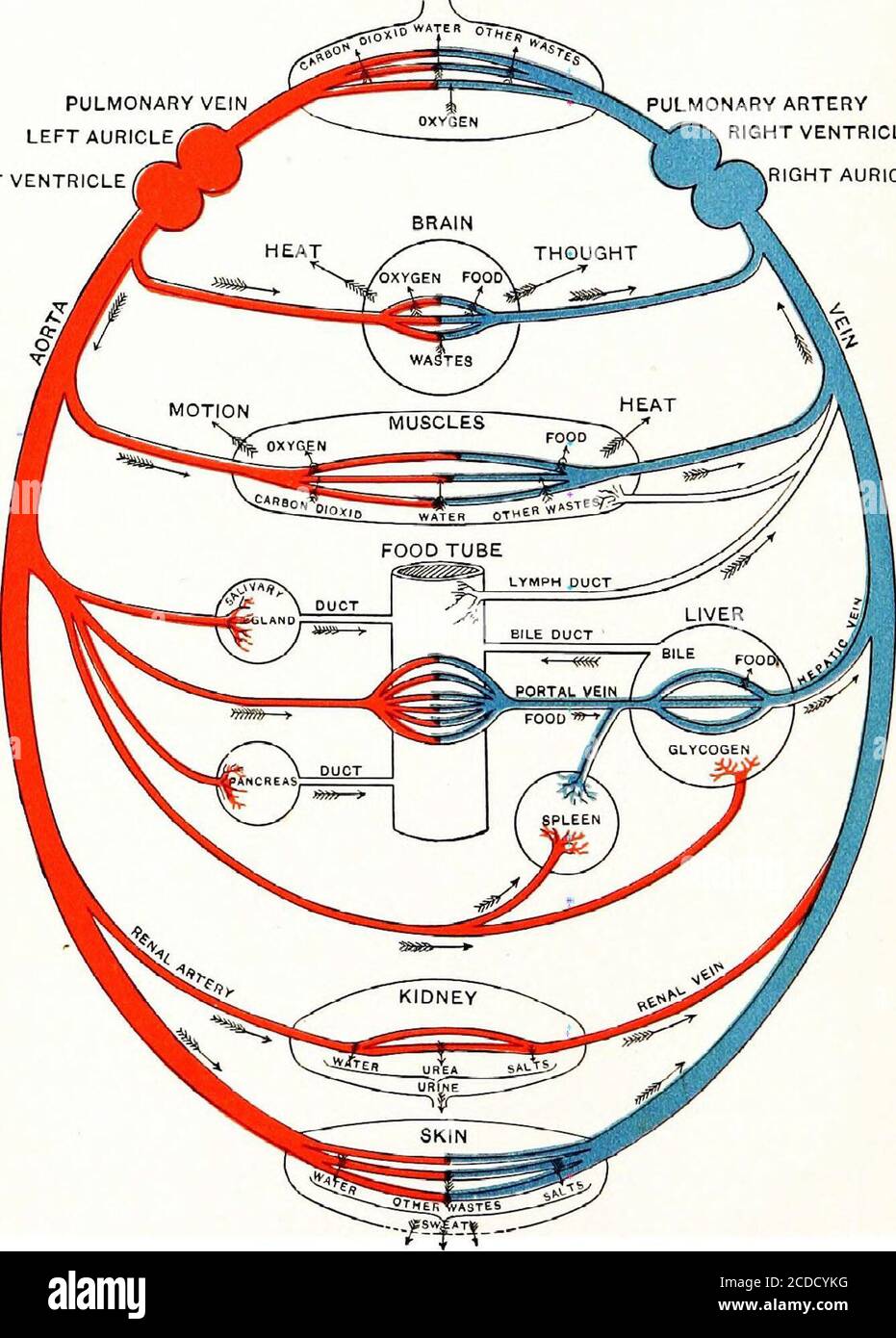 . Physiology, experimental and descriptive . Pulmonary Artery Right AuricleRight Ventricle — Caval Vein Body Capillaries Fig. 73. Diagram of the Circulation, representing the Right and Left Halves separated,showing that the Blood makes but one Circuit. {Dorsal View.) skin to be purified. Yet, as this mixed blood flows througheach organ, that organ, so long as it is in health, takes fromit only what it should take. The kidney takes, during health, 2U THE CIRCULATION- OF THE BLOOD. PULMONARY VEINLEFT AURICLE LEFT VENTRICLE PULMONARY ARTERY RIGHT VENTRICLE RIGHT AURICLE. Fig. 71. Diagram of the C Stock Photohttps://www.alamy.com/image-license-details/?v=1https://www.alamy.com/physiology-experimental-and-descriptive-pulmonary-artery-right-auricleright-ventricle-caval-vein-body-capillaries-fig-73-diagram-of-the-circulation-representing-the-right-and-left-halves-separatedshowing-that-the-blood-makes-but-one-circuit-dorsal-view-skin-to-be-purified-yet-as-this-mixed-blood-flows-througheach-organ-that-organ-so-long-as-it-is-in-health-takes-fromit-only-what-it-should-take-the-kidney-takes-during-health-2u-the-circulation-of-the-blood-pulmonary-veinleft-auricle-left-ventricle-pulmonary-artery-right-ventricle-right-auricle-fig-71-diagram-of-the-c-image369693396.html
RM2CDCYKG–. Physiology, experimental and descriptive . Pulmonary Artery Right AuricleRight Ventricle — Caval Vein Body Capillaries Fig. 73. Diagram of the Circulation, representing the Right and Left Halves separated,showing that the Blood makes but one Circuit. {Dorsal View.) skin to be purified. Yet, as this mixed blood flows througheach organ, that organ, so long as it is in health, takes fromit only what it should take. The kidney takes, during health, 2U THE CIRCULATION- OF THE BLOOD. PULMONARY VEINLEFT AURICLE LEFT VENTRICLE PULMONARY ARTERY RIGHT VENTRICLE RIGHT AURICLE. Fig. 71. Diagram of the C
 Fish bladders are being prepared for sale. Stock Photohttps://www.alamy.com/image-license-details/?v=1https://www.alamy.com/fish-bladders-are-being-prepared-for-sale-image626735687.html
RM2YBJ7NB–Fish bladders are being prepared for sale.
 . General surgical pathology and therapeutics, in fifty-one lectures .. . in the organ, so the part in which theabove coagulation occurs must be shaped like a Diagram: a, central end ofwedge or cone. In pathological anatomy thesecoagulations due to embolism have been called red or hemorrhagic wedge-shaped infarctions.Frequently as these wedge-shaped infarctionsoccur, they are not a necessary result of embo-lism ; for, when the arterial collateral circulation is strong enoughin the ischemic part to drive the blood through the capillaries,as is the case in otherwise healthy persons and in animal Stock Photohttps://www.alamy.com/image-license-details/?v=1https://www.alamy.com/general-surgical-pathology-and-therapeutics-in-fifty-one-lectures-in-the-organ-so-the-part-in-which-theabove-coagulation-occurs-must-be-shaped-like-a-diagram-a-central-end-ofwedge-or-cone-in-pathological-anatomy-thesecoagulations-due-to-embolism-have-been-called-red-or-hemorrhagic-wedge-shaped-infarctionsfrequently-as-these-wedge-shaped-infarctionsoccur-they-are-not-a-necessary-result-of-embo-lism-for-when-the-arterial-collateral-circulation-is-strong-enoughin-the-ischemic-part-to-drive-the-blood-through-the-capillariesas-is-the-case-in-otherwise-healthy-persons-and-in-animal-image370151602.html
RM2CE5T42–. General surgical pathology and therapeutics, in fifty-one lectures .. . in the organ, so the part in which theabove coagulation occurs must be shaped like a Diagram: a, central end ofwedge or cone. In pathological anatomy thesecoagulations due to embolism have been called red or hemorrhagic wedge-shaped infarctions.Frequently as these wedge-shaped infarctionsoccur, they are not a necessary result of embo-lism ; for, when the arterial collateral circulation is strong enoughin the ischemic part to drive the blood through the capillaries,as is the case in otherwise healthy persons and in animal
 . Text-book of normal histology: including an account of the development of the tissues and of the organs. Section of human kidney part a ly injected a interlobular artery giving off afferent tw g (i), c, efferent vesselpassing into intertubular capillaries [d); e, convolutedcapillaries of glomerulus ; /, outer layer of Bowmans cap-sule, the nuclei of whose cells show at g: h, uriniferoustubule in transverse section, /, in oblique section. 194 NORMAL HISTOLOGY.Fig. 237.. Pelvic Diagram of the kidney, showing the course of the uriniferous tubules and of the blood vessels ; forconvenience the me Stock Photohttps://www.alamy.com/image-license-details/?v=1https://www.alamy.com/text-book-of-normal-histology-including-an-account-of-the-development-of-the-tissues-and-of-the-organs-section-of-human-kidney-part-a-ly-injected-a-interlobular-artery-giving-off-afferent-tw-g-i-c-efferent-vesselpassing-into-intertubular-capillaries-d-e-convolutedcapillaries-of-glomerulus-outer-layer-of-bowmans-cap-sule-the-nuclei-of-whose-cells-show-at-g-h-uriniferoustubule-in-transverse-section-in-oblique-section-194-normal-histologyfig-237-pelvic-diagram-of-the-kidney-showing-the-course-of-the-uriniferous-tubules-and-of-the-blood-vessels-forconvenience-the-me-image370382308.html
RM2CEGABG–. Text-book of normal histology: including an account of the development of the tissues and of the organs. Section of human kidney part a ly injected a interlobular artery giving off afferent tw g (i), c, efferent vesselpassing into intertubular capillaries [d); e, convolutedcapillaries of glomerulus ; /, outer layer of Bowmans cap-sule, the nuclei of whose cells show at g: h, uriniferoustubule in transverse section, /, in oblique section. 194 NORMAL HISTOLOGY.Fig. 237.. Pelvic Diagram of the kidney, showing the course of the uriniferous tubules and of the blood vessels ; forconvenience the me
 . A text-book of comparative physiology for students and practitioners of comparative (veterinary) medicine . wIfi iqo CaDillarv blood-vessels (Landois). The cement substance between the en-dothelium h^Len rendered dark by silver nitrate, and the nuclei made prominentby staining. smaller end toward the heart aud the widest portions repre-senting the capillaries.. Pig. 193—Diagram to illustrate the relative proportions of the ^egate sectional we.; of the different parts of the vascular system (after eo). A, aorta, C, capillars.V, veins. 222 COMPARATIVE PHYSIOLOGY, THE ACTION OF THE MAMMALIAN Stock Photohttps://www.alamy.com/image-license-details/?v=1https://www.alamy.com/a-text-book-of-comparative-physiology-for-students-and-practitioners-of-comparative-veterinary-medicine-wifi-iqo-cadillarv-blood-vessels-landois-the-cement-substance-between-the-en-dothelium-hlen-rendered-dark-by-silver-nitrate-and-the-nuclei-made-prominentby-staining-smaller-end-toward-the-heart-aud-the-widest-portions-repre-senting-the-capillaries-pig-193diagram-to-illustrate-the-relative-proportions-of-the-egate-sectional-we-of-the-different-parts-of-the-vascular-system-after-eo-a-aorta-c-capillarsv-veins-222-comparative-physiology-the-action-of-the-mammalian-image372529995.html
RM2CJ25PK–. A text-book of comparative physiology for students and practitioners of comparative (veterinary) medicine . wIfi iqo CaDillarv blood-vessels (Landois). The cement substance between the en-dothelium h^Len rendered dark by silver nitrate, and the nuclei made prominentby staining. smaller end toward the heart aud the widest portions repre-senting the capillaries.. Pig. 193—Diagram to illustrate the relative proportions of the ^egate sectional we.; of the different parts of the vascular system (after eo). A, aorta, C, capillars.V, veins. 222 COMPARATIVE PHYSIOLOGY, THE ACTION OF THE MAMMALIAN
 . A text-book of comparative physiology for students and practitioners of comparative (veterinary) medicine . in different partsof the vascular system (after Yeo). h, heart; a, arterioles ; v, small veins; A,arteries ; C, capillaries ; V, large veins; H, V, representing the zero-line, i. e., at-mospheric pressure ; the blood-pressure is indicated by the height of the curve.The numbers on the left give the pressure approximately, in mm. of mercury. Relative Time occupied by the Various Phases of the CardiacCycle.—The old and valuable diagram reproduced below ismeant to convey through the eye th Stock Photohttps://www.alamy.com/image-license-details/?v=1https://www.alamy.com/a-text-book-of-comparative-physiology-for-students-and-practitioners-of-comparative-veterinary-medicine-in-different-partsof-the-vascular-system-after-yeo-h-heart-a-arterioles-v-small-veins-aarteries-c-capillaries-v-large-veins-h-v-representing-the-zero-line-i-e-at-mospheric-pressure-the-blood-pressure-is-indicated-by-the-height-of-the-curvethe-numbers-on-the-left-give-the-pressure-approximately-in-mm-of-mercury-relative-time-occupied-by-the-various-phases-of-the-cardiaccyclethe-old-and-valuable-diagram-reproduced-below-ismeant-to-convey-through-the-eye-th-image372519274.html
RM2CJ1M3P–. A text-book of comparative physiology for students and practitioners of comparative (veterinary) medicine . in different partsof the vascular system (after Yeo). h, heart; a, arterioles ; v, small veins; A,arteries ; C, capillaries ; V, large veins; H, V, representing the zero-line, i. e., at-mospheric pressure ; the blood-pressure is indicated by the height of the curve.The numbers on the left give the pressure approximately, in mm. of mercury. Relative Time occupied by the Various Phases of the CardiacCycle.—The old and valuable diagram reproduced below ismeant to convey through the eye th
 . Human physiology. Fig. 122. -Capillary Blood-vessels in the Web of aFrogs Foot, as seen withthe microscope. a, small artery ; b, capillaries ; c, smallvein. The arrows show the courseof the blood. Fig. 123.—Diagram showingthe Valves of Veins. a, part of a vein laid open, with twopairs of valves. b, longitudinal section of a vein,showing the valves closed. c, portion of a distended vein, ex-hibiting a swelling at a pair of valves. Most veins are also provided with valves like the semilunarvalves of the heart, sometimes arranged at very short intervals.All these valves are situated with their Stock Photohttps://www.alamy.com/image-license-details/?v=1https://www.alamy.com/human-physiology-fig-122-capillary-blood-vessels-in-the-web-of-afrogs-foot-as-seen-withthe-microscope-a-small-artery-b-capillaries-c-smallvein-the-arrows-show-the-courseof-the-blood-fig-123diagram-showingthe-valves-of-veins-a-part-of-a-vein-laid-open-with-twopairs-of-valves-b-longitudinal-section-of-a-veinshowing-the-valves-closed-c-portion-of-a-distended-vein-ex-hibiting-a-swelling-at-a-pair-of-valves-most-veins-are-also-provided-with-valves-like-the-semilunarvalves-of-the-heart-sometimes-arranged-at-very-short-intervalsall-these-valves-are-situated-with-their-image370643163.html
RM2CF073R–. Human physiology. Fig. 122. -Capillary Blood-vessels in the Web of aFrogs Foot, as seen withthe microscope. a, small artery ; b, capillaries ; c, smallvein. The arrows show the courseof the blood. Fig. 123.—Diagram showingthe Valves of Veins. a, part of a vein laid open, with twopairs of valves. b, longitudinal section of a vein,showing the valves closed. c, portion of a distended vein, ex-hibiting a swelling at a pair of valves. Most veins are also provided with valves like the semilunarvalves of the heart, sometimes arranged at very short intervals.All these valves are situated with their
 . Physiology, experimental and descriptive . Artery -Vein CapillariesFig. 72. Diagram of the Heart and Blood Tubes.(Dorsal View.) main points in the blood circuit, and the losses and gains inits course. In the common blood streams are combined the good andthe bad. The newly digested food is received into a currentof impure blood returning from the muscles (Caval Veins). PLAN OF CIRCULATION. 243 The blood from the kidneys, probably the purest blood inthe body, joins the same impure stream. From the Aorta, redblood, usually called pure, is sent to the kidneys.and to the Lung Capillaries Pulmonar Stock Photohttps://www.alamy.com/image-license-details/?v=1https://www.alamy.com/physiology-experimental-and-descriptive-artery-vein-capillariesfig-72-diagram-of-the-heart-and-blood-tubesdorsal-view-main-points-in-the-blood-circuit-and-the-losses-and-gains-inits-course-in-the-common-blood-streams-are-combined-the-good-andthe-bad-the-newly-digested-food-is-received-into-a-currentof-impure-blood-returning-from-the-muscles-caval-veins-plan-of-circulation-243-the-blood-from-the-kidneys-probably-the-purest-blood-inthe-body-joins-the-same-impure-stream-from-the-aorta-redblood-usually-called-pure-is-sent-to-the-kidneysand-to-the-lung-capillaries-pulmonar-image369693519.html
RM2CDCYRY–. Physiology, experimental and descriptive . Artery -Vein CapillariesFig. 72. Diagram of the Heart and Blood Tubes.(Dorsal View.) main points in the blood circuit, and the losses and gains inits course. In the common blood streams are combined the good andthe bad. The newly digested food is received into a currentof impure blood returning from the muscles (Caval Veins). PLAN OF CIRCULATION. 243 The blood from the kidneys, probably the purest blood inthe body, joins the same impure stream. From the Aorta, redblood, usually called pure, is sent to the kidneys.and to the Lung Capillaries Pulmonar
 . The human body and health : an elementary text-book of essential anatomy, applied physiology and practical hygiene for schools . he proteidsof the food. Intestinal Glands.— The mucousmembrane of theintestine is com-posed largely ofsimple, tubelikeglands standingside by side, withtheir mouths open-ing at the bases of the villi which stick ^^^ 57. —Diagram of a tiny block of tissue smaller than a pins head, cut from the wall ofout into the cavity ^he stomach, a, mucous membrane; c and d,of the intestine. muscular coats; e, mouths of gastric glands; h, gastric glands; i, capillaries supplying t Stock Photohttps://www.alamy.com/image-license-details/?v=1https://www.alamy.com/the-human-body-and-health-an-elementary-text-book-of-essential-anatomy-applied-physiology-and-practical-hygiene-for-schools-he-proteidsof-the-food-intestinal-glands-the-mucousmembrane-of-theintestine-is-com-posed-largely-ofsimple-tubelikeglands-standingside-by-side-withtheir-mouths-open-ing-at-the-bases-of-the-villi-which-stick-57-diagram-of-a-tiny-block-of-tissue-smaller-than-a-pins-head-cut-from-the-wall-ofout-into-the-cavity-he-stomach-a-mucous-membrane-c-and-dof-the-intestine-muscular-coats-e-mouths-of-gastric-glands-h-gastric-glands-i-capillaries-supplying-t-image369998361.html
RM2CDXTK5–. The human body and health : an elementary text-book of essential anatomy, applied physiology and practical hygiene for schools . he proteidsof the food. Intestinal Glands.— The mucousmembrane of theintestine is com-posed largely ofsimple, tubelikeglands standingside by side, withtheir mouths open-ing at the bases of the villi which stick ^^^ 57. —Diagram of a tiny block of tissue smaller than a pins head, cut from the wall ofout into the cavity ^he stomach, a, mucous membrane; c and d,of the intestine. muscular coats; e, mouths of gastric glands; h, gastric glands; i, capillaries supplying t
 . Essentials of biology presented in problems. Biology. 376 THE BLOOD AND ITS CIRCULATION. Diagram showing the exchange between blood and the cells of the body. up its food material to the lymph. This it does by passing it through the walls of the capillaries. The food is in turn given up to the tissue cells which are bathed by the IjTnph. Some of the amoeboid corpuscles from the blood make their way between the cells forming the walls of the capillaries. Lymph, then, is practically blood- plasma plus some colorless corpuscles. It acts as the medium of exchange between the blood proper and the Stock Photohttps://www.alamy.com/image-license-details/?v=1https://www.alamy.com/essentials-of-biology-presented-in-problems-biology-376-the-blood-and-its-circulation-diagram-showing-the-exchange-between-blood-and-the-cells-of-the-body-up-its-food-material-to-the-lymph-this-it-does-by-passing-it-through-the-walls-of-the-capillaries-the-food-is-in-turn-given-up-to-the-tissue-cells-which-are-bathed-by-the-ijtnph-some-of-the-amoeboid-corpuscles-from-the-blood-make-their-way-between-the-cells-forming-the-walls-of-the-capillaries-lymph-then-is-practically-blood-plasma-plus-some-colorless-corpuscles-it-acts-as-the-medium-of-exchange-between-the-blood-proper-and-the-image232339739.html
RMRDYYKR–. Essentials of biology presented in problems. Biology. 376 THE BLOOD AND ITS CIRCULATION. Diagram showing the exchange between blood and the cells of the body. up its food material to the lymph. This it does by passing it through the walls of the capillaries. The food is in turn given up to the tissue cells which are bathed by the IjTnph. Some of the amoeboid corpuscles from the blood make their way between the cells forming the walls of the capillaries. Lymph, then, is practically blood- plasma plus some colorless corpuscles. It acts as the medium of exchange between the blood proper and the
 . Anatomical technology as applied to the domestic cat; an introduction to human, veterinary, and comparative anatomy. Cats; Dissection; Mammals. THE CARDIAC CAVITIES. 323 Fig. 93.—Diagrammatic representation of the heart, the great vessels, the pulmonic and systemic capillaries. In this diagram, as in Fig. 91, the heart is seen from its dorsal (posterior) aspect; hence Us right and left portions correspond in position with the right and left of the observer (§ 56). Most of the parts are shown by outlines only, but the ventricular and auricular walls are shaded, and the lines representing the Stock Photohttps://www.alamy.com/image-license-details/?v=1https://www.alamy.com/anatomical-technology-as-applied-to-the-domestic-cat-an-introduction-to-human-veterinary-and-comparative-anatomy-cats-dissection-mammals-the-cardiac-cavities-323-fig-93diagrammatic-representation-of-the-heart-the-great-vessels-the-pulmonic-and-systemic-capillaries-in-this-diagram-as-in-fig-91-the-heart-is-seen-from-its-dorsal-posterior-aspect-hence-us-right-and-left-portions-correspond-in-position-with-the-right-and-left-of-the-observer-56-most-of-the-parts-are-shown-by-outlines-only-but-the-ventricular-and-auricular-walls-are-shaded-and-the-lines-representing-the-image232347952.html
RMRE0A54–. Anatomical technology as applied to the domestic cat; an introduction to human, veterinary, and comparative anatomy. Cats; Dissection; Mammals. THE CARDIAC CAVITIES. 323 Fig. 93.—Diagrammatic representation of the heart, the great vessels, the pulmonic and systemic capillaries. In this diagram, as in Fig. 91, the heart is seen from its dorsal (posterior) aspect; hence Us right and left portions correspond in position with the right and left of the observer (§ 56). Most of the parts are shown by outlines only, but the ventricular and auricular walls are shaded, and the lines representing the
 . Essentials of biology presented in problems. Biology. Diagram showing the exchange between blood and the cells of the body. up its food material to the lymph. This it does by passing it through the walls of the capillaries. The food is in turn given up to the tissue cells which are bathed by the IjTnph. Some of the amoeboid corpuscles from the blood make their way between the cells forming the walls of the capillaries. Lymph, then, is practically blood- plasma plus some colorless corpuscles. It acts as the medium of exchange between the blood proper and the cells in the tissues of the body. Stock Photohttps://www.alamy.com/image-license-details/?v=1https://www.alamy.com/essentials-of-biology-presented-in-problems-biology-diagram-showing-the-exchange-between-blood-and-the-cells-of-the-body-up-its-food-material-to-the-lymph-this-it-does-by-passing-it-through-the-walls-of-the-capillaries-the-food-is-in-turn-given-up-to-the-tissue-cells-which-are-bathed-by-the-ijtnph-some-of-the-amoeboid-corpuscles-from-the-blood-make-their-way-between-the-cells-forming-the-walls-of-the-capillaries-lymph-then-is-practically-blood-plasma-plus-some-colorless-corpuscles-it-acts-as-the-medium-of-exchange-between-the-blood-proper-and-the-cells-in-the-tissues-of-the-body-image232339735.html
RMRDYYKK–. Essentials of biology presented in problems. Biology. Diagram showing the exchange between blood and the cells of the body. up its food material to the lymph. This it does by passing it through the walls of the capillaries. The food is in turn given up to the tissue cells which are bathed by the IjTnph. Some of the amoeboid corpuscles from the blood make their way between the cells forming the walls of the capillaries. Lymph, then, is practically blood- plasma plus some colorless corpuscles. It acts as the medium of exchange between the blood proper and the cells in the tissues of the body.
 . A text-book in general physiology and anatomy. Physiology, Comparative; Anatomy. 196 CIECULATION monary arteries convey the blood from the right ventricle directly to the lungs. In the many capillaries, into which. Fig. 74 — Diagram of the circulatory system. the branches of the pulmonary artery divide in the lungs, the blood takes on a new supply of oxygen and loses its carbon dioxide. These capillaries are collected into larger. Please note that these images are extracted from scanned page images that may have been digitally enhanced for readability - coloration and appearance of these ill Stock Photohttps://www.alamy.com/image-license-details/?v=1https://www.alamy.com/a-text-book-in-general-physiology-and-anatomy-physiology-comparative-anatomy-196-cieculation-monary-arteries-convey-the-blood-from-the-right-ventricle-directly-to-the-lungs-in-the-many-capillaries-into-which-fig-74-diagram-of-the-circulatory-system-the-branches-of-the-pulmonary-artery-divide-in-the-lungs-the-blood-takes-on-a-new-supply-of-oxygen-and-loses-its-carbon-dioxide-these-capillaries-are-collected-into-larger-please-note-that-these-images-are-extracted-from-scanned-page-images-that-may-have-been-digitally-enhanced-for-readability-coloration-and-appearance-of-these-ill-image232125031.html
RMRDJ5RK–. A text-book in general physiology and anatomy. Physiology, Comparative; Anatomy. 196 CIECULATION monary arteries convey the blood from the right ventricle directly to the lungs. In the many capillaries, into which. Fig. 74 — Diagram of the circulatory system. the branches of the pulmonary artery divide in the lungs, the blood takes on a new supply of oxygen and loses its carbon dioxide. These capillaries are collected into larger. Please note that these images are extracted from scanned page images that may have been digitally enhanced for readability - coloration and appearance of these ill
 . First lessons in zoology. Zoology. FOOD AND AJR TURNED INTO FLESH AND ENERGY. 95 distribute it to the capillaries of tlie general body-tissues. From these it is gathered by the veins and carried back to the auricle to begin again. In the course of circu- lation the blood reaches every part of the body, picking up certain substances here, leaving others there, thus accomplishing the results already pointed out as the objects of the circulation. In the circulation of the higher vertebrates the most striking difference from that of the fish is in the structure. Fig. 59.—Diagram of circulatory s Stock Photohttps://www.alamy.com/image-license-details/?v=1https://www.alamy.com/first-lessons-in-zoology-zoology-food-and-ajr-turned-into-flesh-and-energy-95-distribute-it-to-the-capillaries-of-tlie-general-body-tissues-from-these-it-is-gathered-by-the-veins-and-carried-back-to-the-auricle-to-begin-again-in-the-course-of-circu-lation-the-blood-reaches-every-part-of-the-body-picking-up-certain-substances-here-leaving-others-there-thus-accomplishing-the-results-already-pointed-out-as-the-objects-of-the-circulation-in-the-circulation-of-the-higher-vertebrates-the-most-striking-difference-from-that-of-the-fish-is-in-the-structure-fig-59diagram-of-circulatory-s-image232255936.html
RMRDT4PT–. First lessons in zoology. Zoology. FOOD AND AJR TURNED INTO FLESH AND ENERGY. 95 distribute it to the capillaries of tlie general body-tissues. From these it is gathered by the veins and carried back to the auricle to begin again. In the course of circu- lation the blood reaches every part of the body, picking up certain substances here, leaving others there, thus accomplishing the results already pointed out as the objects of the circulation. In the circulation of the higher vertebrates the most striking difference from that of the fish is in the structure. Fig. 59.—Diagram of circulatory s
![. A text-book of comparative physiology for students and practitioners of comparative (veterinary) medicine. Physiology, Comparative. 222 COMPAKATIVB PHYSIOLOGY, THE ACTION OF THE MAMMALIAN HEART. What takes place may be thus very briefly stated : The right auricle contracting squeezes the blood through the au- ricular-ventricular opening into the right ventricle, never quite Superior Vena ' CaTa. Capillaries of the Head, etc. Pulmonary Ca- , pillaries.. ]rain Arterial Trunk. Fig. 194.—Diagram of tlie circulation. The arrows indicate the course of the blood. Thongh the pulmonary, the lower a Stock Photo . A text-book of comparative physiology for students and practitioners of comparative (veterinary) medicine. Physiology, Comparative. 222 COMPAKATIVB PHYSIOLOGY, THE ACTION OF THE MAMMALIAN HEART. What takes place may be thus very briefly stated : The right auricle contracting squeezes the blood through the au- ricular-ventricular opening into the right ventricle, never quite Superior Vena ' CaTa. Capillaries of the Head, etc. Pulmonary Ca- , pillaries.. ]rain Arterial Trunk. Fig. 194.—Diagram of tlie circulation. The arrows indicate the course of the blood. Thongh the pulmonary, the lower a Stock Photo](https://c8.alamy.com/comp/RE0405/a-text-book-of-comparative-physiology-for-students-and-practitioners-of-comparative-veterinary-medicine-physiology-comparative-222-compakativb-physiology-the-action-of-the-mammalian-heart-what-takes-place-may-be-thus-very-briefly-stated-the-right-auricle-contracting-squeezes-the-blood-through-the-au-ricular-ventricular-opening-into-the-right-ventricle-never-quite-superior-vena-cata-capillaries-of-the-head-etc-pulmonary-ca-pillaries-rain-arterial-trunk-fig-194diagram-of-tlie-circulation-the-arrows-indicate-the-course-of-the-blood-thongh-the-pulmonary-the-lower-a-RE0405.jpg) . A text-book of comparative physiology for students and practitioners of comparative (veterinary) medicine. Physiology, Comparative. 222 COMPAKATIVB PHYSIOLOGY, THE ACTION OF THE MAMMALIAN HEART. What takes place may be thus very briefly stated : The right auricle contracting squeezes the blood through the au- ricular-ventricular opening into the right ventricle, never quite Superior Vena ' CaTa. Capillaries of the Head, etc. Pulmonary Ca- , pillaries.. ]rain Arterial Trunk. Fig. 194.—Diagram of tlie circulation. The arrows indicate the course of the blood. Thongh the pulmonary, the lower a Stock Photohttps://www.alamy.com/image-license-details/?v=1https://www.alamy.com/a-text-book-of-comparative-physiology-for-students-and-practitioners-of-comparative-veterinary-medicine-physiology-comparative-222-compakativb-physiology-the-action-of-the-mammalian-heart-what-takes-place-may-be-thus-very-briefly-stated-the-right-auricle-contracting-squeezes-the-blood-through-the-au-ricular-ventricular-opening-into-the-right-ventricle-never-quite-superior-vena-cata-capillaries-of-the-head-etc-pulmonary-ca-pillaries-rain-arterial-trunk-fig-194diagram-of-tlie-circulation-the-arrows-indicate-the-course-of-the-blood-thongh-the-pulmonary-the-lower-a-image232343109.html
. A text-book of comparative physiology for students and practitioners of comparative (veterinary) medicine. Physiology, Comparative. 222 COMPAKATIVB PHYSIOLOGY, THE ACTION OF THE MAMMALIAN HEART. What takes place may be thus very briefly stated : The right auricle contracting squeezes the blood through the au- ricular-ventricular opening into the right ventricle, never quite Superior Vena ' CaTa. Capillaries of the Head, etc. Pulmonary Ca- , pillaries.. ]rain Arterial Trunk. Fig. 194.—Diagram of tlie circulation. The arrows indicate the course of the blood. Thongh the pulmonary, the lower a Stock Photohttps://www.alamy.com/image-license-details/?v=1https://www.alamy.com/a-text-book-of-comparative-physiology-for-students-and-practitioners-of-comparative-veterinary-medicine-physiology-comparative-222-compakativb-physiology-the-action-of-the-mammalian-heart-what-takes-place-may-be-thus-very-briefly-stated-the-right-auricle-contracting-squeezes-the-blood-through-the-au-ricular-ventricular-opening-into-the-right-ventricle-never-quite-superior-vena-cata-capillaries-of-the-head-etc-pulmonary-ca-pillaries-rain-arterial-trunk-fig-194diagram-of-tlie-circulation-the-arrows-indicate-the-course-of-the-blood-thongh-the-pulmonary-the-lower-a-image232343109.htmlRMRE0405–. A text-book of comparative physiology for students and practitioners of comparative (veterinary) medicine. Physiology, Comparative. 222 COMPAKATIVB PHYSIOLOGY, THE ACTION OF THE MAMMALIAN HEART. What takes place may be thus very briefly stated : The right auricle contracting squeezes the blood through the au- ricular-ventricular opening into the right ventricle, never quite Superior Vena ' CaTa. Capillaries of the Head, etc. Pulmonary Ca- , pillaries.. ]rain Arterial Trunk. Fig. 194.—Diagram of tlie circulation. The arrows indicate the course of the blood. Thongh the pulmonary, the lower a
 . Anatomischer Anzeiger. Anatomy, Comparative; Anatomy, Comparative. 262 arterio-venous connection is the source of capillaries. They branch freely over the surfaces of organs, lying in an abundant connective tissue. In their double origin from both an artery and a vein, and in their relation to connective tissue they ditfer from sinusoids. These features are represented in the diagram. Fig. 1. On the right is the pancreas with its capillaries; on the left the liver and sinusoids. If the gland tissue of the pancreas were entirely removed, artery, vein, and capillaries would remain unchanged; b Stock Photohttps://www.alamy.com/image-license-details/?v=1https://www.alamy.com/anatomischer-anzeiger-anatomy-comparative-anatomy-comparative-262-arterio-venous-connection-is-the-source-of-capillaries-they-branch-freely-over-the-surfaces-of-organs-lying-in-an-abundant-connective-tissue-in-their-double-origin-from-both-an-artery-and-a-vein-and-in-their-relation-to-connective-tissue-they-ditfer-from-sinusoids-these-features-are-represented-in-the-diagram-fig-1-on-the-right-is-the-pancreas-with-its-capillaries-on-the-left-the-liver-and-sinusoids-if-the-gland-tissue-of-the-pancreas-were-entirely-removed-artery-vein-and-capillaries-would-remain-unchanged-b-image236830518.html
RMRN8FMP–. Anatomischer Anzeiger. Anatomy, Comparative; Anatomy, Comparative. 262 arterio-venous connection is the source of capillaries. They branch freely over the surfaces of organs, lying in an abundant connective tissue. In their double origin from both an artery and a vein, and in their relation to connective tissue they ditfer from sinusoids. These features are represented in the diagram. Fig. 1. On the right is the pancreas with its capillaries; on the left the liver and sinusoids. If the gland tissue of the pancreas were entirely removed, artery, vein, and capillaries would remain unchanged; b
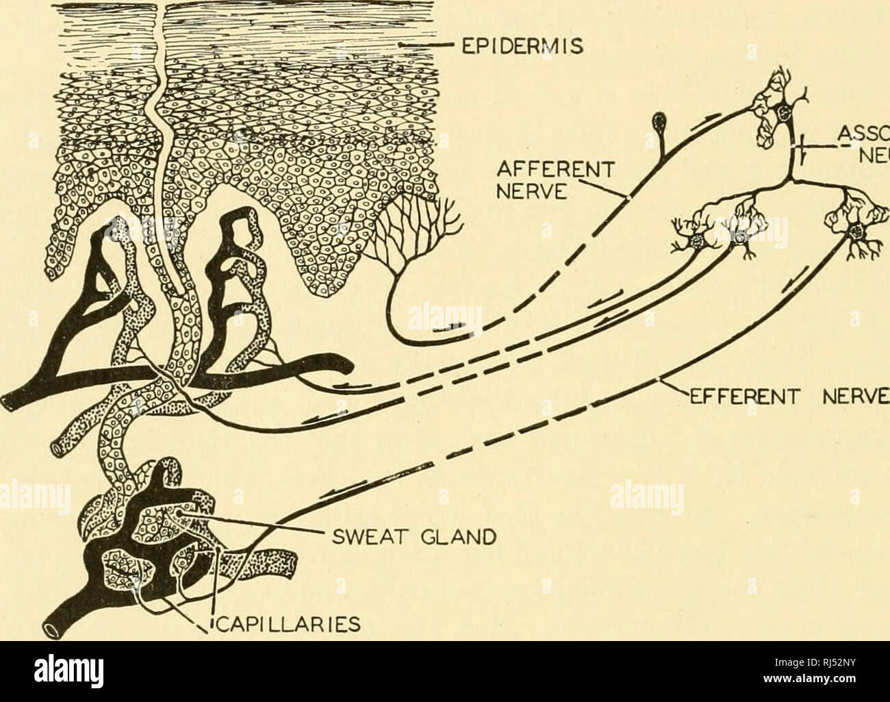 . Chordate anatomy. Chordata; Anatomy, Comparative. 126 CHORDATE ANATOMY secreting area; but those of the axillae are branched and greatly enlarged. They are of the "vitally secretory" type, that is, the cell protoplasm merely produces the secretion, but is not converted into it, and the cell continues alive indefinitely. The sweat is usually oily but, in man, becomes watery under the influence of the nerves. See Fig. 117.. ASSOCIATION NEURONE EFFERENT NERVE SWEAT GLAND CAPILLARIES Fig. 117.—A diagram illustrating the nervous mechanism of temperature regulation in man. The quantity o Stock Photohttps://www.alamy.com/image-license-details/?v=1https://www.alamy.com/chordate-anatomy-chordata-anatomy-comparative-126-chordate-anatomy-secreting-area-but-those-of-the-axillae-are-branched-and-greatly-enlarged-they-are-of-the-quotvitally-secretoryquot-type-that-is-the-cell-protoplasm-merely-produces-the-secretion-but-is-not-converted-into-it-and-the-cell-continues-alive-indefinitely-the-sweat-is-usually-oily-but-in-man-becomes-watery-under-the-influence-of-the-nerves-see-fig-117-association-neurone-efferent-nerve-sweat-gland-capillaries-fig-117a-diagram-illustrating-the-nervous-mechanism-of-temperature-regulation-in-man-the-quantity-o-image234910535.html
RMRJ52NY–. Chordate anatomy. Chordata; Anatomy, Comparative. 126 CHORDATE ANATOMY secreting area; but those of the axillae are branched and greatly enlarged. They are of the "vitally secretory" type, that is, the cell protoplasm merely produces the secretion, but is not converted into it, and the cell continues alive indefinitely. The sweat is usually oily but, in man, becomes watery under the influence of the nerves. See Fig. 117.. ASSOCIATION NEURONE EFFERENT NERVE SWEAT GLAND CAPILLARIES Fig. 117.—A diagram illustrating the nervous mechanism of temperature regulation in man. The quantity o
 . Comparative anatomy of vertebrates. Anatomy, Comparative; Vertebrates. 388 COMPARATIVE ANATOMY and finally disappear as the latter divide up into finer and finer branches. The ultimate bronchioles open into small terminal vesicles, the sacculi alvcolarcs or " infundibula " (Fig. 294, B), which are surrounded by a close network of capillaries, and the walls of. FIG. 294.—DIAGRAM OF THE STRUCTURE OF THE LUNG IN A, BIRDS, AND B, MAMMALS. (The whole of the lung is not represented.) A. Br, main bronchus; Srl, secondary bi'onchi: LP, "lung-pipes"' (para- bronchia). The arrows i Stock Photohttps://www.alamy.com/image-license-details/?v=1https://www.alamy.com/comparative-anatomy-of-vertebrates-anatomy-comparative-vertebrates-388-comparative-anatomy-and-finally-disappear-as-the-latter-divide-up-into-finer-and-finer-branches-the-ultimate-bronchioles-open-into-small-terminal-vesicles-the-sacculi-alvcolarcs-or-quot-infundibula-quot-fig-294-b-which-are-surrounded-by-a-close-network-of-capillaries-and-the-walls-of-fig-294diagram-of-the-structure-of-the-lung-in-a-birds-and-b-mammals-the-whole-of-the-lung-is-not-represented-a-br-main-bronchus-srl-secondary-bionchi-lp-quotlung-pipesquot-para-bronchia-the-arrows-i-image232651218.html
RMREE502–. Comparative anatomy of vertebrates. Anatomy, Comparative; Vertebrates. 388 COMPARATIVE ANATOMY and finally disappear as the latter divide up into finer and finer branches. The ultimate bronchioles open into small terminal vesicles, the sacculi alvcolarcs or " infundibula " (Fig. 294, B), which are surrounded by a close network of capillaries, and the walls of. FIG. 294.—DIAGRAM OF THE STRUCTURE OF THE LUNG IN A, BIRDS, AND B, MAMMALS. (The whole of the lung is not represented.) A. Br, main bronchus; Srl, secondary bi'onchi: LP, "lung-pipes"' (para- bronchia). The arrows i
 . A text-book of comparative physiology for students and practitioners of comparative (veterinary) medicine. Physiology, Comparative. Fig. 192.—Capillaiy blood-vessels (Landois). The cement substance between the en- dothelium has been rendered dark by silver nitrate, and the nuclei made prominent by staining. smaller end toward the heart and the widest portions repre- senting the capillaries.. Fig. 193.—Diagram to illustrate the relative proportions of the aggregate sectional area of the different parts of the vascular system (after Yeo). &, aorta; C, capillaries; V, veins.. Please note th Stock Photohttps://www.alamy.com/image-license-details/?v=1https://www.alamy.com/a-text-book-of-comparative-physiology-for-students-and-practitioners-of-comparative-veterinary-medicine-physiology-comparative-fig-192capillaiy-blood-vessels-landois-the-cement-substance-between-the-en-dothelium-has-been-rendered-dark-by-silver-nitrate-and-the-nuclei-made-prominent-by-staining-smaller-end-toward-the-heart-and-the-widest-portions-repre-senting-the-capillaries-fig-193diagram-to-illustrate-the-relative-proportions-of-the-aggregate-sectional-area-of-the-different-parts-of-the-vascular-system-after-yeo-amp-aorta-c-capillaries-v-veins-please-note-th-image232343114.html
RMRE040A–. A text-book of comparative physiology for students and practitioners of comparative (veterinary) medicine. Physiology, Comparative. Fig. 192.—Capillaiy blood-vessels (Landois). The cement substance between the en- dothelium has been rendered dark by silver nitrate, and the nuclei made prominent by staining. smaller end toward the heart and the widest portions repre- senting the capillaries.. Fig. 193.—Diagram to illustrate the relative proportions of the aggregate sectional area of the different parts of the vascular system (after Yeo). &, aorta; C, capillaries; V, veins.. Please note th
 . A text-book of animal physiology, with introductory chapters on general biology and a full treatment of reproduction ... Physiology, Comparative. 224 ANIMAL PHYSIOLOGY. departure from this harmony of rhythm -would lead to serious disturbance. Capillaries of the Head, etc. Superior Vena Cava. Inferior Vena Cava. Capillaries of Liver. Portal Vein.. Pulmonary Capil- laries. Main Arterial Trunk. Capillaries of Splanchnic Area. Capillaries of Trunk and Lower Ex- tremities. Fig. 204.—Diagram of the circulation. The arrows indicate the course of the blood. Though the pulmonary and the upper and the Stock Photohttps://www.alamy.com/image-license-details/?v=1https://www.alamy.com/a-text-book-of-animal-physiology-with-introductory-chapters-on-general-biology-and-a-full-treatment-of-reproduction-physiology-comparative-224-animal-physiology-departure-from-this-harmony-of-rhythm-would-lead-to-serious-disturbance-capillaries-of-the-head-etc-superior-vena-cava-inferior-vena-cava-capillaries-of-liver-portal-vein-pulmonary-capil-laries-main-arterial-trunk-capillaries-of-splanchnic-area-capillaries-of-trunk-and-lower-ex-tremities-fig-204diagram-of-the-circulation-the-arrows-indicate-the-course-of-the-blood-though-the-pulmonary-and-the-upper-and-the-image232418445.html
RMRE3G2N–. A text-book of animal physiology, with introductory chapters on general biology and a full treatment of reproduction ... Physiology, Comparative. 224 ANIMAL PHYSIOLOGY. departure from this harmony of rhythm -would lead to serious disturbance. Capillaries of the Head, etc. Superior Vena Cava. Inferior Vena Cava. Capillaries of Liver. Portal Vein.. Pulmonary Capil- laries. Main Arterial Trunk. Capillaries of Splanchnic Area. Capillaries of Trunk and Lower Ex- tremities. Fig. 204.—Diagram of the circulation. The arrows indicate the course of the blood. Though the pulmonary and the upper and the
 . A text-book of animal physiology, with introductory chapters on general biology and a full treatment of reproduction ... Physiology, Comparative. THE CIRCULATION OF THE BLOOD. 223. Fig. 203.—Diagram to illustrate the relative proportions of the aggregate Bectional area of the different parts of the vascular system (after Yeo). A, aorta; C, capillaries; V, veins. nels or cones "with the smaller end toward the heart and the widest portions representing the capillaries. The Action of the Mammalian Heart. Very briefly what takes place may be thus stated: The right auricle contracting squeez Stock Photohttps://www.alamy.com/image-license-details/?v=1https://www.alamy.com/a-text-book-of-animal-physiology-with-introductory-chapters-on-general-biology-and-a-full-treatment-of-reproduction-physiology-comparative-the-circulation-of-the-blood-223-fig-203diagram-to-illustrate-the-relative-proportions-of-the-aggregate-bectional-area-of-the-different-parts-of-the-vascular-system-after-yeo-a-aorta-c-capillaries-v-veins-nels-or-cones-quotwith-the-smaller-end-toward-the-heart-and-the-widest-portions-representing-the-capillaries-the-action-of-the-mammalian-heart-very-briefly-what-takes-place-may-be-thus-stated-the-right-auricle-contracting-squeez-image232418450.html
RMRE3G2X–. A text-book of animal physiology, with introductory chapters on general biology and a full treatment of reproduction ... Physiology, Comparative. THE CIRCULATION OF THE BLOOD. 223. Fig. 203.—Diagram to illustrate the relative proportions of the aggregate Bectional area of the different parts of the vascular system (after Yeo). A, aorta; C, capillaries; V, veins. nels or cones "with the smaller end toward the heart and the widest portions representing the capillaries. The Action of the Mammalian Heart. Very briefly what takes place may be thus stated: The right auricle contracting squeez
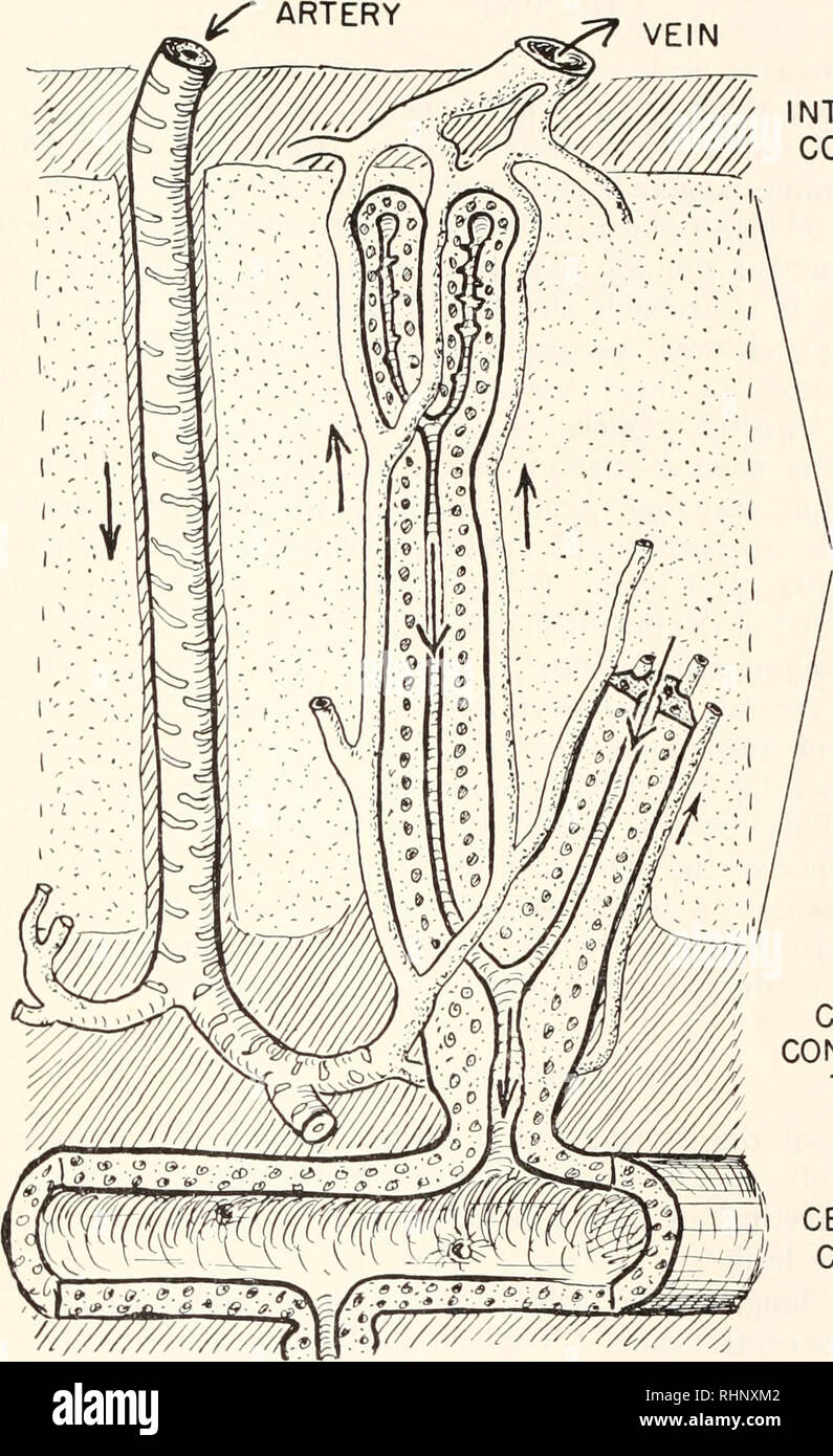 . The Biological bulletin. Biology; Zoology; Biology; Marine Biology. SALT GLAND OF THE GULL 169 ARTERY VEIN. INTERLOBULAR CONNECTIVE TISSUE SECRETORY TUBULES CENTRAL CONNECTIVE TISSUE CENTRAL CANAL FIGURE 9. Diagram of the circulation showing the opposing directions of the flow in the gland tubules and in the capillaries. The tubules branch repeatedly, but for simplicity only tvo ramifications are pictured. liminary histological studies of the salt glands of pelican (Pclecanus*), cormorant (Phalacrocora.v), eider duck (Somateria), petrel (Occanodroma), etc. In these birds the glands have ess Stock Photohttps://www.alamy.com/image-license-details/?v=1https://www.alamy.com/the-biological-bulletin-biology-zoology-biology-marine-biology-salt-gland-of-the-gull-169-artery-vein-interlobular-connective-tissue-secretory-tubules-central-connective-tissue-central-canal-figure-9-diagram-of-the-circulation-showing-the-opposing-directions-of-the-flow-in-the-gland-tubules-and-in-the-capillaries-the-tubules-branch-repeatedly-but-for-simplicity-only-tvo-ramifications-are-pictured-liminary-histological-studies-of-the-salt-glands-of-pelican-pclecanus-cormorant-phalacrocorav-eider-duck-somateria-petrel-occanodroma-etc-in-these-birds-the-glands-have-ess-image234665874.html
RMRHNXM2–. The Biological bulletin. Biology; Zoology; Biology; Marine Biology. SALT GLAND OF THE GULL 169 ARTERY VEIN. INTERLOBULAR CONNECTIVE TISSUE SECRETORY TUBULES CENTRAL CONNECTIVE TISSUE CENTRAL CANAL FIGURE 9. Diagram of the circulation showing the opposing directions of the flow in the gland tubules and in the capillaries. The tubules branch repeatedly, but for simplicity only tvo ramifications are pictured. liminary histological studies of the salt glands of pelican (Pclecanus*), cormorant (Phalacrocora.v), eider duck (Somateria), petrel (Occanodroma), etc. In these birds the glands have ess