Quick filters:
Cataract cortical Stock Photos and Images
 Eye: clouding of the lens in the event of cortical cataracts. Stock Photohttps://www.alamy.com/image-license-details/?v=1https://www.alamy.com/eye-clouding-of-the-lens-in-the-event-of-cortical-cataracts-image476926437.html
RF2JKWTF1–Eye: clouding of the lens in the event of cortical cataracts.
 Cure for Cataract - Red Open Blister Pack Tablets Isolated on White. Stock Photohttps://www.alamy.com/image-license-details/?v=1https://www.alamy.com/stock-photo-cure-for-cataract-red-open-blister-pack-tablets-isolated-on-white-78823363.html
RFEG6M03–Cure for Cataract - Red Open Blister Pack Tablets Isolated on White.
 Cataract Stock Photohttps://www.alamy.com/image-license-details/?v=1https://www.alamy.com/stock-photo-cataract-79771857.html
RFEHNWPW–Cataract
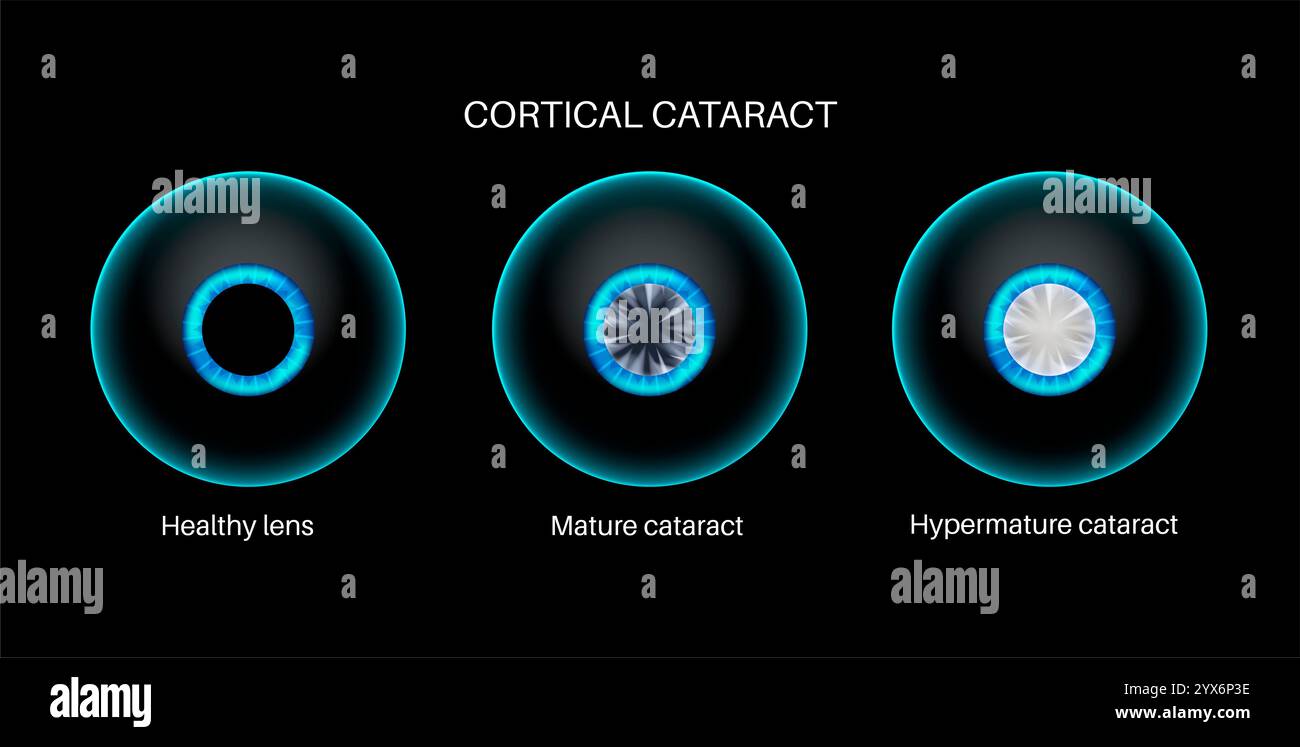 Cataract eye disease, illustration. Stock Photohttps://www.alamy.com/image-license-details/?v=1https://www.alamy.com/cataract-eye-disease-illustration-image635703362.html
RF2YX6P3E–Cataract eye disease, illustration.
 Background concept wordcloud illustration of cataract glowing light Stock Photohttps://www.alamy.com/image-license-details/?v=1https://www.alamy.com/stock-photo-background-concept-wordcloud-illustration-of-cataract-glowing-light-83006407.html
RFER17EF–Background concept wordcloud illustration of cataract glowing light
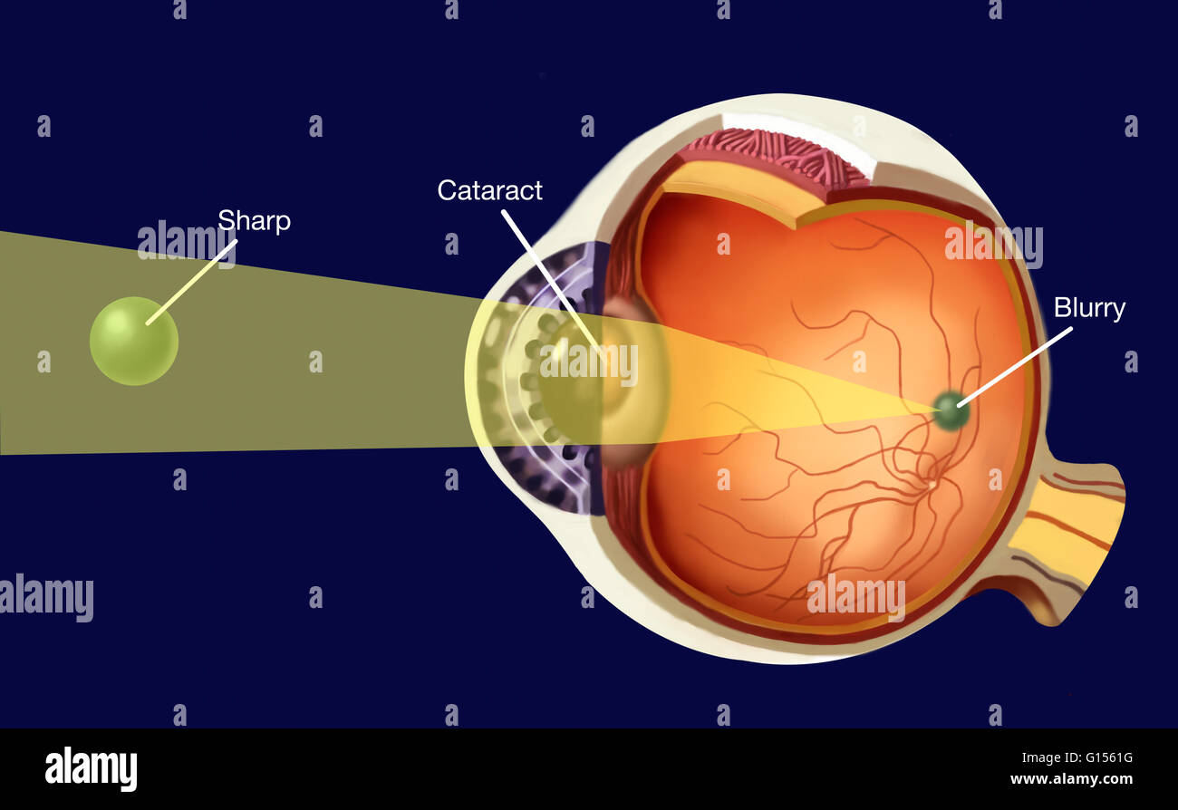 Representation of the opacification of a part of the lens in the case of cortical cataract. Stock Photohttps://www.alamy.com/image-license-details/?v=1https://www.alamy.com/stock-photo-representation-of-the-opacification-of-a-part-of-the-lens-in-the-case-103991372.html
RMG1561G–Representation of the opacification of a part of the lens in the case of cortical cataract.
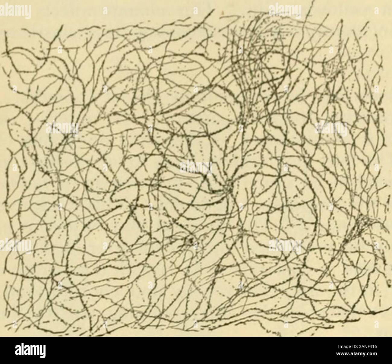 Human anatomy, including structure and development and practical considerations . e at sometimepartial, and they are called according to their location, anterior polar or capsular,posterior polar or capsular, central or nuclear, lamellar, perinuclear and cortical.Cataract occurs sometimes in the young, and is then soft; that is, the lens has nonucleus. The Vitreous Body. The vitreous body (corpus vitreum) fills the space between the lens and theretina, being in close contact with the retina and acting as a support to it as farforward as the ora serrata. Here it becomes separated from the retin Stock Photohttps://www.alamy.com/image-license-details/?v=1https://www.alamy.com/human-anatomy-including-structure-and-development-and-practical-considerations-e-at-sometimepartial-and-they-are-called-according-to-their-location-anterior-polar-or-capsularposterior-polar-or-capsular-central-or-nuclear-lamellar-perinuclear-and-corticalcataract-occurs-sometimes-in-the-young-and-is-then-soft-that-is-the-lens-has-nonucleus-the-vitreous-body-the-vitreous-body-corpus-vitreum-fills-the-space-between-the-lens-and-theretina-being-in-close-contact-with-the-retina-and-acting-as-a-support-to-it-as-farforward-as-the-ora-serrata-here-it-becomes-separated-from-the-retin-image340237218.html
RM2ANF416–Human anatomy, including structure and development and practical considerations . e at sometimepartial, and they are called according to their location, anterior polar or capsular,posterior polar or capsular, central or nuclear, lamellar, perinuclear and cortical.Cataract occurs sometimes in the young, and is then soft; that is, the lens has nonucleus. The Vitreous Body. The vitreous body (corpus vitreum) fills the space between the lens and theretina, being in close contact with the retina and acting as a support to it as farforward as the ora serrata. Here it becomes separated from the retin
 Diseases of the dog and Diseases of the dog and their treatment diseasesofdogthe01ml Year: 1897 CATARACT. 367 should be an unfavorable one. Hereditary cataract shows little inclination to enlargement, as is also the case in senile cataract. In soft cortical cataracts we may see a rapid opacity of the lens in a few days or weeks. The sight is entirely lost and medical treat- ment is of little use. Therapeutic Treatment. A cataract may be removed by an operation, and this is much more advisable in the dog because it is, as a rule, attended without any great danger, and its results are generall Stock Photohttps://www.alamy.com/image-license-details/?v=1https://www.alamy.com/diseases-of-the-dog-and-diseases-of-the-dog-and-their-treatment-diseasesofdogthe01ml-year-1897-cataract-367-should-be-an-unfavorable-one-hereditary-cataract-shows-little-inclination-to-enlargement-as-is-also-the-case-in-senile-cataract-in-soft-cortical-cataracts-we-may-see-a-rapid-opacity-of-the-lens-in-a-few-days-or-weeks-the-sight-is-entirely-lost-and-medical-treat-ment-is-of-little-use-therapeutic-treatment-a-cataract-may-be-removed-by-an-operation-and-this-is-much-more-advisable-in-the-dog-because-it-is-as-a-rule-attended-without-any-great-danger-and-its-results-are-generall-image241966747.html
RMT1JF1F–Diseases of the dog and Diseases of the dog and their treatment diseasesofdogthe01ml Year: 1897 CATARACT. 367 should be an unfavorable one. Hereditary cataract shows little inclination to enlargement, as is also the case in senile cataract. In soft cortical cataracts we may see a rapid opacity of the lens in a few days or weeks. The sight is entirely lost and medical treat- ment is of little use. Therapeutic Treatment. A cataract may be removed by an operation, and this is much more advisable in the dog because it is, as a rule, attended without any great danger, and its results are generall
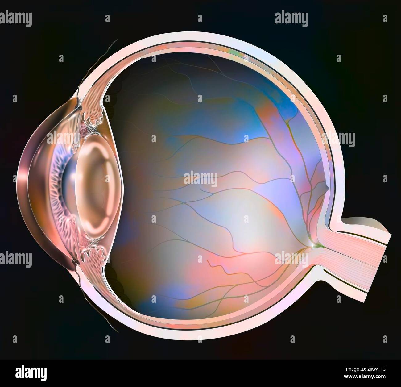 Eye: clouding of the lens in the event of cortical cataracts. Stock Photohttps://www.alamy.com/image-license-details/?v=1https://www.alamy.com/eye-clouding-of-the-lens-in-the-event-of-cortical-cataracts-image476926452.html
RF2JKWTFG–Eye: clouding of the lens in the event of cortical cataracts.
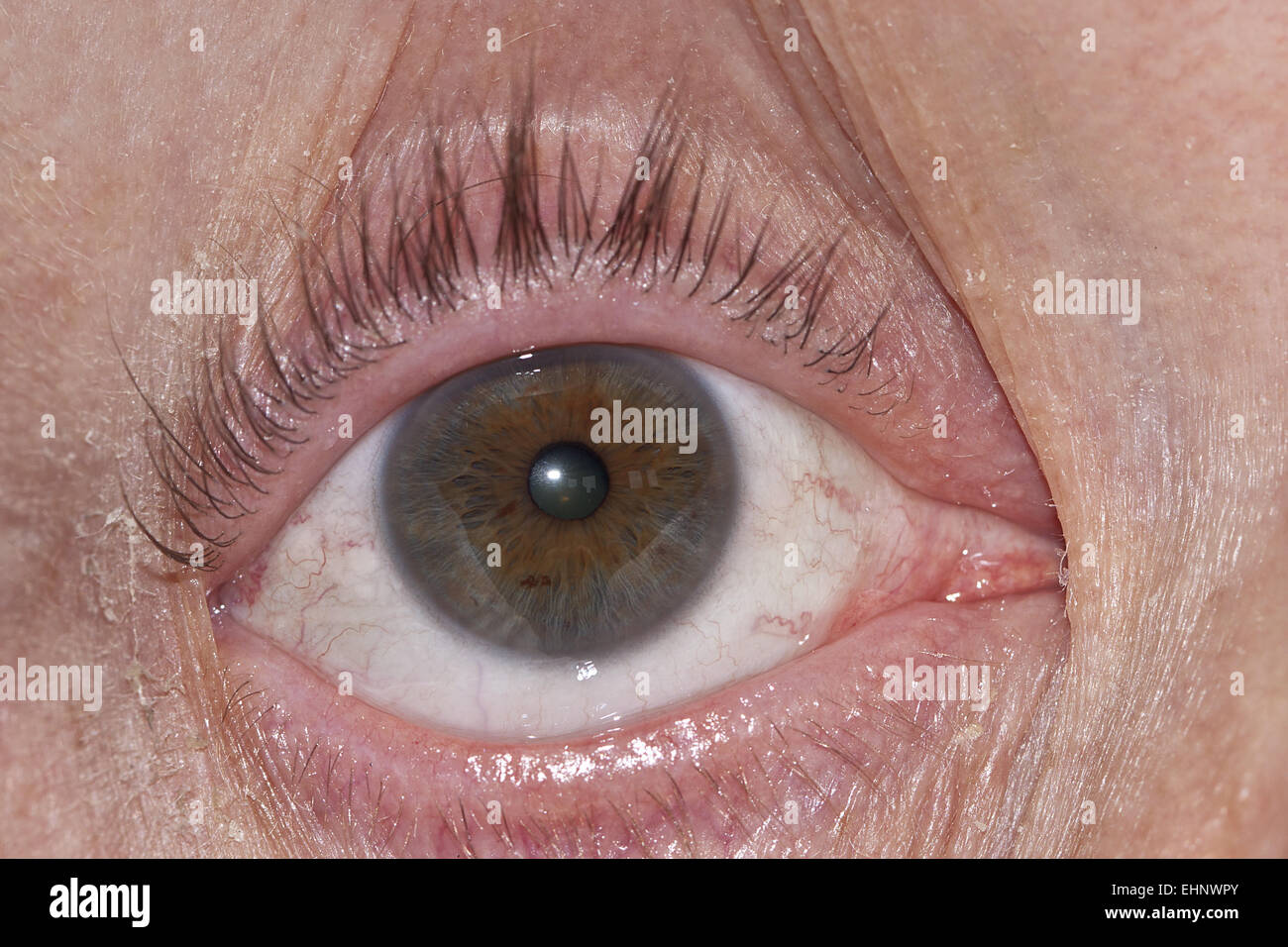 Cataract Stock Photohttps://www.alamy.com/image-license-details/?v=1https://www.alamy.com/stock-photo-cataract-79771859.html
RFEHNWPY–Cataract
 Cataract eye disease, illustration. Stock Photohttps://www.alamy.com/image-license-details/?v=1https://www.alamy.com/cataract-eye-disease-illustration-image635703373.html
RF2YX6P3W–Cataract eye disease, illustration.
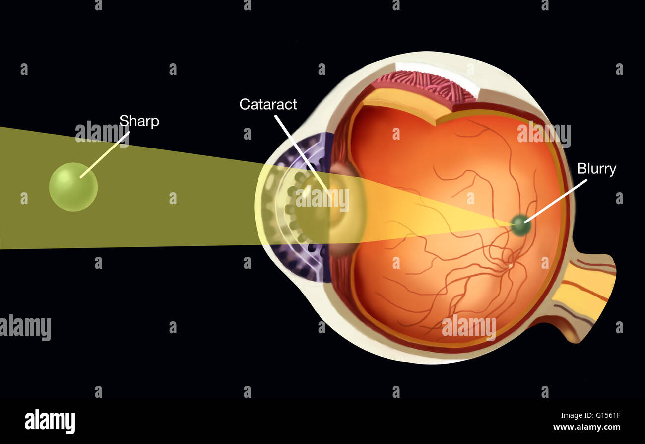 Representation of the opacification of a part of the lens in the case of cortical cataract. Stock Photohttps://www.alamy.com/image-license-details/?v=1https://www.alamy.com/stock-photo-representation-of-the-opacification-of-a-part-of-the-lens-in-the-case-103991371.html
RMG1561F–Representation of the opacification of a part of the lens in the case of cortical cataract.
 Atlas and epitome of operative ophthalmology . d into the upper part of the anterior chamber,making a corneal incision, the size of which is gauged bythe size of the nucleus of the cataract and the size of thecornea. The darker and more yellow the nucleus appearsthrough the small quantity of cortical matter, the largerit will be. If, on the other hand, the cortical materialrepresents a thick layer surrounding the nucleus, as, forexample, in young individuals, such a condition betraysitself by the fact that the grayish, translucent, rathermilky coloration extends deep into the cataract. Thediam Stock Photohttps://www.alamy.com/image-license-details/?v=1https://www.alamy.com/atlas-and-epitome-of-operative-ophthalmology-d-into-the-upper-part-of-the-anterior-chambermaking-a-corneal-incision-the-size-of-which-is-gauged-bythe-size-of-the-nucleus-of-the-cataract-and-the-size-of-thecornea-the-darker-and-more-yellow-the-nucleus-appearsthrough-the-small-quantity-of-cortical-matter-the-largerit-will-be-if-on-the-other-hand-the-cortical-materialrepresents-a-thick-layer-surrounding-the-nucleus-as-forexample-in-young-individuals-such-a-condition-betraysitself-by-the-fact-that-the-grayish-translucent-rathermilky-coloration-extends-deep-into-the-cataract-thediam-image339108543.html
RM2AKKMBB–Atlas and epitome of operative ophthalmology . d into the upper part of the anterior chamber,making a corneal incision, the size of which is gauged bythe size of the nucleus of the cataract and the size of thecornea. The darker and more yellow the nucleus appearsthrough the small quantity of cortical matter, the largerit will be. If, on the other hand, the cortical materialrepresents a thick layer surrounding the nucleus, as, forexample, in young individuals, such a condition betraysitself by the fact that the grayish, translucent, rathermilky coloration extends deep into the cataract. Thediam
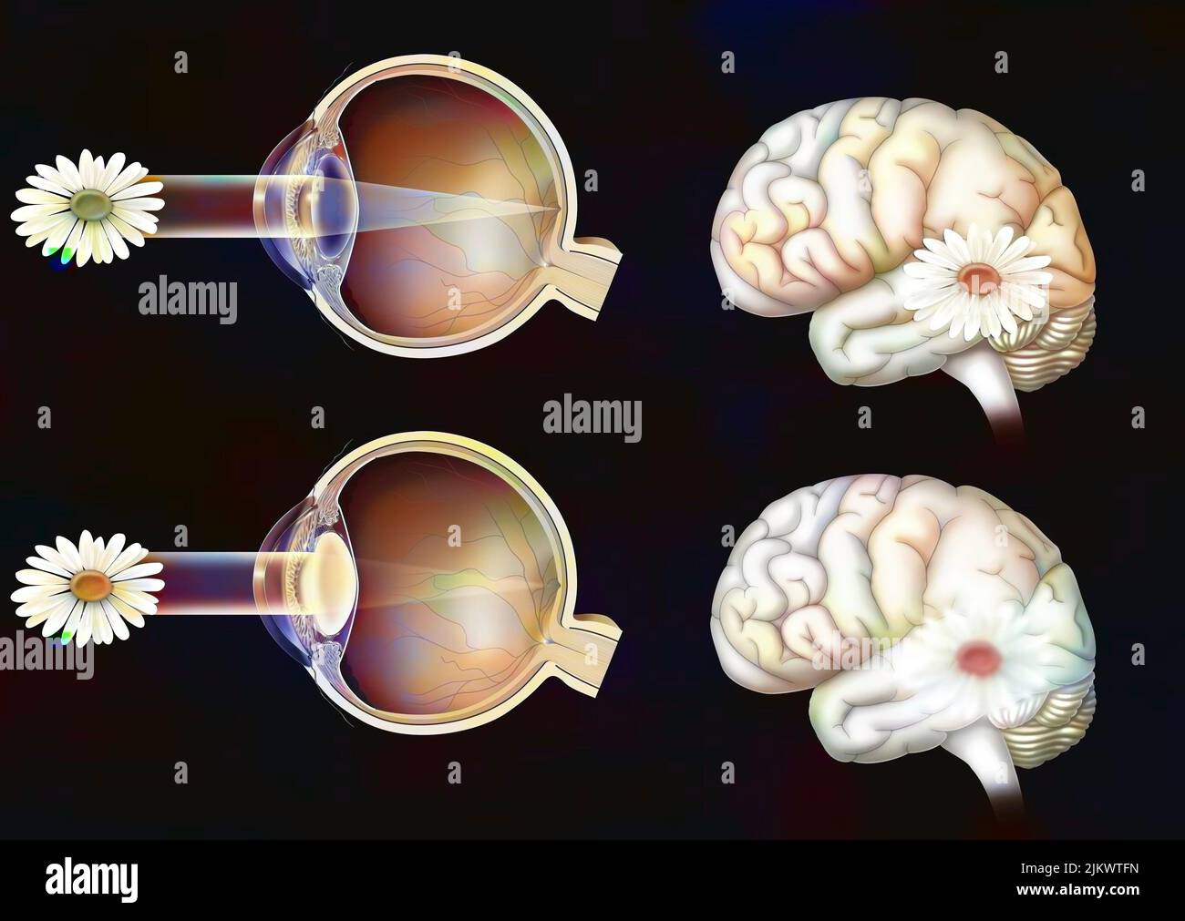 Comparison between normal vision and that of an eye with cataract. Stock Photohttps://www.alamy.com/image-license-details/?v=1https://www.alamy.com/comparison-between-normal-vision-and-that-of-an-eye-with-cataract-image476926457.html
RF2JKWTFN–Comparison between normal vision and that of an eye with cataract.
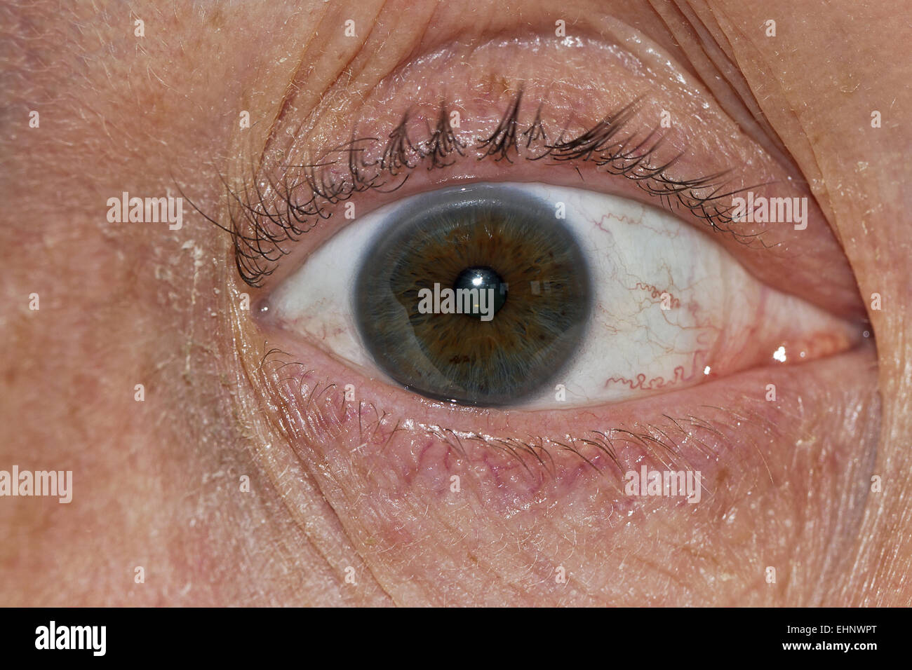 Cataract Stock Photohttps://www.alamy.com/image-license-details/?v=1https://www.alamy.com/stock-photo-cataract-79771856.html
RFEHNWPT–Cataract
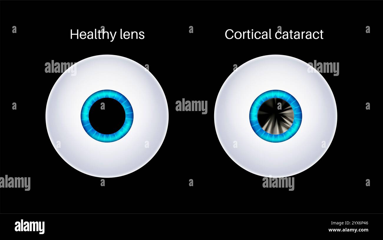 Cataract eye disease, illustration. Stock Photohttps://www.alamy.com/image-license-details/?v=1https://www.alamy.com/cataract-eye-disease-illustration-image635703382.html
RF2YX6P46–Cataract eye disease, illustration.
 A reference handbook of the medical sciences, embracing the entire range of scientific and practical medicine and allied science . per cent., and which may be followed by * Whites ointment causes irritation in a small number of cases. 682 uveitis, astigmatism, or an irregular pupil. More-over, in simple extraction a larger incision is requiredto deliver the lens, and greater difficulty is experiencedin its removal. Cortical matter more frequentlyremains, producing secondary cataract. Greaterskill is also needed on the part of the operator. The advantages of simple extraction are cosmetic.and o Stock Photohttps://www.alamy.com/image-license-details/?v=1https://www.alamy.com/a-reference-handbook-of-the-medical-sciences-embracing-the-entire-range-of-scientific-and-practical-medicine-and-allied-science-per-cent-and-which-may-be-followed-by-whites-ointment-causes-irritation-in-a-small-number-of-cases-682-uveitis-astigmatism-or-an-irregular-pupil-more-over-in-simple-extraction-a-larger-incision-is-requiredto-deliver-the-lens-and-greater-difficulty-is-experiencedin-its-removal-cortical-matter-more-frequentlyremains-producing-secondary-cataract-greaterskill-is-also-needed-on-the-part-of-the-operator-the-advantages-of-simple-extraction-are-cosmeticand-o-image338457694.html
RM2AJJ26P–A reference handbook of the medical sciences, embracing the entire range of scientific and practical medicine and allied science . per cent., and which may be followed by * Whites ointment causes irritation in a small number of cases. 682 uveitis, astigmatism, or an irregular pupil. More-over, in simple extraction a larger incision is requiredto deliver the lens, and greater difficulty is experiencedin its removal. Cortical matter more frequentlyremains, producing secondary cataract. Greaterskill is also needed on the part of the operator. The advantages of simple extraction are cosmetic.and o
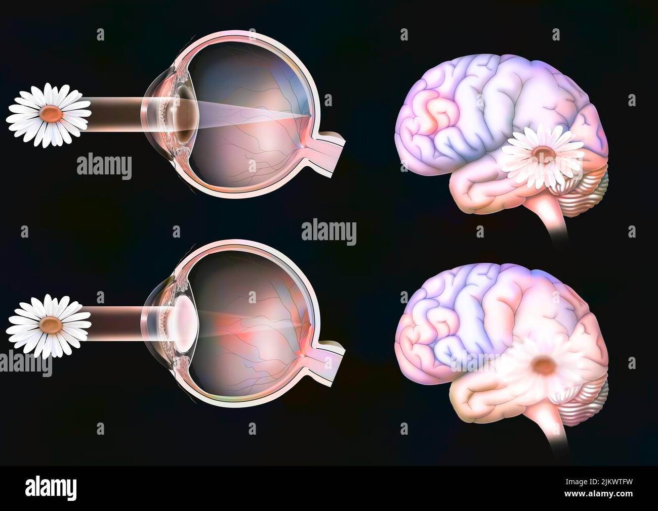 Comparison between normal vision and that of an eye with cataract. Stock Photohttps://www.alamy.com/image-license-details/?v=1https://www.alamy.com/comparison-between-normal-vision-and-that-of-an-eye-with-cataract-image476926461.html
RF2JKWTFW–Comparison between normal vision and that of an eye with cataract.
 Cataract eye disease, illustration. Stock Photohttps://www.alamy.com/image-license-details/?v=1https://www.alamy.com/cataract-eye-disease-illustration-image635703321.html
RF2YX6P21–Cataract eye disease, illustration.
 The student's guide to diseases of the eye . gh giving some help, do notremove the defect. If, as is usual, he be presbyopic, 154 CATARACT he will be likely to choose over-strong spectacles, andto place objects too close to his eyes, so as to obtainlarger retinal images (p. 13), and thus compensatefor want of clearness. In nuclear cataract, as theaxial rays of light are most obstructed, sight is oftenbetter when the pupil is rather large, and suchpatients tell us that they see better in a dull light orwith their backs to the window, or when shading theeyes with the hand. In the cortical and mo Stock Photohttps://www.alamy.com/image-license-details/?v=1https://www.alamy.com/the-students-guide-to-diseases-of-the-eye-gh-giving-some-help-do-notremove-the-defect-if-as-is-usual-he-be-presbyopic-154-cataract-he-will-be-likely-to-choose-over-strong-spectacles-andto-place-objects-too-close-to-his-eyes-so-as-to-obtainlarger-retinal-images-p-13-and-thus-compensatefor-want-of-clearness-in-nuclear-cataract-as-theaxial-rays-of-light-are-most-obstructed-sight-is-oftenbetter-when-the-pupil-is-rather-large-and-suchpatients-tell-us-that-they-see-better-in-a-dull-light-orwith-their-backs-to-the-window-or-when-shading-theeyes-with-the-hand-in-the-cortical-and-mo-image342769443.html
RM2AWJDWR–The student's guide to diseases of the eye . gh giving some help, do notremove the defect. If, as is usual, he be presbyopic, 154 CATARACT he will be likely to choose over-strong spectacles, andto place objects too close to his eyes, so as to obtainlarger retinal images (p. 13), and thus compensatefor want of clearness. In nuclear cataract, as theaxial rays of light are most obstructed, sight is oftenbetter when the pupil is rather large, and suchpatients tell us that they see better in a dull light orwith their backs to the window, or when shading theeyes with the hand. In the cortical and mo
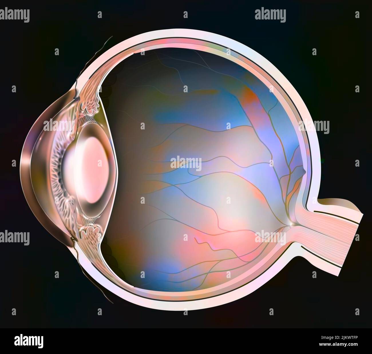 Eye: clouding of the lens in the event of nuclear cataracts. Stock Photohttps://www.alamy.com/image-license-details/?v=1https://www.alamy.com/eye-clouding-of-the-lens-in-the-event-of-nuclear-cataracts-image476926458.html
RF2JKWTFP–Eye: clouding of the lens in the event of nuclear cataracts.
 Cataract eye disease, illustration. Stock Photohttps://www.alamy.com/image-license-details/?v=1https://www.alamy.com/cataract-eye-disease-illustration-image635703385.html
RF2YX6P49–Cataract eye disease, illustration.
 Skiascopy and its practical application to the study of refraction . Fig. ii. Fig. 12. caused by irregular astigmatism following corneal diseaseis shown in figure 11. That due to changes in the lenssuch as may precede cortical senile cataract is shown infigure 12, in which the black lines represent fixed spiculesof actual opacity, while the other parts of the pupil indicatemerely refractive differences, and change from light to dark,or dark to light, as the inclination of the mirror is varied.Some such appearance is sometimes presented by youngpersons, indicating a congenital defect which may Stock Photohttps://www.alamy.com/image-license-details/?v=1https://www.alamy.com/skiascopy-and-its-practical-application-to-the-study-of-refraction-fig-ii-fig-12-caused-by-irregular-astigmatism-following-corneal-diseaseis-shown-in-figure-11-that-due-to-changes-in-the-lenssuch-as-may-precede-cortical-senile-cataract-is-shown-infigure-12-in-which-the-black-lines-represent-fixed-spiculesof-actual-opacity-while-the-other-parts-of-the-pupil-indicatemerely-refractive-differences-and-change-from-light-to-darkor-dark-to-light-as-the-inclination-of-the-mirror-is-variedsome-such-appearance-is-sometimes-presented-by-youngpersons-indicating-a-congenital-defect-which-may-image342847242.html
RM2AWP14A–Skiascopy and its practical application to the study of refraction . Fig. ii. Fig. 12. caused by irregular astigmatism following corneal diseaseis shown in figure 11. That due to changes in the lenssuch as may precede cortical senile cataract is shown infigure 12, in which the black lines represent fixed spiculesof actual opacity, while the other parts of the pupil indicatemerely refractive differences, and change from light to dark,or dark to light, as the inclination of the mirror is varied.Some such appearance is sometimes presented by youngpersons, indicating a congenital defect which may
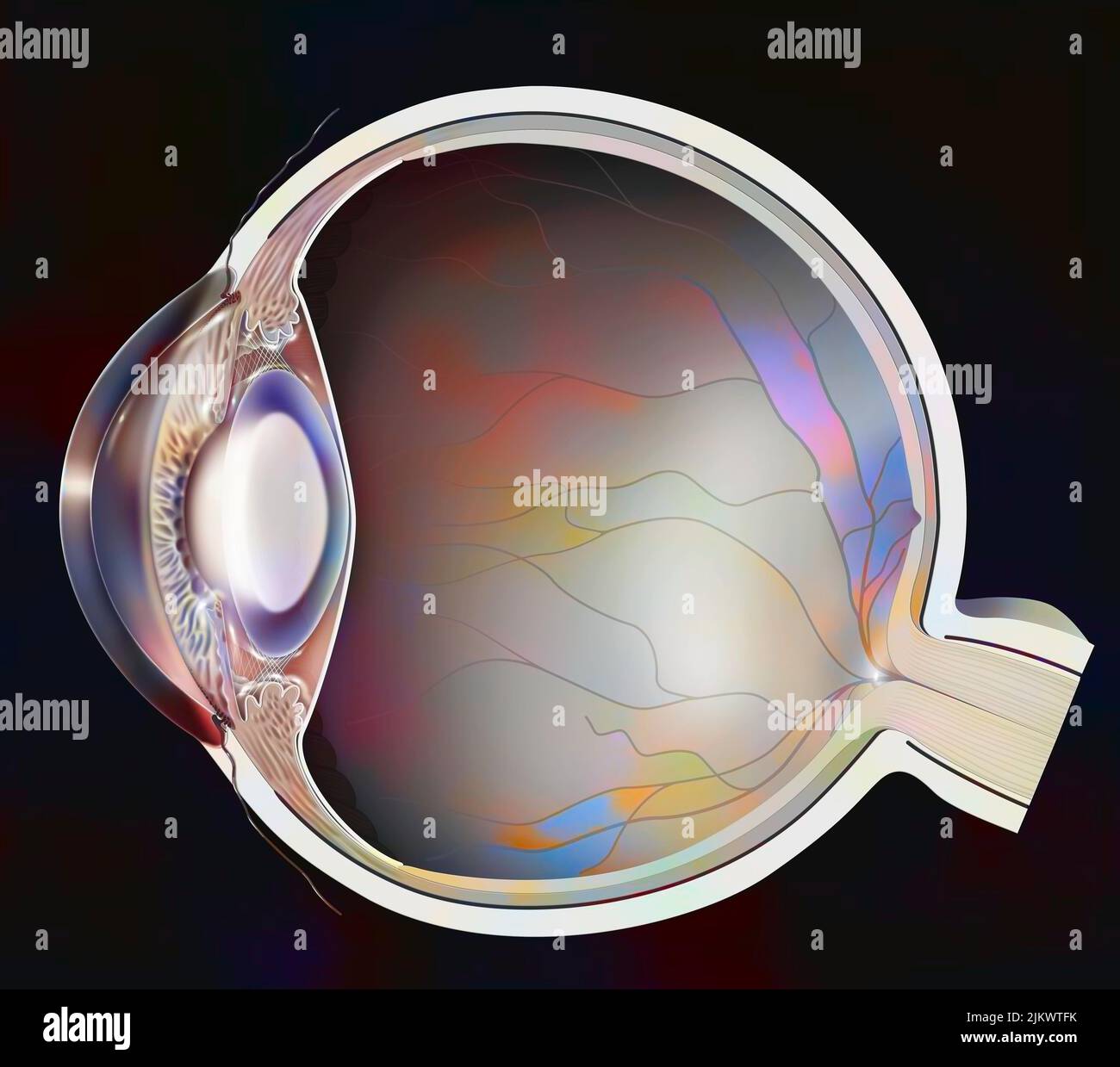 Eye: clouding of the lens in the event of nuclear cataracts. Stock Photohttps://www.alamy.com/image-license-details/?v=1https://www.alamy.com/eye-clouding-of-the-lens-in-the-event-of-nuclear-cataracts-image476926455.html
RF2JKWTFK–Eye: clouding of the lens in the event of nuclear cataracts.
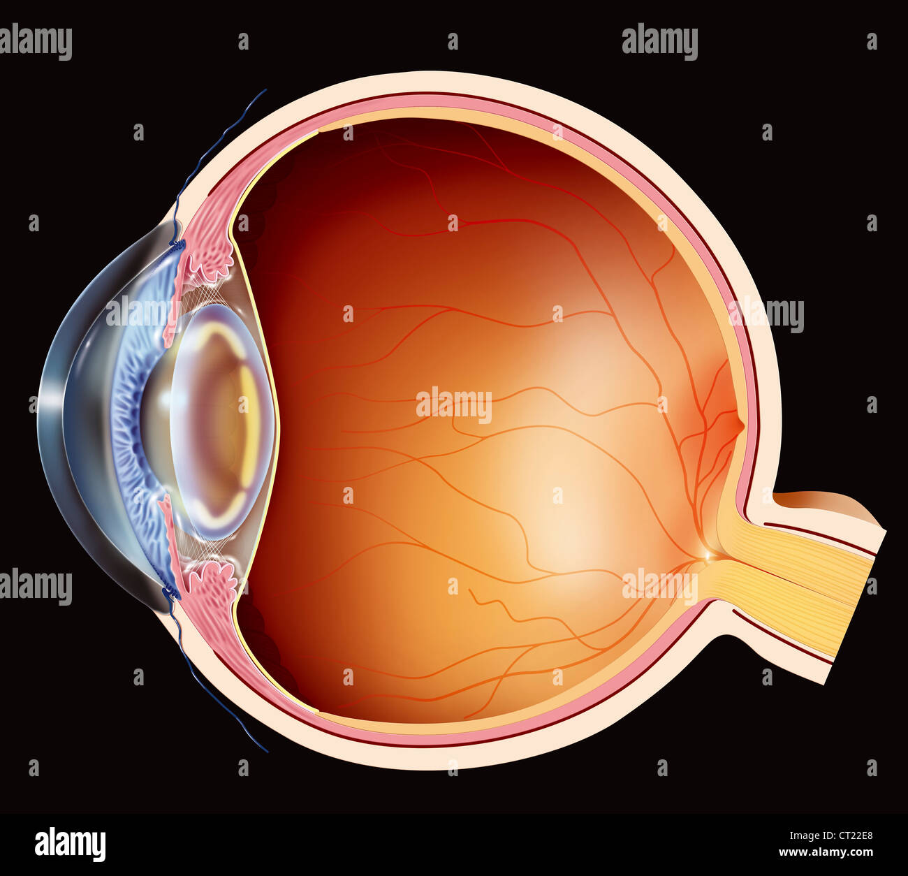 CATARACT, DRAWING Stock Photohttps://www.alamy.com/image-license-details/?v=1https://www.alamy.com/stock-photo-cataract-drawing-49218352.html
RMCT22E8–CATARACT, DRAWING
 Cataract eye disease, illustration. Stock Photohttps://www.alamy.com/image-license-details/?v=1https://www.alamy.com/cataract-eye-disease-illustration-image635703383.html
RF2YX6P47–Cataract eye disease, illustration.
 Text-book of ophthalmology . is not the case until late in life (in the sixthdecade or later). [In some cases of retinitis pigmentosa the main disturbance of sight, especially forreading, is caused by a cortical cataract. The densest part of this lenticular opacitylies righl in the center of the pupillary area, and hence these patients see worst whenthe pupil is contracted, i. e., in a strong light. They therefore form an exception to therule thai persons with retinitis pigmentosa see worst when the illumination is reduced.These cases can be recognized by the fact that dilatation of the pupil Stock Photohttps://www.alamy.com/image-license-details/?v=1https://www.alamy.com/text-book-of-ophthalmology-is-not-the-case-until-late-in-life-in-the-sixthdecade-or-later-in-some-cases-of-retinitis-pigmentosa-the-main-disturbance-of-sight-especially-forreading-is-caused-by-a-cortical-cataract-the-densest-part-of-this-lenticular-opacitylies-righl-in-the-center-of-the-pupillary-area-and-hence-these-patients-see-worst-whenthe-pupil-is-contracted-i-e-in-a-strong-light-they-therefore-form-an-exception-to-therule-thai-persons-with-retinitis-pigmentosa-see-worst-when-the-illumination-is-reducedthese-cases-can-be-recognized-by-the-fact-that-dilatation-of-the-pupil-image339996520.html
RM2AN450T–Text-book of ophthalmology . is not the case until late in life (in the sixthdecade or later). [In some cases of retinitis pigmentosa the main disturbance of sight, especially forreading, is caused by a cortical cataract. The densest part of this lenticular opacitylies righl in the center of the pupillary area, and hence these patients see worst whenthe pupil is contracted, i. e., in a strong light. They therefore form an exception to therule thai persons with retinitis pigmentosa see worst when the illumination is reduced.These cases can be recognized by the fact that dilatation of the pupil
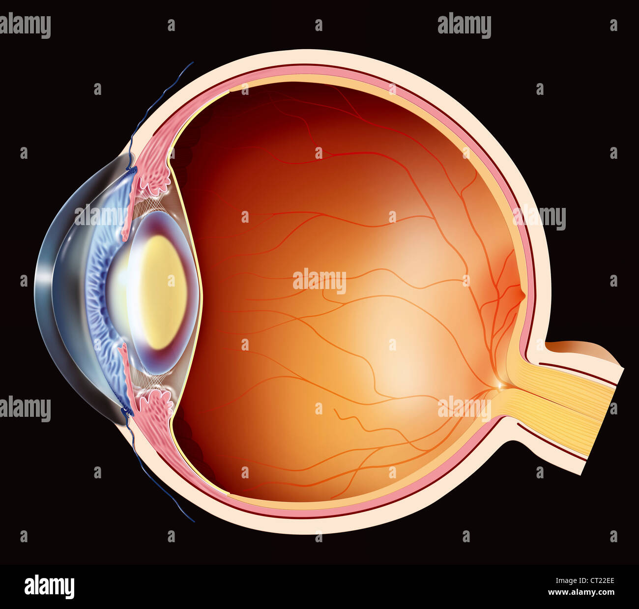 CATARACT, DRAWING Stock Photohttps://www.alamy.com/image-license-details/?v=1https://www.alamy.com/stock-photo-cataract-drawing-49218358.html
RMCT22EE–CATARACT, DRAWING
 Cataract eye disease, illustration. Stock Photohttps://www.alamy.com/image-license-details/?v=1https://www.alamy.com/cataract-eye-disease-illustration-image635703368.html
RF2YX6P3M–Cataract eye disease, illustration.
 The student's guide to diseases of the eye . Fig. 60.—Cortical cataract. References as in preceding figure. according as they are in the anterior or posteriorlayers (3, Fig. 60). With the mirror they appearblack or greyish, and of rather smaller size (2, Fig.60), and if the intervening substance is clear, thedetails of the fundus can be seen sharply betweenthe bars of opacity. Posterior polar opacities are seldom visible withoutcareful focal illumination, when we find a patchy orstellate figure very deeply seated in the axis of thelens (3, Fig. 61) ; if large it looks concave like thebottom of Stock Photohttps://www.alamy.com/image-license-details/?v=1https://www.alamy.com/the-students-guide-to-diseases-of-the-eye-fig-60cortical-cataract-references-as-in-preceding-figure-according-as-they-are-in-the-anterior-or-posteriorlayers-3-fig-60-with-the-mirror-they-appearblack-or-greyish-and-of-rather-smaller-size-2-fig60-and-if-the-intervening-substance-is-clear-thedetails-of-the-fundus-can-be-seen-sharply-betweenthe-bars-of-opacity-posterior-polar-opacities-are-seldom-visible-withoutcareful-focal-illumination-when-we-find-a-patchy-orstellate-figure-very-deeply-seated-in-the-axis-of-thelens-3-fig-61-if-large-it-looks-concave-like-thebottom-of-image342767303.html
RM2AWJB5B–The student's guide to diseases of the eye . Fig. 60.—Cortical cataract. References as in preceding figure. according as they are in the anterior or posteriorlayers (3, Fig. 60). With the mirror they appearblack or greyish, and of rather smaller size (2, Fig.60), and if the intervening substance is clear, thedetails of the fundus can be seen sharply betweenthe bars of opacity. Posterior polar opacities are seldom visible withoutcareful focal illumination, when we find a patchy orstellate figure very deeply seated in the axis of thelens (3, Fig. 61) ; if large it looks concave like thebottom of
 CATARACT, DRAWING Stock Photohttps://www.alamy.com/image-license-details/?v=1https://www.alamy.com/stock-photo-cataract-drawing-49218365.html
RMCT22EN–CATARACT, DRAWING
 Cataract eye disease, illustration. Stock Photohttps://www.alamy.com/image-license-details/?v=1https://www.alamy.com/cataract-eye-disease-illustration-image635703317.html
RF2YX6P1W–Cataract eye disease, illustration.
 The student's guide to diseases of the eye . Fig. 60.—Cortical cataract. References as in preceding figure. according as they are in the anterior or posteriorlayers (3, Fig. 60). With the mirror they appearblack or greyish, and of rather smaller size (2, Fig.60), and if the intervening substance is clear, thedetails of the fundus can be seen sharply betweenthe bars of opacity. Posterior polar opacities are seldom visible withoutcareful focal illumination, when we find a patchy orstellate figure very deeply seated in the axis of thelens (3, Fig. 61) ; if large it looks concave like thebottom of Stock Photohttps://www.alamy.com/image-license-details/?v=1https://www.alamy.com/the-students-guide-to-diseases-of-the-eye-fig-60cortical-cataract-references-as-in-preceding-figure-according-as-they-are-in-the-anterior-or-posteriorlayers-3-fig-60-with-the-mirror-they-appearblack-or-greyish-and-of-rather-smaller-size-2-fig60-and-if-the-intervening-substance-is-clear-thedetails-of-the-fundus-can-be-seen-sharply-betweenthe-bars-of-opacity-posterior-polar-opacities-are-seldom-visible-withoutcareful-focal-illumination-when-we-find-a-patchy-orstellate-figure-very-deeply-seated-in-the-axis-of-thelens-3-fig-61-if-large-it-looks-concave-like-thebottom-of-image342767774.html
RM2AWJBP6–The student's guide to diseases of the eye . Fig. 60.—Cortical cataract. References as in preceding figure. according as they are in the anterior or posteriorlayers (3, Fig. 60). With the mirror they appearblack or greyish, and of rather smaller size (2, Fig.60), and if the intervening substance is clear, thedetails of the fundus can be seen sharply betweenthe bars of opacity. Posterior polar opacities are seldom visible withoutcareful focal illumination, when we find a patchy orstellate figure very deeply seated in the axis of thelens (3, Fig. 61) ; if large it looks concave like thebottom of
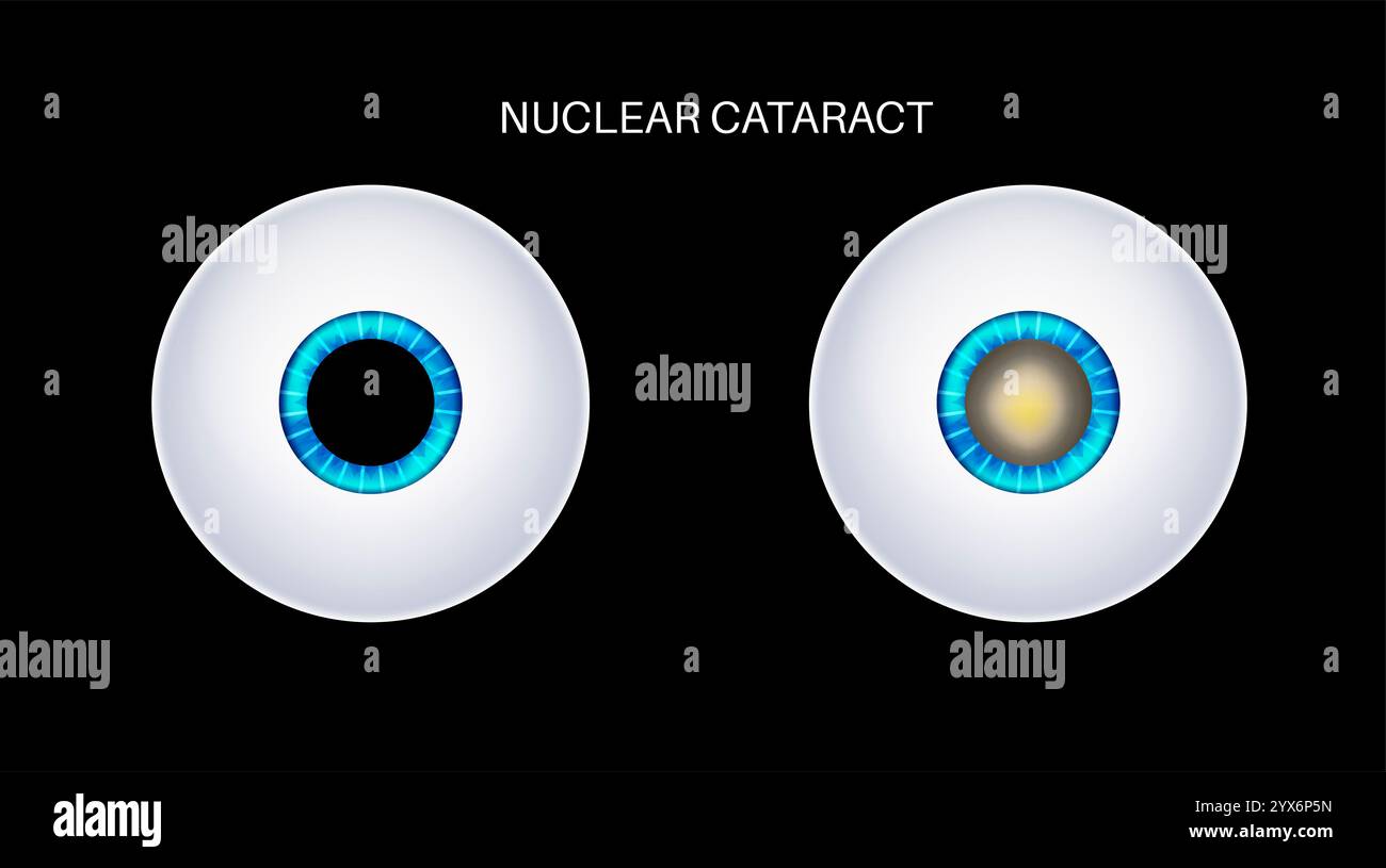 Cataract eye disease, illustration. Stock Photohttps://www.alamy.com/image-license-details/?v=1https://www.alamy.com/cataract-eye-disease-illustration-image635703425.html
RF2YX6P5N–Cataract eye disease, illustration.
 The student's guide to diseases of the eye . FiG. 57.—Pyramidal cataractseen from the front and insection.. Fig. 58.—Magnified section of a pyramidal cataract. The fineparallel shading shows the thickness of the opacity, thedouble (black and white) outline is the capsule; on eachside are the cortical lens-fibres, many being broken up intoglobules beneath the opacity. Lying upon the puckeredcapsule over the opacity is a little fibrous tissue, the result ofiritis. forating ulceration of the cornea in early life, and ofthis ophthalmia neonatorum is nearly always thecause; it is therefore often as Stock Photohttps://www.alamy.com/image-license-details/?v=1https://www.alamy.com/the-students-guide-to-diseases-of-the-eye-fig-57pyramidal-cataractseen-from-the-front-and-insection-fig-58magnified-section-of-a-pyramidal-cataract-the-fineparallel-shading-shows-the-thickness-of-the-opacity-thedouble-black-and-white-outline-is-the-capsule-on-eachside-are-the-cortical-lens-fibres-many-being-broken-up-intoglobules-beneath-the-opacity-lying-upon-the-puckeredcapsule-over-the-opacity-is-a-little-fibrous-tissue-the-result-ofiritis-forating-ulceration-of-the-cornea-in-early-life-and-ofthis-ophthalmia-neonatorum-is-nearly-always-thecause-it-is-therefore-often-as-image342770535.html
RM2AWJF8R–The student's guide to diseases of the eye . FiG. 57.—Pyramidal cataractseen from the front and insection.. Fig. 58.—Magnified section of a pyramidal cataract. The fineparallel shading shows the thickness of the opacity, thedouble (black and white) outline is the capsule; on eachside are the cortical lens-fibres, many being broken up intoglobules beneath the opacity. Lying upon the puckeredcapsule over the opacity is a little fibrous tissue, the result ofiritis. forating ulceration of the cornea in early life, and ofthis ophthalmia neonatorum is nearly always thecause; it is therefore often as
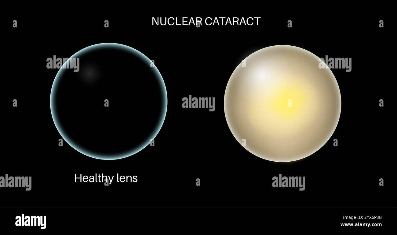 Cataract eye disease, illustration. Stock Photohttps://www.alamy.com/image-license-details/?v=1https://www.alamy.com/cataract-eye-disease-illustration-image635703359.html
RF2YX6P3B–Cataract eye disease, illustration.
 The science and art of surgery : being a treatise on surgical injuries, diseases, and operations . Fig. 571.—Commeacng Catav.ict; Opaquestreaks converging from the margin:the (larlier strije are in the anterior partsof the leas. Fig. 372.—Commeucing Cataract; Opaquestreaks divergin, from the centre. Thelens-nucleus is altogether obscured. the stri.ie, according to the direction of the lens-fil)res, of which someonly have become opaque, always diverge from or converge to tlie centreof the lens. In hard cataracts the cortical parts, as in the normal lens,are always comparatively soft, sometimes Stock Photohttps://www.alamy.com/image-license-details/?v=1https://www.alamy.com/the-science-and-art-of-surgery-being-a-treatise-on-surgical-injuries-diseases-and-operations-fig-571commeacng-catavict-opaquestreaks-converging-from-the-marginthe-larlier-strije-are-in-the-anterior-partsof-the-leas-fig-372commeucing-cataract-opaquestreaks-divergin-from-the-centre-thelens-nucleus-is-altogether-obscured-the-striie-according-to-the-direction-of-the-lens-filres-of-which-someonly-have-become-opaque-always-diverge-from-or-converge-to-tlie-centreof-the-lens-in-hard-cataracts-the-cortical-parts-as-in-the-normal-lensare-always-comparatively-soft-sometimes-image339144507.html
RM2AKNA7R–The science and art of surgery : being a treatise on surgical injuries, diseases, and operations . Fig. 571.—Commeacng Catav.ict; Opaquestreaks converging from the margin:the (larlier strije are in the anterior partsof the leas. Fig. 372.—Commeucing Cataract; Opaquestreaks divergin, from the centre. Thelens-nucleus is altogether obscured. the stri.ie, according to the direction of the lens-fil)res, of which someonly have become opaque, always diverge from or converge to tlie centreof the lens. In hard cataracts the cortical parts, as in the normal lens,are always comparatively soft, sometimes
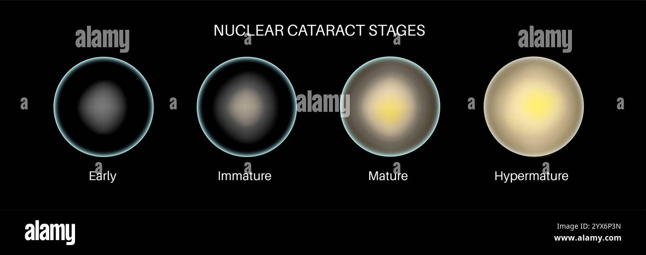 Cataract eye disease, illustration. Stock Photohttps://www.alamy.com/image-license-details/?v=1https://www.alamy.com/cataract-eye-disease-illustration-image635703369.html
RF2YX6P3N–Cataract eye disease, illustration.
 Text-book of ophthalmology . Fig. 233. Fig. 234. Fig. 233.—Incipient Cataract under the form of opaque sectors, which look black when seenby transmitted light with the ophthalmoscope. Fig. 234.—Incipient Cataract under the form of an irregular disk, which is more markedly-opaque at its edges, and which is situated in the posterior layers of the cortex. 3. A disk-shaped opacity which is situated in the posterior layers of the cortex, butwhich, in contradistinction to the typical posterior cortical cataract (Fig. 232), pre-sents an irregular and ill-defined contour and a cobweb-like structure (F Stock Photohttps://www.alamy.com/image-license-details/?v=1https://www.alamy.com/text-book-of-ophthalmology-fig-233-fig-234-fig-233incipient-cataract-under-the-form-of-opaque-sectors-which-look-black-when-seenby-transmitted-light-with-the-ophthalmoscope-fig-234incipient-cataract-under-the-form-of-an-irregular-disk-which-is-more-markedly-opaque-at-its-edges-and-which-is-situated-in-the-posterior-layers-of-the-cortex-3-a-disk-shaped-opacity-which-is-situated-in-the-posterior-layers-of-the-cortex-butwhich-in-contradistinction-to-the-typical-posterior-cortical-cataract-fig-232-pre-sents-an-irregular-and-ill-defined-contour-and-a-cobweb-like-structure-f-image340003586.html
RM2AN4E16–Text-book of ophthalmology . Fig. 233. Fig. 234. Fig. 233.—Incipient Cataract under the form of opaque sectors, which look black when seenby transmitted light with the ophthalmoscope. Fig. 234.—Incipient Cataract under the form of an irregular disk, which is more markedly-opaque at its edges, and which is situated in the posterior layers of the cortex. 3. A disk-shaped opacity which is situated in the posterior layers of the cortex, butwhich, in contradistinction to the typical posterior cortical cataract (Fig. 232), pre-sents an irregular and ill-defined contour and a cobweb-like structure (F
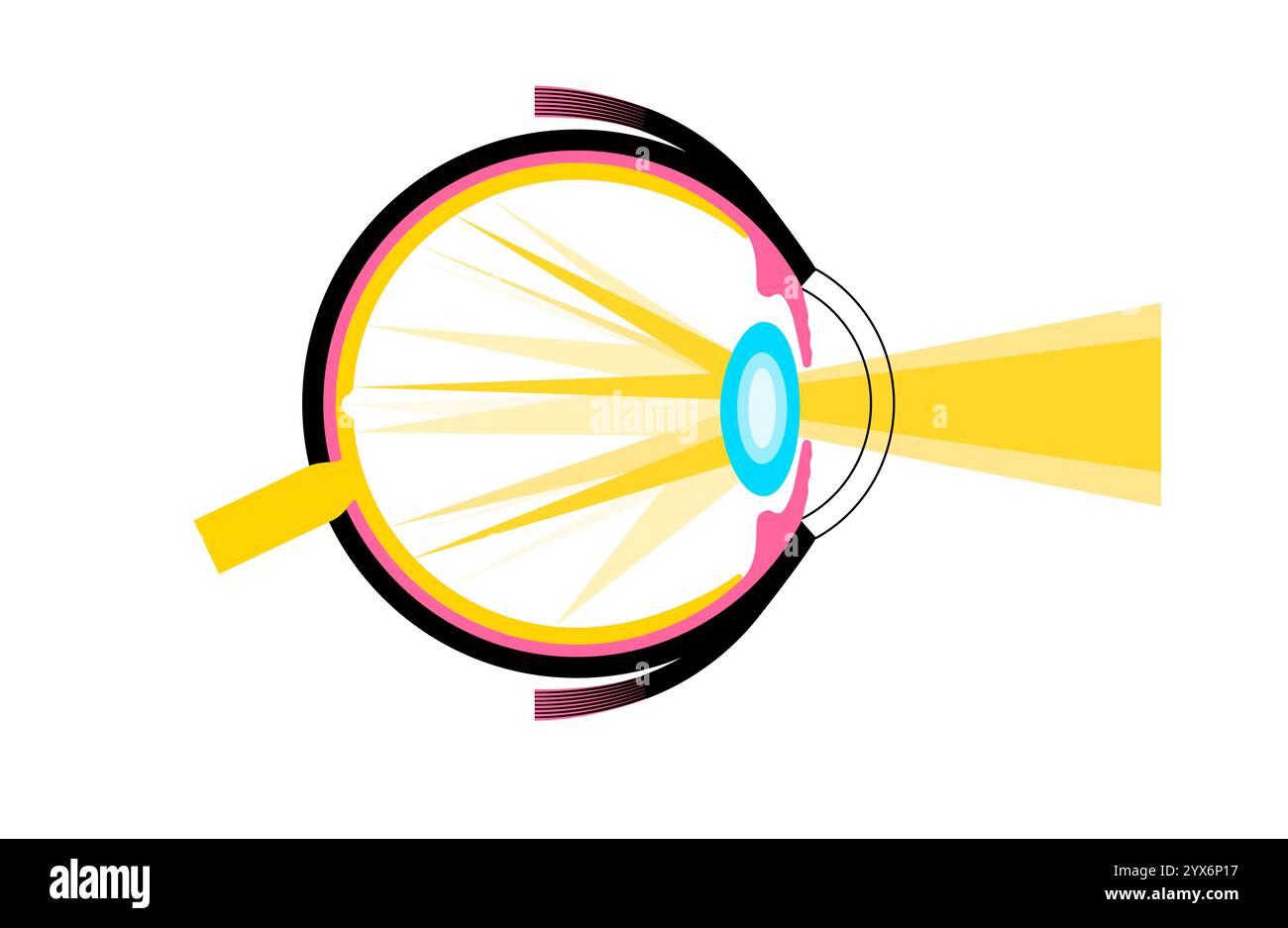 Cataract eye disease, illustration. Stock Photohttps://www.alamy.com/image-license-details/?v=1https://www.alamy.com/cataract-eye-disease-illustration-image635703299.html
RF2YX6P17–Cataract eye disease, illustration.
 . Surgery, its principles and practice . ainsolution, the instillation of the mixture produces at once a blanchingof the tissues as well as a local anesthesia. OPEKATIONS ON THE EYEBALL. 865 OPERATIONS ON THE EYEBALL. Operations for the Extraction of Senile Cataract.—Cataract,an opaque condition of the crystalline lens, is called cortical, if the opaci-ties radiate from the periphery toward the center, and nuclear, if theopacity includes and surrounds the nucleus. The period of growth of acataract from incipiency to full maturity ordinarily consumes from oneto three years, but the rate of incr Stock Photohttps://www.alamy.com/image-license-details/?v=1https://www.alamy.com/surgery-its-principles-and-practice-ainsolution-the-instillation-of-the-mixture-produces-at-once-a-blanchingof-the-tissues-as-well-as-a-local-anesthesia-opekations-on-the-eyeball-865-operations-on-the-eyeball-operations-for-the-extraction-of-senile-cataractcataractan-opaque-condition-of-the-crystalline-lens-is-called-cortical-if-the-opaci-ties-radiate-from-the-periphery-toward-the-center-and-nuclear-if-theopacity-includes-and-surrounds-the-nucleus-the-period-of-growth-of-acataract-from-incipiency-to-full-maturity-ordinarily-consumes-from-oneto-three-years-but-the-rate-of-incr-image369810328.html
RM2CDJ8RM–. Surgery, its principles and practice . ainsolution, the instillation of the mixture produces at once a blanchingof the tissues as well as a local anesthesia. OPEKATIONS ON THE EYEBALL. 865 OPERATIONS ON THE EYEBALL. Operations for the Extraction of Senile Cataract.—Cataract,an opaque condition of the crystalline lens, is called cortical, if the opaci-ties radiate from the periphery toward the center, and nuclear, if theopacity includes and surrounds the nucleus. The period of growth of acataract from incipiency to full maturity ordinarily consumes from oneto three years, but the rate of incr
 Cataract eye disease, illustration. Stock Photohttps://www.alamy.com/image-license-details/?v=1https://www.alamy.com/cataract-eye-disease-illustration-image635703319.html
RF2YX6P1Y–Cataract eye disease, illustration.
 . Annual and analytical cyclopaedia of practical medicine . ?!• Posterior cortical rataraet. {^.>ir/id.) seen associated with uveal disorder as-sociated with lymph-stream disturbanceand liquefaction of the vitreous body.Generally it appears in the stellar formof opacity. In this variety interferencewith vision depends not only iipon thesize of the opacity, but also upon con-comitant and relevant changes. Treat-ment, to be of any avail, must be di-. ^^yss Congeuital cataract with riders. (Skkel.) rected, if possible, toward any existingcause. A third form, although separated intoquite a ser Stock Photohttps://www.alamy.com/image-license-details/?v=1https://www.alamy.com/annual-and-analytical-cyclopaedia-of-practical-medicine-!-posterior-cortical-rataraet-gtirid-seen-associated-with-uveal-disorder-as-sociated-with-lymph-stream-disturbanceand-liquefaction-of-the-vitreous-bodygenerally-it-appears-in-the-stellar-formof-opacity-in-this-variety-interferencewith-vision-depends-not-only-iipon-thesize-of-the-opacity-but-also-upon-con-comitant-and-relevant-changes-treat-ment-to-be-of-any-avail-must-be-di-yss-congeuital-cataract-with-riders-skkel-rected-if-possible-toward-any-existingcause-a-third-form-although-separated-intoquite-a-ser-image369668078.html
RM2CDBRBA–. Annual and analytical cyclopaedia of practical medicine . ?!• Posterior cortical rataraet. {^.>ir/id.) seen associated with uveal disorder as-sociated with lymph-stream disturbanceand liquefaction of the vitreous body.Generally it appears in the stellar formof opacity. In this variety interferencewith vision depends not only iipon thesize of the opacity, but also upon con-comitant and relevant changes. Treat-ment, to be of any avail, must be di-. ^^yss Congeuital cataract with riders. (Skkel.) rected, if possible, toward any existingcause. A third form, although separated intoquite a ser
 Cataract eye disease, illustration. Stock Photohttps://www.alamy.com/image-license-details/?v=1https://www.alamy.com/cataract-eye-disease-illustration-image635703307.html
RF2YX6P1F–Cataract eye disease, illustration.
![. Pathology and bacteriology [electronic resource]. Ophthalmology; Eye; Eye; Bacteriology; Ophthalmology; Eye; Bacteriology; Eye. CATARACTS 613 the cortical fibres. As the result of their shrinkage cleft like spaces are formed between them, in which the inter- fibrillar fluid collects and coagulates into drops or spheroidal bodies, the **Morgagnian globules" (Fig. 274).. ? * Fig. 275.—Shows spreading changes in senile cataract which give rise to peri- nuclear opacity. They have a different refractive index from that of the lens fibres and, therefore, give rise to streaks or patches of opa Stock Photo . Pathology and bacteriology [electronic resource]. Ophthalmology; Eye; Eye; Bacteriology; Ophthalmology; Eye; Bacteriology; Eye. CATARACTS 613 the cortical fibres. As the result of their shrinkage cleft like spaces are formed between them, in which the inter- fibrillar fluid collects and coagulates into drops or spheroidal bodies, the **Morgagnian globules" (Fig. 274).. ? * Fig. 275.—Shows spreading changes in senile cataract which give rise to peri- nuclear opacity. They have a different refractive index from that of the lens fibres and, therefore, give rise to streaks or patches of opa Stock Photo](https://c8.alamy.com/comp/RJN707/pathology-and-bacteriology-electronic-resource-ophthalmology-eye-eye-bacteriology-ophthalmology-eye-bacteriology-eye-cataracts-613-the-cortical-fibres-as-the-result-of-their-shrinkage-cleft-like-spaces-are-formed-between-them-in-which-the-inter-fibrillar-fluid-collects-and-coagulates-into-drops-or-spheroidal-bodies-the-morgagnian-globulesquot-fig-274-fig-275shows-spreading-changes-in-senile-cataract-which-give-rise-to-peri-nuclear-opacity-they-have-a-different-refractive-index-from-that-of-the-lens-fibres-and-therefore-give-rise-to-streaks-or-patches-of-opa-RJN707.jpg) . Pathology and bacteriology [electronic resource]. Ophthalmology; Eye; Eye; Bacteriology; Ophthalmology; Eye; Bacteriology; Eye. CATARACTS 613 the cortical fibres. As the result of their shrinkage cleft like spaces are formed between them, in which the inter- fibrillar fluid collects and coagulates into drops or spheroidal bodies, the **Morgagnian globules" (Fig. 274).. ? * Fig. 275.—Shows spreading changes in senile cataract which give rise to peri- nuclear opacity. They have a different refractive index from that of the lens fibres and, therefore, give rise to streaks or patches of opa Stock Photohttps://www.alamy.com/image-license-details/?v=1https://www.alamy.com/pathology-and-bacteriology-electronic-resource-ophthalmology-eye-eye-bacteriology-ophthalmology-eye-bacteriology-eye-cataracts-613-the-cortical-fibres-as-the-result-of-their-shrinkage-cleft-like-spaces-are-formed-between-them-in-which-the-inter-fibrillar-fluid-collects-and-coagulates-into-drops-or-spheroidal-bodies-the-morgagnian-globulesquot-fig-274-fig-275shows-spreading-changes-in-senile-cataract-which-give-rise-to-peri-nuclear-opacity-they-have-a-different-refractive-index-from-that-of-the-lens-fibres-and-therefore-give-rise-to-streaks-or-patches-of-opa-image235265079.html
. Pathology and bacteriology [electronic resource]. Ophthalmology; Eye; Eye; Bacteriology; Ophthalmology; Eye; Bacteriology; Eye. CATARACTS 613 the cortical fibres. As the result of their shrinkage cleft like spaces are formed between them, in which the inter- fibrillar fluid collects and coagulates into drops or spheroidal bodies, the **Morgagnian globules" (Fig. 274).. ? * Fig. 275.—Shows spreading changes in senile cataract which give rise to peri- nuclear opacity. They have a different refractive index from that of the lens fibres and, therefore, give rise to streaks or patches of opa Stock Photohttps://www.alamy.com/image-license-details/?v=1https://www.alamy.com/pathology-and-bacteriology-electronic-resource-ophthalmology-eye-eye-bacteriology-ophthalmology-eye-bacteriology-eye-cataracts-613-the-cortical-fibres-as-the-result-of-their-shrinkage-cleft-like-spaces-are-formed-between-them-in-which-the-inter-fibrillar-fluid-collects-and-coagulates-into-drops-or-spheroidal-bodies-the-morgagnian-globulesquot-fig-274-fig-275shows-spreading-changes-in-senile-cataract-which-give-rise-to-peri-nuclear-opacity-they-have-a-different-refractive-index-from-that-of-the-lens-fibres-and-therefore-give-rise-to-streaks-or-patches-of-opa-image235265079.htmlRMRJN707–. Pathology and bacteriology [electronic resource]. Ophthalmology; Eye; Eye; Bacteriology; Ophthalmology; Eye; Bacteriology; Eye. CATARACTS 613 the cortical fibres. As the result of their shrinkage cleft like spaces are formed between them, in which the inter- fibrillar fluid collects and coagulates into drops or spheroidal bodies, the **Morgagnian globules" (Fig. 274).. ? * Fig. 275.—Shows spreading changes in senile cataract which give rise to peri- nuclear opacity. They have a different refractive index from that of the lens fibres and, therefore, give rise to streaks or patches of opa
 Cataract eye disease, illustration. Stock Photohttps://www.alamy.com/image-license-details/?v=1https://www.alamy.com/cataract-eye-disease-illustration-image635703302.html
RF2YX6P1A–Cataract eye disease, illustration.
 . Diseases of the dog and their treatment. Dogs. CATARACT. 367 should be an unfavorable one. Hereditary cataract shows little inclination to enlarMment, as is also the case in senile cataract. In soft cortical cataracts we may see a rapid opacity of the lens in a few days or weeks. The sight is entirely lost and medical treat- ment is of little use. Therapeutic Treatment. A cataract may be removed by an operation, and this is much more advisable in the dog because it is, as a rule, attended without any great danger, and its results are generally beneficial, producing a partial restoration of t Stock Photohttps://www.alamy.com/image-license-details/?v=1https://www.alamy.com/diseases-of-the-dog-and-their-treatment-dogs-cataract-367-should-be-an-unfavorable-one-hereditary-cataract-shows-little-inclination-to-enlarmment-as-is-also-the-case-in-senile-cataract-in-soft-cortical-cataracts-we-may-see-a-rapid-opacity-of-the-lens-in-a-few-days-or-weeks-the-sight-is-entirely-lost-and-medical-treat-ment-is-of-little-use-therapeutic-treatment-a-cataract-may-be-removed-by-an-operation-and-this-is-much-more-advisable-in-the-dog-because-it-is-as-a-rule-attended-without-any-great-danger-and-its-results-are-generally-beneficial-producing-a-partial-restoration-of-t-image232348967.html
RMRE0BDB–. Diseases of the dog and their treatment. Dogs. CATARACT. 367 should be an unfavorable one. Hereditary cataract shows little inclination to enlarMment, as is also the case in senile cataract. In soft cortical cataracts we may see a rapid opacity of the lens in a few days or weeks. The sight is entirely lost and medical treat- ment is of little use. Therapeutic Treatment. A cataract may be removed by an operation, and this is much more advisable in the dog because it is, as a rule, attended without any great danger, and its results are generally beneficial, producing a partial restoration of t
 Cataract eye disease, illustration. Stock Photohttps://www.alamy.com/image-license-details/?v=1https://www.alamy.com/cataract-eye-disease-illustration-image635703391.html
RF2YX6P4F–Cataract eye disease, illustration.
![. Pathology and bacteriology [electronic resource]. Ophthalmology; Eye; Eye; Bacteriology; Ophthalmology; Eye; Bacteriology; Eye. CATAEACTS 605 Degenerative changes in the capsular epithehum may be brought about in a variety of ways. Friction of the outer surface of the lens capsule, as in the operation for maturation of immature cataract, has been found to produce changes in the capsule cells resulting in the for-. Fig. 272.—Section through the posterior part of a sub-capsular cataract, showing separation of the cortical fibres from the capsule and one another, the spaces being filled with al Stock Photo . Pathology and bacteriology [electronic resource]. Ophthalmology; Eye; Eye; Bacteriology; Ophthalmology; Eye; Bacteriology; Eye. CATAEACTS 605 Degenerative changes in the capsular epithehum may be brought about in a variety of ways. Friction of the outer surface of the lens capsule, as in the operation for maturation of immature cataract, has been found to produce changes in the capsule cells resulting in the for-. Fig. 272.—Section through the posterior part of a sub-capsular cataract, showing separation of the cortical fibres from the capsule and one another, the spaces being filled with al Stock Photo](https://c8.alamy.com/comp/RJN71C/pathology-and-bacteriology-electronic-resource-ophthalmology-eye-eye-bacteriology-ophthalmology-eye-bacteriology-eye-cataeacts-605-degenerative-changes-in-the-capsular-epithehum-may-be-brought-about-in-a-variety-of-ways-friction-of-the-outer-surface-of-the-lens-capsule-as-in-the-operation-for-maturation-of-immature-cataract-has-been-found-to-produce-changes-in-the-capsule-cells-resulting-in-the-for-fig-272section-through-the-posterior-part-of-a-sub-capsular-cataract-showing-separation-of-the-cortical-fibres-from-the-capsule-and-one-another-the-spaces-being-filled-with-al-RJN71C.jpg) . Pathology and bacteriology [electronic resource]. Ophthalmology; Eye; Eye; Bacteriology; Ophthalmology; Eye; Bacteriology; Eye. CATAEACTS 605 Degenerative changes in the capsular epithehum may be brought about in a variety of ways. Friction of the outer surface of the lens capsule, as in the operation for maturation of immature cataract, has been found to produce changes in the capsule cells resulting in the for-. Fig. 272.—Section through the posterior part of a sub-capsular cataract, showing separation of the cortical fibres from the capsule and one another, the spaces being filled with al Stock Photohttps://www.alamy.com/image-license-details/?v=1https://www.alamy.com/pathology-and-bacteriology-electronic-resource-ophthalmology-eye-eye-bacteriology-ophthalmology-eye-bacteriology-eye-cataeacts-605-degenerative-changes-in-the-capsular-epithehum-may-be-brought-about-in-a-variety-of-ways-friction-of-the-outer-surface-of-the-lens-capsule-as-in-the-operation-for-maturation-of-immature-cataract-has-been-found-to-produce-changes-in-the-capsule-cells-resulting-in-the-for-fig-272section-through-the-posterior-part-of-a-sub-capsular-cataract-showing-separation-of-the-cortical-fibres-from-the-capsule-and-one-another-the-spaces-being-filled-with-al-image235265112.html
. Pathology and bacteriology [electronic resource]. Ophthalmology; Eye; Eye; Bacteriology; Ophthalmology; Eye; Bacteriology; Eye. CATAEACTS 605 Degenerative changes in the capsular epithehum may be brought about in a variety of ways. Friction of the outer surface of the lens capsule, as in the operation for maturation of immature cataract, has been found to produce changes in the capsule cells resulting in the for-. Fig. 272.—Section through the posterior part of a sub-capsular cataract, showing separation of the cortical fibres from the capsule and one another, the spaces being filled with al Stock Photohttps://www.alamy.com/image-license-details/?v=1https://www.alamy.com/pathology-and-bacteriology-electronic-resource-ophthalmology-eye-eye-bacteriology-ophthalmology-eye-bacteriology-eye-cataeacts-605-degenerative-changes-in-the-capsular-epithehum-may-be-brought-about-in-a-variety-of-ways-friction-of-the-outer-surface-of-the-lens-capsule-as-in-the-operation-for-maturation-of-immature-cataract-has-been-found-to-produce-changes-in-the-capsule-cells-resulting-in-the-for-fig-272section-through-the-posterior-part-of-a-sub-capsular-cataract-showing-separation-of-the-cortical-fibres-from-the-capsule-and-one-another-the-spaces-being-filled-with-al-image235265112.htmlRMRJN71C–. Pathology and bacteriology [electronic resource]. Ophthalmology; Eye; Eye; Bacteriology; Ophthalmology; Eye; Bacteriology; Eye. CATAEACTS 605 Degenerative changes in the capsular epithehum may be brought about in a variety of ways. Friction of the outer surface of the lens capsule, as in the operation for maturation of immature cataract, has been found to produce changes in the capsule cells resulting in the for-. Fig. 272.—Section through the posterior part of a sub-capsular cataract, showing separation of the cortical fibres from the capsule and one another, the spaces being filled with al
 Cataract eye disease, illustration. Stock Photohttps://www.alamy.com/image-license-details/?v=1https://www.alamy.com/cataract-eye-disease-illustration-image635703285.html
RF2YX6P0N–Cataract eye disease, illustration.
![. Pathology and bacteriology [electronic resource]. Ophthalmology; Eye; Eye; Bacteriology; Ophthalmology; Eye; Bacteriology; Eye. CONGENITAL PUNCTATE CATARACT 55 plane and it is probable that they are due to some distur- bance situated in them. Congenital Punctate Cataract.—^In congenital punctate cataract small, circumscribed, grey or white opacities are seen in the cortical layers of the lens. They vary in number in different cases and may be exceedingly numerous. They usually remain stationary, showing no tendency to increase Fig. 31.—Section through a lens with congenital punctate cataract Stock Photo . Pathology and bacteriology [electronic resource]. Ophthalmology; Eye; Eye; Bacteriology; Ophthalmology; Eye; Bacteriology; Eye. CONGENITAL PUNCTATE CATARACT 55 plane and it is probable that they are due to some distur- bance situated in them. Congenital Punctate Cataract.—^In congenital punctate cataract small, circumscribed, grey or white opacities are seen in the cortical layers of the lens. They vary in number in different cases and may be exceedingly numerous. They usually remain stationary, showing no tendency to increase Fig. 31.—Section through a lens with congenital punctate cataract Stock Photo](https://c8.alamy.com/comp/RJNCN4/pathology-and-bacteriology-electronic-resource-ophthalmology-eye-eye-bacteriology-ophthalmology-eye-bacteriology-eye-congenital-punctate-cataract-55-plane-and-it-is-probable-that-they-are-due-to-some-distur-bance-situated-in-them-congenital-punctate-cataractin-congenital-punctate-cataract-small-circumscribed-grey-or-white-opacities-are-seen-in-the-cortical-layers-of-the-lens-they-vary-in-number-in-different-cases-and-may-be-exceedingly-numerous-they-usually-remain-stationary-showing-no-tendency-to-increase-fig-31section-through-a-lens-with-congenital-punctate-cataract-RJNCN4.jpg) . Pathology and bacteriology [electronic resource]. Ophthalmology; Eye; Eye; Bacteriology; Ophthalmology; Eye; Bacteriology; Eye. CONGENITAL PUNCTATE CATARACT 55 plane and it is probable that they are due to some distur- bance situated in them. Congenital Punctate Cataract.—^In congenital punctate cataract small, circumscribed, grey or white opacities are seen in the cortical layers of the lens. They vary in number in different cases and may be exceedingly numerous. They usually remain stationary, showing no tendency to increase Fig. 31.—Section through a lens with congenital punctate cataract Stock Photohttps://www.alamy.com/image-license-details/?v=1https://www.alamy.com/pathology-and-bacteriology-electronic-resource-ophthalmology-eye-eye-bacteriology-ophthalmology-eye-bacteriology-eye-congenital-punctate-cataract-55-plane-and-it-is-probable-that-they-are-due-to-some-distur-bance-situated-in-them-congenital-punctate-cataractin-congenital-punctate-cataract-small-circumscribed-grey-or-white-opacities-are-seen-in-the-cortical-layers-of-the-lens-they-vary-in-number-in-different-cases-and-may-be-exceedingly-numerous-they-usually-remain-stationary-showing-no-tendency-to-increase-fig-31section-through-a-lens-with-congenital-punctate-cataract-image235269584.html
. Pathology and bacteriology [electronic resource]. Ophthalmology; Eye; Eye; Bacteriology; Ophthalmology; Eye; Bacteriology; Eye. CONGENITAL PUNCTATE CATARACT 55 plane and it is probable that they are due to some distur- bance situated in them. Congenital Punctate Cataract.—^In congenital punctate cataract small, circumscribed, grey or white opacities are seen in the cortical layers of the lens. They vary in number in different cases and may be exceedingly numerous. They usually remain stationary, showing no tendency to increase Fig. 31.—Section through a lens with congenital punctate cataract Stock Photohttps://www.alamy.com/image-license-details/?v=1https://www.alamy.com/pathology-and-bacteriology-electronic-resource-ophthalmology-eye-eye-bacteriology-ophthalmology-eye-bacteriology-eye-congenital-punctate-cataract-55-plane-and-it-is-probable-that-they-are-due-to-some-distur-bance-situated-in-them-congenital-punctate-cataractin-congenital-punctate-cataract-small-circumscribed-grey-or-white-opacities-are-seen-in-the-cortical-layers-of-the-lens-they-vary-in-number-in-different-cases-and-may-be-exceedingly-numerous-they-usually-remain-stationary-showing-no-tendency-to-increase-fig-31section-through-a-lens-with-congenital-punctate-cataract-image235269584.htmlRMRJNCN4–. Pathology and bacteriology [electronic resource]. Ophthalmology; Eye; Eye; Bacteriology; Ophthalmology; Eye; Bacteriology; Eye. CONGENITAL PUNCTATE CATARACT 55 plane and it is probable that they are due to some distur- bance situated in them. Congenital Punctate Cataract.—^In congenital punctate cataract small, circumscribed, grey or white opacities are seen in the cortical layers of the lens. They vary in number in different cases and may be exceedingly numerous. They usually remain stationary, showing no tendency to increase Fig. 31.—Section through a lens with congenital punctate cataract
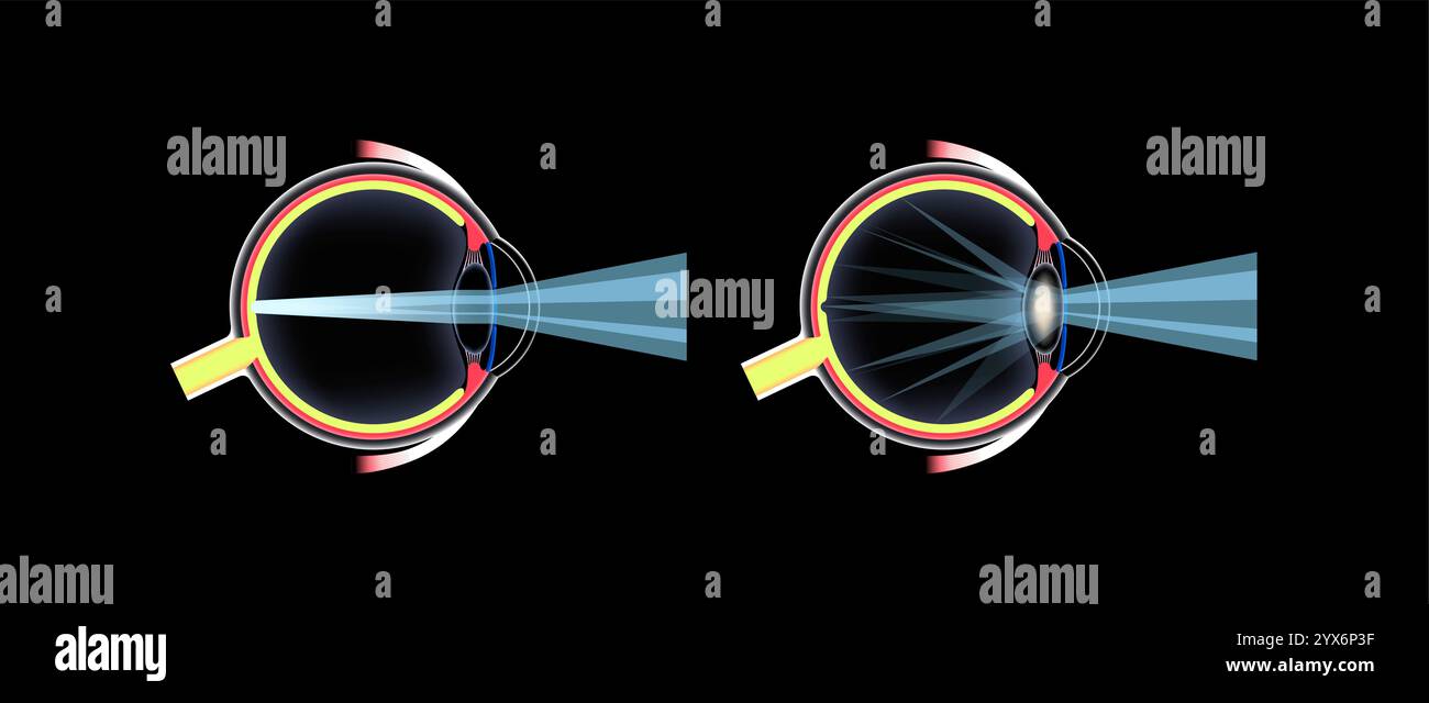 Cataract eye disease, illustration. Stock Photohttps://www.alamy.com/image-license-details/?v=1https://www.alamy.com/cataract-eye-disease-illustration-image635703363.html
RF2YX6P3F–Cataract eye disease, illustration.
 . Diseases of the dog and their treatment. Dogs. CATARACT. 367 should be an unfavorable one. Hereditary cataract shows little inclination to enlargement, as is also the case in senile cataract. In soft cortical cataracts we may see a rapid opacity of the lens in a few days or weeks. The sight is entirely lost and medical treat- ment is of little use. Therapeutic Treatment. A cataract may be removed by an operation, and this is much more advisable in the dog because it is, as a rule, attended without any great danger, and its results are generally beneficial, producing a partial restoration of Stock Photohttps://www.alamy.com/image-license-details/?v=1https://www.alamy.com/diseases-of-the-dog-and-their-treatment-dogs-cataract-367-should-be-an-unfavorable-one-hereditary-cataract-shows-little-inclination-to-enlargement-as-is-also-the-case-in-senile-cataract-in-soft-cortical-cataracts-we-may-see-a-rapid-opacity-of-the-lens-in-a-few-days-or-weeks-the-sight-is-entirely-lost-and-medical-treat-ment-is-of-little-use-therapeutic-treatment-a-cataract-may-be-removed-by-an-operation-and-this-is-much-more-advisable-in-the-dog-because-it-is-as-a-rule-attended-without-any-great-danger-and-its-results-are-generally-beneficial-producing-a-partial-restoration-of-image231379500.html
RMRCC6WG–. Diseases of the dog and their treatment. Dogs. CATARACT. 367 should be an unfavorable one. Hereditary cataract shows little inclination to enlargement, as is also the case in senile cataract. In soft cortical cataracts we may see a rapid opacity of the lens in a few days or weeks. The sight is entirely lost and medical treat- ment is of little use. Therapeutic Treatment. A cataract may be removed by an operation, and this is much more advisable in the dog because it is, as a rule, attended without any great danger, and its results are generally beneficial, producing a partial restoration of
 Cataract eye disease, illustration. Stock Photohttps://www.alamy.com/image-license-details/?v=1https://www.alamy.com/cataract-eye-disease-illustration-image635703324.html
RF2YX6P24–Cataract eye disease, illustration.
 Cataract eye disease, illustration. Stock Photohttps://www.alamy.com/image-license-details/?v=1https://www.alamy.com/cataract-eye-disease-illustration-image635703398.html
RF2YX6P4P–Cataract eye disease, illustration.
 Cataract eye disease, illustration. Stock Photohttps://www.alamy.com/image-license-details/?v=1https://www.alamy.com/cataract-eye-disease-illustration-image635703400.html
RF2YX6P4T–Cataract eye disease, illustration.
 Cataract eye disease, illustration. Stock Photohttps://www.alamy.com/image-license-details/?v=1https://www.alamy.com/cataract-eye-disease-illustration-image635703352.html
RF2YX6P34–Cataract eye disease, illustration.
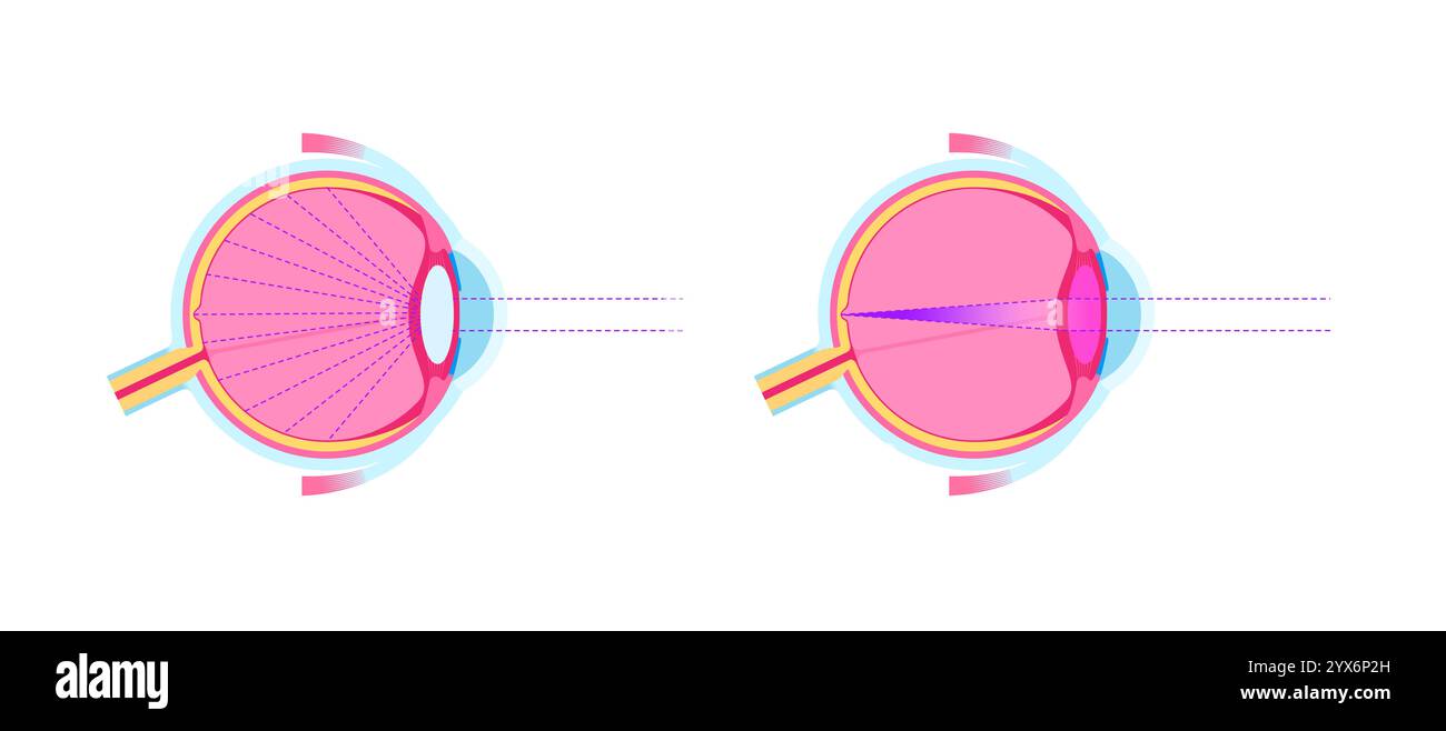 Cataract eye disease, illustration. Stock Photohttps://www.alamy.com/image-license-details/?v=1https://www.alamy.com/cataract-eye-disease-illustration-image635703337.html
RF2YX6P2H–Cataract eye disease, illustration.
 Cataract eye disease, illustration. Stock Photohttps://www.alamy.com/image-license-details/?v=1https://www.alamy.com/cataract-eye-disease-illustration-image635703289.html
RF2YX6P0W–Cataract eye disease, illustration.
 Cataract eye disease, illustration. Stock Photohttps://www.alamy.com/image-license-details/?v=1https://www.alamy.com/cataract-eye-disease-illustration-image635703347.html
RF2YX6P2Y–Cataract eye disease, illustration.
 Cataract eye disease, illustration. Stock Photohttps://www.alamy.com/image-license-details/?v=1https://www.alamy.com/cataract-eye-disease-illustration-image635703300.html
RF2YX6P18–Cataract eye disease, illustration.
 Cataract eye disease, illustration. Stock Photohttps://www.alamy.com/image-license-details/?v=1https://www.alamy.com/cataract-eye-disease-illustration-image635703303.html
RF2YX6P1B–Cataract eye disease, illustration.
 Cataract eye disease, illustration. Stock Photohttps://www.alamy.com/image-license-details/?v=1https://www.alamy.com/cataract-eye-disease-illustration-image635703358.html
RF2YX6P3A–Cataract eye disease, illustration.
 Cataract eye disease, illustration. Stock Photohttps://www.alamy.com/image-license-details/?v=1https://www.alamy.com/cataract-eye-disease-illustration-image635703305.html
RF2YX6P1D–Cataract eye disease, illustration.
 Cataract eye disease, illustration. Stock Photohttps://www.alamy.com/image-license-details/?v=1https://www.alamy.com/cataract-eye-disease-illustration-image635703345.html
RF2YX6P2W–Cataract eye disease, illustration.
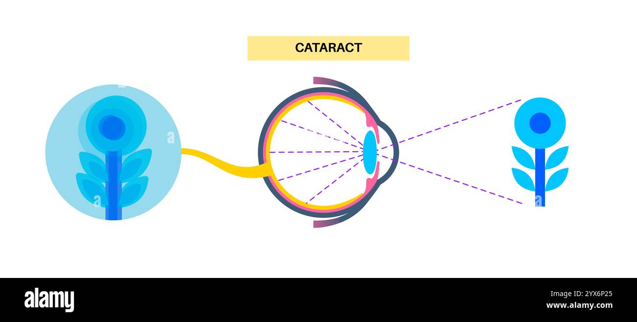 Cataract eye disease, illustration. Stock Photohttps://www.alamy.com/image-license-details/?v=1https://www.alamy.com/cataract-eye-disease-illustration-image635703325.html
RF2YX6P25–Cataract eye disease, illustration.
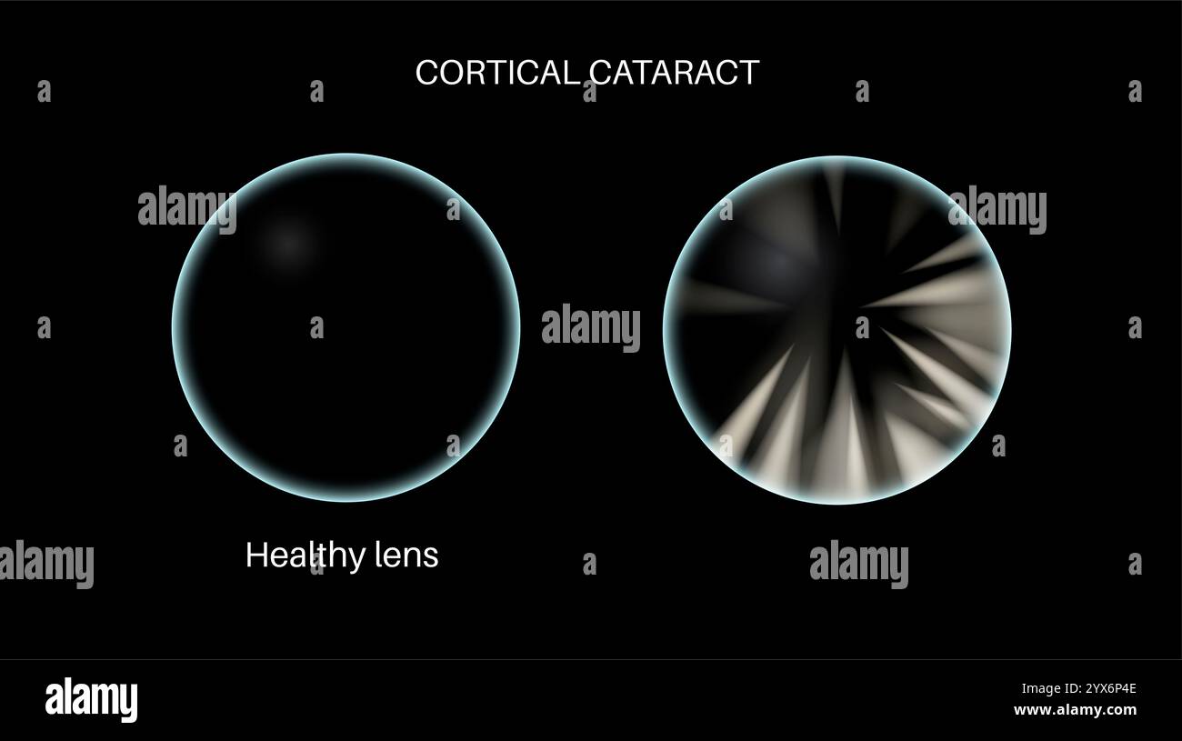 Cataract eye disease, illustration. Stock Photohttps://www.alamy.com/image-license-details/?v=1https://www.alamy.com/cataract-eye-disease-illustration-image635703390.html
RF2YX6P4E–Cataract eye disease, illustration.
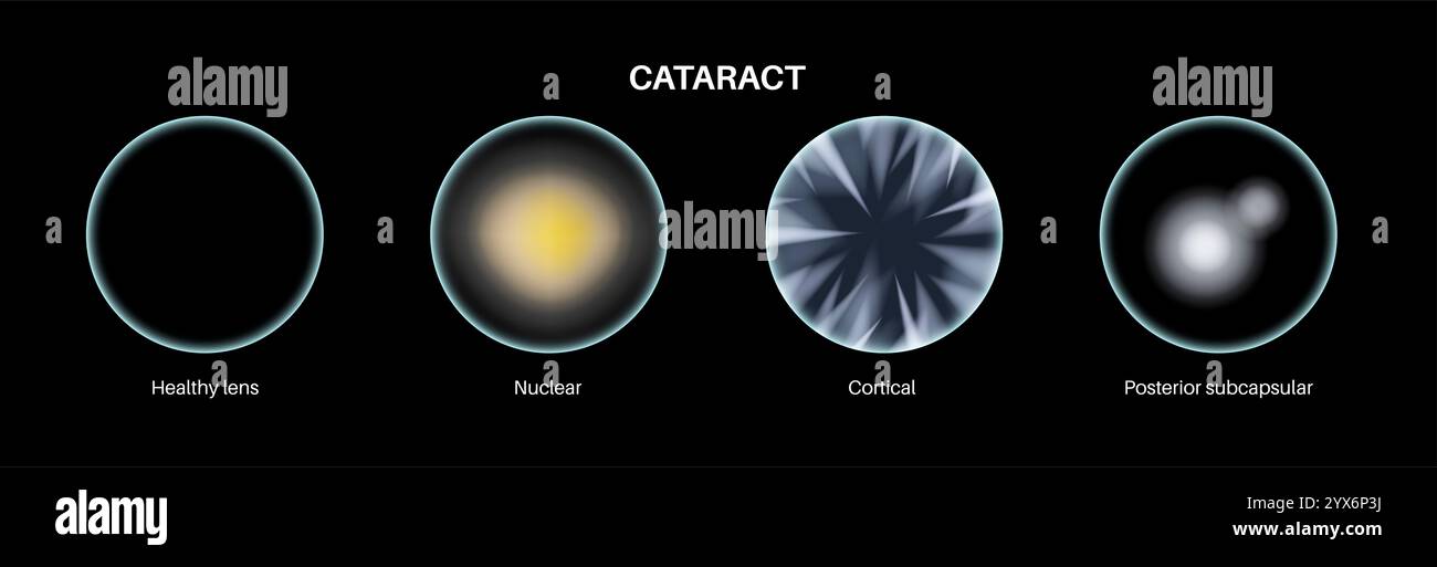 Cataract eye disease, illustration. Stock Photohttps://www.alamy.com/image-license-details/?v=1https://www.alamy.com/cataract-eye-disease-illustration-image635703366.html
RF2YX6P3J–Cataract eye disease, illustration.
 Cataract eye disease, illustration. Stock Photohttps://www.alamy.com/image-license-details/?v=1https://www.alamy.com/cataract-eye-disease-illustration-image635703327.html
RF2YX6P27–Cataract eye disease, illustration.
 Cataract eye disease, illustration. Stock Photohttps://www.alamy.com/image-license-details/?v=1https://www.alamy.com/cataract-eye-disease-illustration-image635703328.html
RF2YX6P28–Cataract eye disease, illustration.
 Cataract eye disease, illustration. Stock Photohttps://www.alamy.com/image-license-details/?v=1https://www.alamy.com/cataract-eye-disease-illustration-image635703386.html
RF2YX6P4A–Cataract eye disease, illustration.
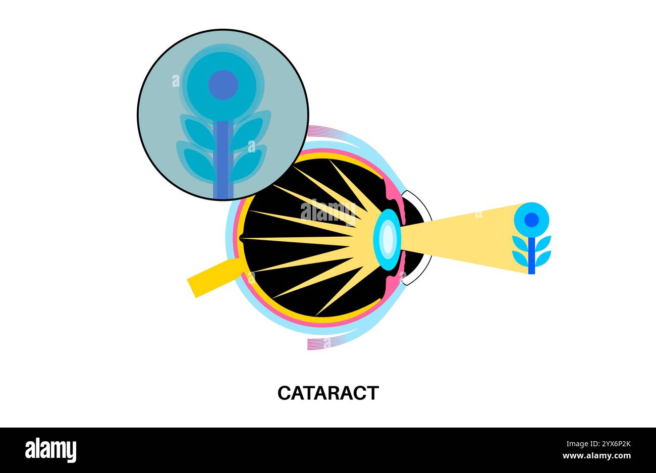 Cataract eye disease, illustration. Stock Photohttps://www.alamy.com/image-license-details/?v=1https://www.alamy.com/cataract-eye-disease-illustration-image635703339.html
RF2YX6P2K–Cataract eye disease, illustration.
 Cataract eye disease, illustration. Stock Photohttps://www.alamy.com/image-license-details/?v=1https://www.alamy.com/cataract-eye-disease-illustration-image635703349.html
RF2YX6P31–Cataract eye disease, illustration.
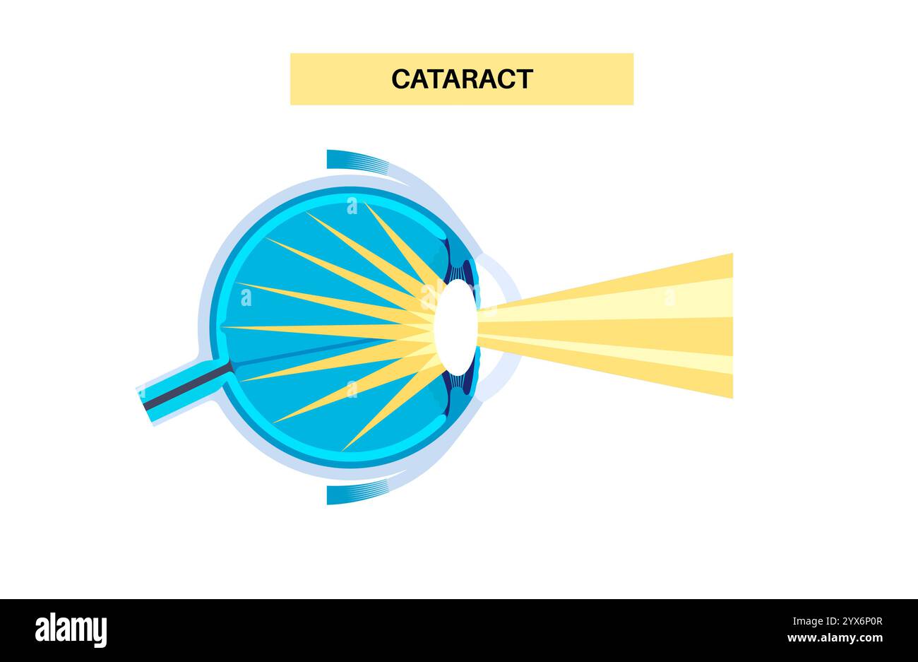 Cataract eye disease, illustration. Stock Photohttps://www.alamy.com/image-license-details/?v=1https://www.alamy.com/cataract-eye-disease-illustration-image635703287.html
RF2YX6P0R–Cataract eye disease, illustration.
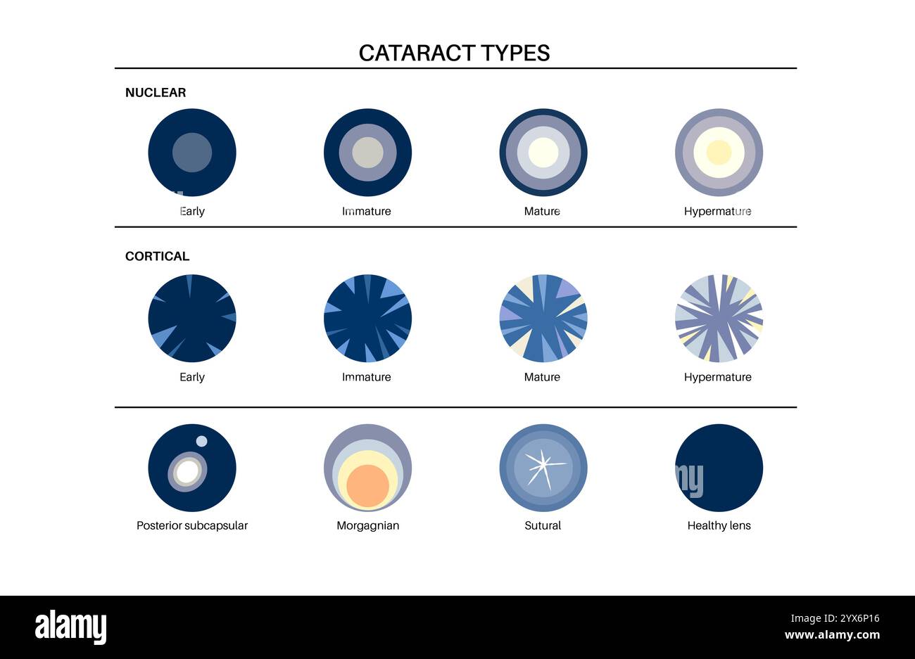 Cataract eye disease, illustration. Stock Photohttps://www.alamy.com/image-license-details/?v=1https://www.alamy.com/cataract-eye-disease-illustration-image635703298.html
RF2YX6P16–Cataract eye disease, illustration.
 Cataract eye disease, illustration. Stock Photohttps://www.alamy.com/image-license-details/?v=1https://www.alamy.com/cataract-eye-disease-illustration-image635703290.html
RF2YX6P0X–Cataract eye disease, illustration.
 Cataract eye disease, illustration. Stock Photohttps://www.alamy.com/image-license-details/?v=1https://www.alamy.com/cataract-eye-disease-illustration-image635703371.html
RF2YX6P3R–Cataract eye disease, illustration.
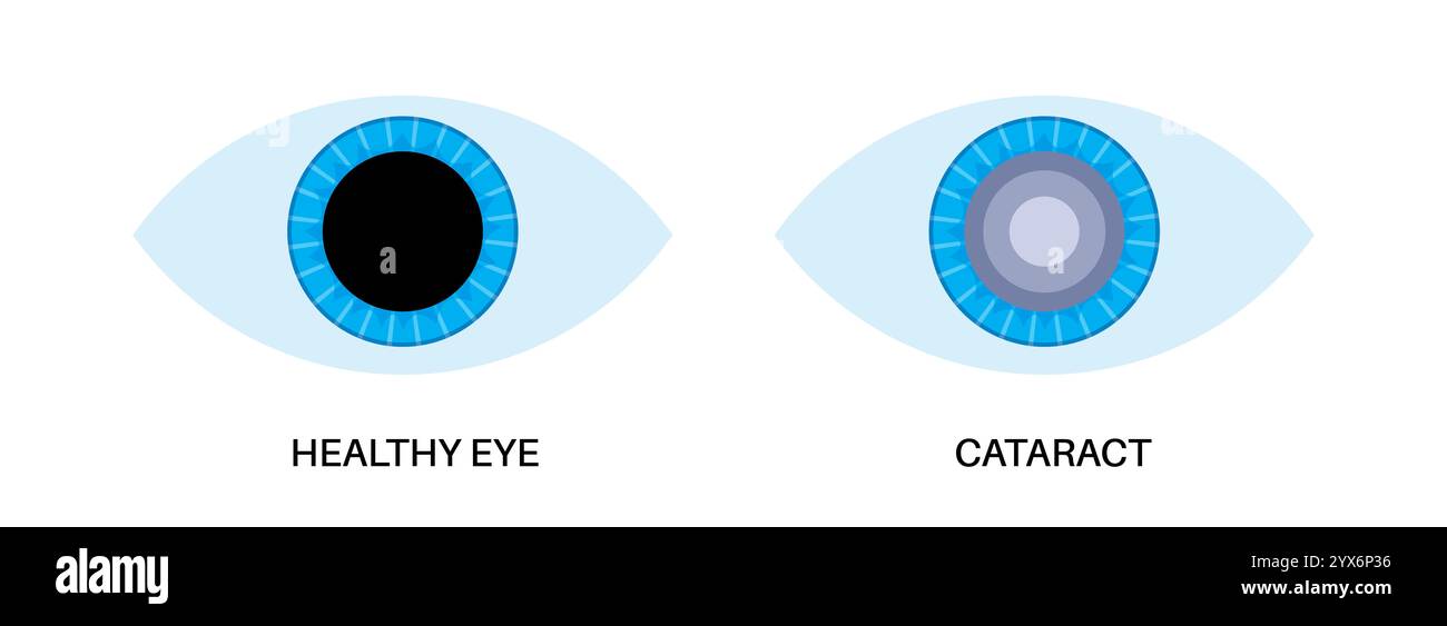 Cataract eye disease, illustration. Stock Photohttps://www.alamy.com/image-license-details/?v=1https://www.alamy.com/cataract-eye-disease-illustration-image635703354.html
RF2YX6P36–Cataract eye disease, illustration.
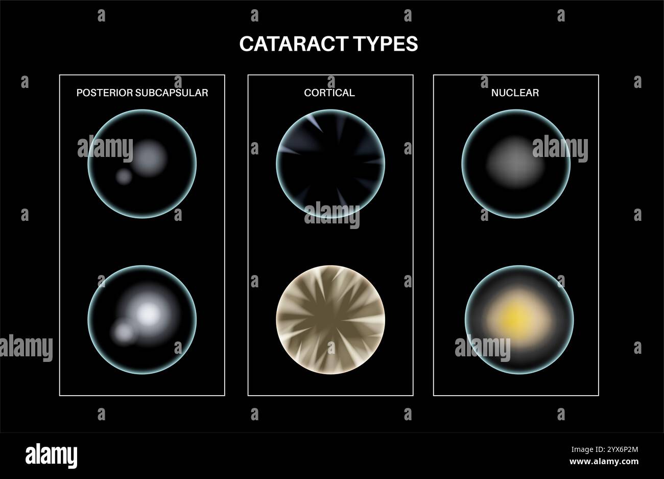 Cataract eye disease, illustration. Stock Photohttps://www.alamy.com/image-license-details/?v=1https://www.alamy.com/cataract-eye-disease-illustration-image635703340.html
RF2YX6P2M–Cataract eye disease, illustration.
 Cataract eye disease, illustration. Stock Photohttps://www.alamy.com/image-license-details/?v=1https://www.alamy.com/cataract-eye-disease-illustration-image635703342.html
RF2YX6P2P–Cataract eye disease, illustration.
 Cataract eye disease, illustration. Stock Photohttps://www.alamy.com/image-license-details/?v=1https://www.alamy.com/cataract-eye-disease-illustration-image635703309.html
RF2YX6P1H–Cataract eye disease, illustration.
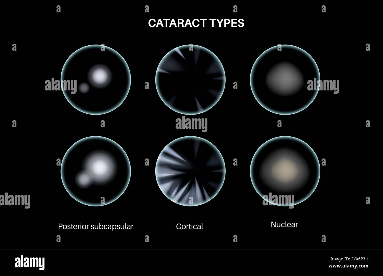 Cataract eye disease, illustration. Stock Photohttps://www.alamy.com/image-license-details/?v=1https://www.alamy.com/cataract-eye-disease-illustration-image635703365.html
RF2YX6P3H–Cataract eye disease, illustration.
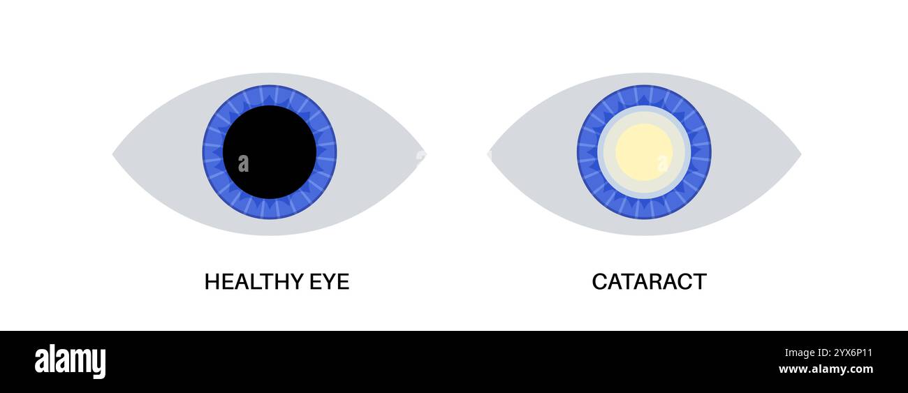 Cataract eye disease, illustration. Stock Photohttps://www.alamy.com/image-license-details/?v=1https://www.alamy.com/cataract-eye-disease-illustration-image635703293.html
RF2YX6P11–Cataract eye disease, illustration.
 Cataract eye disease, illustration. Stock Photohttps://www.alamy.com/image-license-details/?v=1https://www.alamy.com/cataract-eye-disease-illustration-image635703343.html
RF2YX6P2R–Cataract eye disease, illustration.
 Cataract eye disease, illustration. Stock Photohttps://www.alamy.com/image-license-details/?v=1https://www.alamy.com/cataract-eye-disease-illustration-image635703322.html
RF2YX6P22–Cataract eye disease, illustration.
 Cataract eye disease, illustration. Stock Photohttps://www.alamy.com/image-license-details/?v=1https://www.alamy.com/cataract-eye-disease-illustration-image635703311.html
RF2YX6P1K–Cataract eye disease, illustration.
 Cataract eye disease, illustration. Stock Photohttps://www.alamy.com/image-license-details/?v=1https://www.alamy.com/cataract-eye-disease-illustration-image635703336.html
RF2YX6P2G–Cataract eye disease, illustration.
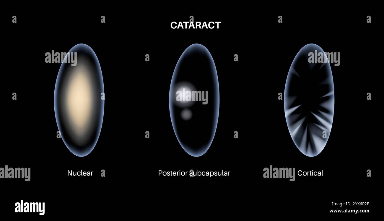 Cataract eye disease, illustration. Stock Photohttps://www.alamy.com/image-license-details/?v=1https://www.alamy.com/cataract-eye-disease-illustration-image635703334.html
RF2YX6P2E–Cataract eye disease, illustration.
 Cataract eye disease, illustration. Stock Photohttps://www.alamy.com/image-license-details/?v=1https://www.alamy.com/cataract-eye-disease-illustration-image635703323.html
RF2YX6P23–Cataract eye disease, illustration.
 Cataract eye disease, illustration. Stock Photohttps://www.alamy.com/image-license-details/?v=1https://www.alamy.com/cataract-eye-disease-illustration-image635703381.html
RF2YX6P45–Cataract eye disease, illustration.
 Cataract eye disease, illustration. Stock Photohttps://www.alamy.com/image-license-details/?v=1https://www.alamy.com/cataract-eye-disease-illustration-image635703341.html
RF2YX6P2N–Cataract eye disease, illustration.
 Cataract eye disease, illustration. Stock Photohttps://www.alamy.com/image-license-details/?v=1https://www.alamy.com/cataract-eye-disease-illustration-image635703286.html
RF2YX6P0P–Cataract eye disease, illustration.
 Cataract eye disease, illustration. Stock Photohttps://www.alamy.com/image-license-details/?v=1https://www.alamy.com/cataract-eye-disease-illustration-image635703306.html
RF2YX6P1E–Cataract eye disease, illustration.
