Quick filters:
Cell labelled diagram Stock Photos and Images
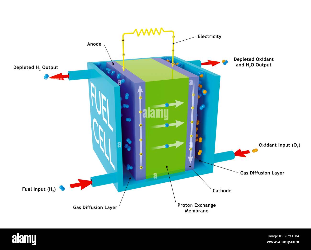 Hydrogen fuel cell, illustration Stock Photohttps://www.alamy.com/image-license-details/?v=1https://www.alamy.com/hydrogen-fuel-cell-illustration-image430103048.html
RF2FYMTR4–Hydrogen fuel cell, illustration
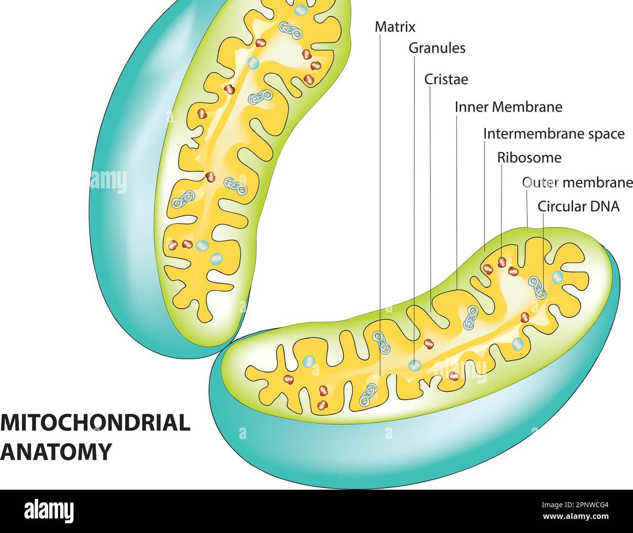 Mitochondria diagram Stock Vectorhttps://www.alamy.com/image-license-details/?v=1https://www.alamy.com/mitochondria-diagram-image546987844.html
RF2PNWCG4–Mitochondria diagram
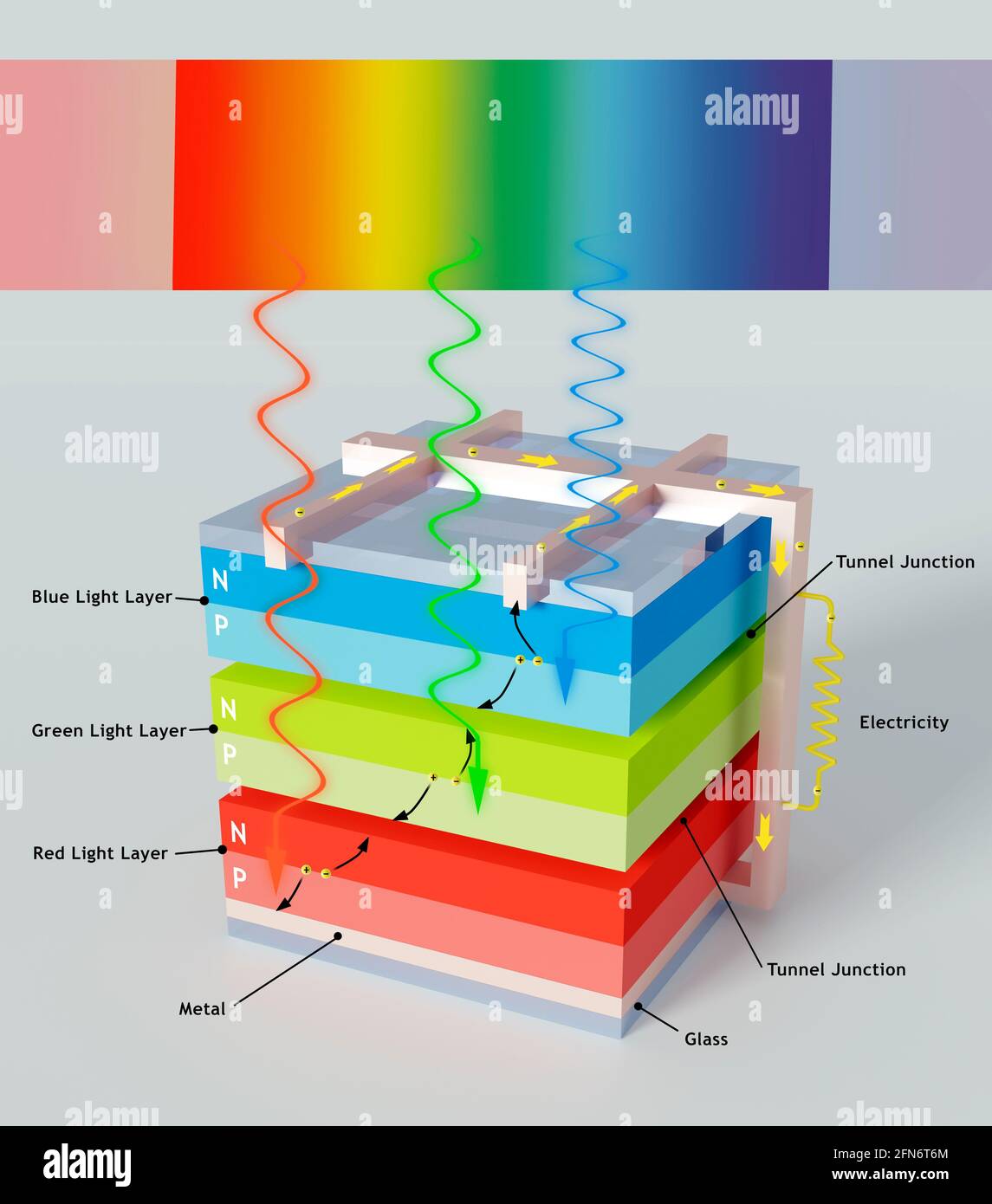 Multi-junction solar cell, illustration Stock Photohttps://www.alamy.com/image-license-details/?v=1https://www.alamy.com/multi-junction-solar-cell-illustration-image426107324.html
RF2FN6T6M–Multi-junction solar cell, illustration
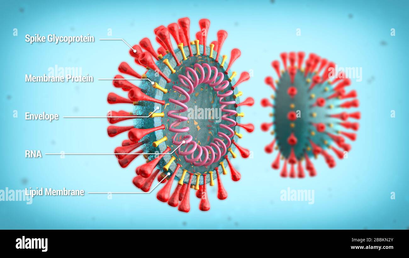 Labelled diagram of COVID-19 cell structure showing spike glycoprotein, membrane protein, envelope, RNA. 3d illustration schematic of coronavirus. Stock Photohttps://www.alamy.com/image-license-details/?v=1https://www.alamy.com/labelled-diagram-of-covid-19-cell-structure-showing-spike-glycoprotein-membrane-protein-envelope-rna-3d-illustration-schematic-of-coronavirus-image351402211.html
RF2BBKN2Y–Labelled diagram of COVID-19 cell structure showing spike glycoprotein, membrane protein, envelope, RNA. 3d illustration schematic of coronavirus.
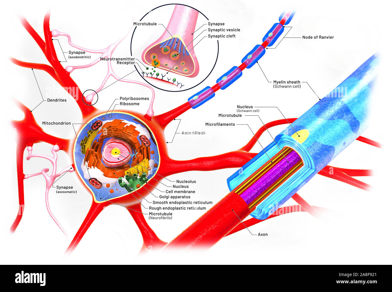 Nerve cell anatomy, illustration Stock Photohttps://www.alamy.com/image-license-details/?v=1https://www.alamy.com/nerve-cell-anatomy-illustration-image332250633.html
RF2A8F921–Nerve cell anatomy, illustration
 Simple electrochemical or galvanic cell. The Daniell cell. Stock Photohttps://www.alamy.com/image-license-details/?v=1https://www.alamy.com/stock-photo-simple-electrochemical-or-galvanic-cell-the-daniell-cell-74405627.html
RFE91D3R–Simple electrochemical or galvanic cell. The Daniell cell.
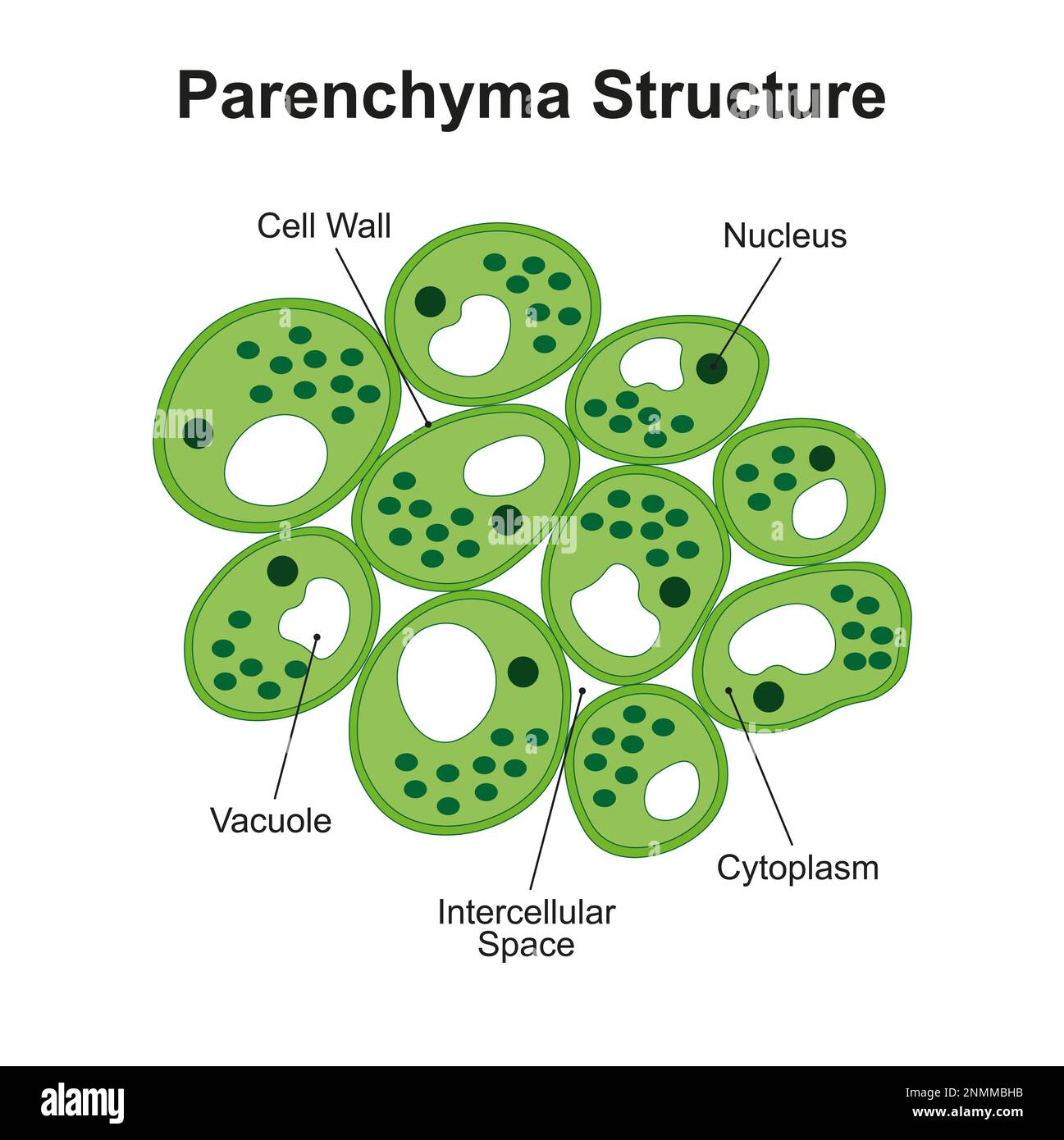 Plant parenchyma structure, illustration Stock Photohttps://www.alamy.com/image-license-details/?v=1https://www.alamy.com/plant-parenchyma-structure-illustration-image529052311.html
RF2NMMBHB–Plant parenchyma structure, illustration
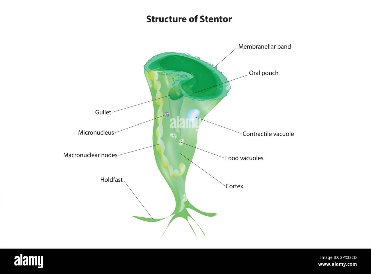 stentor Stock Vectorhttps://www.alamy.com/image-license-details/?v=1https://www.alamy.com/stentor-image549569957.html
RF2PX322D–stentor
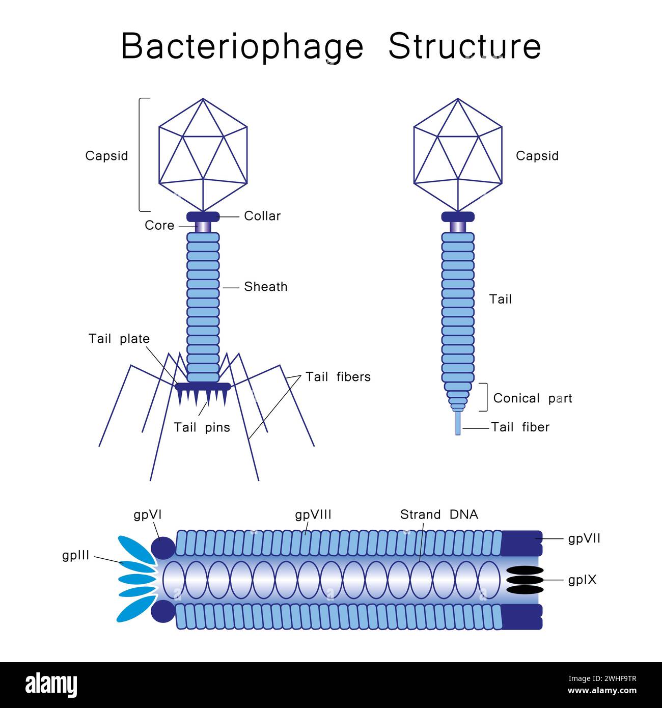 Bacteriophage, illustration Stock Photohttps://www.alamy.com/image-license-details/?v=1https://www.alamy.com/bacteriophage-illustration-image595938695.html
RF2WHF9TR–Bacteriophage, illustration
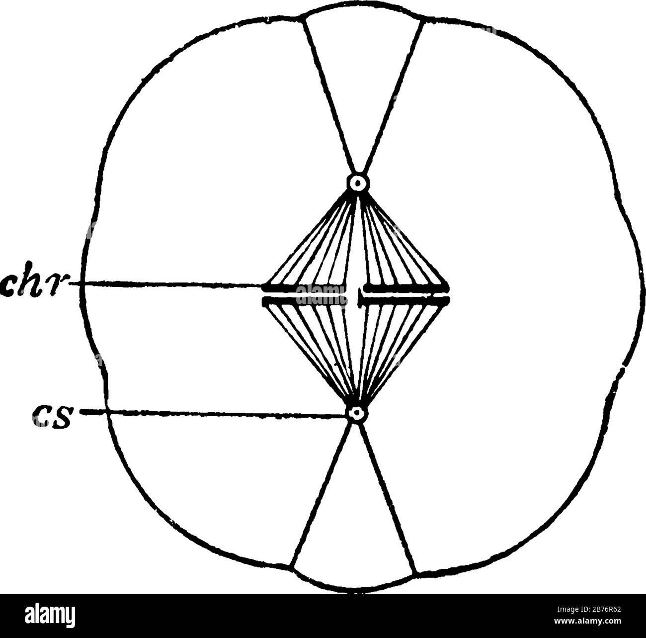 Diagram of cell division, with the parts labelled, chr, chromosomes forming an equatorial plate; and cs, centrosome, vintage line drawing or engraving Stock Vectorhttps://www.alamy.com/image-license-details/?v=1https://www.alamy.com/diagram-of-cell-division-with-the-parts-labelled-chr-chromosomes-forming-an-equatorial-plate-and-cs-centrosome-vintage-line-drawing-or-engraving-image348659866.html
RF2B76R62–Diagram of cell division, with the parts labelled, chr, chromosomes forming an equatorial plate; and cs, centrosome, vintage line drawing or engraving
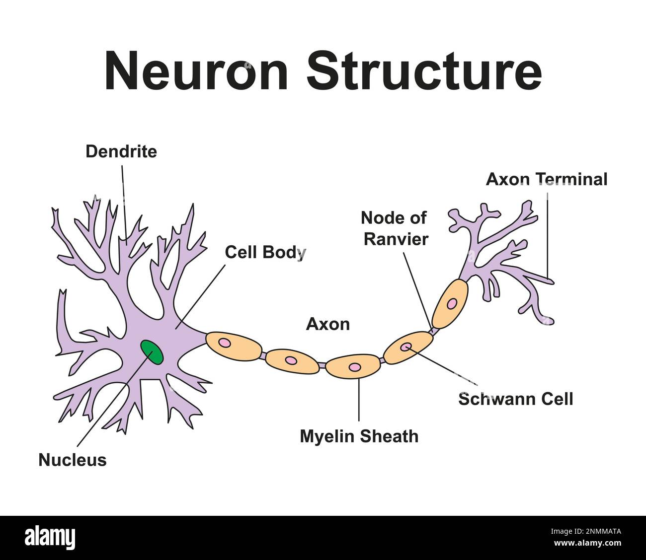 Nerve cell, illustration Stock Photohttps://www.alamy.com/image-license-details/?v=1https://www.alamy.com/nerve-cell-illustration-image529051722.html
RF2NMMATA–Nerve cell, illustration
 Diagram of a neuron, with its parts labelled as, 'A', representing axon arising from the cell-body and branching at its termination; 'D', representing Stock Vectorhttps://www.alamy.com/image-license-details/?v=1https://www.alamy.com/diagram-of-a-neuron-with-its-parts-labelled-as-a-representing-axon-arising-from-the-cell-body-and-branching-at-its-termination-d-representing-image359323478.html
RF2BTGGNA–Diagram of a neuron, with its parts labelled as, 'A', representing axon arising from the cell-body and branching at its termination; 'D', representing
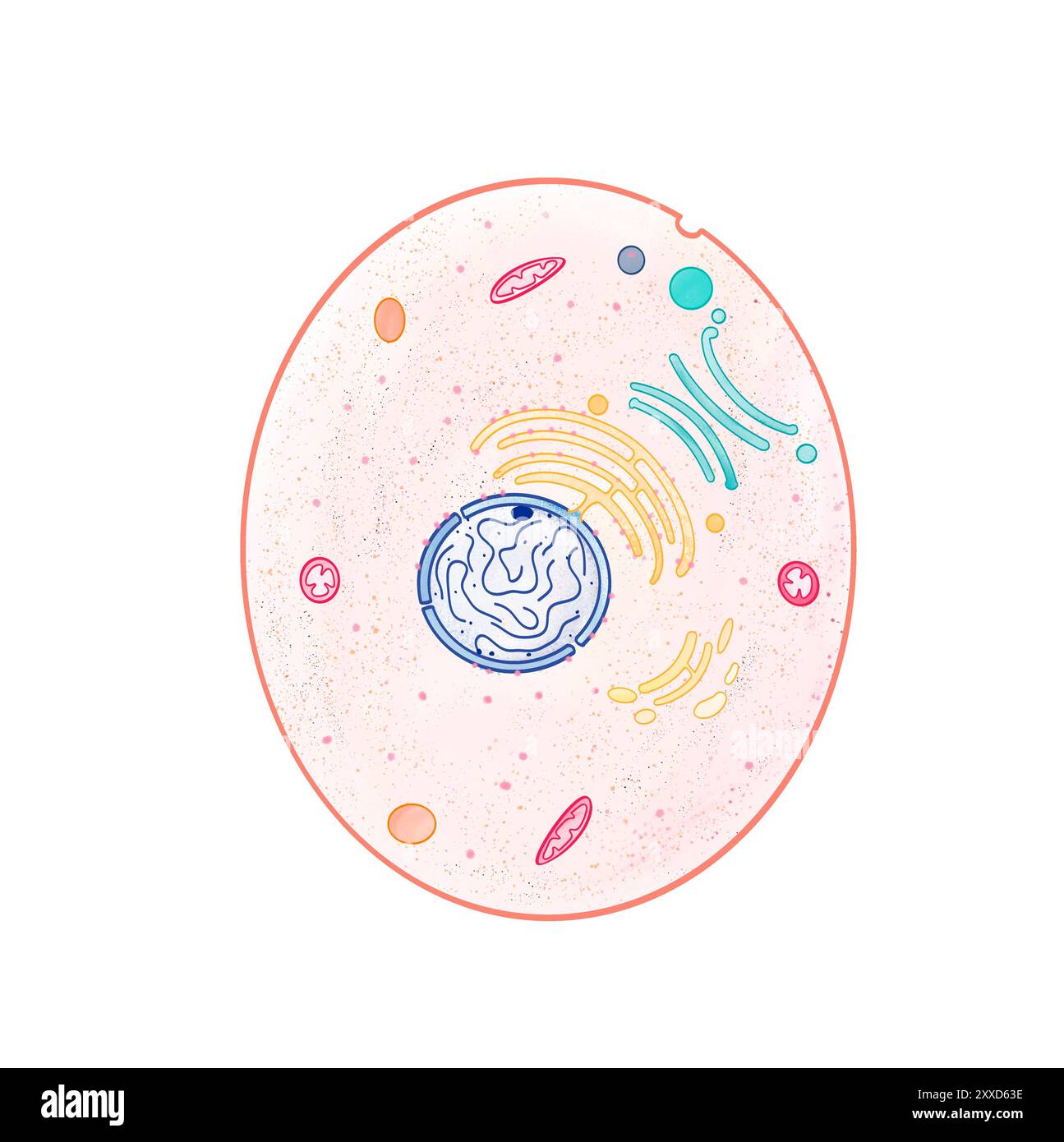 Animal cell, illustration. Animal cells are eukaryotic cells (whose nucleus is bound by a nuclear membrane). Stock Photohttps://www.alamy.com/image-license-details/?v=1https://www.alamy.com/animal-cell-illustration-animal-cells-are-eukaryotic-cells-whose-nucleus-is-bound-by-a-nuclear-membrane-image618634114.html
RF2XXD63E–Animal cell, illustration. Animal cells are eukaryotic cells (whose nucleus is bound by a nuclear membrane).
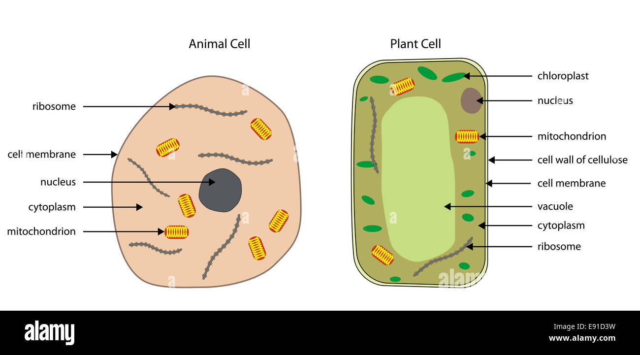 Labeled diagrams of typical animal and plant cells with editable layers. Stock Photohttps://www.alamy.com/image-license-details/?v=1https://www.alamy.com/stock-photo-labeled-diagrams-of-typical-animal-and-plant-cells-with-editable-layers-74405629.html
RFE91D3W–Labeled diagrams of typical animal and plant cells with editable layers.
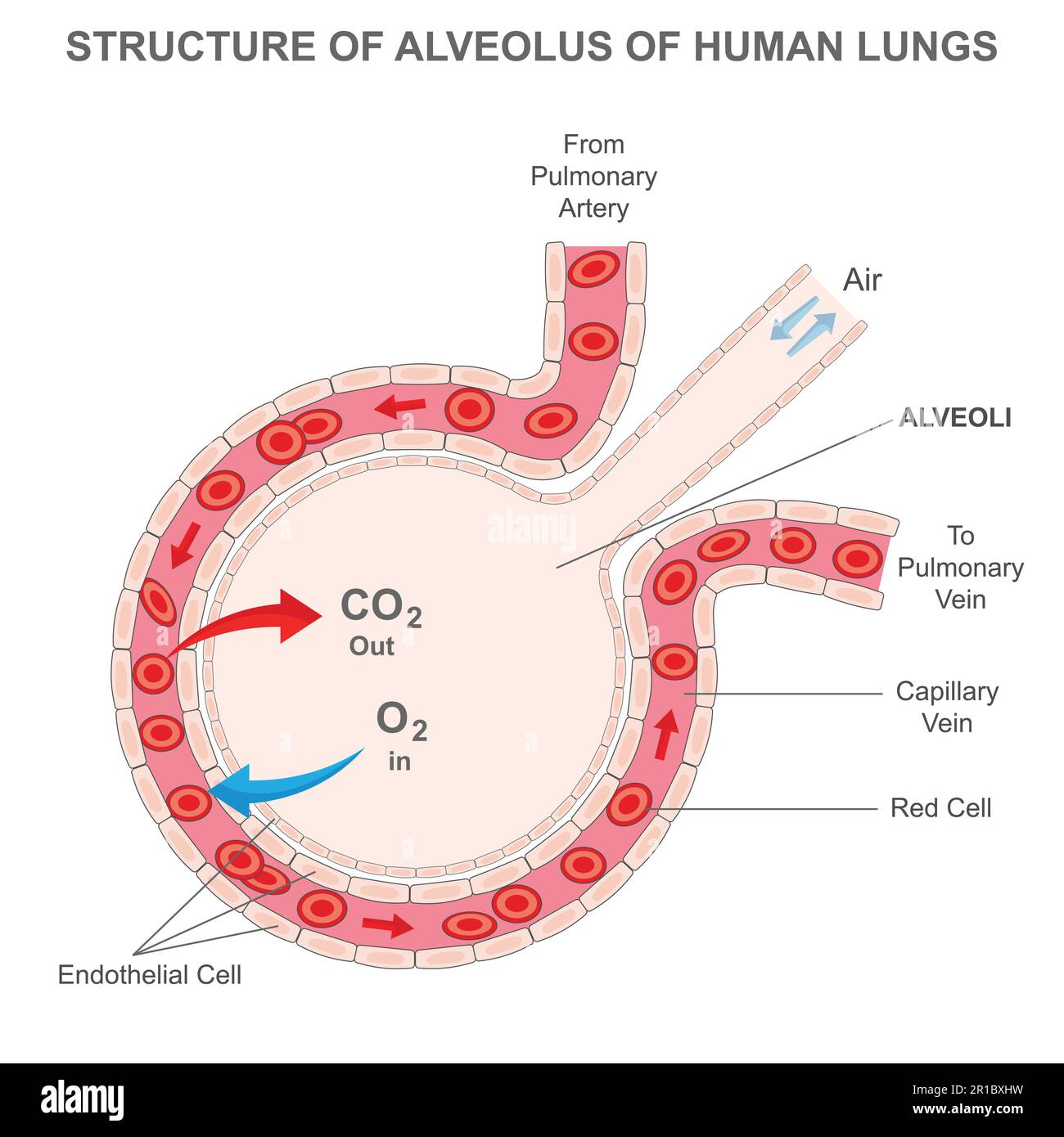 Structure of alveolus of human lungs. Labelled diagram of the alveolus in the lungs showing gaseous exchange. Pulmonary alveolus. alveoli and capillar Stock Vectorhttps://www.alamy.com/image-license-details/?v=1https://www.alamy.com/structure-of-alveolus-of-human-lungs-labelled-diagram-of-the-alveolus-in-the-lungs-showing-gaseous-exchange-pulmonary-alveolus-alveoli-and-capillar-image551608789.html
RF2R1BXHW–Structure of alveolus of human lungs. Labelled diagram of the alveolus in the lungs showing gaseous exchange. Pulmonary alveolus. alveoli and capillar
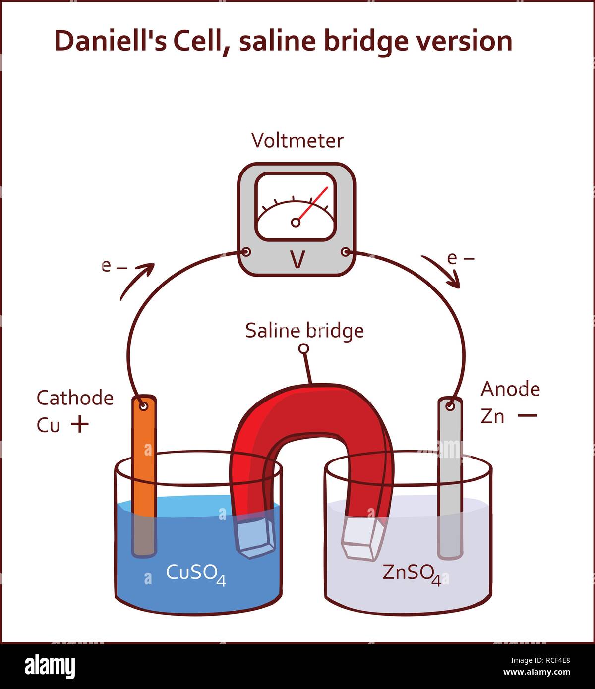 Daniell's Cell, saline bridge version vector illustration Stock Vectorhttps://www.alamy.com/image-license-details/?v=1https://www.alamy.com/daniells-cell-saline-bridge-version-vector-illustration-image231443472.html
RFRCF4E8–Daniell's Cell, saline bridge version vector illustration
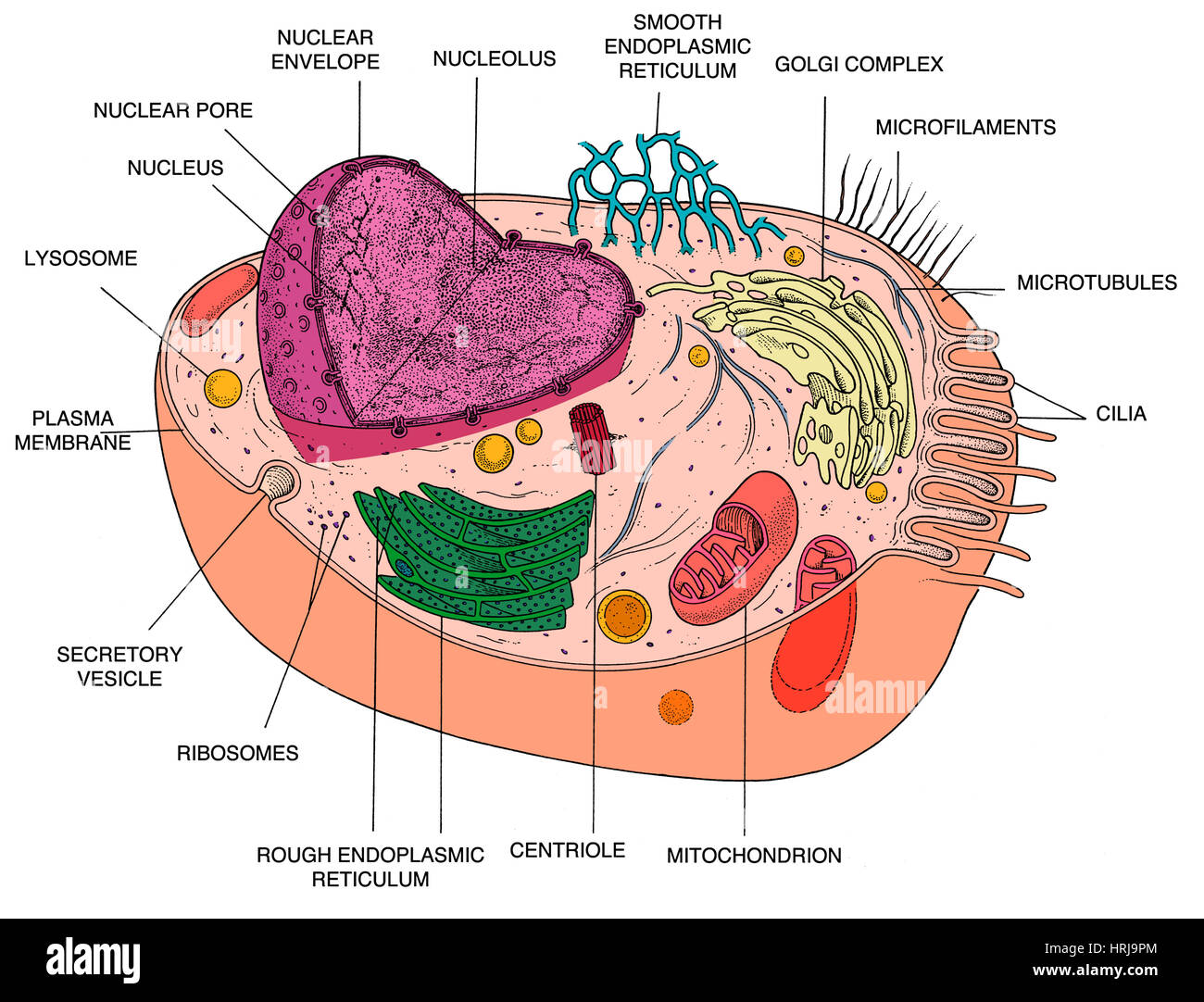 Animal Cell Diagram Stock Photohttps://www.alamy.com/image-license-details/?v=1https://www.alamy.com/stock-photo-animal-cell-diagram-135012492.html
RMHRJ9PM–Animal Cell Diagram
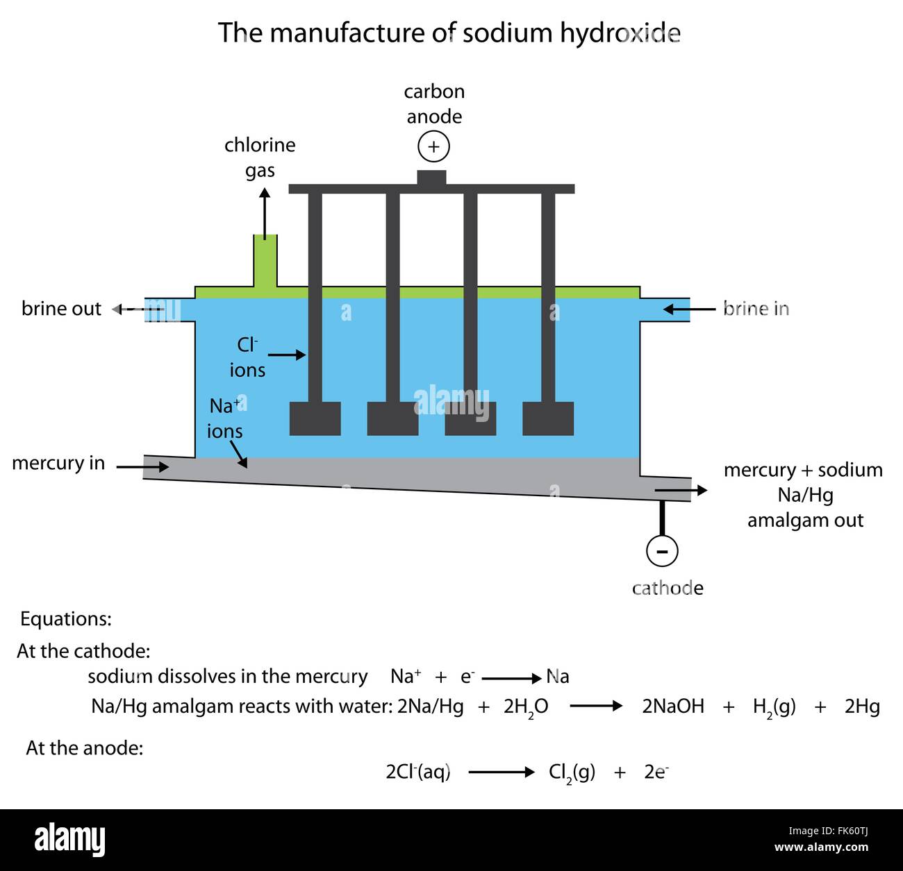 Labelled diagram of the industrial manufacture of sodium hydroxide in the flowing mercury cell in vector format. Stock Vectorhttps://www.alamy.com/image-license-details/?v=1https://www.alamy.com/stock-photo-labelled-diagram-of-the-industrial-manufacture-of-sodium-hydroxide-97862706.html
RFFK60TJ–Labelled diagram of the industrial manufacture of sodium hydroxide in the flowing mercury cell in vector format.
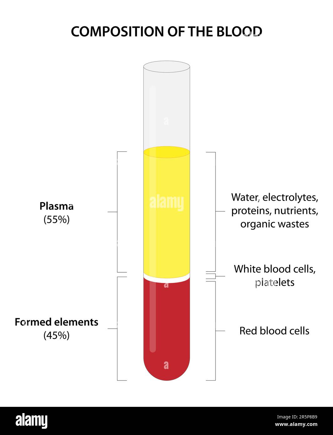 Whole blood consists of red blood cells (erythrocytes), white blood cells (leukocytes), platelets (thrombocytes), and plasma. Stock Vectorhttps://www.alamy.com/image-license-details/?v=1https://www.alamy.com/whole-blood-consists-of-red-blood-cells-erythrocytes-white-blood-cells-leukocytes-platelets-thrombocytes-and-plasma-image554294589.html
RF2R5P8B9–Whole blood consists of red blood cells (erythrocytes), white blood cells (leukocytes), platelets (thrombocytes), and plasma.
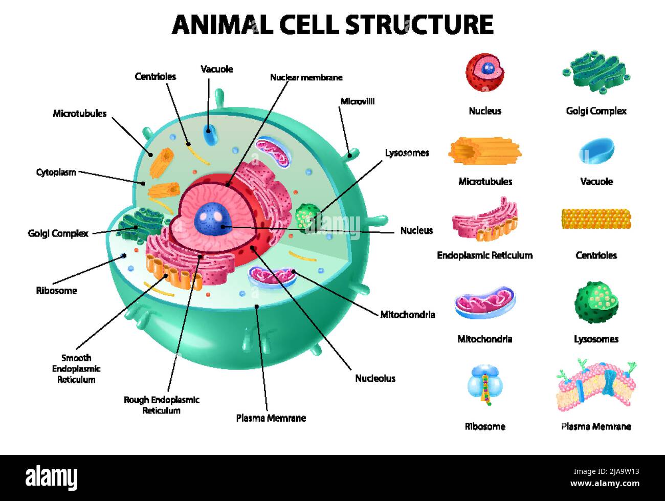 Animal cell anatomy infographics with detailed educative diagram and labelled elements realistic vector illustration Stock Vectorhttps://www.alamy.com/image-license-details/?v=1https://www.alamy.com/animal-cell-anatomy-infographics-with-detailed-educative-diagram-and-labelled-elements-realistic-vector-illustration-image471043695.html
RF2JA9W13–Animal cell anatomy infographics with detailed educative diagram and labelled elements realistic vector illustration
 Animal cell anatomy infographics with detailed educative diagram and labelled elements realistic vector illustration Stock Vectorhttps://www.alamy.com/image-license-details/?v=1https://www.alamy.com/animal-cell-anatomy-infographics-with-detailed-educative-diagram-and-labelled-elements-realistic-vector-illustration-image471323345.html
RF2JAPHMH–Animal cell anatomy infographics with detailed educative diagram and labelled elements realistic vector illustration
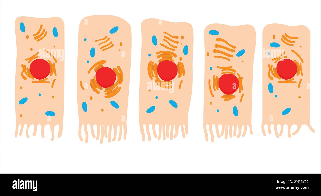 Parts of the Cell, columnar epithelial and goblet cell, Human cell anatomy Stock Vectorhttps://www.alamy.com/image-license-details/?v=1https://www.alamy.com/parts-of-the-cell-columnar-epithelial-and-goblet-cell-human-cell-anatomy-image634082990.html
RF2YRGY92–Parts of the Cell, columnar epithelial and goblet cell, Human cell anatomy
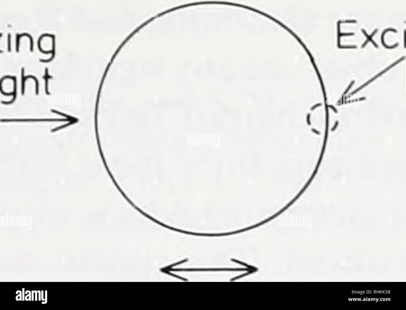 . The Biological bulletin. Biology; Zoology; Biology; Marine Biology. VISUALIZING CYTOPLASMIC CALCIUM 15 Polarizing light. Excitation beam Figure 1. Schematic diagram of polarizing fucus zygote showing method of excitation of fluorescentlv labelled cell. gradients in polarized and polarizing zygotes using the fluorescent Ca2+ indicator fura-2. The spectral properties of fura-2 are such that the ratio of fluorescence following excitation with 350 (or 360) and 385 nm light is calcium- dependent. (Grynkievicz el al, 1985; Tsien el ai. 1985; Cobbold and Rink. 1987). Materials and Methods Zygote gr Stock Photohttps://www.alamy.com/image-license-details/?v=1https://www.alamy.com/the-biological-bulletin-biology-zoology-biology-marine-biology-visualizing-cytoplasmic-calcium-15-polarizing-light-excitation-beam-figure-1-schematic-diagram-of-polarizing-fucus-zygote-showing-method-of-excitation-of-fluorescentlv-labelled-cell-gradients-in-polarized-and-polarizing-zygotes-using-the-fluorescent-ca2-indicator-fura-2-the-spectral-properties-of-fura-2-are-such-that-the-ratio-of-fluorescence-following-excitation-with-350-or-360-and-385-nm-light-is-calcium-dependent-grynkievicz-el-al-1985-tsien-el-ai-1985-cobbold-and-rink-1987-materials-and-methods-zygote-gr-image234616071.html
RMRHKK5B–. The Biological bulletin. Biology; Zoology; Biology; Marine Biology. VISUALIZING CYTOPLASMIC CALCIUM 15 Polarizing light. Excitation beam Figure 1. Schematic diagram of polarizing fucus zygote showing method of excitation of fluorescentlv labelled cell. gradients in polarized and polarizing zygotes using the fluorescent Ca2+ indicator fura-2. The spectral properties of fura-2 are such that the ratio of fluorescence following excitation with 350 (or 360) and 385 nm light is calcium- dependent. (Grynkievicz el al, 1985; Tsien el ai. 1985; Cobbold and Rink. 1987). Materials and Methods Zygote gr
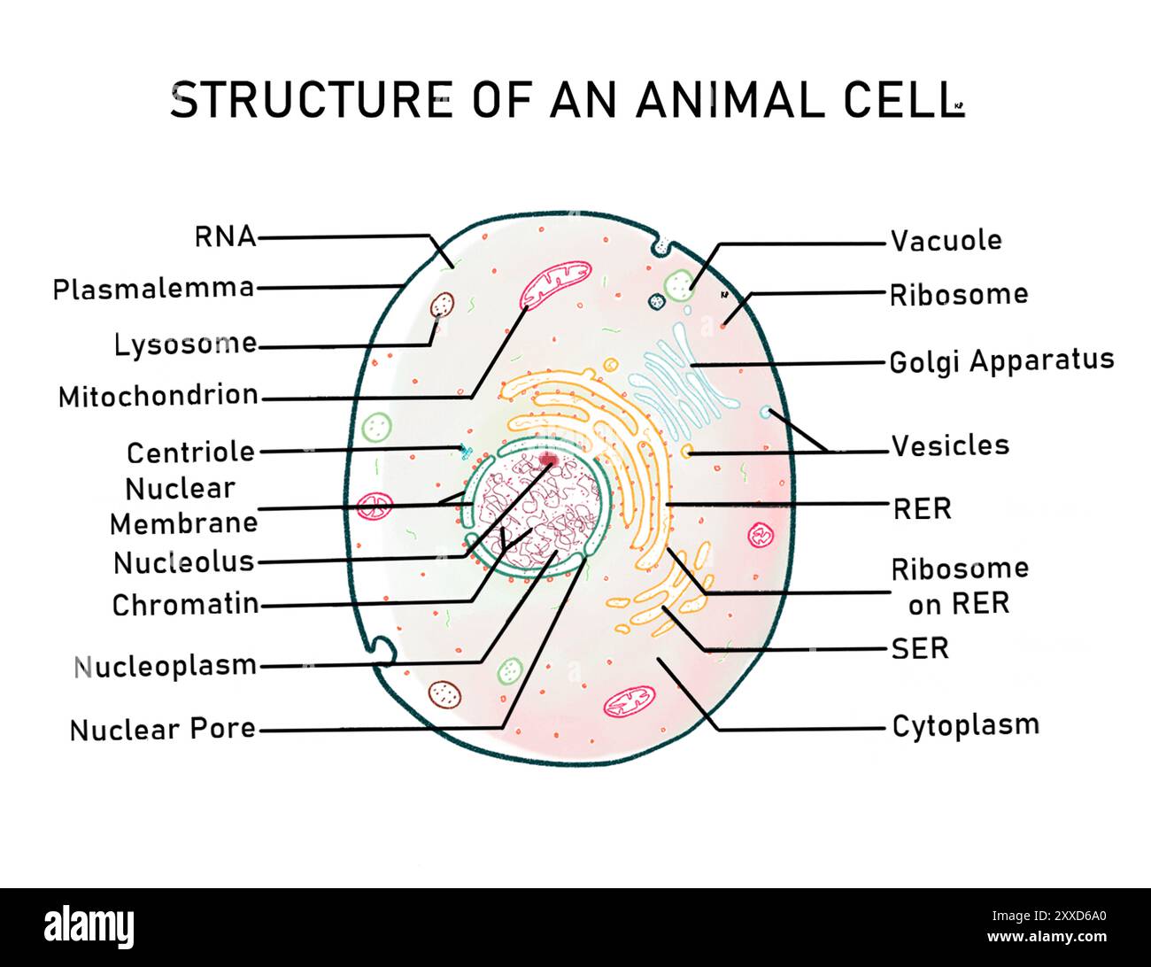 Structure of an animal cell, illustration. Animal cells are eukaryotic cells, which are cells whose nucleus is bound by a nuclear membrane. Stock Photohttps://www.alamy.com/image-license-details/?v=1https://www.alamy.com/structure-of-an-animal-cell-illustration-animal-cells-are-eukaryotic-cells-which-are-cells-whose-nucleus-is-bound-by-a-nuclear-membrane-image618634296.html
RF2XXD6A0–Structure of an animal cell, illustration. Animal cells are eukaryotic cells, which are cells whose nucleus is bound by a nuclear membrane.
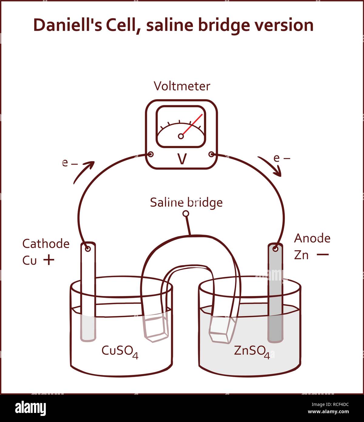 Daniell's Cell, saline bridge version vector illustration Stock Vectorhttps://www.alamy.com/image-license-details/?v=1https://www.alamy.com/daniells-cell-saline-bridge-version-vector-illustration-image231443448.html
RFRCF4DC–Daniell's Cell, saline bridge version vector illustration
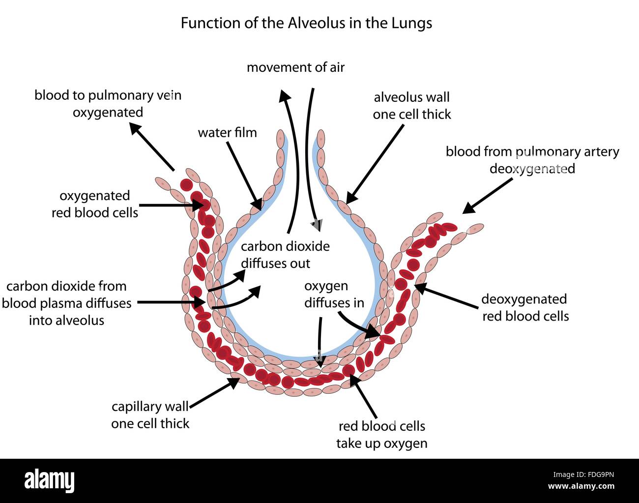 Fully labelled diagram of the alveolus in the lungs showing gaseous exchange. Stock Vectorhttps://www.alamy.com/image-license-details/?v=1https://www.alamy.com/stock-photo-fully-labelled-diagram-of-the-alveolus-in-the-lungs-showing-gaseous-94401293.html
RFFDG9PN–Fully labelled diagram of the alveolus in the lungs showing gaseous exchange.
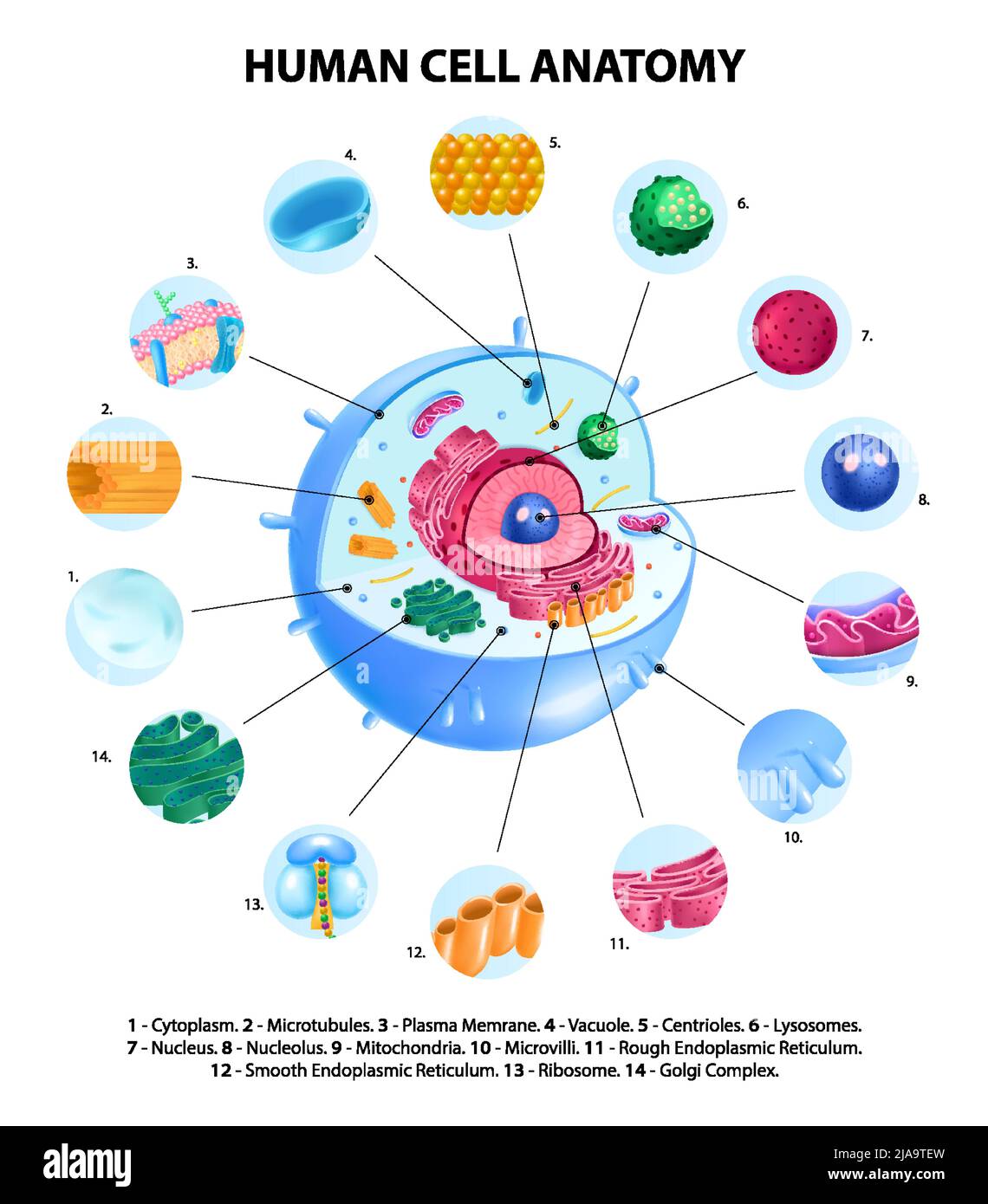 Human cell anatomy realistic infographics with labelled educational diagram on white background vector illustration Stock Vectorhttps://www.alamy.com/image-license-details/?v=1https://www.alamy.com/human-cell-anatomy-realistic-infographics-with-labelled-educational-diagram-on-white-background-vector-illustration-image471043297.html
RF2JA9TEW–Human cell anatomy realistic infographics with labelled educational diagram on white background vector illustration
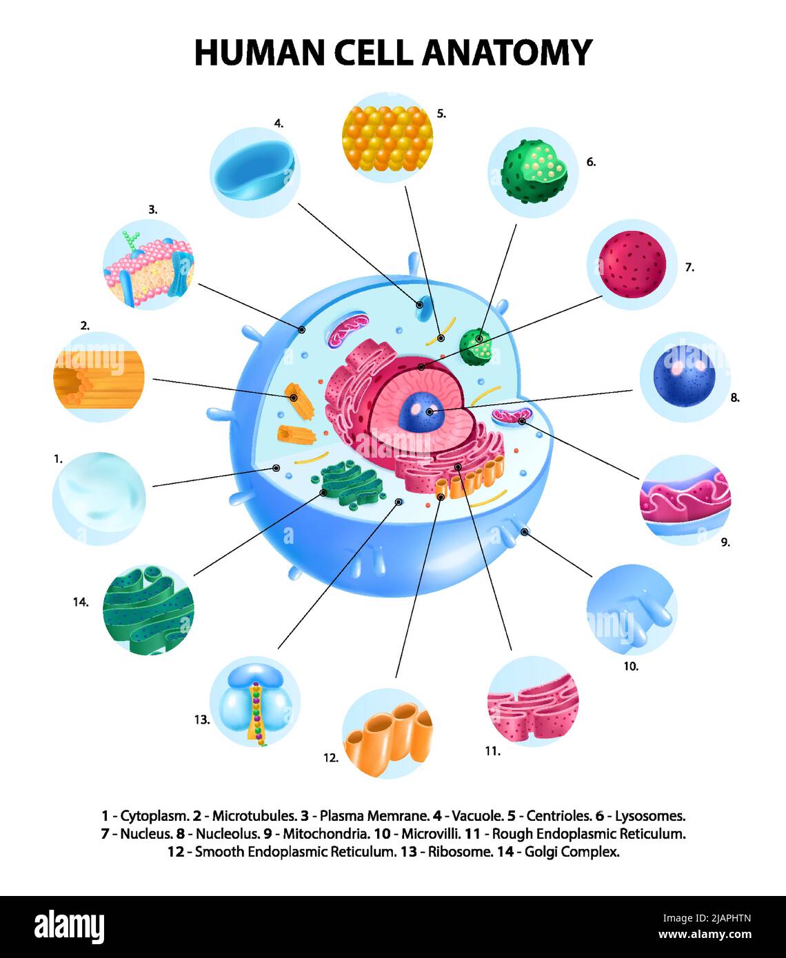 Human cell anatomy realistic infographics with labelled educational diagram on white background vector illustration Stock Vectorhttps://www.alamy.com/image-license-details/?v=1https://www.alamy.com/human-cell-anatomy-realistic-infographics-with-labelled-educational-diagram-on-white-background-vector-illustration-image471323461.html
RF2JAPHTN–Human cell anatomy realistic infographics with labelled educational diagram on white background vector illustration
 . The Biological bulletin. Biology; Zoology; Biology; Marine Biology. Excitation beam Figure 1. Schematic diagram of polarizing fucus zygote showing method of excitation of fluorescentlv labelled cell. gradients in polarized and polarizing zygotes using the fluorescent Ca2+ indicator fura-2. The spectral properties of fura-2 are such that the ratio of fluorescence following excitation with 350 (or 360) and 385 nm light is calcium- dependent. (Grynkievicz el al, 1985; Tsien el ai. 1985; Cobbold and Rink. 1987). Materials and Methods Zygote growth and dye loading Zygotes of Fucus serratus could Stock Photohttps://www.alamy.com/image-license-details/?v=1https://www.alamy.com/the-biological-bulletin-biology-zoology-biology-marine-biology-excitation-beam-figure-1-schematic-diagram-of-polarizing-fucus-zygote-showing-method-of-excitation-of-fluorescentlv-labelled-cell-gradients-in-polarized-and-polarizing-zygotes-using-the-fluorescent-ca2-indicator-fura-2-the-spectral-properties-of-fura-2-are-such-that-the-ratio-of-fluorescence-following-excitation-with-350-or-360-and-385-nm-light-is-calcium-dependent-grynkievicz-el-al-1985-tsien-el-ai-1985-cobbold-and-rink-1987-materials-and-methods-zygote-growth-and-dye-loading-zygotes-of-fucus-serratus-could-image234616061.html
RMRHKK51–. The Biological bulletin. Biology; Zoology; Biology; Marine Biology. Excitation beam Figure 1. Schematic diagram of polarizing fucus zygote showing method of excitation of fluorescentlv labelled cell. gradients in polarized and polarizing zygotes using the fluorescent Ca2+ indicator fura-2. The spectral properties of fura-2 are such that the ratio of fluorescence following excitation with 350 (or 360) and 385 nm light is calcium- dependent. (Grynkievicz el al, 1985; Tsien el ai. 1985; Cobbold and Rink. 1987). Materials and Methods Zygote growth and dye loading Zygotes of Fucus serratus could
 Cell components, illustration. A cell is made up of three parts: the cell membrane, the nucleus, and the cytoplasm that lies between the two. Stock Photohttps://www.alamy.com/image-license-details/?v=1https://www.alamy.com/cell-components-illustration-a-cell-is-made-up-of-three-parts-the-cell-membrane-the-nucleus-and-the-cytoplasm-that-lies-between-the-two-image618634084.html
RF2XXD62C–Cell components, illustration. A cell is made up of three parts: the cell membrane, the nucleus, and the cytoplasm that lies between the two.
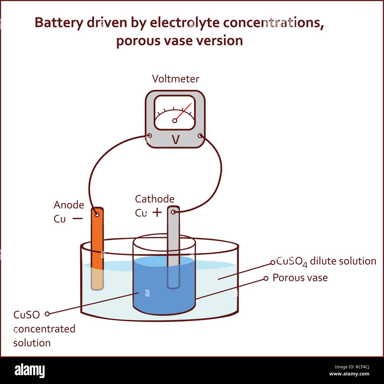 Battery driven by electrolyte concentrations porous vase version Stock Vectorhttps://www.alamy.com/image-license-details/?v=1https://www.alamy.com/battery-driven-by-electrolyte-concentrations-porous-vase-version-image231443426.html
RFRCF4CJ–Battery driven by electrolyte concentrations porous vase version
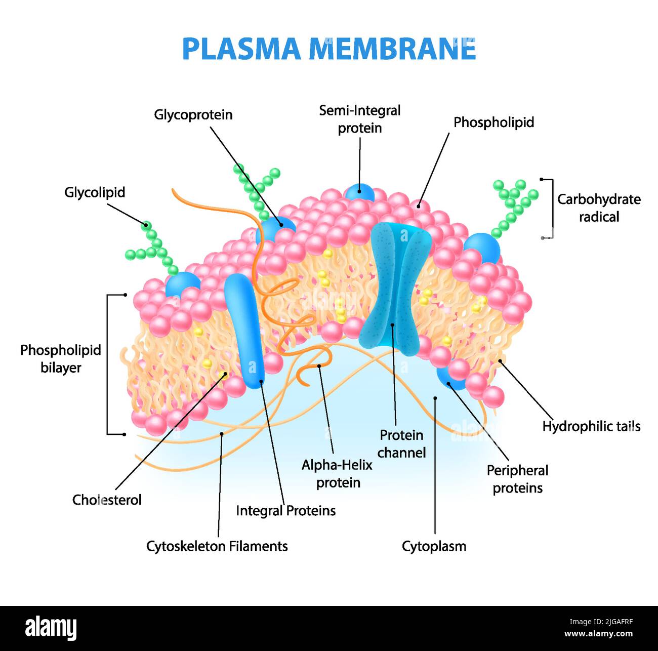 Realistic human cell anatomy infographics with diagram showing plasma membrane structure with labelled elements vector illustration Stock Vectorhttps://www.alamy.com/image-license-details/?v=1https://www.alamy.com/realistic-human-cell-anatomy-infographics-with-diagram-showing-plasma-membrane-structure-with-labelled-elements-vector-illustration-image474746371.html
RF2JGAFRF–Realistic human cell anatomy infographics with diagram showing plasma membrane structure with labelled elements vector illustration
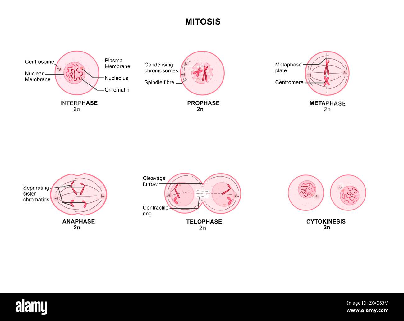 Mitosis in an animal cell, illustration. Mitosis is the process by which a cell replicates its chromosomes and then segregates them, producing two identical nuclei in preparation for cell division. Stock Photohttps://www.alamy.com/image-license-details/?v=1https://www.alamy.com/mitosis-in-an-animal-cell-illustration-mitosis-is-the-process-by-which-a-cell-replicates-its-chromosomes-and-then-segregates-them-producing-two-identical-nuclei-in-preparation-for-cell-division-image618634120.html
RF2XXD63M–Mitosis in an animal cell, illustration. Mitosis is the process by which a cell replicates its chromosomes and then segregates them, producing two identical nuclei in preparation for cell division.
 Battery driven by electrolyte concentrations porous vase version Stock Vectorhttps://www.alamy.com/image-license-details/?v=1https://www.alamy.com/battery-driven-by-electrolyte-concentrations-porous-vase-version-image231443417.html
RFRCF4C9–Battery driven by electrolyte concentrations porous vase version
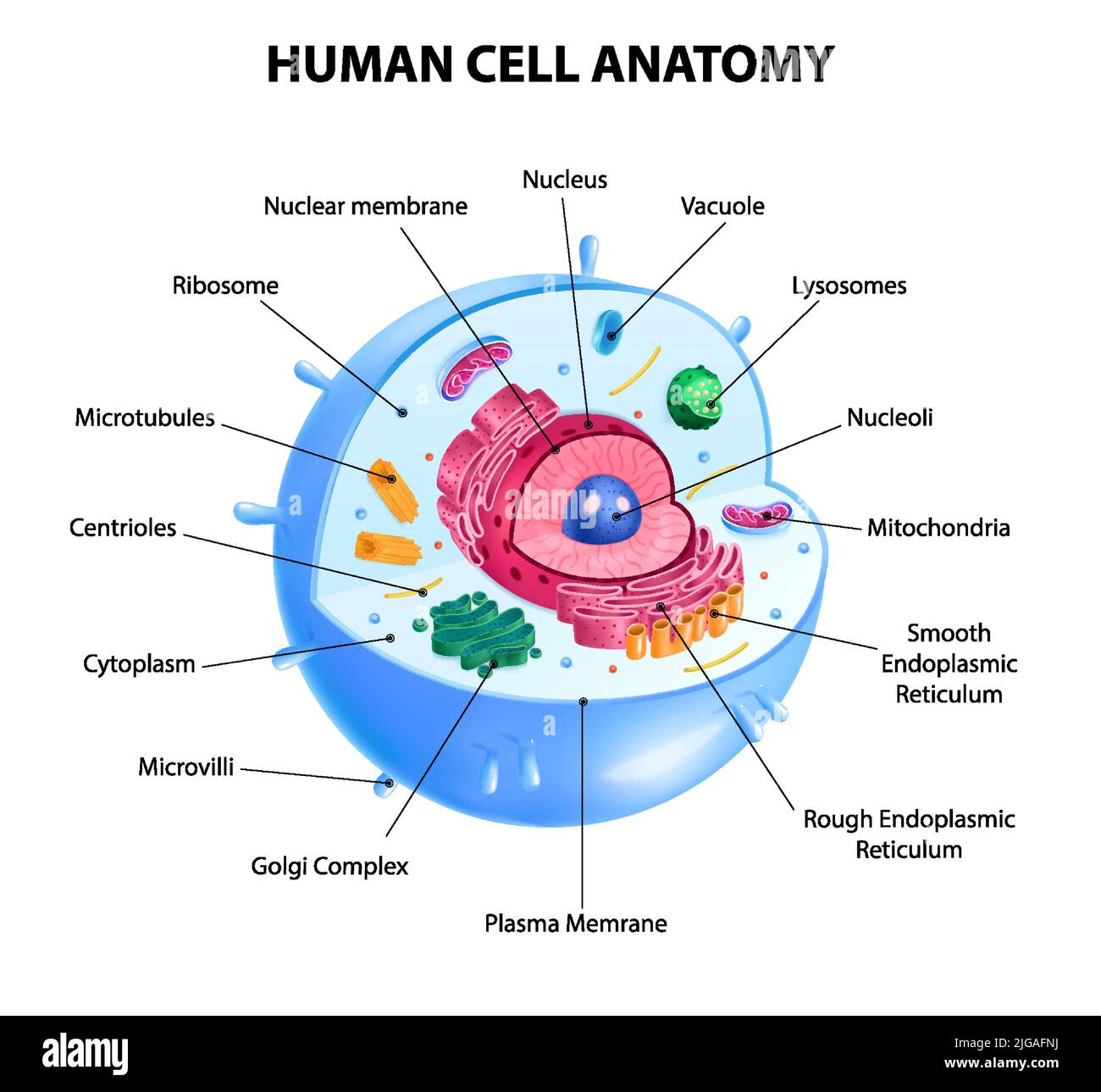 Realistic human cell anatomy diagram infographic poster vector illustration Stock Vectorhttps://www.alamy.com/image-license-details/?v=1https://www.alamy.com/realistic-human-cell-anatomy-diagram-infographic-poster-vector-illustration-image474746318.html
RF2JGAFNJ–Realistic human cell anatomy diagram infographic poster vector illustration
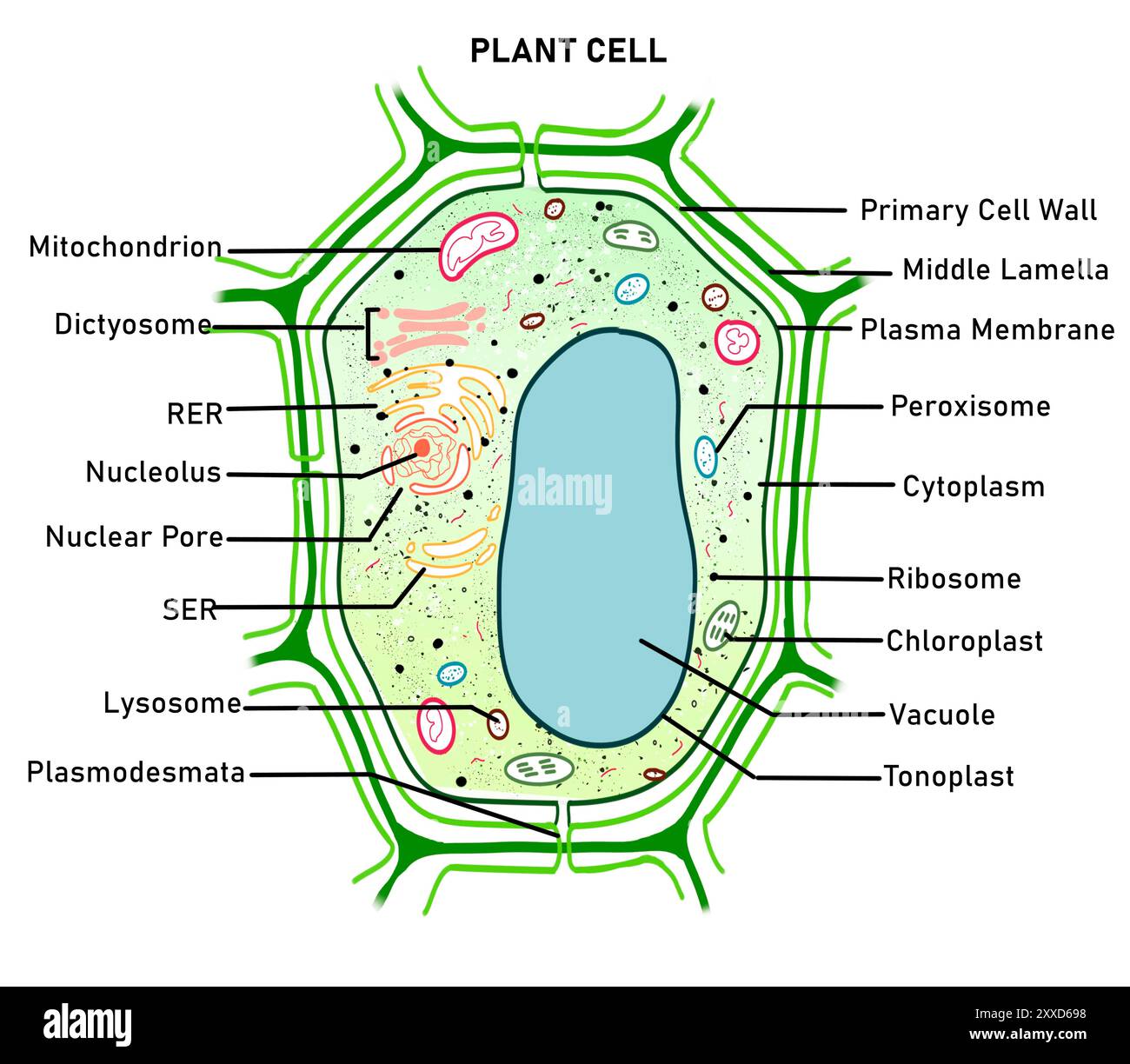 Structure of a plant cell, illustration. Plant cells differ from animal cells in the presence of specialised organelles called plastids and having a rigid cell wall, large vacuole and plasmodesmata. Stock Photohttps://www.alamy.com/image-license-details/?v=1https://www.alamy.com/structure-of-a-plant-cell-illustration-plant-cells-differ-from-animal-cells-in-the-presence-of-specialised-organelles-called-plastids-and-having-a-rigid-cell-wall-large-vacuole-and-plasmodesmata-image618634276.html
RF2XXD698–Structure of a plant cell, illustration. Plant cells differ from animal cells in the presence of specialised organelles called plastids and having a rigid cell wall, large vacuole and plasmodesmata.
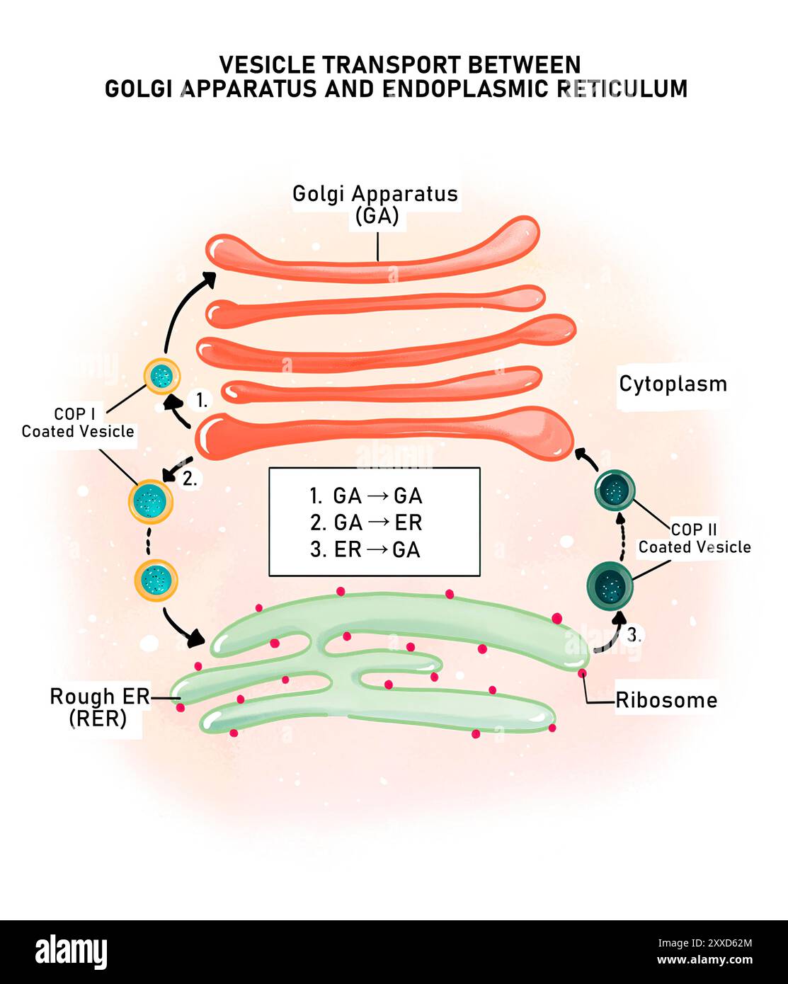 Vesicle transport in a cell, illustration. Vesicles move proteins between the Golgi apparatus and endoplasmic reticulum. The vesicles are coated with a coatomer (COP) protein that gives the vesicle an address to reach their destination. Stock Photohttps://www.alamy.com/image-license-details/?v=1https://www.alamy.com/vesicle-transport-in-a-cell-illustration-vesicles-move-proteins-between-the-golgi-apparatus-and-endoplasmic-reticulum-the-vesicles-are-coated-with-a-coatomer-cop-protein-that-gives-the-vesicle-an-address-to-reach-their-destination-image618634092.html
RF2XXD62M–Vesicle transport in a cell, illustration. Vesicles move proteins between the Golgi apparatus and endoplasmic reticulum. The vesicles are coated with a coatomer (COP) protein that gives the vesicle an address to reach their destination.
 Cotranslational protein targeting, illustration. Protein targeting is the process of targeting newly synthesised proteins to endoplasmic Reticulum lumen so that the proteins can be sorted for their final destination in or outside the cell. In the above diagram, In the above diagram, Co-translational (while the protein is being synthesised) targeting is seen for a secretory protein where the protein translation occurs simultaneously to it entering ER lumen. Stock Photohttps://www.alamy.com/image-license-details/?v=1https://www.alamy.com/cotranslational-protein-targeting-illustration-protein-targeting-is-the-process-of-targeting-newly-synthesised-proteins-to-endoplasmic-reticulum-lumen-so-that-the-proteins-can-be-sorted-for-their-final-destination-in-or-outside-the-cell-in-the-above-diagram-in-the-above-diagram-co-translational-while-the-protein-is-being-synthesised-targeting-is-seen-for-a-secretory-protein-where-the-protein-translation-occurs-simultaneously-to-it-entering-er-lumen-image618634058.html
RF2XXD61E–Cotranslational protein targeting, illustration. Protein targeting is the process of targeting newly synthesised proteins to endoplasmic Reticulum lumen so that the proteins can be sorted for their final destination in or outside the cell. In the above diagram, In the above diagram, Co-translational (while the protein is being synthesised) targeting is seen for a secretory protein where the protein translation occurs simultaneously to it entering ER lumen.
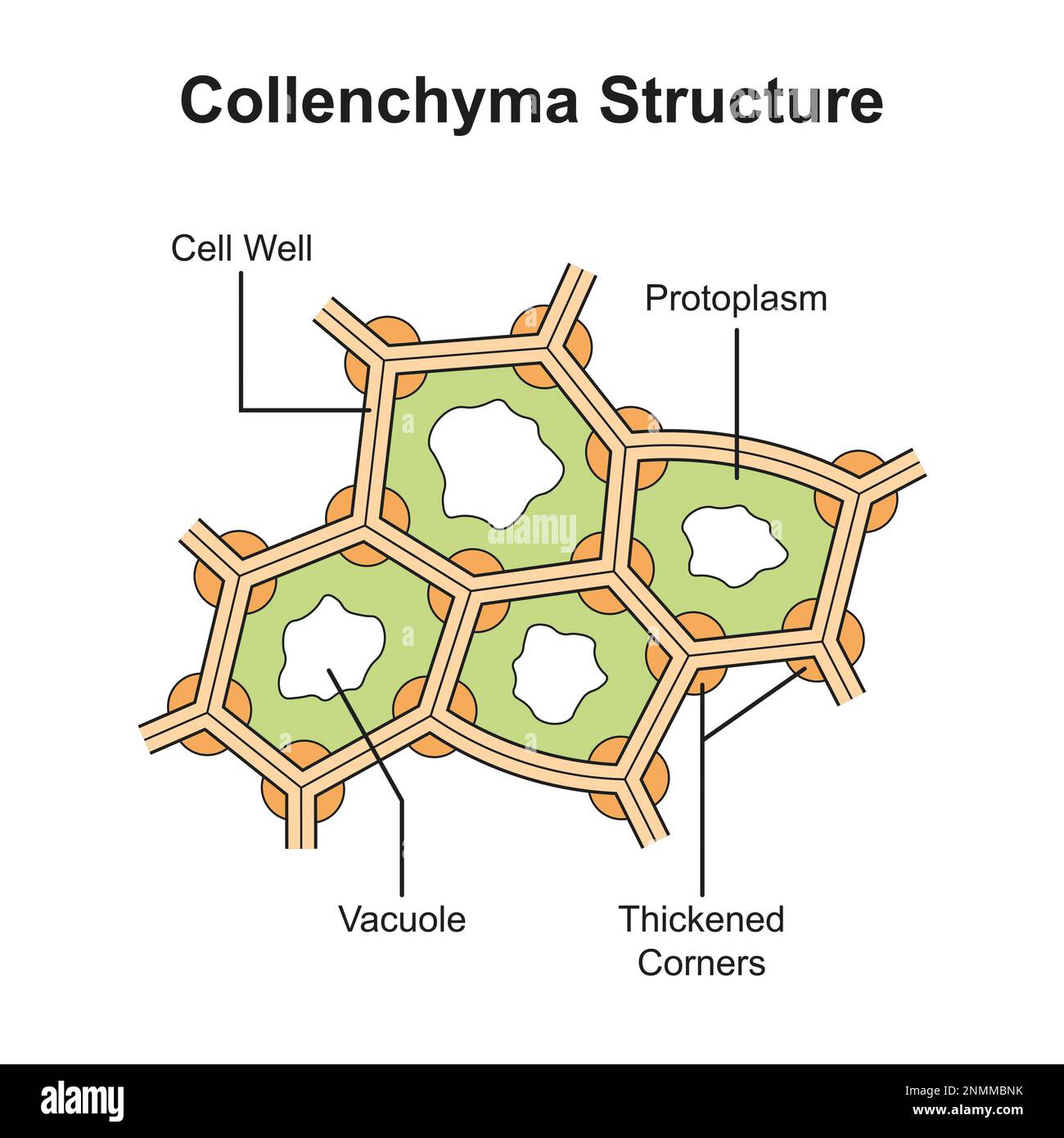 Collenchyma structure, illustration Stock Photohttps://www.alamy.com/image-license-details/?v=1https://www.alamy.com/collenchyma-structure-illustration-image529052431.html
RF2NMMBNK–Collenchyma structure, illustration
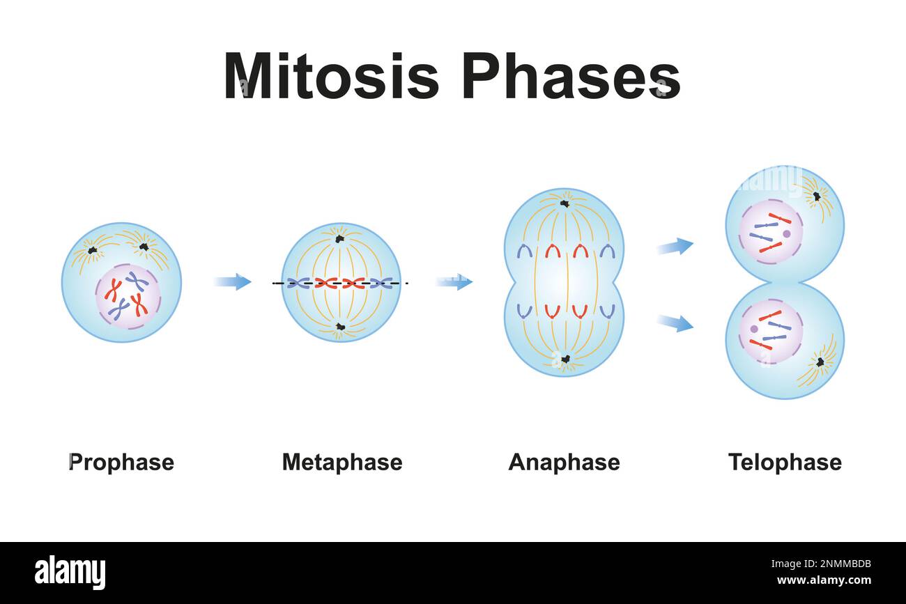 Mitosis phases, illustration Stock Photohttps://www.alamy.com/image-license-details/?v=1https://www.alamy.com/mitosis-phases-illustration-image529052199.html
RF2NMMBDB–Mitosis phases, illustration
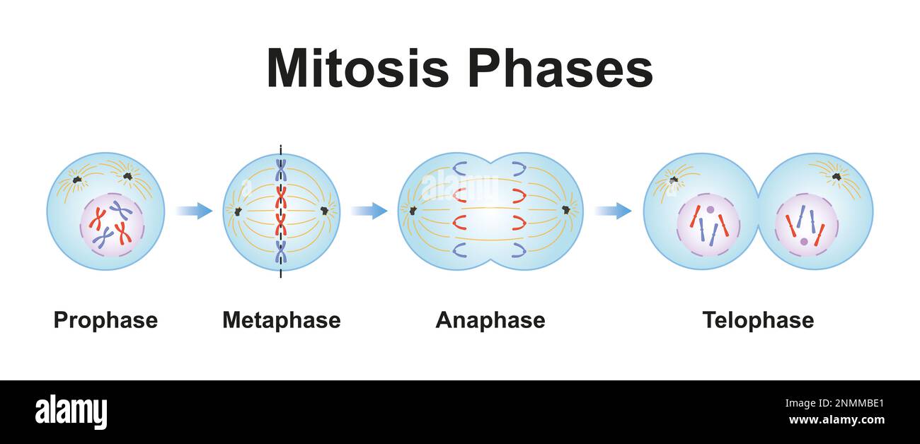 Mitosis phases, illustration Stock Photohttps://www.alamy.com/image-license-details/?v=1https://www.alamy.com/mitosis-phases-illustration-image529052217.html
RF2NMMBE1–Mitosis phases, illustration
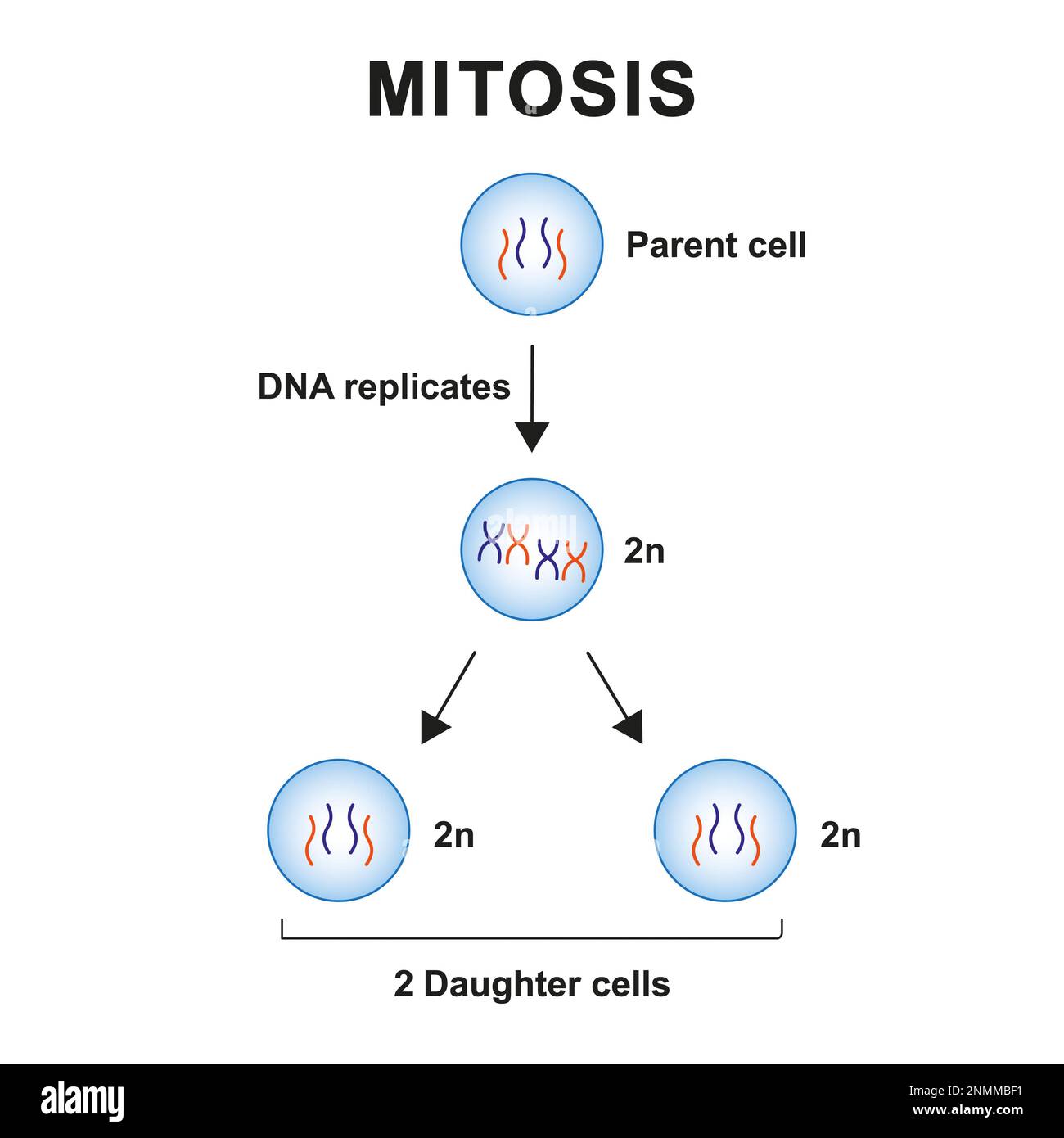 Mitosis phases, illustration Stock Photohttps://www.alamy.com/image-license-details/?v=1https://www.alamy.com/mitosis-phases-illustration-image529052245.html
RF2NMMBF1–Mitosis phases, illustration
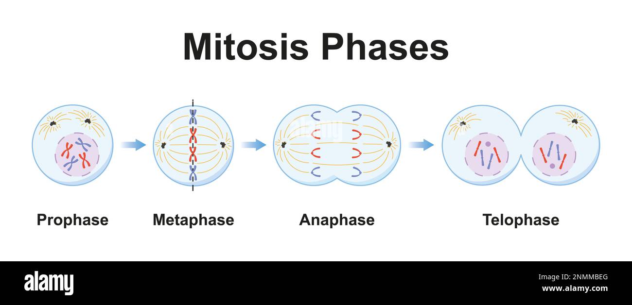 Mitosis phases, illustration Stock Photohttps://www.alamy.com/image-license-details/?v=1https://www.alamy.com/mitosis-phases-illustration-image529052232.html
RF2NMMBEG–Mitosis phases, illustration
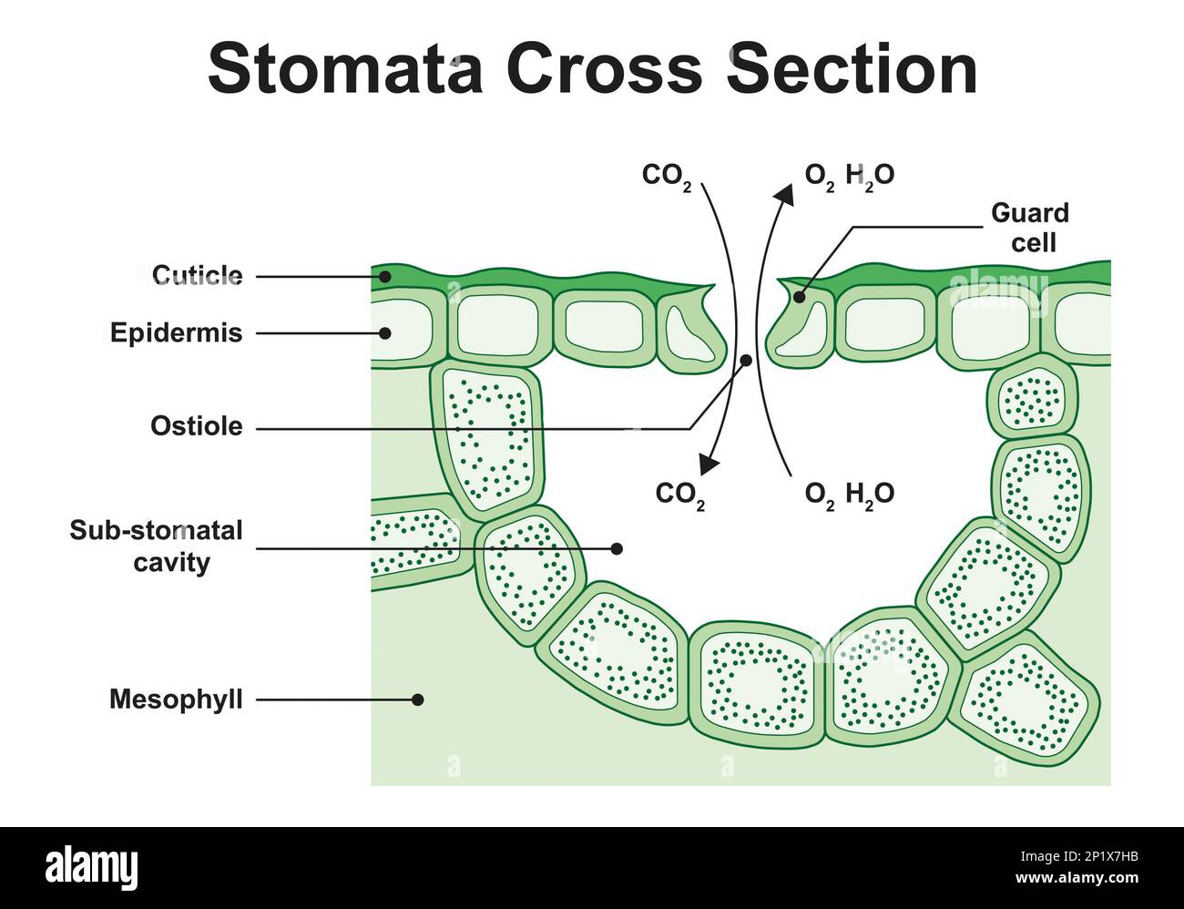 Stoma structure, illustration Stock Photohttps://www.alamy.com/image-license-details/?v=1https://www.alamy.com/stoma-structure-illustration-image534712791.html
RF2P1X7HB–Stoma structure, illustration
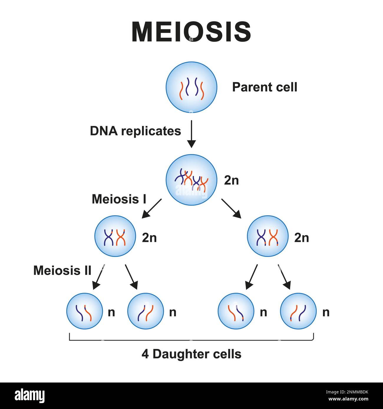 Meiosis phases, illustration Stock Photohttps://www.alamy.com/image-license-details/?v=1https://www.alamy.com/meiosis-phases-illustration-image529052207.html
RF2NMMBDK–Meiosis phases, illustration
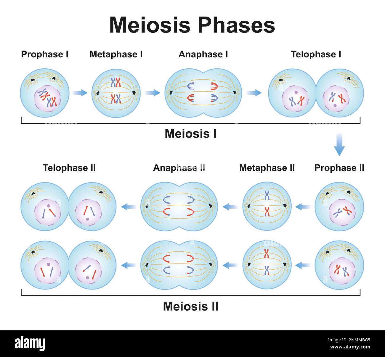 Meiosis phases, illustration Stock Photohttps://www.alamy.com/image-license-details/?v=1https://www.alamy.com/meiosis-phases-illustration-image529052277.html
RF2NMMBG5–Meiosis phases, illustration
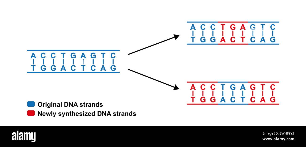 Dispersive replication of DNA , illustration. Stock Photohttps://www.alamy.com/image-license-details/?v=1https://www.alamy.com/dispersive-replication-of-dna-illustration-image595938759.html
RF2WHF9Y3–Dispersive replication of DNA , illustration.
 Dispersive replication of DNA , illustration. Stock Photohttps://www.alamy.com/image-license-details/?v=1https://www.alamy.com/dispersive-replication-of-dna-illustration-image595938800.html
RF2WHFA0G–Dispersive replication of DNA , illustration.
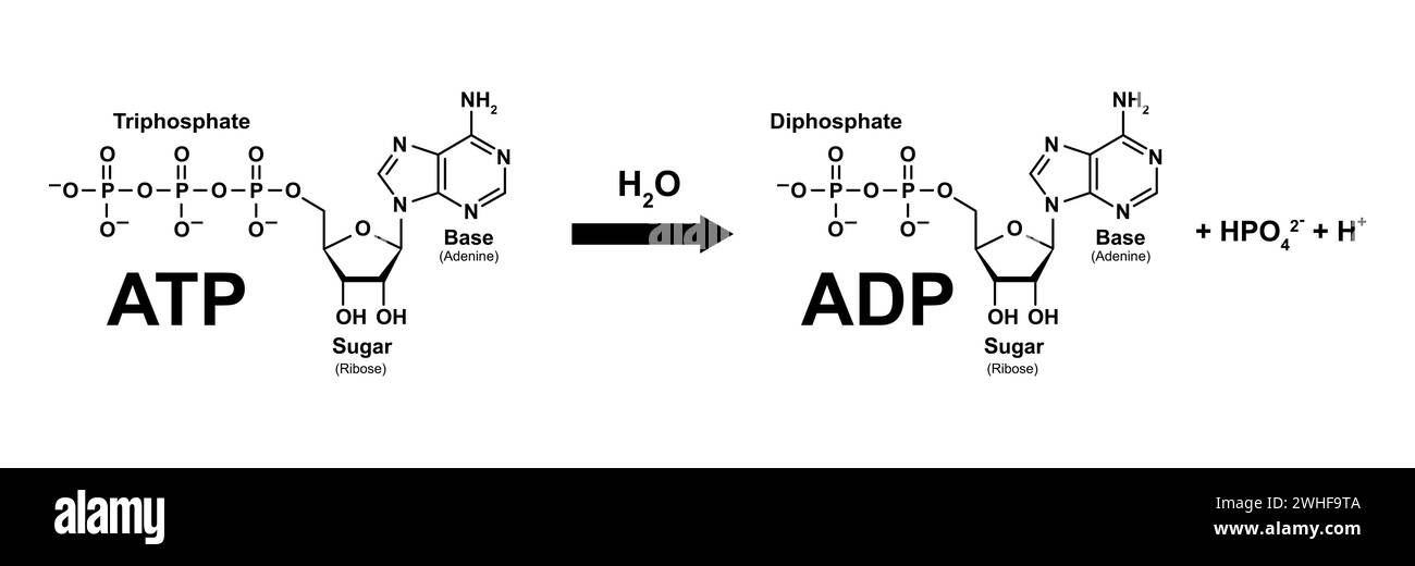 ATP-ADP cycle, illustration Stock Photohttps://www.alamy.com/image-license-details/?v=1https://www.alamy.com/atp-adp-cycle-illustration-image595938682.html
RF2WHF9TA–ATP-ADP cycle, illustration
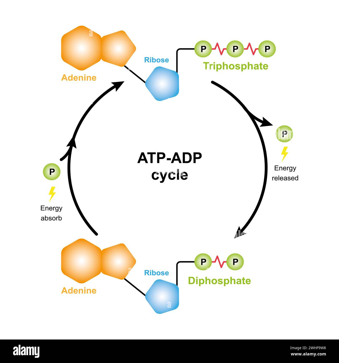 ATP-ADP cycle, illustration Stock Photohttps://www.alamy.com/image-license-details/?v=1https://www.alamy.com/atp-adp-cycle-illustration-image595938708.html
RF2WHF9W8–ATP-ADP cycle, illustration
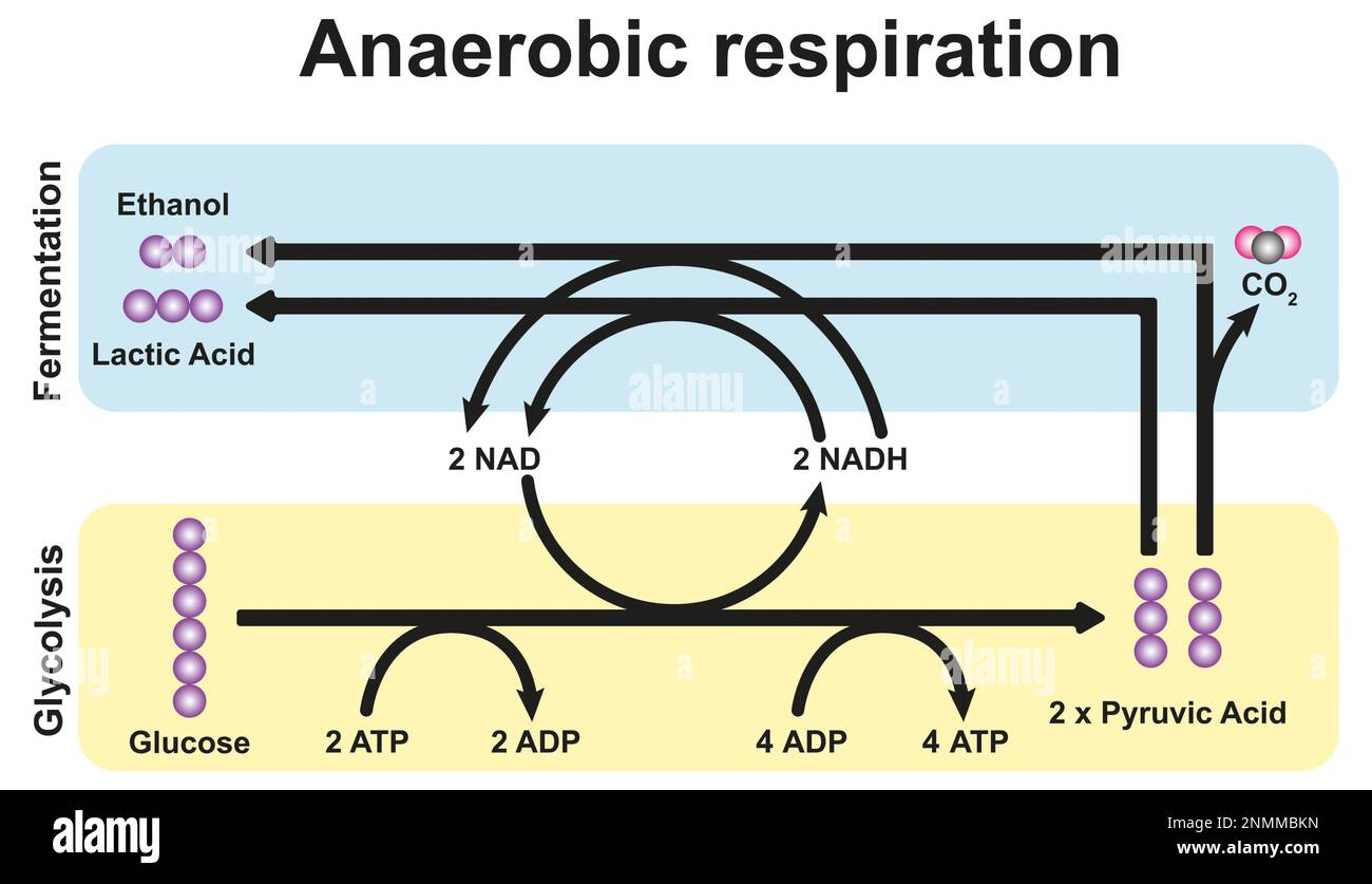 Anaerobic respiration, illustration Stock Photohttps://www.alamy.com/image-license-details/?v=1https://www.alamy.com/anaerobic-respiration-illustration-image529052377.html
RF2NMMBKN–Anaerobic respiration, illustration
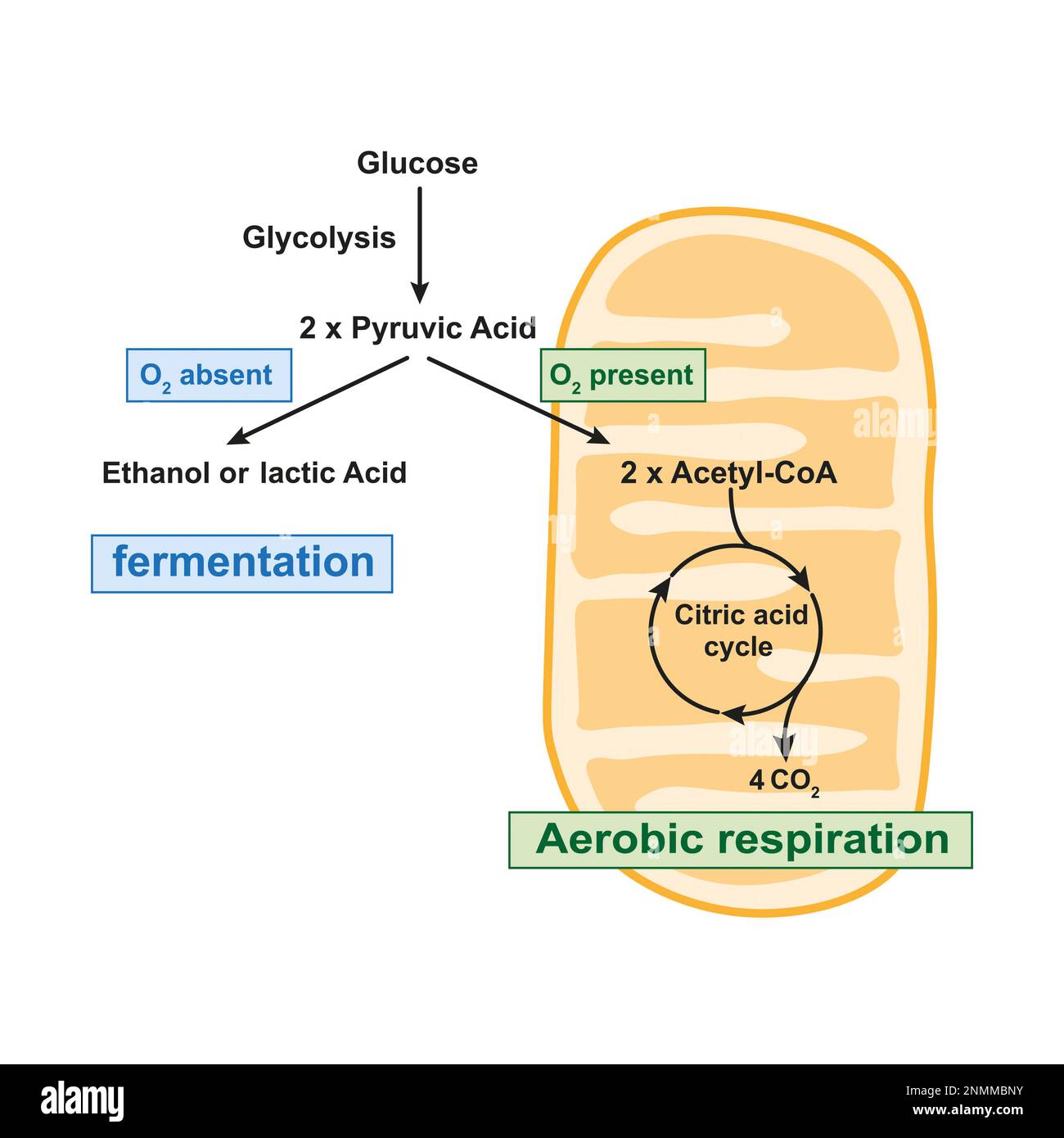 Aerobic respiration and anaerobic fermentation, illustration Stock Photohttps://www.alamy.com/image-license-details/?v=1https://www.alamy.com/aerobic-respiration-and-anaerobic-fermentation-illustration-image529052439.html
RF2NMMBNY–Aerobic respiration and anaerobic fermentation, illustration
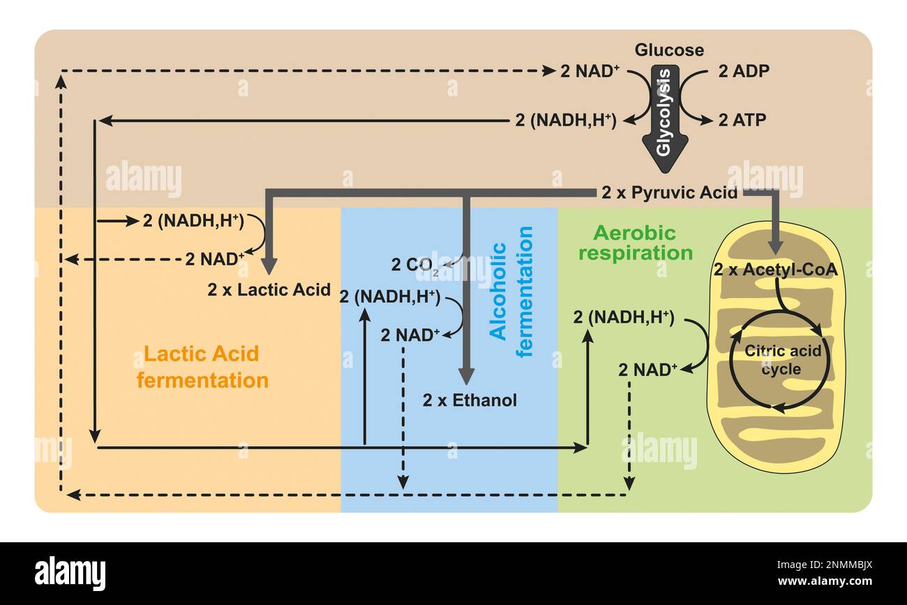 Aerobic respiration and anaerobic fermentation, illustration Stock Photohttps://www.alamy.com/image-license-details/?v=1https://www.alamy.com/aerobic-respiration-and-anaerobic-fermentation-illustration-image529052354.html
RF2NMMBJX–Aerobic respiration and anaerobic fermentation, illustration
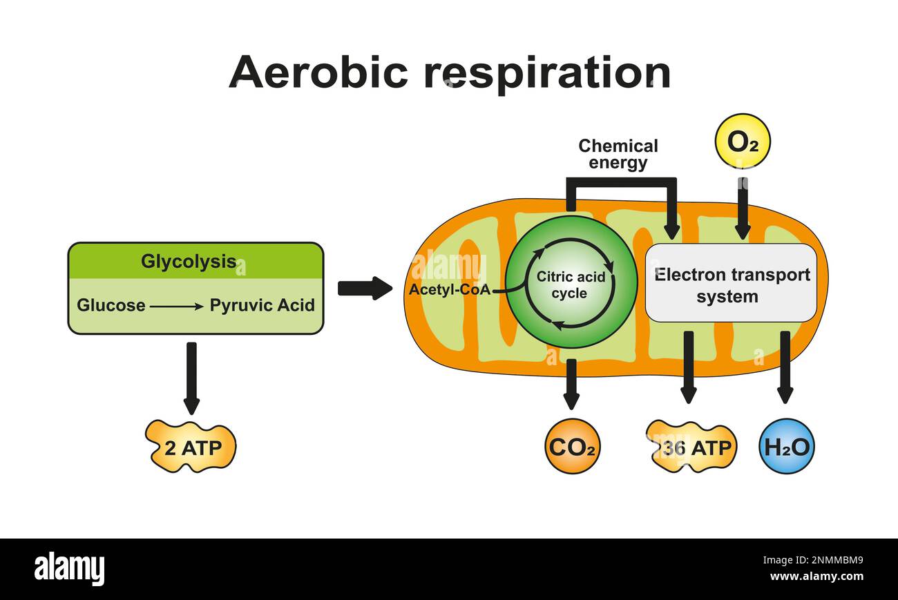 Aerobic respiration, illustration Stock Photohttps://www.alamy.com/image-license-details/?v=1https://www.alamy.com/aerobic-respiration-illustration-image529052393.html
RF2NMMBM9–Aerobic respiration, illustration
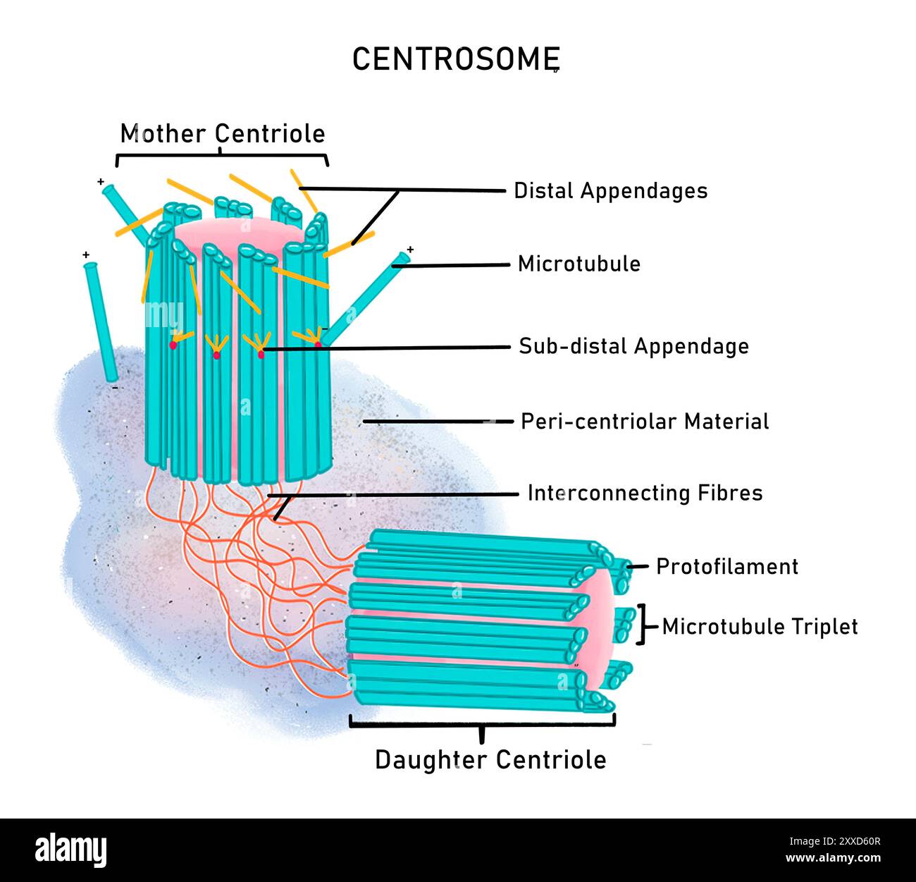 Structure of a centrosome, illustration. The centrosome functions as the main microtubule organising centre (MTOC) of the animal cell. It is structurally composed of two centrioles (mother centriole and daughter centriole), interconnecting fibres and pericentriolar material. Stock Photohttps://www.alamy.com/image-license-details/?v=1https://www.alamy.com/structure-of-a-centrosome-illustration-the-centrosome-functions-as-the-main-microtubule-organising-centre-mtoc-of-the-animal-cell-it-is-structurally-composed-of-two-centrioles-mother-centriole-and-daughter-centriole-interconnecting-fibres-and-pericentriolar-material-image618634039.html
RF2XXD60R–Structure of a centrosome, illustration. The centrosome functions as the main microtubule organising centre (MTOC) of the animal cell. It is structurally composed of two centrioles (mother centriole and daughter centriole), interconnecting fibres and pericentriolar material.
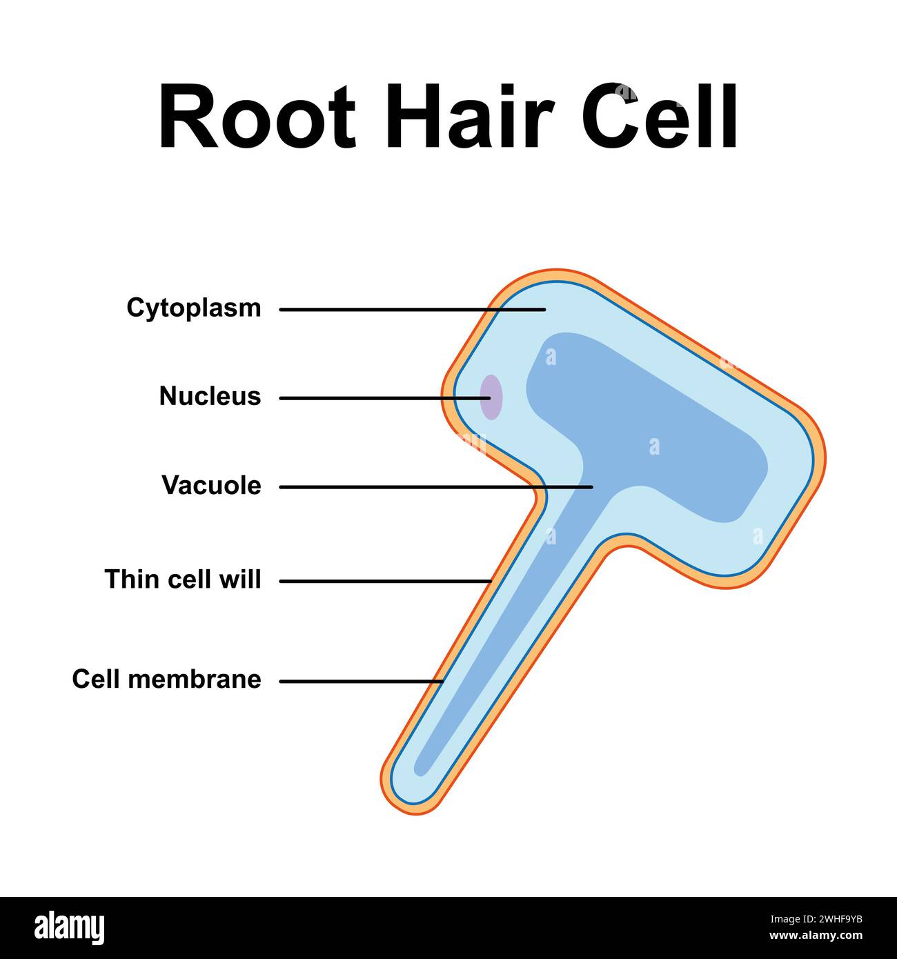 Root hair cell, illustration Stock Photohttps://www.alamy.com/image-license-details/?v=1https://www.alamy.com/root-hair-cell-illustration-image595938767.html
RF2WHF9YB–Root hair cell, illustration
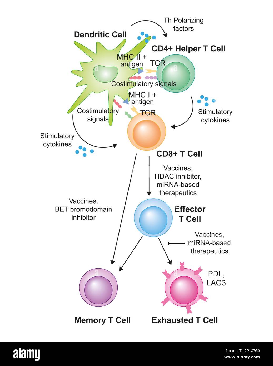 T cell activation, illustration Stock Photohttps://www.alamy.com/image-license-details/?v=1https://www.alamy.com/t-cell-activation-illustration-image534712752.html
RF2P1X7G0–T cell activation, illustration
 Intermediate filaments, illustration. Intermediate filaments are part of the cytoskeleton, which maintains cell shape and the motility of organelles. Stock Photohttps://www.alamy.com/image-license-details/?v=1https://www.alamy.com/intermediate-filaments-illustration-intermediate-filaments-are-part-of-the-cytoskeleton-which-maintains-cell-shape-and-the-motility-of-organelles-image618634080.html
RF2XXD628–Intermediate filaments, illustration. Intermediate filaments are part of the cytoskeleton, which maintains cell shape and the motility of organelles.
 Hypotonic, isotonic and hypertonic solutions, illustration. In a hypotonic solution, water moves into the cell and in a hypertonic solution water moves out of it. Stock Photohttps://www.alamy.com/image-license-details/?v=1https://www.alamy.com/hypotonic-isotonic-and-hypertonic-solutions-illustration-in-a-hypotonic-solution-water-moves-into-the-cell-and-in-a-hypertonic-solution-water-moves-out-of-it-image618634101.html
RF2XXD631–Hypotonic, isotonic and hypertonic solutions, illustration. In a hypotonic solution, water moves into the cell and in a hypertonic solution water moves out of it.
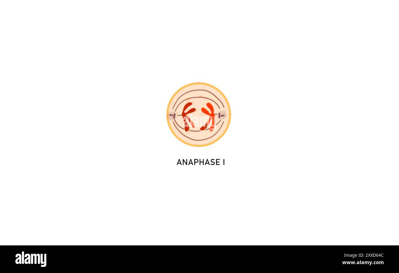 Meiosis I anaphase I, illustration. During anaphase I the chromosomes move to the opposite poles of the cell. Stock Photohttps://www.alamy.com/image-license-details/?v=1https://www.alamy.com/meiosis-i-anaphase-i-illustration-during-anaphase-i-the-chromosomes-move-to-the-opposite-poles-of-the-cell-image618634140.html
RF2XXD64C–Meiosis I anaphase I, illustration. During anaphase I the chromosomes move to the opposite poles of the cell.
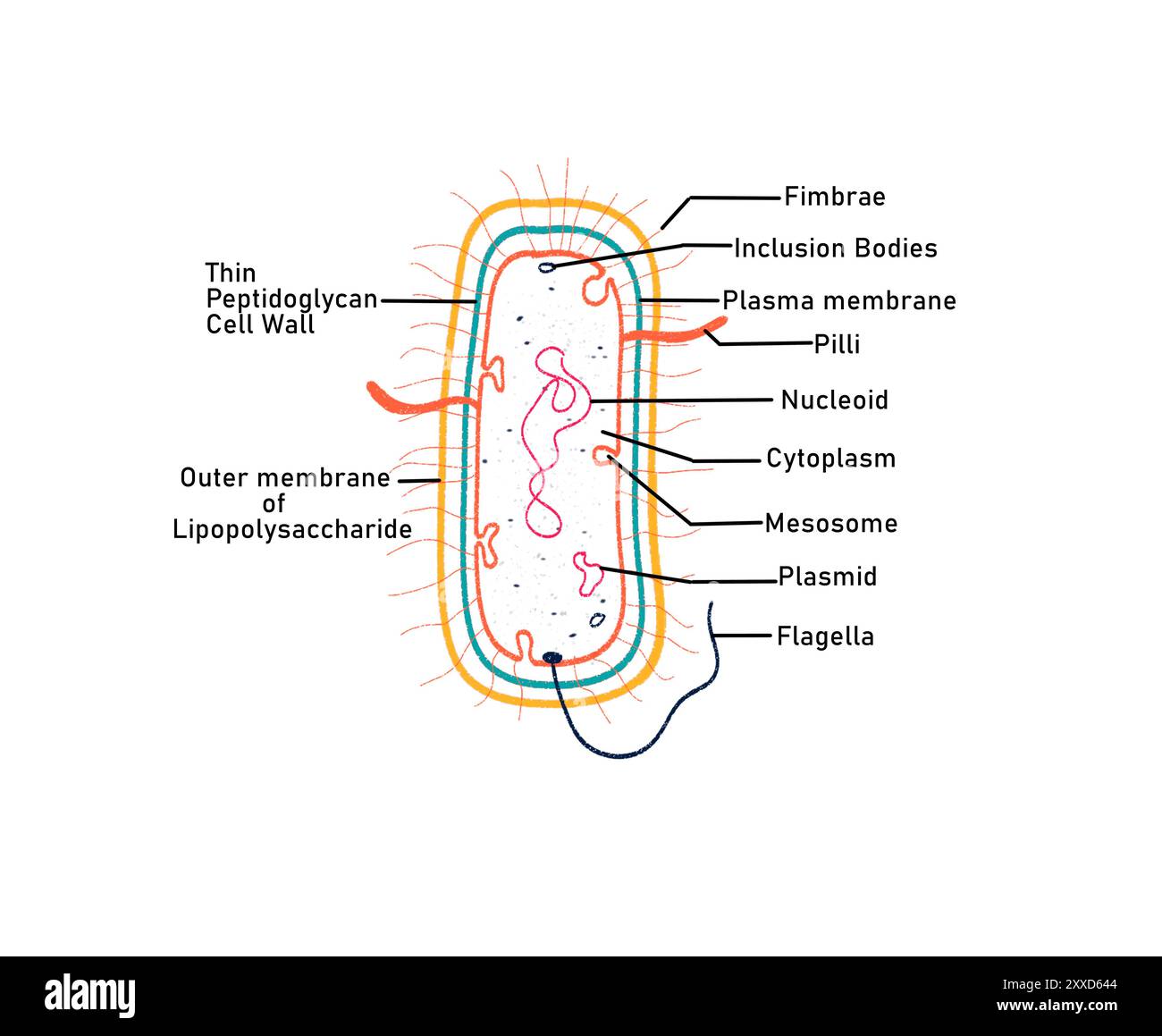 Gram negative bacterium, illustration. Gram negative bacteria are characterised by the presence of a thin peptidoglycan cell wall. Stock Photohttps://www.alamy.com/image-license-details/?v=1https://www.alamy.com/gram-negative-bacterium-illustration-gram-negative-bacteria-are-characterised-by-the-presence-of-a-thin-peptidoglycan-cell-wall-image618634132.html
RF2XXD644–Gram negative bacterium, illustration. Gram negative bacteria are characterised by the presence of a thin peptidoglycan cell wall.
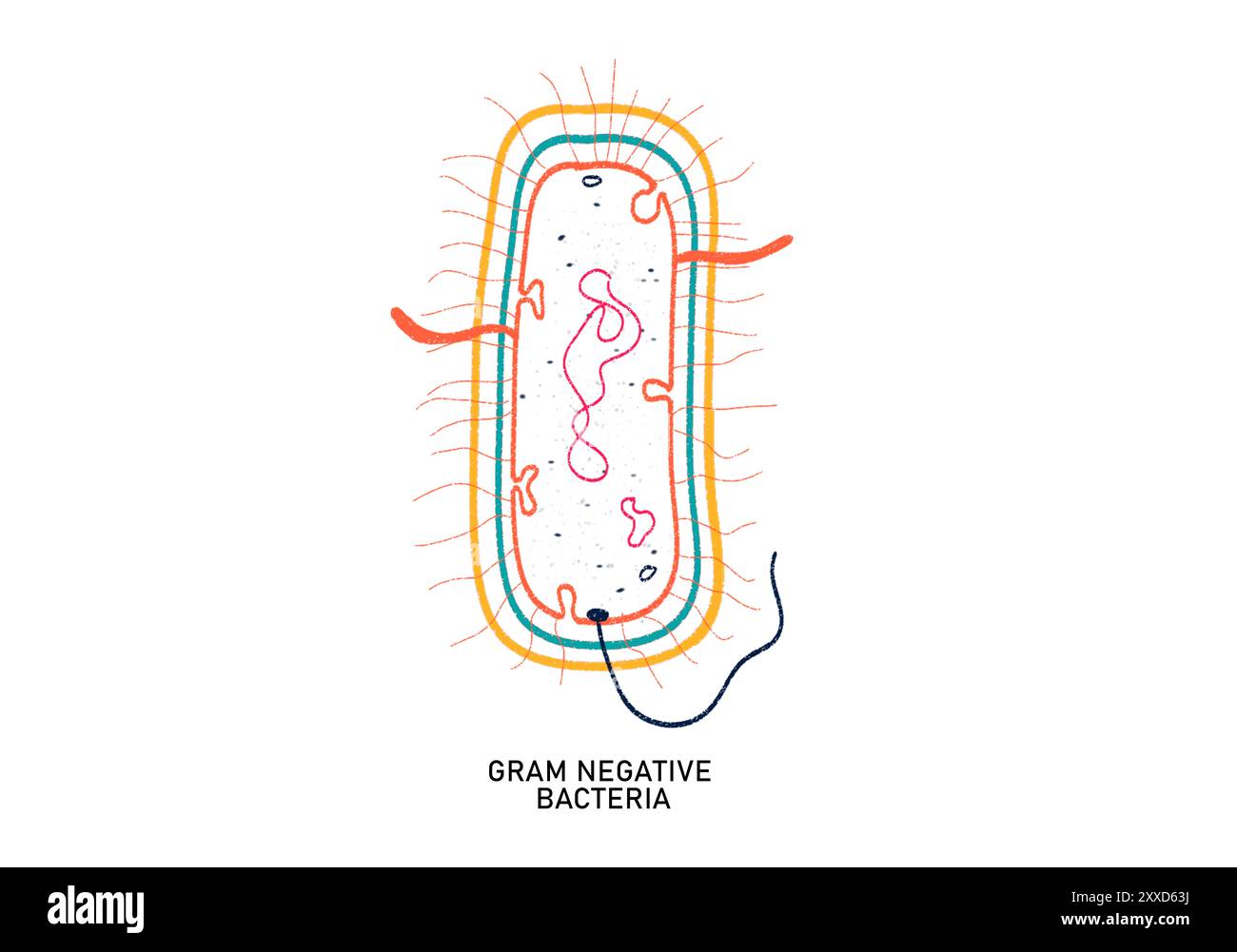 Gram negative bacterium, illustration. Gram negative bacteria are characterised by the presence of a thin peptidoglycan cell wall. Stock Photohttps://www.alamy.com/image-license-details/?v=1https://www.alamy.com/gram-negative-bacterium-illustration-gram-negative-bacteria-are-characterised-by-the-presence-of-a-thin-peptidoglycan-cell-wall-image618634118.html
RF2XXD63J–Gram negative bacterium, illustration. Gram negative bacteria are characterised by the presence of a thin peptidoglycan cell wall.
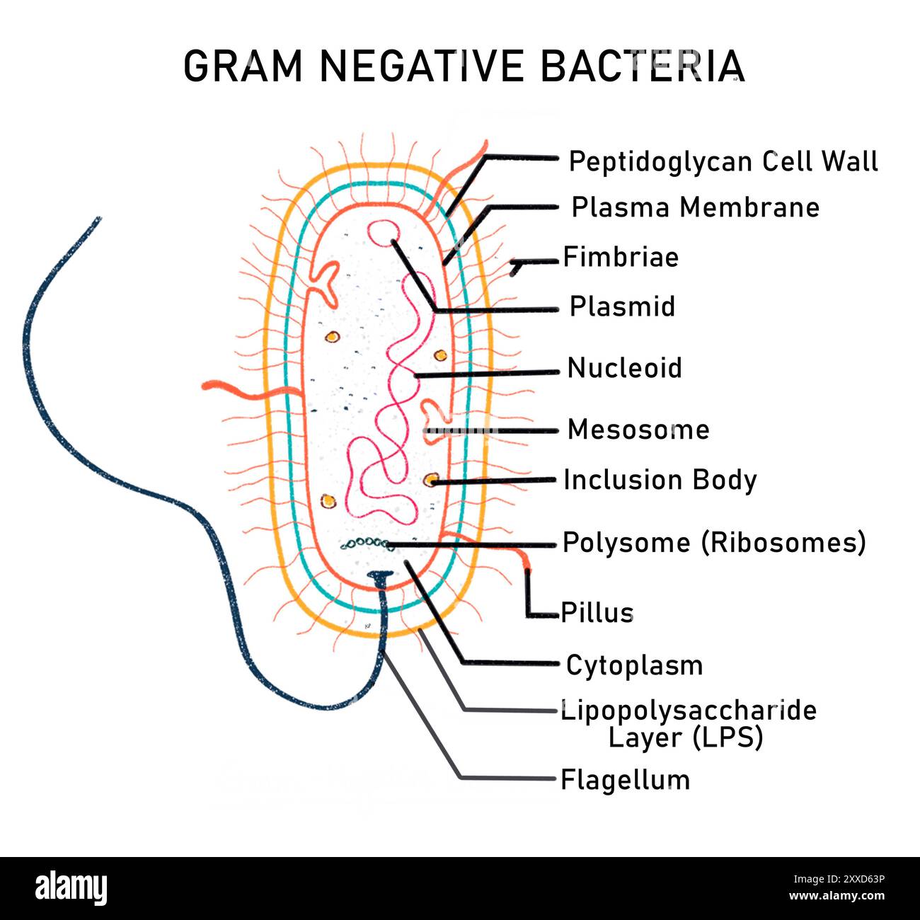 Gram negative bacterium, illustration. Gram negative bacteria are characterised by the presence of a thin peptidoglycan cell wall. Stock Photohttps://www.alamy.com/image-license-details/?v=1https://www.alamy.com/gram-negative-bacterium-illustration-gram-negative-bacteria-are-characterised-by-the-presence-of-a-thin-peptidoglycan-cell-wall-image618634122.html
RF2XXD63P–Gram negative bacterium, illustration. Gram negative bacteria are characterised by the presence of a thin peptidoglycan cell wall.
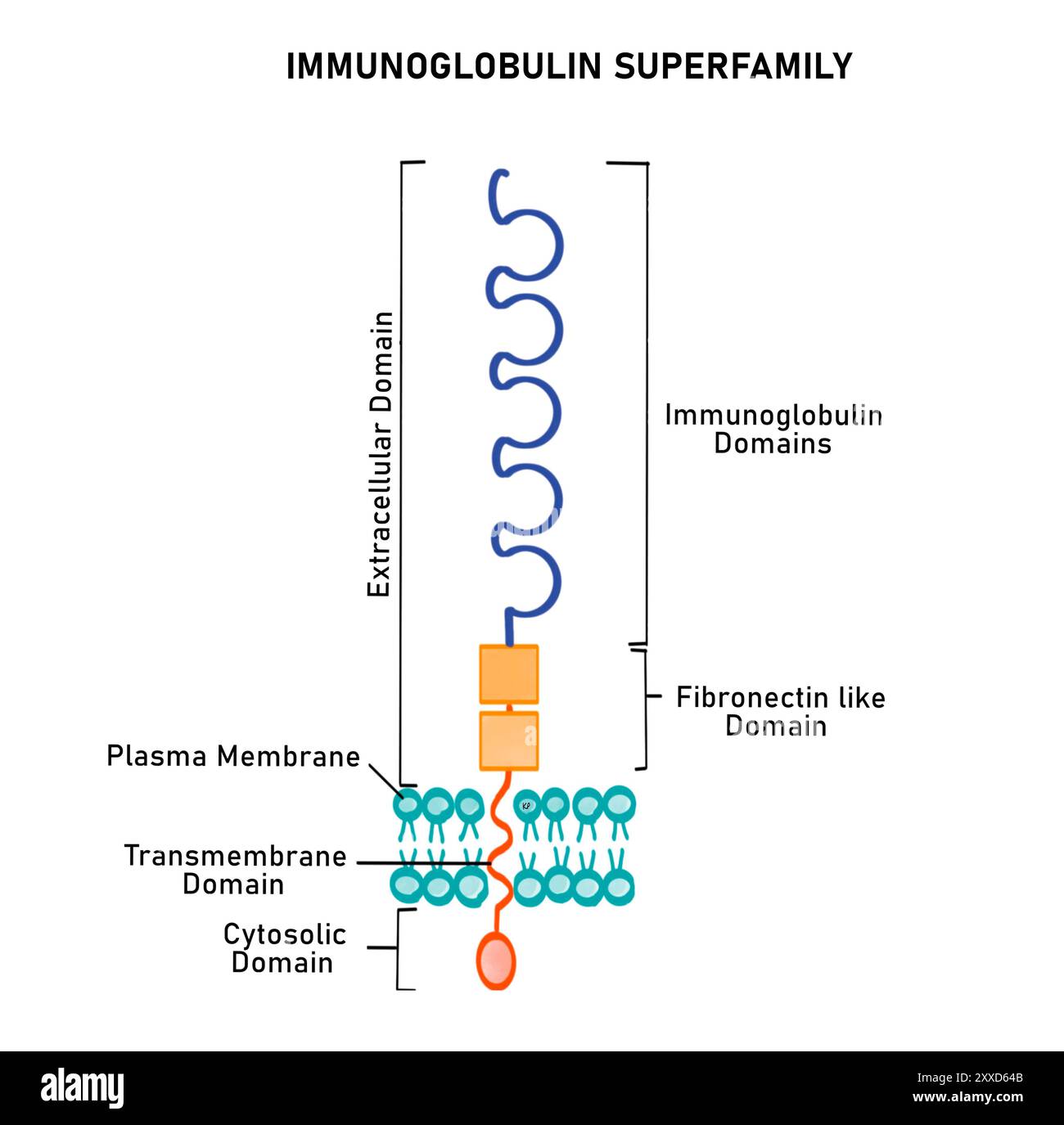 Immunoglobulin superfamily, illustration. The immunoglobulin superfamily (IgSF) is a large family of integral proteins that help in cell recognition and cell binding. IgSFs have immunoglobin domains and fibronectin type domain. Stock Photohttps://www.alamy.com/image-license-details/?v=1https://www.alamy.com/immunoglobulin-superfamily-illustration-the-immunoglobulin-superfamily-igsf-is-a-large-family-of-integral-proteins-that-help-in-cell-recognition-and-cell-binding-igsfs-have-immunoglobin-domains-and-fibronectin-type-domain-image618634139.html
RF2XXD64B–Immunoglobulin superfamily, illustration. The immunoglobulin superfamily (IgSF) is a large family of integral proteins that help in cell recognition and cell binding. IgSFs have immunoglobin domains and fibronectin type domain.
 PXo bodies, illustration. PXo bodies are a newly discovered cell organelle. Stock Photohttps://www.alamy.com/image-license-details/?v=1https://www.alamy.com/pxo-bodies-illustration-pxo-bodies-are-a-newly-discovered-cell-organelle-image618634293.html
RF2XXD69W–PXo bodies, illustration. PXo bodies are a newly discovered cell organelle.
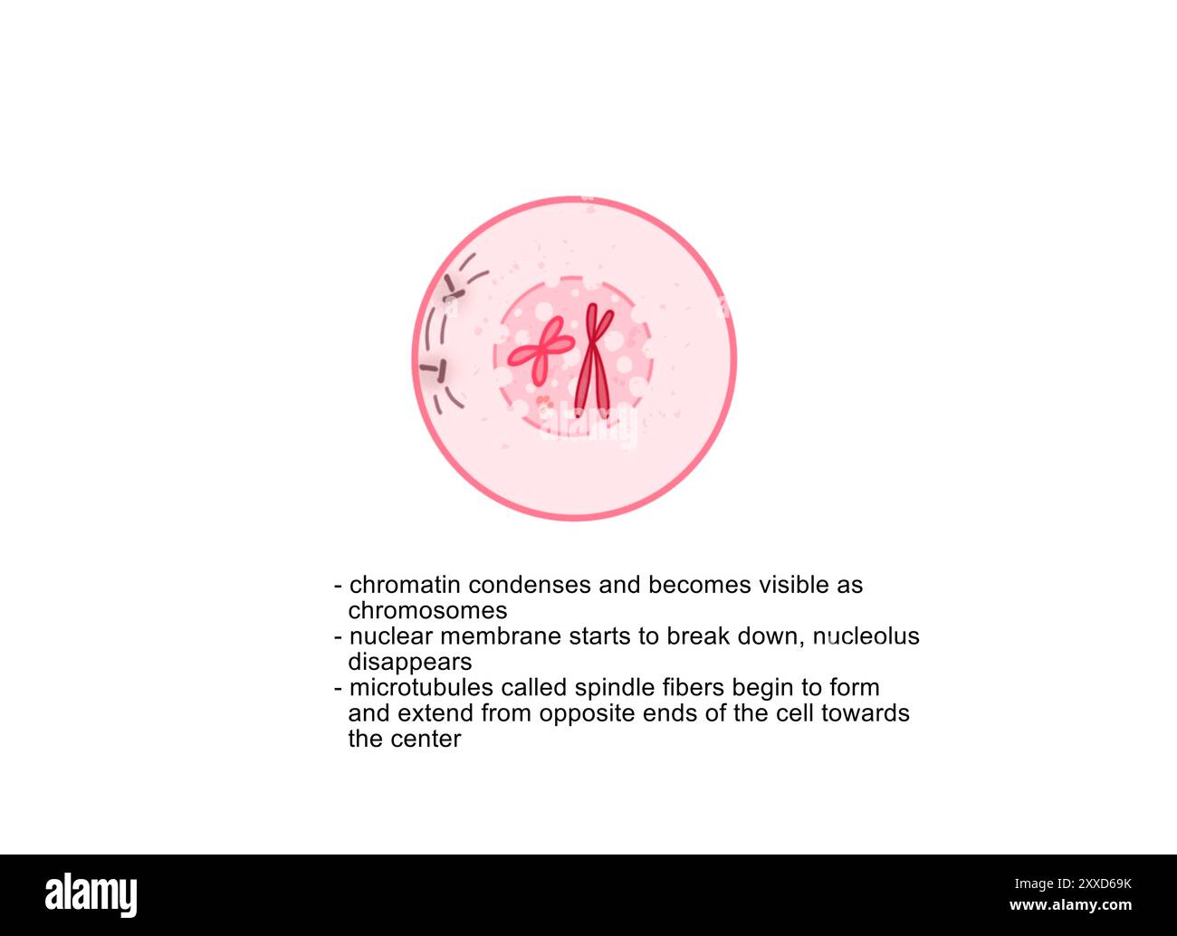 Prophase, illustration. Prophase is the first phase of mitosis, the process that separates the duplicated genetic material carried in the nucleus of a parent cell into two identical daughter cells. Stock Photohttps://www.alamy.com/image-license-details/?v=1https://www.alamy.com/prophase-illustration-prophase-is-the-first-phase-of-mitosis-the-process-that-separates-the-duplicated-genetic-material-carried-in-the-nucleus-of-a-parent-cell-into-two-identical-daughter-cells-image618634287.html
RF2XXD69K–Prophase, illustration. Prophase is the first phase of mitosis, the process that separates the duplicated genetic material carried in the nucleus of a parent cell into two identical daughter cells.
 Meiosis II telophase II, illustration. Each daughter cell is haploid (has half the total number of chromosomes of the original cell). Stock Photohttps://www.alamy.com/image-license-details/?v=1https://www.alamy.com/meiosis-ii-telophase-ii-illustration-each-daughter-cell-is-haploid-has-half-the-total-number-of-chromosomes-of-the-original-cell-image618634143.html
RF2XXD64F–Meiosis II telophase II, illustration. Each daughter cell is haploid (has half the total number of chromosomes of the original cell).
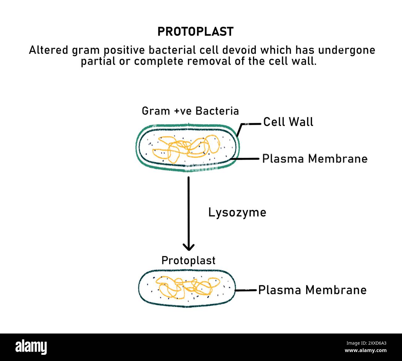 Protoplast, illustration. The protoplast is formed by degradation of the cell wall in a Gram positive bacteria. Stock Photohttps://www.alamy.com/image-license-details/?v=1https://www.alamy.com/protoplast-illustration-the-protoplast-is-formed-by-degradation-of-the-cell-wall-in-a-gram-positive-bacteria-image618634299.html
RF2XXD6A3–Protoplast, illustration. The protoplast is formed by degradation of the cell wall in a Gram positive bacteria.
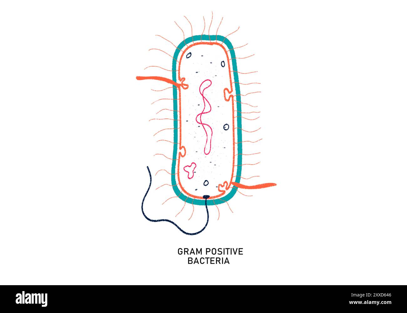 Gram positive bacterium, illustration. Gram positive bacteria are characterised by the presence of a multilayered peptidoglycan cell wall that appears purple after Gram Staining. Stock Photohttps://www.alamy.com/image-license-details/?v=1https://www.alamy.com/gram-positive-bacterium-illustration-gram-positive-bacteria-are-characterised-by-the-presence-of-a-multilayered-peptidoglycan-cell-wall-that-appears-purple-after-gram-staining-image618634134.html
RF2XXD646–Gram positive bacterium, illustration. Gram positive bacteria are characterised by the presence of a multilayered peptidoglycan cell wall that appears purple after Gram Staining.
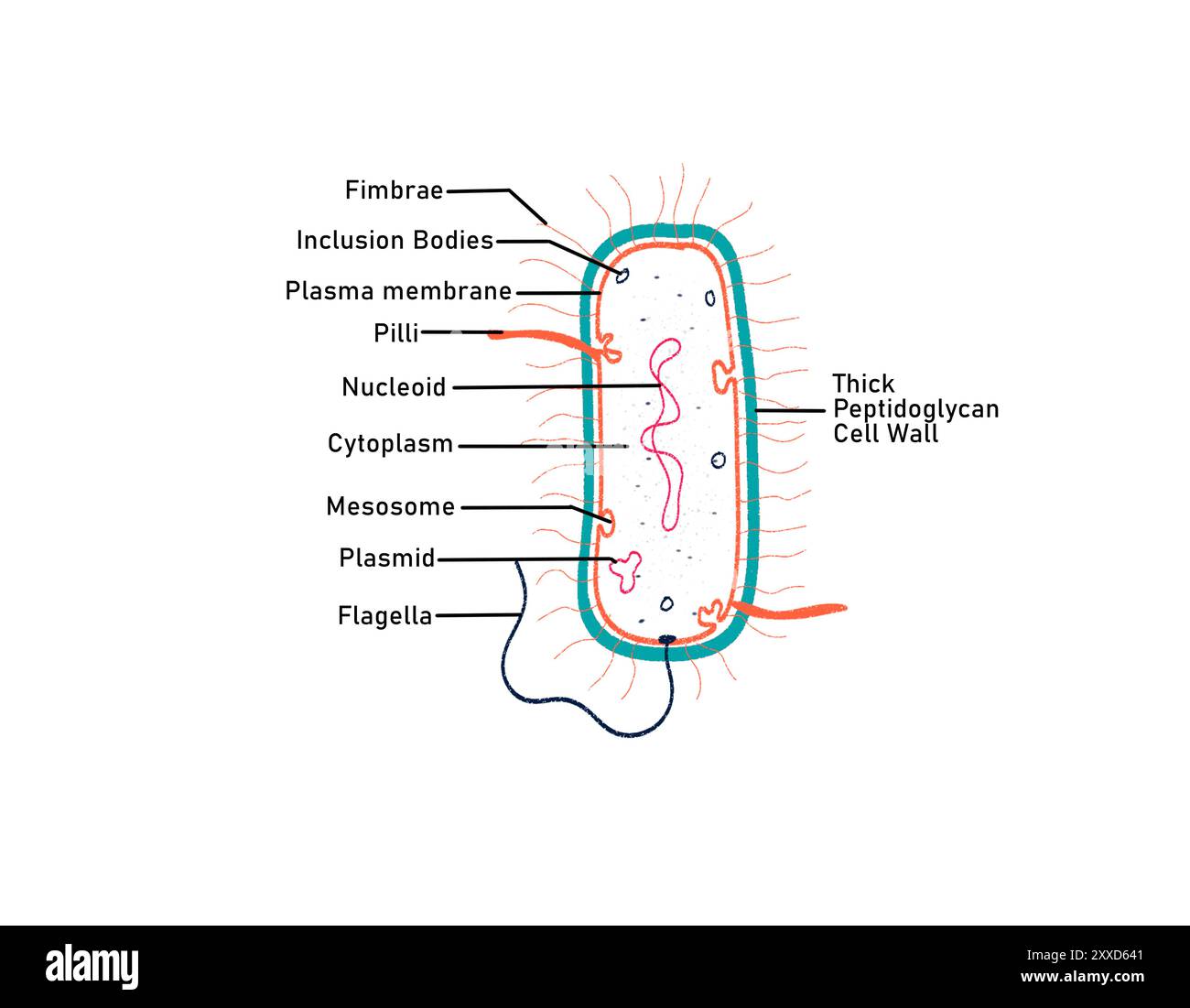 Gram positive bacterium, illustration. Gram positive bacteria are characterised by the presence of a multilayered peptidoglycan cell wall that appears purple after Gram Staining. Stock Photohttps://www.alamy.com/image-license-details/?v=1https://www.alamy.com/gram-positive-bacterium-illustration-gram-positive-bacteria-are-characterised-by-the-presence-of-a-multilayered-peptidoglycan-cell-wall-that-appears-purple-after-gram-staining-image618634129.html
RF2XXD641–Gram positive bacterium, illustration. Gram positive bacteria are characterised by the presence of a multilayered peptidoglycan cell wall that appears purple after Gram Staining.
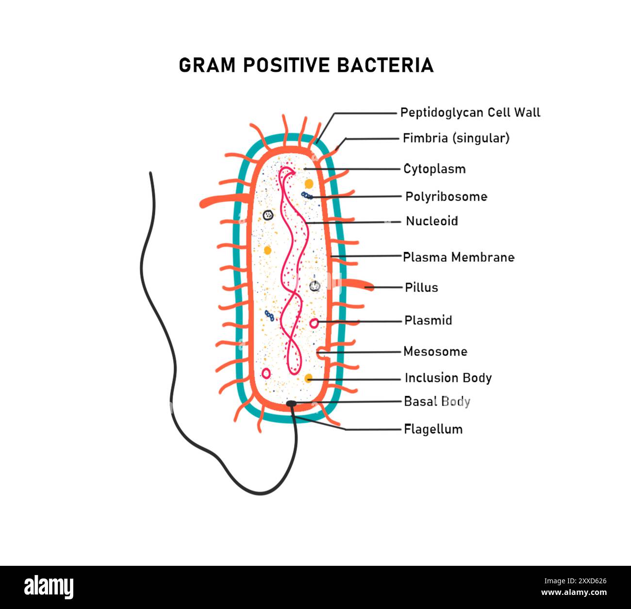 Gram positive bacterium, illustration. Gram positive bacteria are characterised by the presence of a multilayered peptidoglycan cell wall that appears purple after Gram Staining. Stock Photohttps://www.alamy.com/image-license-details/?v=1https://www.alamy.com/gram-positive-bacterium-illustration-gram-positive-bacteria-are-characterised-by-the-presence-of-a-multilayered-peptidoglycan-cell-wall-that-appears-purple-after-gram-staining-image618634078.html
RF2XXD626–Gram positive bacterium, illustration. Gram positive bacteria are characterised by the presence of a multilayered peptidoglycan cell wall that appears purple after Gram Staining.
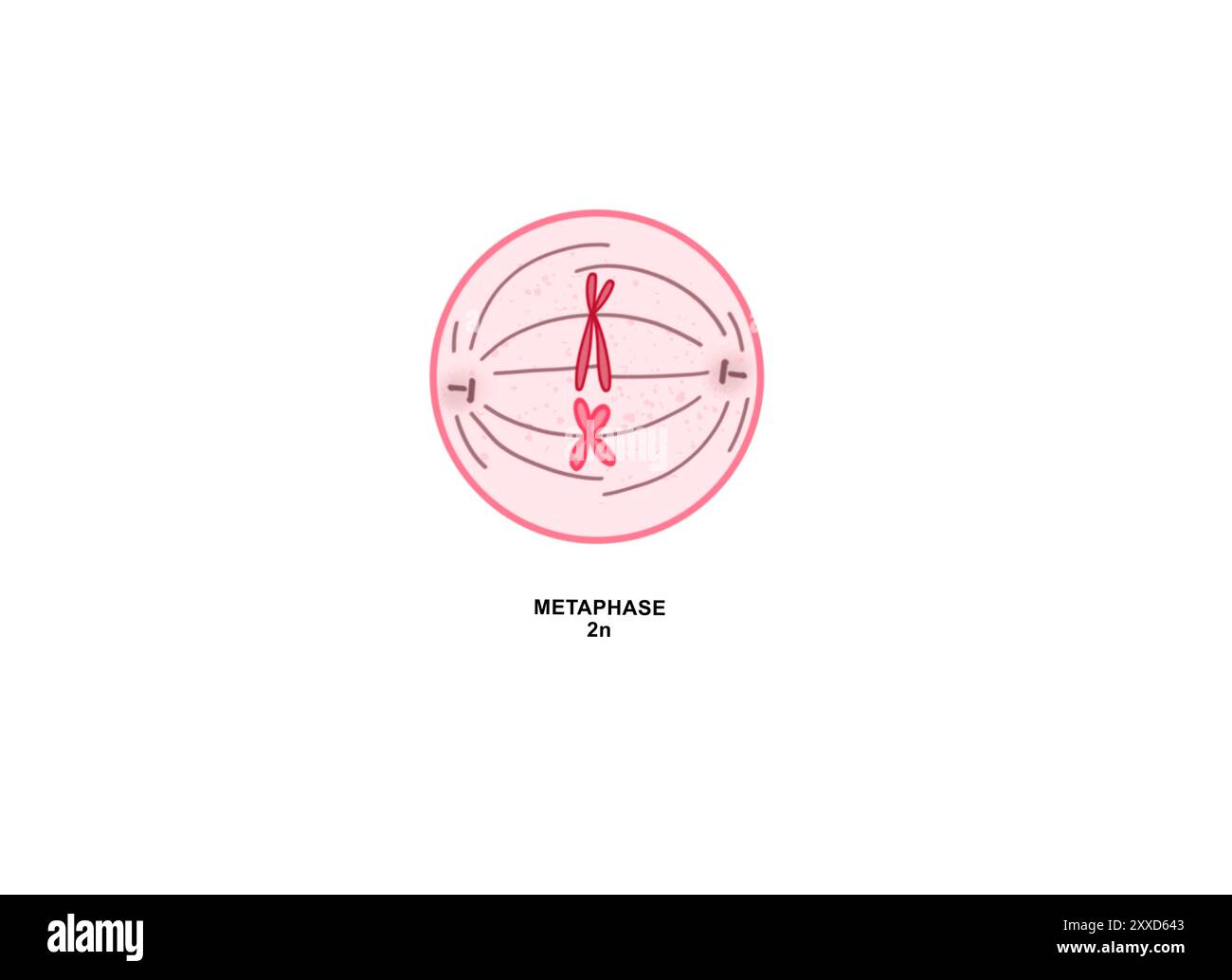 Metaphase, illustration. Metaphase is the third phase of mitosis, the process that separates duplicated genetic material carried in the nucleus of a parent cell into two identical daughter cells. Stock Photohttps://www.alamy.com/image-license-details/?v=1https://www.alamy.com/metaphase-illustration-metaphase-is-the-third-phase-of-mitosis-the-process-that-separates-duplicated-genetic-material-carried-in-the-nucleus-of-a-parent-cell-into-two-identical-daughter-cells-image618634131.html
RF2XXD643–Metaphase, illustration. Metaphase is the third phase of mitosis, the process that separates duplicated genetic material carried in the nucleus of a parent cell into two identical daughter cells.
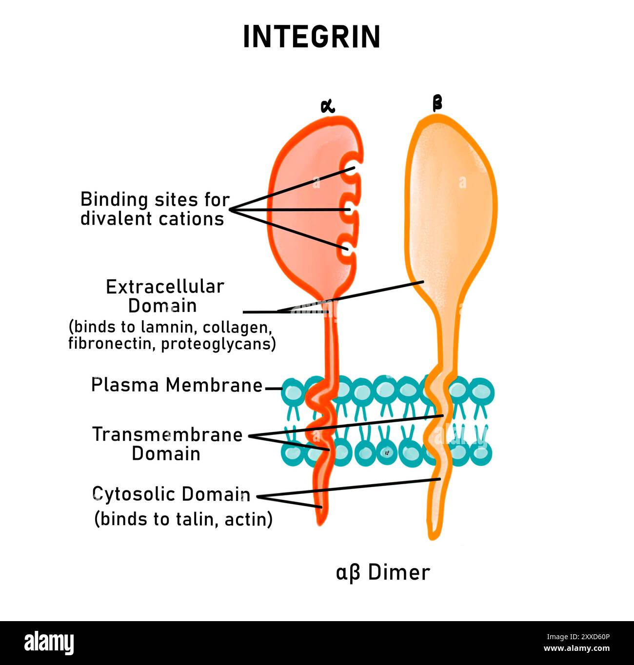 Integrins, illustration. Integrins are type of cell adhesion molecule (CAM). The are integral membrane proteins, made up of two polypeptide chains; alpha and beta. Integrin becomes active when these two units dimerise. Stock Photohttps://www.alamy.com/image-license-details/?v=1https://www.alamy.com/integrins-illustration-integrins-are-type-of-cell-adhesion-molecule-cam-the-are-integral-membrane-proteins-made-up-of-two-polypeptide-chains-alpha-and-beta-integrin-becomes-active-when-these-two-units-dimerise-image618634038.html
RF2XXD60P–Integrins, illustration. Integrins are type of cell adhesion molecule (CAM). The are integral membrane proteins, made up of two polypeptide chains; alpha and beta. Integrin becomes active when these two units dimerise.
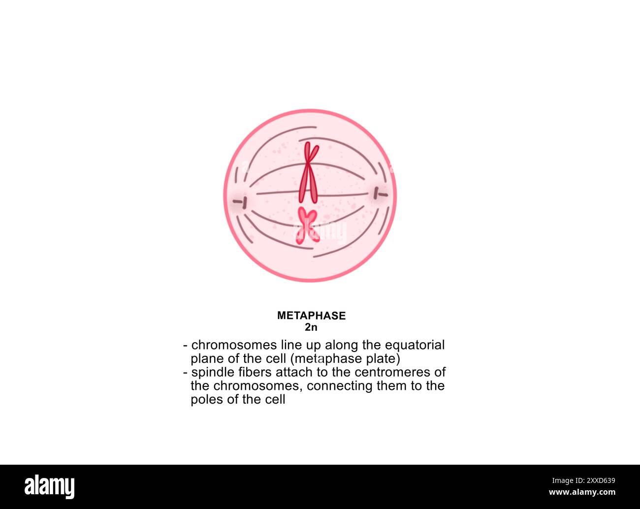 Metaphase of mitosis, illustration. Metaphase is the third phase of mitosis, the process that separates duplicated genetic material carried in the nucleus of a parent cell into two identical daughter cells. Stock Photohttps://www.alamy.com/image-license-details/?v=1https://www.alamy.com/metaphase-of-mitosis-illustration-metaphase-is-the-third-phase-of-mitosis-the-process-that-separates-duplicated-genetic-material-carried-in-the-nucleus-of-a-parent-cell-into-two-identical-daughter-cells-image618634109.html
RF2XXD639–Metaphase of mitosis, illustration. Metaphase is the third phase of mitosis, the process that separates duplicated genetic material carried in the nucleus of a parent cell into two identical daughter cells.
 Meiosis II prophase II, illustration. During prophase II the chromosomes condense and a new set of spindle fibres form. The chromosomes begin moving towards the equator of the cell. Stock Photohttps://www.alamy.com/image-license-details/?v=1https://www.alamy.com/meiosis-ii-prophase-ii-illustration-during-prophase-ii-the-chromosomes-condense-and-a-new-set-of-spindle-fibres-form-the-chromosomes-begin-moving-towards-the-equator-of-the-cell-image618634126.html
RF2XXD63X–Meiosis II prophase II, illustration. During prophase II the chromosomes condense and a new set of spindle fibres form. The chromosomes begin moving towards the equator of the cell.
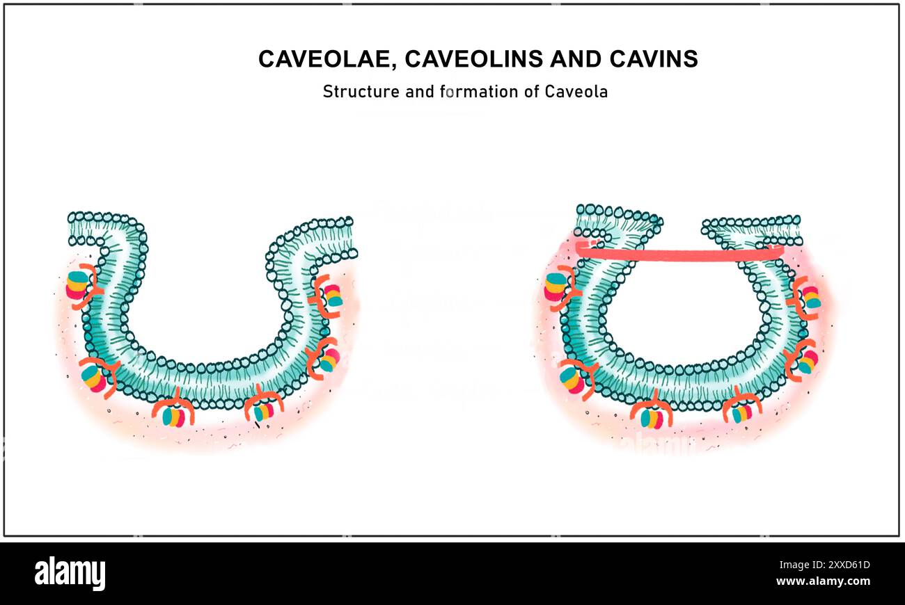 Caveolae, illustration. Caveolae or 'little caves' are small invaginations of the cell plasma membrane. They function during endocytosis, signal transduction and lipid and cholesterol regulation. Stock Photohttps://www.alamy.com/image-license-details/?v=1https://www.alamy.com/caveolae-illustration-caveolae-or-little-caves-are-small-invaginations-of-the-cell-plasma-membrane-they-function-during-endocytosis-signal-transduction-and-lipid-and-cholesterol-regulation-image618634057.html
RF2XXD61D–Caveolae, illustration. Caveolae or 'little caves' are small invaginations of the cell plasma membrane. They function during endocytosis, signal transduction and lipid and cholesterol regulation.
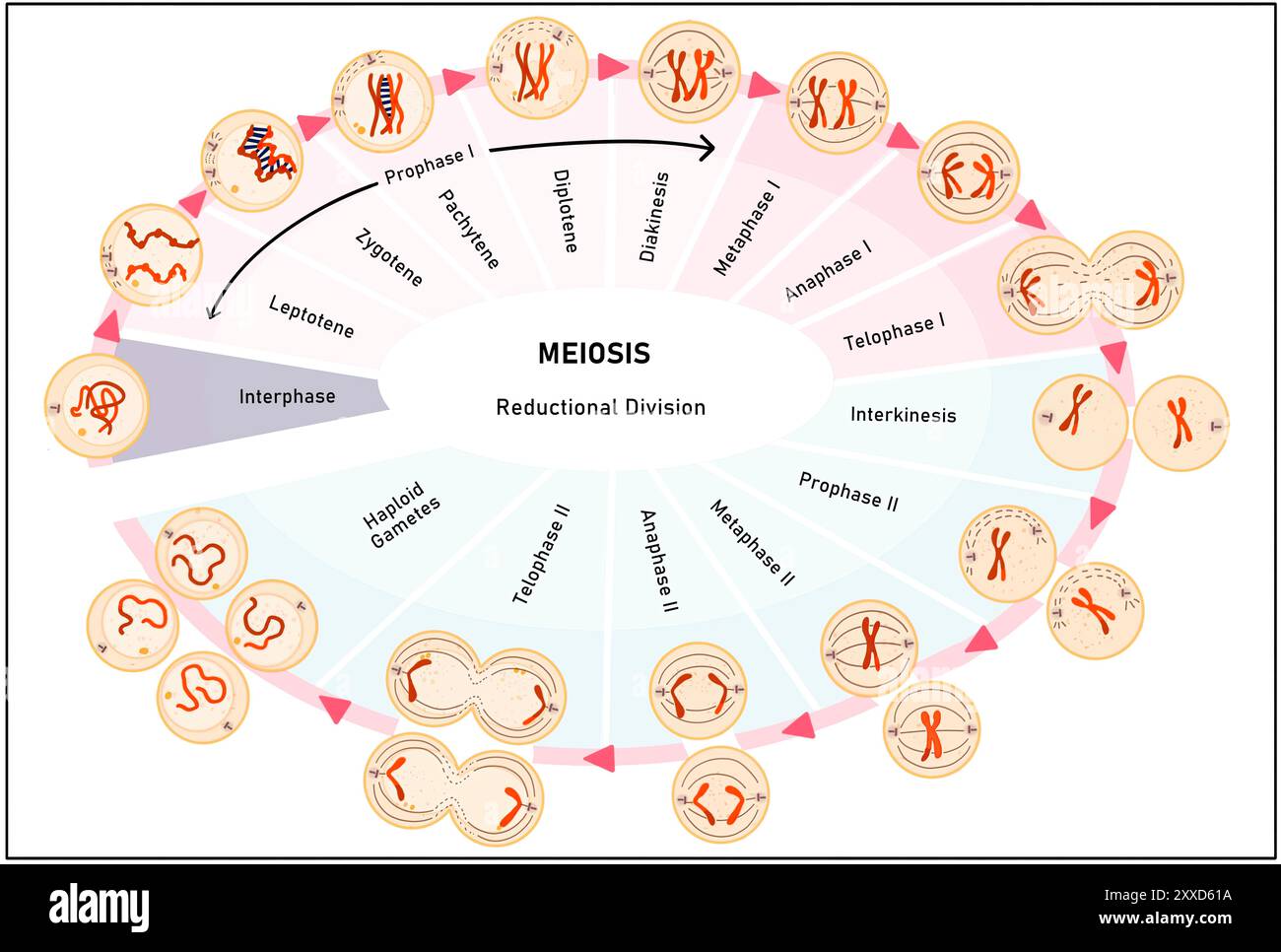 Meiosis, illustration. Meiosis is the process by which a single cell divides twice to form four haploid daughter cells. Stock Photohttps://www.alamy.com/image-license-details/?v=1https://www.alamy.com/meiosis-illustration-meiosis-is-the-process-by-which-a-single-cell-divides-twice-to-form-four-haploid-daughter-cells-image618634054.html
RF2XXD61A–Meiosis, illustration. Meiosis is the process by which a single cell divides twice to form four haploid daughter cells.
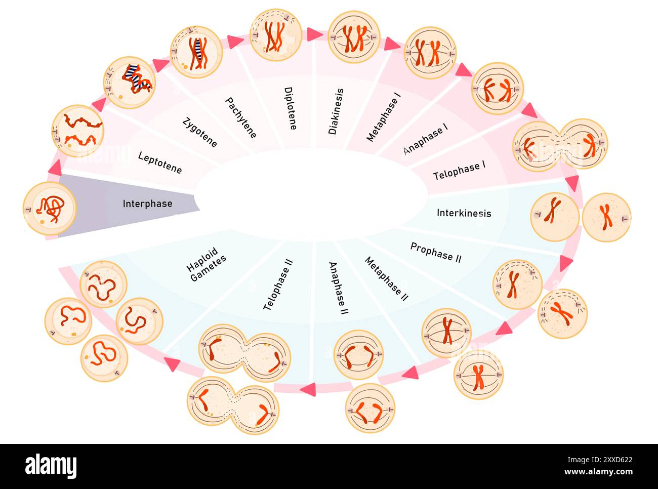 Meiosis, illustration. Meiosis is the process by which a single cell divides twice to form four haploid daughter cells. Stock Photohttps://www.alamy.com/image-license-details/?v=1https://www.alamy.com/meiosis-illustration-meiosis-is-the-process-by-which-a-single-cell-divides-twice-to-form-four-haploid-daughter-cells-image618634074.html
RF2XXD622–Meiosis, illustration. Meiosis is the process by which a single cell divides twice to form four haploid daughter cells.
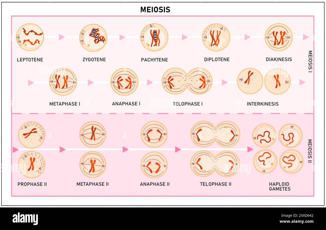 Meiosis, illustration. Meiosis is the process by which a single cell divides twice to form four haploid daughter cells. Stock Photohttps://www.alamy.com/image-license-details/?v=1https://www.alamy.com/meiosis-illustration-meiosis-is-the-process-by-which-a-single-cell-divides-twice-to-form-four-haploid-daughter-cells-image618634130.html
RF2XXD642–Meiosis, illustration. Meiosis is the process by which a single cell divides twice to form four haploid daughter cells.
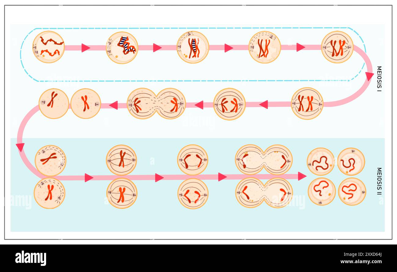 Meiosis, illustration. Meiosis is the process by which a single cell divides twice to form four haploid daughter cells. Stock Photohttps://www.alamy.com/image-license-details/?v=1https://www.alamy.com/meiosis-illustration-meiosis-is-the-process-by-which-a-single-cell-divides-twice-to-form-four-haploid-daughter-cells-image618634146.html
RF2XXD64J–Meiosis, illustration. Meiosis is the process by which a single cell divides twice to form four haploid daughter cells.
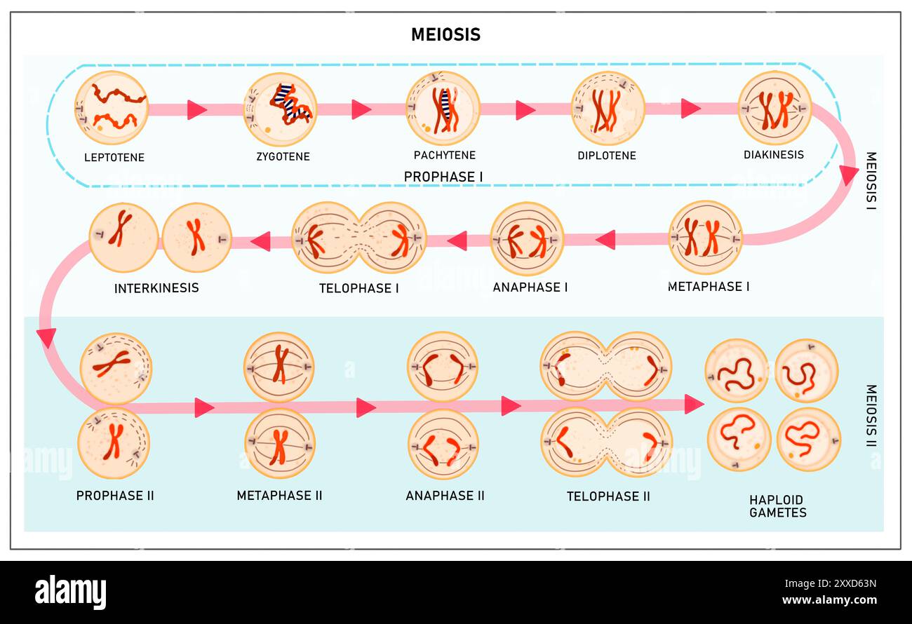 Meiosis, illustration. Meiosis is the process by which a single cell divides twice to form four haploid daughter cells. Stock Photohttps://www.alamy.com/image-license-details/?v=1https://www.alamy.com/meiosis-illustration-meiosis-is-the-process-by-which-a-single-cell-divides-twice-to-form-four-haploid-daughter-cells-image618634121.html
RF2XXD63N–Meiosis, illustration. Meiosis is the process by which a single cell divides twice to form four haploid daughter cells.
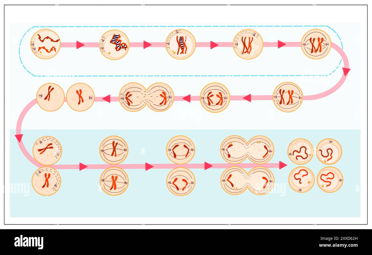 Meiosis, illustration. Meiosis is the process by which a single cell divides twice to form four haploid daughter cells. Stock Photohttps://www.alamy.com/image-license-details/?v=1https://www.alamy.com/meiosis-illustration-meiosis-is-the-process-by-which-a-single-cell-divides-twice-to-form-four-haploid-daughter-cells-image618634089.html
RF2XXD62H–Meiosis, illustration. Meiosis is the process by which a single cell divides twice to form four haploid daughter cells.
 Meiosis, illustration. Meiosis is the process by which a single cell divides twice to form four haploid daughter cells. Stock Photohttps://www.alamy.com/image-license-details/?v=1https://www.alamy.com/meiosis-illustration-meiosis-is-the-process-by-which-a-single-cell-divides-twice-to-form-four-haploid-daughter-cells-image618634036.html
RF2XXD60M–Meiosis, illustration. Meiosis is the process by which a single cell divides twice to form four haploid daughter cells.
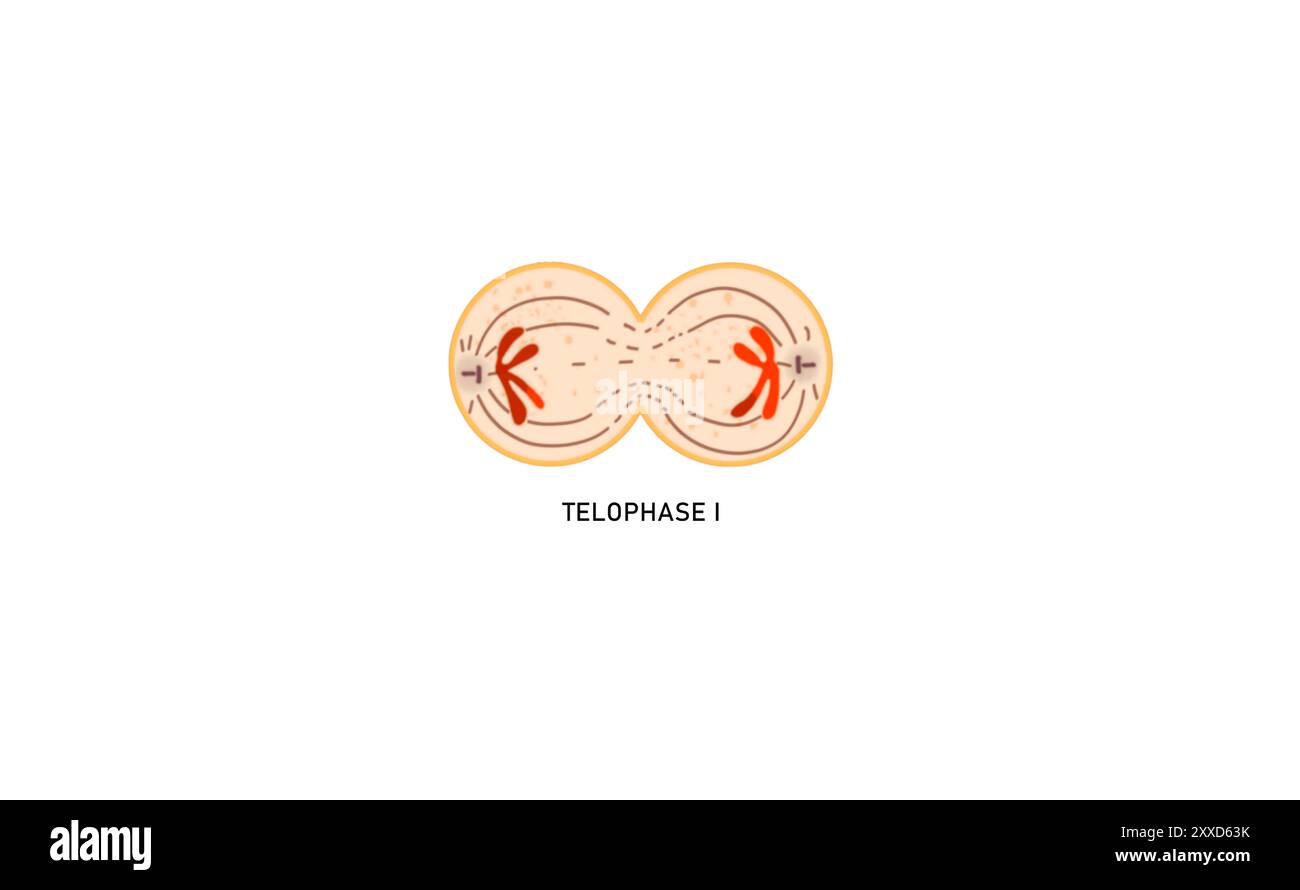 Meiosis I telophase I, illustration. During telophase , the chromosomes are enclosed in the nucleus. The cell then undergoes cytokinesis, which divides the cytoplasm of the original cell into two daughter cells. Stock Photohttps://www.alamy.com/image-license-details/?v=1https://www.alamy.com/meiosis-i-telophase-i-illustration-during-telophase-the-chromosomes-are-enclosed-in-the-nucleus-the-cell-then-undergoes-cytokinesis-which-divides-the-cytoplasm-of-the-original-cell-into-two-daughter-cells-image618634119.html
RF2XXD63K–Meiosis I telophase I, illustration. During telophase , the chromosomes are enclosed in the nucleus. The cell then undergoes cytokinesis, which divides the cytoplasm of the original cell into two daughter cells.
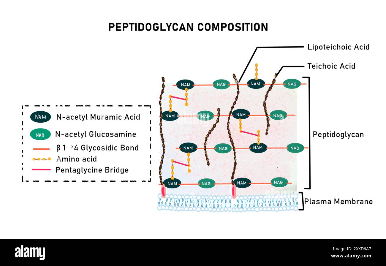 Structure of peptidoglycan, illustration. Peptidoglycan is a component of bacterial cell walls. Structurally it is a heteropolysaccharide where carbohydrates (N-acetylglucosamine and N-acetylmuramic acid)are conjugated with amino acids. Stock Photohttps://www.alamy.com/image-license-details/?v=1https://www.alamy.com/structure-of-peptidoglycan-illustration-peptidoglycan-is-a-component-of-bacterial-cell-walls-structurally-it-is-a-heteropolysaccharide-where-carbohydrates-n-acetylglucosamine-and-n-acetylmuramic-acidare-conjugated-with-amino-acids-image618634303.html
RF2XXD6A7–Structure of peptidoglycan, illustration. Peptidoglycan is a component of bacterial cell walls. Structurally it is a heteropolysaccharide where carbohydrates (N-acetylglucosamine and N-acetylmuramic acid)are conjugated with amino acids.
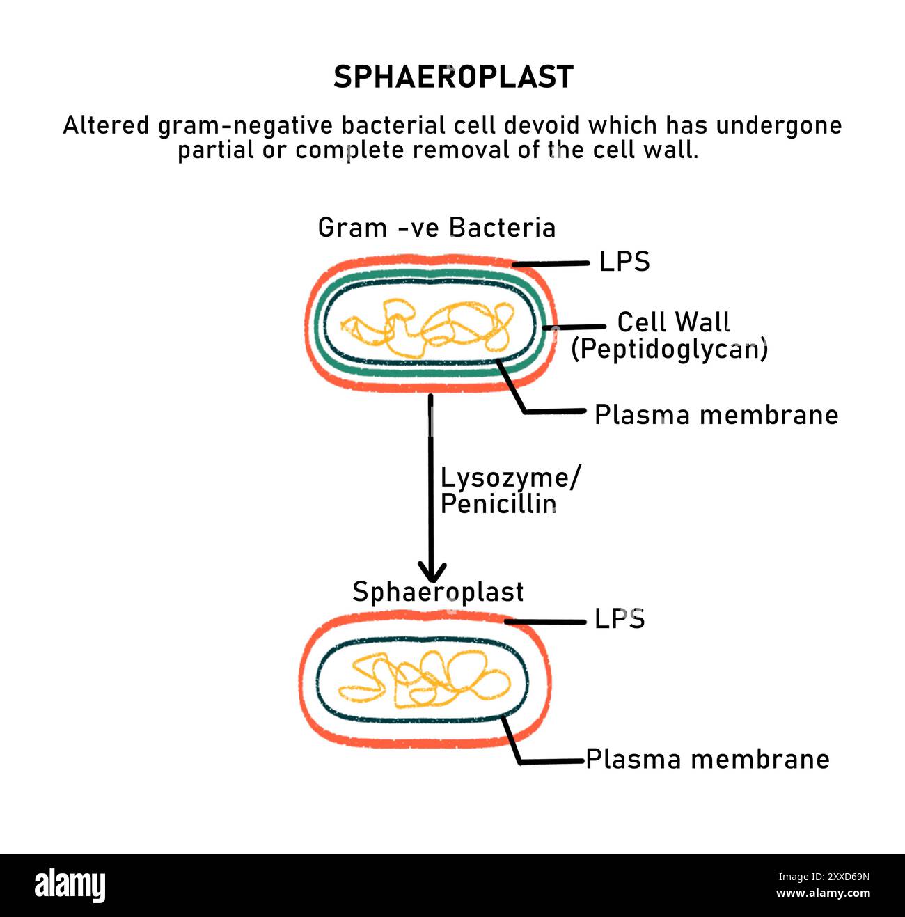 Sphaeroplast, illustration. A sphaeroplast is a Gram negative bacterium whose cell wall has been degraded by enzymes leaving the cell intact with a cell membrane and lipopolysaccharide layer. Stock Photohttps://www.alamy.com/image-license-details/?v=1https://www.alamy.com/sphaeroplast-illustration-a-sphaeroplast-is-a-gram-negative-bacterium-whose-cell-wall-has-been-degraded-by-enzymes-leaving-the-cell-intact-with-a-cell-membrane-and-lipopolysaccharide-layer-image618634289.html
RF2XXD69N–Sphaeroplast, illustration. A sphaeroplast is a Gram negative bacterium whose cell wall has been degraded by enzymes leaving the cell intact with a cell membrane and lipopolysaccharide layer.
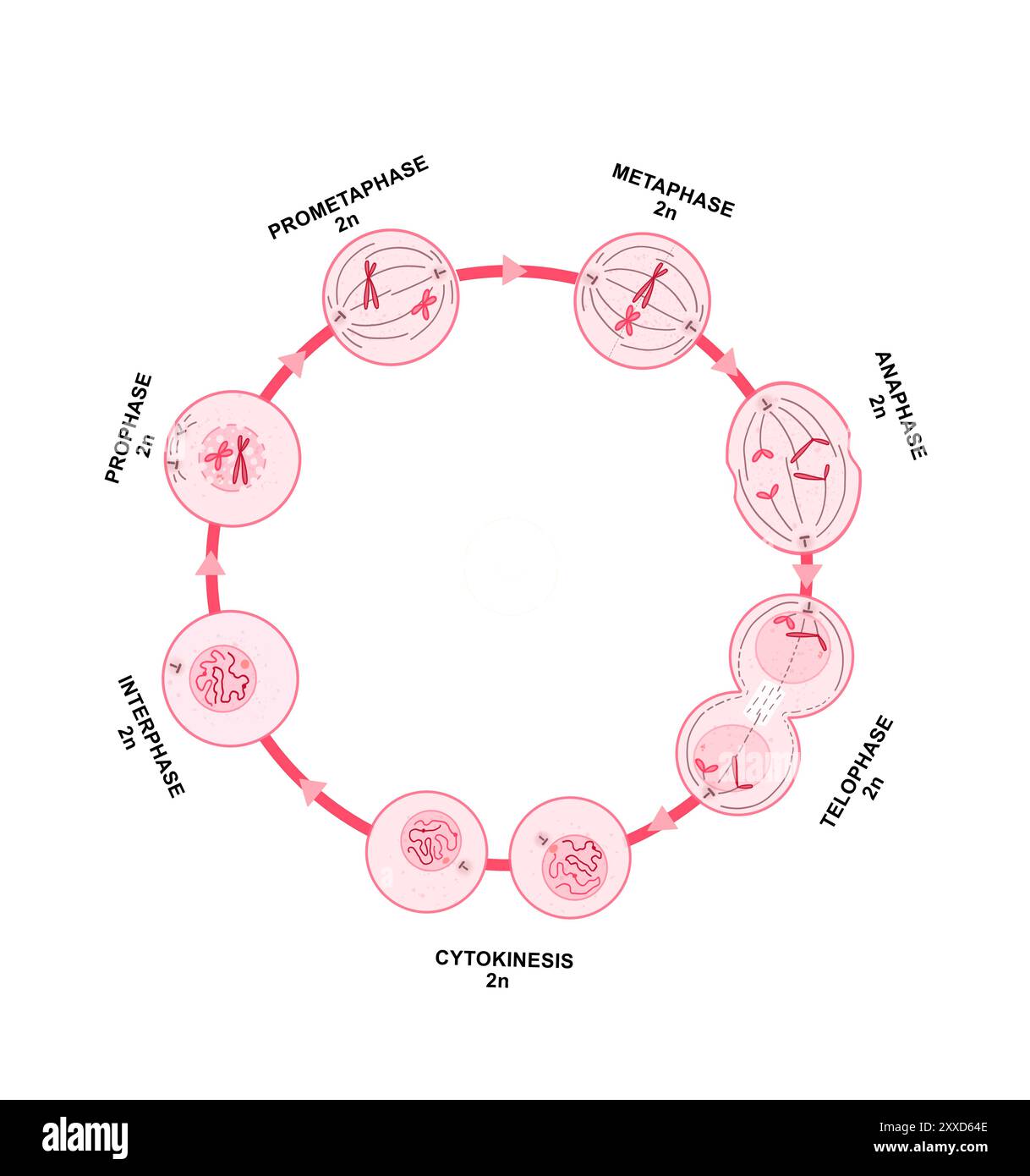 Mitosis, illustration. Mitosis is the process by which a cell replicates its chromosomes and then segregates them, producing two identical nuclei in preparation for cell division. Stock Photohttps://www.alamy.com/image-license-details/?v=1https://www.alamy.com/mitosis-illustration-mitosis-is-the-process-by-which-a-cell-replicates-its-chromosomes-and-then-segregates-them-producing-two-identical-nuclei-in-preparation-for-cell-division-image618634142.html
RF2XXD64E–Mitosis, illustration. Mitosis is the process by which a cell replicates its chromosomes and then segregates them, producing two identical nuclei in preparation for cell division.
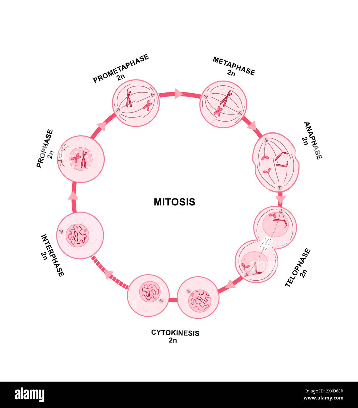 Mitosis, illustration. Mitosis is the process by which a cell replicates its chromosomes and then segregates them, producing two identical nuclei in preparation for cell division. Stock Photohttps://www.alamy.com/image-license-details/?v=1https://www.alamy.com/mitosis-illustration-mitosis-is-the-process-by-which-a-cell-replicates-its-chromosomes-and-then-segregates-them-producing-two-identical-nuclei-in-preparation-for-cell-division-image618634263.html
RF2XXD68R–Mitosis, illustration. Mitosis is the process by which a cell replicates its chromosomes and then segregates them, producing two identical nuclei in preparation for cell division.
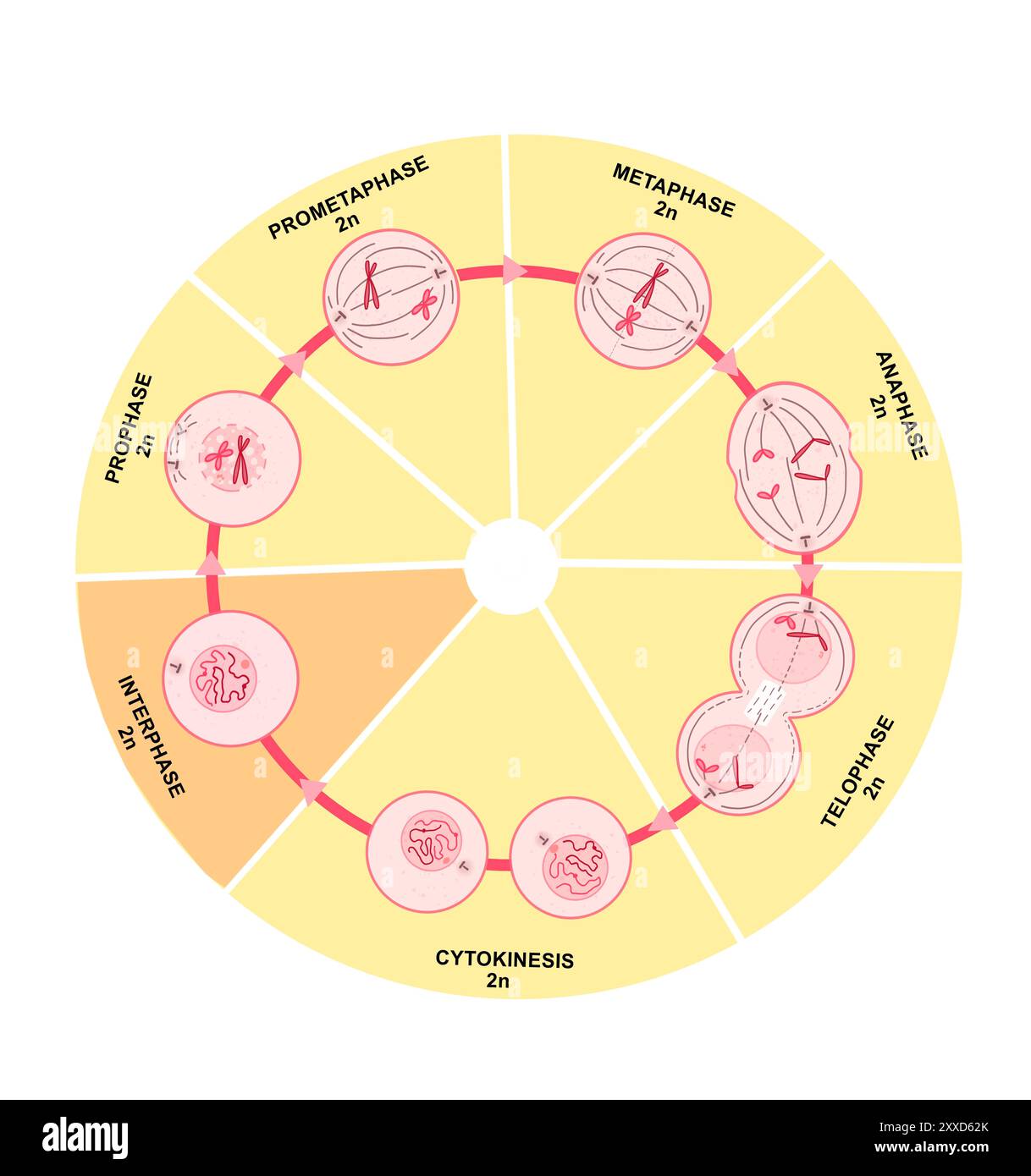 Mitosis, illustration. Mitosis is the process by which a cell replicates its chromosomes and then segregates them, producing two identical nuclei in preparation for cell division. Stock Photohttps://www.alamy.com/image-license-details/?v=1https://www.alamy.com/mitosis-illustration-mitosis-is-the-process-by-which-a-cell-replicates-its-chromosomes-and-then-segregates-them-producing-two-identical-nuclei-in-preparation-for-cell-division-image618634091.html
RF2XXD62K–Mitosis, illustration. Mitosis is the process by which a cell replicates its chromosomes and then segregates them, producing two identical nuclei in preparation for cell division.
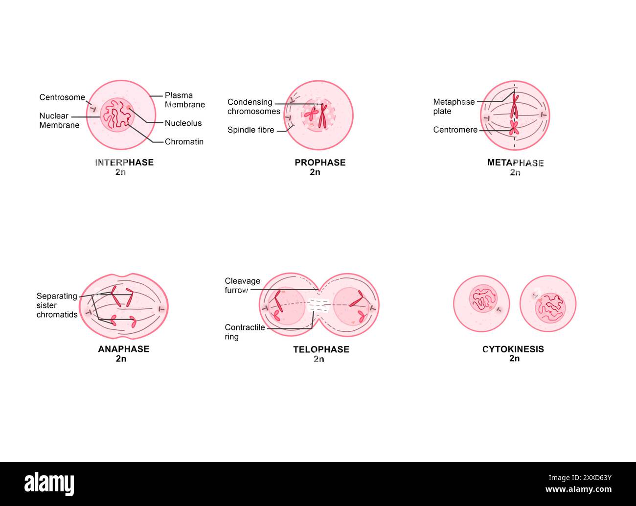 Mitosis, illustration. Mitosis is the process by which a cell replicates its chromosomes and then segregates them, producing two identical nuclei in preparation for cell division. Stock Photohttps://www.alamy.com/image-license-details/?v=1https://www.alamy.com/mitosis-illustration-mitosis-is-the-process-by-which-a-cell-replicates-its-chromosomes-and-then-segregates-them-producing-two-identical-nuclei-in-preparation-for-cell-division-image618634127.html
RF2XXD63Y–Mitosis, illustration. Mitosis is the process by which a cell replicates its chromosomes and then segregates them, producing two identical nuclei in preparation for cell division.
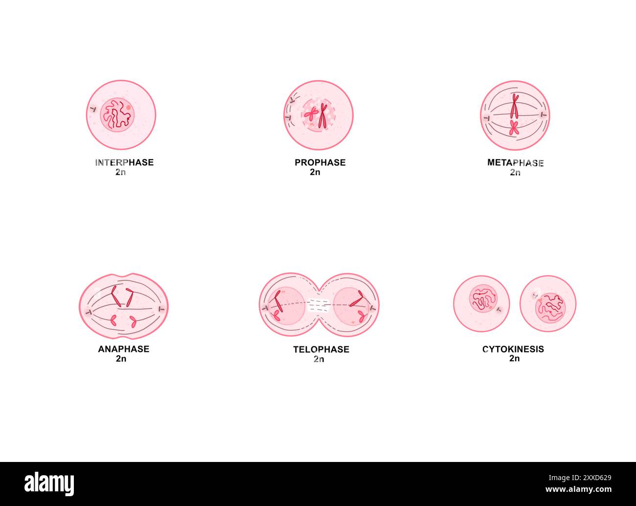 Mitosis, illustration. Mitosis is the process by which a cell replicates its chromosomes and then segregates them, producing two identical nuclei in preparation for cell division. Stock Photohttps://www.alamy.com/image-license-details/?v=1https://www.alamy.com/mitosis-illustration-mitosis-is-the-process-by-which-a-cell-replicates-its-chromosomes-and-then-segregates-them-producing-two-identical-nuclei-in-preparation-for-cell-division-image618634081.html
RF2XXD629–Mitosis, illustration. Mitosis is the process by which a cell replicates its chromosomes and then segregates them, producing two identical nuclei in preparation for cell division.
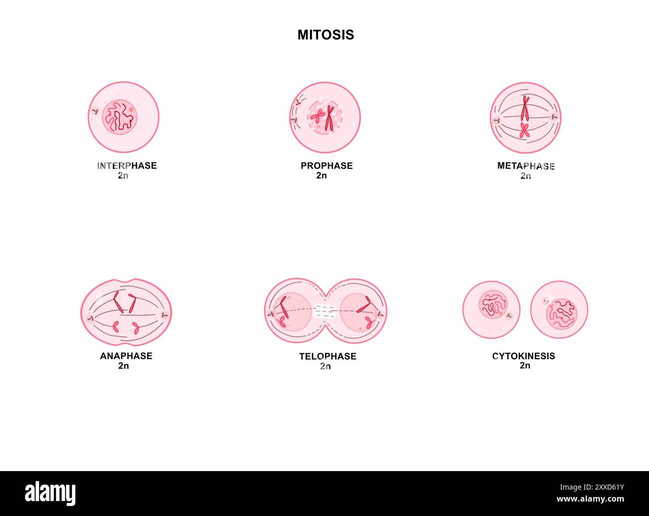 Mitosis, illustration. Mitosis is the process by which a cell replicates its chromosomes and then segregates them, producing two identical nuclei in preparation for cell division. Stock Photohttps://www.alamy.com/image-license-details/?v=1https://www.alamy.com/mitosis-illustration-mitosis-is-the-process-by-which-a-cell-replicates-its-chromosomes-and-then-segregates-them-producing-two-identical-nuclei-in-preparation-for-cell-division-image618634071.html
RF2XXD61Y–Mitosis, illustration. Mitosis is the process by which a cell replicates its chromosomes and then segregates them, producing two identical nuclei in preparation for cell division.
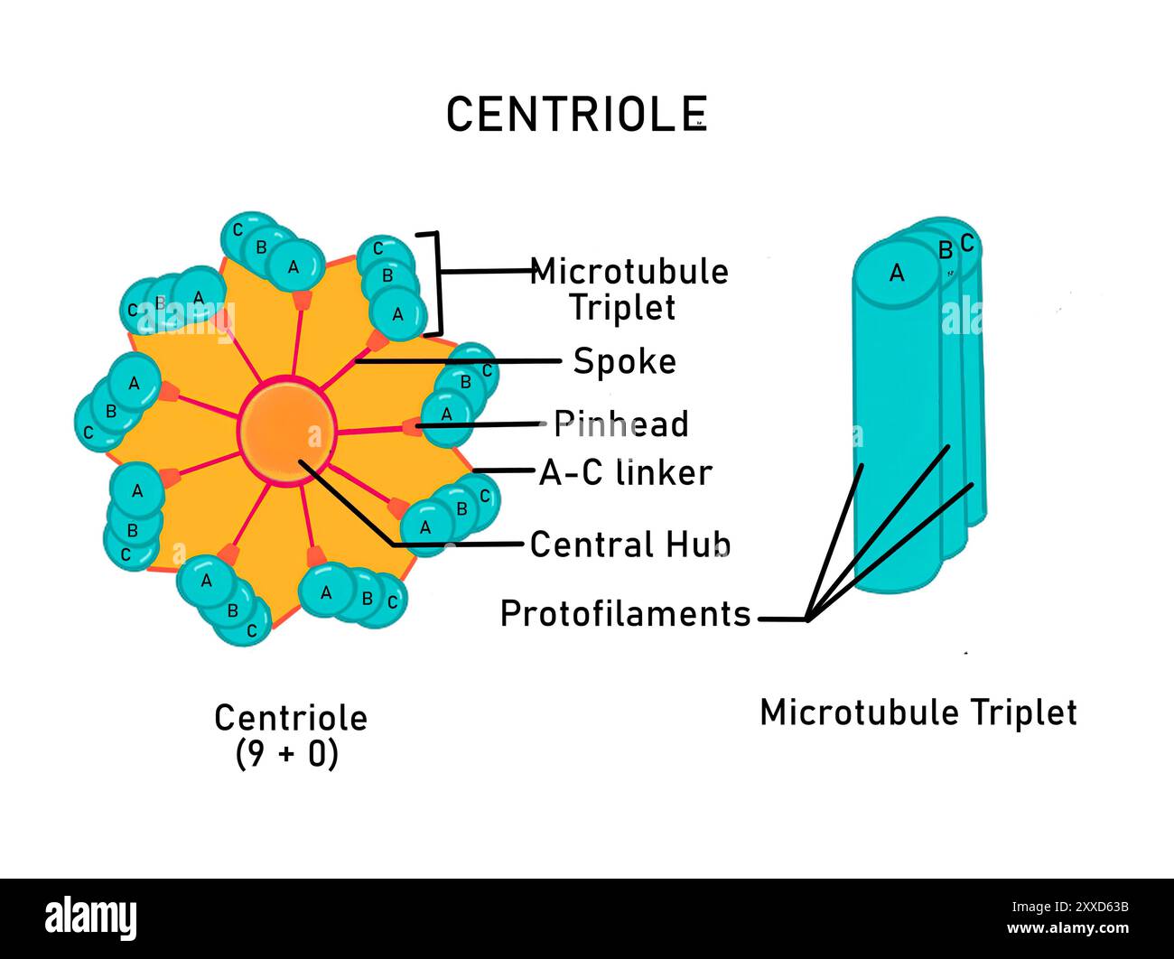 Structure of a centriole, illustration. A centriole is a cylindrical organelle that is a component of the centrosome (microtubule organizing centres). Centrioles are composed of protein tubulin and play an important role in organising microtubules during cell division. Stock Photohttps://www.alamy.com/image-license-details/?v=1https://www.alamy.com/structure-of-a-centriole-illustration-a-centriole-is-a-cylindrical-organelle-that-is-a-component-of-the-centrosome-microtubule-organizing-centres-centrioles-are-composed-of-protein-tubulin-and-play-an-important-role-in-organising-microtubules-during-cell-division-image618634111.html
RF2XXD63B–Structure of a centriole, illustration. A centriole is a cylindrical organelle that is a component of the centrosome (microtubule organizing centres). Centrioles are composed of protein tubulin and play an important role in organising microtubules during cell division.
 Structure of a bacterial flagellum, illustration. Different flagellin proteins are arranged in a helical manner. The filament is embedded in the cell via a basal body. Flagella help in locomotion and can also have sensory functions. Stock Photohttps://www.alamy.com/image-license-details/?v=1https://www.alamy.com/structure-of-a-bacterial-flagellum-illustration-different-flagellin-proteins-are-arranged-in-a-helical-manner-the-filament-is-embedded-in-the-cell-via-a-basal-body-flagella-help-in-locomotion-and-can-also-have-sensory-functions-image618634309.html
RF2XXD6AD–Structure of a bacterial flagellum, illustration. Different flagellin proteins are arranged in a helical manner. The filament is embedded in the cell via a basal body. Flagella help in locomotion and can also have sensory functions.
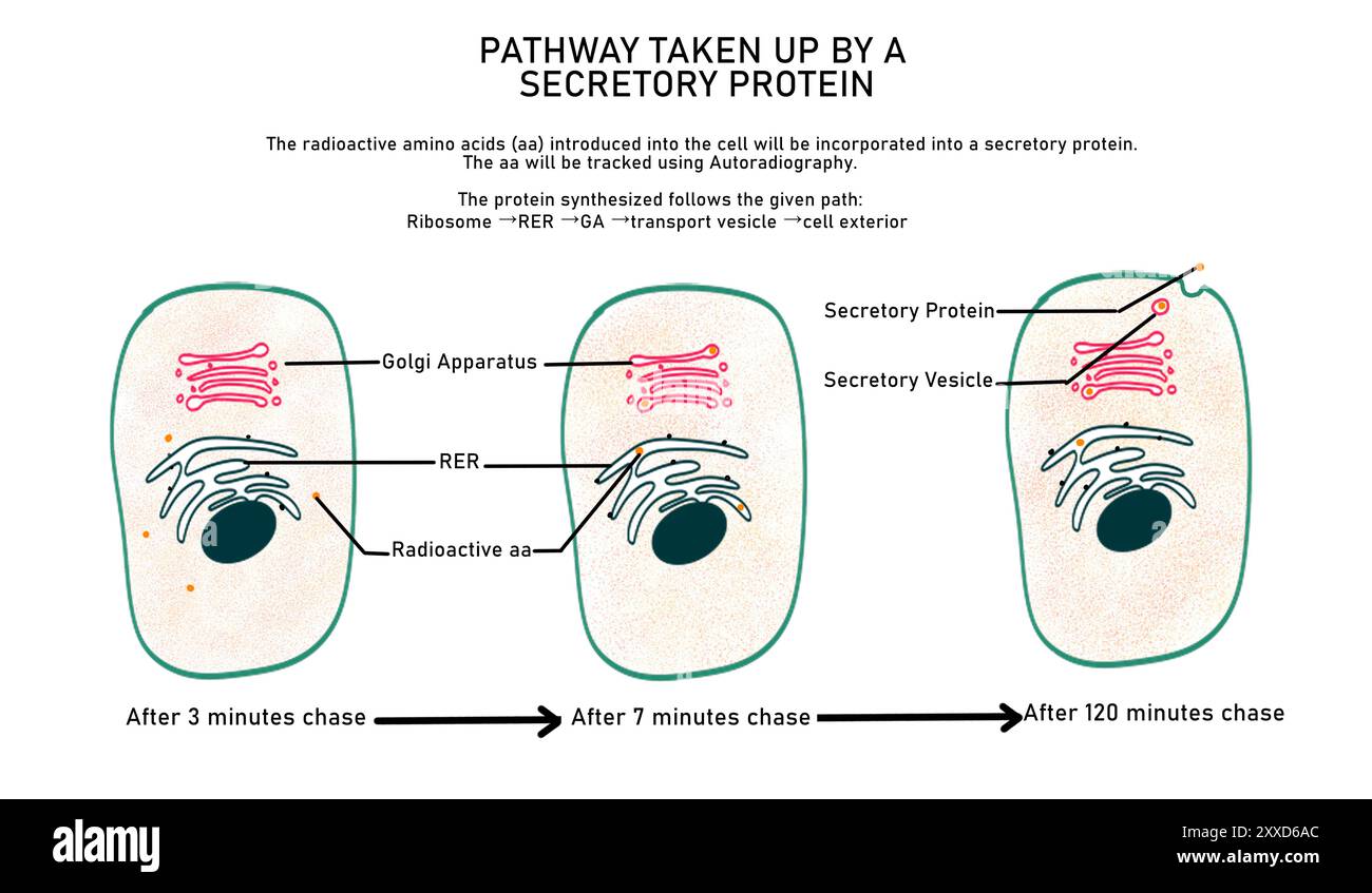 Illustration showing the pathway taken by a secretory protein after it has been synthesised. They move from the cytoplasm to the endoplasmic reticulum, Golgi apparatus, transport vesicle and finally the exterior of the cell. Stock Photohttps://www.alamy.com/image-license-details/?v=1https://www.alamy.com/illustration-showing-the-pathway-taken-by-a-secretory-protein-after-it-has-been-synthesised-they-move-from-the-cytoplasm-to-the-endoplasmic-reticulum-golgi-apparatus-transport-vesicle-and-finally-the-exterior-of-the-cell-image618634308.html
RF2XXD6AC–Illustration showing the pathway taken by a secretory protein after it has been synthesised. They move from the cytoplasm to the endoplasmic reticulum, Golgi apparatus, transport vesicle and finally the exterior of the cell.
 Selectins, illustration. Selectins are a type of calcium dependent cell adhesion molecule (CAM). They participate in attachment to carbohydrates present on the surface of other cells and participate in heterophilic adhesion. They are known to take part in lymphocyte homing. Stock Photohttps://www.alamy.com/image-license-details/?v=1https://www.alamy.com/selectins-illustration-selectins-are-a-type-of-calcium-dependent-cell-adhesion-molecule-cam-they-participate-in-attachment-to-carbohydrates-present-on-the-surface-of-other-cells-and-participate-in-heterophilic-adhesion-they-are-known-to-take-part-in-lymphocyte-homing-image618634283.html
RF2XXD69F–Selectins, illustration. Selectins are a type of calcium dependent cell adhesion molecule (CAM). They participate in attachment to carbohydrates present on the surface of other cells and participate in heterophilic adhesion. They are known to take part in lymphocyte homing.
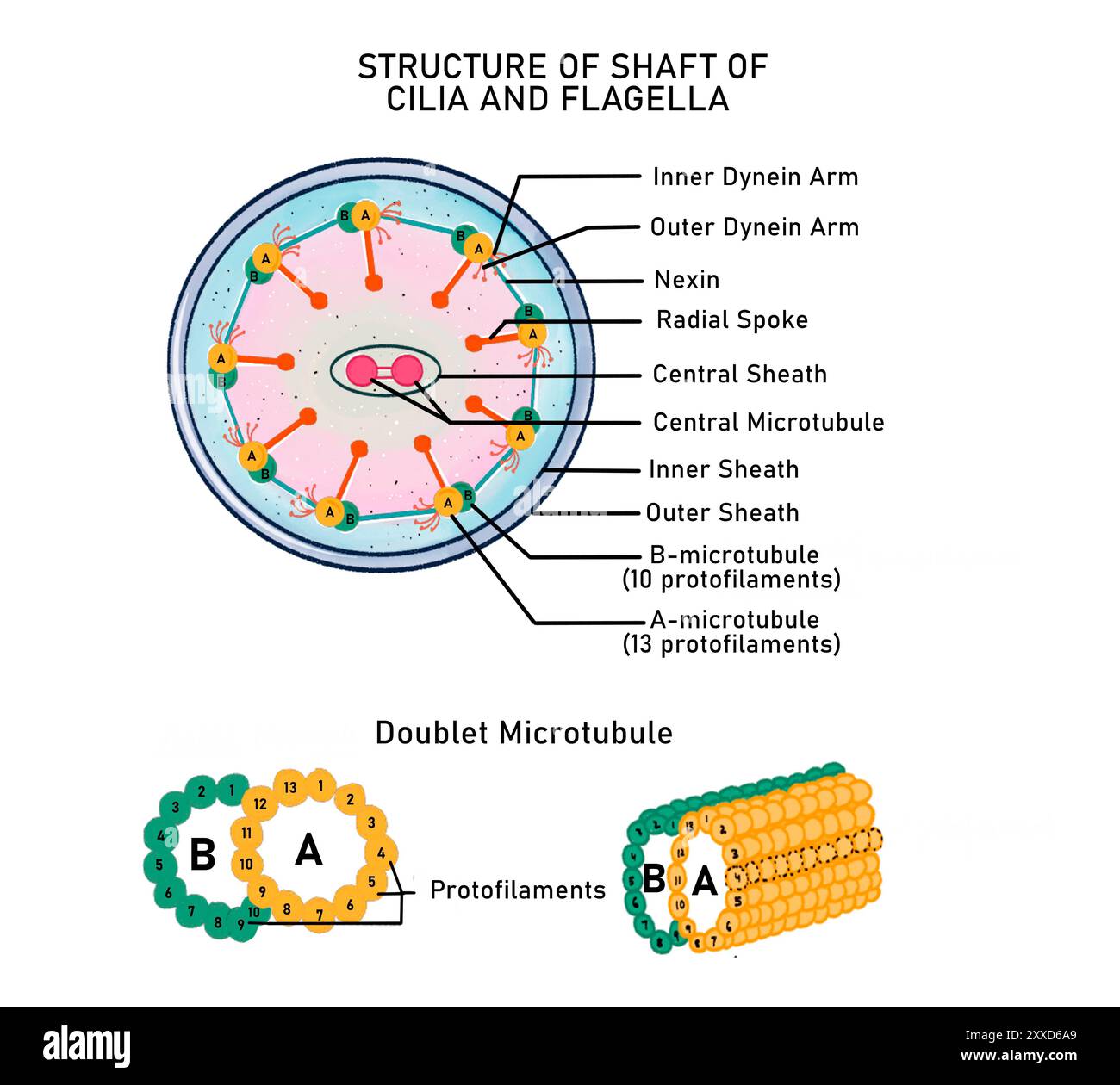 Structure of a bacterial flagellum, illustration. Different flagellin proteins are arranged in a helical manner. The filament is embedded in the cell via a basal body. Flagella help in locomotion and can also have sensory functions. Stock Photohttps://www.alamy.com/image-license-details/?v=1https://www.alamy.com/structure-of-a-bacterial-flagellum-illustration-different-flagellin-proteins-are-arranged-in-a-helical-manner-the-filament-is-embedded-in-the-cell-via-a-basal-body-flagella-help-in-locomotion-and-can-also-have-sensory-functions-image618634305.html
RF2XXD6A9–Structure of a bacterial flagellum, illustration. Different flagellin proteins are arranged in a helical manner. The filament is embedded in the cell via a basal body. Flagella help in locomotion and can also have sensory functions.
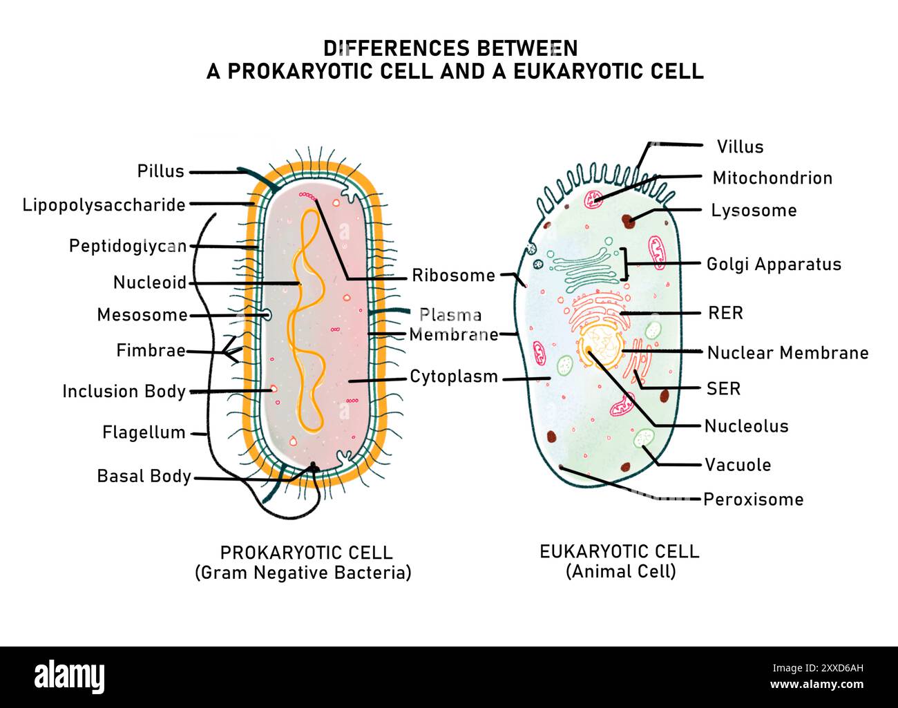 Illustration showing the differences between a prokaryotic cell and eukaryotic cell with respect to the presence or absence of a cell wall, membrane bound organelles and the organisation of nuclear material. Stock Photohttps://www.alamy.com/image-license-details/?v=1https://www.alamy.com/illustration-showing-the-differences-between-a-prokaryotic-cell-and-eukaryotic-cell-with-respect-to-the-presence-or-absence-of-a-cell-wall-membrane-bound-organelles-and-the-organisation-of-nuclear-material-image618634313.html
RF2XXD6AH–Illustration showing the differences between a prokaryotic cell and eukaryotic cell with respect to the presence or absence of a cell wall, membrane bound organelles and the organisation of nuclear material.
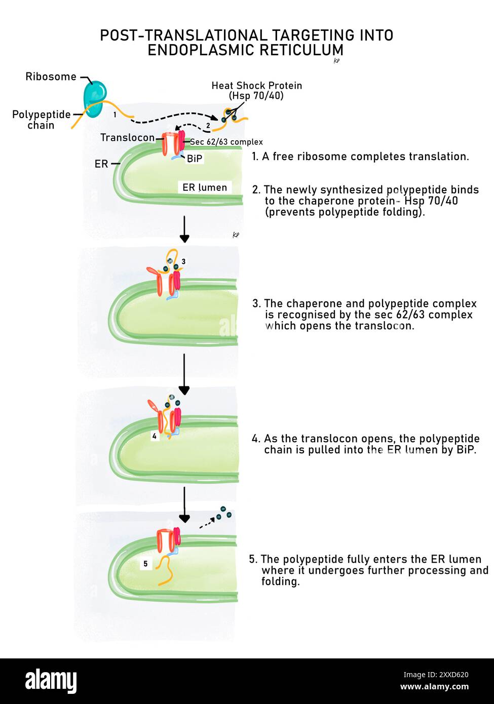 Posttranslational protein targeting, illustration. Protein targeting is the process of sending newly synthesised proteins to the endoplasmic reticulum lumen so that they can be sorted for their final destination in or outside the cell. Stock Photohttps://www.alamy.com/image-license-details/?v=1https://www.alamy.com/posttranslational-protein-targeting-illustration-protein-targeting-is-the-process-of-sending-newly-synthesised-proteins-to-the-endoplasmic-reticulum-lumen-so-that-they-can-be-sorted-for-their-final-destination-in-or-outside-the-cell-image618634072.html
RF2XXD620–Posttranslational protein targeting, illustration. Protein targeting is the process of sending newly synthesised proteins to the endoplasmic reticulum lumen so that they can be sorted for their final destination in or outside the cell.
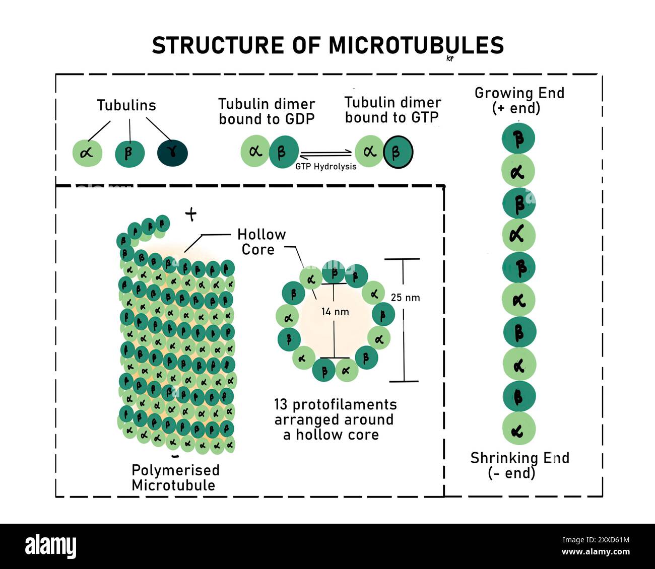 Structure and functions of microtubule, illustration. Microtubules, part of the cytoskeleton, are formed from dimers of the protein tubulin. Three monomers of tubulin are present; alpha, beta and gamma. The functions of microtubules include the formation of centrioles, attachment to centromere of chromosomes during cell division, providing motility and transport of substances from one part of cell to another. Stock Photohttps://www.alamy.com/image-license-details/?v=1https://www.alamy.com/structure-and-functions-of-microtubule-illustration-microtubules-part-of-the-cytoskeleton-are-formed-from-dimers-of-the-protein-tubulin-three-monomers-of-tubulin-are-present-alpha-beta-and-gamma-the-functions-of-microtubules-include-the-formation-of-centrioles-attachment-to-centromere-of-chromosomes-during-cell-division-providing-motility-and-transport-of-substances-from-one-part-of-cell-to-another-image618634064.html
RF2XXD61M–Structure and functions of microtubule, illustration. Microtubules, part of the cytoskeleton, are formed from dimers of the protein tubulin. Three monomers of tubulin are present; alpha, beta and gamma. The functions of microtubules include the formation of centrioles, attachment to centromere of chromosomes during cell division, providing motility and transport of substances from one part of cell to another.