Quick filters:
Cell pigment Stock Photos and Images
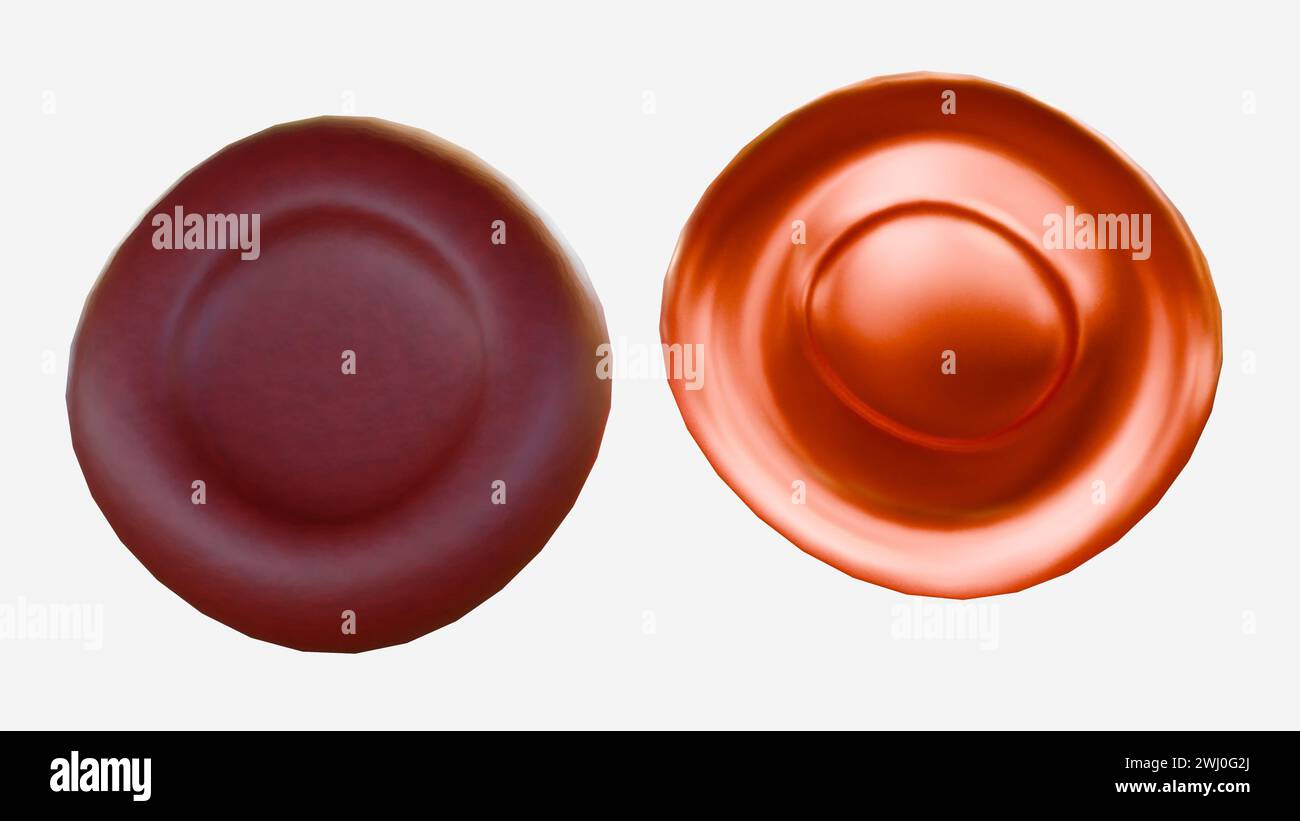 3d rendering of hypochromic red blood cells are red blood cells that have less color than normal when examined under a microscope. Stock Photohttps://www.alamy.com/image-license-details/?v=1https://www.alamy.com/3d-rendering-of-hypochromic-red-blood-cells-are-red-blood-cells-that-have-less-color-than-normal-when-examined-under-a-microscope-image596228938.html
RF2WJ0G2J–3d rendering of hypochromic red blood cells are red blood cells that have less color than normal when examined under a microscope.
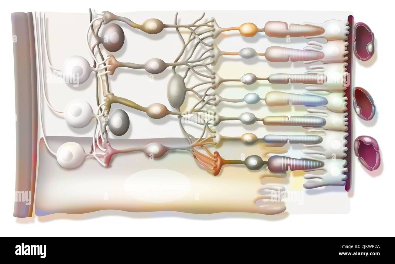 Zoom on the structure of the retina with vitreous body, internal limiting membrane, ganglion cells. Stock Photohttps://www.alamy.com/image-license-details/?v=1https://www.alamy.com/zoom-on-the-structure-of-the-retina-with-vitreous-body-internal-limiting-membrane-ganglion-cells-image476925298.html
RF2JKWR2A–Zoom on the structure of the retina with vitreous body, internal limiting membrane, ganglion cells.
 3d rendering, abstract, all, background, beautiful, biochemistry, blue, celebration, cell pigment, christmas, color, colorful, computer graphics, conf Stock Photohttps://www.alamy.com/image-license-details/?v=1https://www.alamy.com/3d-rendering-abstract-all-background-beautiful-biochemistry-blue-celebration-cell-pigment-christmas-color-colorful-computer-graphics-conf-image454988484.html
RF2HC6ECM–3d rendering, abstract, all, background, beautiful, biochemistry, blue, celebration, cell pigment, christmas, color, colorful, computer graphics, conf
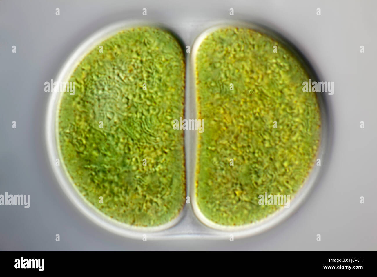 blue-green alga (Chroococcus spec.), cell division Stock Photohttps://www.alamy.com/image-license-details/?v=1https://www.alamy.com/stock-photo-blue-green-alga-chroococcus-spec-cell-division-97255217.html
RMFJ6A0H–blue-green alga (Chroococcus spec.), cell division
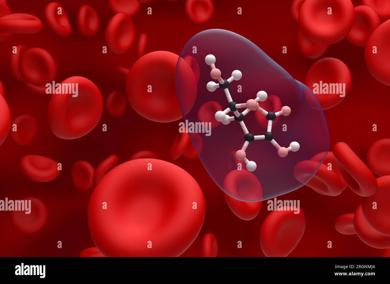 Vitamin c (ascorbic acid) structure in the blood flow ball and stick closeup view 3d illustration Stock Photohttps://www.alamy.com/image-license-details/?v=1https://www.alamy.com/vitamin-c-ascorbic-acid-structure-in-the-blood-flow-ball-and-stick-closeup-view-3d-illustration-image561043452.html
RF2RGNMJ4–Vitamin c (ascorbic acid) structure in the blood flow ball and stick closeup view 3d illustration
 Couple taking selfie covered in pigment powder Stock Photohttps://www.alamy.com/image-license-details/?v=1https://www.alamy.com/stock-photo-couple-taking-selfie-covered-in-pigment-powder-86030926.html
RFEYY192–Couple taking selfie covered in pigment powder
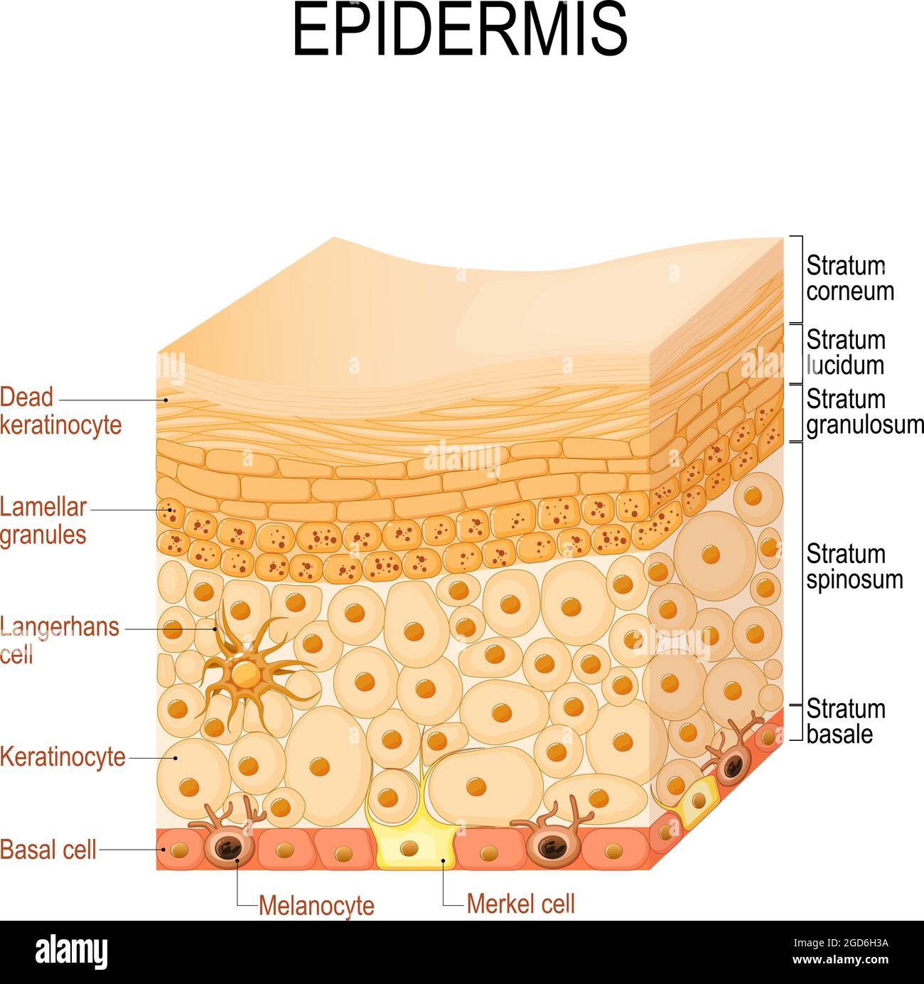 epidermis anatomy. layers and Cell structure of the human skin. Close-up of epidermis. Vector illustration Stock Vectorhttps://www.alamy.com/image-license-details/?v=1https://www.alamy.com/epidermis-anatomy-layers-and-cell-structure-of-the-human-skin-close-up-of-epidermis-vector-illustration-image438394862.html
RF2GD6H3A–epidermis anatomy. layers and Cell structure of the human skin. Close-up of epidermis. Vector illustration
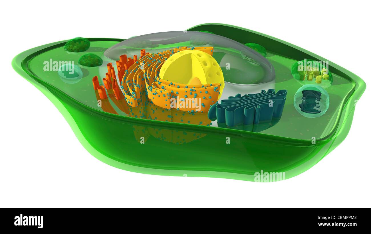 Computer illustration showing the internal structure of a plant cell. In addition to the chloroplast, the mitochondrion and the nucleus are cut through revealing their internal structures. Stock Photohttps://www.alamy.com/image-license-details/?v=1https://www.alamy.com/computer-illustration-showing-the-internal-structure-of-a-plant-cell-in-addition-to-the-chloroplast-the-mitochondrion-and-the-nucleus-are-cut-through-revealing-their-internal-structures-image357001235.html
RF2BMPPM3–Computer illustration showing the internal structure of a plant cell. In addition to the chloroplast, the mitochondrion and the nucleus are cut through revealing their internal structures.
 macro of round drops of oil mixed with green and red pigment floating on water, isolated on white Stock Photohttps://www.alamy.com/image-license-details/?v=1https://www.alamy.com/stock-photo-macro-of-round-drops-of-oil-mixed-with-green-and-red-pigment-floating-54423520.html
RFD4F5N4–macro of round drops of oil mixed with green and red pigment floating on water, isolated on white
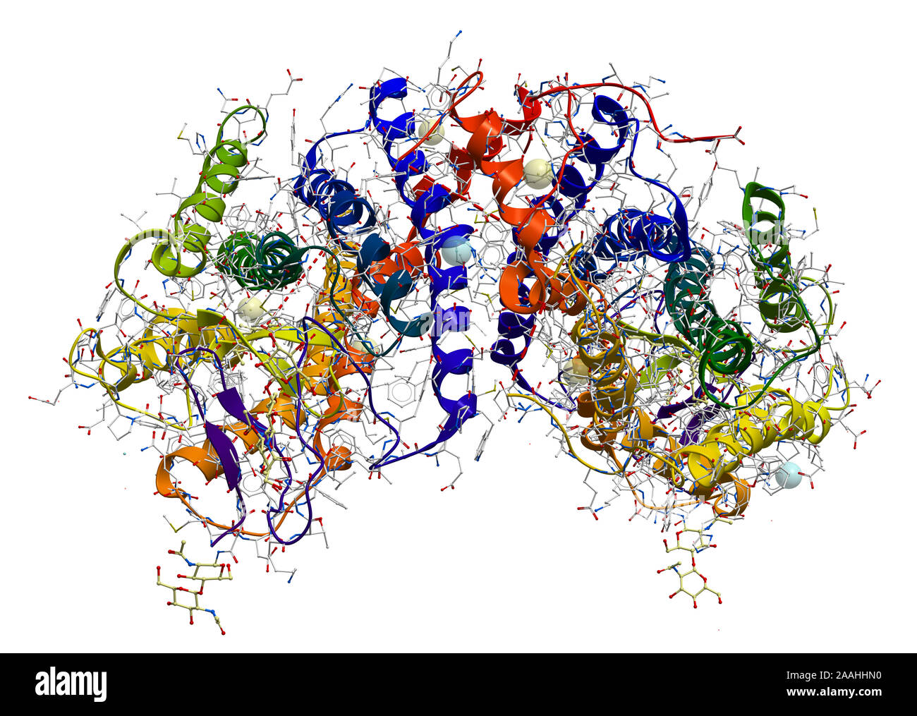 Rhodopsin (the extremely sensitive to light pigment involved in vision process) protein structure Stock Photohttps://www.alamy.com/image-license-details/?v=1https://www.alamy.com/rhodopsin-the-extremely-sensitive-to-light-pigment-involved-in-vision-process-protein-structure-image333530652.html
RF2AAHHN0–Rhodopsin (the extremely sensitive to light pigment involved in vision process) protein structure
RM2BE0GCG–Blood cells and platelets. Scanning electron micrograph (SEM) of human blood showing red and white cells and platelets. Red blood cells (erythrocytes) have a characteristic biconcave-disc shape and are numerous. These large cells contain hemoglobin, a red pigment by which oxygen is transported around the body. They are more numerous than white blood cells one of which is visible in this sample. White blood cells (leukocytes) are rounded cells with microvilli projections from the cell surface. Leucocytes play an important role in the immune response of the body. Platelets are smaller cells that
 3D image of Resazurin skeletal formula - molecular chemical structure of phenoxazine dye Alamar Blue isolated on white background Stock Photohttps://www.alamy.com/image-license-details/?v=1https://www.alamy.com/3d-image-of-resazurin-skeletal-formula-molecular-chemical-structure-of-phenoxazine-dye-alamar-blue-isolated-on-white-background-image500162026.html
RF2M1M9NE–3D image of Resazurin skeletal formula - molecular chemical structure of phenoxazine dye Alamar Blue isolated on white background
 Cellphone colour burst Stock Photohttps://www.alamy.com/image-license-details/?v=1https://www.alamy.com/stock-photo-cellphone-colour-burst-100909500.html
RFFT4R2M–Cellphone colour burst
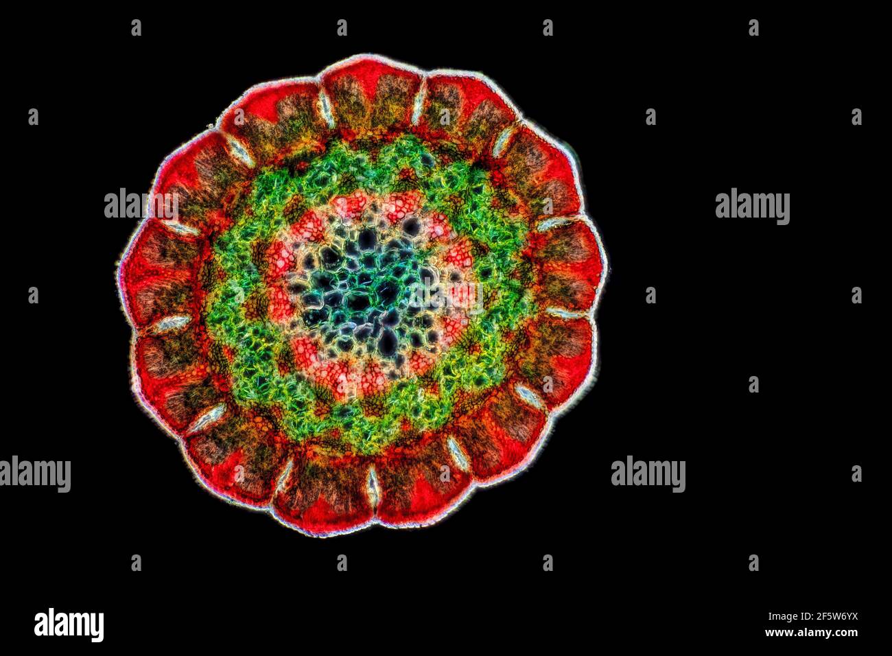 Coastal she-oak (Casuarina equisetifolia), leaf t.s., staining Wacker 3A, darkfield illumination Stock Photohttps://www.alamy.com/image-license-details/?v=1https://www.alamy.com/coastal-she-oak-casuarina-equisetifolia-leaf-ts-staining-wacker-3a-darkfield-illumination-image416676398.html
RF2F5W6YX–Coastal she-oak (Casuarina equisetifolia), leaf t.s., staining Wacker 3A, darkfield illumination
 Abstract illustration of green chlorophyll plant cells as pattern seamless Stock Photohttps://www.alamy.com/image-license-details/?v=1https://www.alamy.com/abstract-illustration-of-green-chlorophyll-plant-cells-as-pattern-seamless-image238712927.html
RFRTA8NK–Abstract illustration of green chlorophyll plant cells as pattern seamless
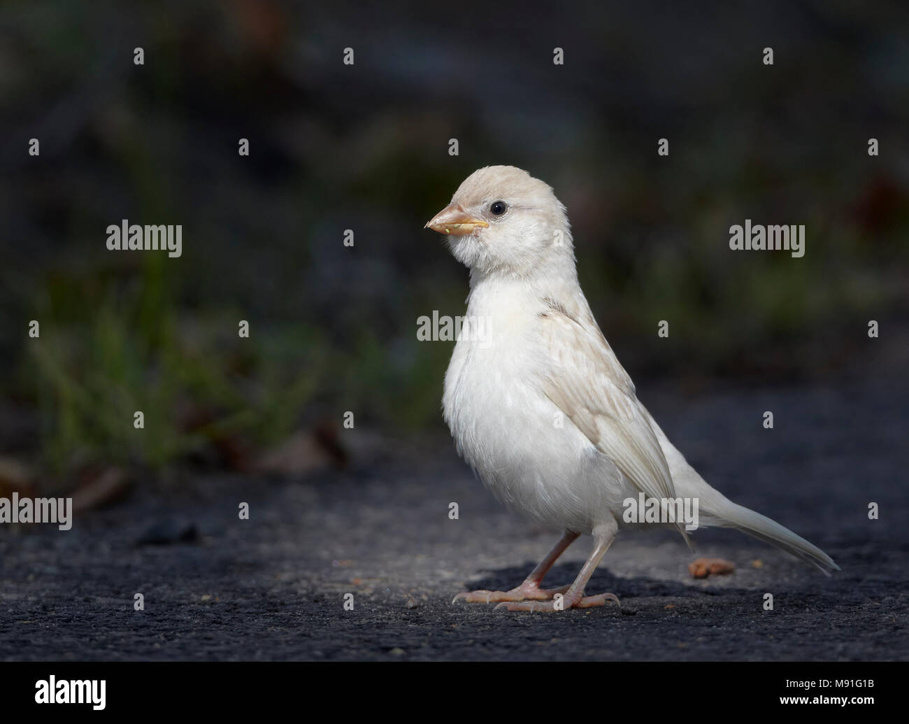 Leucistische onvolwassen Huismus; House Sparrow Finland July juvenile, leukism a general term for the phenotype resulting from defects in pigment cell Stock Photohttps://www.alamy.com/image-license-details/?v=1https://www.alamy.com/leucistische-onvolwassen-huismus-house-sparrow-finland-july-juvenile-leukism-a-general-term-for-the-phenotype-resulting-from-defects-in-pigment-cell-image177670119.html
RMM91G1B–Leucistische onvolwassen Huismus; House Sparrow Finland July juvenile, leukism a general term for the phenotype resulting from defects in pigment cell
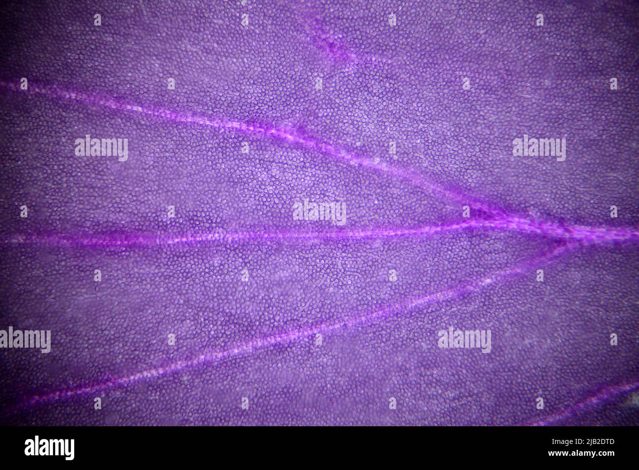 Microscope image of a flower petal showing individual cells. Frame size c5mm across Stock Photohttps://www.alamy.com/image-license-details/?v=1https://www.alamy.com/microscope-image-of-a-flower-petal-showing-individual-cells-frame-size-c5mm-across-image471495933.html
RM2JB2DTD–Microscope image of a flower petal showing individual cells. Frame size c5mm across
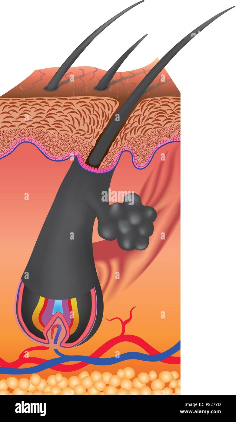 Hair follows a specific growth cycle with three distinct and concurrent phases anagen, catagen, and telogen phases Stock Vectorhttps://www.alamy.com/image-license-details/?v=1https://www.alamy.com/hair-follows-a-specific-growth-cycle-with-three-distinct-and-concurrent-phases-anagen-catagen-and-telogen-phases-image211491825.html
RFP827YD–Hair follows a specific growth cycle with three distinct and concurrent phases anagen, catagen, and telogen phases
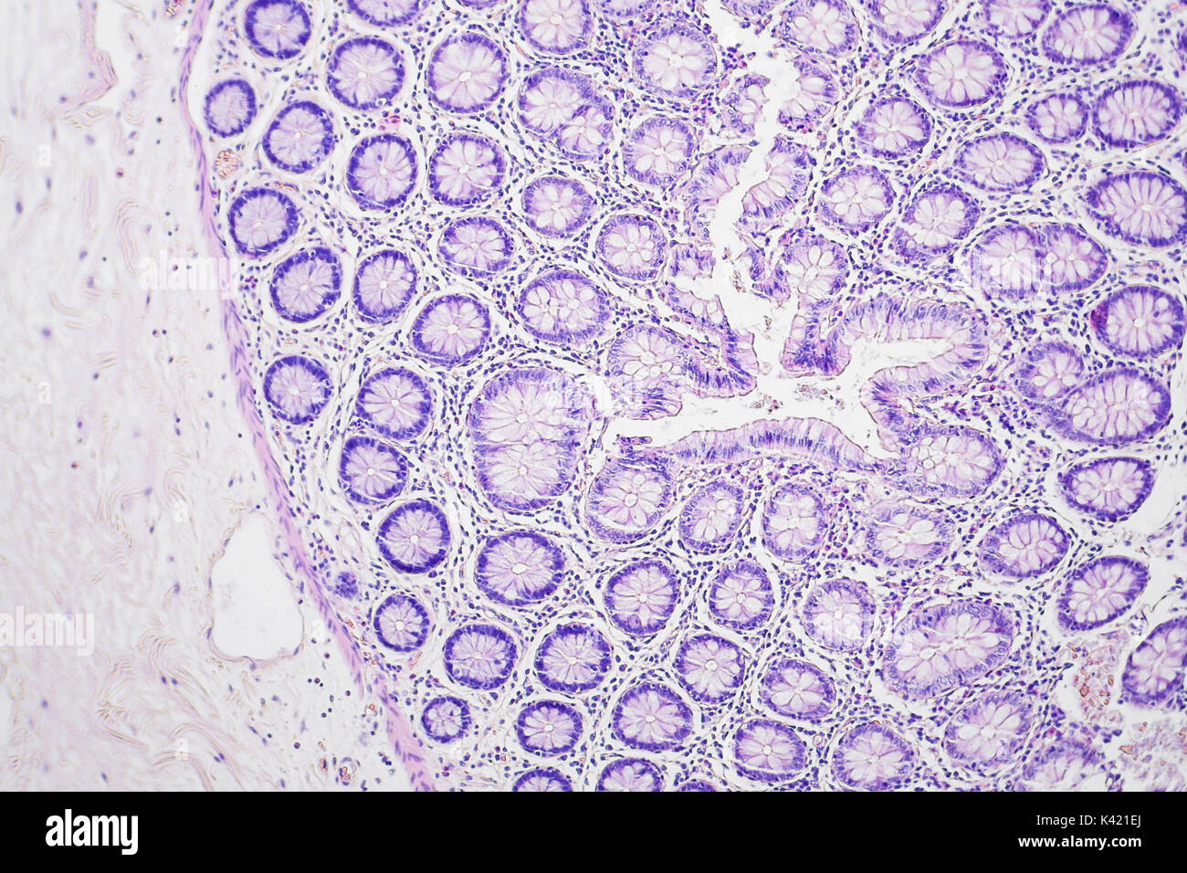 Colon cancer microscopic photography, magnification x100 Stock Photohttps://www.alamy.com/image-license-details/?v=1https://www.alamy.com/colon-cancer-microscopic-photography-magnification-x100-image157397034.html
RFK421EJ–Colon cancer microscopic photography, magnification x100
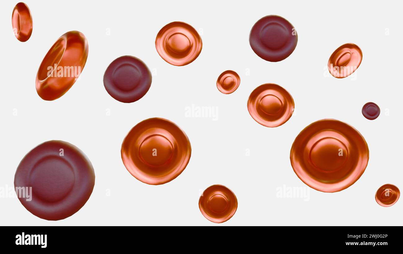 3d rendering of hypochromic red blood cells are red blood cells that have less color than normal when examined under a microscope. Stock Photohttps://www.alamy.com/image-license-details/?v=1https://www.alamy.com/3d-rendering-of-hypochromic-red-blood-cells-are-red-blood-cells-that-have-less-color-than-normal-when-examined-under-a-microscope-image596228942.html
RF2WJ0G2P–3d rendering of hypochromic red blood cells are red blood cells that have less color than normal when examined under a microscope.
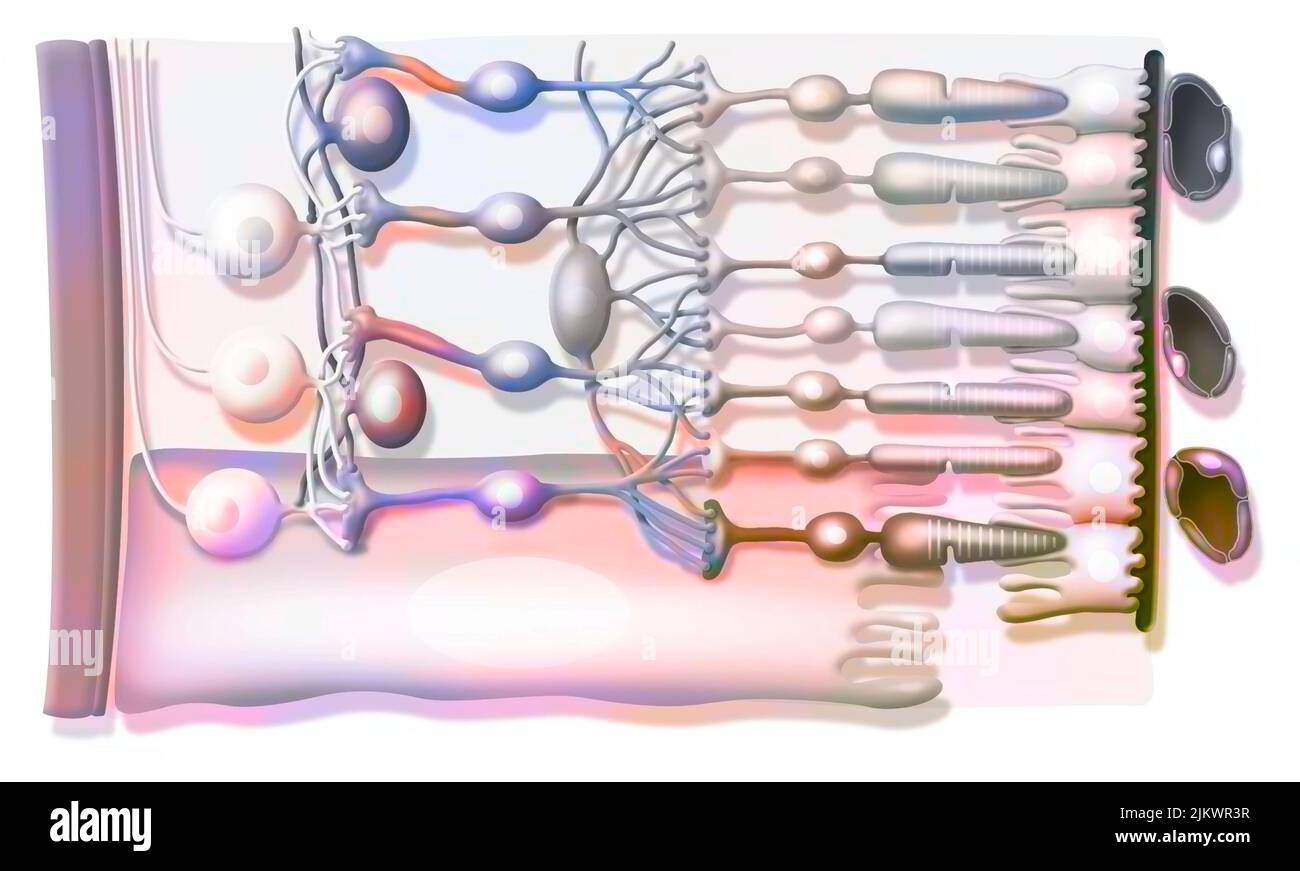 Zoom on the structure of the retina with vitreous body, internal limiting membrane, ganglion cells. Stock Photohttps://www.alamy.com/image-license-details/?v=1https://www.alamy.com/zoom-on-the-structure-of-the-retina-with-vitreous-body-internal-limiting-membrane-ganglion-cells-image476925339.html
RF2JKWR3R–Zoom on the structure of the retina with vitreous body, internal limiting membrane, ganglion cells.
 3d rendering, abstract, all, background, beautiful, biochemistry, blue, celebration, cell pigment, christmas, color, colorful, computer graphics, conf Stock Photohttps://www.alamy.com/image-license-details/?v=1https://www.alamy.com/3d-rendering-abstract-all-background-beautiful-biochemistry-blue-celebration-cell-pigment-christmas-color-colorful-computer-graphics-conf-image454733878.html
RF2HBPWKJ–3d rendering, abstract, all, background, beautiful, biochemistry, blue, celebration, cell pigment, christmas, color, colorful, computer graphics, conf
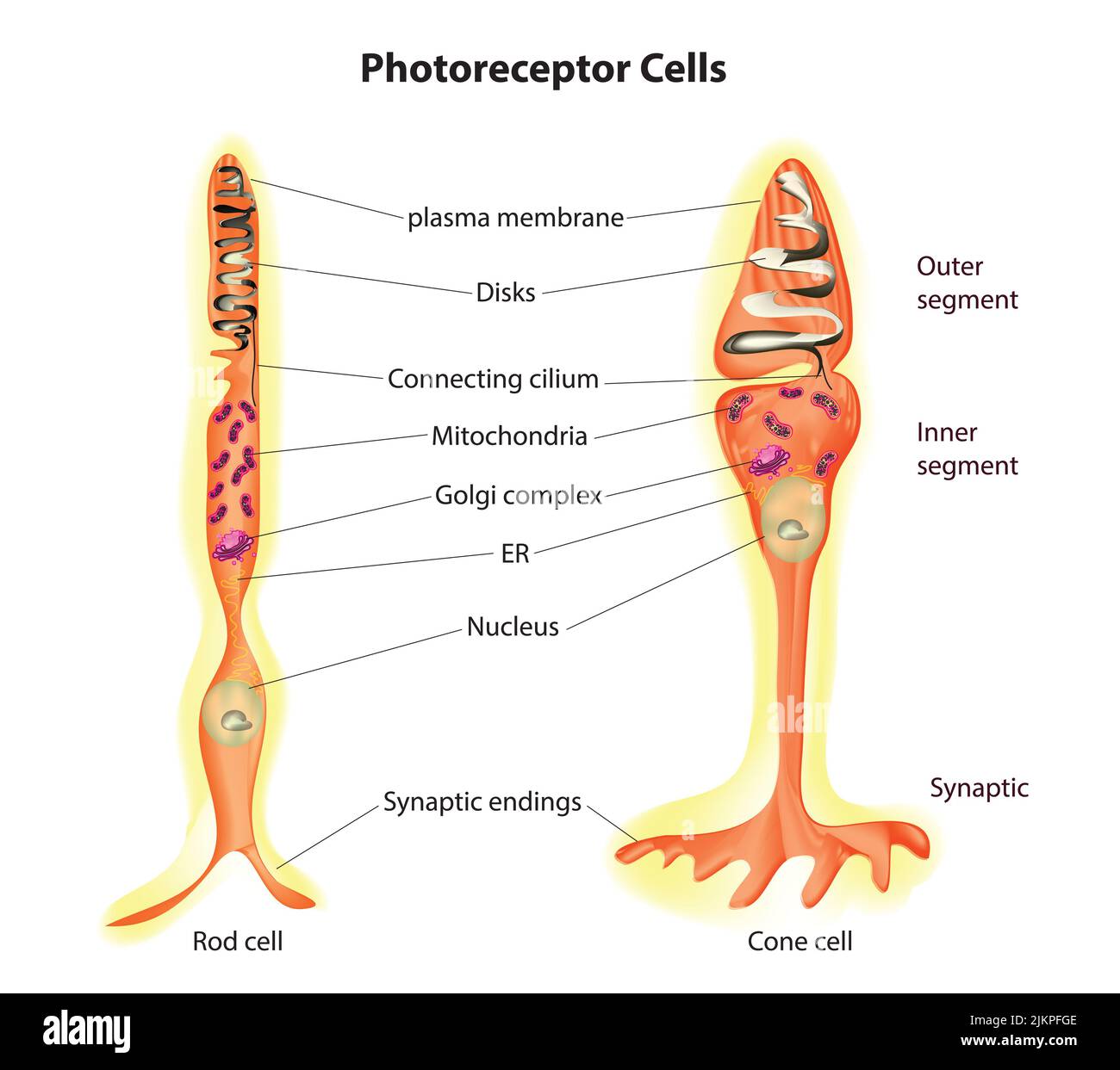 Photoreceptor cells (rods and cones) Stock Photohttps://www.alamy.com/image-license-details/?v=1https://www.alamy.com/photoreceptor-cells-rods-and-cones-image476853566.html
RF2JKPFGE–Photoreceptor cells (rods and cones)
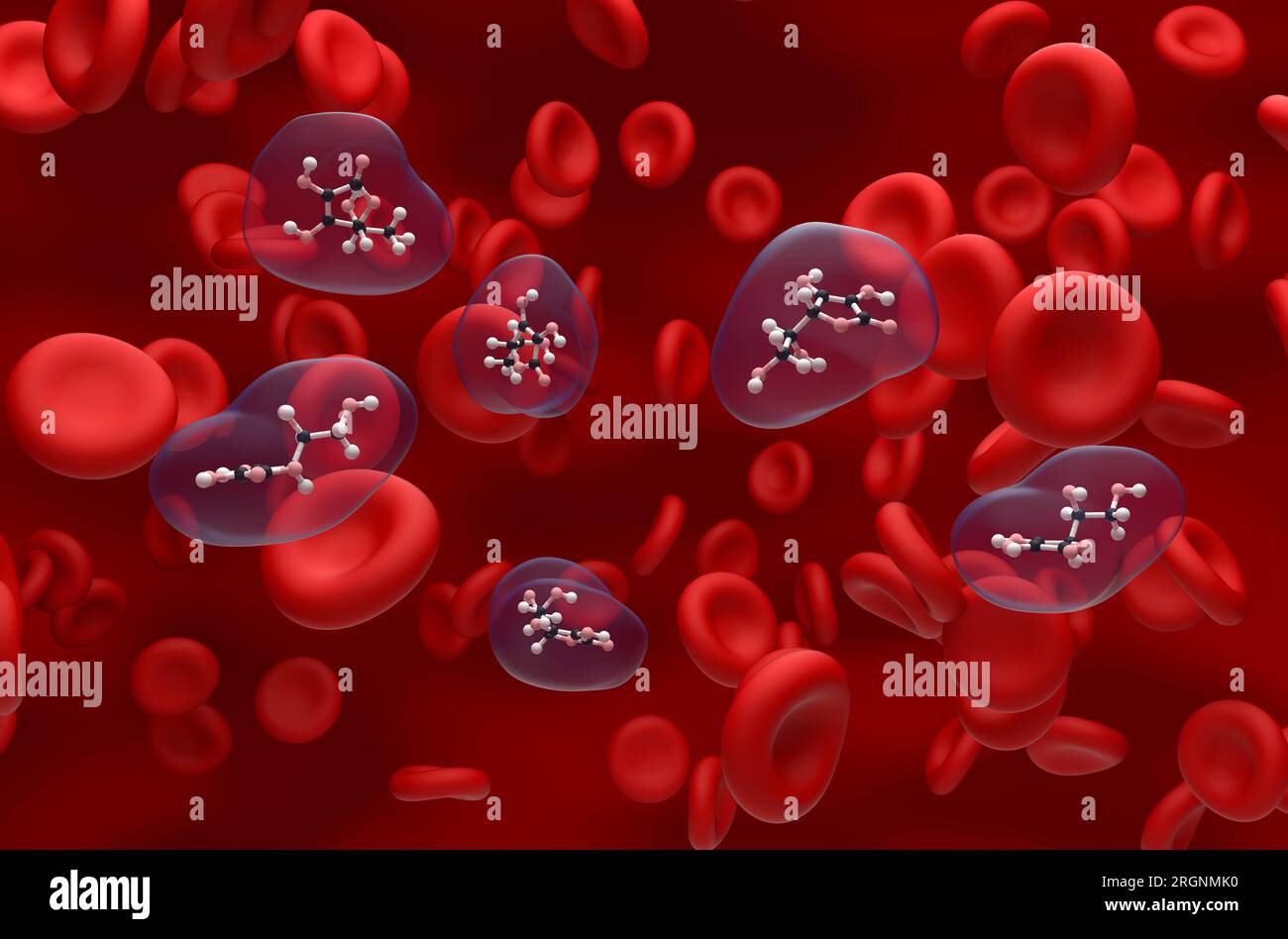 Vitamin c (ascorbic acid) structure in the blood flow ball and stick isometric view 3d illustration Stock Photohttps://www.alamy.com/image-license-details/?v=1https://www.alamy.com/vitamin-c-ascorbic-acid-structure-in-the-blood-flow-ball-and-stick-isometric-view-3d-illustration-image561043476.html
RF2RGNMK0–Vitamin c (ascorbic acid) structure in the blood flow ball and stick isometric view 3d illustration
 Friends taking selfie covered in pigment powder Stock Photohttps://www.alamy.com/image-license-details/?v=1https://www.alamy.com/stock-photo-friends-taking-selfie-covered-in-pigment-powder-86030925.html
RFEYY191–Friends taking selfie covered in pigment powder
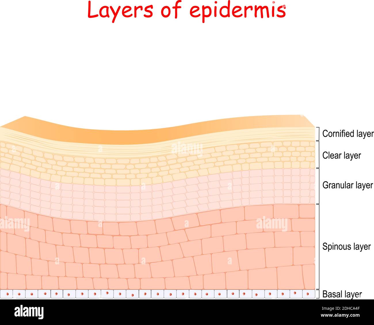 epidermis. Cell structure of layers: stratum corneum, lucidum, stratum granulosum, spinosum, and germinativum. Vector illustration Stock Vectorhttps://www.alamy.com/image-license-details/?v=1https://www.alamy.com/epidermis-cell-structure-of-layers-stratum-corneum-lucidum-stratum-granulosum-spinosum-and-germinativum-vector-illustration-image389348639.html
RF2DHCA4F–epidermis. Cell structure of layers: stratum corneum, lucidum, stratum granulosum, spinosum, and germinativum. Vector illustration
 Skin pigmentation, illustration Stock Photohttps://www.alamy.com/image-license-details/?v=1https://www.alamy.com/skin-pigmentation-illustration-image478059167.html
RF2JNND9K–Skin pigmentation, illustration
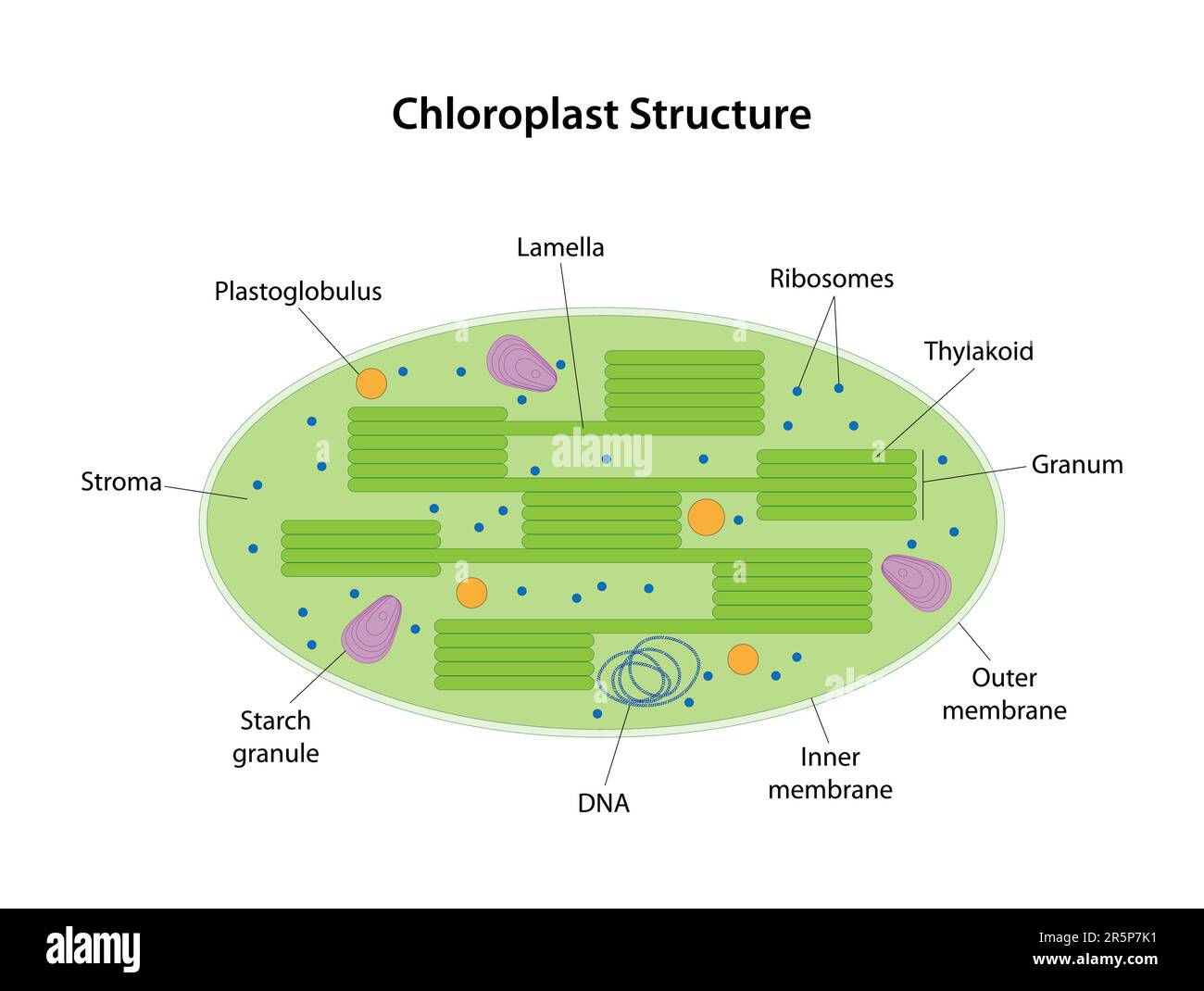 Chloroplast is a two-membrane organelle of a plant cell. On the inner membranes of the chloroplast contains the green pigment chlorophyll. Stock Vectorhttps://www.alamy.com/image-license-details/?v=1https://www.alamy.com/chloroplast-is-a-two-membrane-organelle-of-a-plant-cell-on-the-inner-membranes-of-the-chloroplast-contains-the-green-pigment-chlorophyll-image554294021.html
RF2R5P7K1–Chloroplast is a two-membrane organelle of a plant cell. On the inner membranes of the chloroplast contains the green pigment chlorophyll.
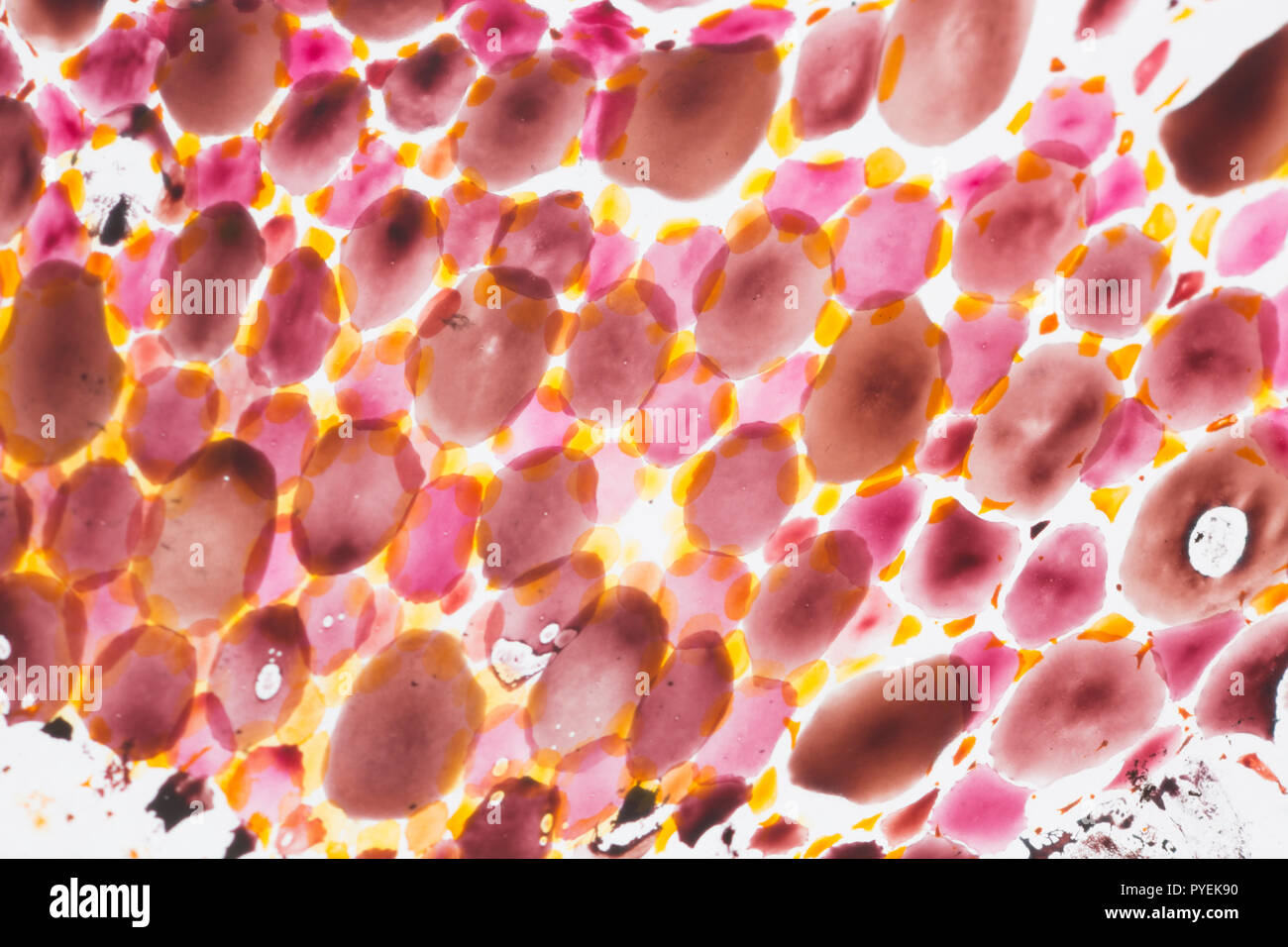 A close up/macro photograph of chromatophores in the skin of a Loligo vulgaris squid caught on rod and line in the UK. The chromatophores are cells th Stock Photohttps://www.alamy.com/image-license-details/?v=1https://www.alamy.com/a-close-upmacro-photograph-of-chromatophores-in-the-skin-of-a-loligo-vulgaris-squid-caught-on-rod-and-line-in-the-uk-the-chromatophores-are-cells-th-image223442604.html
RMPYEK90–A close up/macro photograph of chromatophores in the skin of a Loligo vulgaris squid caught on rod and line in the UK. The chromatophores are cells th
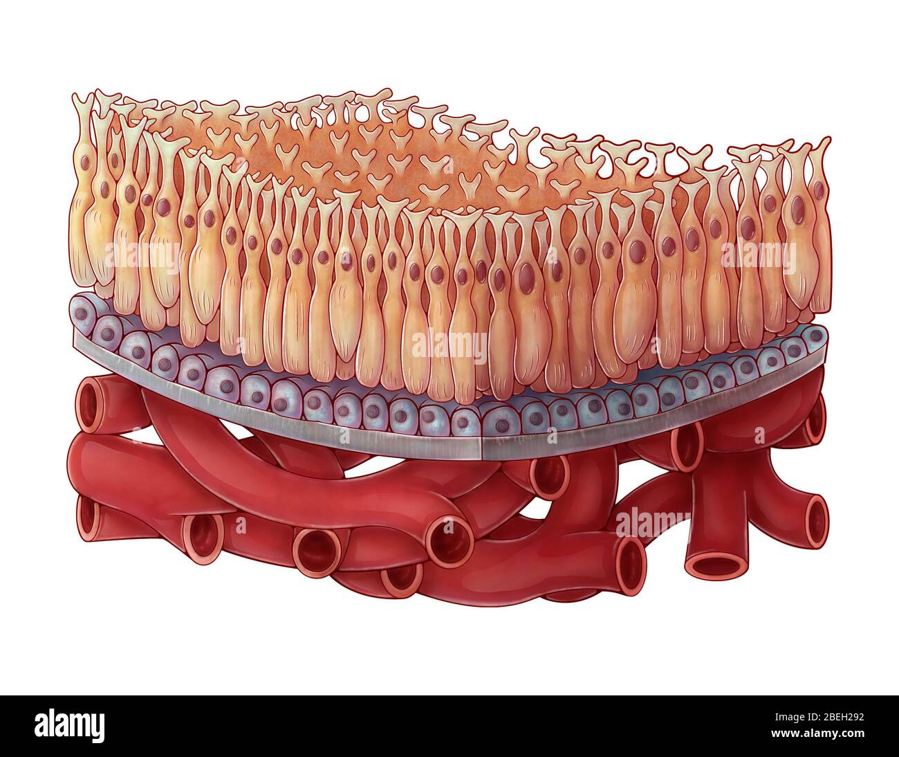 Macula of the Retina, Illustration Stock Photohttps://www.alamy.com/image-license-details/?v=1https://www.alamy.com/macula-of-the-retina-illustration-image353187550.html
RM2BEH292–Macula of the Retina, Illustration
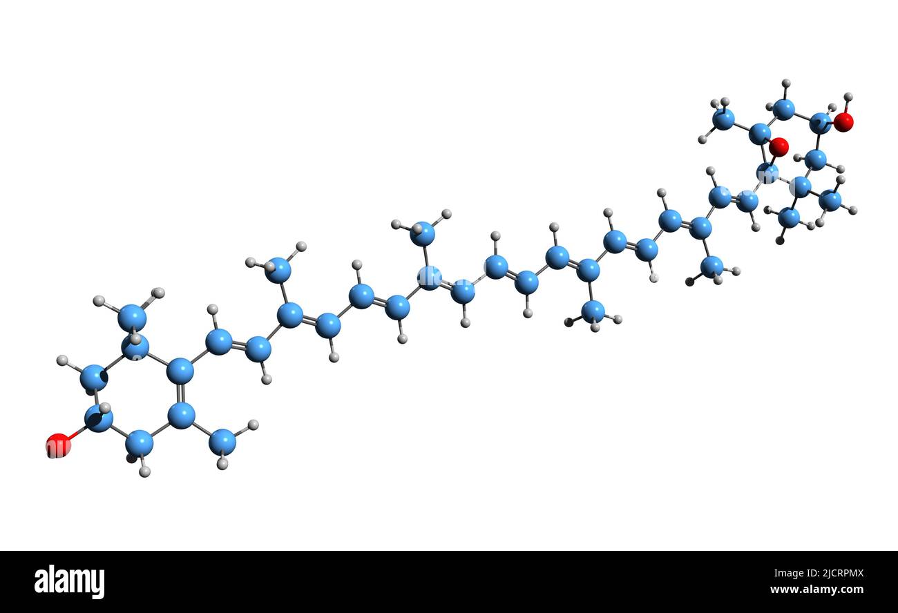 3D image of Diadinoxanthin skeletal formula - molecular chemical structure of phytoplankton pigment isolated on white background Stock Photohttps://www.alamy.com/image-license-details/?v=1https://www.alamy.com/3d-image-of-diadinoxanthin-skeletal-formula-molecular-chemical-structure-of-phytoplankton-pigment-isolated-on-white-background-image472578538.html
RF2JCRPMX–3D image of Diadinoxanthin skeletal formula - molecular chemical structure of phytoplankton pigment isolated on white background
 Cellphone colour burst Stock Photohttps://www.alamy.com/image-license-details/?v=1https://www.alamy.com/stock-photo-cellphone-colour-burst-109775265.html
RFGAGKD5–Cellphone colour burst
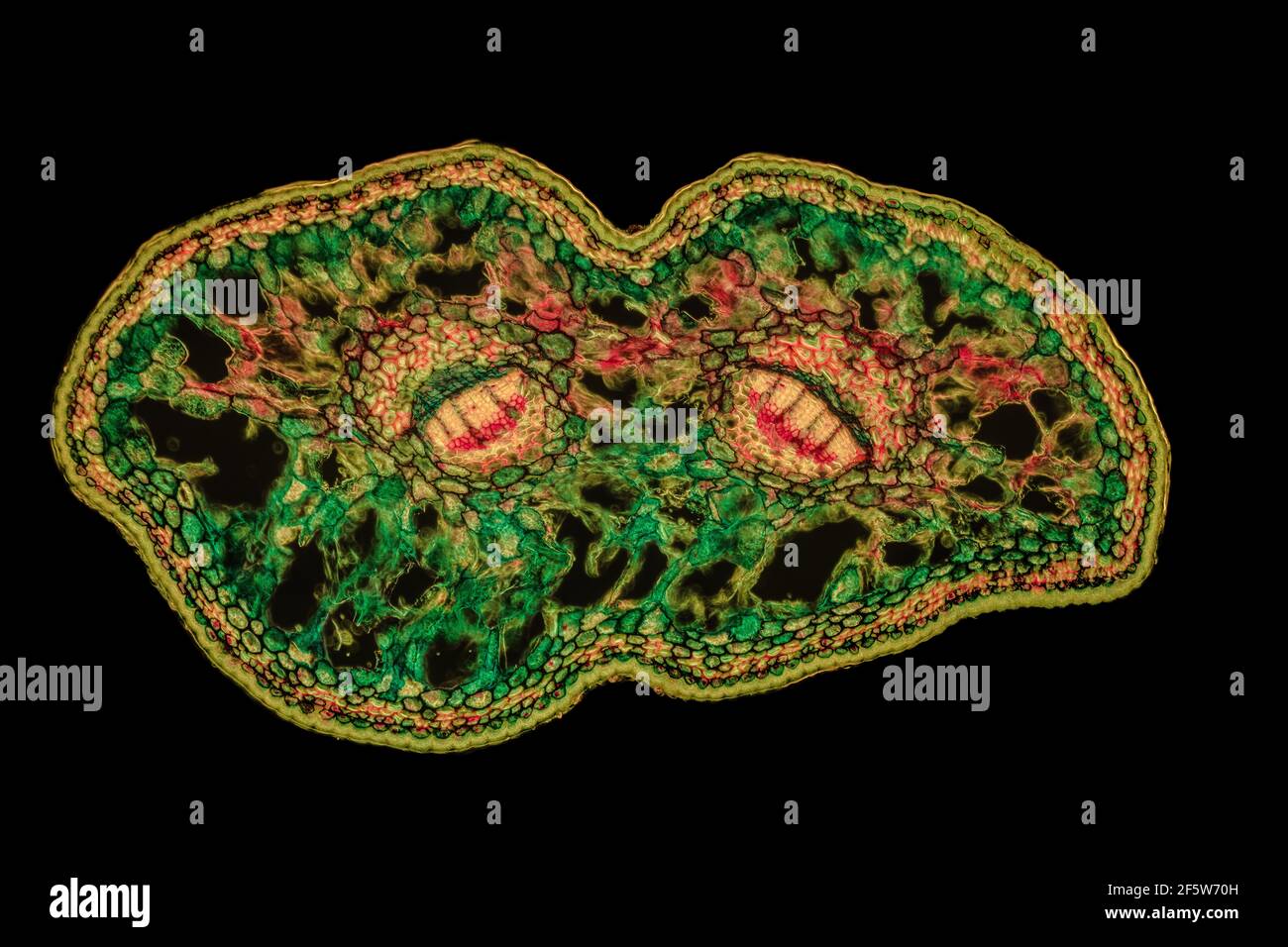 Japanese umbrella fir (Sciadopitys verticillata), needle t.s., staining Wacker 3A, fluorescence and darkfield transmitted light Stock Photohttps://www.alamy.com/image-license-details/?v=1https://www.alamy.com/japanese-umbrella-fir-sciadopitys-verticillata-needle-ts-staining-wacker-3a-fluorescence-and-darkfield-transmitted-light-image416676417.html
RF2F5W70H–Japanese umbrella fir (Sciadopitys verticillata), needle t.s., staining Wacker 3A, fluorescence and darkfield transmitted light
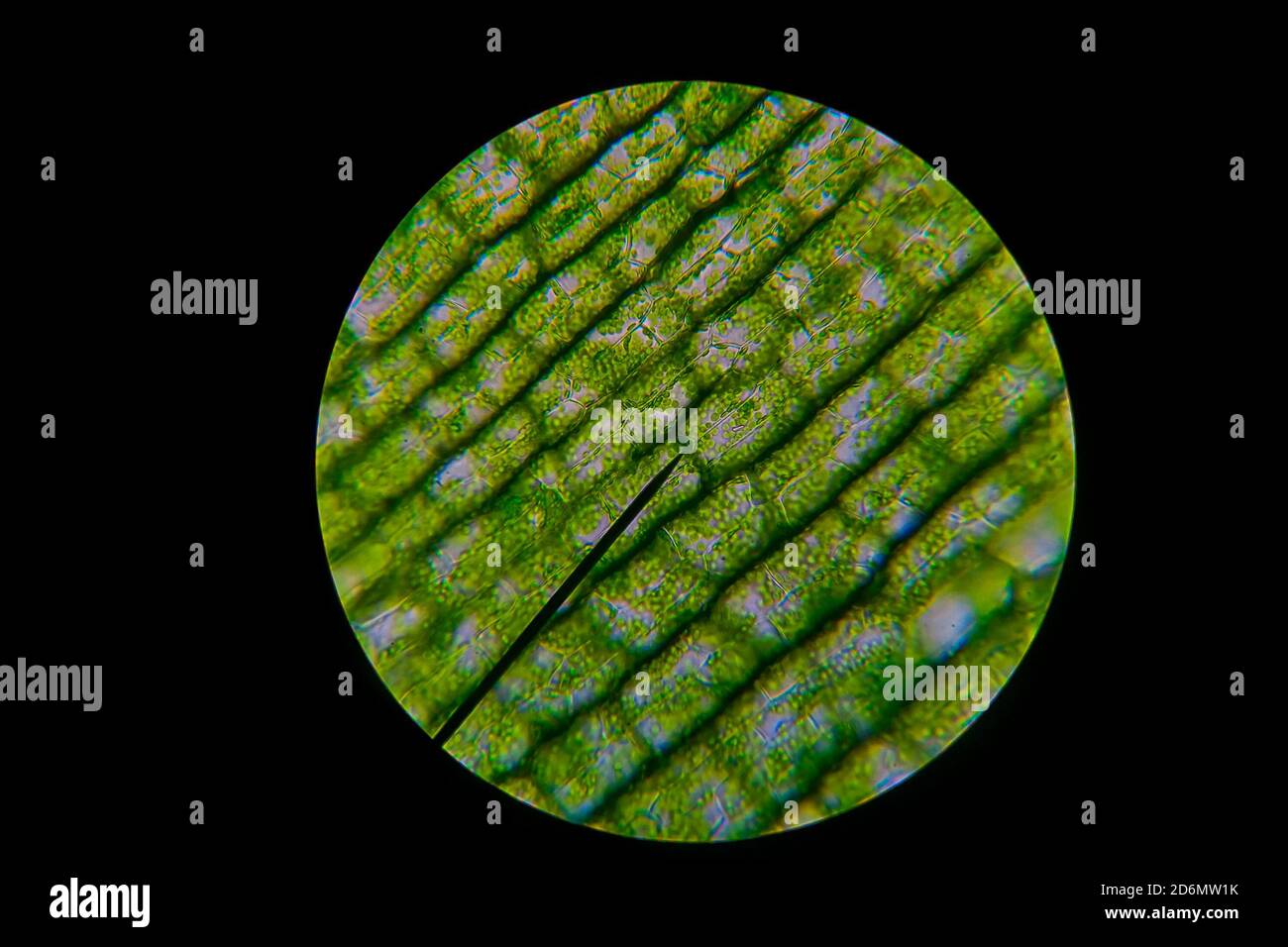 Green leaf grains also known as chloroplasts in cells of a waterweed seen through a microscope. Biology experiment. Stock Photohttps://www.alamy.com/image-license-details/?v=1https://www.alamy.com/green-leaf-grains-also-known-as-chloroplasts-in-cells-of-a-waterweed-seen-through-a-microscope-biology-experiment-image382774719.html
RF2D6MW1K–Green leaf grains also known as chloroplasts in cells of a waterweed seen through a microscope. Biology experiment.
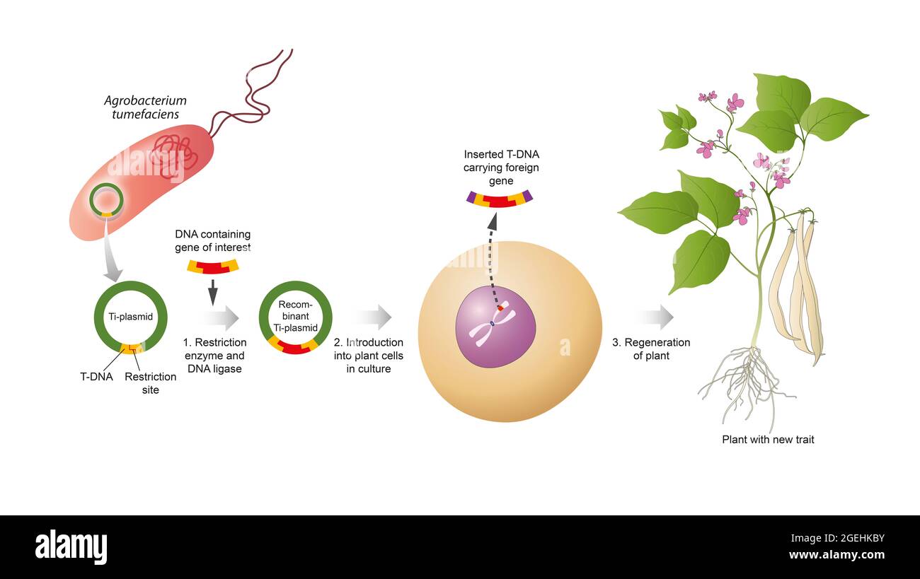 Plant genetic engineering, also known as plant genetic modification or manipulation Stock Photohttps://www.alamy.com/image-license-details/?v=1https://www.alamy.com/plant-genetic-engineering-also-known-as-plant-genetic-modification-or-manipulation-image439252799.html
RF2GEHKBY–Plant genetic engineering, also known as plant genetic modification or manipulation
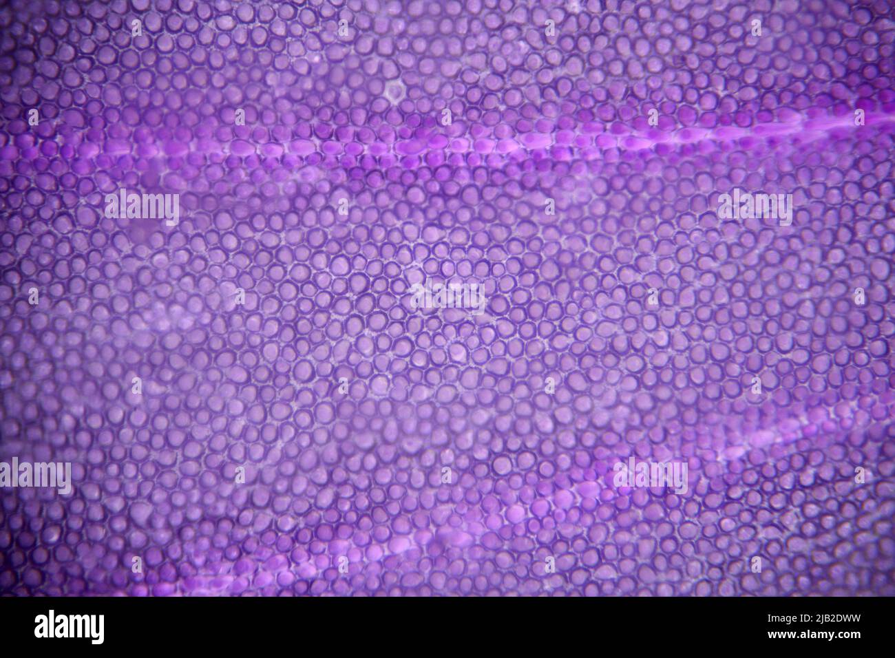 Microscope image of a flower petal showing individual cells. Frame size c2mm across Stock Photohttps://www.alamy.com/image-license-details/?v=1https://www.alamy.com/microscope-image-of-a-flower-petal-showing-individual-cells-frame-size-c2mm-across-image471495973.html
RM2JB2DWW–Microscope image of a flower petal showing individual cells. Frame size c2mm across
 Professional watercolor paints in box with brushes. Stock Photohttps://www.alamy.com/image-license-details/?v=1https://www.alamy.com/professional-watercolor-paints-in-box-with-brushes-image226248791.html
RFR42EHY–Professional watercolor paints in box with brushes.
 Metastatic melanoma cells inside body Stock Photohttps://www.alamy.com/image-license-details/?v=1https://www.alamy.com/metastatic-melanoma-cells-inside-body-image606796169.html
RF2X75XKN–Metastatic melanoma cells inside body
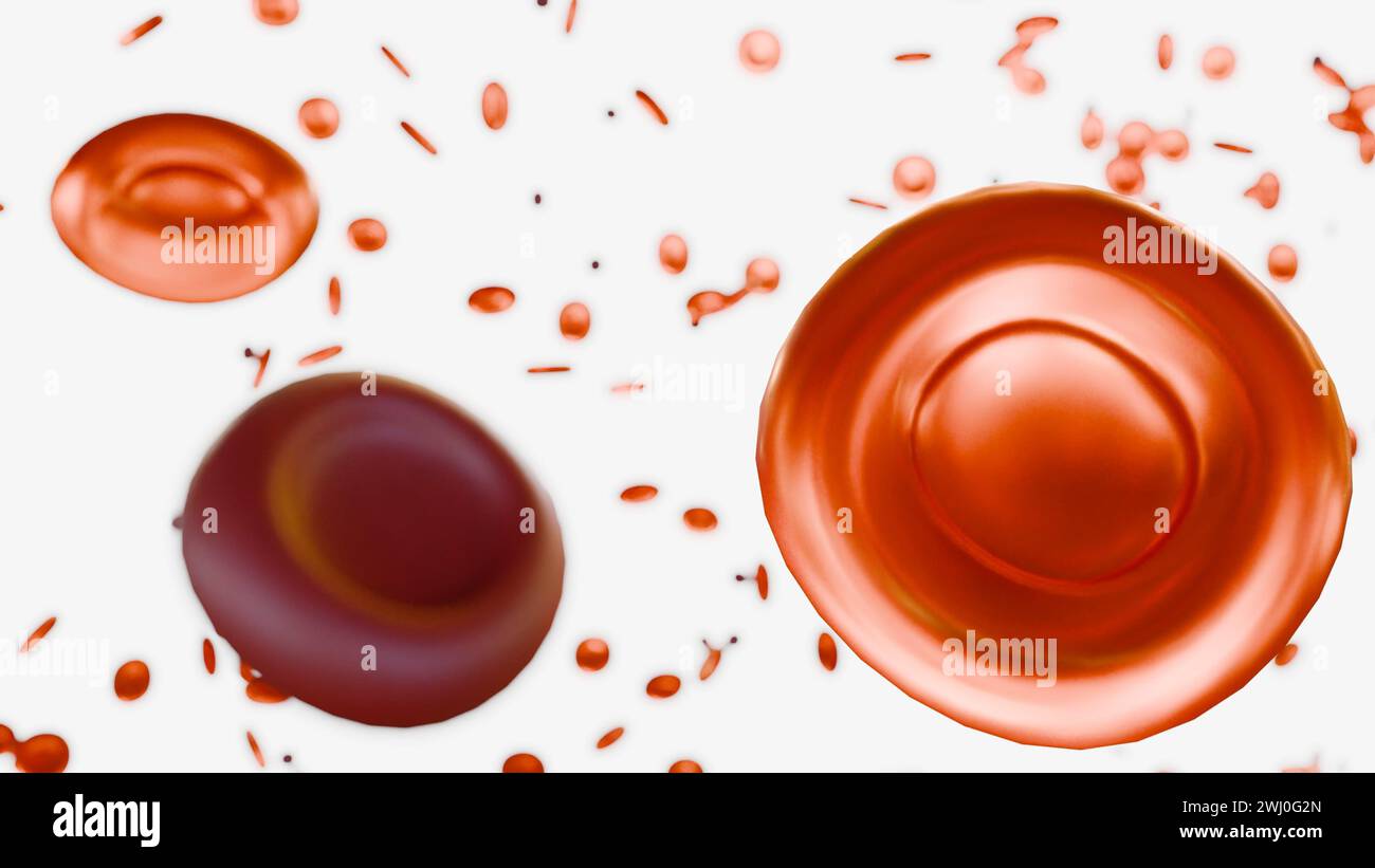 3d rendering of hypochromic red blood cells are red blood cells that have less color than normal when examined under a microscope. Stock Photohttps://www.alamy.com/image-license-details/?v=1https://www.alamy.com/3d-rendering-of-hypochromic-red-blood-cells-are-red-blood-cells-that-have-less-color-than-normal-when-examined-under-a-microscope-image596228941.html
RF2WJ0G2N–3d rendering of hypochromic red blood cells are red blood cells that have less color than normal when examined under a microscope.
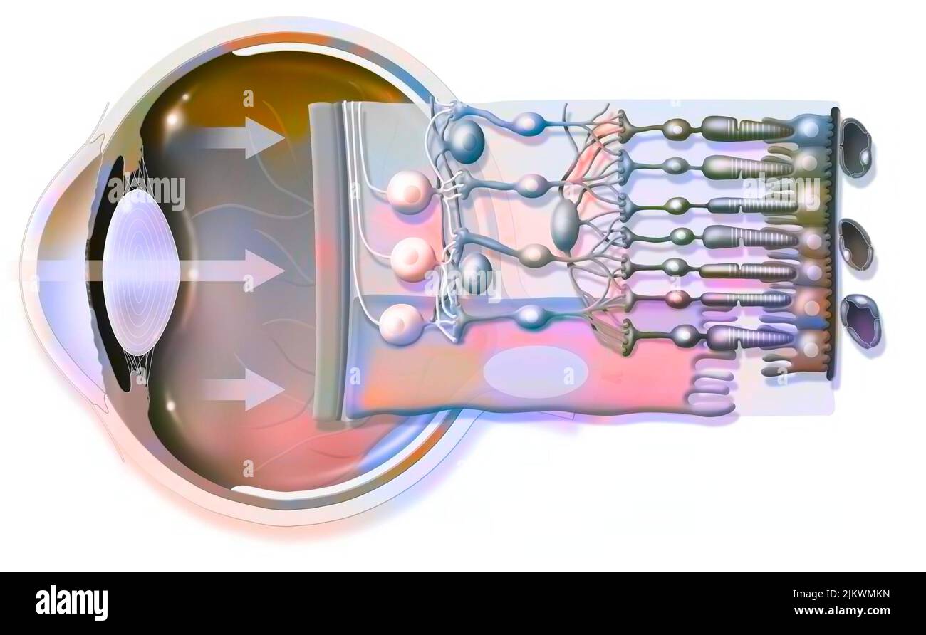 The eye and retina with the vitreous, the internal limiting membrane. Stock Photohttps://www.alamy.com/image-license-details/?v=1https://www.alamy.com/the-eye-and-retina-with-the-vitreous-the-internal-limiting-membrane-image476923433.html
RF2JKWMKN–The eye and retina with the vitreous, the internal limiting membrane.
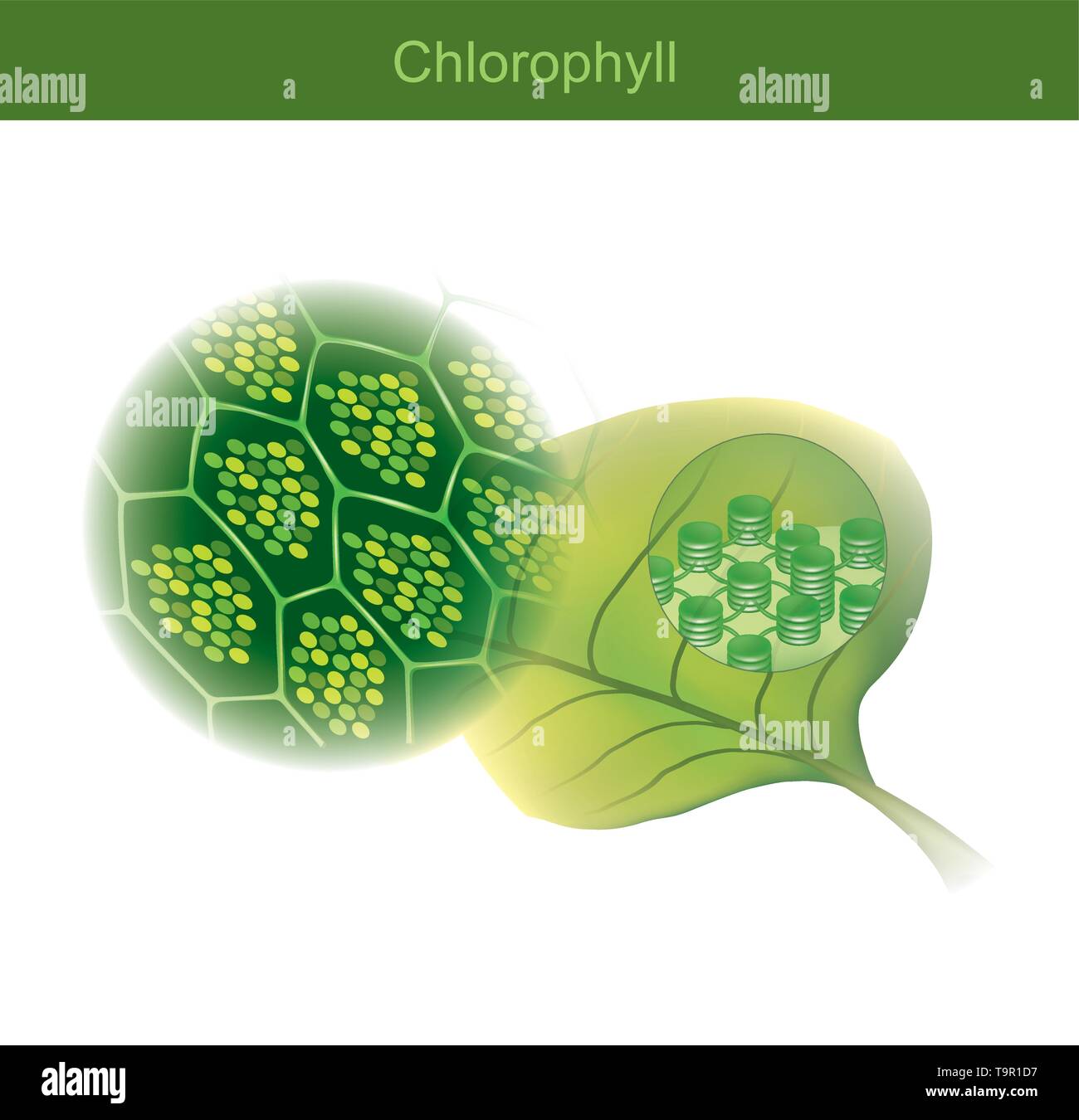 Chlorophyll is a green photosynthetic pigment found in plants, Chlorophyll molecules are specifically arranged in and around pigment protein complexes Stock Vectorhttps://www.alamy.com/image-license-details/?v=1https://www.alamy.com/chlorophyll-is-a-green-photosynthetic-pigment-found-in-plants-chlorophyll-molecules-are-specifically-arranged-in-and-around-pigment-protein-complexes-image246983107.html
RFT9R1D7–Chlorophyll is a green photosynthetic pigment found in plants, Chlorophyll molecules are specifically arranged in and around pigment protein complexes
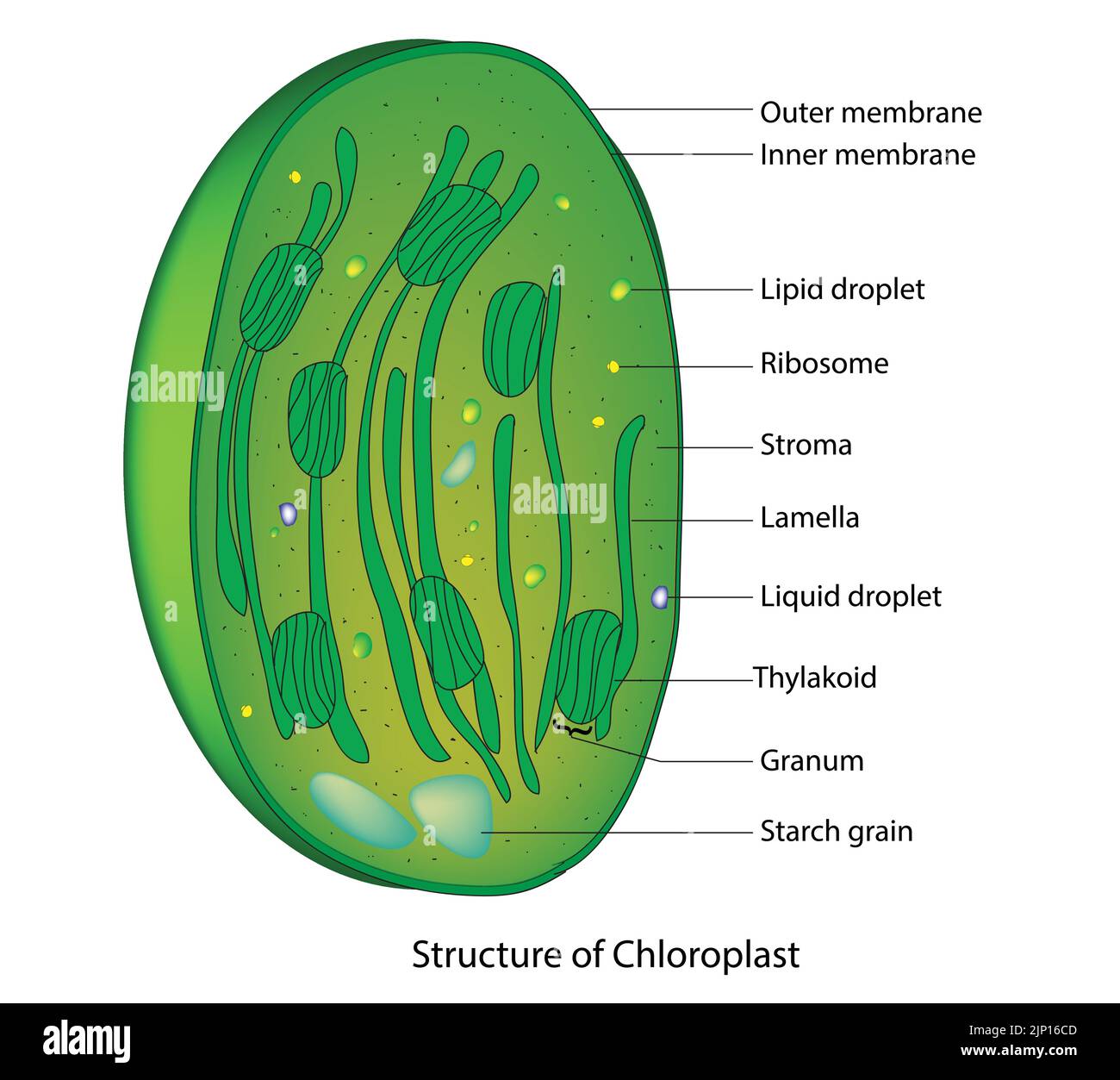 Chloroplast Anatomy Stock Vectorhttps://www.alamy.com/image-license-details/?v=1https://www.alamy.com/chloroplast-anatomy-image478229373.html
RF2JP16CD–Chloroplast Anatomy
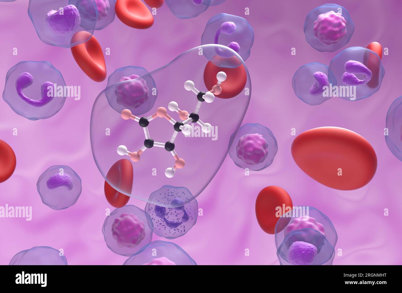 Vitamin c (ascorbic acid) structure in the blood flow ball and stick closeup view 3d illustration Stock Photohttps://www.alamy.com/image-license-details/?v=1https://www.alamy.com/vitamin-c-ascorbic-acid-structure-in-the-blood-flow-ball-and-stick-closeup-view-3d-illustration-image561043444.html
RF2RGNMHT–Vitamin c (ascorbic acid) structure in the blood flow ball and stick closeup view 3d illustration
 Friends covered in pigment powder using selfie stick Stock Photohttps://www.alamy.com/image-license-details/?v=1https://www.alamy.com/stock-photo-friends-covered-in-pigment-powder-using-selfie-stick-86030942.html
RFEYY19J–Friends covered in pigment powder using selfie stick
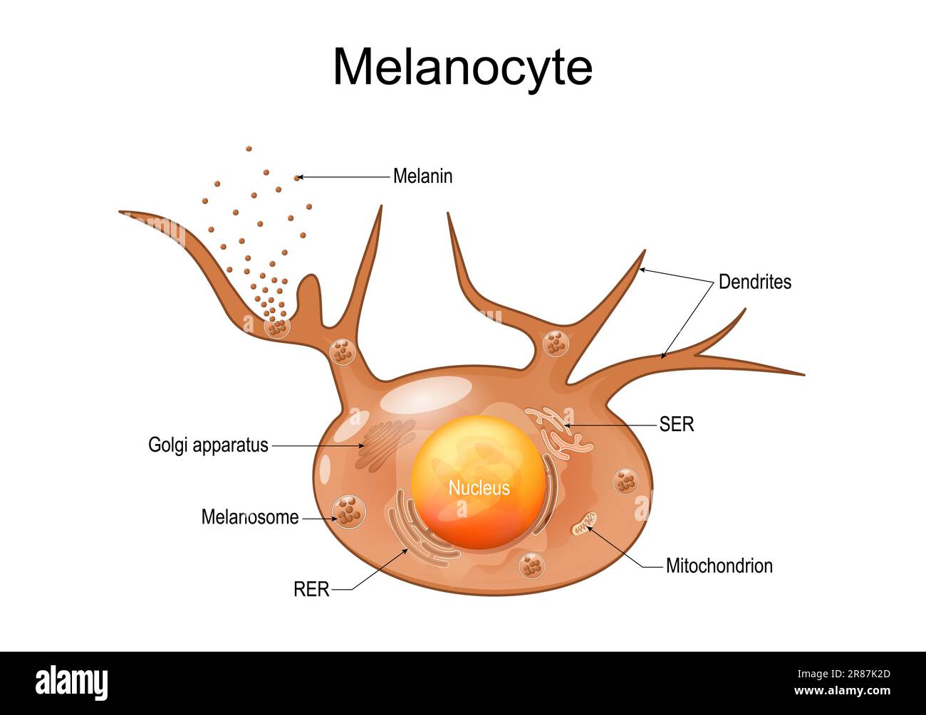 Melanocyte structure and anatomy. melanin producing cells. Melanin is the pigment responsible for skin color. vector poster Stock Vectorhttps://www.alamy.com/image-license-details/?v=1https://www.alamy.com/melanocyte-structure-and-anatomy-melanin-producing-cells-melanin-is-the-pigment-responsible-for-skin-color-vector-poster-image555817653.html
RF2R87K2D–Melanocyte structure and anatomy. melanin producing cells. Melanin is the pigment responsible for skin color. vector poster
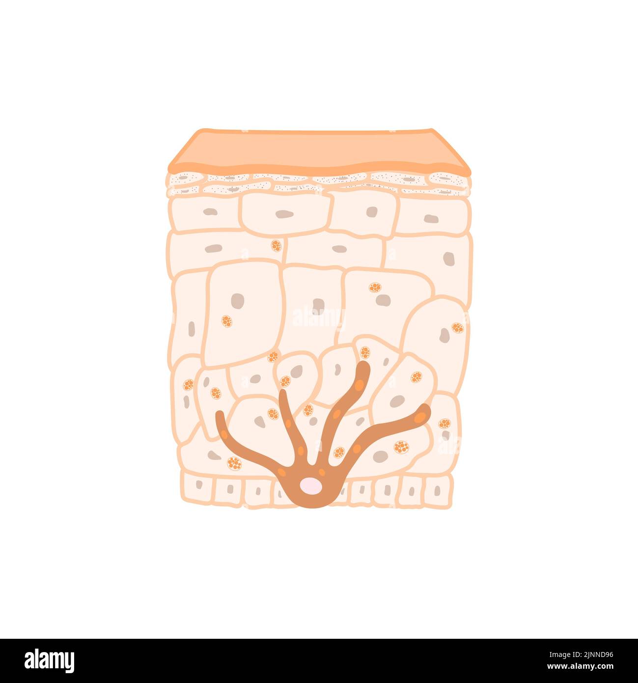 Skin pigmentation, illustration Stock Photohttps://www.alamy.com/image-license-details/?v=1https://www.alamy.com/skin-pigmentation-illustration-image478059154.html
RF2JNND96–Skin pigmentation, illustration
 macro of oil drops and pigment on water surface with bright background Stock Photohttps://www.alamy.com/image-license-details/?v=1https://www.alamy.com/stock-photo-macro-of-oil-drops-and-pigment-on-water-surface-with-bright-background-74999788.html
RFEA0EYT–macro of oil drops and pigment on water surface with bright background
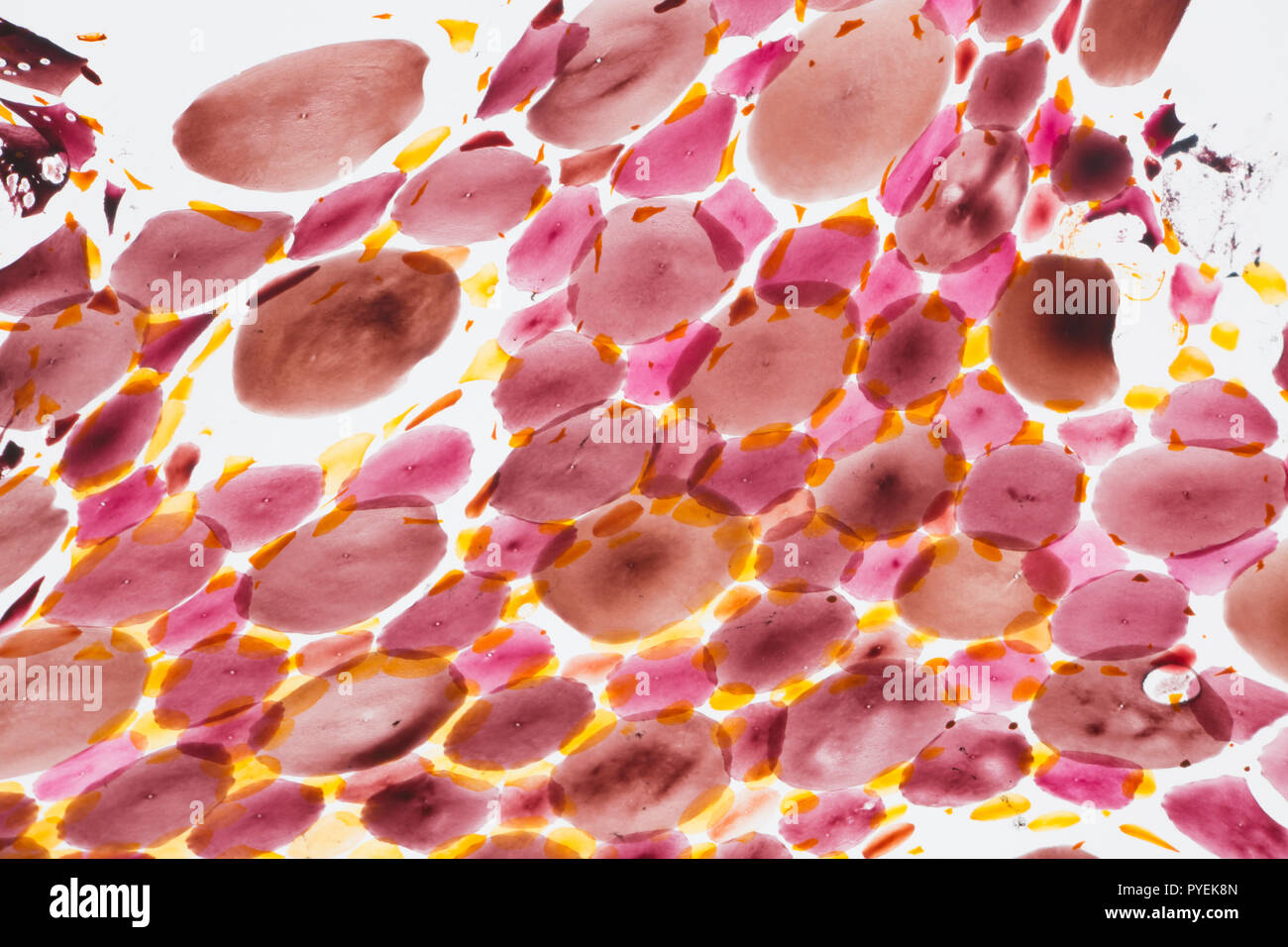 A close up/macro photograph of chromatophores in the skin of a Loligo vulgaris squid caught on rod and line in the UK. The chromatophores are cells th Stock Photohttps://www.alamy.com/image-license-details/?v=1https://www.alamy.com/a-close-upmacro-photograph-of-chromatophores-in-the-skin-of-a-loligo-vulgaris-squid-caught-on-rod-and-line-in-the-uk-the-chromatophores-are-cells-th-image223442597.html
RMPYEK8N–A close up/macro photograph of chromatophores in the skin of a Loligo vulgaris squid caught on rod and line in the UK. The chromatophores are cells th
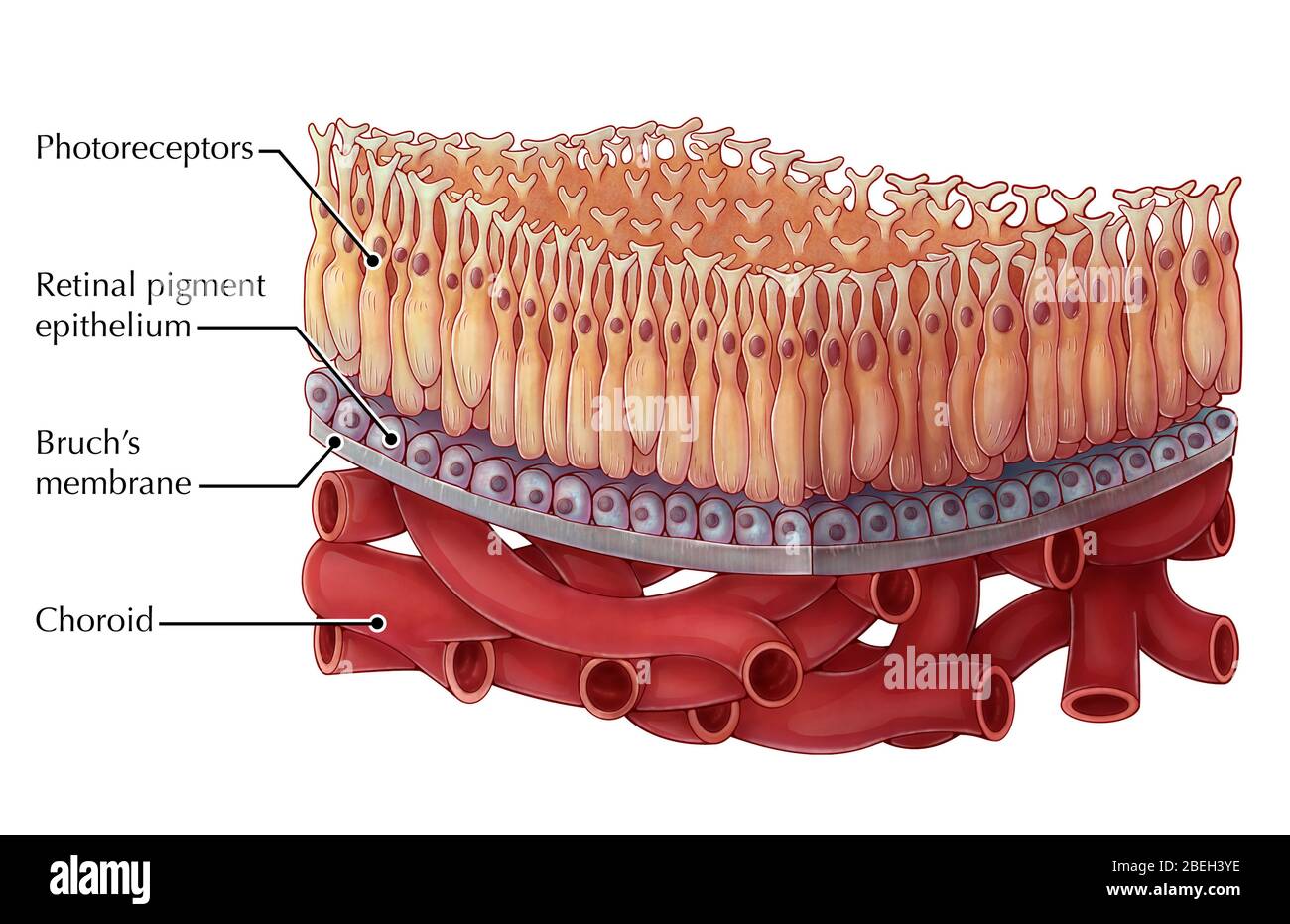 Macula of the Retina, Illustration Stock Photohttps://www.alamy.com/image-license-details/?v=1https://www.alamy.com/macula-of-the-retina-illustration-image353188850.html
RM2BEH3YE–Macula of the Retina, Illustration
 3D image of Rubrofusarin skeletal formula - molecular chemical structure of benzochromenone isolated on white background Stock Photohttps://www.alamy.com/image-license-details/?v=1https://www.alamy.com/3d-image-of-rubrofusarin-skeletal-formula-molecular-chemical-structure-of-benzochromenone-isolated-on-white-background-image500163176.html
RF2M1MB6G–3D image of Rubrofusarin skeletal formula - molecular chemical structure of benzochromenone isolated on white background
 Cellphone colorful multimedia Stock Photohttps://www.alamy.com/image-license-details/?v=1https://www.alamy.com/stock-photo-cellphone-colorful-multimedia-101206449.html
RFFTJ9T1–Cellphone colorful multimedia
 European Birthwort n Stock Photohttps://www.alamy.com/image-license-details/?v=1https://www.alamy.com/european-birthwort-n-image416676425.html
RF2F5W70W–European Birthwort n
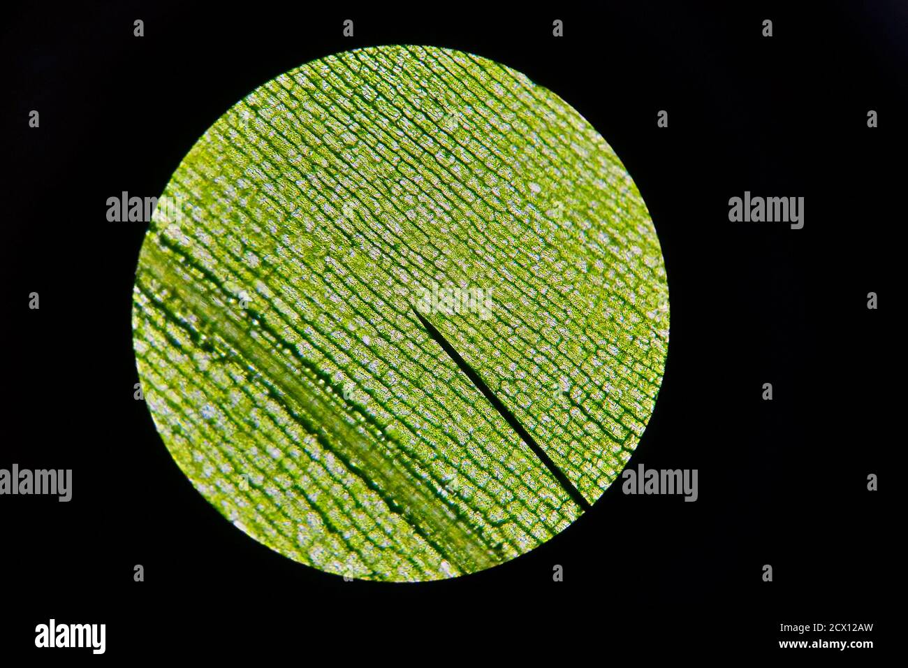 Detailed view of the cells of a leaf of waterweed as seen through a microscope. Biology experiment. Stock Photohttps://www.alamy.com/image-license-details/?v=1https://www.alamy.com/detailed-view-of-the-cells-of-a-leaf-of-waterweed-as-seen-through-a-microscope-biology-experiment-image377422609.html
RF2CX12AW–Detailed view of the cells of a leaf of waterweed as seen through a microscope. Biology experiment.
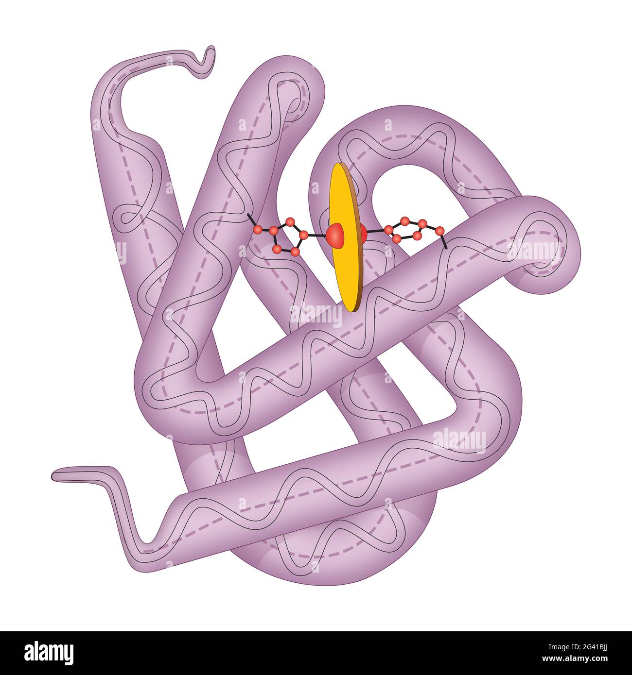 Structure of human myoglobin molecule Stock Photohttps://www.alamy.com/image-license-details/?v=1https://www.alamy.com/structure-of-human-myoglobin-molecule-image432748922.html
RF2G41BJJ–Structure of human myoglobin molecule
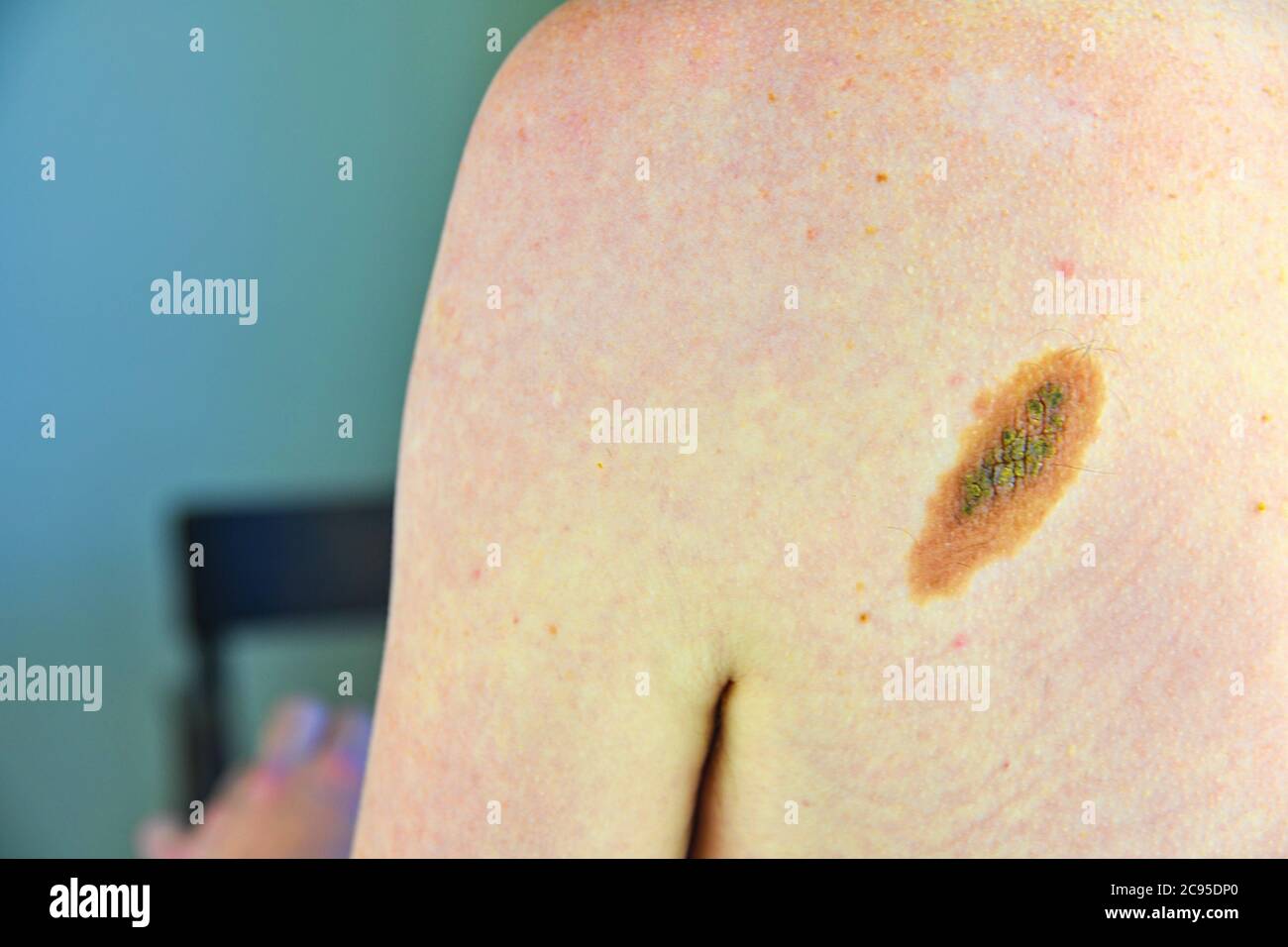 Close up picture of dangerous brown nevus on human skin - melanoma Stock Photohttps://www.alamy.com/image-license-details/?v=1https://www.alamy.com/close-up-picture-of-dangerous-brown-nevus-on-human-skin-melanoma-image367070200.html
RF2C95DP0–Close up picture of dangerous brown nevus on human skin - melanoma
 Professional watercolor paints in box with brushes. Stock Photohttps://www.alamy.com/image-license-details/?v=1https://www.alamy.com/professional-watercolor-paints-in-box-with-brushes-image226248792.html
RFR42EJ0–Professional watercolor paints in box with brushes.
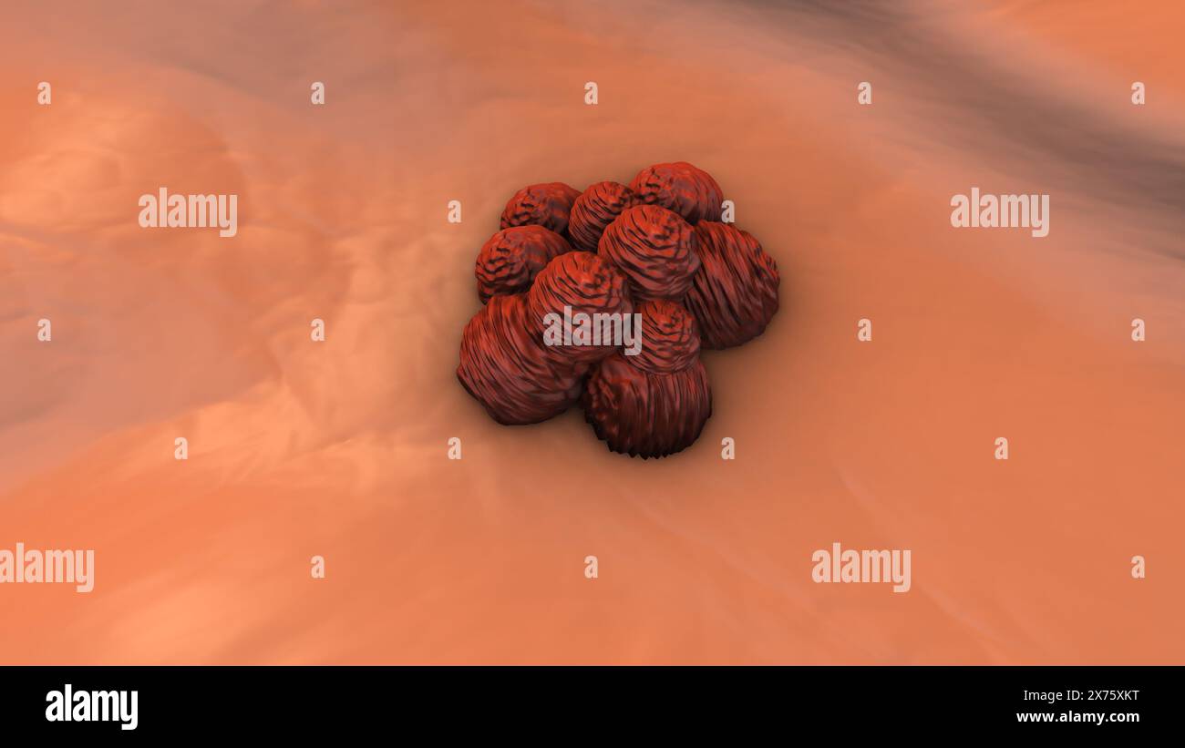 Internal metastases of melanoma cells Stock Photohttps://www.alamy.com/image-license-details/?v=1https://www.alamy.com/internal-metastases-of-melanoma-cells-image606796172.html
RF2X75XKT–Internal metastases of melanoma cells
 A large green leaf in closeup, Thailand Stock Photohttps://www.alamy.com/image-license-details/?v=1https://www.alamy.com/a-large-green-leaf-in-closeup-thailand-image626260996.html
RM2YATJ84–A large green leaf in closeup, Thailand
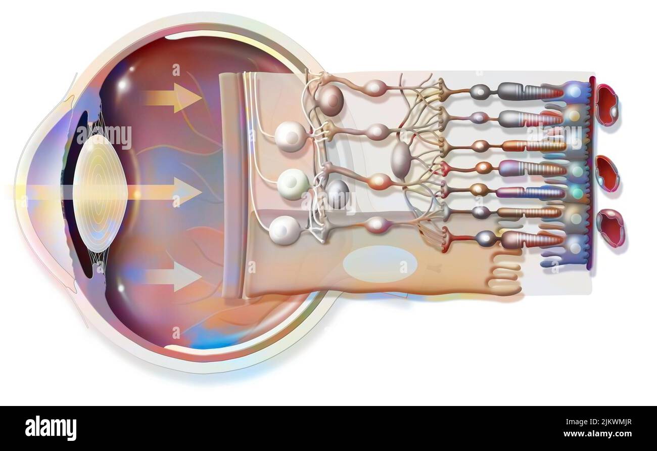 The eye and retina with the vitreous, the internal limiting membrane. Stock Photohttps://www.alamy.com/image-license-details/?v=1https://www.alamy.com/the-eye-and-retina-with-the-vitreous-the-internal-limiting-membrane-image476923407.html
RF2JKWMJR–The eye and retina with the vitreous, the internal limiting membrane.
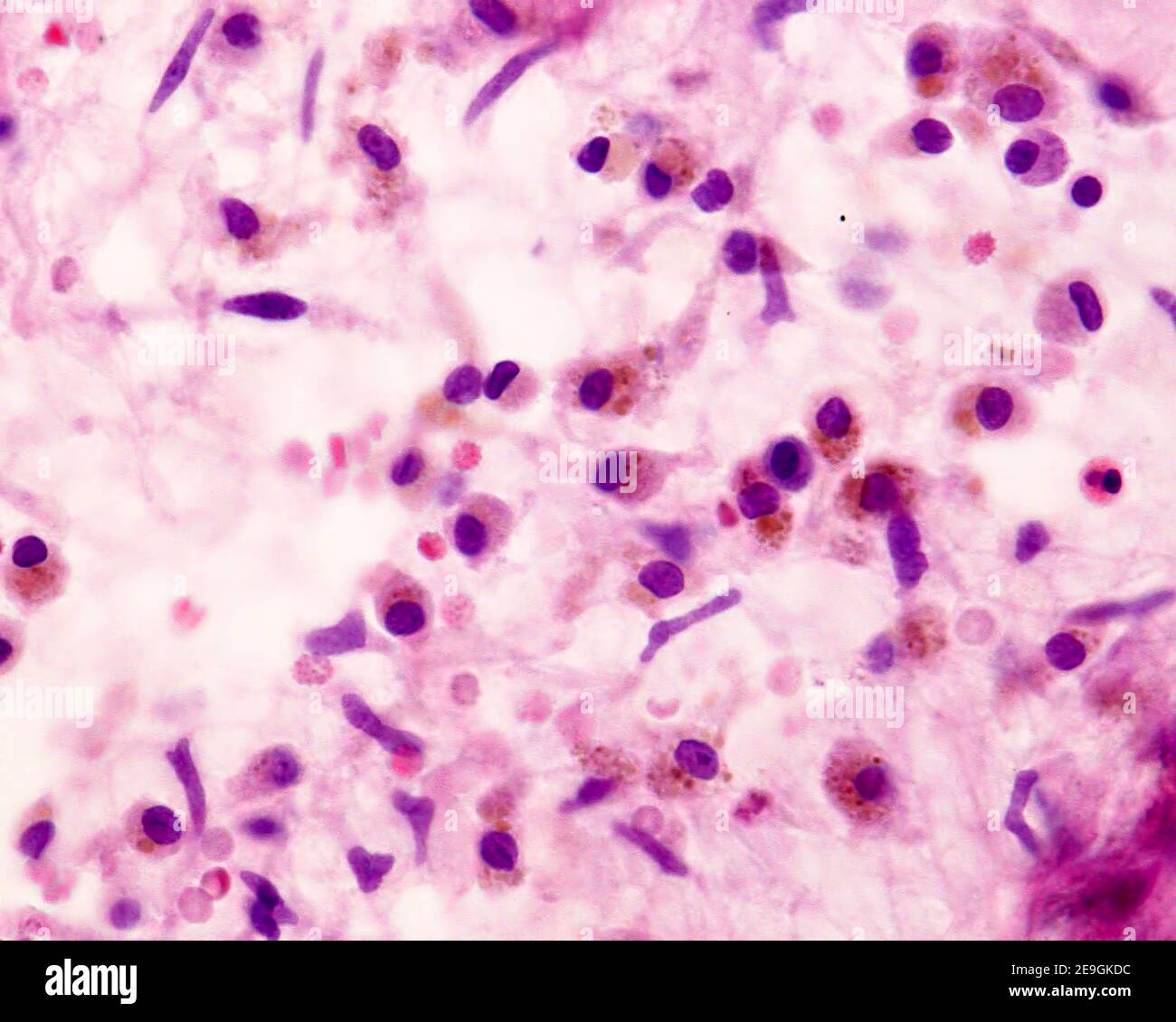 Histiocytes (macrophages) loaded with brown pigment granules of hemosiderin. These cells appear in tissues following hemorrages. Stock Photohttps://www.alamy.com/image-license-details/?v=1https://www.alamy.com/histiocytes-macrophages-loaded-with-brown-pigment-granules-of-hemosiderin-these-cells-appear-in-tissues-following-hemorrages-image401736872.html
RF2E9GKDC–Histiocytes (macrophages) loaded with brown pigment granules of hemosiderin. These cells appear in tissues following hemorrages.
 Abstract fluid art background black and navy blue colors. Liquid acrylic painting on canvas with gradient and splash. Watercolor backdrop with cell pa Stock Photohttps://www.alamy.com/image-license-details/?v=1https://www.alamy.com/abstract-fluid-art-background-black-and-navy-blue-colors-liquid-acrylic-painting-on-canvas-with-gradient-and-splash-watercolor-backdrop-with-cell-pa-image451643178.html
RF2H6P3DE–Abstract fluid art background black and navy blue colors. Liquid acrylic painting on canvas with gradient and splash. Watercolor backdrop with cell pa
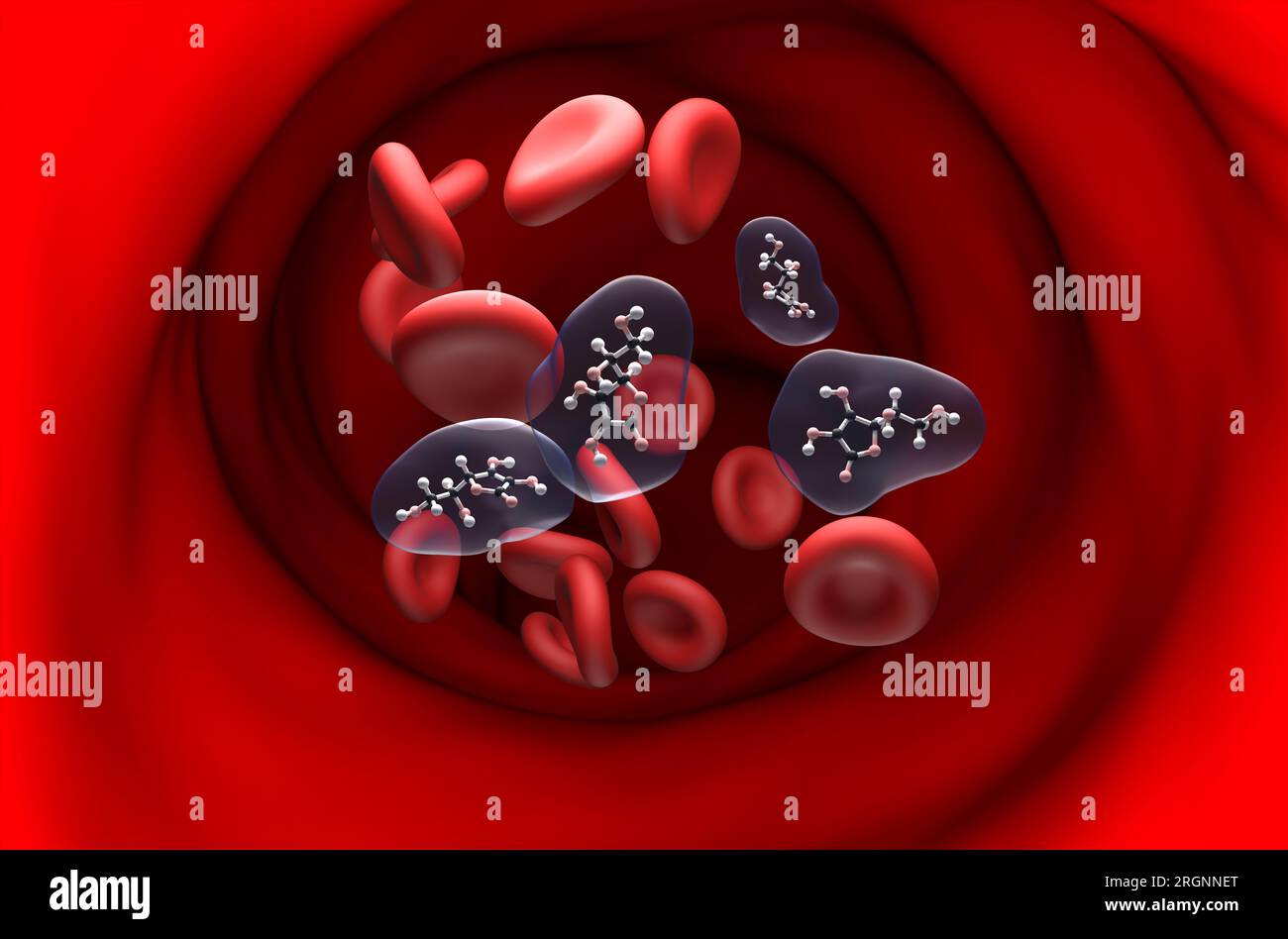 Vitamin c (ascorbic acid) structure in the blood flow ball and stick section view 3d illustration Stock Photohttps://www.alamy.com/image-license-details/?v=1https://www.alamy.com/vitamin-c-ascorbic-acid-structure-in-the-blood-flow-ball-and-stick-section-view-3d-illustration-image561044144.html
RF2RGNNET–Vitamin c (ascorbic acid) structure in the blood flow ball and stick section view 3d illustration
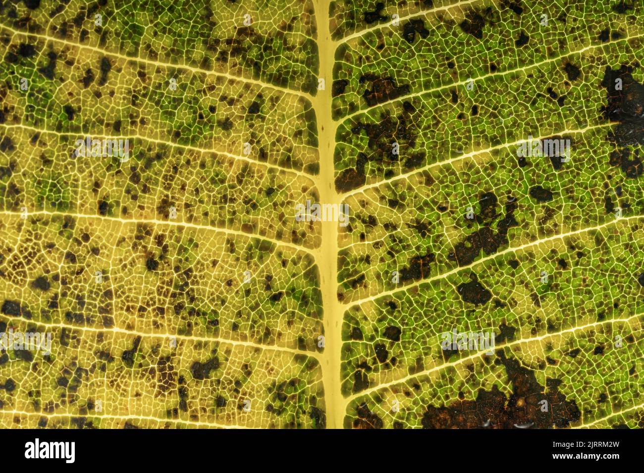 Autumn leaf texture, pigment structure, macro photography. Top view Stock Photohttps://www.alamy.com/image-license-details/?v=1https://www.alamy.com/autumn-leaf-texture-pigment-structure-macro-photography-top-view-image479337681.html
RF2JRRM2W–Autumn leaf texture, pigment structure, macro photography. Top view
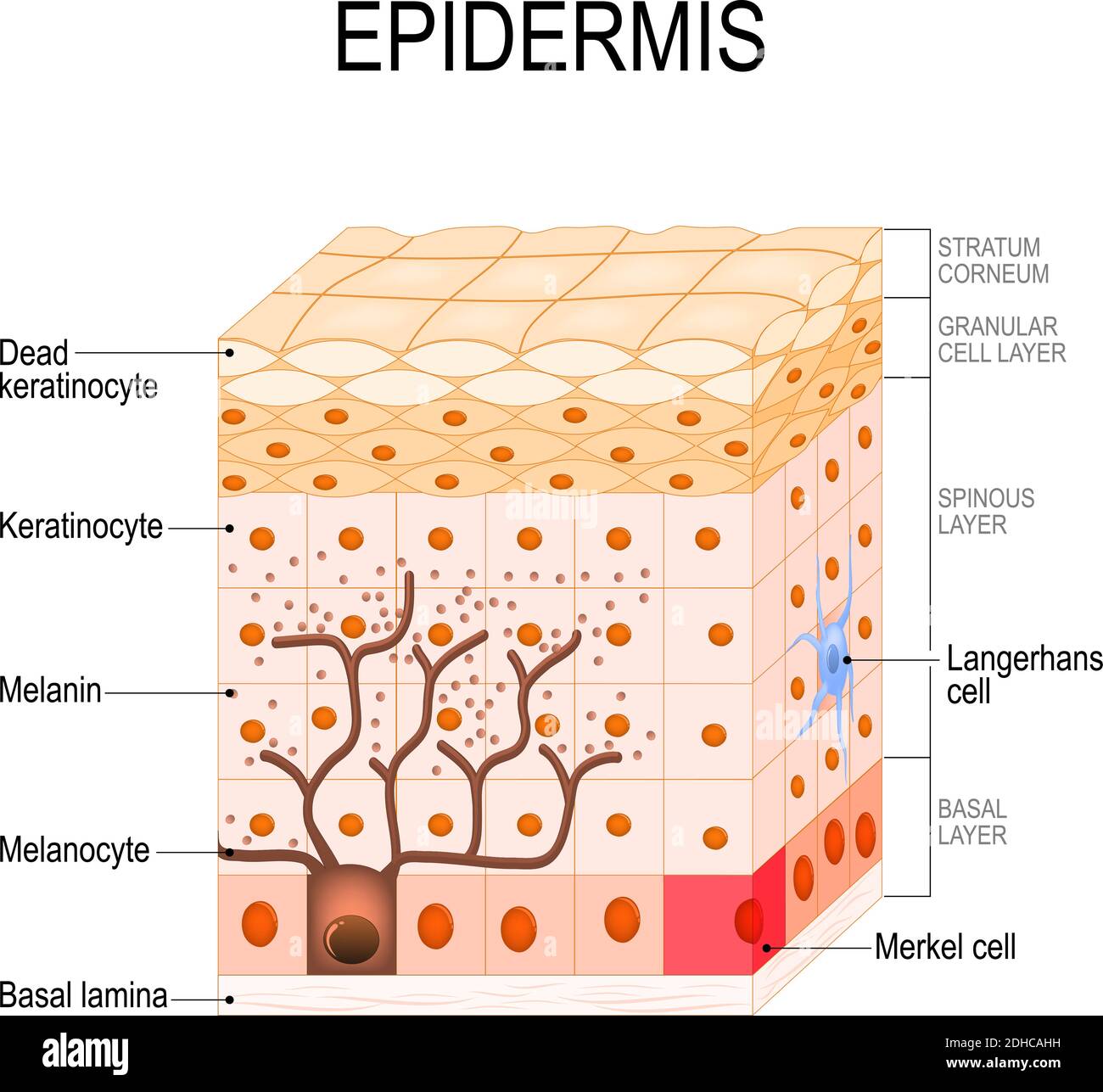 epidermis structure. Cell, and layers of a human skin. vector illustration for medical, educational, biologycal and science use. Skin care Stock Vectorhttps://www.alamy.com/image-license-details/?v=1https://www.alamy.com/epidermis-structure-cell-and-layers-of-a-human-skin-vector-illustration-for-medical-educational-biologycal-and-science-use-skin-care-image389349005.html
RF2DHCAHH–epidermis structure. Cell, and layers of a human skin. vector illustration for medical, educational, biologycal and science use. Skin care
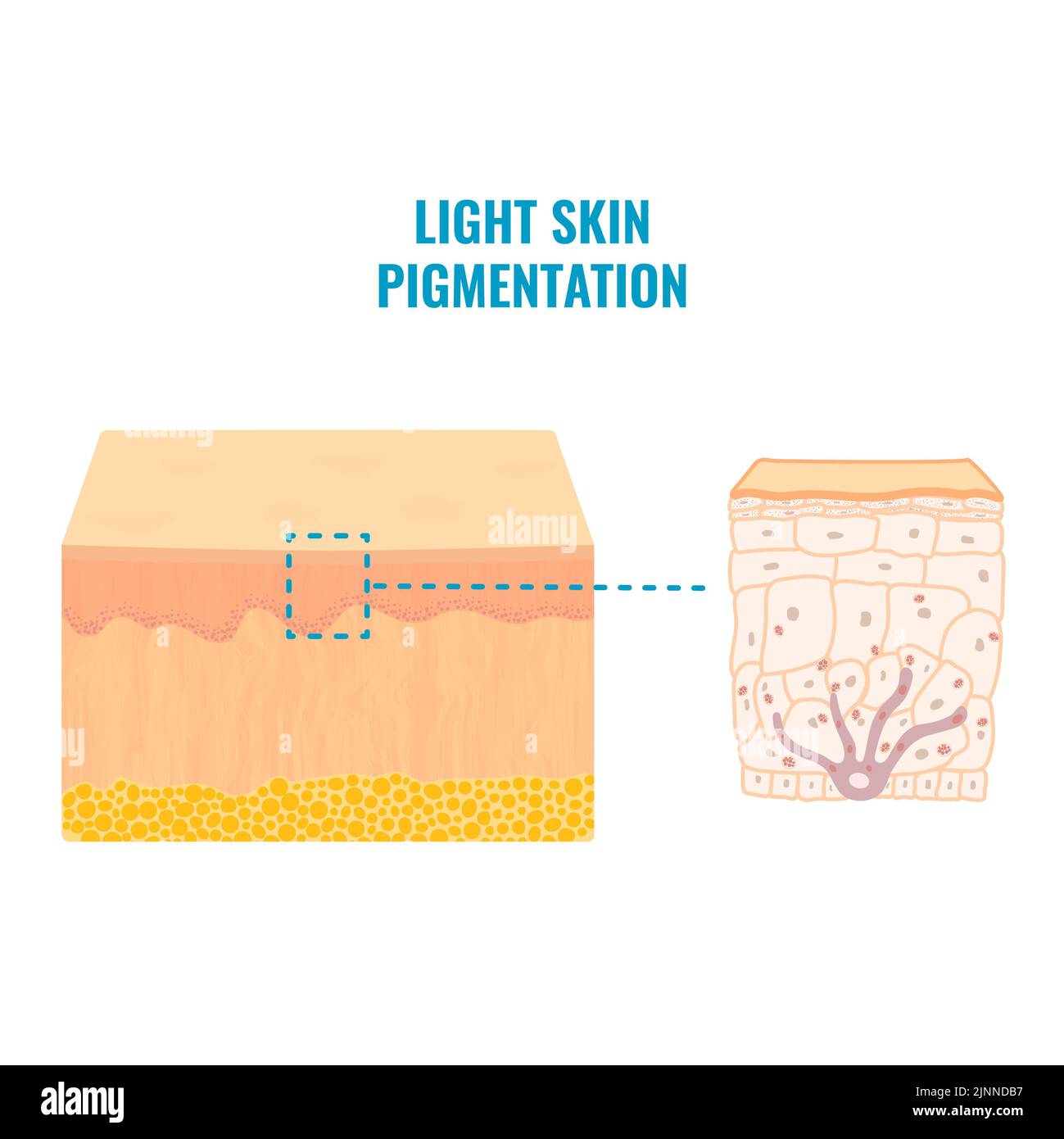 Skin pigmentation, illustration Stock Photohttps://www.alamy.com/image-license-details/?v=1https://www.alamy.com/skin-pigmentation-illustration-image478059211.html
RF2JNNDB7–Skin pigmentation, illustration
 macro of oil drops and pigment on water surface with bright background Stock Photohttps://www.alamy.com/image-license-details/?v=1https://www.alamy.com/stock-photo-macro-of-oil-drops-and-pigment-on-water-surface-with-bright-background-74999787.html
RFEA0EYR–macro of oil drops and pigment on water surface with bright background
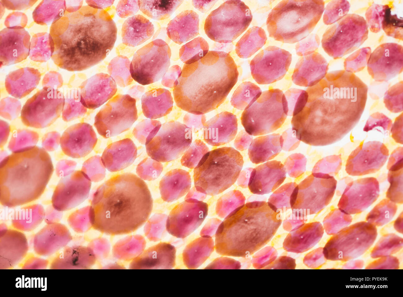 A close up/macro photograph of chromatophores in the skin of a Loligo vulgaris squid caught on rod and line in the UK. The chromatophores are cells th Stock Photohttps://www.alamy.com/image-license-details/?v=1https://www.alamy.com/a-close-upmacro-photograph-of-chromatophores-in-the-skin-of-a-loligo-vulgaris-squid-caught-on-rod-and-line-in-the-uk-the-chromatophores-are-cells-th-image223442623.html
RMPYEK9K–A close up/macro photograph of chromatophores in the skin of a Loligo vulgaris squid caught on rod and line in the UK. The chromatophores are cells th
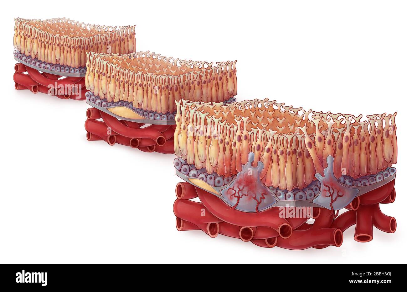 Age-related Macular Degeneration, Illustration Stock Photohttps://www.alamy.com/image-license-details/?v=1https://www.alamy.com/age-related-macular-degeneration-illustration-image353188546.html
RM2BEH3GJ–Age-related Macular Degeneration, Illustration
 3D image of Bixin skeletal formula - molecular chemical structure of norbixin isolated on white background Stock Photohttps://www.alamy.com/image-license-details/?v=1https://www.alamy.com/3d-image-of-bixin-skeletal-formula-molecular-chemical-structure-of-norbixin-isolated-on-white-background-image487601959.html
RF2K9857K–3D image of Bixin skeletal formula - molecular chemical structure of norbixin isolated on white background
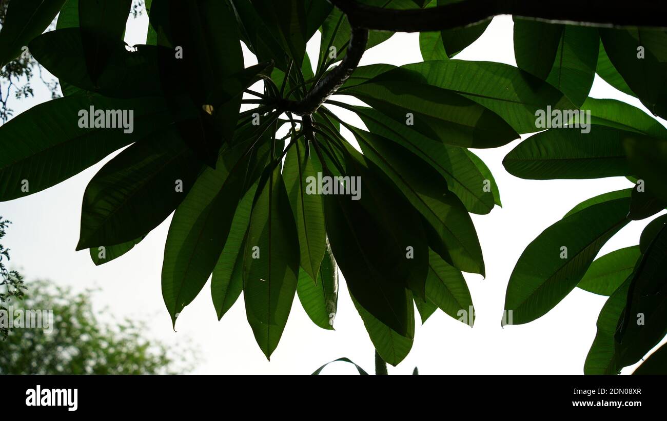 Fresh new budding green leaves view. Broad leaves of shadowed plant. Thick covered leaves with greenish pigment. Stock Photohttps://www.alamy.com/image-license-details/?v=1https://www.alamy.com/fresh-new-budding-green-leaves-view-broad-leaves-of-shadowed-plant-thick-covered-leaves-with-greenish-pigment-image391542895.html
RF2DN08XR–Fresh new budding green leaves view. Broad leaves of shadowed plant. Thick covered leaves with greenish pigment.
 Chestnut (Castanea sativa), shoot t.s., colouring Wacker 3A Stock Photohttps://www.alamy.com/image-license-details/?v=1https://www.alamy.com/chestnut-castanea-sativa-shoot-ts-colouring-wacker-3a-image416676392.html
RF2F5W6YM–Chestnut (Castanea sativa), shoot t.s., colouring Wacker 3A
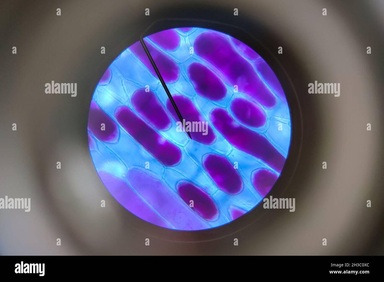 Detailed view of the cells of a red onion as seen through a microscope. Biology experiment. Stock Photohttps://www.alamy.com/image-license-details/?v=1https://www.alamy.com/detailed-view-of-the-cells-of-a-red-onion-as-seen-through-a-microscope-biology-experiment-image449577700.html
RF2H3C0XC–Detailed view of the cells of a red onion as seen through a microscope. Biology experiment.
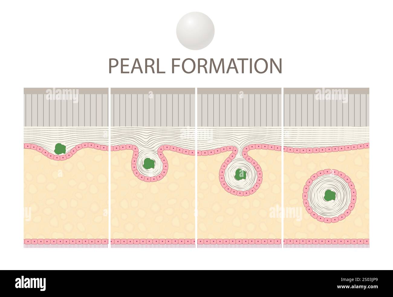 Pearl Formation in Bivalve Molluscs Stock Photohttps://www.alamy.com/image-license-details/?v=1https://www.alamy.com/pearl-formation-in-bivalve-molluscs-image636864209.html
RF2S03JP9–Pearl Formation in Bivalve Molluscs
 Large mole close-up. Macro shot of benign skin lesion on caucasian, human skin. Proliferation of pigment derma cells, melanocytic pigmented nevus Stock Photohttps://www.alamy.com/image-license-details/?v=1https://www.alamy.com/large-mole-close-up-macro-shot-of-benign-skin-lesion-on-caucasian-human-skin-proliferation-of-pigment-derma-cells-melanocytic-pigmented-nevus-image349690605.html
RF2B8WNX5–Large mole close-up. Macro shot of benign skin lesion on caucasian, human skin. Proliferation of pigment derma cells, melanocytic pigmented nevus
 Professional watercolor paints in box with brushes. Stock Photohttps://www.alamy.com/image-license-details/?v=1https://www.alamy.com/professional-watercolor-paints-in-box-with-brushes-image226239254.html
RFR422DA–Professional watercolor paints in box with brushes.
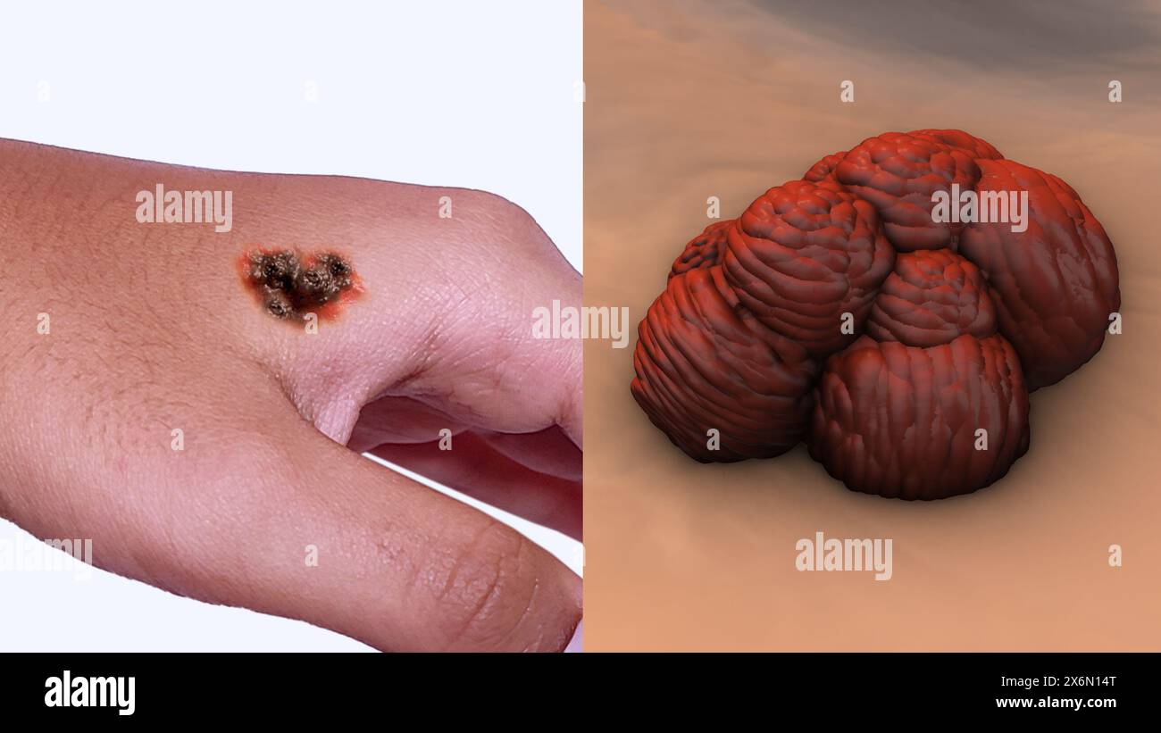 Metastatic melanoma cells on hand Stock Photohttps://www.alamy.com/image-license-details/?v=1https://www.alamy.com/metastatic-melanoma-cells-on-hand-image606512728.html
RF2X6N14T–Metastatic melanoma cells on hand
 Abstract orange background with yellow bubbles on top left. Perfect organic circles made with oil in water. Simulating cell system and space image Stock Photohttps://www.alamy.com/image-license-details/?v=1https://www.alamy.com/abstract-orange-background-with-yellow-bubbles-on-top-left-perfect-organic-circles-made-with-oil-in-water-simulating-cell-system-and-space-image-image559455366.html
RF2RE5B0P–Abstract orange background with yellow bubbles on top left. Perfect organic circles made with oil in water. Simulating cell system and space image
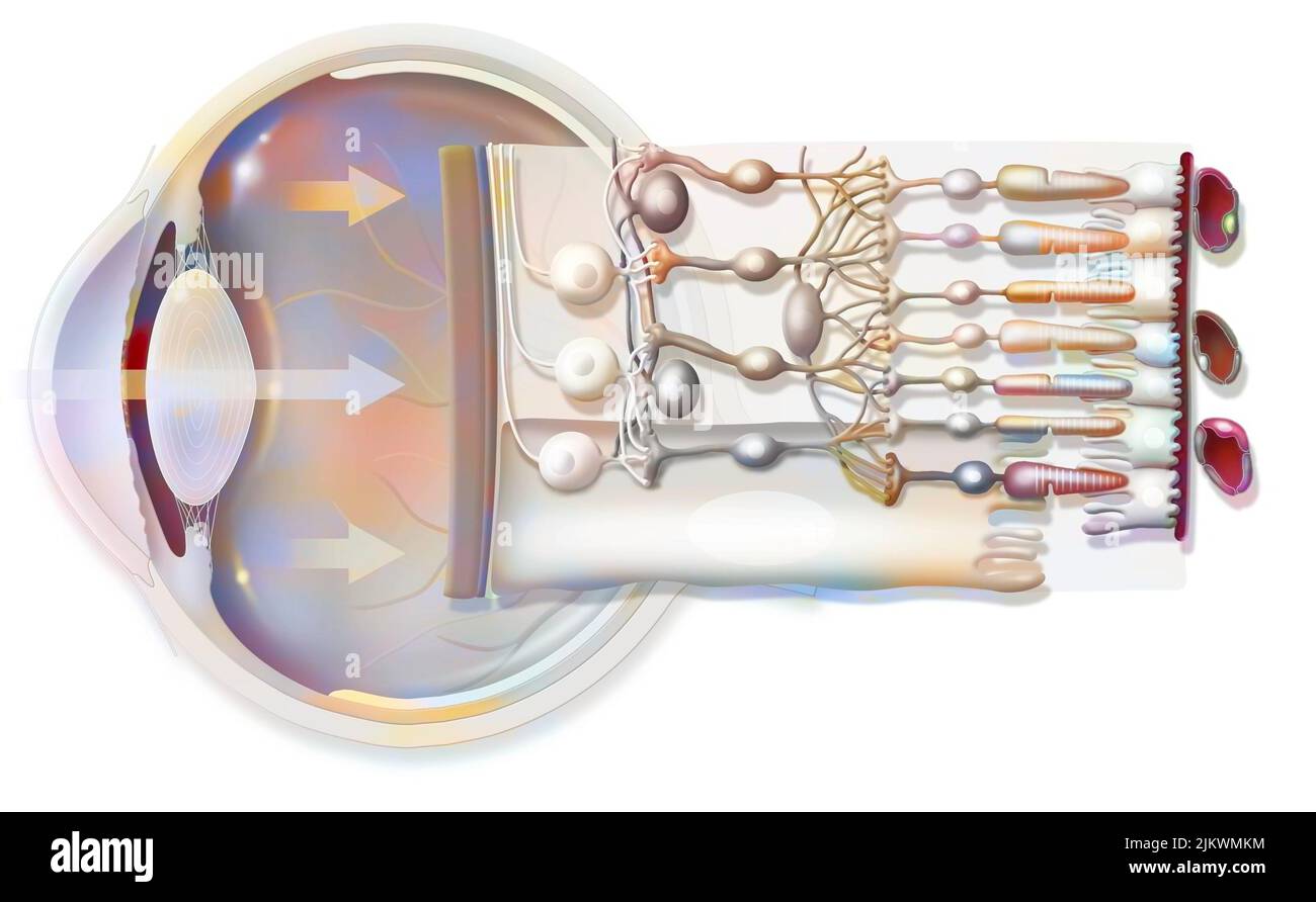 The eye and retina with the vitreous, the internal limiting membrane. Stock Photohttps://www.alamy.com/image-license-details/?v=1https://www.alamy.com/the-eye-and-retina-with-the-vitreous-the-internal-limiting-membrane-image476923432.html
RF2JKWMKM–The eye and retina with the vitreous, the internal limiting membrane.
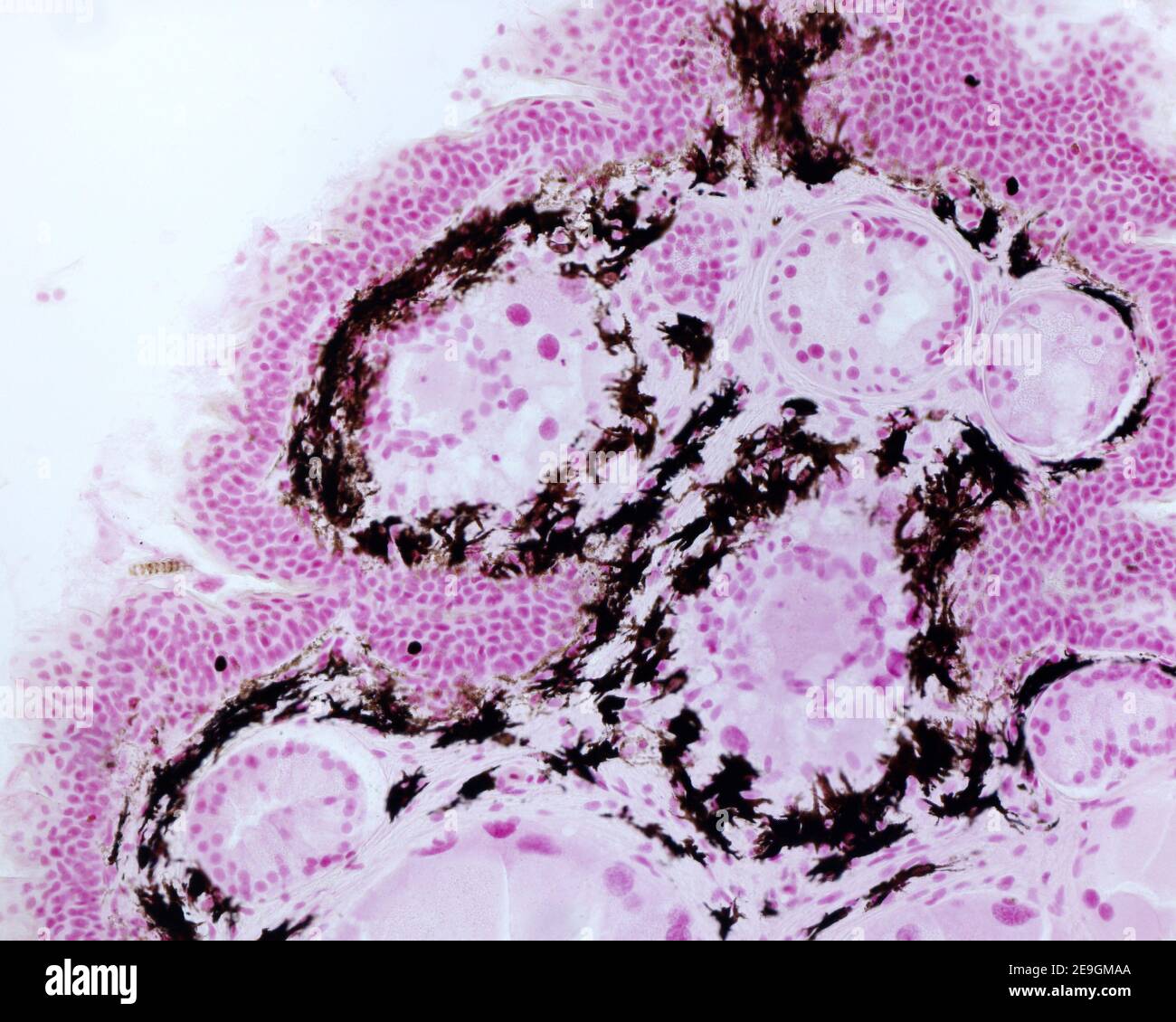 Photomicrograph of pigment cells (chromatophores) in the skin of a newt, (Pleurodeles waltl). Chromatophore are cells loaded of pigment grains, locate Stock Photohttps://www.alamy.com/image-license-details/?v=1https://www.alamy.com/photomicrograph-of-pigment-cells-chromatophores-in-the-skin-of-a-newt-pleurodeles-waltl-chromatophore-are-cells-loaded-of-pigment-grains-locate-image401737570.html
RF2E9GMAA–Photomicrograph of pigment cells (chromatophores) in the skin of a newt, (Pleurodeles waltl). Chromatophore are cells loaded of pigment grains, locate
 Festive colorful star confetti background. Stock Photohttps://www.alamy.com/image-license-details/?v=1https://www.alamy.com/festive-colorful-star-confetti-background-image454733912.html
RF2HBPWMT–Festive colorful star confetti background.
 health supplementary product, lutein Stock Photohttps://www.alamy.com/image-license-details/?v=1https://www.alamy.com/health-supplementary-product-lutein-image401523964.html
RF2E96YWG–health supplementary product, lutein
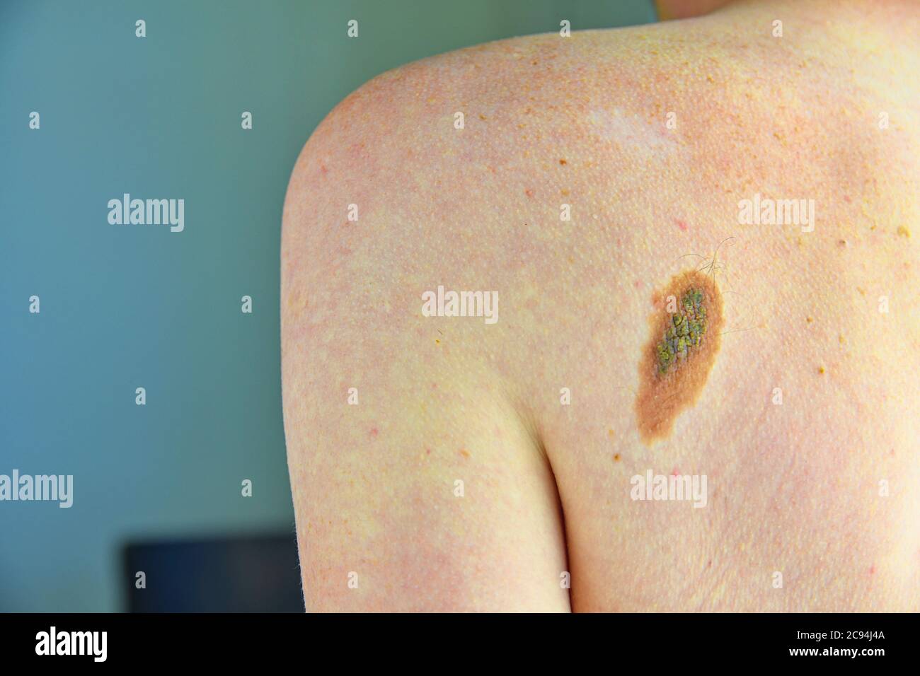 Hairy skin mole. Close up picture of dangerous brown nevus on human skin - melanoma Stock Photohttps://www.alamy.com/image-license-details/?v=1https://www.alamy.com/hairy-skin-mole-close-up-picture-of-dangerous-brown-nevus-on-human-skin-melanoma-image367051674.html
RF2C94J4A–Hairy skin mole. Close up picture of dangerous brown nevus on human skin - melanoma
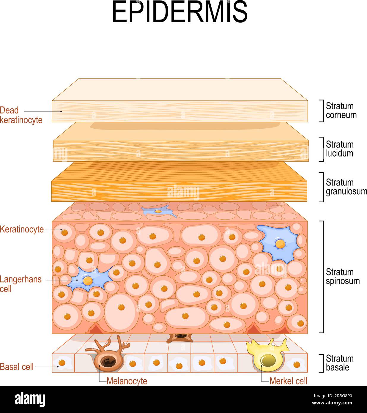 epidermis structure. Skin anatomy. Cell, and layers of a human skin. Cross section of the epidermis. Skin care. vector illustration. Stock Vectorhttps://www.alamy.com/image-license-details/?v=1https://www.alamy.com/epidermis-structure-skin-anatomy-cell-and-layers-of-a-human-skin-cross-section-of-the-epidermis-skin-care-vector-illustration-image554163176.html
RF2R5G8P0–epidermis structure. Skin anatomy. Cell, and layers of a human skin. Cross section of the epidermis. Skin care. vector illustration.
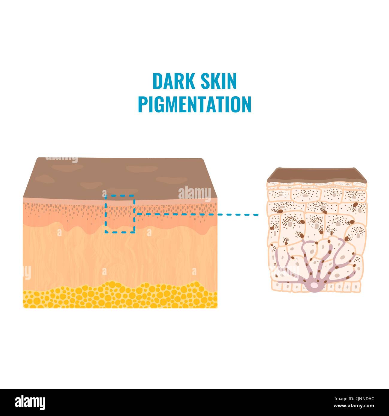 Skin pigmentation, illustration Stock Photohttps://www.alamy.com/image-license-details/?v=1https://www.alamy.com/skin-pigmentation-illustration-image478059188.html
RF2JNNDAC–Skin pigmentation, illustration
 macro of oil drops and pigment on water surface with bright background Stock Photohttps://www.alamy.com/image-license-details/?v=1https://www.alamy.com/stock-photo-macro-of-oil-drops-and-pigment-on-water-surface-with-bright-background-74999789.html
RFEA0EYW–macro of oil drops and pigment on water surface with bright background
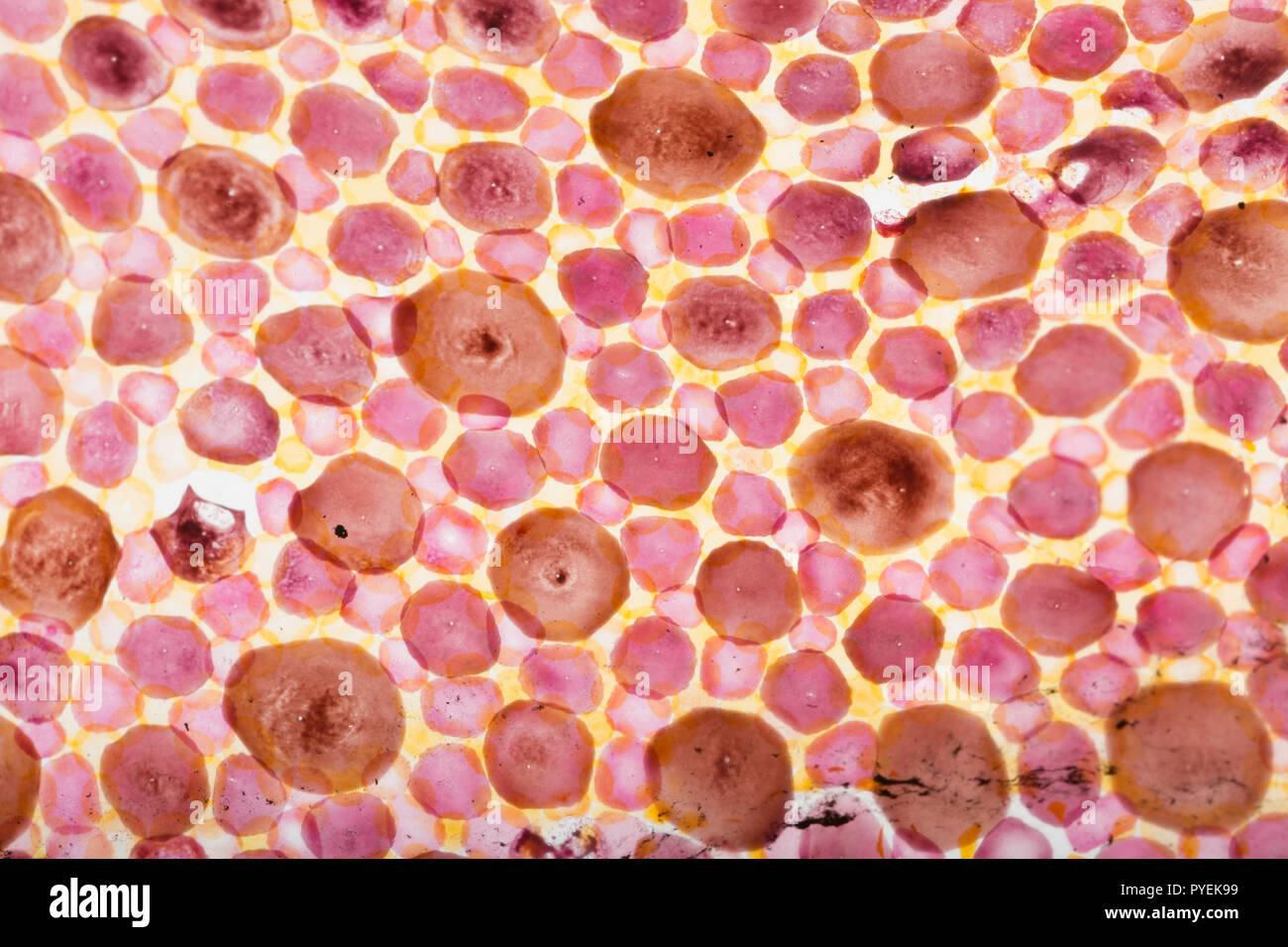 A close up/macro photograph of chromatophores in the skin of a Loligo vulgaris squid caught on rod and line in the UK. The chromatophores are cells th Stock Photohttps://www.alamy.com/image-license-details/?v=1https://www.alamy.com/a-close-upmacro-photograph-of-chromatophores-in-the-skin-of-a-loligo-vulgaris-squid-caught-on-rod-and-line-in-the-uk-the-chromatophores-are-cells-th-image223442613.html
RMPYEK99–A close up/macro photograph of chromatophores in the skin of a Loligo vulgaris squid caught on rod and line in the UK. The chromatophores are cells th
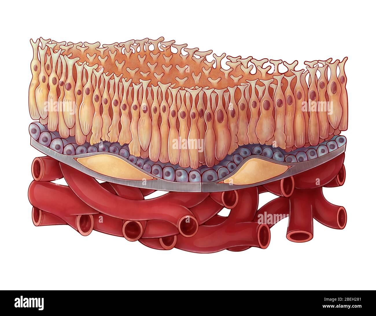 Dry AMD, Illustration Stock Photohttps://www.alamy.com/image-license-details/?v=1https://www.alamy.com/dry-amd-illustration-image353187521.html
RM2BEH281–Dry AMD, Illustration
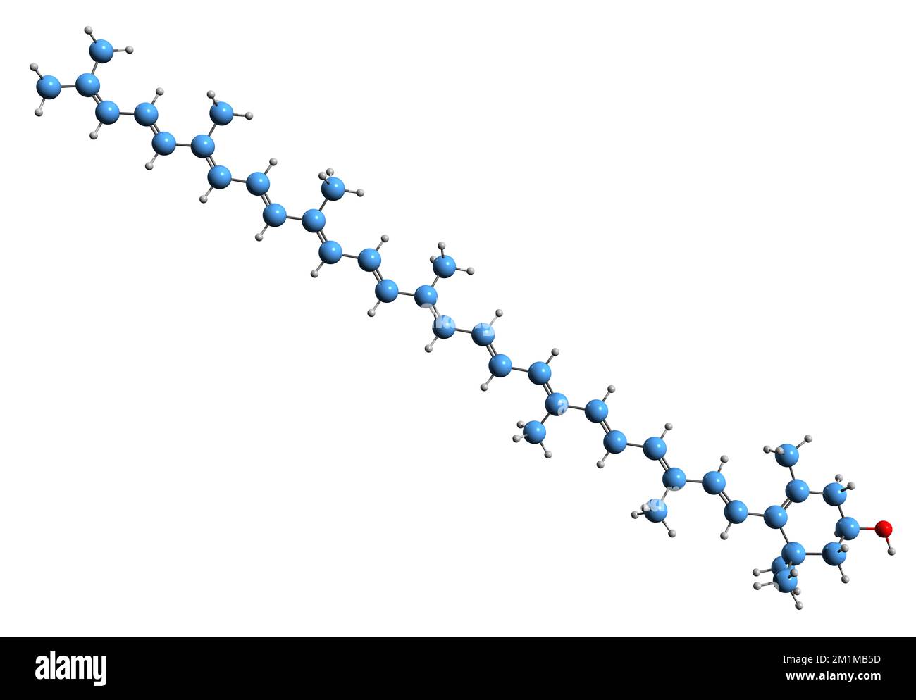 3D image of Rubixanthin skeletal formula - molecular chemical structure of food coloring natural yellow 27 isolated on white background Stock Photohttps://www.alamy.com/image-license-details/?v=1https://www.alamy.com/3d-image-of-rubixanthin-skeletal-formula-molecular-chemical-structure-of-food-coloring-natural-yellow-27-isolated-on-white-background-image500163145.html
RF2M1MB5D–3D image of Rubixanthin skeletal formula - molecular chemical structure of food coloring natural yellow 27 isolated on white background
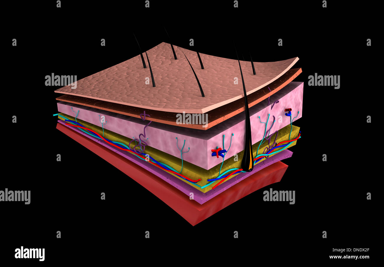
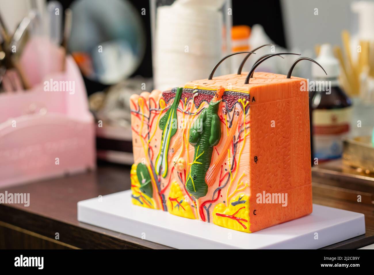 Plastic model of human skin and hair structure for education Stock Photohttps://www.alamy.com/image-license-details/?v=1https://www.alamy.com/plastic-model-of-human-skin-and-hair-structure-for-education-image466181575.html
RF2J2CB9Y–Plastic model of human skin and hair structure for education
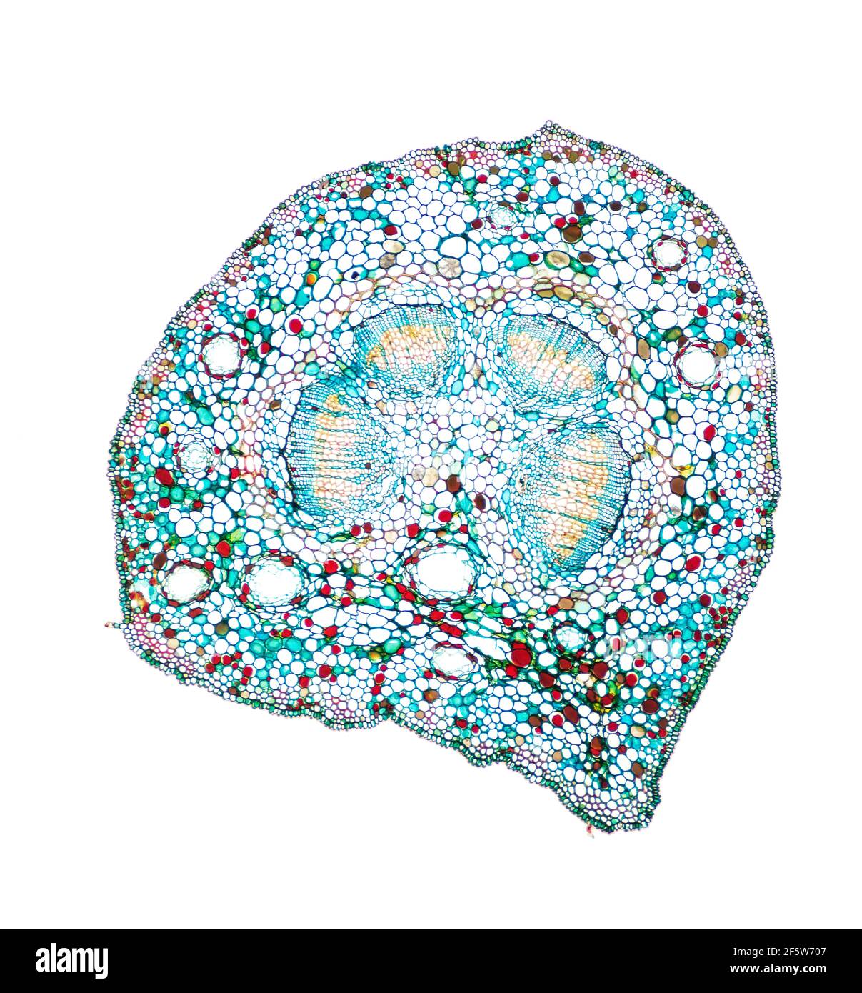 Gingko (Gingko biloba), petiole t.s., staining Wacker 3A Stock Photohttps://www.alamy.com/image-license-details/?v=1https://www.alamy.com/gingko-gingko-biloba-petiole-ts-staining-wacker-3a-image416676407.html
RF2F5W707–Gingko (Gingko biloba), petiole t.s., staining Wacker 3A
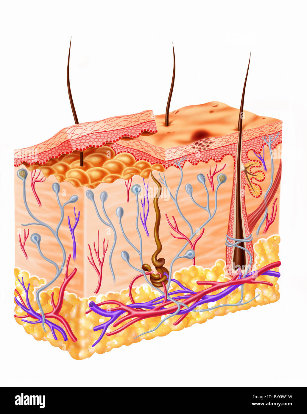 Human skin section diagram Stock Photohttps://www.alamy.com/image-license-details/?v=1https://www.alamy.com/stock-photo-human-skin-section-diagram-34176965.html
RFBYGW1W–Human skin section diagram
 Portrait of redheaded young woman lying on bench using cell phone Stock Photohttps://www.alamy.com/image-license-details/?v=1https://www.alamy.com/portrait-of-redheaded-young-woman-lying-on-bench-using-cell-phone-image223065901.html
RFPXWER9–Portrait of redheaded young woman lying on bench using cell phone
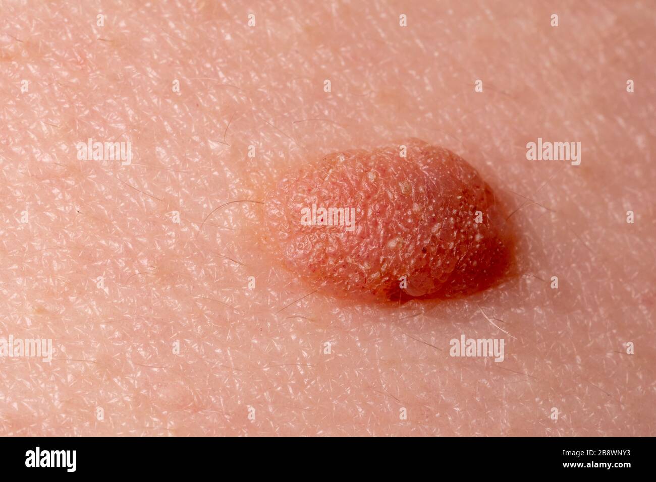 Large mole close-up. Macro shot of benign skin lesion on caucasian, human skin. Proliferation of pigment derma cells, melanocytic pigmented nevus Stock Photohttps://www.alamy.com/image-license-details/?v=1https://www.alamy.com/large-mole-close-up-macro-shot-of-benign-skin-lesion-on-caucasian-human-skin-proliferation-of-pigment-derma-cells-melanocytic-pigmented-nevus-image349690631.html
RF2B8WNY3–Large mole close-up. Macro shot of benign skin lesion on caucasian, human skin. Proliferation of pigment derma cells, melanocytic pigmented nevus
 Professional watercolor paints in box with brushes. Stock Photohttps://www.alamy.com/image-license-details/?v=1https://www.alamy.com/professional-watercolor-paints-in-box-with-brushes-image226239251.html
RFR422D7–Professional watercolor paints in box with brushes.
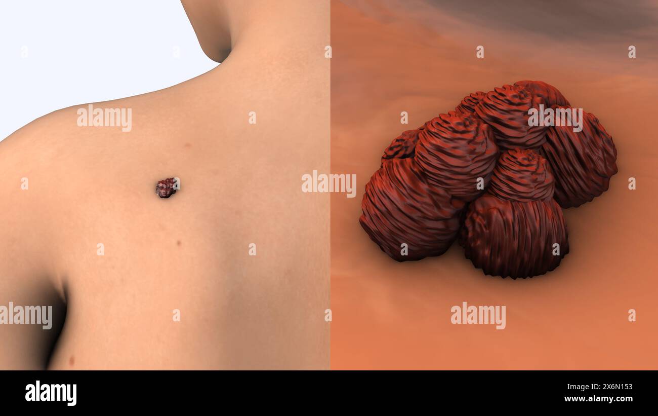 Metastatic melanoma cells medical animation Stock Photohttps://www.alamy.com/image-license-details/?v=1https://www.alamy.com/metastatic-melanoma-cells-medical-animation-image606512735.html
RF2X6N153–Metastatic melanoma cells medical animation
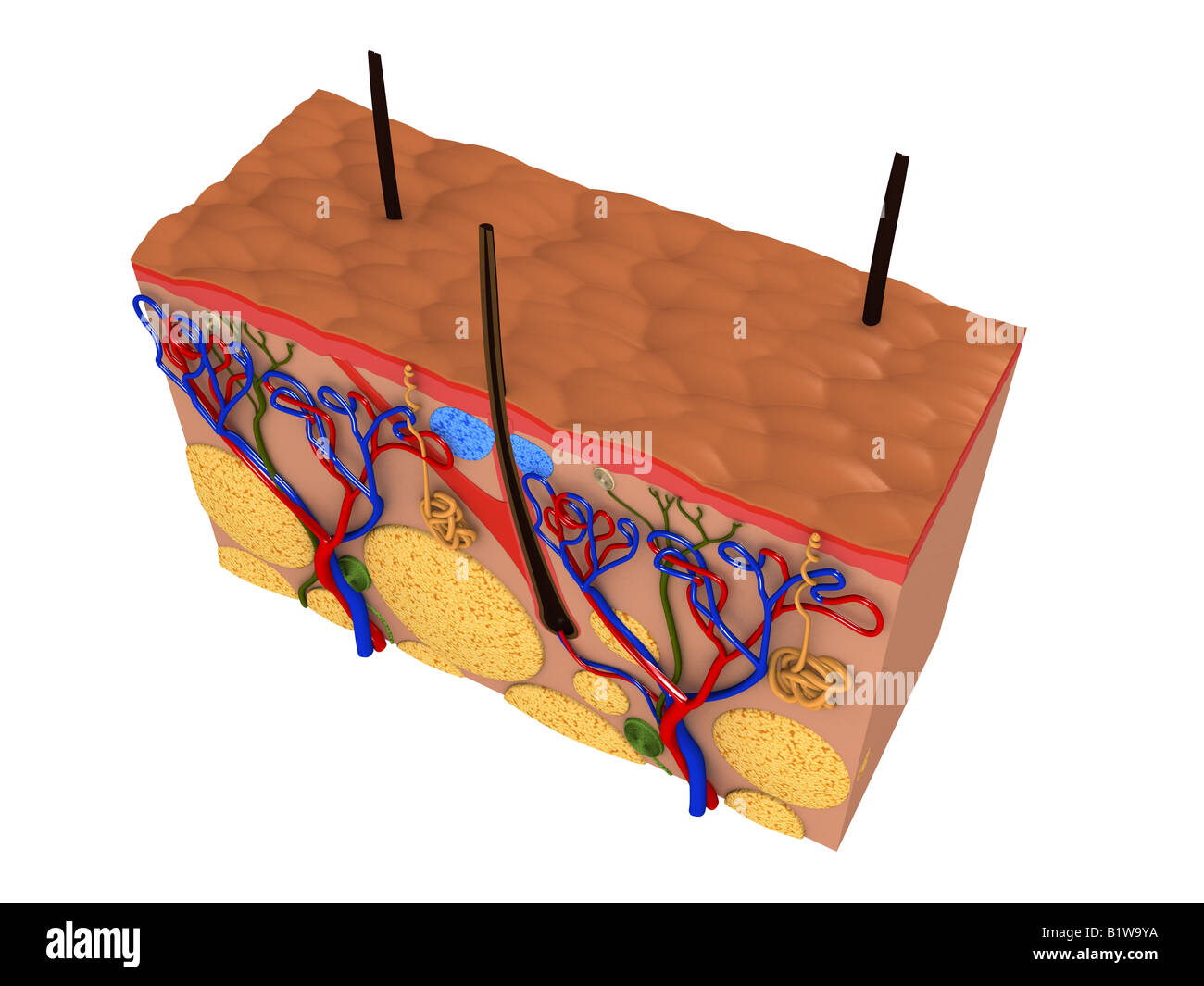 skin cut Stock Photohttps://www.alamy.com/image-license-details/?v=1https://www.alamy.com/stock-photo-skin-cut-18381646.html
RFB1W9YA–skin cut
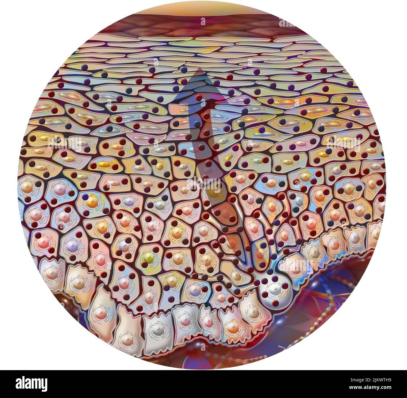 Dark skin type with lots of melanin grains. Stock Photohttps://www.alamy.com/image-license-details/?v=1https://www.alamy.com/dark-skin-type-with-lots-of-melanin-grains-image476926501.html
RF2JKWTH9–Dark skin type with lots of melanin grains.
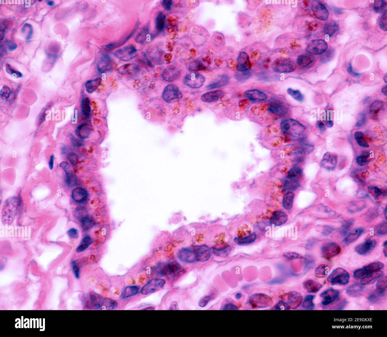 Secretory epithelium of the seminal vesicle. Is a columnar epithelium, which show a highly characteristic golden-yellow pigment. Stock Photohttps://www.alamy.com/image-license-details/?v=1https://www.alamy.com/secretory-epithelium-of-the-seminal-vesicle-is-a-columnar-epithelium-which-show-a-highly-characteristic-golden-yellow-pigment-image401737238.html
RF2E9GKXE–Secretory epithelium of the seminal vesicle. Is a columnar epithelium, which show a highly characteristic golden-yellow pigment.
 Decorative sugar sprinkles on a pink backdrop. Festive background for Birthday, holiday, party Stock Photohttps://www.alamy.com/image-license-details/?v=1https://www.alamy.com/decorative-sugar-sprinkles-on-a-pink-backdrop-festive-background-for-birthday-holiday-party-image454733827.html
RF2HBPWHR–Decorative sugar sprinkles on a pink backdrop. Festive background for Birthday, holiday, party
