Quick filters:
Cerebral trunk Stock Photos and Images
 The arterial blood supply to the neck (carotids and vertebral arteries). Stock Photohttps://www.alamy.com/image-license-details/?v=1https://www.alamy.com/the-arterial-blood-supply-to-the-neck-carotids-and-vertebral-arteries-image476924402.html
RF2JKWNXA–The arterial blood supply to the neck (carotids and vertebral arteries).
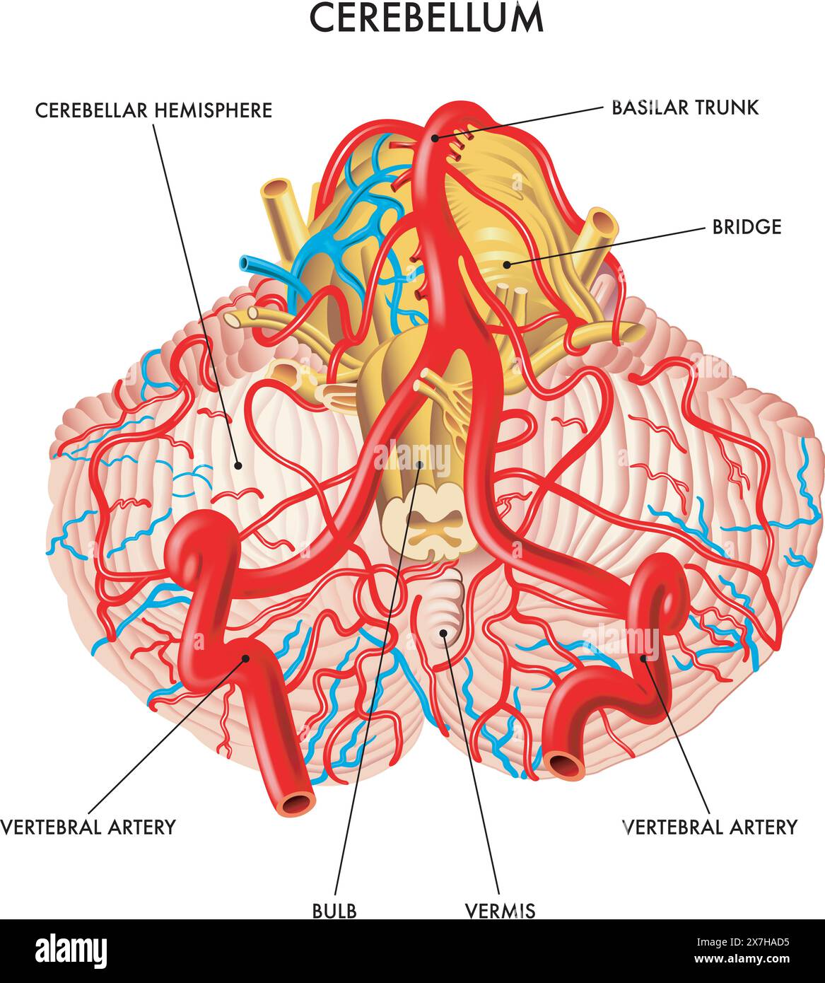 Medical illustration shows the blood circulation of the cerebellum, with annotations. Stock Vectorhttps://www.alamy.com/image-license-details/?v=1https://www.alamy.com/medical-illustration-shows-the-blood-circulation-of-the-cerebellum-with-annotations-image607046865.html
RF2X7HAD5–Medical illustration shows the blood circulation of the cerebellum, with annotations.
 Close up of Black jelly fungus, Exidia nigricans on dead trunk of a birch tree Stock Photohttps://www.alamy.com/image-license-details/?v=1https://www.alamy.com/close-up-of-black-jelly-fungus-exidia-nigricans-on-dead-trunk-of-a-birch-tree-image544325599.html
RF2PHG4RY–Close up of Black jelly fungus, Exidia nigricans on dead trunk of a birch tree
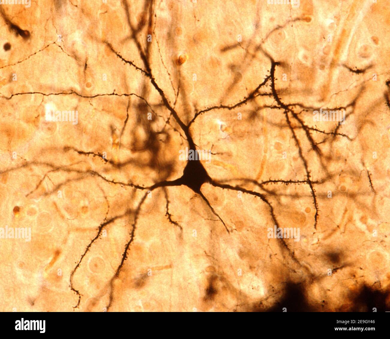 Pyramidal neuron. Golgi’s silver chromate. The apical dendritic trunk and the basilar dendrites contain many spines. From the bottom arises the axon Stock Photohttps://www.alamy.com/image-license-details/?v=1https://www.alamy.com/pyramidal-neuron-golgis-silver-chromate-the-apical-dendritic-trunk-and-the-basilar-dendrites-contain-many-spines-from-the-bottom-arises-the-axon-image401742886.html
RF2E9GY46–Pyramidal neuron. Golgi’s silver chromate. The apical dendritic trunk and the basilar dendrites contain many spines. From the bottom arises the axon
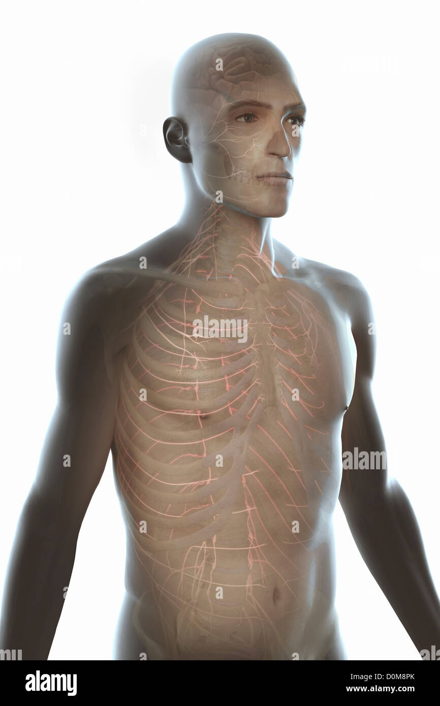 Three-quarter view of the male trunk with the nervous system and brain. Stock Photohttps://www.alamy.com/image-license-details/?v=1https://www.alamy.com/stock-photo-three-quarter-view-of-the-male-trunk-with-the-nervous-system-and-brain-52077051.html
RMD0M8PK–Three-quarter view of the male trunk with the nervous system and brain.
 EXCLUSIVE. Seven-year-old Manon Beauvais pictured in the garden of her family's house in Fecamp-Normandy-France on April 29, 2004. One year ago, Manon was struck down by a cancer of the cheek and of the cerebral trunk. As no doctor was able to operate her in France, the unique chance to save the little girl was to send her to the USA. To finance the travel, the operation and the hospitalization at the Schneider Children's Hospital in New York, her parents created the association 'Manon demain' ('Manon Tomorrow') and raised 170.000 euros in two weeks. Manon was operated successfully by the Amer Stock Photohttps://www.alamy.com/image-license-details/?v=1https://www.alamy.com/exclusive-seven-year-old-manon-beauvais-pictured-in-the-garden-of-her-familys-house-in-fecamp-normandy-france-on-april-29-2004-one-year-ago-manon-was-struck-down-by-a-cancer-of-the-cheek-and-of-the-cerebral-trunk-as-no-doctor-was-able-to-operate-her-in-france-the-unique-chance-to-save-the-little-girl-was-to-send-her-to-the-usa-to-finance-the-travel-the-operation-and-the-hospitalization-at-the-schneider-childrens-hospital-in-new-york-her-parents-created-the-association-manon-demain-manon-tomorrow-and-raised-170000-euros-in-two-weeks-manon-was-operated-successfully-by-the-amer-image402161370.html
RF2EA80X2–EXCLUSIVE. Seven-year-old Manon Beauvais pictured in the garden of her family's house in Fecamp-Normandy-France on April 29, 2004. One year ago, Manon was struck down by a cancer of the cheek and of the cerebral trunk. As no doctor was able to operate her in France, the unique chance to save the little girl was to send her to the USA. To finance the travel, the operation and the hospitalization at the Schneider Children's Hospital in New York, her parents created the association 'Manon demain' ('Manon Tomorrow') and raised 170.000 euros in two weeks. Manon was operated successfully by the Amer
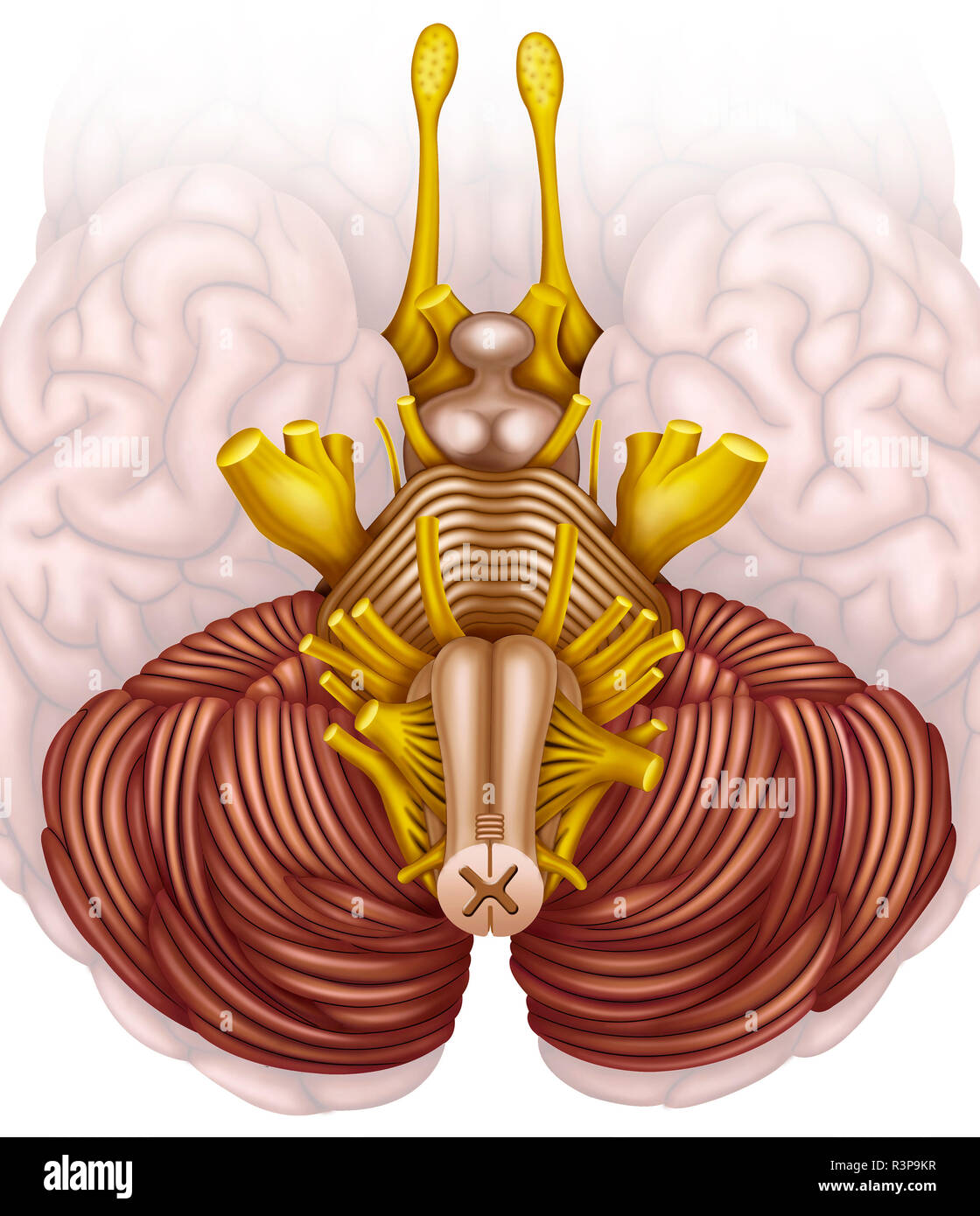 The brain stem, or human brain stem, is the major communication pathway for the brain, spinal cord, and peripheral nerves. Stock Photohttps://www.alamy.com/image-license-details/?v=1https://www.alamy.com/the-brain-stem-or-human-brain-stem-is-the-major-communication-pathway-for-the-brain-spinal-cord-and-peripheral-nerves-image226069307.html
RFR3P9KR–The brain stem, or human brain stem, is the major communication pathway for the brain, spinal cord, and peripheral nerves.
 . St. Thomas's Hospital reports. atal seizure. The three cases which have been detailed correspond withthe larger number of the similar cases that have been placed onrecord. In all of them the aneurism involved the internalcarotid artery or its branches ; in two, indeed, the sac wassituated at the termination of that vessel immediately beforeit divided into its branches ; and in the third it arose from theend of the middle cerebral trunk. In the majority of cases theaneurisms arise in these situations, though they are nearly asfrequent in the basilar artery. In all the cases also theaneurisms Stock Photohttps://www.alamy.com/image-license-details/?v=1https://www.alamy.com/st-thomass-hospital-reports-atal-seizure-the-three-cases-which-have-been-detailed-correspond-withthe-larger-number-of-the-similar-cases-that-have-been-placed-onrecord-in-all-of-them-the-aneurism-involved-the-internalcarotid-artery-or-its-branches-in-two-indeed-the-sac-wassituated-at-the-termination-of-that-vessel-immediately-beforeit-divided-into-its-branches-and-in-the-third-it-arose-from-theend-of-the-middle-cerebral-trunk-in-the-majority-of-cases-theaneurisms-arise-in-these-situations-though-they-are-nearly-asfrequent-in-the-basilar-artery-in-all-the-cases-also-theaneurisms-image336712088.html
RM2AFPFKM–. St. Thomas's Hospital reports. atal seizure. The three cases which have been detailed correspond withthe larger number of the similar cases that have been placed onrecord. In all of them the aneurism involved the internalcarotid artery or its branches ; in two, indeed, the sac wassituated at the termination of that vessel immediately beforeit divided into its branches ; and in the third it arose from theend of the middle cerebral trunk. In the majority of cases theaneurisms arise in these situations, though they are nearly asfrequent in the basilar artery. In all the cases also theaneurisms
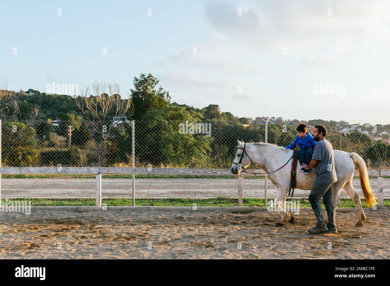 Child during an equine therapy session outdoors Stock Photohttps://www.alamy.com/image-license-details/?v=1https://www.alamy.com/child-during-an-equine-therapy-session-outdoors-image506126530.html
RF2MBC1FE–Child during an equine therapy session outdoors
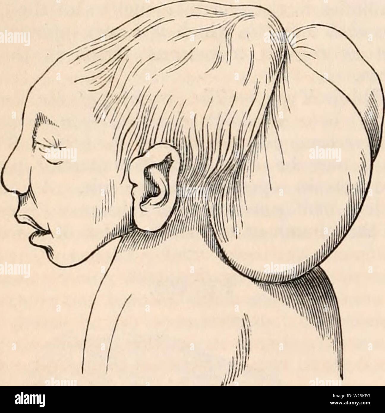 Archive image from page 171 of The cyclopædia of anatomy and. The cyclopædia of anatomy and physiology cyclopdiaofana0402todd Year: 1849 956 TERATOLOGY. a hernia. The parietal bones are sometimes present, together with flat frontal bones, and a perpendicular occipital bone, so that the summit of the skull is quite closed, with the exception of a small opening. Fig. 609. shows how the malformed cerebral substance is applied to the medulla spinalis. All the cerebral nerves are present. This form of monstrosity has in general a less brute-like aspect; the trunk is more evolved, and the whole bo Stock Photohttps://www.alamy.com/image-license-details/?v=1https://www.alamy.com/archive-image-from-page-171-of-the-cyclopdia-of-anatomy-and-the-cyclopdia-of-anatomy-and-physiology-cyclopdiaofana0402todd-year-1849-956-teratology-a-hernia-the-parietal-bones-are-sometimes-present-together-with-flat-frontal-bones-and-a-perpendicular-occipital-bone-so-that-the-summit-of-the-skull-is-quite-closed-with-the-exception-of-a-small-opening-fig-609-shows-how-the-malformed-cerebral-substance-is-applied-to-the-medulla-spinalis-all-the-cerebral-nerves-are-present-this-form-of-monstrosity-has-in-general-a-less-brute-like-aspect-the-trunk-is-more-evolved-and-the-whole-bo-image259466216.html
RMW23KPG–Archive image from page 171 of The cyclopædia of anatomy and. The cyclopædia of anatomy and physiology cyclopdiaofana0402todd Year: 1849 956 TERATOLOGY. a hernia. The parietal bones are sometimes present, together with flat frontal bones, and a perpendicular occipital bone, so that the summit of the skull is quite closed, with the exception of a small opening. Fig. 609. shows how the malformed cerebral substance is applied to the medulla spinalis. All the cerebral nerves are present. This form of monstrosity has in general a less brute-like aspect; the trunk is more evolved, and the whole bo
 Human Heart Anatomy For Medical Concept 3D Illustration Stock Photohttps://www.alamy.com/image-license-details/?v=1https://www.alamy.com/human-heart-anatomy-for-medical-concept-3d-illustration-image368852363.html
RF2CC2JXK–Human Heart Anatomy For Medical Concept 3D Illustration
 . The cyclopædia of anatomy and physiology. Anatomy; Physiology; Zoology. 956 TERATOLOGY. a hernia. The parietal bones are sometimes present, together with flat frontal bones, and a perpendicular occipital bone, so that the summit of the skull is quite closed, with the exception of a small opening. Fig. 609. shows how the malformed cerebral substance is applied to the medulla spinalis. All the cerebral nerves are present. This form of monstrosity has in general a less brute-like aspect; the trunk is more evolved, and the whole body in general very heavy. Fourth Type.—The skull flat, more evolv Stock Photohttps://www.alamy.com/image-license-details/?v=1https://www.alamy.com/the-cyclopdia-of-anatomy-and-physiology-anatomy-physiology-zoology-956-teratology-a-hernia-the-parietal-bones-are-sometimes-present-together-with-flat-frontal-bones-and-a-perpendicular-occipital-bone-so-that-the-summit-of-the-skull-is-quite-closed-with-the-exception-of-a-small-opening-fig-609-shows-how-the-malformed-cerebral-substance-is-applied-to-the-medulla-spinalis-all-the-cerebral-nerves-are-present-this-form-of-monstrosity-has-in-general-a-less-brute-like-aspect-the-trunk-is-more-evolved-and-the-whole-body-in-general-very-heavy-fourth-typethe-skull-flat-more-evolv-image216208725.html
RMPFN4C5–. The cyclopædia of anatomy and physiology. Anatomy; Physiology; Zoology. 956 TERATOLOGY. a hernia. The parietal bones are sometimes present, together with flat frontal bones, and a perpendicular occipital bone, so that the summit of the skull is quite closed, with the exception of a small opening. Fig. 609. shows how the malformed cerebral substance is applied to the medulla spinalis. All the cerebral nerves are present. This form of monstrosity has in general a less brute-like aspect; the trunk is more evolved, and the whole body in general very heavy. Fourth Type.—The skull flat, more evolv
 . The elasmobranch fishes . Fig. 162. Dorsal view of afferent and efferent arteries, Basyatis dipterura. (Blanche Lilli- bridge, orig.) ac, accessory branchial arteries; hr.ef., branchial efferent arteries; c.tr., cross-trunk; ce., coeliac axis; efc, efferent-collector; e.c, external carotid; hy.ef., hyoidean efferent; p.c, posterior cerebral artery; ps., pseudobranchial artery; s.cl., subclavian artery; s.m., su- perior mesenteric; I, first gill cleft. Stock Photohttps://www.alamy.com/image-license-details/?v=1https://www.alamy.com/the-elasmobranch-fishes-fig-162-dorsal-view-of-afferent-and-efferent-arteries-basyatis-dipterura-blanche-lilli-bridge-orig-ac-accessory-branchial-arteries-href-branchial-efferent-arteries-ctr-cross-trunk-ce-coeliac-axis-efc-efferent-collector-ec-external-carotid-hyef-hyoidean-efferent-pc-posterior-cerebral-artery-ps-pseudobranchial-artery-scl-subclavian-artery-sm-su-perior-mesenteric-i-first-gill-cleft-image178413510.html
RMMA7C72–. The elasmobranch fishes . Fig. 162. Dorsal view of afferent and efferent arteries, Basyatis dipterura. (Blanche Lilli- bridge, orig.) ac, accessory branchial arteries; hr.ef., branchial efferent arteries; c.tr., cross-trunk; ce., coeliac axis; efc, efferent-collector; e.c, external carotid; hy.ef., hyoidean efferent; p.c, posterior cerebral artery; ps., pseudobranchial artery; s.cl., subclavian artery; s.m., su- perior mesenteric; I, first gill cleft.
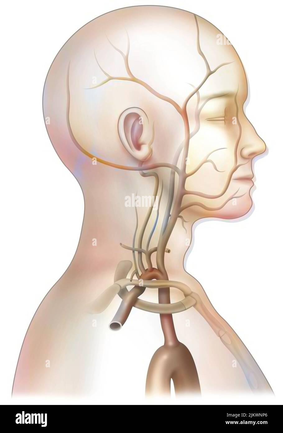 The arterial blood supply to the neck (carotids and vertebral arteries). Stock Photohttps://www.alamy.com/image-license-details/?v=1https://www.alamy.com/the-arterial-blood-supply-to-the-neck-carotids-and-vertebral-arteries-image476924286.html
RF2JKWNP6–The arterial blood supply to the neck (carotids and vertebral arteries).
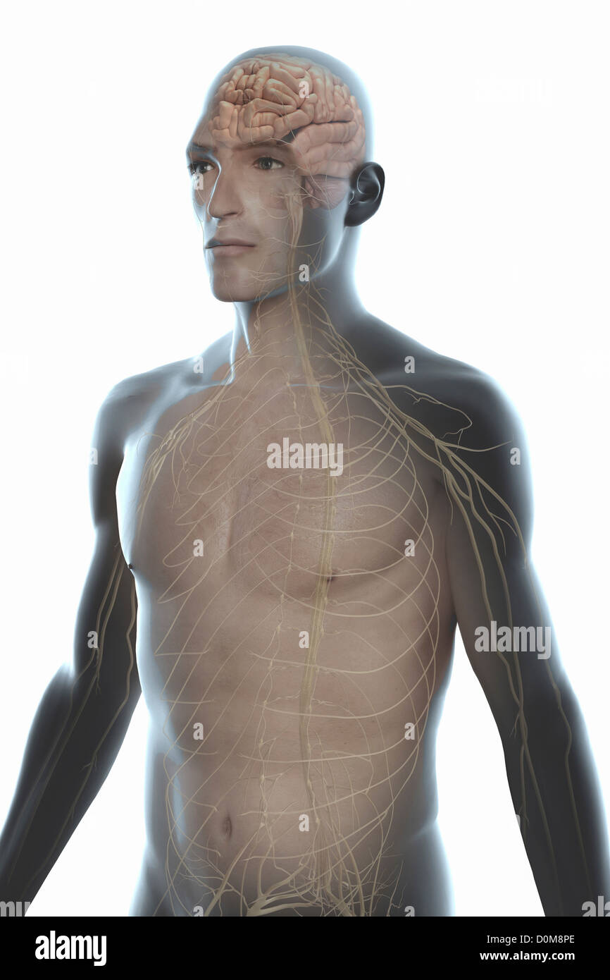 Three-quarter view of the male trunk with the nervous system and brain. Stock Photohttps://www.alamy.com/image-license-details/?v=1https://www.alamy.com/stock-photo-three-quarter-view-of-the-male-trunk-with-the-nervous-system-and-brain-52077046.html
RMD0M8PE–Three-quarter view of the male trunk with the nervous system and brain.
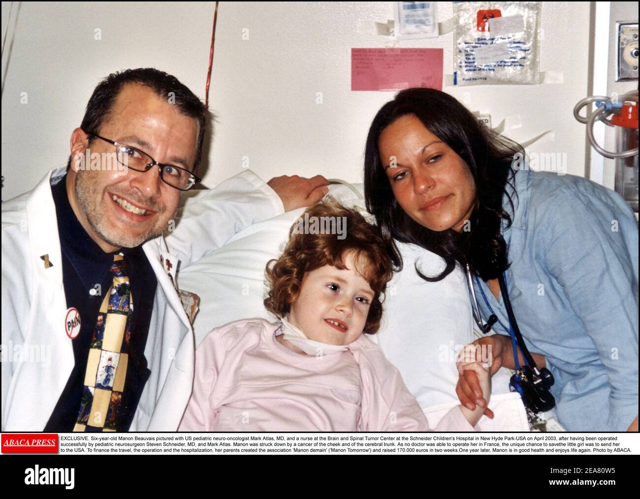 EXCLUSIVE. Six-year-old Manon Beauvais pictured with US pediatric neuro-oncologist Mark Atlas, MD, and a nurse at the Brain and Spinal Tumor Center at the Schneider Children's Hospital in New Hyde Park-USA on April 2003, after having been operated successfully by pediatric neurosurgeon Steven Schneider, MD, and Mark Atlas. Manon was struck down by a cancer of the cheek and of the cerebral trunk. As no doctor was able to operate her in France, the unique chance to savethe little girl was to send her to the USA. To finance the travel, the operation and the hospitalization, her parents created th Stock Photohttps://www.alamy.com/image-license-details/?v=1https://www.alamy.com/exclusive-six-year-old-manon-beauvais-pictured-with-us-pediatric-neuro-oncologist-mark-atlas-md-and-a-nurse-at-the-brain-and-spinal-tumor-center-at-the-schneider-childrens-hospital-in-new-hyde-park-usa-on-april-2003-after-having-been-operated-successfully-by-pediatric-neurosurgeon-steven-schneider-md-and-mark-atlas-manon-was-struck-down-by-a-cancer-of-the-cheek-and-of-the-cerebral-trunk-as-no-doctor-was-able-to-operate-her-in-france-the-unique-chance-to-savethe-little-girl-was-to-send-her-to-the-usa-to-finance-the-travel-the-operation-and-the-hospitalization-her-parents-created-th-image402161345.html
RF2EA80W5–EXCLUSIVE. Six-year-old Manon Beauvais pictured with US pediatric neuro-oncologist Mark Atlas, MD, and a nurse at the Brain and Spinal Tumor Center at the Schneider Children's Hospital in New Hyde Park-USA on April 2003, after having been operated successfully by pediatric neurosurgeon Steven Schneider, MD, and Mark Atlas. Manon was struck down by a cancer of the cheek and of the cerebral trunk. As no doctor was able to operate her in France, the unique chance to savethe little girl was to send her to the USA. To finance the travel, the operation and the hospitalization, her parents created th
 Organic and functional nervous diseases; a text-book of neurology . astomosing vessels is extensive. In the ganglia andcapsule it is very imperfect, as the arteries are terminal. Hence thepermanent effect of occlusion is more serious in lesions of the arteriesof the base than in those of the branches in the cortex. If a largevessel in the cortex — e. g., a main branch of a Sylvian artery, or themiddle cerebral trunk itself—is plugged, the area of softening may beextensive. When a vessel is occluded a clot forms within it whichextends backward to the next large branch. In some cases a secondemb Stock Photohttps://www.alamy.com/image-license-details/?v=1https://www.alamy.com/organic-and-functional-nervous-diseases-a-text-book-of-neurology-astomosing-vessels-is-extensive-in-the-ganglia-andcapsule-it-is-very-imperfect-as-the-arteries-are-terminal-hence-thepermanent-effect-of-occlusion-is-more-serious-in-lesions-of-the-arteriesof-the-base-than-in-those-of-the-branches-in-the-cortex-if-a-largevessel-in-the-cortex-e-g-a-main-branch-of-a-sylvian-artery-or-themiddle-cerebral-trunk-itselfis-plugged-the-area-of-softening-may-beextensive-when-a-vessel-is-occluded-a-clot-forms-within-it-whichextends-backward-to-the-next-large-branch-in-some-cases-a-secondemb-image339928816.html
RM2AN12JT–Organic and functional nervous diseases; a text-book of neurology . astomosing vessels is extensive. In the ganglia andcapsule it is very imperfect, as the arteries are terminal. Hence thepermanent effect of occlusion is more serious in lesions of the arteriesof the base than in those of the branches in the cortex. If a largevessel in the cortex — e. g., a main branch of a Sylvian artery, or themiddle cerebral trunk itself—is plugged, the area of softening may beextensive. When a vessel is occluded a clot forms within it whichextends backward to the next large branch. In some cases a secondemb
 Human Heart With Circulatory System Anatomy For Medical Concept 3D Illustration Stock Photohttps://www.alamy.com/image-license-details/?v=1https://www.alamy.com/human-heart-with-circulatory-system-anatomy-for-medical-concept-3d-illustration-image368852425.html
RF2CC2K0W–Human Heart With Circulatory System Anatomy For Medical Concept 3D Illustration
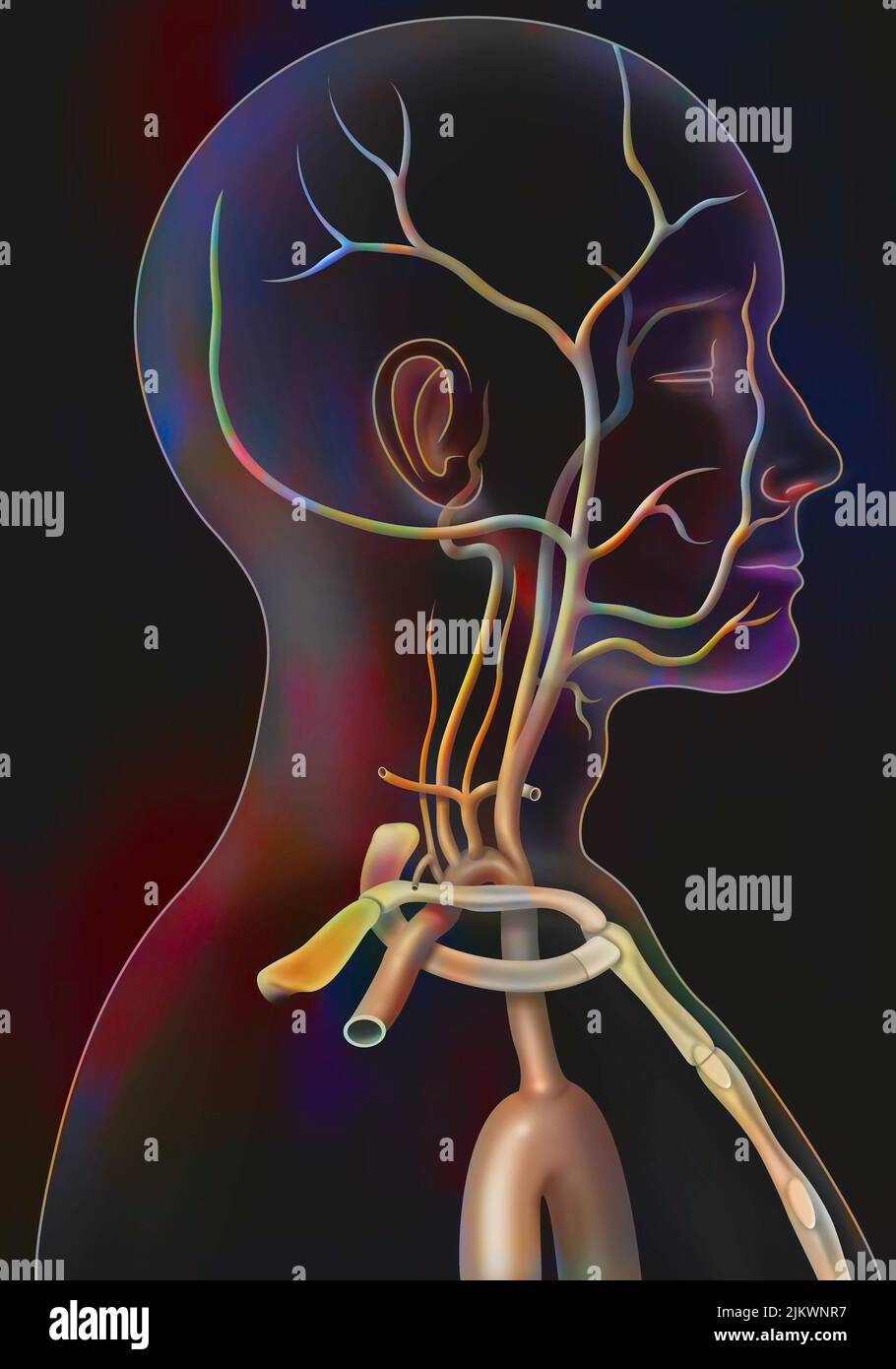 The arterial blood supply to the neck (carotids and vertebral arteries). Stock Photohttps://www.alamy.com/image-license-details/?v=1https://www.alamy.com/the-arterial-blood-supply-to-the-neck-carotids-and-vertebral-arteries-image476924315.html
RF2JKWNR7–The arterial blood supply to the neck (carotids and vertebral arteries).
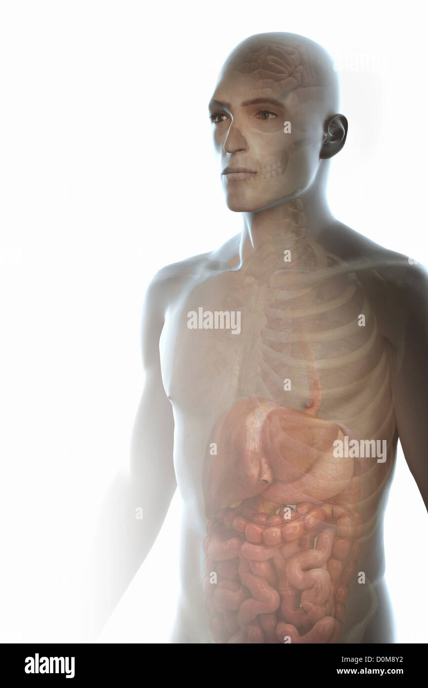 Three-quarter view of the male trunk with the digestive system, skeleton and brain. Stock Photohttps://www.alamy.com/image-license-details/?v=1https://www.alamy.com/stock-photo-three-quarter-view-of-the-male-trunk-with-the-digestive-system-skeleton-52077174.html
RMD0M8Y2–Three-quarter view of the male trunk with the digestive system, skeleton and brain.
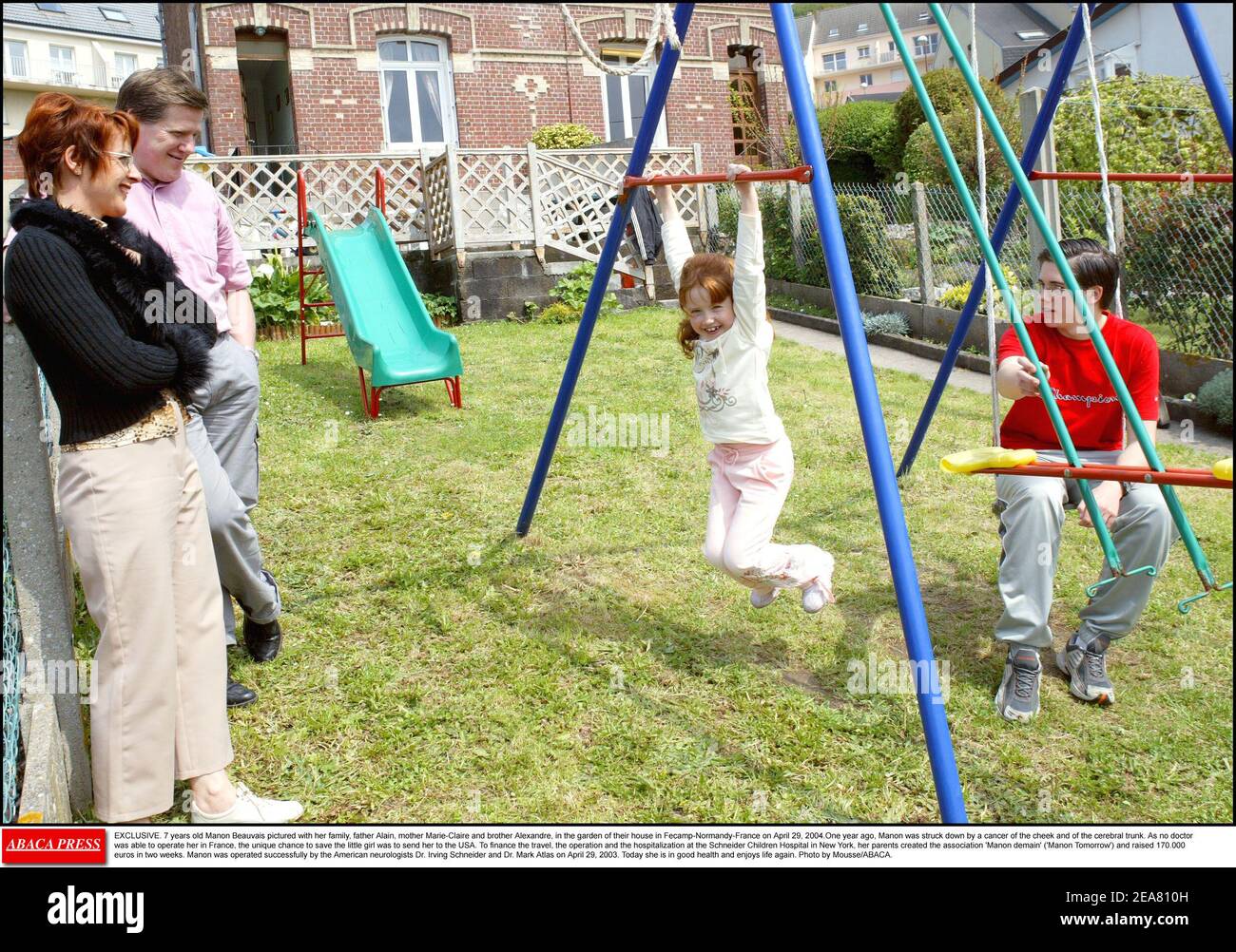 EXCLUSIVE. Seven-year-old Manon Beauvais pictured with her family, father Alain, mother Marie-Claire and brother Alexandre, in the garden of their house in Fecamp-Normandy-France on April 29, 2004. One year ago, Manon was struck down by a cancer of the cheek and of the cerebral trunk. As no doctor was able to operate her in France, the unique chance to save the little girl was to send her to the USA. To finance the travel, the operation and the hospitalization at the Schneider Children's Hospital in New York, her parents created the association 'Manon demain' ('Manon Tomorrow') and raised 170. Stock Photohttps://www.alamy.com/image-license-details/?v=1https://www.alamy.com/exclusive-seven-year-old-manon-beauvais-pictured-with-her-family-father-alain-mother-marie-claire-and-brother-alexandre-in-the-garden-of-their-house-in-fecamp-normandy-france-on-april-29-2004-one-year-ago-manon-was-struck-down-by-a-cancer-of-the-cheek-and-of-the-cerebral-trunk-as-no-doctor-was-able-to-operate-her-in-france-the-unique-chance-to-save-the-little-girl-was-to-send-her-to-the-usa-to-finance-the-travel-the-operation-and-the-hospitalization-at-the-schneider-childrens-hospital-in-new-york-her-parents-created-the-association-manon-demain-manon-tomorrow-and-raised-170-image402161441.html
RF2EA810H–EXCLUSIVE. Seven-year-old Manon Beauvais pictured with her family, father Alain, mother Marie-Claire and brother Alexandre, in the garden of their house in Fecamp-Normandy-France on April 29, 2004. One year ago, Manon was struck down by a cancer of the cheek and of the cerebral trunk. As no doctor was able to operate her in France, the unique chance to save the little girl was to send her to the USA. To finance the travel, the operation and the hospitalization at the Schneider Children's Hospital in New York, her parents created the association 'Manon demain' ('Manon Tomorrow') and raised 170.
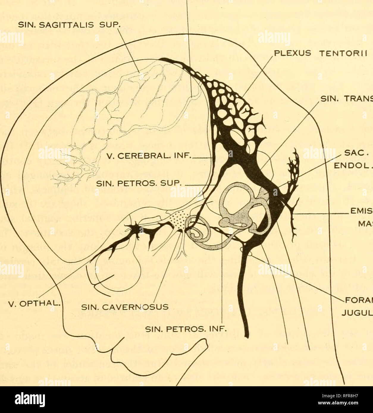 . Carnegie Institution of Washington publication. Of the brain of the human embryo. 29 the large ophthalmic and maxillary tributaries in front. In figure 27 it receives a large terminal trunk lateral to the infundibulum made up of tributaries coming from the region of the Sylvian fissure; this corresponds to the middle cerebral vein of the adult. Caudally the cavernous sinus communicates with the main blood-stream by means of the superior and inferior petrosal sinuses. The superior petrosal sinus passes over the cochlear part of the otic capsule and empties above SIN. RECTUS SIN. SAGITTALIS SU Stock Photohttps://www.alamy.com/image-license-details/?v=1https://www.alamy.com/carnegie-institution-of-washington-publication-of-the-brain-of-the-human-embryo-29-the-large-ophthalmic-and-maxillary-tributaries-in-front-in-figure-27-it-receives-a-large-terminal-trunk-lateral-to-the-infundibulum-made-up-of-tributaries-coming-from-the-region-of-the-sylvian-fissure-this-corresponds-to-the-middle-cerebral-vein-of-the-adult-caudally-the-cavernous-sinus-communicates-with-the-main-blood-stream-by-means-of-the-superior-and-inferior-petrosal-sinuses-the-superior-petrosal-sinus-passes-over-the-cochlear-part-of-the-otic-capsule-and-empties-above-sin-rectus-sin-sagittalis-su-image233466275.html
RMRFR8H7–. Carnegie Institution of Washington publication. Of the brain of the human embryo. 29 the large ophthalmic and maxillary tributaries in front. In figure 27 it receives a large terminal trunk lateral to the infundibulum made up of tributaries coming from the region of the Sylvian fissure; this corresponds to the middle cerebral vein of the adult. Caudally the cavernous sinus communicates with the main blood-stream by means of the superior and inferior petrosal sinuses. The superior petrosal sinus passes over the cochlear part of the otic capsule and empties above SIN. RECTUS SIN. SAGITTALIS SU
 Human Heart With Circulatory System Anatomy For Medical Concept 3D Illustration Stock Photohttps://www.alamy.com/image-license-details/?v=1https://www.alamy.com/human-heart-with-circulatory-system-anatomy-for-medical-concept-3d-illustration-image368852424.html
RF2CC2K0T–Human Heart With Circulatory System Anatomy For Medical Concept 3D Illustration
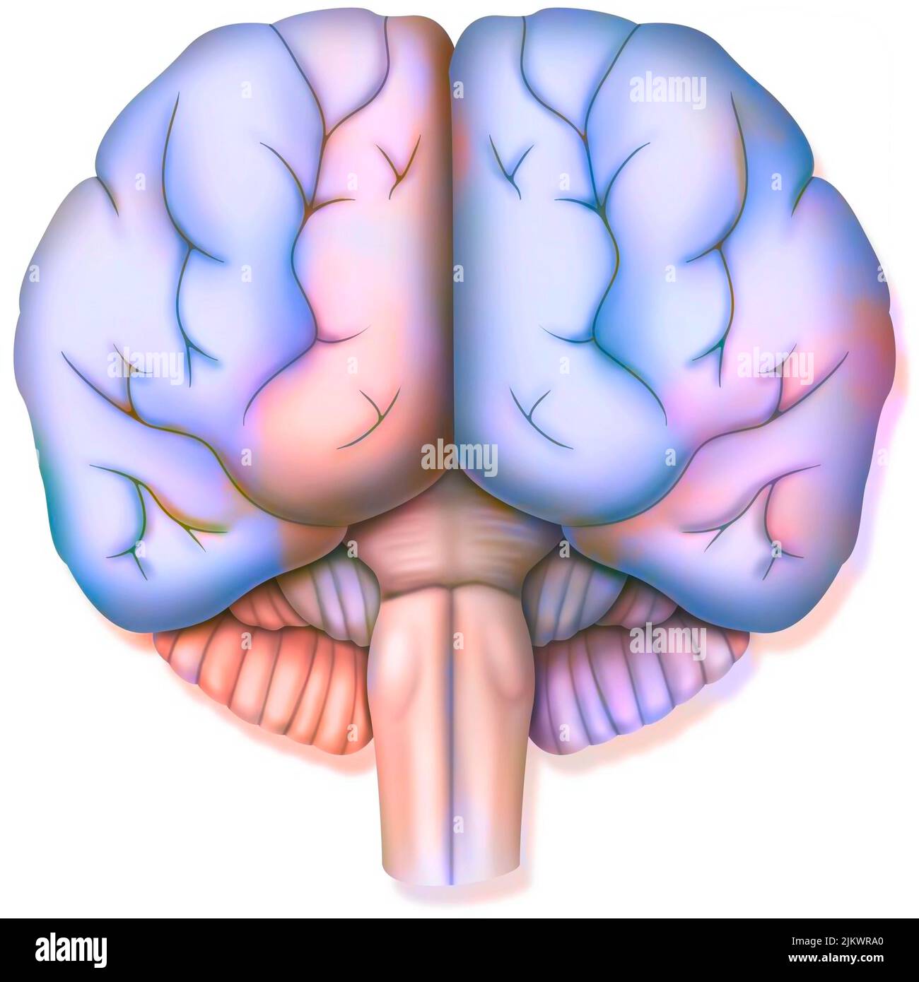 Brain, with the two cerebral hemispheres, the cerebellum and the brainstem. Stock Photohttps://www.alamy.com/image-license-details/?v=1https://www.alamy.com/brain-with-the-two-cerebral-hemispheres-the-cerebellum-and-the-brainstem-image476925512.html
RF2JKWRA0–Brain, with the two cerebral hemispheres, the cerebellum and the brainstem.
 EXCLUSIVE. Seven-year-old Manon Beauvais pictured with her brother Alexandre, in the garden of their family's house in Fecamp-Normandy-France on April 29, 2004. One year ago, Manon was struck down by a cancer of the cheek and of the cerebral trunk. As no doctor was able to operate her in France, the unique chance to save the little girl was to send her to the USA. To finance the travel, the operation and the hospitalization at the Schneider Children's Hospital in New York, her parents created the association 'Manon demain' ('Manon Tomorrow') and raised 170.000 euros in two weeks. Manon was ope Stock Photohttps://www.alamy.com/image-license-details/?v=1https://www.alamy.com/exclusive-seven-year-old-manon-beauvais-pictured-with-her-brother-alexandre-in-the-garden-of-their-familys-house-in-fecamp-normandy-france-on-april-29-2004-one-year-ago-manon-was-struck-down-by-a-cancer-of-the-cheek-and-of-the-cerebral-trunk-as-no-doctor-was-able-to-operate-her-in-france-the-unique-chance-to-save-the-little-girl-was-to-send-her-to-the-usa-to-finance-the-travel-the-operation-and-the-hospitalization-at-the-schneider-childrens-hospital-in-new-york-her-parents-created-the-association-manon-demain-manon-tomorrow-and-raised-170000-euros-in-two-weeks-manon-was-ope-image402161416.html
RF2EA80YM–EXCLUSIVE. Seven-year-old Manon Beauvais pictured with her brother Alexandre, in the garden of their family's house in Fecamp-Normandy-France on April 29, 2004. One year ago, Manon was struck down by a cancer of the cheek and of the cerebral trunk. As no doctor was able to operate her in France, the unique chance to save the little girl was to send her to the USA. To finance the travel, the operation and the hospitalization at the Schneider Children's Hospital in New York, her parents created the association 'Manon demain' ('Manon Tomorrow') and raised 170.000 euros in two weeks. Manon was ope
 Nervous and mental diseases . he cerebral cortex cutaneous sensoryrepresentation is related quite closely to themotor fields, but is not identical with them,being placed mainly just behind the strictlymotor territory. Cortical lesions in this fieldlead to paresthetic disturbances of sensationthat have functional rather than anatom-ical limits, just as electrical stinuilation ofthe cortex leads to purposive or groupedmuscular movements, and not to those sub-served by any spinal segment or nerve-trunk. In hysterical anesthesia a similardistribution is noted, the affected area oftenhaving the out Stock Photohttps://www.alamy.com/image-license-details/?v=1https://www.alamy.com/nervous-and-mental-diseases-he-cerebral-cortex-cutaneous-sensoryrepresentation-is-related-quite-closely-to-themotor-fields-but-is-not-identical-with-thembeing-placed-mainly-just-behind-the-strictlymotor-territory-cortical-lesions-in-this-fieldlead-to-paresthetic-disturbances-of-sensationthat-have-functional-rather-than-anatom-ical-limits-just-as-electrical-stinuilation-ofthe-cortex-leads-to-purposive-or-groupedmuscular-movements-and-not-to-those-sub-served-by-any-spinal-segment-or-nerve-trunk-in-hysterical-anesthesia-a-similardistribution-is-noted-the-affected-area-oftenhaving-the-out-image340050548.html
RM2AN6HXC–Nervous and mental diseases . he cerebral cortex cutaneous sensoryrepresentation is related quite closely to themotor fields, but is not identical with them,being placed mainly just behind the strictlymotor territory. Cortical lesions in this fieldlead to paresthetic disturbances of sensationthat have functional rather than anatom-ical limits, just as electrical stinuilation ofthe cortex leads to purposive or groupedmuscular movements, and not to those sub-served by any spinal segment or nerve-trunk. In hysterical anesthesia a similardistribution is noted, the affected area oftenhaving the out
 Human Heart With Circulatory System Anatomy For Medical Concept 3D Illustration Stock Photohttps://www.alamy.com/image-license-details/?v=1https://www.alamy.com/human-heart-with-circulatory-system-anatomy-for-medical-concept-3d-illustration-image368852439.html
RF2CC2K1B–Human Heart With Circulatory System Anatomy For Medical Concept 3D Illustration
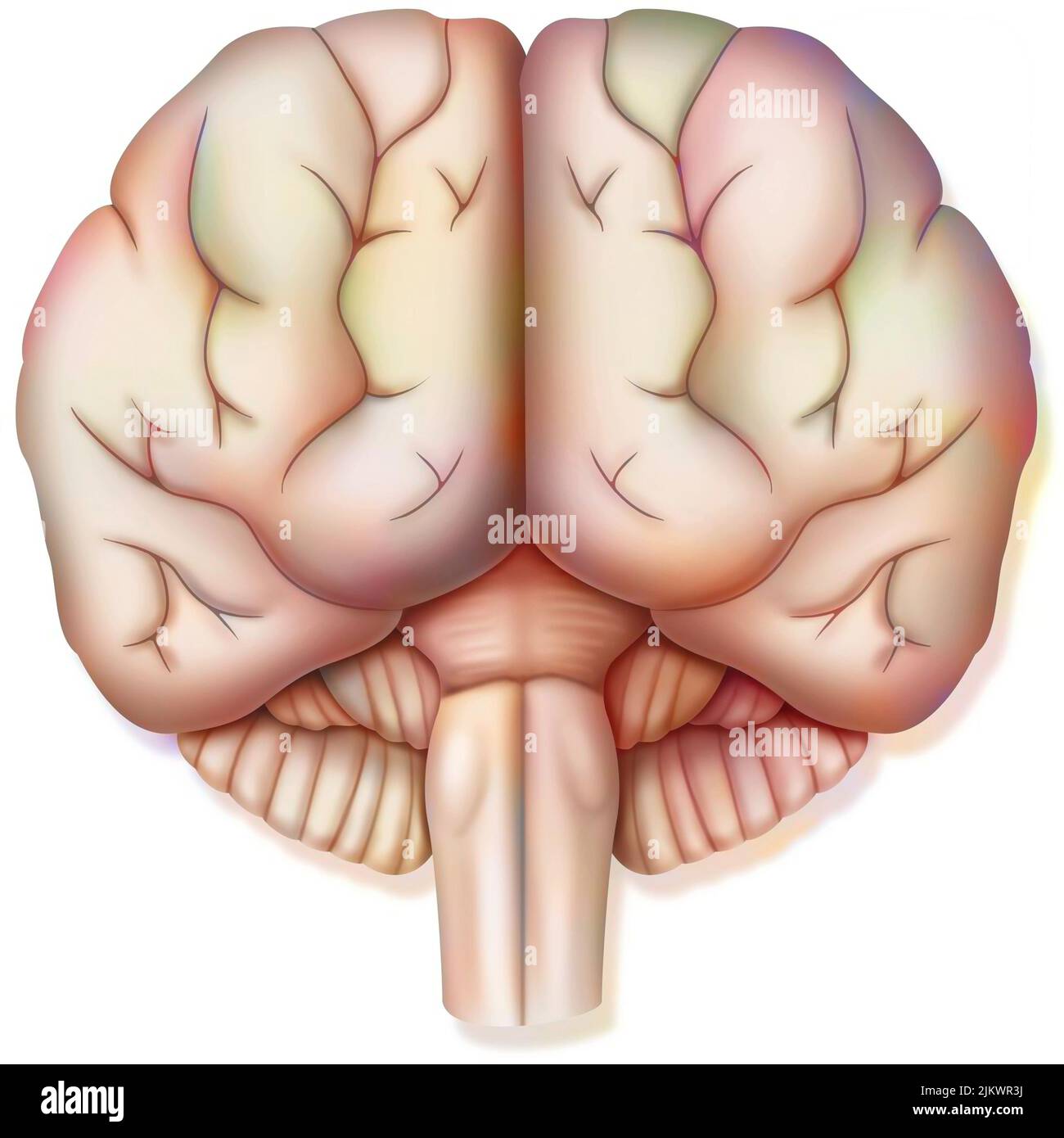 Brain, with the two cerebral hemispheres, the cerebellum and the brainstem. Stock Photohttps://www.alamy.com/image-license-details/?v=1https://www.alamy.com/brain-with-the-two-cerebral-hemispheres-the-cerebellum-and-the-brainstem-image476925334.html
RF2JKWR3J–Brain, with the two cerebral hemispheres, the cerebellum and the brainstem.
 EXCLUSIVE. Seven-year-old Manon Beauvais pictured in the garden of her family's house in Fecamp-Normandy-France on April 29, 2004. One year ago, Manon was struck down by a cancer of the cheek and of the cerebral trunk. As no doctor was able to operate her in France, the unique chance to save the little girl was to send her to the USA. To finance the travel, the operation and the hospitalization at the Schneider Children's Hospital in New York, her parents created the association 'Manon demain' ('Manon Tomorrow') and raised 170.000 euros in two weeks. Manon was operated successfully by the Amer Stock Photohttps://www.alamy.com/image-license-details/?v=1https://www.alamy.com/exclusive-seven-year-old-manon-beauvais-pictured-in-the-garden-of-her-familys-house-in-fecamp-normandy-france-on-april-29-2004-one-year-ago-manon-was-struck-down-by-a-cancer-of-the-cheek-and-of-the-cerebral-trunk-as-no-doctor-was-able-to-operate-her-in-france-the-unique-chance-to-save-the-little-girl-was-to-send-her-to-the-usa-to-finance-the-travel-the-operation-and-the-hospitalization-at-the-schneider-childrens-hospital-in-new-york-her-parents-created-the-association-manon-demain-manon-tomorrow-and-raised-170000-euros-in-two-weeks-manon-was-operated-successfully-by-the-amer-image402161407.html
RF2EA80YB–EXCLUSIVE. Seven-year-old Manon Beauvais pictured in the garden of her family's house in Fecamp-Normandy-France on April 29, 2004. One year ago, Manon was struck down by a cancer of the cheek and of the cerebral trunk. As no doctor was able to operate her in France, the unique chance to save the little girl was to send her to the USA. To finance the travel, the operation and the hospitalization at the Schneider Children's Hospital in New York, her parents created the association 'Manon demain' ('Manon Tomorrow') and raised 170.000 euros in two weeks. Manon was operated successfully by the Amer
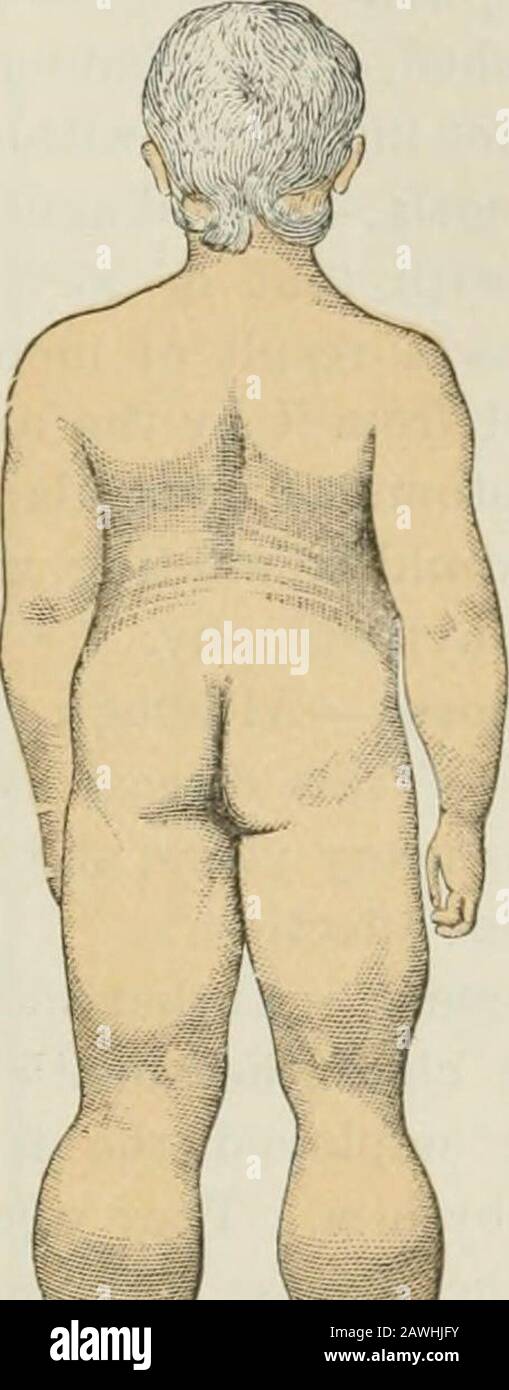 Lectures on nervous diseases from the standpoint of cerebral and spinal localization, and the later methods employed in the diagnosis and treatment of these affections . the. Fig. 112.—Side View of Attitude of Pseudo-hypertrophic Paralysis. (Duchenne.) Fig. 113.—Rear View of Attitude of Pseudo-hypertrophic Paralysis. (Duchenne.) arm/ This movement of climbing up the thighs, as it has beentermed, is an indication of weakness in the muscles which straightenthe knee, and also those which extend the trunk upon the thigh,—theextensors of the hip-joint. (See Figs. 52 and 53.) The (jait of these iiat Stock Photohttps://www.alamy.com/image-license-details/?v=1https://www.alamy.com/lectures-on-nervous-diseases-from-the-standpoint-of-cerebral-and-spinal-localization-and-the-later-methods-employed-in-the-diagnosis-and-treatment-of-these-affections-the-fig-112side-view-of-attitude-of-pseudo-hypertrophic-paralysis-duchenne-fig-113rear-view-of-attitude-of-pseudo-hypertrophic-paralysis-duchenne-arm-this-movement-of-climbing-up-the-thighs-as-it-has-beentermed-is-an-indication-of-weakness-in-the-muscles-which-straightenthe-knee-and-also-those-which-extend-the-trunk-upon-the-thightheextensors-of-the-hip-joint-see-figs-52-and-53-the-jait-of-these-iiat-image342751135.html
RM2AWHJFY–Lectures on nervous diseases from the standpoint of cerebral and spinal localization, and the later methods employed in the diagnosis and treatment of these affections . the. Fig. 112.—Side View of Attitude of Pseudo-hypertrophic Paralysis. (Duchenne.) Fig. 113.—Rear View of Attitude of Pseudo-hypertrophic Paralysis. (Duchenne.) arm/ This movement of climbing up the thighs, as it has beentermed, is an indication of weakness in the muscles which straightenthe knee, and also those which extend the trunk upon the thigh,—theextensors of the hip-joint. (See Figs. 52 and 53.) The (jait of these iiat
 Human Heart With Circulatory System Anatomy For Medical Concept 3D Illustration Stock Photohttps://www.alamy.com/image-license-details/?v=1https://www.alamy.com/human-heart-with-circulatory-system-anatomy-for-medical-concept-3d-illustration-image368852444.html
RF2CC2K1G–Human Heart With Circulatory System Anatomy For Medical Concept 3D Illustration
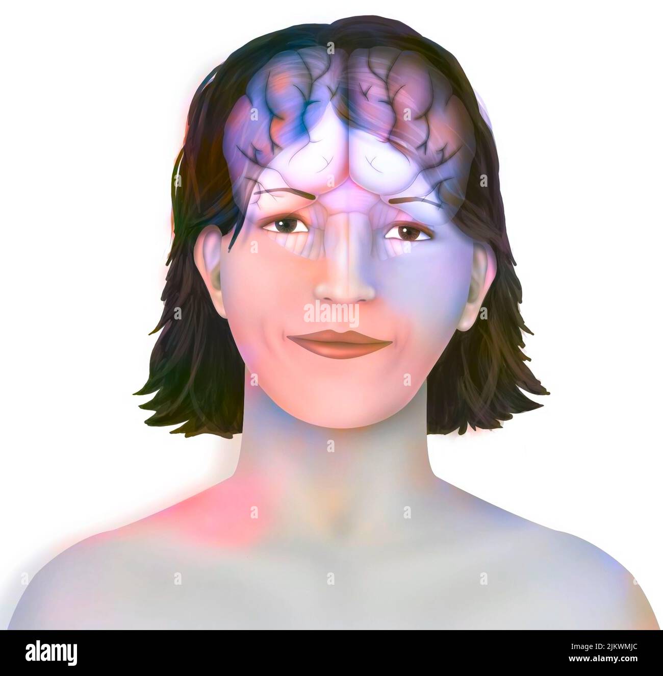 Brain: cerebral hemispheres with longitudinal fissure, cerebellum and brainstem). Stock Photohttps://www.alamy.com/image-license-details/?v=1https://www.alamy.com/brain-cerebral-hemispheres-with-longitudinal-fissure-cerebellum-and-brainstem-image476923396.html
RF2JKWMJC–Brain: cerebral hemispheres with longitudinal fissure, cerebellum and brainstem).
 EXCLUSIVE. Seven-year-old Manon Beauvais pictured in the garden of her family's house in Fecamp-Normandy-France on April 29, 2004. One year ago, Manon was struck down by a cancer of the cheek and of the cerebral trunk. As no doctor was able to operate her in France, the unique chance to save the little girl was to send her to the USA. To finance the travel, the operation and the hospitalization at the Schneider Children's Hospital in New York, her parents created the association 'Manon demain' ('Manon Tomorrow') and raised 170.000 euros in two weeks. Manon was operated successfully by the Amer Stock Photohttps://www.alamy.com/image-license-details/?v=1https://www.alamy.com/exclusive-seven-year-old-manon-beauvais-pictured-in-the-garden-of-her-familys-house-in-fecamp-normandy-france-on-april-29-2004-one-year-ago-manon-was-struck-down-by-a-cancer-of-the-cheek-and-of-the-cerebral-trunk-as-no-doctor-was-able-to-operate-her-in-france-the-unique-chance-to-save-the-little-girl-was-to-send-her-to-the-usa-to-finance-the-travel-the-operation-and-the-hospitalization-at-the-schneider-childrens-hospital-in-new-york-her-parents-created-the-association-manon-demain-manon-tomorrow-and-raised-170000-euros-in-two-weeks-manon-was-operated-successfully-by-the-amer-image402161320.html
RF2EA80T8–EXCLUSIVE. Seven-year-old Manon Beauvais pictured in the garden of her family's house in Fecamp-Normandy-France on April 29, 2004. One year ago, Manon was struck down by a cancer of the cheek and of the cerebral trunk. As no doctor was able to operate her in France, the unique chance to save the little girl was to send her to the USA. To finance the travel, the operation and the hospitalization at the Schneider Children's Hospital in New York, her parents created the association 'Manon demain' ('Manon Tomorrow') and raised 170.000 euros in two weeks. Manon was operated successfully by the Amer
 Outlines of comparative physiology touching the structure and development of the races of animals, living and extinct : for the use of schools and colleges . TV. Brain of Cercopithecus Sabeeuslaid open. a Base of the same brain, showingthe cerebral nerves. [§ 97. CEREBEAL NERVES. We have shown in fig. 20 theprimary course of the cerebral nerves, and their union withthe brain. The olfactory ganglia are large in the cold-bloodedvertebrata, but very small in man, consisting merely of an en-largement of the trunk of the olfactory nerves (1), which arethe first pair that unite with the brain. From Stock Photohttps://www.alamy.com/image-license-details/?v=1https://www.alamy.com/outlines-of-comparative-physiology-touching-the-structure-and-development-of-the-races-of-animals-living-and-extinct-for-the-use-of-schools-and-colleges-tv-brain-of-cercopithecus-sabeeuslaid-open-a-base-of-the-same-brain-showingthe-cerebral-nerves-97-cerebeal-nerves-we-have-shown-in-fig-20-theprimary-course-of-the-cerebral-nerves-and-their-union-withthe-brain-the-olfactory-ganglia-are-large-in-the-cold-bloodedvertebrata-but-very-small-in-man-consisting-merely-of-an-en-largement-of-the-trunk-of-the-olfactory-nerves-1-which-arethe-first-pair-that-unite-with-the-brain-from-image339081308.html
RM2AKJDJM–Outlines of comparative physiology touching the structure and development of the races of animals, living and extinct : for the use of schools and colleges . TV. Brain of Cercopithecus Sabeeuslaid open. a Base of the same brain, showingthe cerebral nerves. [§ 97. CEREBEAL NERVES. We have shown in fig. 20 theprimary course of the cerebral nerves, and their union withthe brain. The olfactory ganglia are large in the cold-bloodedvertebrata, but very small in man, consisting merely of an en-largement of the trunk of the olfactory nerves (1), which arethe first pair that unite with the brain. From
 Human Heart With Circulatory System Anatomy For Medical Concept 3D Illustration Stock Photohttps://www.alamy.com/image-license-details/?v=1https://www.alamy.com/human-heart-with-circulatory-system-anatomy-for-medical-concept-3d-illustration-image368852414.html
RF2CC2K0E–Human Heart With Circulatory System Anatomy For Medical Concept 3D Illustration
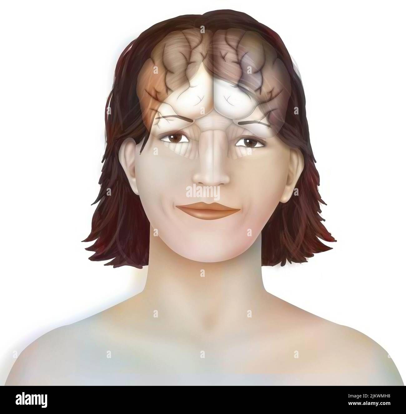 Brain: cerebral hemispheres with longitudinal fissure, cerebellum and brainstem). Stock Photohttps://www.alamy.com/image-license-details/?v=1https://www.alamy.com/brain-cerebral-hemispheres-with-longitudinal-fissure-cerebellum-and-brainstem-image476923364.html
RF2JKWMH8–Brain: cerebral hemispheres with longitudinal fissure, cerebellum and brainstem).
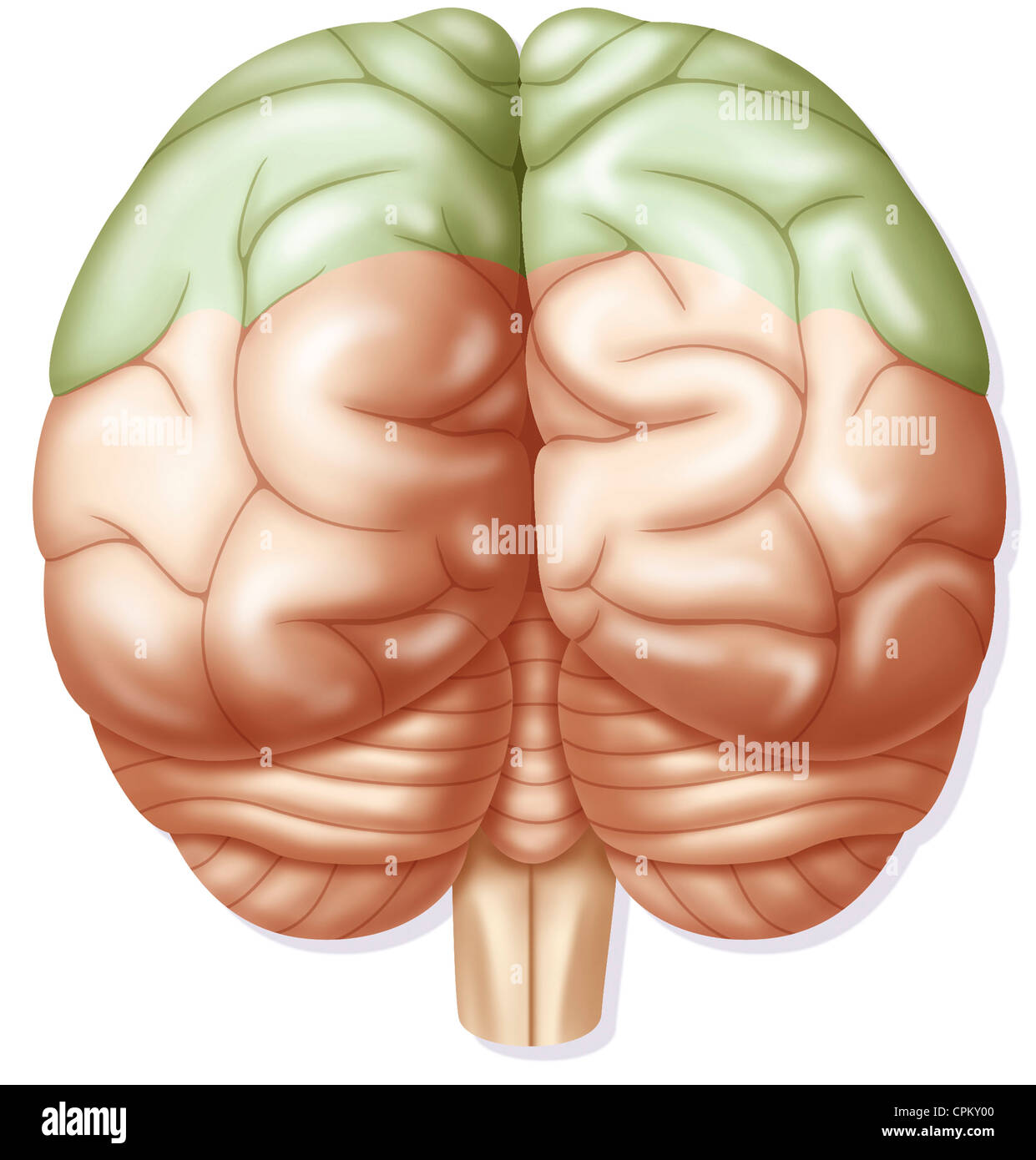 CEREBRAL LOBE Stock Photohttps://www.alamy.com/image-license-details/?v=1https://www.alamy.com/stock-photo-cerebral-lobe-48381424.html
RMCPKY00–CEREBRAL LOBE
 EXCLUSIVE. Seven-year-old Manon Beauvais pictured in the garden of her family's house in Fecamp-Normandy-France on April 29, 2004. One year ago, Manon was struck down by a cancer of the cheek and of the cerebral trunk. As no doctor was able to operate her in France, the unique chance to save the little girl was to send her to the USA. To finance the travel, the operation and the hospitalization at the Schneider Children's Hospital in New York, her parents created the association 'Manon demain' ('Manon Tomorrow') and raised 170.000 euros in two weeks. Manon was operated successfully by the Amer Stock Photohttps://www.alamy.com/image-license-details/?v=1https://www.alamy.com/exclusive-seven-year-old-manon-beauvais-pictured-in-the-garden-of-her-familys-house-in-fecamp-normandy-france-on-april-29-2004-one-year-ago-manon-was-struck-down-by-a-cancer-of-the-cheek-and-of-the-cerebral-trunk-as-no-doctor-was-able-to-operate-her-in-france-the-unique-chance-to-save-the-little-girl-was-to-send-her-to-the-usa-to-finance-the-travel-the-operation-and-the-hospitalization-at-the-schneider-childrens-hospital-in-new-york-her-parents-created-the-association-manon-demain-manon-tomorrow-and-raised-170000-euros-in-two-weeks-manon-was-operated-successfully-by-the-amer-image402161431.html
RF2EA8107–EXCLUSIVE. Seven-year-old Manon Beauvais pictured in the garden of her family's house in Fecamp-Normandy-France on April 29, 2004. One year ago, Manon was struck down by a cancer of the cheek and of the cerebral trunk. As no doctor was able to operate her in France, the unique chance to save the little girl was to send her to the USA. To finance the travel, the operation and the hospitalization at the Schneider Children's Hospital in New York, her parents created the association 'Manon demain' ('Manon Tomorrow') and raised 170.000 euros in two weeks. Manon was operated successfully by the Amer
 . Local and regional anesthesia; with chapters on spinal, epidural, paravertebral, and parasacral analgesia, and other applications of local and regional anesthesia to the surgery of the eye, ear, nose and throat, and to dental practice. Fig. 212.—Base of the skull, inner or cerebral surface. (After Gray.) turing through the chief trunk of the trigeminus through the cistern£eof the posterior cranial fossa (cisterna pontis). This has actuallyhappened to us in the hving subject, and emission of fluid resulted.One secures, if one immediately draws the needle back somewhat andthen injects slowly, Stock Photohttps://www.alamy.com/image-license-details/?v=1https://www.alamy.com/local-and-regional-anesthesia-with-chapters-on-spinal-epidural-paravertebral-and-parasacral-analgesia-and-other-applications-of-local-and-regional-anesthesia-to-the-surgery-of-the-eye-ear-nose-and-throat-and-to-dental-practice-fig-212base-of-the-skull-inner-or-cerebral-surface-after-gray-turing-through-the-chief-trunk-of-the-trigeminus-through-the-cisterneof-the-posterior-cranial-fossa-cisterna-pontis-this-has-actuallyhappened-to-us-in-the-hving-subject-and-emission-of-fluid-resultedone-secures-if-one-immediately-draws-the-needle-back-somewhat-andthen-injects-slowly-image337102738.html
RM2AGC9YE–. Local and regional anesthesia; with chapters on spinal, epidural, paravertebral, and parasacral analgesia, and other applications of local and regional anesthesia to the surgery of the eye, ear, nose and throat, and to dental practice. Fig. 212.—Base of the skull, inner or cerebral surface. (After Gray.) turing through the chief trunk of the trigeminus through the cistern£eof the posterior cranial fossa (cisterna pontis). This has actuallyhappened to us in the hving subject, and emission of fluid resulted.One secures, if one immediately draws the needle back somewhat andthen injects slowly,
 Human Heart Anatomy For Medical Concept 3D Illustration Stock Photohttps://www.alamy.com/image-license-details/?v=1https://www.alamy.com/human-heart-anatomy-for-medical-concept-3d-illustration-image368852423.html
RF2CC2K0R–Human Heart Anatomy For Medical Concept 3D Illustration
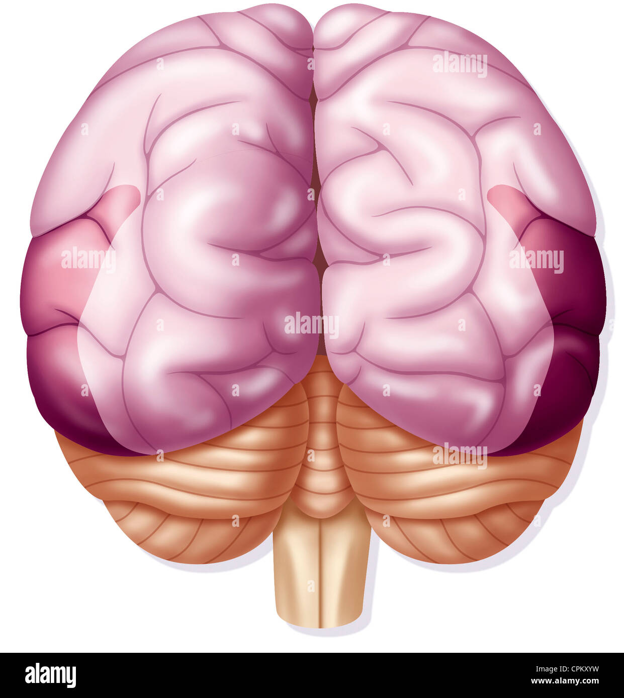 CEREBRAL LOBE Stock Photohttps://www.alamy.com/image-license-details/?v=1https://www.alamy.com/stock-photo-cerebral-lobe-48381421.html
RMCPKXYW–CEREBRAL LOBE
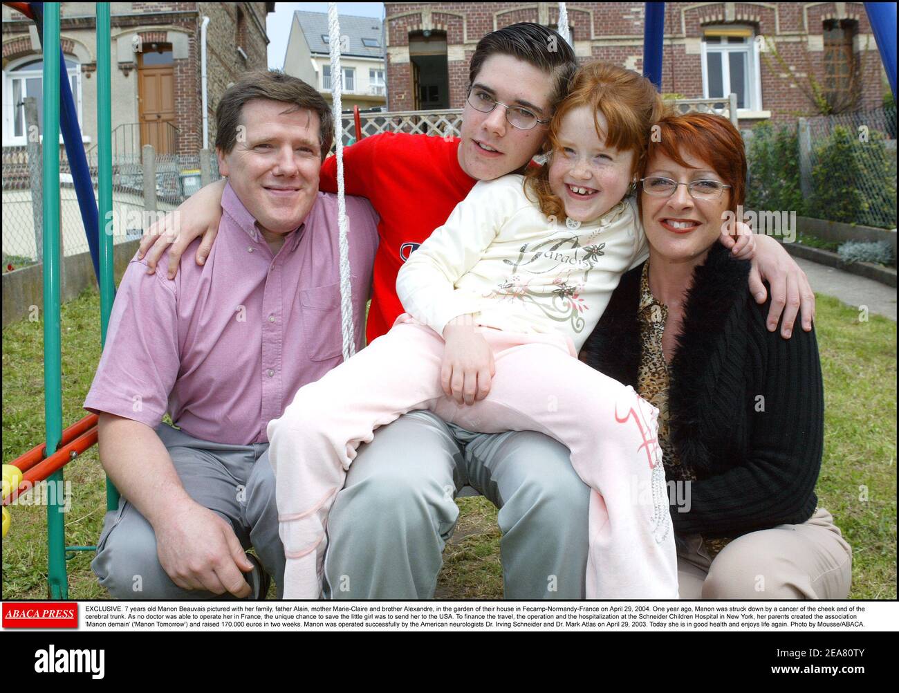 EXCLUSIVE. Seven-year-old Manon Beauvais pictured with her family, father Alain, mother Marie-Claire and brother Alexandre, in the garden of their house in Fecamp-Normandy-France on April 29, 2004. One year ago, Manon was struck down by a cancer of the cheek and of the cerebral trunk. As no doctor was able to operate her in France, the unique chance to save the little girl was to send her to the USA. To finance the travel, the operation and the hospitalization at the Schneider Children's Hospital in New York, her parents created the association 'Manon demain' ('Manon Tomorrow') and raised 170. Stock Photohttps://www.alamy.com/image-license-details/?v=1https://www.alamy.com/exclusive-seven-year-old-manon-beauvais-pictured-with-her-family-father-alain-mother-marie-claire-and-brother-alexandre-in-the-garden-of-their-house-in-fecamp-normandy-france-on-april-29-2004-one-year-ago-manon-was-struck-down-by-a-cancer-of-the-cheek-and-of-the-cerebral-trunk-as-no-doctor-was-able-to-operate-her-in-france-the-unique-chance-to-save-the-little-girl-was-to-send-her-to-the-usa-to-finance-the-travel-the-operation-and-the-hospitalization-at-the-schneider-childrens-hospital-in-new-york-her-parents-created-the-association-manon-demain-manon-tomorrow-and-raised-170-image402161339.html
RF2EA80TY–EXCLUSIVE. Seven-year-old Manon Beauvais pictured with her family, father Alain, mother Marie-Claire and brother Alexandre, in the garden of their house in Fecamp-Normandy-France on April 29, 2004. One year ago, Manon was struck down by a cancer of the cheek and of the cerebral trunk. As no doctor was able to operate her in France, the unique chance to save the little girl was to send her to the USA. To finance the travel, the operation and the hospitalization at the Schneider Children's Hospital in New York, her parents created the association 'Manon demain' ('Manon Tomorrow') and raised 170.
 A reference handbook of the medical sciences, embracing the entire range of scientific and practical medicine and allied science . n thissystem of vessels and the cerebral—except through thebasilar trunk—is by small branches of the precere-bellars and postcerebrals across the crura cerebri. 347 Brain, Circulation In REFERENCE HANDBOOK OF THE MEDICAL SCIENCES CiKCLE OF Willis.—The carotid ends at the outerangle of the chiasma in two main branches, the pre-and medicerebral arteries. At about one centimeterin front of the chiasm the two precerebrals are con-nected by the precommunicant. From eith Stock Photohttps://www.alamy.com/image-license-details/?v=1https://www.alamy.com/a-reference-handbook-of-the-medical-sciences-embracing-the-entire-range-of-scientific-and-practical-medicine-and-allied-science-n-thissystem-of-vessels-and-the-cerebralexcept-through-thebasilar-trunkis-by-small-branches-of-the-precere-bellars-and-postcerebrals-across-the-crura-cerebri-347-brain-circulation-in-reference-handbook-of-the-medical-sciences-cikcle-of-willisthe-carotid-ends-at-the-outerangle-of-the-chiasma-in-two-main-branches-the-pre-and-medicerebral-arteries-at-about-one-centimeterin-front-of-the-chiasm-the-two-precerebrals-are-con-nected-by-the-precommunicant-from-eith-image338896673.html
RM2AKA24H–A reference handbook of the medical sciences, embracing the entire range of scientific and practical medicine and allied science . n thissystem of vessels and the cerebral—except through thebasilar trunk—is by small branches of the precere-bellars and postcerebrals across the crura cerebri. 347 Brain, Circulation In REFERENCE HANDBOOK OF THE MEDICAL SCIENCES CiKCLE OF Willis.—The carotid ends at the outerangle of the chiasma in two main branches, the pre-and medicerebral arteries. At about one centimeterin front of the chiasm the two precerebrals are con-nected by the precommunicant. From eith
 Human Heart Anatomy For Medical Concept 3D Illustration Stock Photohttps://www.alamy.com/image-license-details/?v=1https://www.alamy.com/human-heart-anatomy-for-medical-concept-3d-illustration-image368852416.html
RF2CC2K0G–Human Heart Anatomy For Medical Concept 3D Illustration
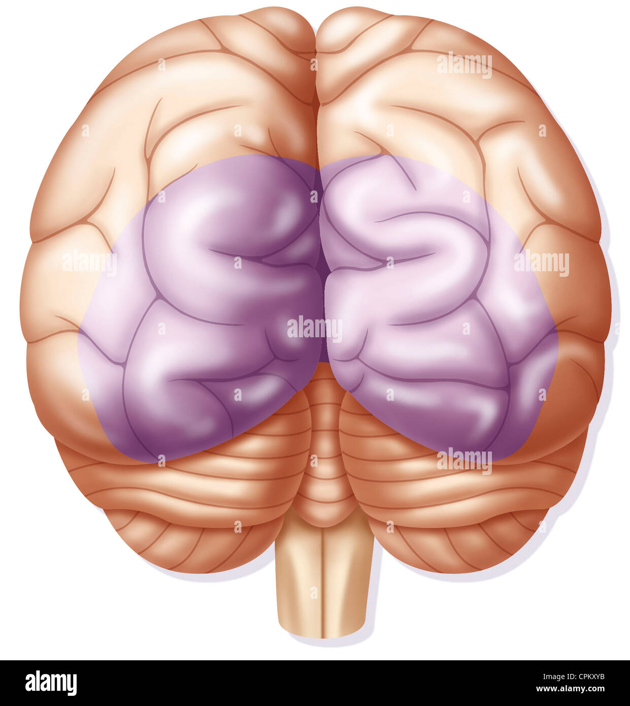 CEREBRAL LOBE Stock Photohttps://www.alamy.com/image-license-details/?v=1https://www.alamy.com/stock-photo-cerebral-lobe-48381407.html
RMCPKXYB–CEREBRAL LOBE
 EXCLUSIVE. Seven-year-old Manon Beauvais pictured in the garden of her family's house in Fecamp-Normandy-France on April 29, 2004. One year ago, Manon was struck down by a cancer of the cheek and of the cerebral trunk. As no doctor was able to operate her in France, the unique chance to save the little girl was to send her to the USA. To finance the travel, the operation and the hospitalization at the Schneider Children's Hospital in New York, her parents created the association 'Manon demain' ('Manon Tomorrow') and raised 170.000 euros in two weeks. Manon was operated successfully by the Amer Stock Photohttps://www.alamy.com/image-license-details/?v=1https://www.alamy.com/exclusive-seven-year-old-manon-beauvais-pictured-in-the-garden-of-her-familys-house-in-fecamp-normandy-france-on-april-29-2004-one-year-ago-manon-was-struck-down-by-a-cancer-of-the-cheek-and-of-the-cerebral-trunk-as-no-doctor-was-able-to-operate-her-in-france-the-unique-chance-to-save-the-little-girl-was-to-send-her-to-the-usa-to-finance-the-travel-the-operation-and-the-hospitalization-at-the-schneider-childrens-hospital-in-new-york-her-parents-created-the-association-manon-demain-manon-tomorrow-and-raised-170000-euros-in-two-weeks-manon-was-operated-successfully-by-the-amer-image402161330.html
RF2EA80TJ–EXCLUSIVE. Seven-year-old Manon Beauvais pictured in the garden of her family's house in Fecamp-Normandy-France on April 29, 2004. One year ago, Manon was struck down by a cancer of the cheek and of the cerebral trunk. As no doctor was able to operate her in France, the unique chance to save the little girl was to send her to the USA. To finance the travel, the operation and the hospitalization at the Schneider Children's Hospital in New York, her parents created the association 'Manon demain' ('Manon Tomorrow') and raised 170.000 euros in two weeks. Manon was operated successfully by the Amer
 . The physiology of the Invertebrata. ing like a hood the superior ganglion,and from certain experiments of M. Paivre,t the cerebralganglion has, like the cerebral hemispheres of the Vertebrates,the property of being insensible to punctures and lacerations. The nervous system of the Insecta (speaking in generalterms) consists of a cerebral ganglion connected to a gan-glionated nerve-trunk or trunks, which passes backwards alongthe ventral surface of the animal. Lateral nerves are givenofE from these ganglia to the organs of sense, limbs, viscera,&c. Fig. 63 represents the nervous systems of va Stock Photohttps://www.alamy.com/image-license-details/?v=1https://www.alamy.com/the-physiology-of-the-invertebrata-ing-like-a-hood-the-superior-ganglionand-from-certain-experiments-of-m-paivret-the-cerebralganglion-has-like-the-cerebral-hemispheres-of-the-vertebratesthe-property-of-being-insensible-to-punctures-and-lacerations-the-nervous-system-of-the-insecta-speaking-in-generalterms-consists-of-a-cerebral-ganglion-connected-to-a-gan-glionated-nerve-trunk-or-trunks-which-passes-backwards-alongthe-ventral-surface-of-the-animal-lateral-nerves-are-givenofe-from-these-ganglia-to-the-organs-of-sense-limbs-viscerac-fig-63-represents-the-nervous-systems-of-va-image336622038.html
RM2AFJCRJ–. The physiology of the Invertebrata. ing like a hood the superior ganglion,and from certain experiments of M. Paivre,t the cerebralganglion has, like the cerebral hemispheres of the Vertebrates,the property of being insensible to punctures and lacerations. The nervous system of the Insecta (speaking in generalterms) consists of a cerebral ganglion connected to a gan-glionated nerve-trunk or trunks, which passes backwards alongthe ventral surface of the animal. Lateral nerves are givenofE from these ganglia to the organs of sense, limbs, viscera,&c. Fig. 63 represents the nervous systems of va
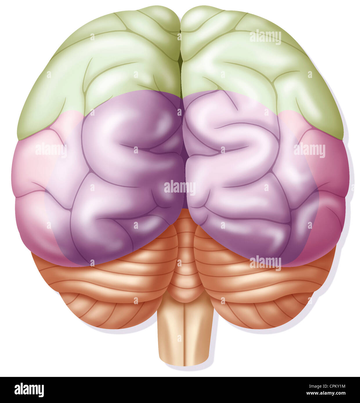 CEREBRAL LOBE Stock Photohttps://www.alamy.com/image-license-details/?v=1https://www.alamy.com/stock-photo-cerebral-lobe-48381472.html
RMCPKY1M–CEREBRAL LOBE
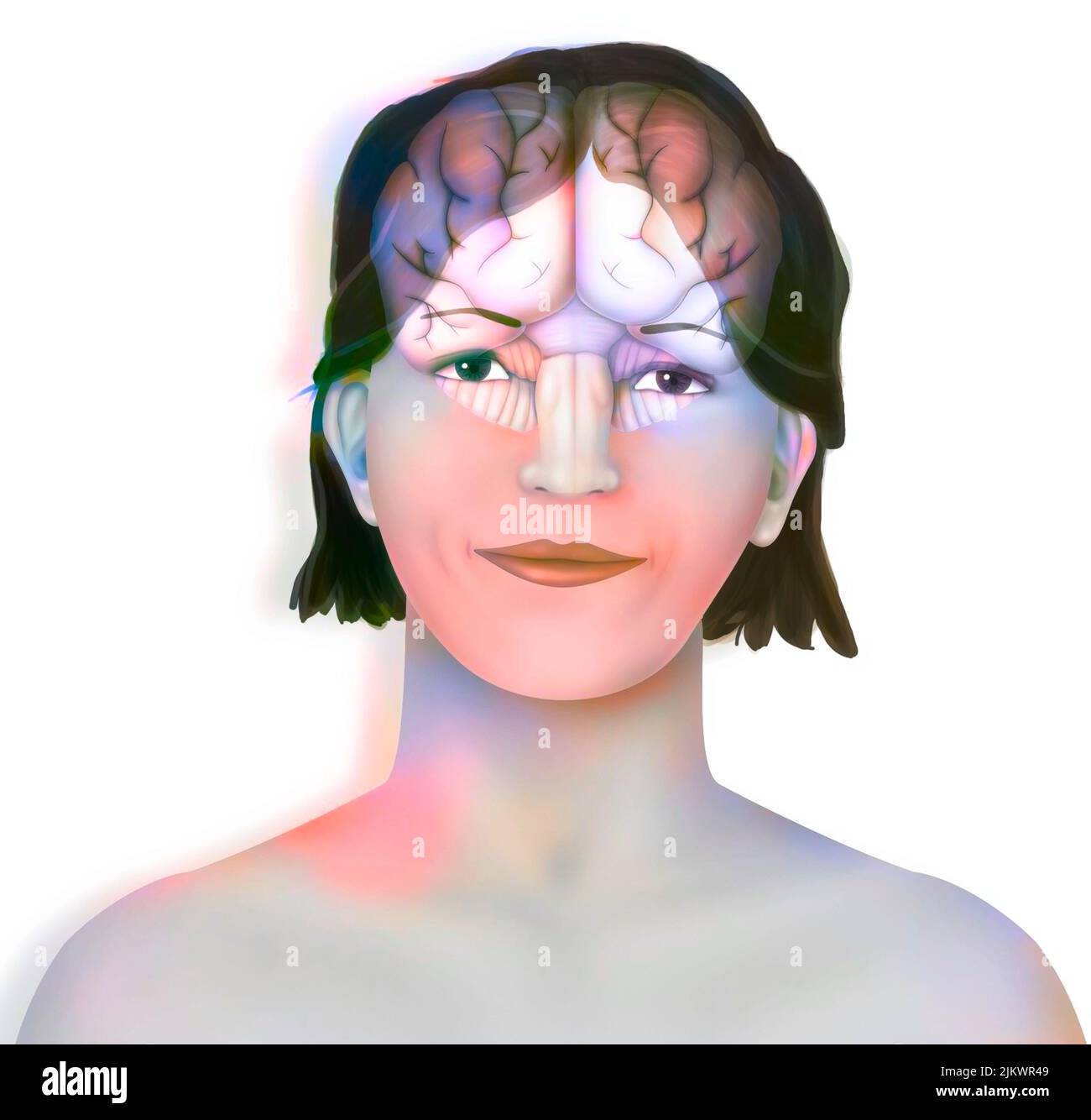 Brain (right and left cerebral hemispheres, cerebellum and brainstem) in a woman's face. Stock Photohttps://www.alamy.com/image-license-details/?v=1https://www.alamy.com/brain-right-and-left-cerebral-hemispheres-cerebellum-and-brainstem-in-a-womans-face-image476925353.html
RF2JKWR49–Brain (right and left cerebral hemispheres, cerebellum and brainstem) in a woman's face.
 EXCLUSIVE. Seven-year-old Manon Beauvais pictured in the garden of her family's house in Fecamp-Normandy-France on April 29, 2004. One year ago, Manon was struck down by a cancer of the cheek and of the cerebral trunk. As no doctor was able to operate her in France, the unique chance to save the little girl was to send her to the USA. To finance the travel, the operation and the hospitalization at the Schneider Children's Hospital in New York, her parents created the association 'Manon demain' ('Manon Tomorrow') and raised 170.000 euros in two weeks. Manon was operated successfully by the Amer Stock Photohttps://www.alamy.com/image-license-details/?v=1https://www.alamy.com/exclusive-seven-year-old-manon-beauvais-pictured-in-the-garden-of-her-familys-house-in-fecamp-normandy-france-on-april-29-2004-one-year-ago-manon-was-struck-down-by-a-cancer-of-the-cheek-and-of-the-cerebral-trunk-as-no-doctor-was-able-to-operate-her-in-france-the-unique-chance-to-save-the-little-girl-was-to-send-her-to-the-usa-to-finance-the-travel-the-operation-and-the-hospitalization-at-the-schneider-childrens-hospital-in-new-york-her-parents-created-the-association-manon-demain-manon-tomorrow-and-raised-170000-euros-in-two-weeks-manon-was-operated-successfully-by-the-amer-image402161412.html
RF2EA80YG–EXCLUSIVE. Seven-year-old Manon Beauvais pictured in the garden of her family's house in Fecamp-Normandy-France on April 29, 2004. One year ago, Manon was struck down by a cancer of the cheek and of the cerebral trunk. As no doctor was able to operate her in France, the unique chance to save the little girl was to send her to the USA. To finance the travel, the operation and the hospitalization at the Schneider Children's Hospital in New York, her parents created the association 'Manon demain' ('Manon Tomorrow') and raised 170.000 euros in two weeks. Manon was operated successfully by the Amer
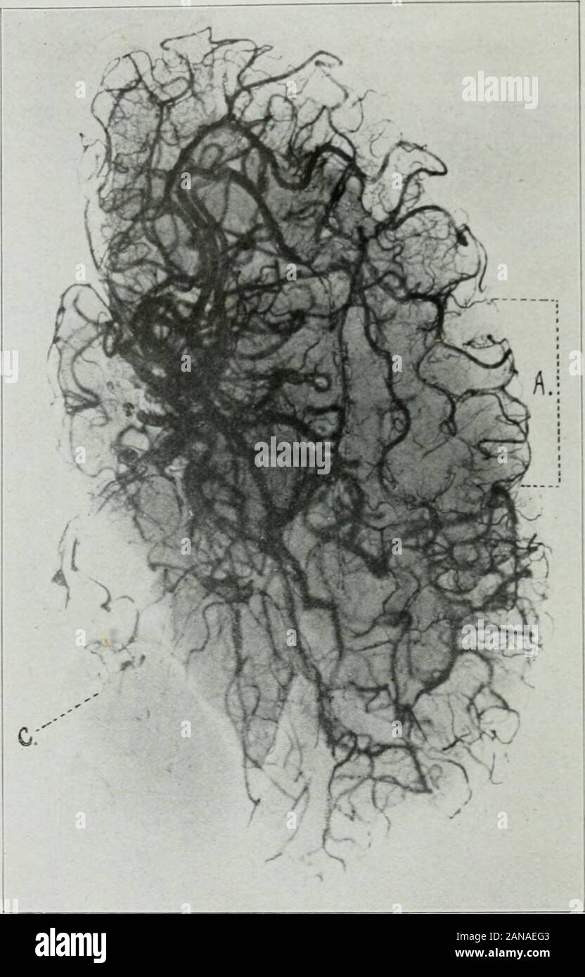 The Journal of laboratory and clinical medicine . Fig. 3-A.—Case No. A-2997. cerebral artery at a point in its m;iin trunk, as shown by an apparent projectioninto the intima of this artery of a part of the clot, when scon stereoscopically;thrombosis of the posterior cerebellar arteries, as seen by the absence of the in-jection mass in these vessels; thrombosis of the terminal liranchcs of the ascendingfrontal artery on the left side, as seen by the absence oi iho injection mass inthese vessels; possible multiple miliary aneurisnial formations in the rightcerelirum. To demonstrate these finding Stock Photohttps://www.alamy.com/image-license-details/?v=1https://www.alamy.com/the-journal-of-laboratory-and-clinical-medicine-fig-3-acase-no-a-2997-cerebral-artery-at-a-point-in-its-miin-trunk-as-shown-by-an-apparent-projectioninto-the-intima-of-this-artery-of-a-part-of-the-clot-when-scon-stereoscopicallythrombosis-of-the-posterior-cerebellar-arteries-as-seen-by-the-absence-of-the-in-jection-mass-in-these-vessels-thrombosis-of-the-terminal-liranchcs-of-the-ascendingfrontal-artery-on-the-left-side-as-seen-by-the-absence-oi-iho-injection-mass-inthese-vessels-possible-multiple-miliary-aneurisnial-formations-in-the-rightcerelirum-to-demonstrate-these-finding-image340135715.html
RM2ANAEG3–The Journal of laboratory and clinical medicine . Fig. 3-A.—Case No. A-2997. cerebral artery at a point in its m;iin trunk, as shown by an apparent projectioninto the intima of this artery of a part of the clot, when scon stereoscopically;thrombosis of the posterior cerebellar arteries, as seen by the absence of the in-jection mass in these vessels; thrombosis of the terminal liranchcs of the ascendingfrontal artery on the left side, as seen by the absence oi iho injection mass inthese vessels; possible multiple miliary aneurisnial formations in the rightcerelirum. To demonstrate these finding
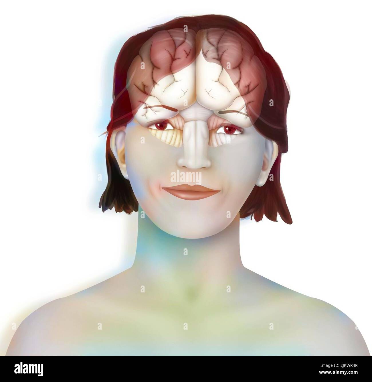 Brain (right and left cerebral hemispheres, cerebellum and brainstem) in a woman's face. Stock Photohttps://www.alamy.com/image-license-details/?v=1https://www.alamy.com/brain-right-and-left-cerebral-hemispheres-cerebellum-and-brainstem-in-a-womans-face-image476925367.html
RF2JKWR4R–Brain (right and left cerebral hemispheres, cerebellum and brainstem) in a woman's face.
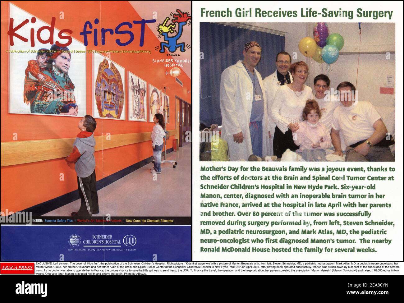 EXCLUSIVE. Left picture : The cover of 'Kids first', the publication of the Schneider Children's Hospital. Right picture : 'Kids first' page two with a picture of Manon Beauvais with, from left, Steven Schneider, MD, a pediatric neurosurgeon, Mark Atlas, MD, a pediatric neuro-oncologist, her mother Marie-Claire, her brother Alexandre and her father Alain at the Brain and Spinal Tumor Center at the Schneider Children's Hospital in New Hyde Park-USA on April 2003, after having been operated successfully. Manon was struck down by a cancer of the cheek and of the cerebral trunk. As no doctor was a Stock Photohttps://www.alamy.com/image-license-details/?v=1https://www.alamy.com/exclusive-left-picture-the-cover-of-kids-first-the-publication-of-the-schneider-childrens-hospital-right-picture-kids-first-page-two-with-a-picture-of-manon-beauvais-with-from-left-steven-schneider-md-a-pediatric-neurosurgeon-mark-atlas-md-a-pediatric-neuro-oncologist-her-mother-marie-claire-her-brother-alexandre-and-her-father-alain-at-the-brain-and-spinal-tumor-center-at-the-schneider-childrens-hospital-in-new-hyde-park-usa-on-april-2003-after-having-been-operated-successfully-manon-was-struck-down-by-a-cancer-of-the-cheek-and-of-the-cerebral-trunk-as-no-doctor-was-a-image402161417.html
RF2EA80YN–EXCLUSIVE. Left picture : The cover of 'Kids first', the publication of the Schneider Children's Hospital. Right picture : 'Kids first' page two with a picture of Manon Beauvais with, from left, Steven Schneider, MD, a pediatric neurosurgeon, Mark Atlas, MD, a pediatric neuro-oncologist, her mother Marie-Claire, her brother Alexandre and her father Alain at the Brain and Spinal Tumor Center at the Schneider Children's Hospital in New Hyde Park-USA on April 2003, after having been operated successfully. Manon was struck down by a cancer of the cheek and of the cerebral trunk. As no doctor was a
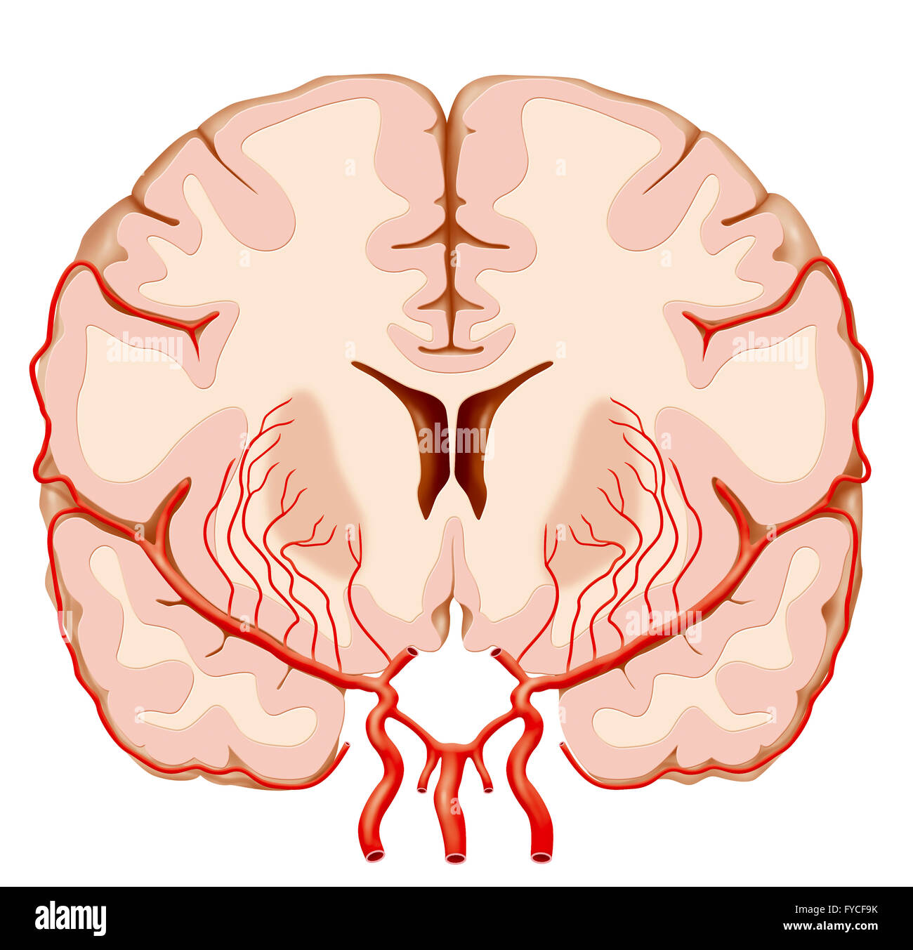 CEREBRAL ARTERY, ILLUSTRATION Stock Photohttps://www.alamy.com/image-license-details/?v=1https://www.alamy.com/stock-photo-cerebral-artery-illustration-102923007.html
RMFYCF9K–CEREBRAL ARTERY, ILLUSTRATION
 Lectures on nervous diseases from the standpoint of cerebral and spinal localization, and the later methods employed in the diagnosis and treatment of these affections . A Dfaokam of the Regions of Cutaneous Nerve Distrirution in thkAnterior Surface op the Upper Extremity and Trunk. (Modifiedfrom Flower.) 1, rcffioii suiJiilied by the sujjra-clavicular nerve (branch of the oervioal iilexus); 2,region supplied l)y the circumflex nerve; 3, region supplied by the intercosto-luimeral nerve ; 1. region supplied by the intercostal nerve (lateral branch); 5,regiou supplied V)y the lesser internal cut Stock Photohttps://www.alamy.com/image-license-details/?v=1https://www.alamy.com/lectures-on-nervous-diseases-from-the-standpoint-of-cerebral-and-spinal-localization-and-the-later-methods-employed-in-the-diagnosis-and-treatment-of-these-affections-a-dfaokam-of-the-regions-of-cutaneous-nerve-distrirution-in-thkanterior-surface-op-the-upper-extremity-and-trunk-modifiedfrom-flower-1-rcffioii-suijiilied-by-the-sujjra-clavicular-nerve-branch-of-the-oervioal-iilexus-2region-supplied-ly-the-circumflex-nerve-3-region-supplied-by-the-intercosto-luimeral-nerve-1-region-supplied-by-the-intercostal-nerve-lateral-branch-5regiou-supplied-vy-the-lesser-internal-cut-image342716253.html
RM2AWG225–Lectures on nervous diseases from the standpoint of cerebral and spinal localization, and the later methods employed in the diagnosis and treatment of these affections . A Dfaokam of the Regions of Cutaneous Nerve Distrirution in thkAnterior Surface op the Upper Extremity and Trunk. (Modifiedfrom Flower.) 1, rcffioii suiJiilied by the sujjra-clavicular nerve (branch of the oervioal iilexus); 2,region supplied l)y the circumflex nerve; 3, region supplied by the intercosto-luimeral nerve ; 1. region supplied by the intercostal nerve (lateral branch); 5,regiou supplied V)y the lesser internal cut
 CEREBRAL ARTERY, ILLUSTRATION Stock Photohttps://www.alamy.com/image-license-details/?v=1https://www.alamy.com/stock-photo-cerebral-artery-illustration-102923008.html
RMFYCF9M–CEREBRAL ARTERY, ILLUSTRATION
 . A treatise on the nervous diseases of children : for physicians and students. ulation, but it is often suspected to be the cause in cases in vi^hich the defect is due to a distinct cerebral lesion. Sensory disturbances of the face are rare in children, but, if suspected, tests should be made carefully with cotton, with the head and point of a pin, and by application of hot and cold ob-jects. Subjective sensory disturbances(neuralgia) may occur as in adults, andwill vary in the distribution accordingto the branches affected. (Fig. 6.) In examining the trunk andthe four extremities no fact is Stock Photohttps://www.alamy.com/image-license-details/?v=1https://www.alamy.com/a-treatise-on-the-nervous-diseases-of-children-for-physicians-and-students-ulation-but-it-is-often-suspected-to-be-the-cause-in-cases-in-vihich-the-defect-is-due-to-a-distinct-cerebral-lesion-sensory-disturbances-of-the-face-are-rare-in-children-but-if-suspected-tests-should-be-made-carefully-with-cotton-with-the-head-and-point-of-a-pin-and-by-application-of-hot-and-cold-ob-jects-subjective-sensory-disturbancesneuralgia-may-occur-as-in-adults-andwill-vary-in-the-distribution-accordingto-the-branches-affected-fig-6-in-examining-the-trunk-andthe-four-extremities-no-fact-is-image337081093.html
RM2AGBAAD–. A treatise on the nervous diseases of children : for physicians and students. ulation, but it is often suspected to be the cause in cases in vi^hich the defect is due to a distinct cerebral lesion. Sensory disturbances of the face are rare in children, but, if suspected, tests should be made carefully with cotton, with the head and point of a pin, and by application of hot and cold ob-jects. Subjective sensory disturbances(neuralgia) may occur as in adults, andwill vary in the distribution accordingto the branches affected. (Fig. 6.) In examining the trunk andthe four extremities no fact is
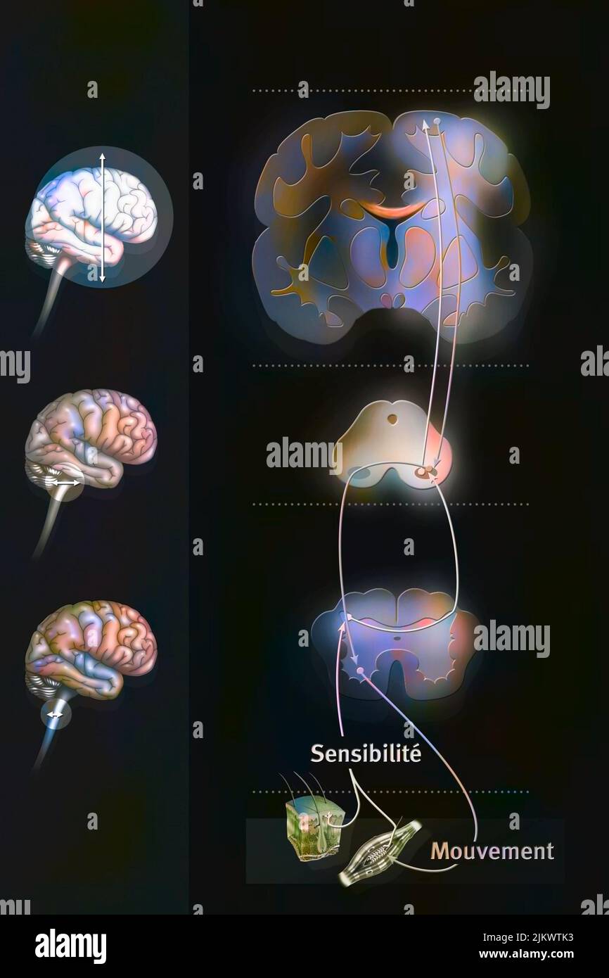 Sensorimotor loop: control of the brain to link sensations to motor reactions. Stock Photohttps://www.alamy.com/image-license-details/?v=1https://www.alamy.com/sensorimotor-loop-control-of-the-brain-to-link-sensations-to-motor-reactions-image476926551.html
RF2JKWTK3–Sensorimotor loop: control of the brain to link sensations to motor reactions.
 . Local and regional anesthesia; with chapters on spinal, epidural, paravertebral, and parasacral analgesia, and other applications of local and regional anesthesia to the surgery of the eye, ear, nose and throat, and to dental practice. tt^-rvs For^ hiffrruni jjirdiu/iu. Cvi/tct of Curatuf, Canal JVcMfui Audittrr. iTcfernua Slit foT Durn-Mat^r ^ujl. P£tro£nl grcai€ Fnr. Tacerum paaUriiu Anterior CondyloLdJTi^ii Aqurdt4ot. Ve^ti&uh PffStcriar Condyloid Fo^ JtaJtoid Foz. Fig. 212.—Base of the skull, inner or cerebral surface. (After Gray.) turing through the chief trunk of the trigeminus throug Stock Photohttps://www.alamy.com/image-license-details/?v=1https://www.alamy.com/local-and-regional-anesthesia-with-chapters-on-spinal-epidural-paravertebral-and-parasacral-analgesia-and-other-applications-of-local-and-regional-anesthesia-to-the-surgery-of-the-eye-ear-nose-and-throat-and-to-dental-practice-tt-rvs-for-hiffrruni-jjirdiuiu-cvitct-of-curatuf-canal-jvcmfui-audittrr-itcfernua-slit-fot-durn-matr-ujl-ptronl-grcai-fnr-tacerum-paauriiu-anterior-condyloldjtiii-aqurdt4ot-vetiuh-pffstcriar-condyloid-fo-jtajtoid-foz-fig-212base-of-the-skull-inner-or-cerebral-surface-after-gray-turing-through-the-chief-trunk-of-the-trigeminus-throug-image337116618.html
RM2AGCYK6–. Local and regional anesthesia; with chapters on spinal, epidural, paravertebral, and parasacral analgesia, and other applications of local and regional anesthesia to the surgery of the eye, ear, nose and throat, and to dental practice. tt^-rvs For^ hiffrruni jjirdiu/iu. Cvi/tct of Curatuf, Canal JVcMfui Audittrr. iTcfernua Slit foT Durn-Mat^r ^ujl. P£tro£nl grcai€ Fnr. Tacerum paaUriiu Anterior CondyloLdJTi^ii Aqurdt4ot. Ve^ti&uh PffStcriar Condyloid Fo^ JtaJtoid Foz. Fig. 212.—Base of the skull, inner or cerebral surface. (After Gray.) turing through the chief trunk of the trigeminus throug
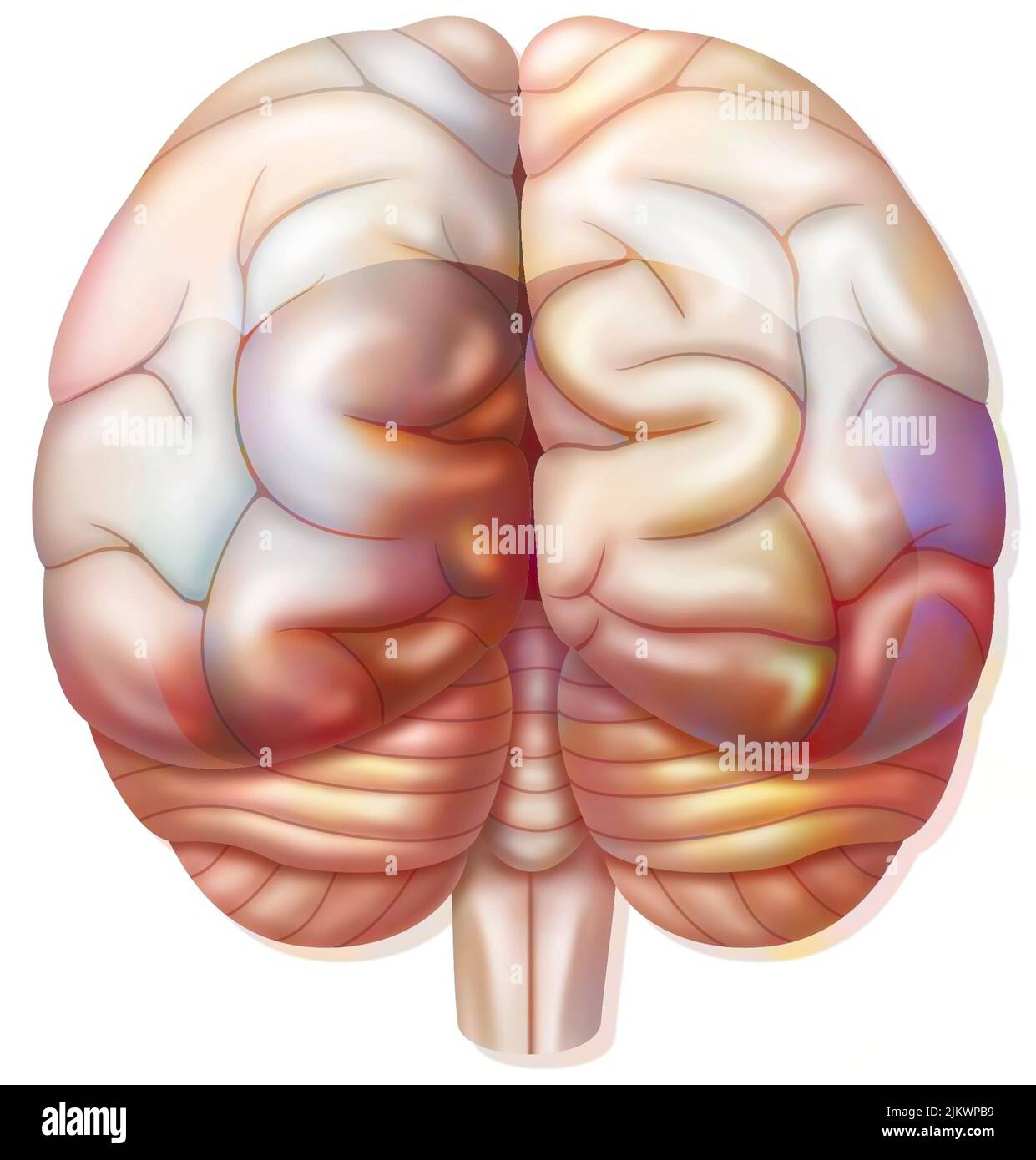 Brain with occipital, parietal, temporal lobes, cerebellum. Stock Photohttps://www.alamy.com/image-license-details/?v=1https://www.alamy.com/brain-with-occipital-parietal-temporal-lobes-cerebellum-image476924765.html
RF2JKWPB9–Brain with occipital, parietal, temporal lobes, cerebellum.
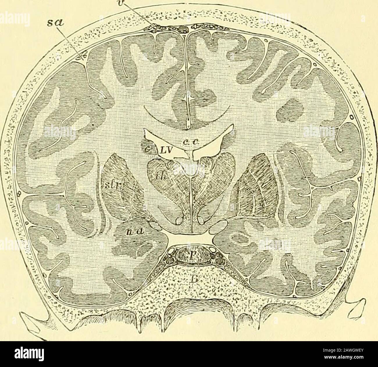 Quain's elements of anatomy . d into the straight sinus, having generally first unitedinto a single trunk. The corpora striata {ganglia of the cerebral hemispheres), situated infront and to the outer side of the optic thalami, are two large ovoid Fig. 306. — Trans- Fig. 306. VEKSE SECTION THROUGH THE BRAINAND SKULL MADE WHILST FROZEN (Keyand Retzius). J c, c, corpus callo-sum ; below its middlepart the septum luci-dum, and below thatagain the fornix ; L V,lateral ventricle ; th,thalamus; between thetwo thalami the thirdventricle is seen; belowthe thalamus is thesubstantia innominata;sir, lenti Stock Photohttps://www.alamy.com/image-license-details/?v=1https://www.alamy.com/quains-elements-of-anatomy-d-into-the-straight-sinus-having-generally-first-unitedinto-a-single-trunk-the-corpora-striata-ganglia-of-the-cerebral-hemispheres-situated-infront-and-to-the-outer-side-of-the-optic-thalami-are-two-large-ovoid-fig-306-trans-fig-306-vekse-section-through-the-brainand-skull-made-whilst-frozen-keyand-retzius-j-c-c-corpus-callo-sum-below-its-middlepart-the-septum-luci-dum-and-below-thatagain-the-fornix-l-vlateral-ventricle-ththalamus-between-thetwo-thalami-the-thirdventricle-is-seen-belowthe-thalamus-is-thesubstantia-innominatasir-lenti-image342734643.html
RM2AWGWEY–Quain's elements of anatomy . d into the straight sinus, having generally first unitedinto a single trunk. The corpora striata {ganglia of the cerebral hemispheres), situated infront and to the outer side of the optic thalami, are two large ovoid Fig. 306. — Trans- Fig. 306. VEKSE SECTION THROUGH THE BRAINAND SKULL MADE WHILST FROZEN (Keyand Retzius). J c, c, corpus callo-sum ; below its middlepart the septum luci-dum, and below thatagain the fornix ; L V,lateral ventricle ; th,thalamus; between thetwo thalami the thirdventricle is seen; belowthe thalamus is thesubstantia innominata;sir, lenti
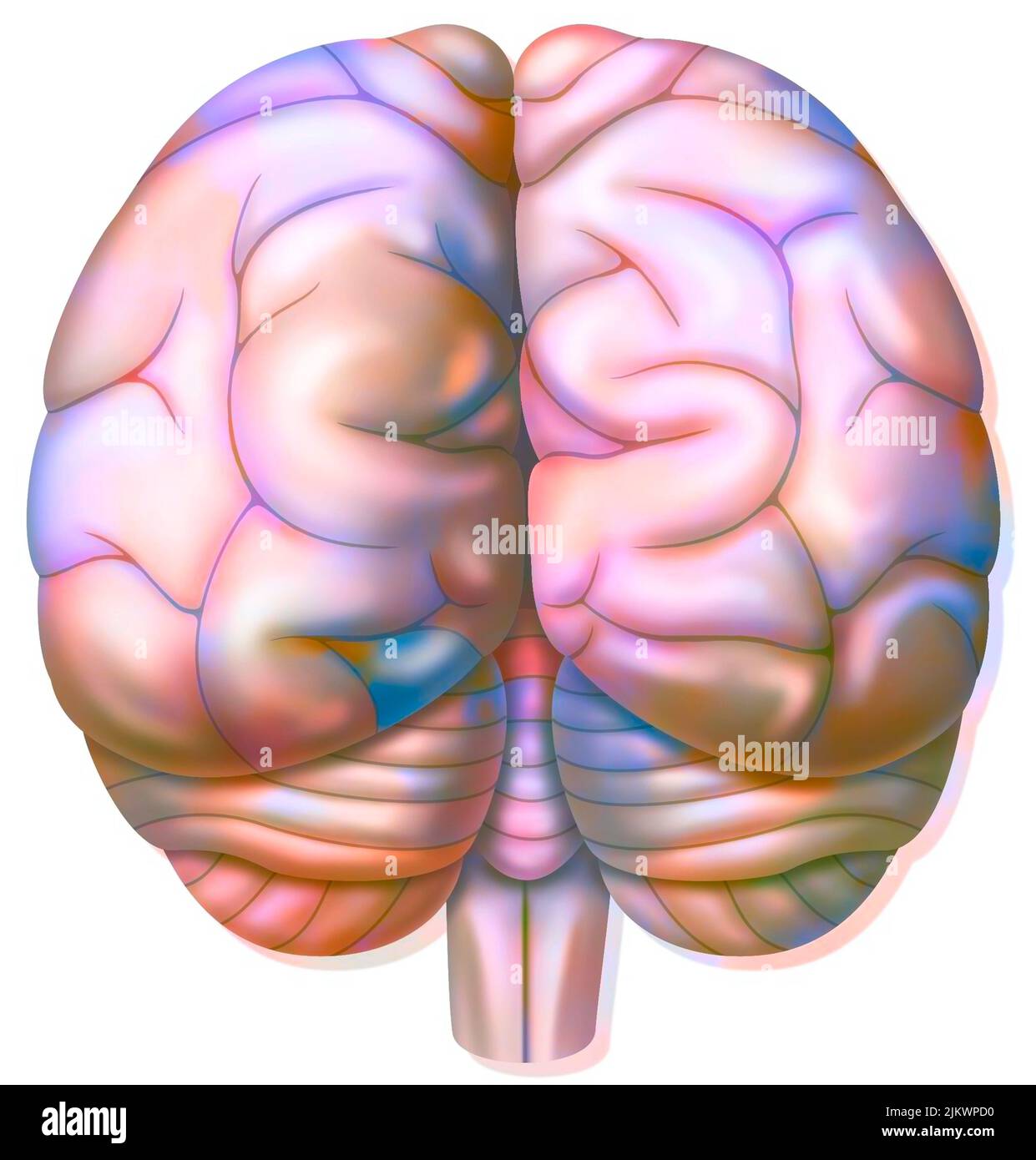 Brain with occipital, parietal, temporal lobes, cerebellum. Stock Photohttps://www.alamy.com/image-license-details/?v=1https://www.alamy.com/brain-with-occipital-parietal-temporal-lobes-cerebellum-image476924812.html
RF2JKWPD0–Brain with occipital, parietal, temporal lobes, cerebellum.
 . The practice of medicine; a text-book for practitioners and students, with special reference to diagnosis and treatment . and,lowest of all, the thumb. The center for the leg lies in the uppermost part CEREBRAL LOCALIZATION 947 of the central convolutions, but mostly in the paracentral lobule. Mostanterior is the hip, next the knee and ankle, next the great toe, the centerfor the movement of which surrounds the upper end of the fissure of Ro-lando; still further back are the centers for the small toes. The centerfor the trunk is situated in the precentral convolution between those forthe upp Stock Photohttps://www.alamy.com/image-license-details/?v=1https://www.alamy.com/the-practice-of-medicine-a-text-book-for-practitioners-and-students-with-special-reference-to-diagnosis-and-treatment-andlowest-of-all-the-thumb-the-center-for-the-leg-lies-in-the-uppermost-part-cerebral-localization-947-of-the-central-convolutions-but-mostly-in-the-paracentral-lobule-mostanterior-is-the-hip-next-the-knee-and-ankle-next-the-great-toe-the-centerfor-the-movement-of-which-surrounds-the-upper-end-of-the-fissure-of-ro-lando-still-further-back-are-the-centers-for-the-small-toes-the-centerfor-the-trunk-is-situated-in-the-precentral-convolution-between-those-forthe-upp-image369644702.html
RM2CDANGE–. The practice of medicine; a text-book for practitioners and students, with special reference to diagnosis and treatment . and,lowest of all, the thumb. The center for the leg lies in the uppermost part CEREBRAL LOCALIZATION 947 of the central convolutions, but mostly in the paracentral lobule. Mostanterior is the hip, next the knee and ankle, next the great toe, the centerfor the movement of which surrounds the upper end of the fissure of Ro-lando; still further back are the centers for the small toes. The centerfor the trunk is situated in the precentral convolution between those forthe upp
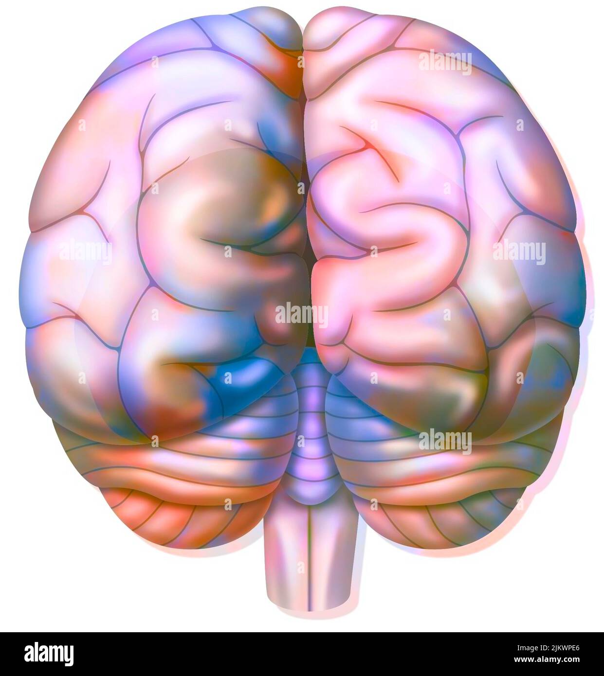 The occipital lobes of the brain in posterior view. Stock Photohttps://www.alamy.com/image-license-details/?v=1https://www.alamy.com/the-occipital-lobes-of-the-brain-in-posterior-view-image476924846.html
RF2JKWPE6–The occipital lobes of the brain in posterior view.
 . Diseases of the nervous system : for the general practitioner and student. er three chiefportions of entire motor zone corresponding to the limbs and head. Thecenter for the lower extremities occupies the anterior part of the para-central lobule and the upper fourth of the ascending frontal convolution,and that of Rolandic fissure. The two middle fourths represent the center CEREBRAL LOCALIZATIONS 77 for the upper extremities. The center of the head lies in the lower fourthand in the rolandic operculum. The center for the trunk lies betweenthose of the extremities. Clinical observations on f Stock Photohttps://www.alamy.com/image-license-details/?v=1https://www.alamy.com/diseases-of-the-nervous-system-for-the-general-practitioner-and-student-er-three-chiefportions-of-entire-motor-zone-corresponding-to-the-limbs-and-head-thecenter-for-the-lower-extremities-occupies-the-anterior-part-of-the-para-central-lobule-and-the-upper-fourth-of-the-ascending-frontal-convolutionand-that-of-rolandic-fissure-the-two-middle-fourths-represent-the-center-cerebral-localizations-77-for-the-upper-extremities-the-center-of-the-head-lies-in-the-lower-fourthand-in-the-rolandic-operculum-the-center-for-the-trunk-lies-betweenthose-of-the-extremities-clinical-observations-on-f-image370470710.html
RM2CEMB4P–. Diseases of the nervous system : for the general practitioner and student. er three chiefportions of entire motor zone corresponding to the limbs and head. Thecenter for the lower extremities occupies the anterior part of the para-central lobule and the upper fourth of the ascending frontal convolution,and that of Rolandic fissure. The two middle fourths represent the center CEREBRAL LOCALIZATIONS 77 for the upper extremities. The center of the head lies in the lower fourthand in the rolandic operculum. The center for the trunk lies betweenthose of the extremities. Clinical observations on f
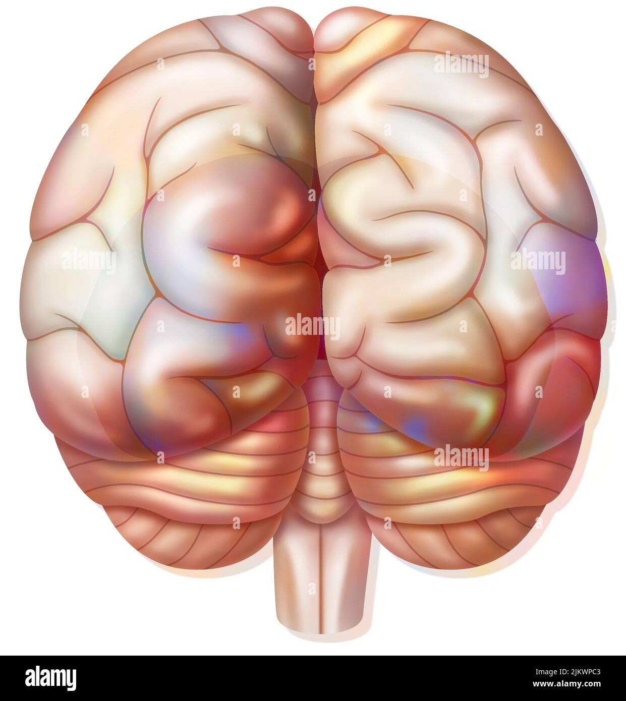 The occipital lobes of the brain in posterior view. Stock Photohttps://www.alamy.com/image-license-details/?v=1https://www.alamy.com/the-occipital-lobes-of-the-brain-in-posterior-view-image476924787.html
RF2JKWPC3–The occipital lobes of the brain in posterior view.
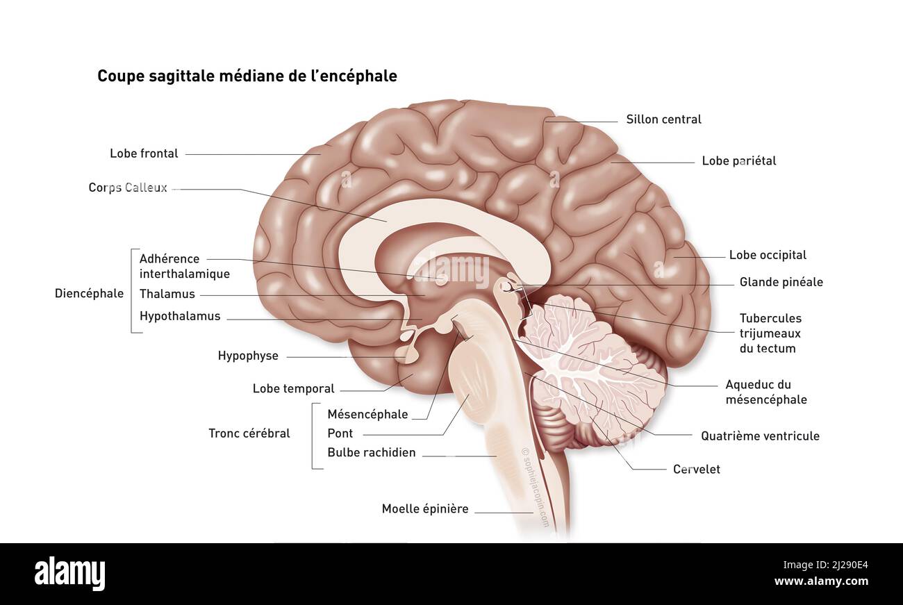 Encephalus - median cup Stock Photohttps://www.alamy.com/image-license-details/?v=1https://www.alamy.com/encephalus-median-cup-image466107212.html
RM2J290E4–Encephalus - median cup
 . Quain's elements of anatomy . d into the straight sinus, having generally first unitedinto a single trunk. The corpora striata {ganglia of the cerebral liemispheres), situated infront and to the outer side of the oj)tic thalami, are two large ovoid Fig. 306. — Trans- Fig. 306. VEKSE SECTION THROUGH THE BRAINAND SKULIi MADE WHILST FROZEN (Keyand Retzius). ^ c, c, corpus caUo-sum ; below its middlepart the septum luci-dum, and below thatagain the fomiK ; L V,lateral ventricle ; th,thalamus; between thetwo thalami the thirdventricle is seen; belowthe thalamus is thesubstantia innominata;str, le Stock Photohttps://www.alamy.com/image-license-details/?v=1https://www.alamy.com/quains-elements-of-anatomy-d-into-the-straight-sinus-having-generally-first-unitedinto-a-single-trunk-the-corpora-striata-ganglia-of-the-cerebral-liemispheres-situated-infront-and-to-the-outer-side-of-the-ojtic-thalami-are-two-large-ovoid-fig-306-trans-fig-306-vekse-section-through-the-brainand-skulii-made-whilst-frozen-keyand-retzius-c-c-corpus-cauo-sum-below-its-middlepart-the-septum-luci-dum-and-below-thatagain-the-fomik-l-vlateral-ventricle-ththalamus-between-thetwo-thalami-the-thirdventricle-is-seen-belowthe-thalamus-is-thesubstantia-innominatastr-le-image372458416.html
RM2CHXXE8–. Quain's elements of anatomy . d into the straight sinus, having generally first unitedinto a single trunk. The corpora striata {ganglia of the cerebral liemispheres), situated infront and to the outer side of the oj)tic thalami, are two large ovoid Fig. 306. — Trans- Fig. 306. VEKSE SECTION THROUGH THE BRAINAND SKULIi MADE WHILST FROZEN (Keyand Retzius). ^ c, c, corpus caUo-sum ; below its middlepart the septum luci-dum, and below thatagain the fomiK ; L V,lateral ventricle ; th,thalamus; between thetwo thalami the thirdventricle is seen; belowthe thalamus is thesubstantia innominata;str, le
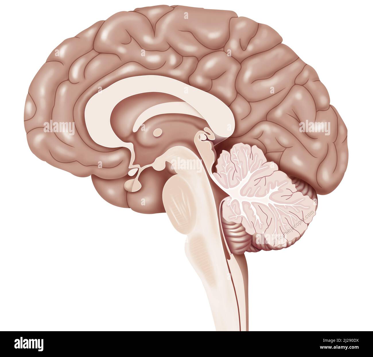 Sagittal cup of encephalus Stock Photohttps://www.alamy.com/image-license-details/?v=1https://www.alamy.com/sagittal-cup-of-encephalus-image466107206.html
RM2J290DX–Sagittal cup of encephalus
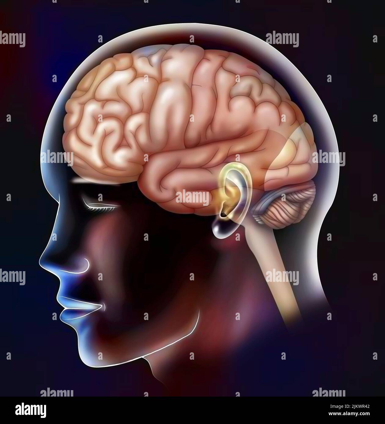 The lobes of the brain: frontal, parietal, temporal and occipital lobes. Stock Photohttps://www.alamy.com/image-license-details/?v=1https://www.alamy.com/the-lobes-of-the-brain-frontal-parietal-temporal-and-occipital-lobes-image476925346.html
RF2JKWR42–The lobes of the brain: frontal, parietal, temporal and occipital lobes.
 . Anatomischer Anzeiger. Anatomy, Comparative; Anatomy, Comparative. 520 earlier stage ^), when the motor nerve-trunk is a strand of protoplasm perfectly continuous with the protoplasm of the myotome. The discussion of the great significance of these facts would hardly be relevant to the main purpose of this communication. But I may remark in passing Lamina tcrmiiialis. Nasal Sac X ' Cerebral Hemisphere Thalam- encephalon Fig. 4.. Please note that these images are extracted from scanned page images that may have been digitally enhanced for readability - coloration and appearance of these illus Stock Photohttps://www.alamy.com/image-license-details/?v=1https://www.alamy.com/anatomischer-anzeiger-anatomy-comparative-anatomy-comparative-520-earlier-stage-when-the-motor-nerve-trunk-is-a-strand-of-protoplasm-perfectly-continuous-with-the-protoplasm-of-the-myotome-the-discussion-of-the-great-significance-of-these-facts-would-hardly-be-relevant-to-the-main-purpose-of-this-communication-but-i-may-remark-in-passing-lamina-tcrmiiialis-nasal-sac-x-cerebral-hemisphere-thalam-encephalon-fig-4-please-note-that-these-images-are-extracted-from-scanned-page-images-that-may-have-been-digitally-enhanced-for-readability-coloration-and-appearance-of-these-illus-image236824989.html
RMRN88K9–. Anatomischer Anzeiger. Anatomy, Comparative; Anatomy, Comparative. 520 earlier stage ^), when the motor nerve-trunk is a strand of protoplasm perfectly continuous with the protoplasm of the myotome. The discussion of the great significance of these facts would hardly be relevant to the main purpose of this communication. But I may remark in passing Lamina tcrmiiialis. Nasal Sac X ' Cerebral Hemisphere Thalam- encephalon Fig. 4.. Please note that these images are extracted from scanned page images that may have been digitally enhanced for readability - coloration and appearance of these illus
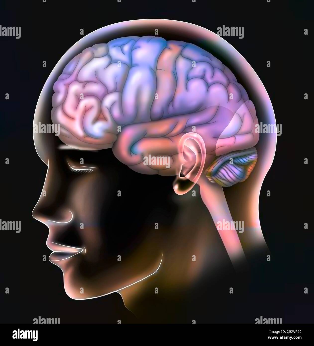 The lobes of the brain: frontal, parietal, temporal and occipital lobes. Stock Photohttps://www.alamy.com/image-license-details/?v=1https://www.alamy.com/the-lobes-of-the-brain-frontal-parietal-temporal-and-occipital-lobes-image476925400.html
RF2JKWR60–The lobes of the brain: frontal, parietal, temporal and occipital lobes.
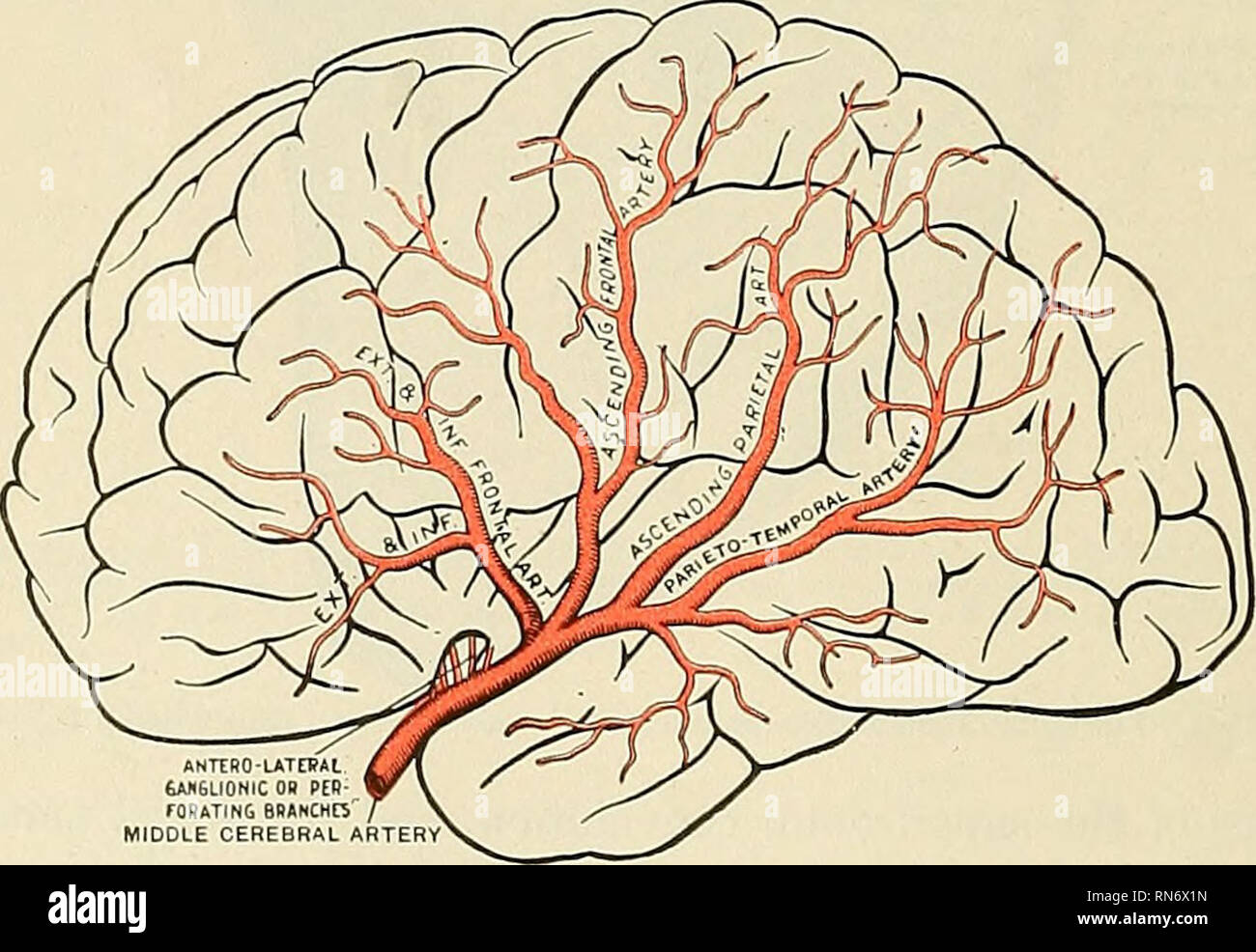 . Anatomy, descriptive and applied. Anatomy. INTERNAL POSTERIOR POSTERIOR CAROTID COMMUNICATING CEREBRAL ARTERY ARTERY ARTERY Fig. 451.—The arteries of the medial surface of the right cerebral hemisphere. (Spalteholz.) The middle cerebral artery (a. cerebri media) (Fig. 452), the largest branch of the internal carotid, passes obliquely outward along the sylvian fissure, and divides on the surface of the insula into its terminal branches.. Fig. 452.—The distribution of the middle cerebral artery. The trunk of the middle cerebral artery Ues i depths of the sylvian cleft. (After Charcot.) Branche Stock Photohttps://www.alamy.com/image-license-details/?v=1https://www.alamy.com/anatomy-descriptive-and-applied-anatomy-internal-posterior-posterior-carotid-communicating-cerebral-artery-artery-artery-fig-451the-arteries-of-the-medial-surface-of-the-right-cerebral-hemisphere-spalteholz-the-middle-cerebral-artery-a-cerebri-media-fig-452-the-largest-branch-of-the-internal-carotid-passes-obliquely-outward-along-the-sylvian-fissure-and-divides-on-the-surface-of-the-insula-into-its-terminal-branches-fig-452the-distribution-of-the-middle-cerebral-artery-the-trunk-of-the-middle-cerebral-artery-ues-i-depths-of-the-sylvian-cleft-after-charcot-branche-image236794705.html
RMRN6X1N–. Anatomy, descriptive and applied. Anatomy. INTERNAL POSTERIOR POSTERIOR CAROTID COMMUNICATING CEREBRAL ARTERY ARTERY ARTERY Fig. 451.—The arteries of the medial surface of the right cerebral hemisphere. (Spalteholz.) The middle cerebral artery (a. cerebri media) (Fig. 452), the largest branch of the internal carotid, passes obliquely outward along the sylvian fissure, and divides on the surface of the insula into its terminal branches.. Fig. 452.—The distribution of the middle cerebral artery. The trunk of the middle cerebral artery Ues i depths of the sylvian cleft. (After Charcot.) Branche
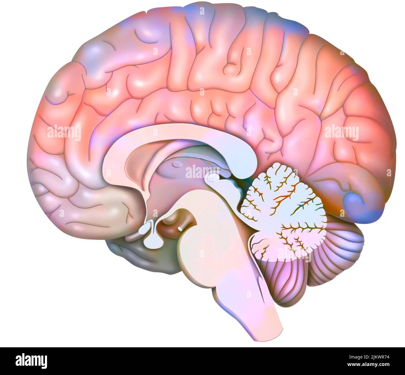 Median sagittal section of the brain with cerebellum and beginning of the brainstem. Stock Photohttps://www.alamy.com/image-license-details/?v=1https://www.alamy.com/median-sagittal-section-of-the-brain-with-cerebellum-and-beginning-of-the-brainstem-image476925432.html
RF2JKWR74–Median sagittal section of the brain with cerebellum and beginning of the brainstem.
 . A manual of zoology. Zoology. /. CH^fJTO GXA TUI. 29T pair 01 fused ovd.- cerebral ganglia (fig. 2fiv!). in the trunk segment a large ventral ganglion, and these are connected "'"• by long (esophageal commissures. Of interest, because characteristic of nematodes and many annelids, are the relations of the musculature, which consists of longitudinal fibres alone. The body cavity is lined with epithelivim (fig. 260), which, so far as it abuts against the ali- mentary tract, is called splanchnic (or visceral) mesoderm; that on the side of the ccelom to- wards the ectoderm is the somat Stock Photohttps://www.alamy.com/image-license-details/?v=1https://www.alamy.com/a-manual-of-zoology-zoology-chfjto-gxa-tui-29t-pair-01-fused-ovd-cerebral-ganglia-fig-2fiv!-in-the-trunk-segment-a-large-ventral-ganglion-and-these-are-connected-quotquot-by-long-esophageal-commissures-of-interest-because-characteristic-of-nematodes-and-many-annelids-are-the-relations-of-the-musculature-which-consists-of-longitudinal-fibres-alone-the-body-cavity-is-lined-with-epithelivim-fig-260-which-so-far-as-it-abuts-against-the-ali-mentary-tract-is-called-splanchnic-or-visceral-mesoderm-that-on-the-side-of-the-ccelom-to-wards-the-ectoderm-is-the-somat-image232351737.html
RMRE0F09–. A manual of zoology. Zoology. /. CH^fJTO GXA TUI. 29T pair 01 fused ovd.- cerebral ganglia (fig. 2fiv!). in the trunk segment a large ventral ganglion, and these are connected "'"• by long (esophageal commissures. Of interest, because characteristic of nematodes and many annelids, are the relations of the musculature, which consists of longitudinal fibres alone. The body cavity is lined with epithelivim (fig. 260), which, so far as it abuts against the ali- mentary tract, is called splanchnic (or visceral) mesoderm; that on the side of the ccelom to- wards the ectoderm is the somat
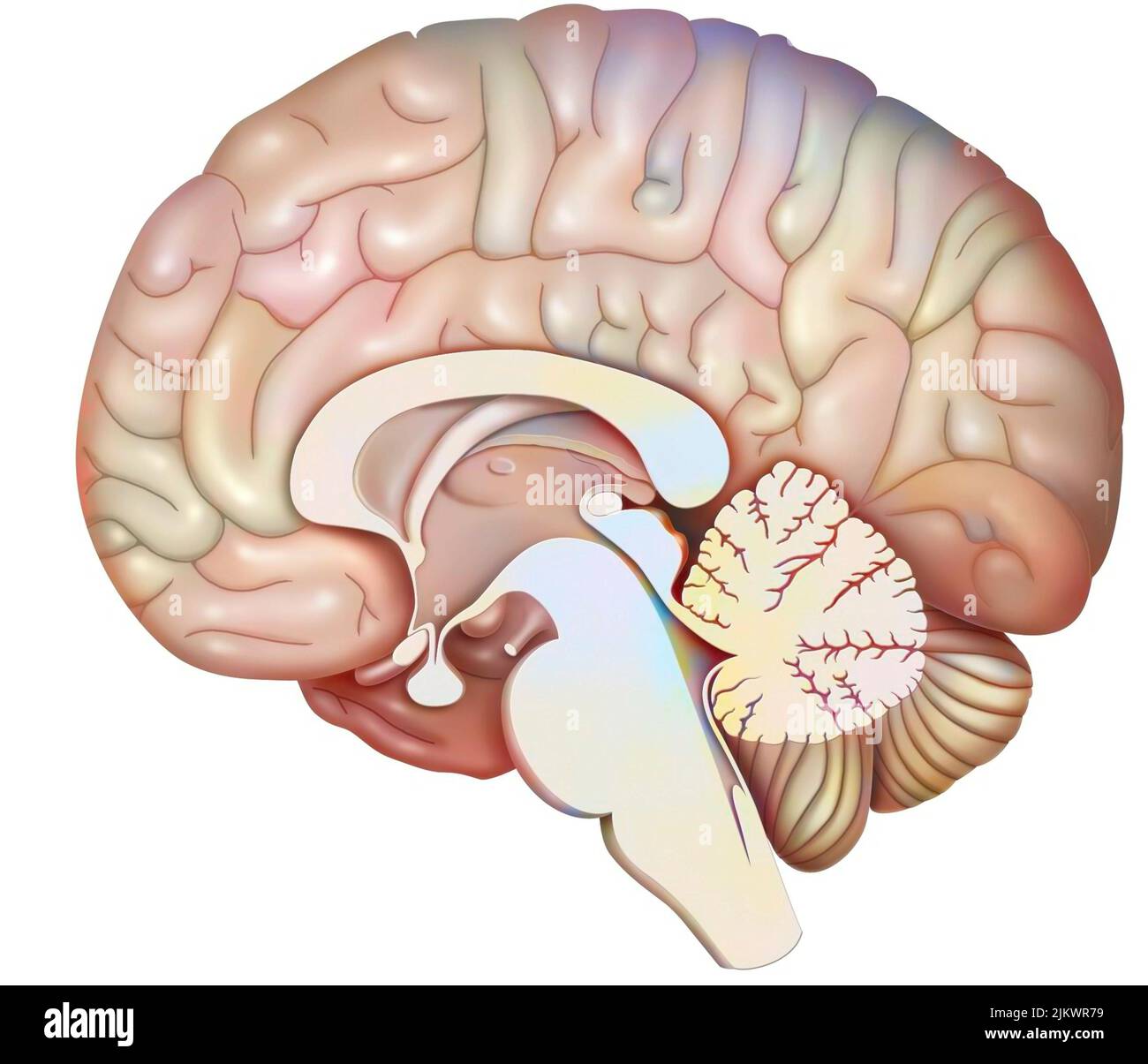 Median sagittal section of the brain with cerebellum and beginning of the brainstem. Stock Photohttps://www.alamy.com/image-license-details/?v=1https://www.alamy.com/median-sagittal-section-of-the-brain-with-cerebellum-and-beginning-of-the-brainstem-image476925437.html
RF2JKWR79–Median sagittal section of the brain with cerebellum and beginning of the brainstem.
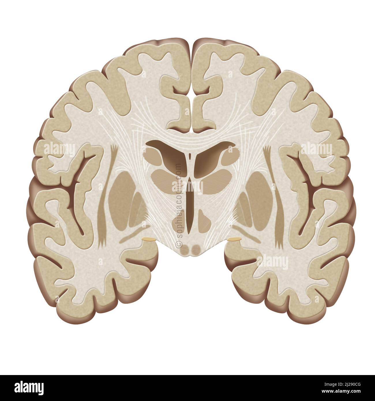 Frontal brain-cut Stock Photohttps://www.alamy.com/image-license-details/?v=1https://www.alamy.com/frontal-brain-cut-image466107168.html
RM2J290CG–Frontal brain-cut
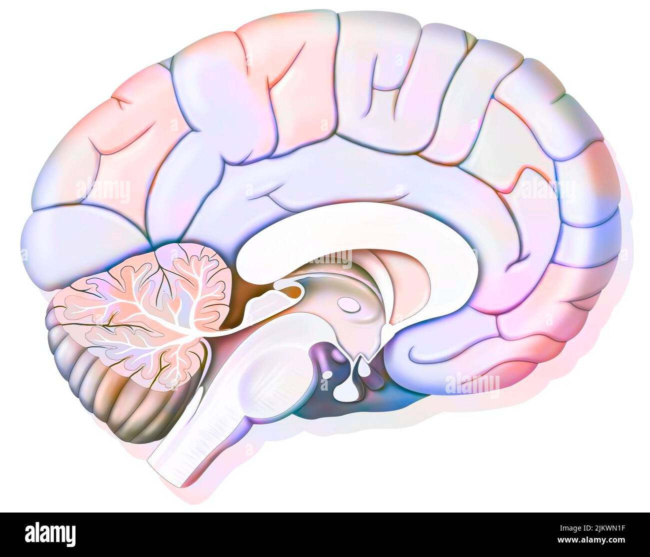 Mid sagittal section of the human brain showing the hypothalamus. Stock Photohttps://www.alamy.com/image-license-details/?v=1https://www.alamy.com/mid-sagittal-section-of-the-human-brain-showing-the-hypothalamus-image476923707.html
RF2JKWN1F–Mid sagittal section of the human brain showing the hypothalamus.
 . The cyclopædia of anatomy and physiology. Anatomy; Physiology; Zoology. 956 TERATOLOGY. a hernia. The parietal bones are sometimes present, together with flat frontal bones, and a perpendicular occipital bone, so that the summit of the skull is quite closed, with the exception of a small opening. Fig. 609. shows how the malformed cerebral substance is applied to the medulla spinalis. All the cerebral nerves are present. This form of monstrosity has in general a less brute-like aspect; the trunk is more evolved, and the whole body in general very heavy. Fourth Type.—The skull flat, more evolv Stock Photohttps://www.alamy.com/image-license-details/?v=1https://www.alamy.com/the-cyclopdia-of-anatomy-and-physiology-anatomy-physiology-zoology-956-teratology-a-hernia-the-parietal-bones-are-sometimes-present-together-with-flat-frontal-bones-and-a-perpendicular-occipital-bone-so-that-the-summit-of-the-skull-is-quite-closed-with-the-exception-of-a-small-opening-fig-609-shows-how-the-malformed-cerebral-substance-is-applied-to-the-medulla-spinalis-all-the-cerebral-nerves-are-present-this-form-of-monstrosity-has-in-general-a-less-brute-like-aspect-the-trunk-is-more-evolved-and-the-whole-body-in-general-very-heavy-fourth-typethe-skull-flat-more-evolv-image231839091.html
RMRD553F–. The cyclopædia of anatomy and physiology. Anatomy; Physiology; Zoology. 956 TERATOLOGY. a hernia. The parietal bones are sometimes present, together with flat frontal bones, and a perpendicular occipital bone, so that the summit of the skull is quite closed, with the exception of a small opening. Fig. 609. shows how the malformed cerebral substance is applied to the medulla spinalis. All the cerebral nerves are present. This form of monstrosity has in general a less brute-like aspect; the trunk is more evolved, and the whole body in general very heavy. Fourth Type.—The skull flat, more evolv
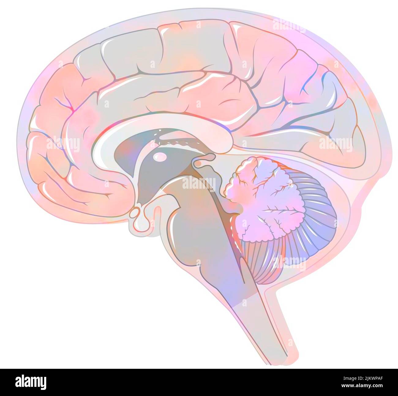 Sagittal section of the brain with meninges and cerebrospinal fluid. Stock Photohttps://www.alamy.com/image-license-details/?v=1https://www.alamy.com/sagittal-section-of-the-brain-with-meninges-and-cerebrospinal-fluid-image476924743.html
RF2JKWPAF–Sagittal section of the brain with meninges and cerebrospinal fluid.
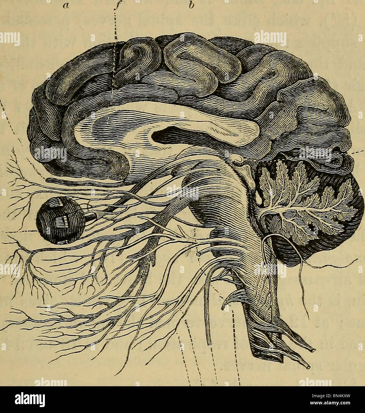 . Animal physiology. Physiology, Comparative; Physiology, Comparative. BRAIN, AND NERVES PROCEEDING FROM IT. 351 at g the optic lobes or ganglia, buried as it were beneath the cerebral hemispheres. The olfactory ganglia are also very small,. 7 11 9 10 6 e Fig. 185.—Section of the Brain of Man. consisting merely of bulbous enlargements upon the trunk of the olfactory nerves (1), which are the first pair given off from the brain. From these bulbous enlargements (which, though covered in by the cerebrum, are separated from it by foot-stalks, as in the Shark, §. 453), are given off the small branc Stock Photohttps://www.alamy.com/image-license-details/?v=1https://www.alamy.com/animal-physiology-physiology-comparative-physiology-comparative-brain-and-nerves-proceeding-from-it-351-at-g-the-optic-lobes-or-ganglia-buried-as-it-were-beneath-the-cerebral-hemispheres-the-olfactory-ganglia-are-also-very-small-7-11-9-10-6-e-fig-185section-of-the-brain-of-man-consisting-merely-of-bulbous-enlargements-upon-the-trunk-of-the-olfactory-nerves-1-which-are-the-first-pair-given-off-from-the-brain-from-these-bulbous-enlargements-which-though-covered-in-by-the-cerebrum-are-separated-from-it-by-foot-stalks-as-in-the-shark-453-are-given-off-the-small-branc-image236746017.html
RMRN4KXW–. Animal physiology. Physiology, Comparative; Physiology, Comparative. BRAIN, AND NERVES PROCEEDING FROM IT. 351 at g the optic lobes or ganglia, buried as it were beneath the cerebral hemispheres. The olfactory ganglia are also very small,. 7 11 9 10 6 e Fig. 185.—Section of the Brain of Man. consisting merely of bulbous enlargements upon the trunk of the olfactory nerves (1), which are the first pair given off from the brain. From these bulbous enlargements (which, though covered in by the cerebrum, are separated from it by foot-stalks, as in the Shark, §. 453), are given off the small branc
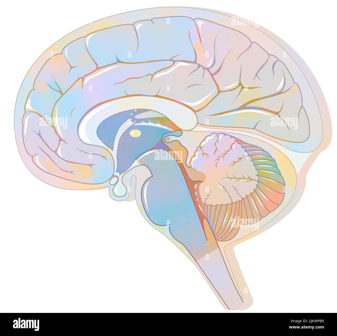 Sagittal section of the brain with meninges and cerebrospinal fluid. Stock Photohttps://www.alamy.com/image-license-details/?v=1https://www.alamy.com/sagittal-section-of-the-brain-with-meninges-and-cerebrospinal-fluid-image476924756.html
RF2JKWPB0–Sagittal section of the brain with meninges and cerebrospinal fluid.
 . Annotationes zoologicae japonenses / Nihon do?butsugaku iho?. NOTES ON TWO NEW SPECIES OF JAPANESE POLYCLADS. 597 to that of other species of the genus. The broadly rounded head is without tentacles of any sort, and passes behind into the trunk, from which it is distinctly marked off. When fully extended the trunk is elongate-slender, and is nearly uniformly broad for a great part of its length, but tapers at the posterior end to a point. The dorsal surface is slightly raised in the median parts from behind cerebral eyes. The sucker and the mouth occur as usual in the median line, the former Stock Photohttps://www.alamy.com/image-license-details/?v=1https://www.alamy.com/annotationes-zoologicae-japonenses-nihon-dobutsugaku-iho-notes-on-two-new-species-of-japanese-polyclads-597-to-that-of-other-species-of-the-genus-the-broadly-rounded-head-is-without-tentacles-of-any-sort-and-passes-behind-into-the-trunk-from-which-it-is-distinctly-marked-off-when-fully-extended-the-trunk-is-elongate-slender-and-is-nearly-uniformly-broad-for-a-great-part-of-its-length-but-tapers-at-the-posterior-end-to-a-point-the-dorsal-surface-is-slightly-raised-in-the-median-parts-from-behind-cerebral-eyes-the-sucker-and-the-mouth-occur-as-usual-in-the-median-line-the-former-image236359070.html
RMRMF2BA–. Annotationes zoologicae japonenses / Nihon do?butsugaku iho?. NOTES ON TWO NEW SPECIES OF JAPANESE POLYCLADS. 597 to that of other species of the genus. The broadly rounded head is without tentacles of any sort, and passes behind into the trunk, from which it is distinctly marked off. When fully extended the trunk is elongate-slender, and is nearly uniformly broad for a great part of its length, but tapers at the posterior end to a point. The dorsal surface is slightly raised in the median parts from behind cerebral eyes. The sucker and the mouth occur as usual in the median line, the former
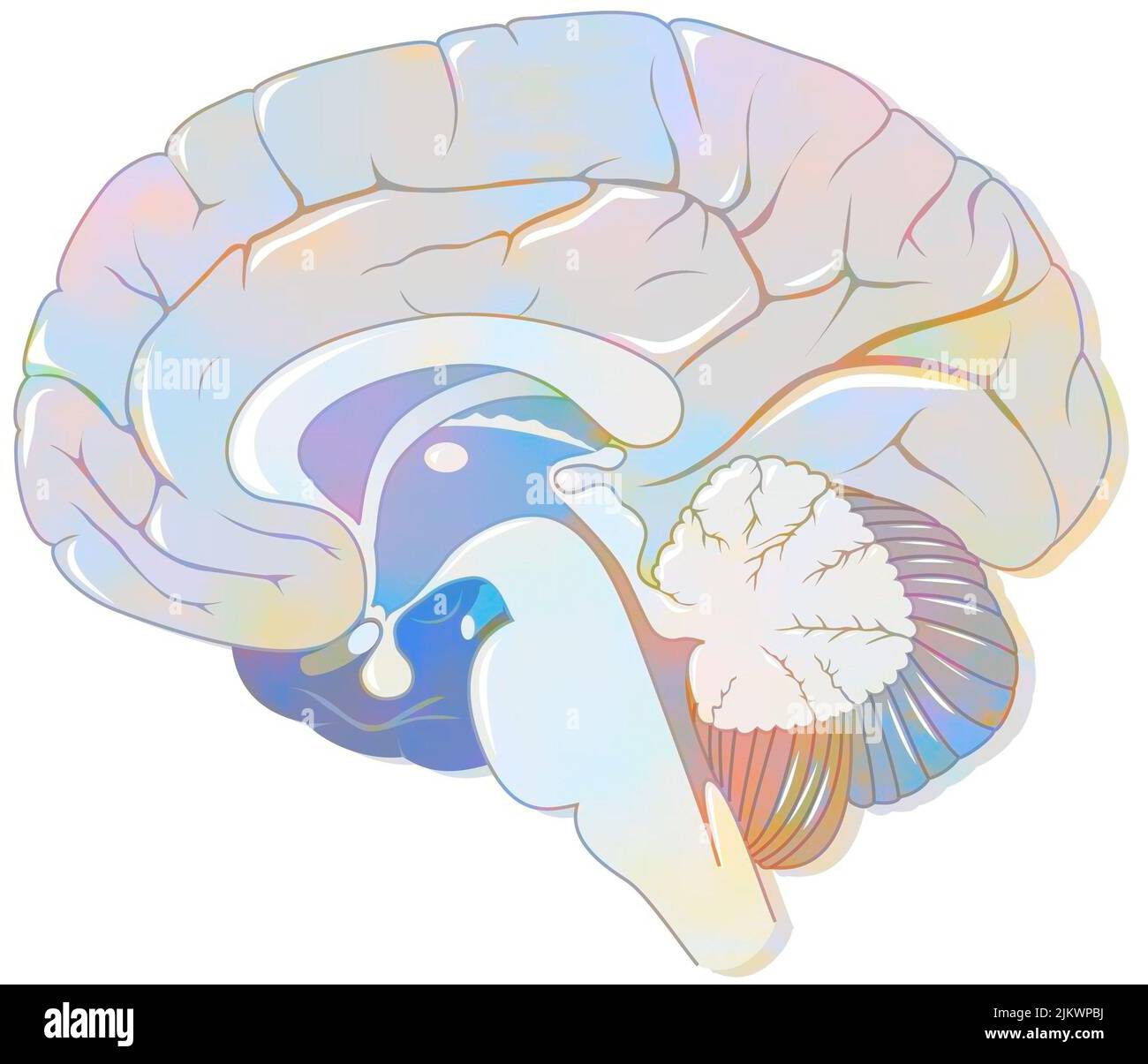 Sagittal section of the brain with meninges and cerebrospinal fluid. Stock Photohttps://www.alamy.com/image-license-details/?v=1https://www.alamy.com/sagittal-section-of-the-brain-with-meninges-and-cerebrospinal-fluid-image476924774.html
RF2JKWPBJ–Sagittal section of the brain with meninges and cerebrospinal fluid.
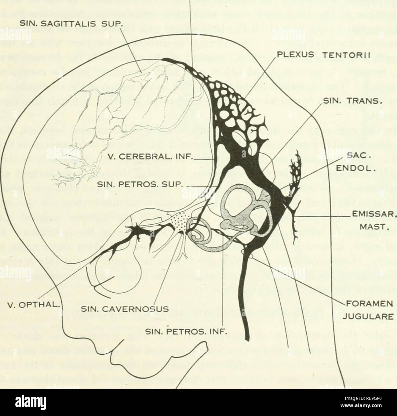 . Contributions to embryology. Embryology. OF THE BRAIN OF THE HUMAN EMBRYO. 29 the large ophthalmic and maxillary tributaries in front. In figure 27 it receives a large terminal trunk lateral to the infundibulum made up of tributaries coming from the region of the Sylvian fissure; this corresponds to the middle cerebral vein of the adult. Caudally the cavernous sinus communicates with the main blood-stream by means of the superior and inferior petrosal sinuses. The superior petrosal sinus passes over the cochlear part of the otic capsule and empties above SIN. RECTUS SIN. SAGITTALIS SUP.. V. Stock Photohttps://www.alamy.com/image-license-details/?v=1https://www.alamy.com/contributions-to-embryology-embryology-of-the-brain-of-the-human-embryo-29-the-large-ophthalmic-and-maxillary-tributaries-in-front-in-figure-27-it-receives-a-large-terminal-trunk-lateral-to-the-infundibulum-made-up-of-tributaries-coming-from-the-region-of-the-sylvian-fissure-this-corresponds-to-the-middle-cerebral-vein-of-the-adult-caudally-the-cavernous-sinus-communicates-with-the-main-blood-stream-by-means-of-the-superior-and-inferior-petrosal-sinuses-the-superior-petrosal-sinus-passes-over-the-cochlear-part-of-the-otic-capsule-and-empties-above-sin-rectus-sin-sagittalis-sup-v-image232550696.html
RMRE9GP0–. Contributions to embryology. Embryology. OF THE BRAIN OF THE HUMAN EMBRYO. 29 the large ophthalmic and maxillary tributaries in front. In figure 27 it receives a large terminal trunk lateral to the infundibulum made up of tributaries coming from the region of the Sylvian fissure; this corresponds to the middle cerebral vein of the adult. Caudally the cavernous sinus communicates with the main blood-stream by means of the superior and inferior petrosal sinuses. The superior petrosal sinus passes over the cochlear part of the otic capsule and empties above SIN. RECTUS SIN. SAGITTALIS SUP.. V.
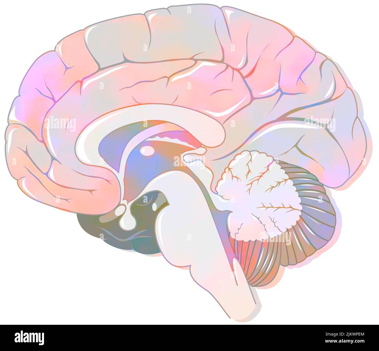 Sagittal section of the brain with meninges and cerebrospinal fluid. Stock Photohttps://www.alamy.com/image-license-details/?v=1https://www.alamy.com/sagittal-section-of-the-brain-with-meninges-and-cerebrospinal-fluid-image476924860.html
RF2JKWPEM–Sagittal section of the brain with meninges and cerebrospinal fluid.
 . The Biological bulletin. Biology; Zoology; Biology; Marine Biology. NEOMENIOID APLACOPHORAN DEVELOPMENT 97. Figure 24. Larvae during the completion of metamorphosis (9- to 11-day-old larvae), (a) The larva covered by spicules on the trunk. The apical cap has diminished, and the apical tuft is being resorhed at the apical end. (bl Post-metamorphic juvenile. The apical cap and caudal regions have been completely engulfed by the trunk, leaving the trunk as the only definitive juvenile structure. A. anterior; CD, cerebral depression; EP; epidermal papilla; Sp. spicule; P. posterior; PGr. pedal g Stock Photohttps://www.alamy.com/image-license-details/?v=1https://www.alamy.com/the-biological-bulletin-biology-zoology-biology-marine-biology-neomenioid-aplacophoran-development-97-figure-24-larvae-during-the-completion-of-metamorphosis-9-to-11-day-old-larvae-a-the-larva-covered-by-spicules-on-the-trunk-the-apical-cap-has-diminished-and-the-apical-tuft-is-being-resorhed-at-the-apical-end-bl-post-metamorphic-juvenile-the-apical-cap-and-caudal-regions-have-been-completely-engulfed-by-the-trunk-leaving-the-trunk-as-the-only-definitive-juvenile-structure-a-anterior-cd-cerebral-depression-ep-epidermal-papilla-sp-spicule-p-posterior-pgr-pedal-g-image234630090.html
RMRHM922–. The Biological bulletin. Biology; Zoology; Biology; Marine Biology. NEOMENIOID APLACOPHORAN DEVELOPMENT 97. Figure 24. Larvae during the completion of metamorphosis (9- to 11-day-old larvae), (a) The larva covered by spicules on the trunk. The apical cap has diminished, and the apical tuft is being resorhed at the apical end. (bl Post-metamorphic juvenile. The apical cap and caudal regions have been completely engulfed by the trunk, leaving the trunk as the only definitive juvenile structure. A. anterior; CD, cerebral depression; EP; epidermal papilla; Sp. spicule; P. posterior; PGr. pedal g
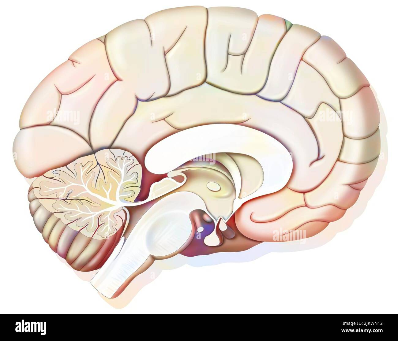 Mid sagittal section of the human brain showing the hypothalamus. Stock Photohttps://www.alamy.com/image-license-details/?v=1https://www.alamy.com/mid-sagittal-section-of-the-human-brain-showing-the-hypothalamus-image476923694.html
RF2JKWN12–Mid sagittal section of the human brain showing the hypothalamus.
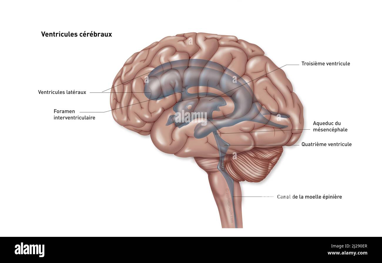 Brain ventricles Stock Photohttps://www.alamy.com/image-license-details/?v=1https://www.alamy.com/brain-ventricles-image466107231.html
RM2J290ER–Brain ventricles
 . The Biological bulletin. Biology; Zoology; Biology; Marine Biology. 96 A. OKUSU. Figure 23. Beginning of the gradual process of metamorphosis (7- to 8-day-old larvae), (a) The post-trochal region of the apical cap has completely disappeared. The trunk has become covered with numerous large epidermal papillae, (b) A larva with its apical region withdrawn into the trunk, (e) Aragonitic larval spicules. restricted to the trunk region, have started to appear underneath the cuticle, (d) Regularly arranged spicules underneath the cuticle beside the foot. AT, apical tuft; CD. cerebral depression: E Stock Photohttps://www.alamy.com/image-license-details/?v=1https://www.alamy.com/the-biological-bulletin-biology-zoology-biology-marine-biology-96-a-okusu-figure-23-beginning-of-the-gradual-process-of-metamorphosis-7-to-8-day-old-larvae-a-the-post-trochal-region-of-the-apical-cap-has-completely-disappeared-the-trunk-has-become-covered-with-numerous-large-epidermal-papillae-b-a-larva-with-its-apical-region-withdrawn-into-the-trunk-e-aragonitic-larval-spicules-restricted-to-the-trunk-region-have-started-to-appear-underneath-the-cuticle-d-regularly-arranged-spicules-underneath-the-cuticle-beside-the-foot-at-apical-tuft-cd-cerebral-depression-e-image234630111.html
RMRHM92R–. The Biological bulletin. Biology; Zoology; Biology; Marine Biology. 96 A. OKUSU. Figure 23. Beginning of the gradual process of metamorphosis (7- to 8-day-old larvae), (a) The post-trochal region of the apical cap has completely disappeared. The trunk has become covered with numerous large epidermal papillae, (b) A larva with its apical region withdrawn into the trunk, (e) Aragonitic larval spicules. restricted to the trunk region, have started to appear underneath the cuticle, (d) Regularly arranged spicules underneath the cuticle beside the foot. AT, apical tuft; CD. cerebral depression: E
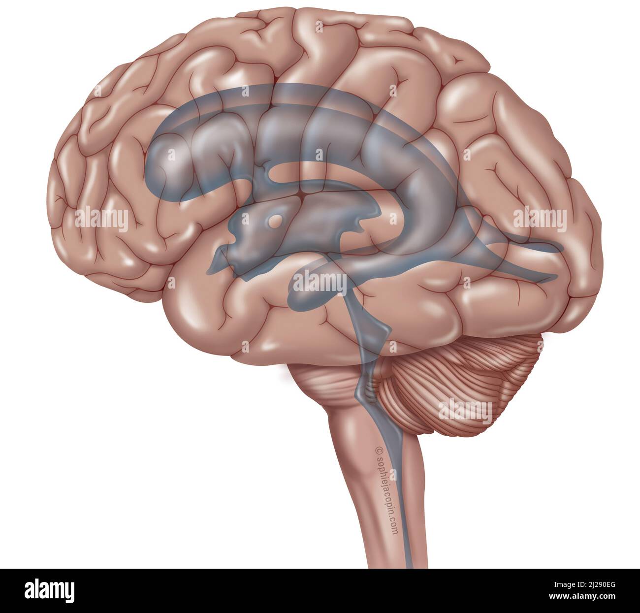 Encephal ventricular system Stock Photohttps://www.alamy.com/image-license-details/?v=1https://www.alamy.com/encephal-ventricular-system-image466107224.html
RM2J290EG–Encephal ventricular system
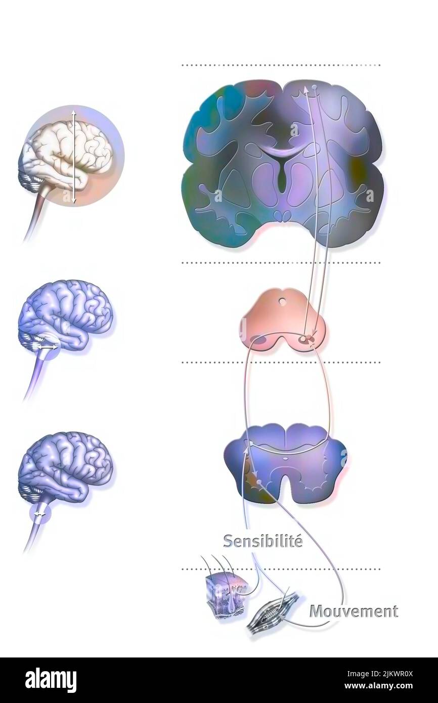 Sensorimotor loop: control of the brain to motor reactions. Stock Photohttps://www.alamy.com/image-license-details/?v=1https://www.alamy.com/sensorimotor-loop-control-of-the-brain-to-motor-reactions-image476925258.html
RF2JKWR0X–Sensorimotor loop: control of the brain to motor reactions.
 . The Biological bulletin. Biology; Zoology; Biology; Marine Biology. NEPHRIDIUM OF THE MITRARIA LARVA 317 nc es. 100pm FIGURE 1. Diagram of the mitraria larva of Owenia fusiformis (after Wilson, 1932). an, anus; br, cerebral ganglion; cb. prototrochal ciliary band; em, esophageal muscle; ep, episphere; es, esophagus; hp, hyposphere; in, intestine; 1m, dorsal levator muscles; mo, mouth; nc, circumesophageal nerve cord; nd, nephridioduct; ne, nephridium; ss, setal sac; st, stomach; tr, trunk rudiment. Transmission electron microscopic observations indicate that each nephridial sac is lined by m Stock Photohttps://www.alamy.com/image-license-details/?v=1https://www.alamy.com/the-biological-bulletin-biology-zoology-biology-marine-biology-nephridium-of-the-mitraria-larva-317-nc-es-100pm-figure-1-diagram-of-the-mitraria-larva-of-owenia-fusiformis-after-wilson-1932-an-anus-br-cerebral-ganglion-cb-prototrochal-ciliary-band-em-esophageal-muscle-ep-episphere-es-esophagus-hp-hyposphere-in-intestine-1m-dorsal-levator-muscles-mo-mouth-nc-circumesophageal-nerve-cord-nd-nephridioduct-ne-nephridium-ss-setal-sac-st-stomach-tr-trunk-rudiment-transmission-electron-microscopic-observations-indicate-that-each-nephridial-sac-is-lined-by-m-image234636197.html
RMRHMGT5–. The Biological bulletin. Biology; Zoology; Biology; Marine Biology. NEPHRIDIUM OF THE MITRARIA LARVA 317 nc es. 100pm FIGURE 1. Diagram of the mitraria larva of Owenia fusiformis (after Wilson, 1932). an, anus; br, cerebral ganglion; cb. prototrochal ciliary band; em, esophageal muscle; ep, episphere; es, esophagus; hp, hyposphere; in, intestine; 1m, dorsal levator muscles; mo, mouth; nc, circumesophageal nerve cord; nd, nephridioduct; ne, nephridium; ss, setal sac; st, stomach; tr, trunk rudiment. Transmission electron microscopic observations indicate that each nephridial sac is lined by m
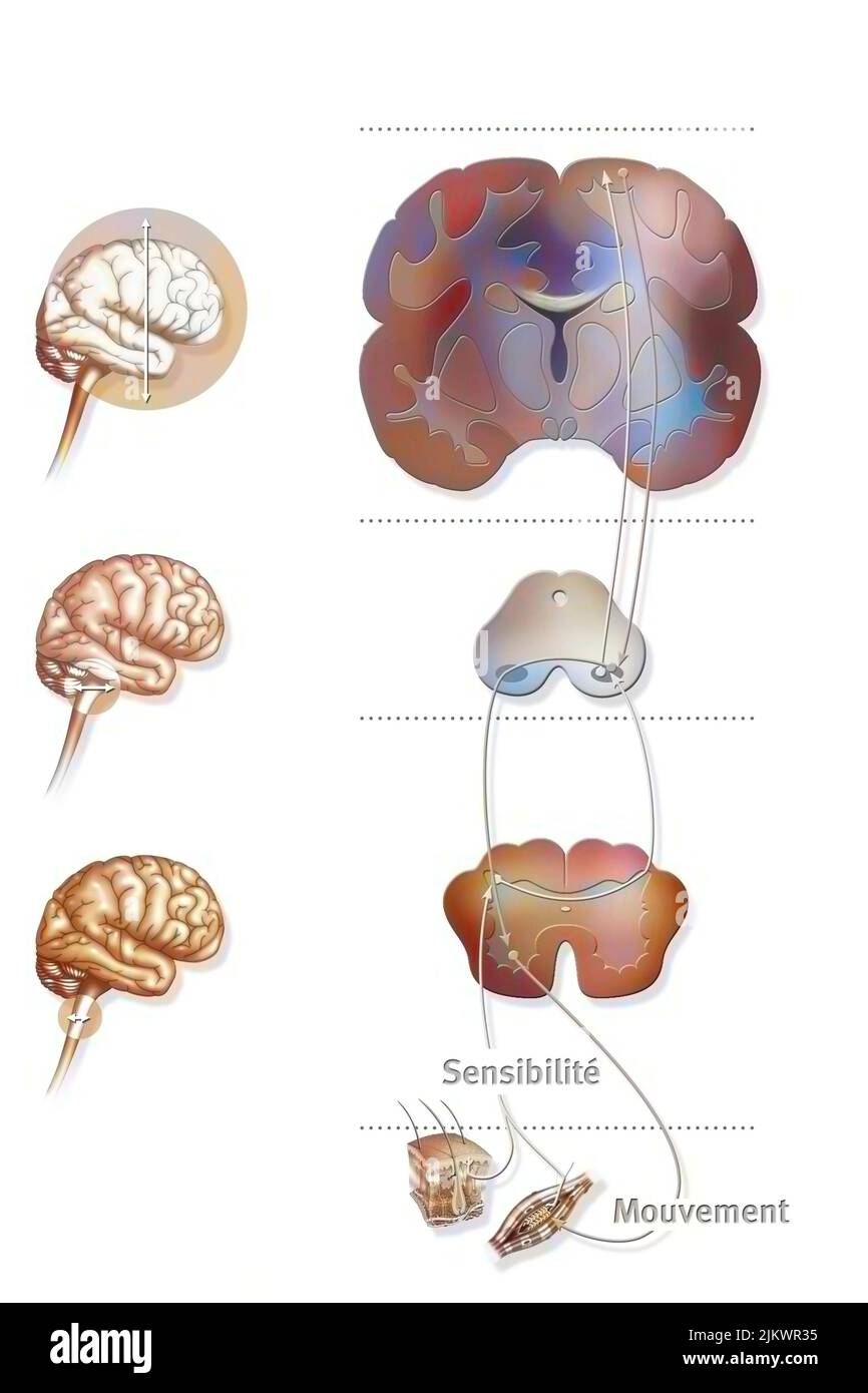 Sensorimotor loop: control of the brain to motor reactions. Stock Photohttps://www.alamy.com/image-license-details/?v=1https://www.alamy.com/sensorimotor-loop-control-of-the-brain-to-motor-reactions-image476925321.html
RF2JKWR35–Sensorimotor loop: control of the brain to motor reactions.
 . The Biological bulletin. Biology; Zoology; Biology; Marine Biology. 92 A. OKUSU. Trunk Cauda! region Figure 14. Larvae after hatching from the egg capsule (3 days after ovipositon. 1-day-old larvae), (a) Ventral view of the larva with three distinct body regions: ciliated pre-oral apical cap, unciliated trunk, and ciliated caudal region, (b) Vegetal (caudal) view of the trochophore larva showing the small imagination of the stomodeum on the ventral side of the trunk. AT, apical tuft; CD, cerebral depression; Pt, prototroch; Sd. stomo- deum; Tt. telotroch. appears to be secretory, because the Stock Photohttps://www.alamy.com/image-license-details/?v=1https://www.alamy.com/the-biological-bulletin-biology-zoology-biology-marine-biology-92-a-okusu-trunk-cauda!-region-figure-14-larvae-after-hatching-from-the-egg-capsule-3-days-after-ovipositon-1-day-old-larvae-a-ventral-view-of-the-larva-with-three-distinct-body-regions-ciliated-pre-oral-apical-cap-unciliated-trunk-and-ciliated-caudal-region-b-vegetal-caudal-view-of-the-trochophore-larva-showing-the-small-imagination-of-the-stomodeum-on-the-ventral-side-of-the-trunk-at-apical-tuft-cd-cerebral-depression-pt-prototroch-sd-stomo-deum-tt-telotroch-appears-to-be-secretory-because-the-image234630170.html
RMRHM94X–. The Biological bulletin. Biology; Zoology; Biology; Marine Biology. 92 A. OKUSU. Trunk Cauda! region Figure 14. Larvae after hatching from the egg capsule (3 days after ovipositon. 1-day-old larvae), (a) Ventral view of the larva with three distinct body regions: ciliated pre-oral apical cap, unciliated trunk, and ciliated caudal region, (b) Vegetal (caudal) view of the trochophore larva showing the small imagination of the stomodeum on the ventral side of the trunk. AT, apical tuft; CD, cerebral depression; Pt, prototroch; Sd. stomo- deum; Tt. telotroch. appears to be secretory, because the
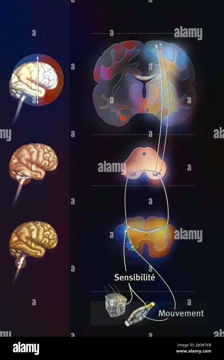 Sensorimotor loop: control of the brain to link sensations to motor reactions. Stock Photohttps://www.alamy.com/image-license-details/?v=1https://www.alamy.com/sensorimotor-loop-control-of-the-brain-to-link-sensations-to-motor-reactions-image476926559.html
RF2JKWTKB–Sensorimotor loop: control of the brain to link sensations to motor reactions.
 . Bulletin of the British Museum (Natural History) Zoology. AFRICAN TURBELLARIANS 57. Fig.7 Cryptocelis capensis: A, eyes in anterior region of body; B, sagittal section of copulatory organs. Family CRYPTOCELIDIDAE Laidlaw, 1903, emend. Poche,1926 Diagnostic features. Elongate oval or discoid forms. Tentacles indistinct or not apparent. Marginal eyes small; additional eyes distributed fanwise anteriorly or disposed in cerebral tentacular and frontal groups. Mouth in mid-third of body; pharynx ruffled; intestinal trunk relatively short. Genital pores in mid or hind third of body. Male copulator Stock Photohttps://www.alamy.com/image-license-details/?v=1https://www.alamy.com/bulletin-of-the-british-museum-natural-history-zoology-african-turbellarians-57-fig7-cryptocelis-capensis-a-eyes-in-anterior-region-of-body-b-sagittal-section-of-copulatory-organs-family-cryptocelididae-laidlaw-1903-emend-poche1926-diagnostic-features-elongate-oval-or-discoid-forms-tentacles-indistinct-or-not-apparent-marginal-eyes-small-additional-eyes-distributed-fanwise-anteriorly-or-disposed-in-cerebral-tentacular-and-frontal-groups-mouth-in-mid-third-of-body-pharynx-ruffled-intestinal-trunk-relatively-short-genital-pores-in-mid-or-hind-third-of-body-male-copulator-image233954162.html
RMRGHEWP–. Bulletin of the British Museum (Natural History) Zoology. AFRICAN TURBELLARIANS 57. Fig.7 Cryptocelis capensis: A, eyes in anterior region of body; B, sagittal section of copulatory organs. Family CRYPTOCELIDIDAE Laidlaw, 1903, emend. Poche,1926 Diagnostic features. Elongate oval or discoid forms. Tentacles indistinct or not apparent. Marginal eyes small; additional eyes distributed fanwise anteriorly or disposed in cerebral tentacular and frontal groups. Mouth in mid-third of body; pharynx ruffled; intestinal trunk relatively short. Genital pores in mid or hind third of body. Male copulator
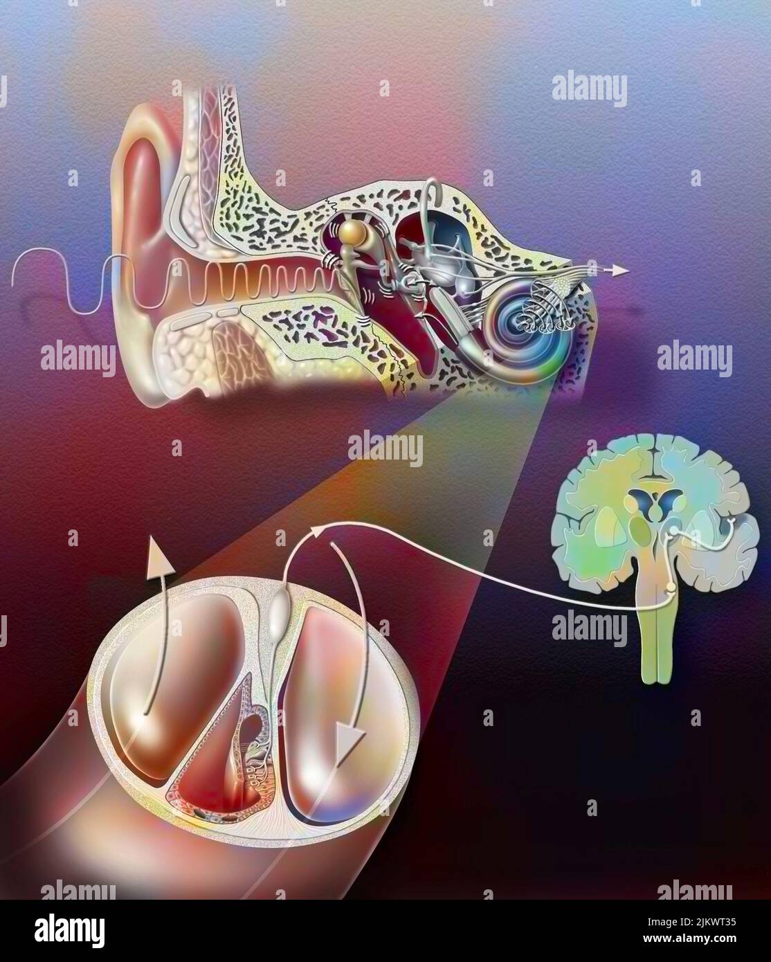 Anatomy of the ear with zoom of the organ of hearing. Stock Photohttps://www.alamy.com/image-license-details/?v=1https://www.alamy.com/anatomy-of-the-ear-with-zoom-of-the-organ-of-hearing-image476926105.html
RF2JKWT35–Anatomy of the ear with zoom of the organ of hearing.
 . Bulletin of the British Museum (Natural History) Zoology. . Fig.44 Prostheceraeus boucheti: A, dorsal surface of body in life; B, tentacles and arrangement of eyes in preserved specimen. Genus CYCLOPORUS Lang, 1884 Diagnostic features. Small oval forms with smooth or papillate dorsal surface. Small marginal tentacles with eyes in their bases and ventral surfaces. Cerebral eyes in two closely associated elongate clusters. Intestinal trunk extends to near posterior end of body, dividing into three branches anteriorly, median branch passing over cerebral organ; 6 to 10 pairs of lateral intestin Stock Photohttps://www.alamy.com/image-license-details/?v=1https://www.alamy.com/bulletin-of-the-british-museum-natural-history-zoology-fig44-prostheceraeus-boucheti-a-dorsal-surface-of-body-in-life-b-tentacles-and-arrangement-of-eyes-in-preserved-specimen-genus-cycloporus-lang-1884-diagnostic-features-small-oval-forms-with-smooth-or-papillate-dorsal-surface-small-marginal-tentacles-with-eyes-in-their-bases-and-ventral-surfaces-cerebral-eyes-in-two-closely-associated-elongate-clusters-intestinal-trunk-extends-to-near-posterior-end-of-body-dividing-into-three-branches-anteriorly-median-branch-passing-over-cerebral-organ-6-to-10-pairs-of-lateral-intestin-image233953426.html
RMRGHDYE–. Bulletin of the British Museum (Natural History) Zoology. . Fig.44 Prostheceraeus boucheti: A, dorsal surface of body in life; B, tentacles and arrangement of eyes in preserved specimen. Genus CYCLOPORUS Lang, 1884 Diagnostic features. Small oval forms with smooth or papillate dorsal surface. Small marginal tentacles with eyes in their bases and ventral surfaces. Cerebral eyes in two closely associated elongate clusters. Intestinal trunk extends to near posterior end of body, dividing into three branches anteriorly, median branch passing over cerebral organ; 6 to 10 pairs of lateral intestin
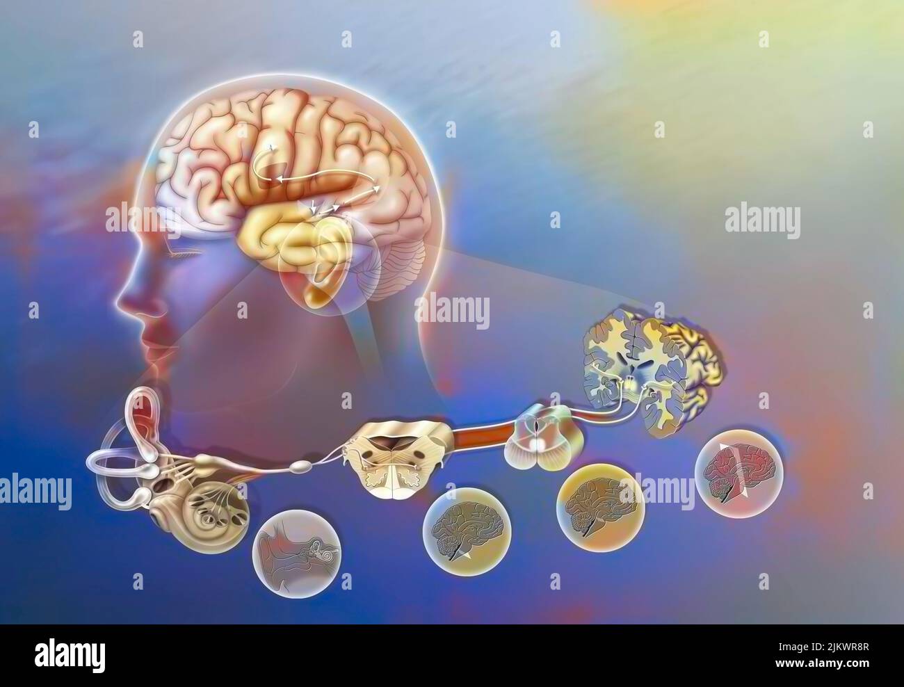 How a sound when it has reached the cochlea spreads through the brain. Stock Photohttps://www.alamy.com/image-license-details/?v=1https://www.alamy.com/how-a-sound-when-it-has-reached-the-cochlea-spreads-through-the-brain-image476925479.html
RF2JKWR8R–How a sound when it has reached the cochlea spreads through the brain.