Cerebrum superior view Stock Photos and Images
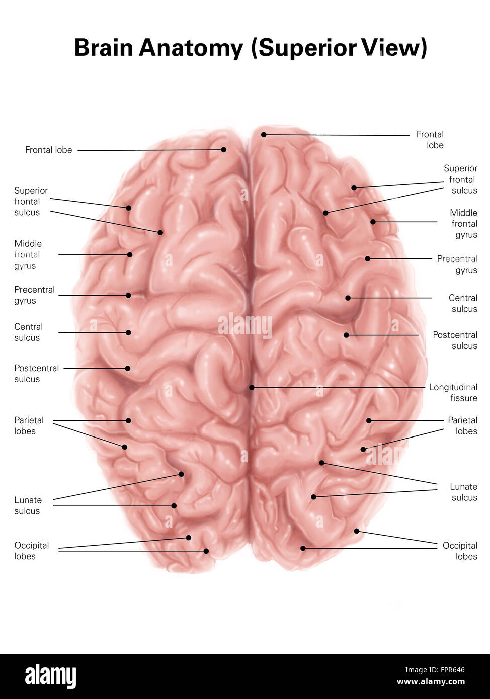 Human brain anatomy, superior view. Stock Photohttps://www.alamy.com/image-license-details/?v=1https://www.alamy.com/stock-photo-human-brain-anatomy-superior-view-100083990.html
RFFPR646–Human brain anatomy, superior view.
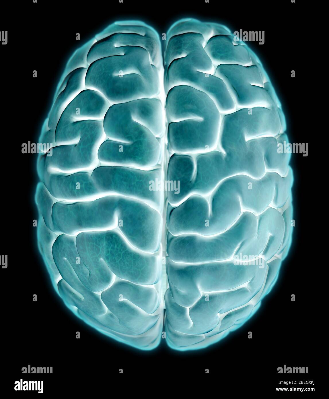 Human Brain, Superior View Stock Photohttps://www.alamy.com/image-license-details/?v=1https://www.alamy.com/human-brain-superior-view-image353184710.html
RM2BEGXKJ–Human Brain, Superior View
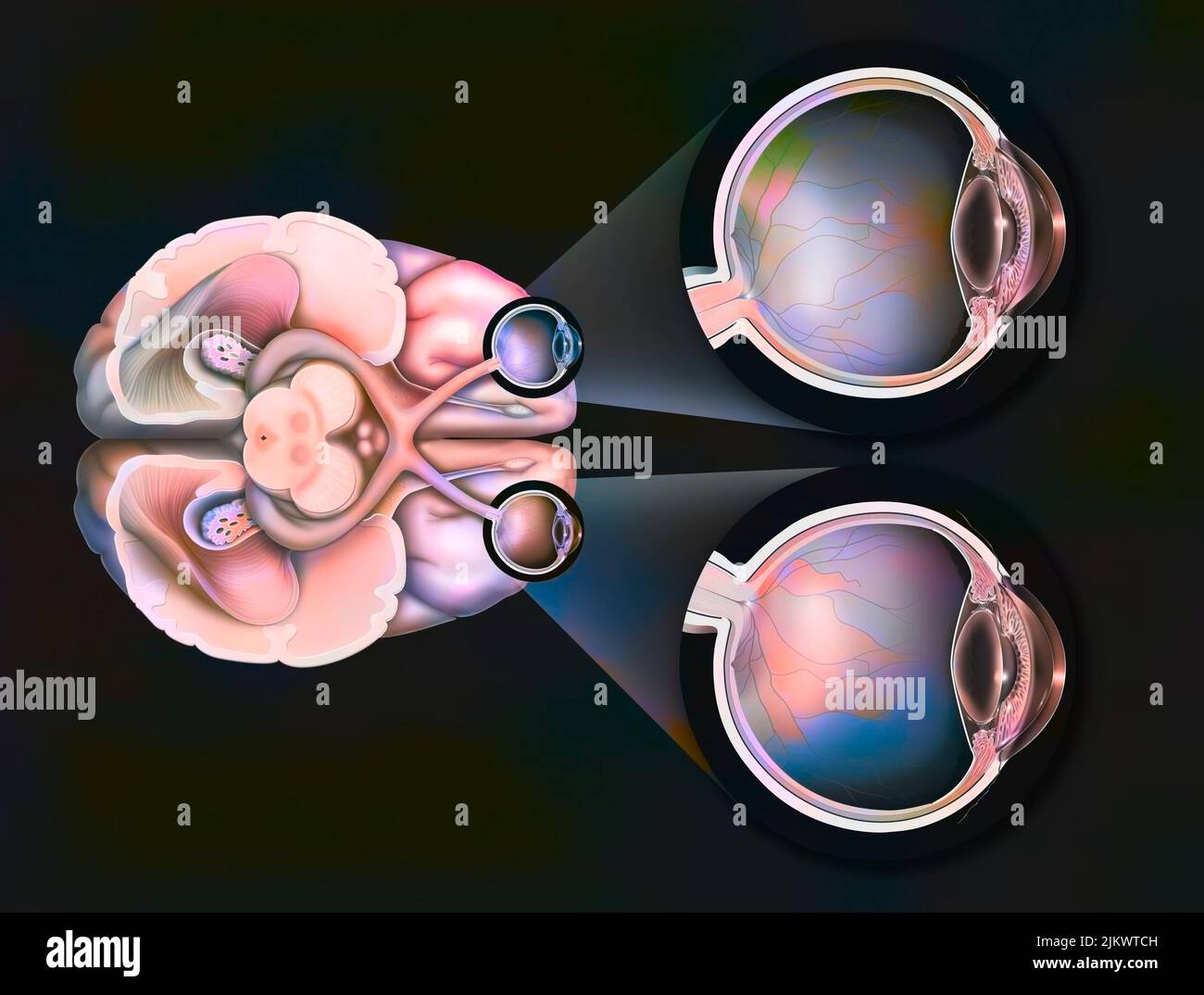 Eye: path of the visual pathways from the eye to the visual areas of the brain. Stock Photohttps://www.alamy.com/image-license-details/?v=1https://www.alamy.com/eye-path-of-the-visual-pathways-from-the-eye-to-the-visual-areas-of-the-brain-image476926369.html
RF2JKWTCH–Eye: path of the visual pathways from the eye to the visual areas of the brain.
 The cerebrum is the most voluminous and most complex part of the brain. It is made up of two hemispheres subdivided into four cerebral lobes, which cover the diencephalon. The most complex functions are completed by the exterior layer of the brain, the cerebral cortex. Stock Photohttps://www.alamy.com/image-license-details/?v=1https://www.alamy.com/the-cerebrum-is-the-most-voluminous-and-most-complex-part-of-the-brain-image156173949.html
RMK229D1–The cerebrum is the most voluminous and most complex part of the brain. It is made up of two hemispheres subdivided into four cerebral lobes, which cover the diencephalon. The most complex functions are completed by the exterior layer of the brain, the cerebral cortex.
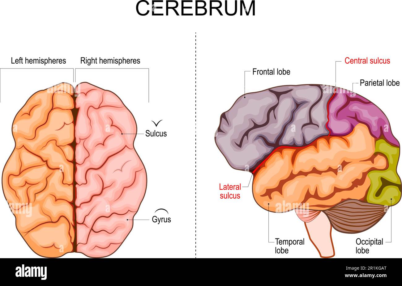 Human brain structure. Hemispheres and lobes of the cerebral cortex. frontal, temporal, occipital, and parietal lobes. lateral and superior view Stock Vectorhttps://www.alamy.com/image-license-details/?v=1https://www.alamy.com/human-brain-structure-hemispheres-and-lobes-of-the-cerebral-cortex-frontal-temporal-occipital-and-parietal-lobes-lateral-and-superior-view-image551776368.html
RF2R1KGAT–Human brain structure. Hemispheres and lobes of the cerebral cortex. frontal, temporal, occipital, and parietal lobes. lateral and superior view
 X-ray superior or top view of the full brain 3D rendering illustration. Human body and nervous system anatomy, medical, healthcare, biology, science, Stock Photohttps://www.alamy.com/image-license-details/?v=1https://www.alamy.com/x-ray-superior-or-top-view-of-the-full-brain-3d-rendering-illustration-human-body-and-nervous-system-anatomy-medical-healthcare-biology-science-image570401527.html
RF2T400Y3–X-ray superior or top view of the full brain 3D rendering illustration. Human body and nervous system anatomy, medical, healthcare, biology, science,
 Illustration of the oculomotor nerve anatomy in the human brain. The oculomotor nerve divides into superior and inferior branches in the anterior part of the cavernous sinus. Stock Photohttps://www.alamy.com/image-license-details/?v=1https://www.alamy.com/illustration-of-the-oculomotor-nerve-anatomy-in-the-human-brain-the-oculomotor-nerve-divides-into-superior-and-inferior-branches-in-the-anterior-part-of-the-cavernous-sinus-image628777686.html
RF2YEY89X–Illustration of the oculomotor nerve anatomy in the human brain. The oculomotor nerve divides into superior and inferior branches in the anterior part of the cavernous sinus.
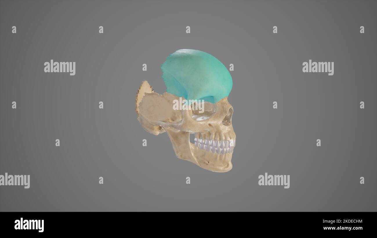 Lateral View of Frontal Bone Stock Photohttps://www.alamy.com/image-license-details/?v=1https://www.alamy.com/lateral-view-of-frontal-bone-image490198064.html
RF2KDECHM–Lateral View of Frontal Bone
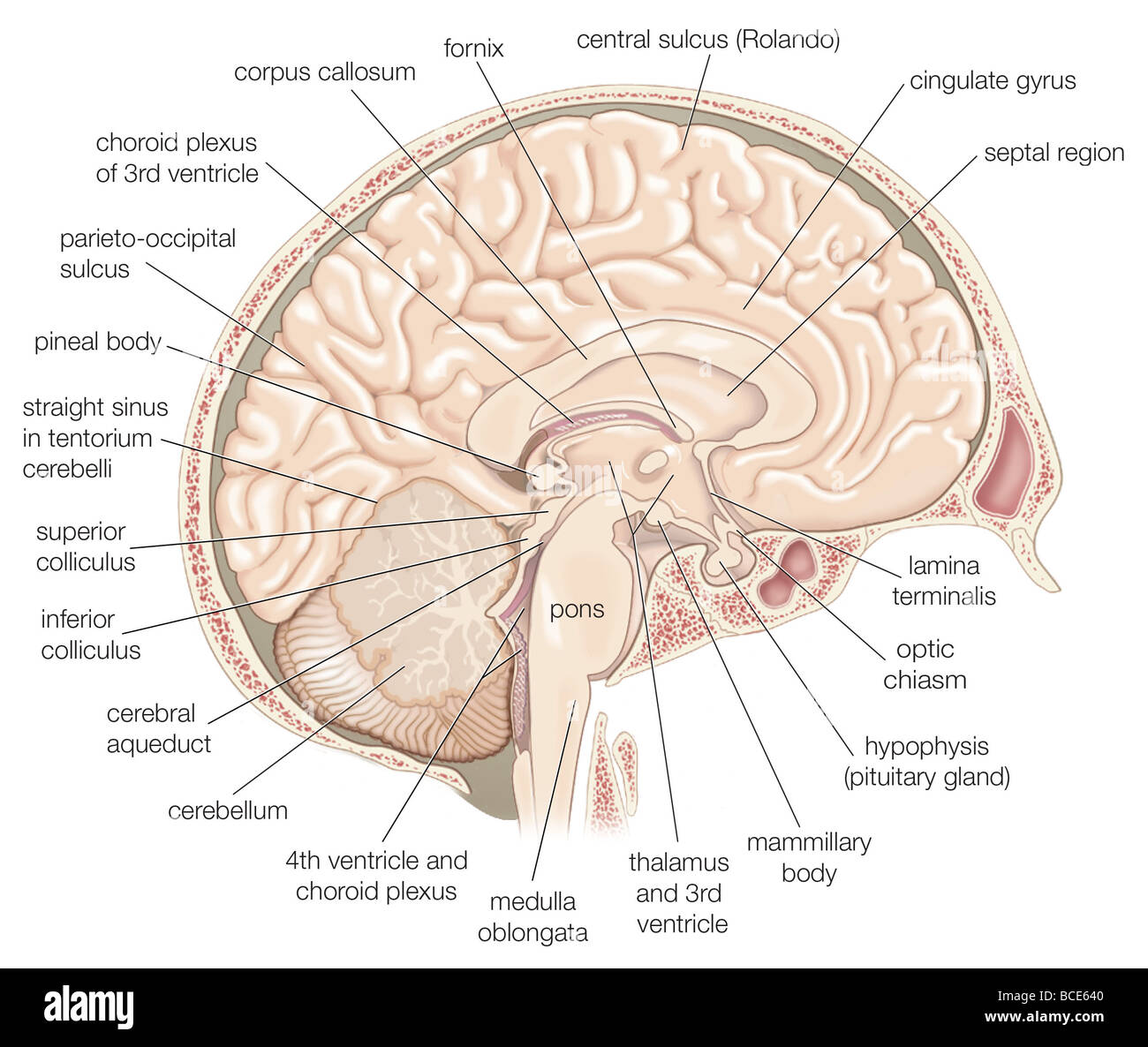 Medial view of the left hemisphere of the human brain. Stock Photohttps://www.alamy.com/image-license-details/?v=1https://www.alamy.com/stock-photo-medial-view-of-the-left-hemisphere-of-the-human-brain-24898384.html
RMBCE640–Medial view of the left hemisphere of the human brain.
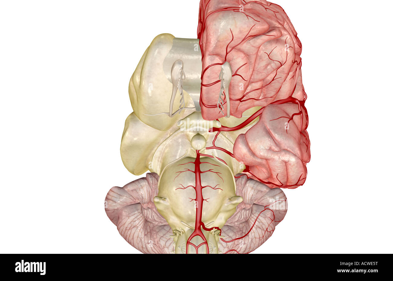 Arteries of the brain Stock Photohttps://www.alamy.com/image-license-details/?v=1https://www.alamy.com/stock-photo-arteries-of-the-brain-13235555.html
RFACWE5T–Arteries of the brain
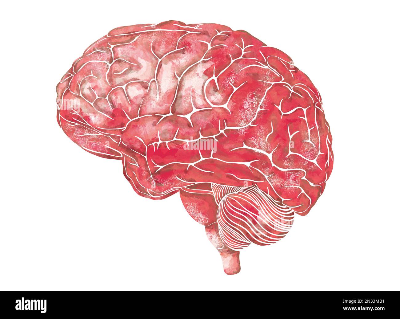 Structure of the human brain. Side Lateral view. Medical watercolor anatomy illustration. Hand drawn elegant anatomical brain art Stock Photohttps://www.alamy.com/image-license-details/?v=1https://www.alamy.com/structure-of-the-human-brain-side-lateral-view-medical-watercolor-anatomy-illustration-hand-drawn-elegant-anatomical-brain-art-image518236853.html
RF2N33MB1–Structure of the human brain. Side Lateral view. Medical watercolor anatomy illustration. Hand drawn elegant anatomical brain art
 A text-book of physiology for medical students and physicians . Fig. 82.—Schema of the projection fibers of the cerebrum and of the peduncles of thecerebellum; lateral view of the internal capsule : A, Tract from the frontal gyri to the ponsnuclei, and so to the cerebellum (frontal cerebro-cortico-pontal tract) ; B, the motor(pyramidal) tract; C, the sensory (lemniscus) tract ; D, the visual tract ; E, the auditorytract; F, the fibers of the superior peduncle of the cerebellum ; G, fibers of the middle pedun-cle uniting with A in the pons ; H, fibers of the inferior peduncle of the cerebellum Stock Photohttps://www.alamy.com/image-license-details/?v=1https://www.alamy.com/a-text-book-of-physiology-for-medical-students-and-physicians-fig-82schema-of-the-projection-fibers-of-the-cerebrum-and-of-the-peduncles-of-thecerebellum-lateral-view-of-the-internal-capsule-a-tract-from-the-frontal-gyri-to-the-ponsnuclei-and-so-to-the-cerebellum-frontal-cerebro-cortico-pontal-tract-b-the-motorpyramidal-tract-c-the-sensory-lemniscus-tract-d-the-visual-tract-e-the-auditorytract-f-the-fibers-of-the-superior-peduncle-of-the-cerebellum-g-fibers-of-the-middle-pedun-cle-uniting-with-a-in-the-pons-h-fibers-of-the-inferior-peduncle-of-the-cerebellum-image342811590.html
RM2AWMBK2–A text-book of physiology for medical students and physicians . Fig. 82.—Schema of the projection fibers of the cerebrum and of the peduncles of thecerebellum; lateral view of the internal capsule : A, Tract from the frontal gyri to the ponsnuclei, and so to the cerebellum (frontal cerebro-cortico-pontal tract) ; B, the motor(pyramidal) tract; C, the sensory (lemniscus) tract ; D, the visual tract ; E, the auditorytract; F, the fibers of the superior peduncle of the cerebellum ; G, fibers of the middle pedun-cle uniting with A in the pons ; H, fibers of the inferior peduncle of the cerebellum
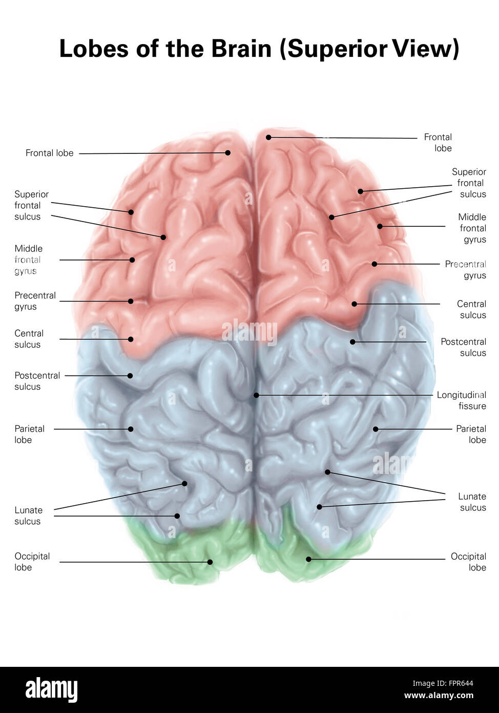 Superior view of human brain with colored lobes and labels. Stock Photohttps://www.alamy.com/image-license-details/?v=1https://www.alamy.com/stock-photo-superior-view-of-human-brain-with-colored-lobes-and-labels-100083988.html
RFFPR644–Superior view of human brain with colored lobes and labels.
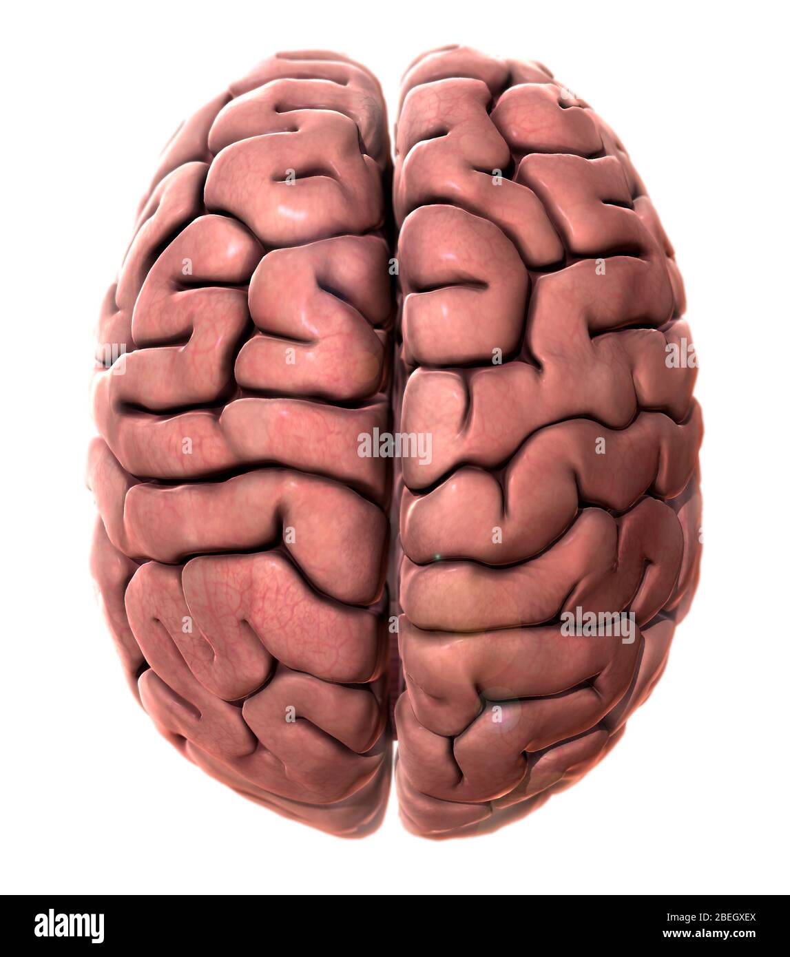 Human Brain, Superior View Stock Photohttps://www.alamy.com/image-license-details/?v=1https://www.alamy.com/human-brain-superior-view-image353184578.html
RM2BEGXEX–Human Brain, Superior View
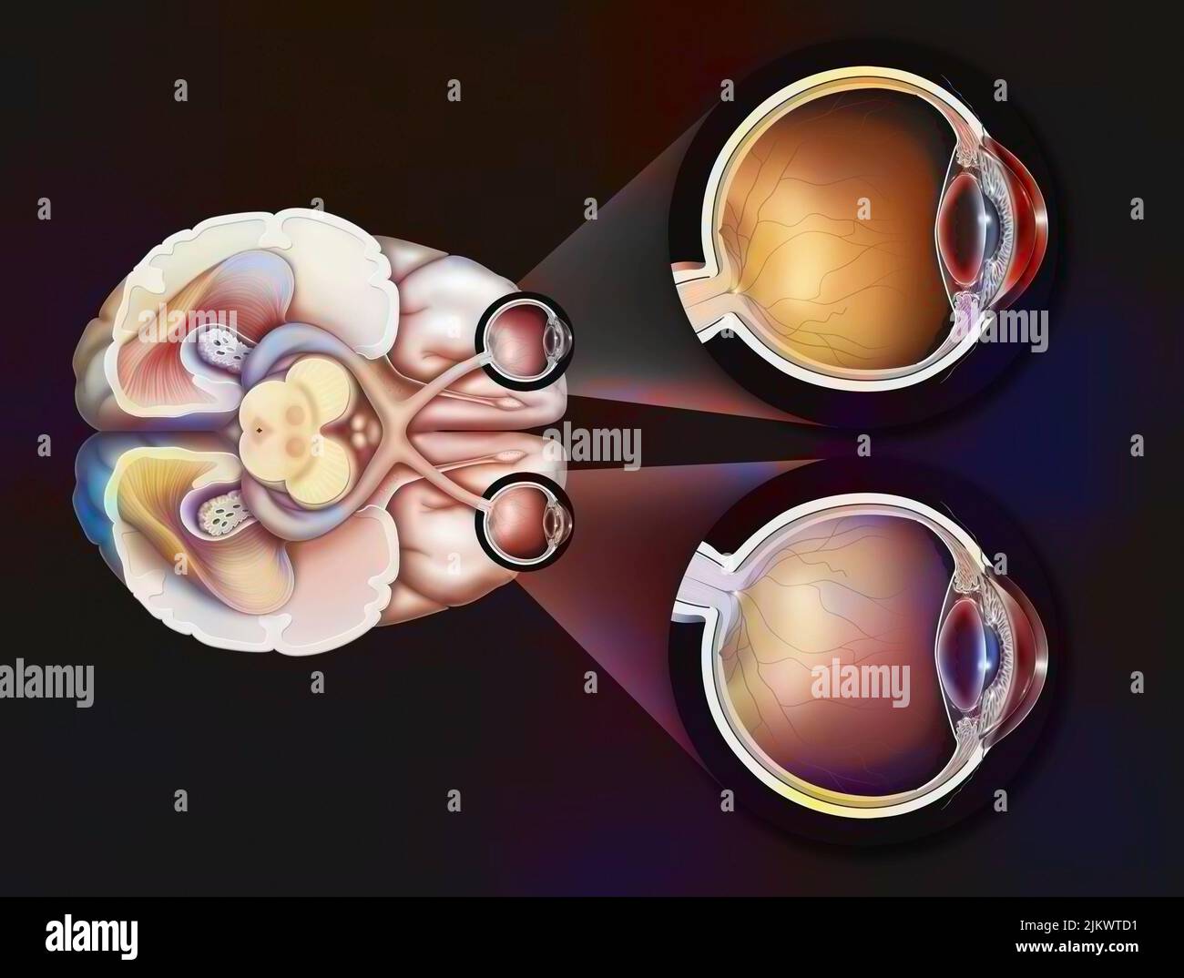 Eye: path of the visual pathways from the eye to the visual areas of the brain. Stock Photohttps://www.alamy.com/image-license-details/?v=1https://www.alamy.com/eye-path-of-the-visual-pathways-from-the-eye-to-the-visual-areas-of-the-brain-image476926381.html
RF2JKWTD1–Eye: path of the visual pathways from the eye to the visual areas of the brain.
 X-ray superior or top view of the full brain whit eyes 3D rendering illustration. Human body and nervous system anatomy, medical, healthcare, biology, Stock Photohttps://www.alamy.com/image-license-details/?v=1https://www.alamy.com/x-ray-superior-or-top-view-of-the-full-brain-whit-eyes-3d-rendering-illustration-human-body-and-nervous-system-anatomy-medical-healthcare-biology-image570401519.html
RF2T400XR–X-ray superior or top view of the full brain whit eyes 3D rendering illustration. Human body and nervous system anatomy, medical, healthcare, biology,
 Illustration of the oculomotor nerve anatomy in the human brain. The oculomotor nerve divides into superior and inferior branches in the anterior part of the cavernous sinus. Stock Photohttps://www.alamy.com/image-license-details/?v=1https://www.alamy.com/illustration-of-the-oculomotor-nerve-anatomy-in-the-human-brain-the-oculomotor-nerve-divides-into-superior-and-inferior-branches-in-the-anterior-part-of-the-cavernous-sinus-image628777692.html
RF2YEY8A4–Illustration of the oculomotor nerve anatomy in the human brain. The oculomotor nerve divides into superior and inferior branches in the anterior part of the cavernous sinus.
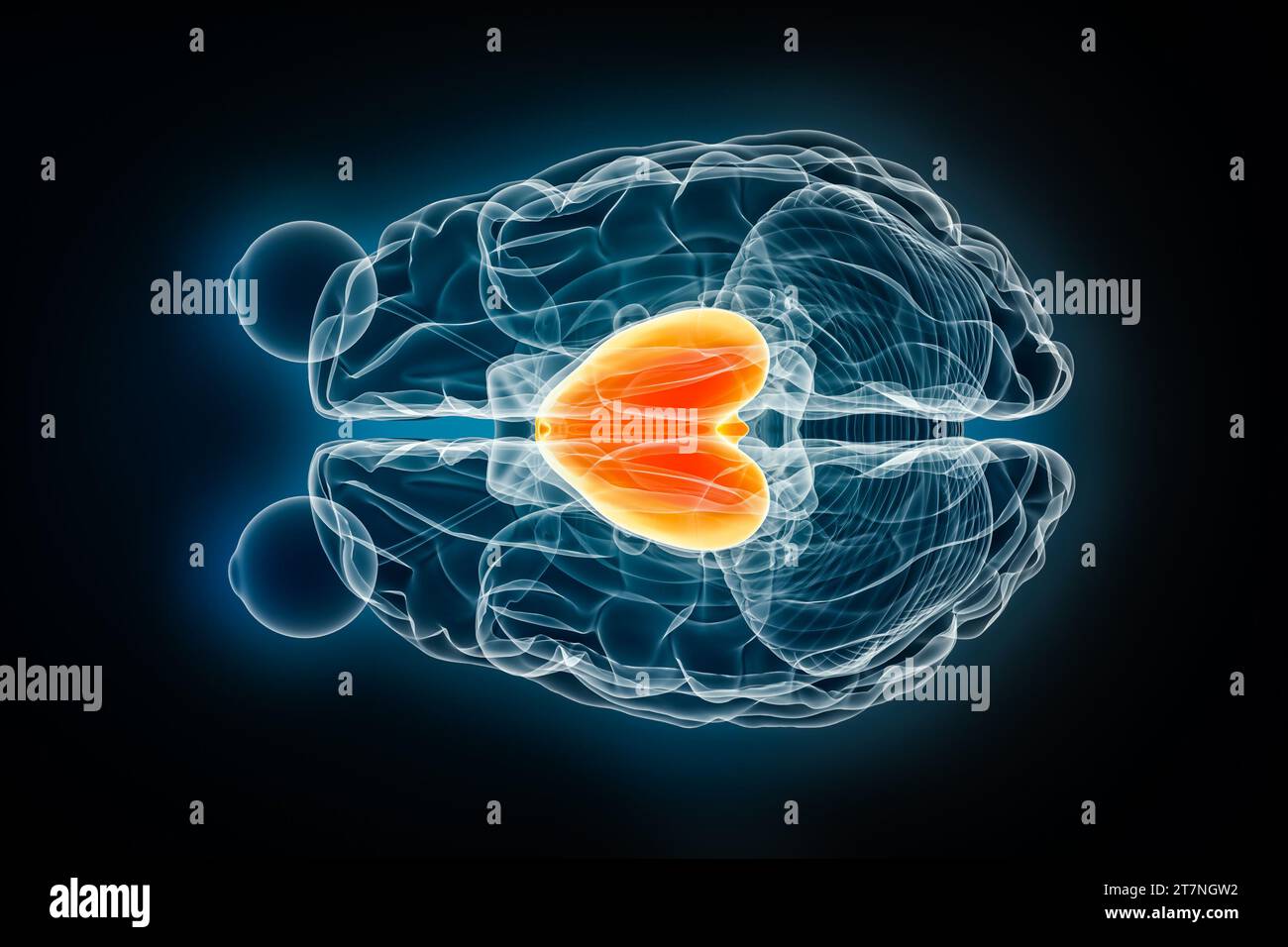 Forebrain or prosencephalon x-ray superior or top view 3D rendering illustration. Human brain and nervous system anatomy, medical, healthcare, biology Stock Photohttps://www.alamy.com/image-license-details/?v=1https://www.alamy.com/forebrain-or-prosencephalon-x-ray-superior-or-top-view-3d-rendering-illustration-human-brain-and-nervous-system-anatomy-medical-healthcare-biology-image572718974.html
RF2T7NGW2–Forebrain or prosencephalon x-ray superior or top view 3D rendering illustration. Human brain and nervous system anatomy, medical, healthcare, biology
 The brainstem Stock Photohttps://www.alamy.com/image-license-details/?v=1https://www.alamy.com/stock-photo-the-brainstem-13233403.html
RFACW7PM–The brainstem
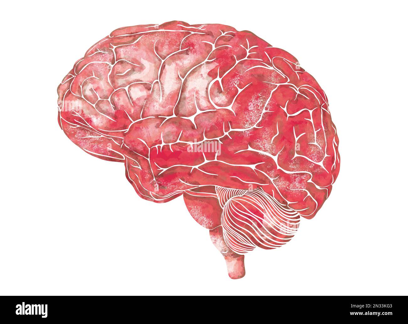 Structure of the human brain. Side Lateral view. Medical watercolor anatomy illustration. Hand drawn elegant anatomical brain art Stock Photohttps://www.alamy.com/image-license-details/?v=1https://www.alamy.com/structure-of-the-human-brain-side-lateral-view-medical-watercolor-anatomy-illustration-hand-drawn-elegant-anatomical-brain-art-image518236211.html
RF2N33KG3–Structure of the human brain. Side Lateral view. Medical watercolor anatomy illustration. Hand drawn elegant anatomical brain art
 A text-book of physiology, for medical students and physicians . Fig. 81.—Schema of the projection fibers of the cerebrum and of the peduncles of thecerebellum; lateral view of the internal capsule : A, Tract from the frontal gyri to the ponsnuclei, and so to the cerebellum (frontal cerebro-cortico-pontal tract) ; B, the motor(pyramidal) tract ; C, the sensory (lemniscus) tract; D, the visual tract ; E, the auditorytract; F, the fibers of the superior peduncle of the cerebellum ; G, fibers of the middle pedun-cle uniting with A in the pons ; H, fibers of the inferior peduncle of the cerebellum Stock Photohttps://www.alamy.com/image-license-details/?v=1https://www.alamy.com/a-text-book-of-physiology-for-medical-students-and-physicians-fig-81schema-of-the-projection-fibers-of-the-cerebrum-and-of-the-peduncles-of-thecerebellum-lateral-view-of-the-internal-capsule-a-tract-from-the-frontal-gyri-to-the-ponsnuclei-and-so-to-the-cerebellum-frontal-cerebro-cortico-pontal-tract-b-the-motorpyramidal-tract-c-the-sensory-lemniscus-tract-d-the-visual-tract-e-the-auditorytract-f-the-fibers-of-the-superior-peduncle-of-the-cerebellum-g-fibers-of-the-middle-pedun-cle-uniting-with-a-in-the-pons-h-fibers-of-the-inferior-peduncle-of-the-cerebellum-image342978199.html
RM2AX005B–A text-book of physiology, for medical students and physicians . Fig. 81.—Schema of the projection fibers of the cerebrum and of the peduncles of thecerebellum; lateral view of the internal capsule : A, Tract from the frontal gyri to the ponsnuclei, and so to the cerebellum (frontal cerebro-cortico-pontal tract) ; B, the motor(pyramidal) tract ; C, the sensory (lemniscus) tract; D, the visual tract ; E, the auditorytract; F, the fibers of the superior peduncle of the cerebellum ; G, fibers of the middle pedun-cle uniting with A in the pons ; H, fibers of the inferior peduncle of the cerebellum
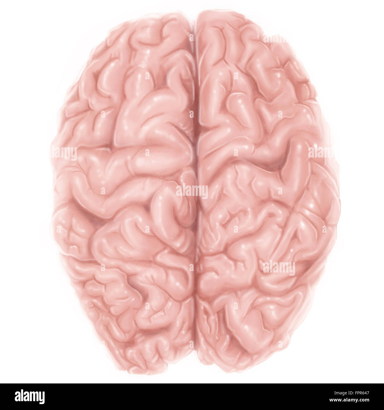 Superior view of human brain. Stock Photohttps://www.alamy.com/image-license-details/?v=1https://www.alamy.com/stock-photo-superior-view-of-human-brain-100083991.html
RFFPR647–Superior view of human brain.
 Human Brain, Superior View Stock Photohttps://www.alamy.com/image-license-details/?v=1https://www.alamy.com/human-brain-superior-view-image353184560.html
RM2BEGXE8–Human Brain, Superior View
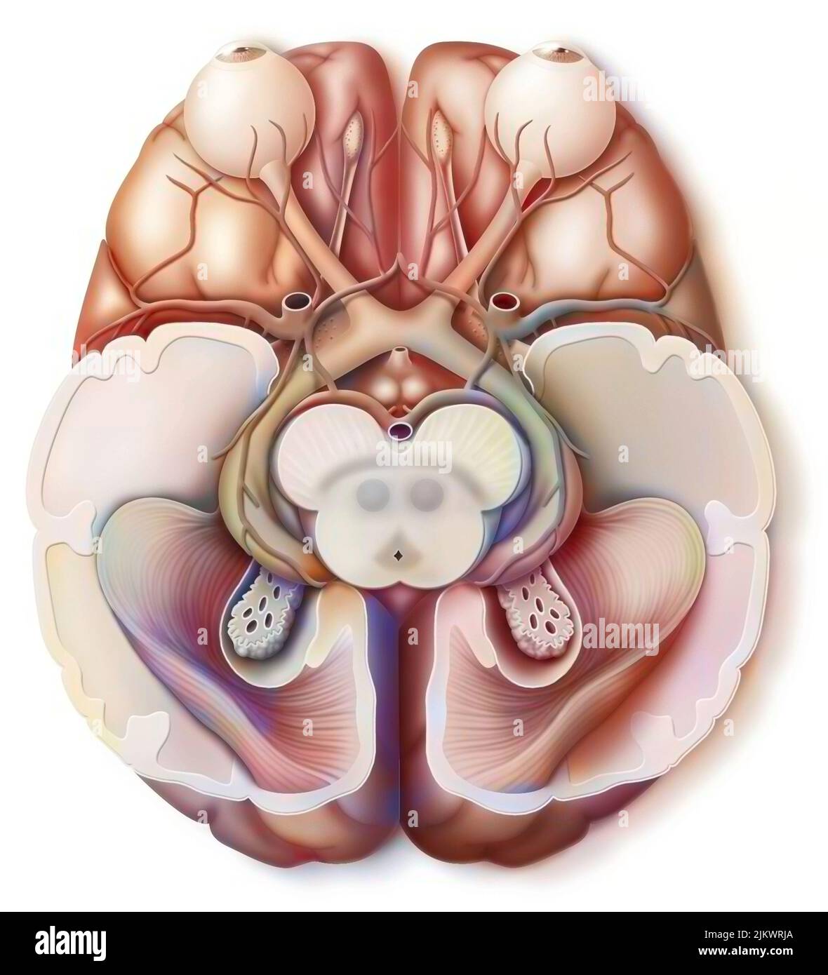 Path traveled by information sent to the brain through the eyes. Stock Photohttps://www.alamy.com/image-license-details/?v=1https://www.alamy.com/path-traveled-by-information-sent-to-the-brain-through-the-eyes-image476925746.html
RF2JKWRJA–Path traveled by information sent to the brain through the eyes.
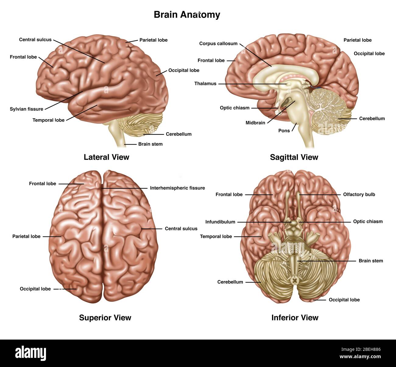 Brain Anatomy, Illustration Stock Photohttps://www.alamy.com/image-license-details/?v=1https://www.alamy.com/brain-anatomy-illustration-image353192230.html
RF2BEH886–Brain Anatomy, Illustration
 Illustration of the oculomotor nerve anatomy in the human brain. The oculomotor nerve divides into superior and inferior branches in the anterior part of the cavernous sinus. Stock Photohttps://www.alamy.com/image-license-details/?v=1https://www.alamy.com/illustration-of-the-oculomotor-nerve-anatomy-in-the-human-brain-the-oculomotor-nerve-divides-into-superior-and-inferior-branches-in-the-anterior-part-of-the-cavernous-sinus-image628777681.html
RF2YEY89N–Illustration of the oculomotor nerve anatomy in the human brain. The oculomotor nerve divides into superior and inferior branches in the anterior part of the cavernous sinus.
 Cerebral amygdala x-ray superior or top view 3D rendering illustration. Human brain, limbic and nervous system anatomy, medical, healthcare, biology, Stock Photohttps://www.alamy.com/image-license-details/?v=1https://www.alamy.com/cerebral-amygdala-x-ray-superior-or-top-view-3d-rendering-illustration-human-brain-limbic-and-nervous-system-anatomy-medical-healthcare-biology-image572718970.html
RF2T7NGTX–Cerebral amygdala x-ray superior or top view 3D rendering illustration. Human brain, limbic and nervous system anatomy, medical, healthcare, biology,
 The brainstem Stock Photohttps://www.alamy.com/image-license-details/?v=1https://www.alamy.com/stock-photo-the-brainstem-13228416.html
RFACTMXW–The brainstem
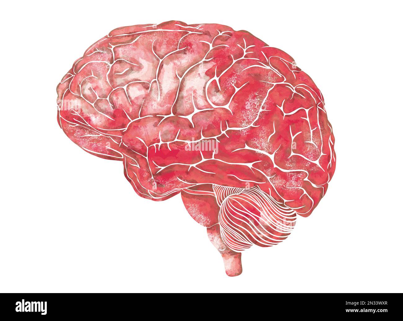 Structure of the human brain. Side Lateral view. Medical watercolor anatomy illustration. Hand drawn elegant anatomical brain art Stock Photohttps://www.alamy.com/image-license-details/?v=1https://www.alamy.com/structure-of-the-human-brain-side-lateral-view-medical-watercolor-anatomy-illustration-hand-drawn-elegant-anatomical-brain-art-image518241215.html
RF2N33WXR–Structure of the human brain. Side Lateral view. Medical watercolor anatomy illustration. Hand drawn elegant anatomical brain art
 . A text-book of physiology : for medical students and physicians . Fig. 82.—Schema of the projection fibers of the cerebrum and of the peduncles of thecerebellum; lateral view of the internal capsule : A, Tract from the frontal gyri to the ponsnuclei, and so to the cerebellum (frontal cerebro-cortico-pontal tract) ; B, the motor(pyramidal) tract; C, the sensory (lemniscus) tract; D, the visual tract ; E, the auditorytract; F, the fibers of the superior peduncle of the cerebellum ; G, fibers of the middle pedun-cle uniting with A in the pons ; H, fibers of the inferior peduncle of the cerebell Stock Photohttps://www.alamy.com/image-license-details/?v=1https://www.alamy.com/a-text-book-of-physiology-for-medical-students-and-physicians-fig-82schema-of-the-projection-fibers-of-the-cerebrum-and-of-the-peduncles-of-thecerebellum-lateral-view-of-the-internal-capsule-a-tract-from-the-frontal-gyri-to-the-ponsnuclei-and-so-to-the-cerebellum-frontal-cerebro-cortico-pontal-tract-b-the-motorpyramidal-tract-c-the-sensory-lemniscus-tract-d-the-visual-tract-e-the-auditorytract-f-the-fibers-of-the-superior-peduncle-of-the-cerebellum-g-fibers-of-the-middle-pedun-cle-uniting-with-a-in-the-pons-h-fibers-of-the-inferior-peduncle-of-the-cerebell-image370189367.html
RM2CE7G8R–. A text-book of physiology : for medical students and physicians . Fig. 82.—Schema of the projection fibers of the cerebrum and of the peduncles of thecerebellum; lateral view of the internal capsule : A, Tract from the frontal gyri to the ponsnuclei, and so to the cerebellum (frontal cerebro-cortico-pontal tract) ; B, the motor(pyramidal) tract; C, the sensory (lemniscus) tract; D, the visual tract ; E, the auditorytract; F, the fibers of the superior peduncle of the cerebellum ; G, fibers of the middle pedun-cle uniting with A in the pons ; H, fibers of the inferior peduncle of the cerebell
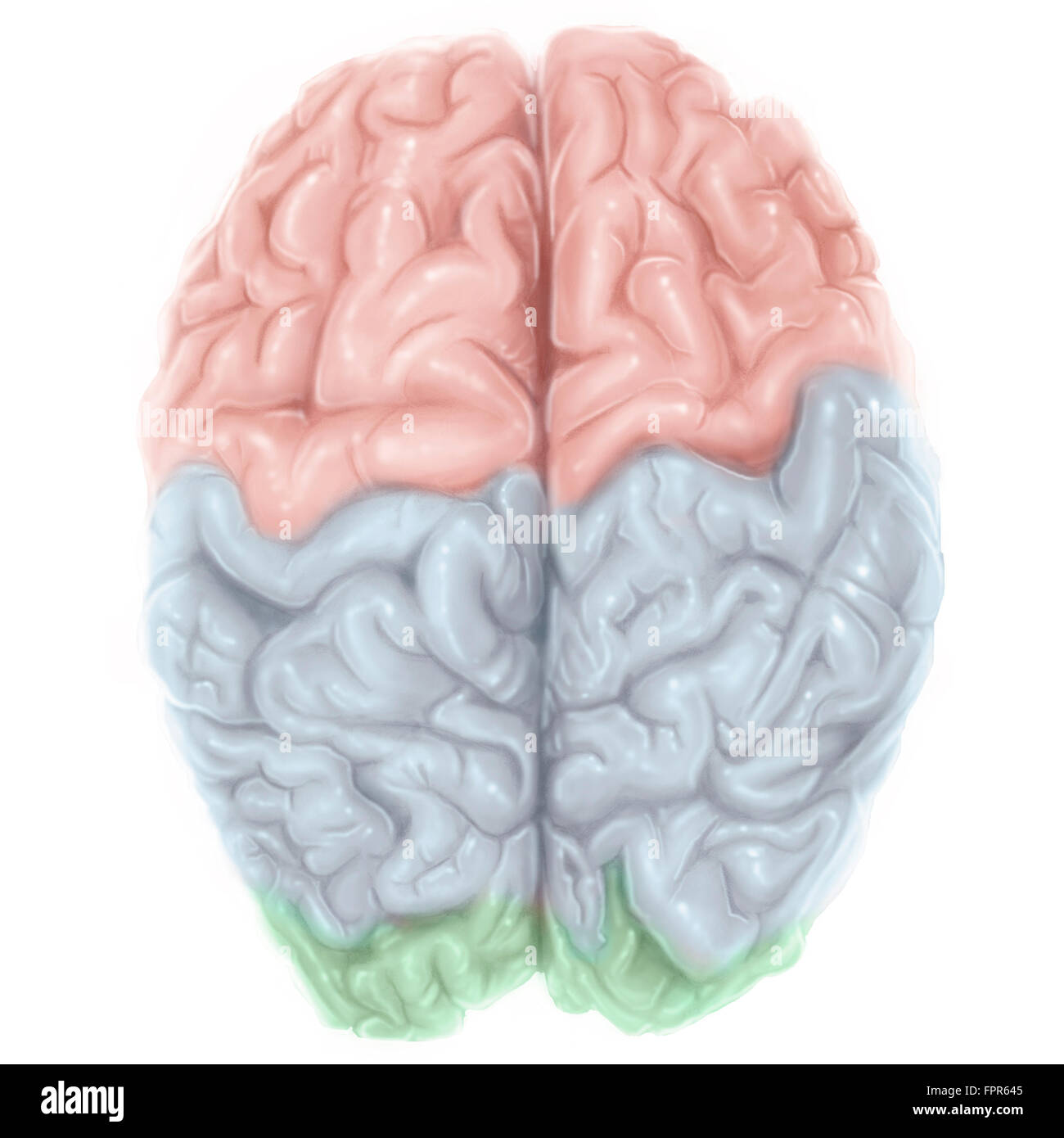 Superior view of human brain with colored lobes. Stock Photohttps://www.alamy.com/image-license-details/?v=1https://www.alamy.com/stock-photo-superior-view-of-human-brain-with-colored-lobes-100083989.html
RFFPR645–Superior view of human brain with colored lobes.
 Superior view of the path and transmission of visual information from the retina. Stock Photohttps://www.alamy.com/image-license-details/?v=1https://www.alamy.com/superior-view-of-the-path-and-transmission-of-visual-information-from-the-retina-image476925265.html
RF2JKWR15–Superior view of the path and transmission of visual information from the retina.
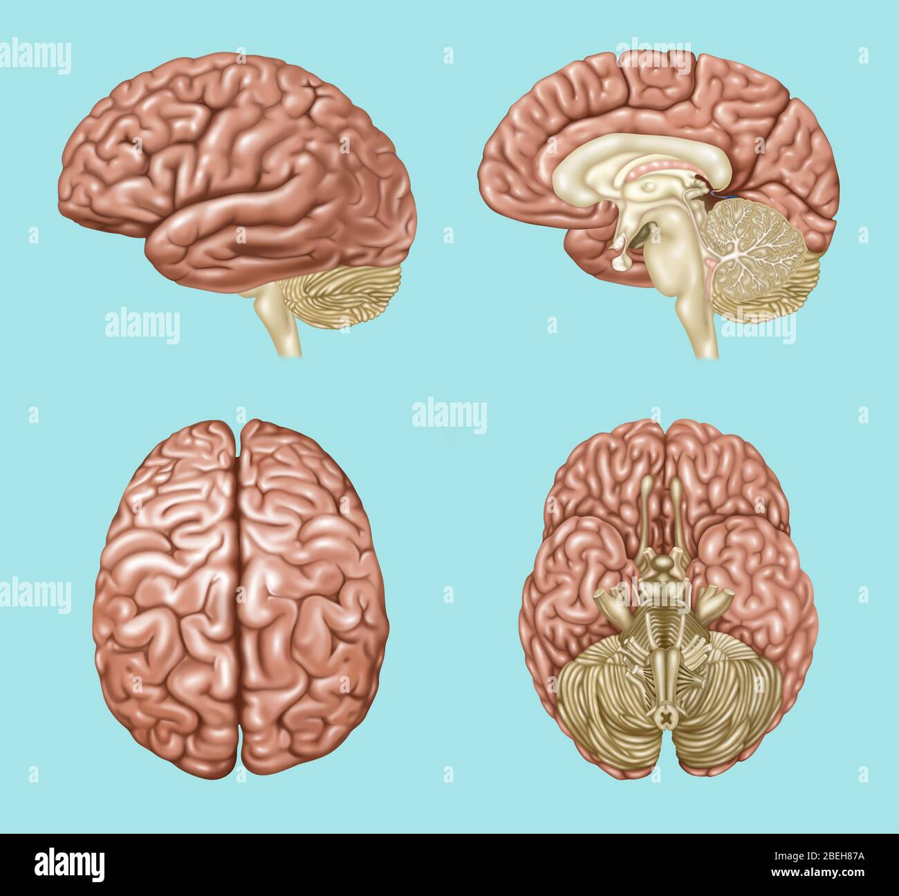 Brain Anatomy, Illustration Stock Photohttps://www.alamy.com/image-license-details/?v=1https://www.alamy.com/brain-anatomy-illustration-image353192206.html
RF2BEH87A–Brain Anatomy, Illustration
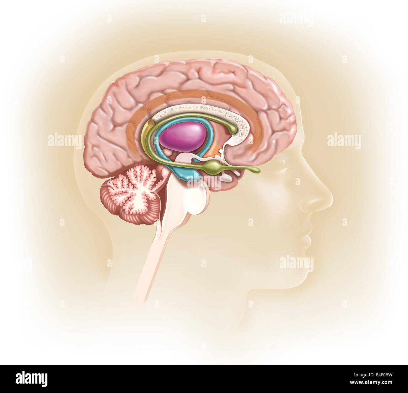 Sagittal view of human brain showing the limbic system. Stock Photohttps://www.alamy.com/image-license-details/?v=1https://www.alamy.com/stock-photo-sagittal-view-of-human-brain-showing-the-limbic-system-71629569.html
RME4F06W–Sagittal view of human brain showing the limbic system.
 Illustration of the oculomotor nerve anatomy in the human brain. The oculomotor nerve divides into superior and inferior branches in the anterior part of the cavernous sinus. Stock Photohttps://www.alamy.com/image-license-details/?v=1https://www.alamy.com/illustration-of-the-oculomotor-nerve-anatomy-in-the-human-brain-the-oculomotor-nerve-divides-into-superior-and-inferior-branches-in-the-anterior-part-of-the-cavernous-sinus-image628777712.html
RF2YEY8AT–Illustration of the oculomotor nerve anatomy in the human brain. The oculomotor nerve divides into superior and inferior branches in the anterior part of the cavernous sinus.
 Thalamus x-ray superior or top view 3D rendering illustration. Human brain, limbic and nervous system anatomy, medical, healthcare, biology, science, Stock Photohttps://www.alamy.com/image-license-details/?v=1https://www.alamy.com/thalamus-x-ray-superior-or-top-view-3d-rendering-illustration-human-brain-limbic-and-nervous-system-anatomy-medical-healthcare-biology-science-image572718985.html
RF2T7NGWD–Thalamus x-ray superior or top view 3D rendering illustration. Human brain, limbic and nervous system anatomy, medical, healthcare, biology, science,
 The brainstem Stock Photohttps://www.alamy.com/image-license-details/?v=1https://www.alamy.com/stock-photo-the-brainstem-13235360.html
RFACWDHN–The brainstem
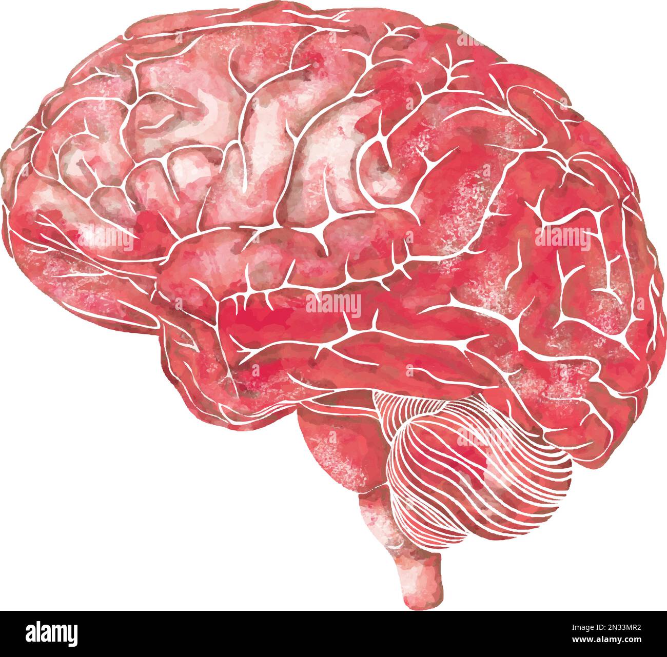 Structure of the human brain. Side Lateral view. Medical watercolor anatomy illustration. Hand drawn elegant anatomical brain art Stock Vectorhttps://www.alamy.com/image-license-details/?v=1https://www.alamy.com/structure-of-the-human-brain-side-lateral-view-medical-watercolor-anatomy-illustration-hand-drawn-elegant-anatomical-brain-art-image518237190.html
RF2N33MR2–Structure of the human brain. Side Lateral view. Medical watercolor anatomy illustration. Hand drawn elegant anatomical brain art
 . A text-book of physiology : for medical students and physicians . V J<. GENERAL PHYSIOLOGY OF THE CEREBRUM. 185. Fig. 82.—Schema of the projection fibers of the cerebrum and of the peduncles of thecerebellum; lateral view of the internal capsule : A, Tract from the frontal gyri to the ponsnuclei, and so to the cerebellum (frontal cerebro-cortico-pontal tract) ; B, the motor(pyramidal) tract; C, the sensory (lemniscus) tract; D, the visual tract ; E, the auditorytract; F, the fibers of the superior peduncle of the cerebellum ; G, fibers of the middle pedun-cle uniting with A in the pons ; Stock Photohttps://www.alamy.com/image-license-details/?v=1https://www.alamy.com/a-text-book-of-physiology-for-medical-students-and-physicians-v-jlt-general-physiology-of-the-cerebrum-185-fig-82schema-of-the-projection-fibers-of-the-cerebrum-and-of-the-peduncles-of-thecerebellum-lateral-view-of-the-internal-capsule-a-tract-from-the-frontal-gyri-to-the-ponsnuclei-and-so-to-the-cerebellum-frontal-cerebro-cortico-pontal-tract-b-the-motorpyramidal-tract-c-the-sensory-lemniscus-tract-d-the-visual-tract-e-the-auditorytract-f-the-fibers-of-the-superior-peduncle-of-the-cerebellum-g-fibers-of-the-middle-pedun-cle-uniting-with-a-in-the-pons-image370189502.html
RM2CE7GDJ–. A text-book of physiology : for medical students and physicians . V J<. GENERAL PHYSIOLOGY OF THE CEREBRUM. 185. Fig. 82.—Schema of the projection fibers of the cerebrum and of the peduncles of thecerebellum; lateral view of the internal capsule : A, Tract from the frontal gyri to the ponsnuclei, and so to the cerebellum (frontal cerebro-cortico-pontal tract) ; B, the motor(pyramidal) tract; C, the sensory (lemniscus) tract; D, the visual tract ; E, the auditorytract; F, the fibers of the superior peduncle of the cerebellum ; G, fibers of the middle pedun-cle uniting with A in the pons ;
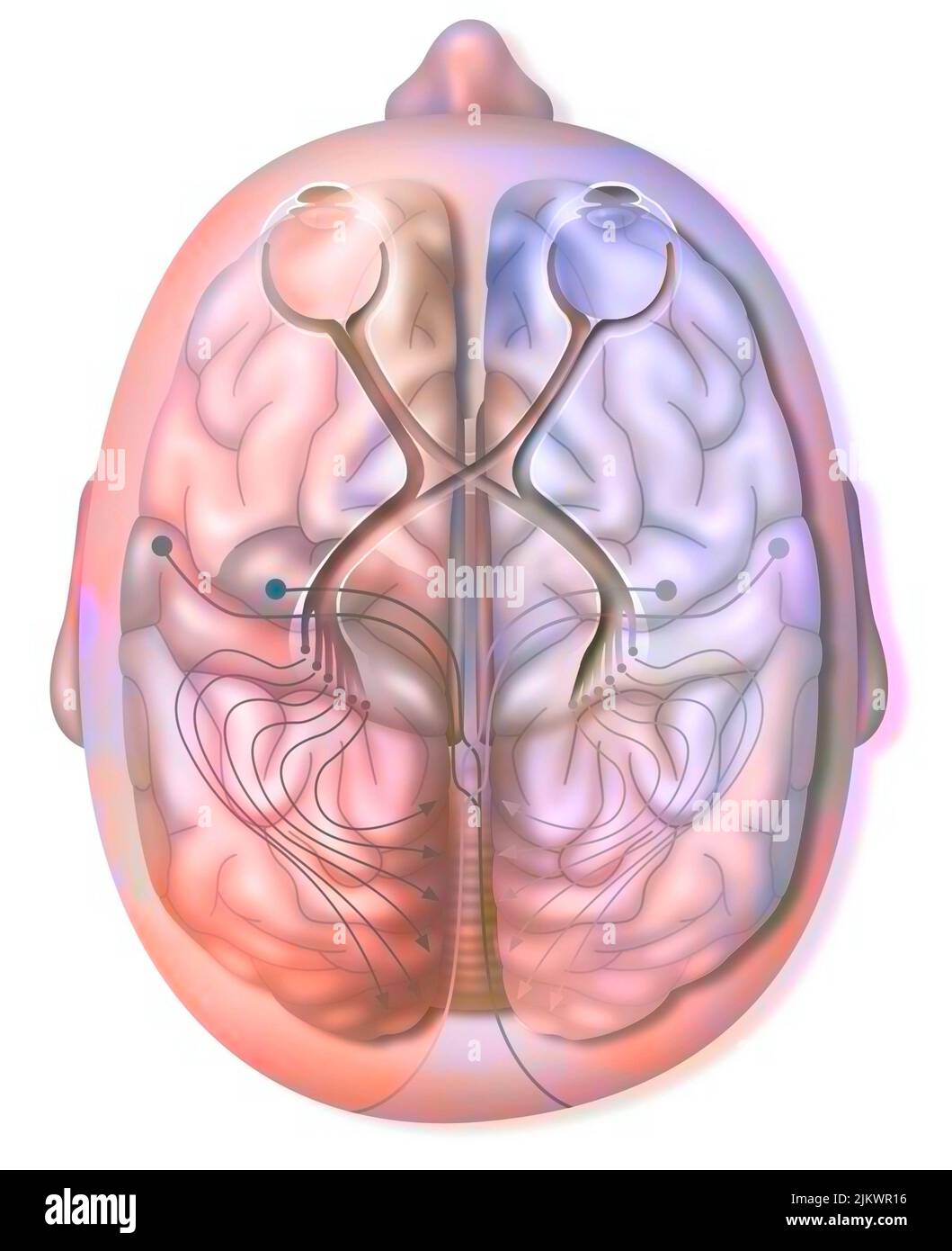 Superior view of the path and transmission of visual information from the retina. Stock Photohttps://www.alamy.com/image-license-details/?v=1https://www.alamy.com/superior-view-of-the-path-and-transmission-of-visual-information-from-the-retina-image476925266.html
RF2JKWR16–Superior view of the path and transmission of visual information from the retina.
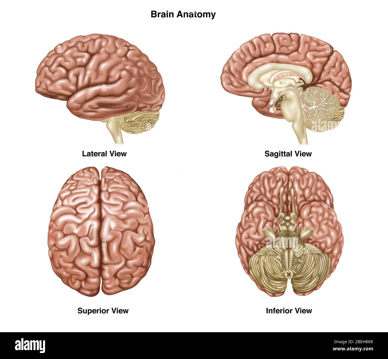 Brain Anatomy, Illustration Stock Photohttps://www.alamy.com/image-license-details/?v=1https://www.alamy.com/brain-anatomy-illustration-image353192191.html
RF2BEH86R–Brain Anatomy, Illustration
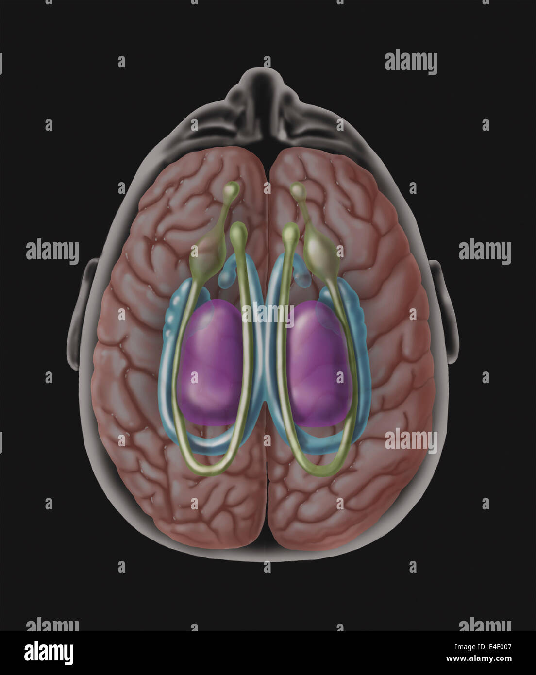 View of limbic system as seen from directly above the head. Stock Photohttps://www.alamy.com/image-license-details/?v=1https://www.alamy.com/stock-photo-view-of-limbic-system-as-seen-from-directly-above-the-head-71629383.html
RME4F007–View of limbic system as seen from directly above the head.
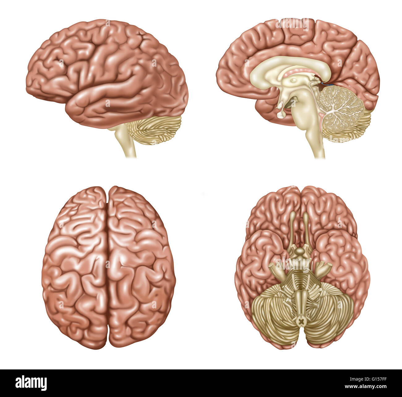 Illustration showing anatomy of a normal brain in lateral, sagittal, superior, and inferior view. Detailed in the various views are: frontal lobe, central sulcus, parietal lobe, sylvian fissure, temporal lobe, occipital lobe, cerebellum, brain stem, corpu Stock Photohttps://www.alamy.com/image-license-details/?v=1https://www.alamy.com/stock-photo-illustration-showing-anatomy-of-a-normal-brain-in-lateral-sagittal-103992547.html
RMG157FF–Illustration showing anatomy of a normal brain in lateral, sagittal, superior, and inferior view. Detailed in the various views are: frontal lobe, central sulcus, parietal lobe, sylvian fissure, temporal lobe, occipital lobe, cerebellum, brain stem, corpu
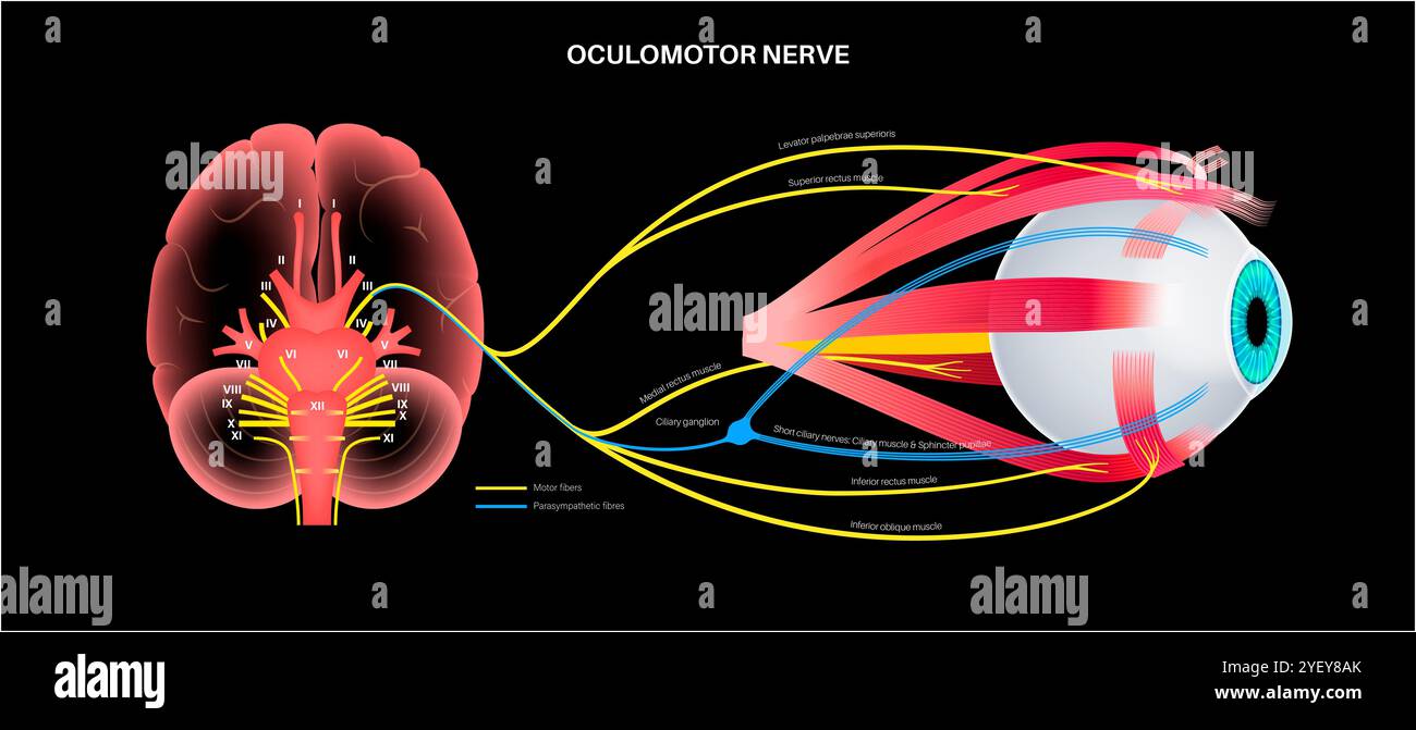 Illustration of the oculomotor nerve anatomy in the human brain. The oculomotor nerve divides into superior and inferior branches in the anterior part of the cavernous sinus. Stock Photohttps://www.alamy.com/image-license-details/?v=1https://www.alamy.com/illustration-of-the-oculomotor-nerve-anatomy-in-the-human-brain-the-oculomotor-nerve-divides-into-superior-and-inferior-branches-in-the-anterior-part-of-the-cavernous-sinus-image628777707.html
RF2YEY8AK–Illustration of the oculomotor nerve anatomy in the human brain. The oculomotor nerve divides into superior and inferior branches in the anterior part of the cavernous sinus.
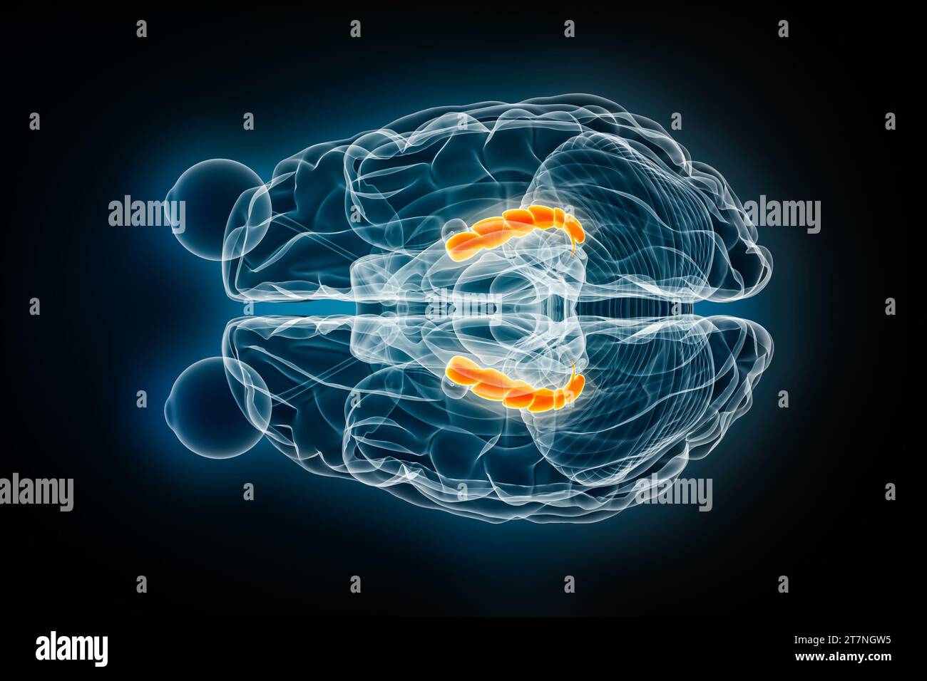 Hippocampus x-ray superior or top view 3D rendering illustration. Human brain, limbic and nervous system anatomy, medical, healthcare, biology, scienc Stock Photohttps://www.alamy.com/image-license-details/?v=1https://www.alamy.com/hippocampus-x-ray-superior-or-top-view-3d-rendering-illustration-human-brain-limbic-and-nervous-system-anatomy-medical-healthcare-biology-scienc-image572718977.html
RF2T7NGW5–Hippocampus x-ray superior or top view 3D rendering illustration. Human brain, limbic and nervous system anatomy, medical, healthcare, biology, scienc
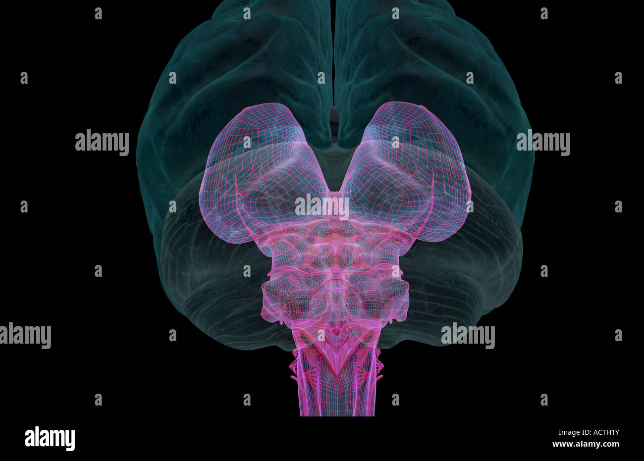 The brainstem Stock Photohttps://www.alamy.com/image-license-details/?v=1https://www.alamy.com/stock-photo-the-brainstem-13227110.html
RFACTH1Y–The brainstem
 . The anatomical record. Anatomy; Anatomy. Cer. Hem. cbl. Vent. IV. Fus. vent. y^ . Fig. 8 Posterior view of the brain of the pig. Coll. sup., coUirulus superior; Coll. inf., colUculus inferior; Hem. cbl., Heniisphaerium cerebelli; Th., thalamic mass; Ve7i(. Ill, vcntriculus tertius; Venl. IV, ventriculus quartus. Fig. 9 Mesial aspect of the brain of the pig. Cblm., cerebellum; Cer., point at which thin cerebrum was detached; Coll., colliculi; Fm^. rent., fused ventricles with medullary velum removed; Th., thalamic mass. Micro.'^copicnl .s7(/r/)/ of ."iedions throiujh (he cord. The medull Stock Photohttps://www.alamy.com/image-license-details/?v=1https://www.alamy.com/the-anatomical-record-anatomy-anatomy-cer-hem-cbl-vent-iv-fus-vent-y-fig-8-posterior-view-of-the-brain-of-the-pig-coll-sup-couirulus-superior-coll-inf-coluculus-inferior-hem-cbl-heniisphaerium-cerebelli-th-thalamic-mass-ve7i-ill-vcntriculus-tertius-venl-iv-ventriculus-quartus-fig-9-mesial-aspect-of-the-brain-of-the-pig-cblm-cerebellum-cer-point-at-which-thin-cerebrum-was-detached-coll-colliculi-fm-rent-fused-ventricles-with-medullary-velum-removed-th-thalamic-mass-microcopicnl-s7r-of-quotiedions-throiujh-he-cord-the-medull-image236874347.html
RMRNAFJ3–. The anatomical record. Anatomy; Anatomy. Cer. Hem. cbl. Vent. IV. Fus. vent. y^ . Fig. 8 Posterior view of the brain of the pig. Coll. sup., coUirulus superior; Coll. inf., colUculus inferior; Hem. cbl., Heniisphaerium cerebelli; Th., thalamic mass; Ve7i(. Ill, vcntriculus tertius; Venl. IV, ventriculus quartus. Fig. 9 Mesial aspect of the brain of the pig. Cblm., cerebellum; Cer., point at which thin cerebrum was detached; Coll., colliculi; Fm^. rent., fused ventricles with medullary velum removed; Th., thalamic mass. Micro.'^copicnl .s7(/r/)/ of ."iedions throiujh (he cord. The medull
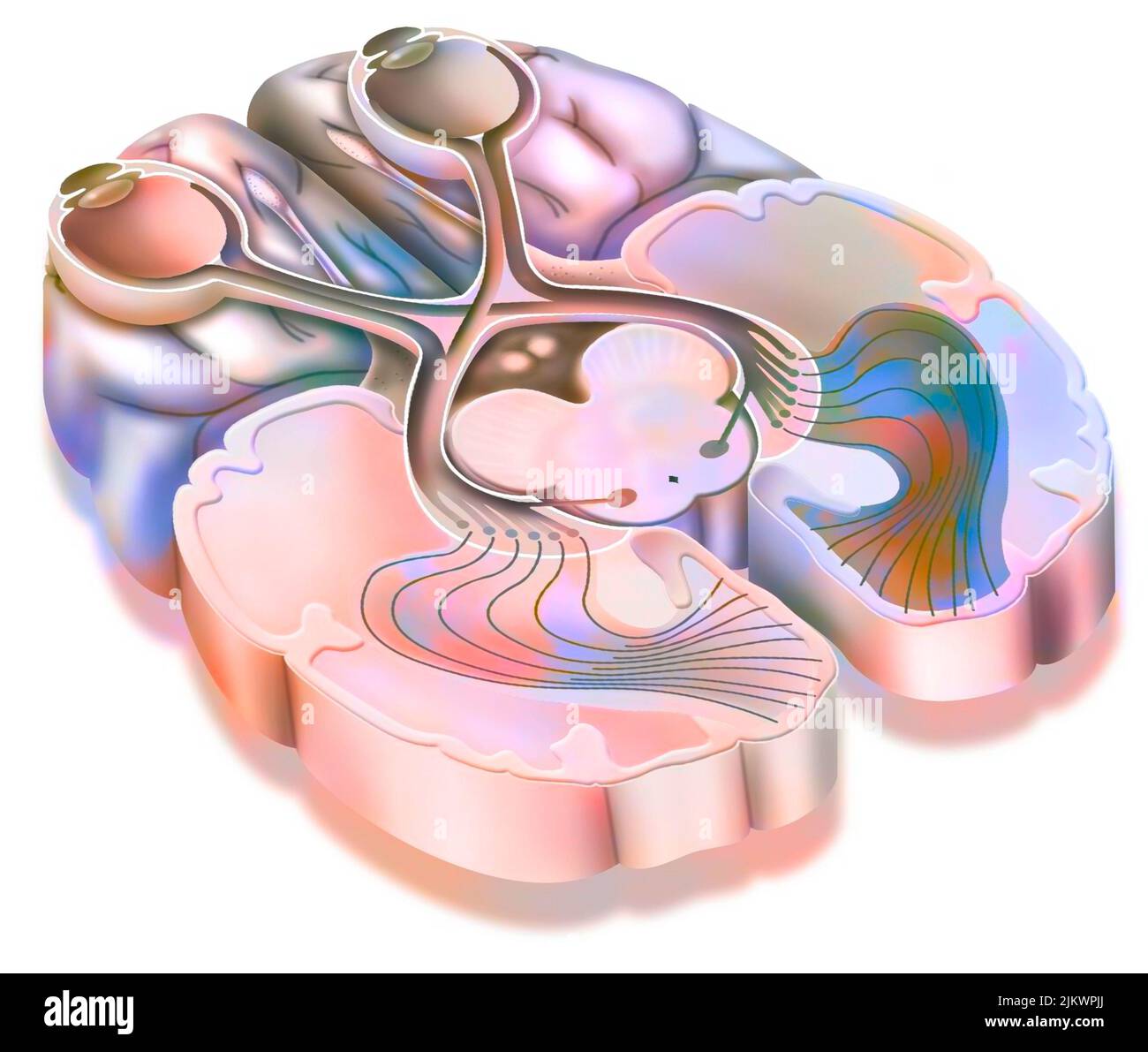 The optic tract: transmission of visual information from the retina to the visual cortex. Stock Photohttps://www.alamy.com/image-license-details/?v=1https://www.alamy.com/the-optic-tract-transmission-of-visual-information-from-the-retina-to-the-visual-cortex-image476924970.html
RF2JKWPJJ–The optic tract: transmission of visual information from the retina to the visual cortex.
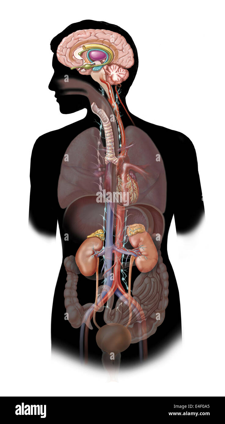 The sympathetic nervous system and the organs of fight-or-flight response. Stock Photohttps://www.alamy.com/image-license-details/?v=1https://www.alamy.com/stock-photo-the-sympathetic-nervous-system-and-the-organs-of-fight-or-flight-response-71629661.html
RME4F0A5–The sympathetic nervous system and the organs of fight-or-flight response.
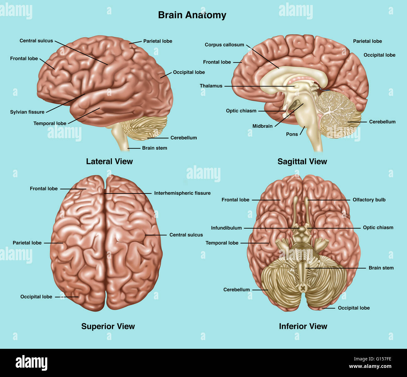 Illustration showing anatomy of a normal brain in lateral, sagittal, superior, and inferior view. Notated are frontal lobe, central sulcus, parietal lobe, sylvian fissure, temporal lobe, occipital lobe, cerebellum, brain stem, corpus callosum, thalamus, o Stock Photohttps://www.alamy.com/image-license-details/?v=1https://www.alamy.com/stock-photo-illustration-showing-anatomy-of-a-normal-brain-in-lateral-sagittal-103992546.html
RMG157FE–Illustration showing anatomy of a normal brain in lateral, sagittal, superior, and inferior view. Notated are frontal lobe, central sulcus, parietal lobe, sylvian fissure, temporal lobe, occipital lobe, cerebellum, brain stem, corpus callosum, thalamus, o
 Illustration of the oculomotor nerve anatomy in the human brain. The oculomotor nerve divides into superior and inferior branches in the anterior part of the cavernous sinus. Stock Photohttps://www.alamy.com/image-license-details/?v=1https://www.alamy.com/illustration-of-the-oculomotor-nerve-anatomy-in-the-human-brain-the-oculomotor-nerve-divides-into-superior-and-inferior-branches-in-the-anterior-part-of-the-cavernous-sinus-image628777715.html
RF2YEY8AY–Illustration of the oculomotor nerve anatomy in the human brain. The oculomotor nerve divides into superior and inferior branches in the anterior part of the cavernous sinus.
 Corpus callosum x-ray superior or top view 3D rendering illustration. Human brain and nervous system anatomy, medical, healthcare, biology, science, n Stock Photohttps://www.alamy.com/image-license-details/?v=1https://www.alamy.com/corpus-callosum-x-ray-superior-or-top-view-3d-rendering-illustration-human-brain-and-nervous-system-anatomy-medical-healthcare-biology-science-n-image572718971.html
RF2T7NGTY–Corpus callosum x-ray superior or top view 3D rendering illustration. Human brain and nervous system anatomy, medical, healthcare, biology, science, n
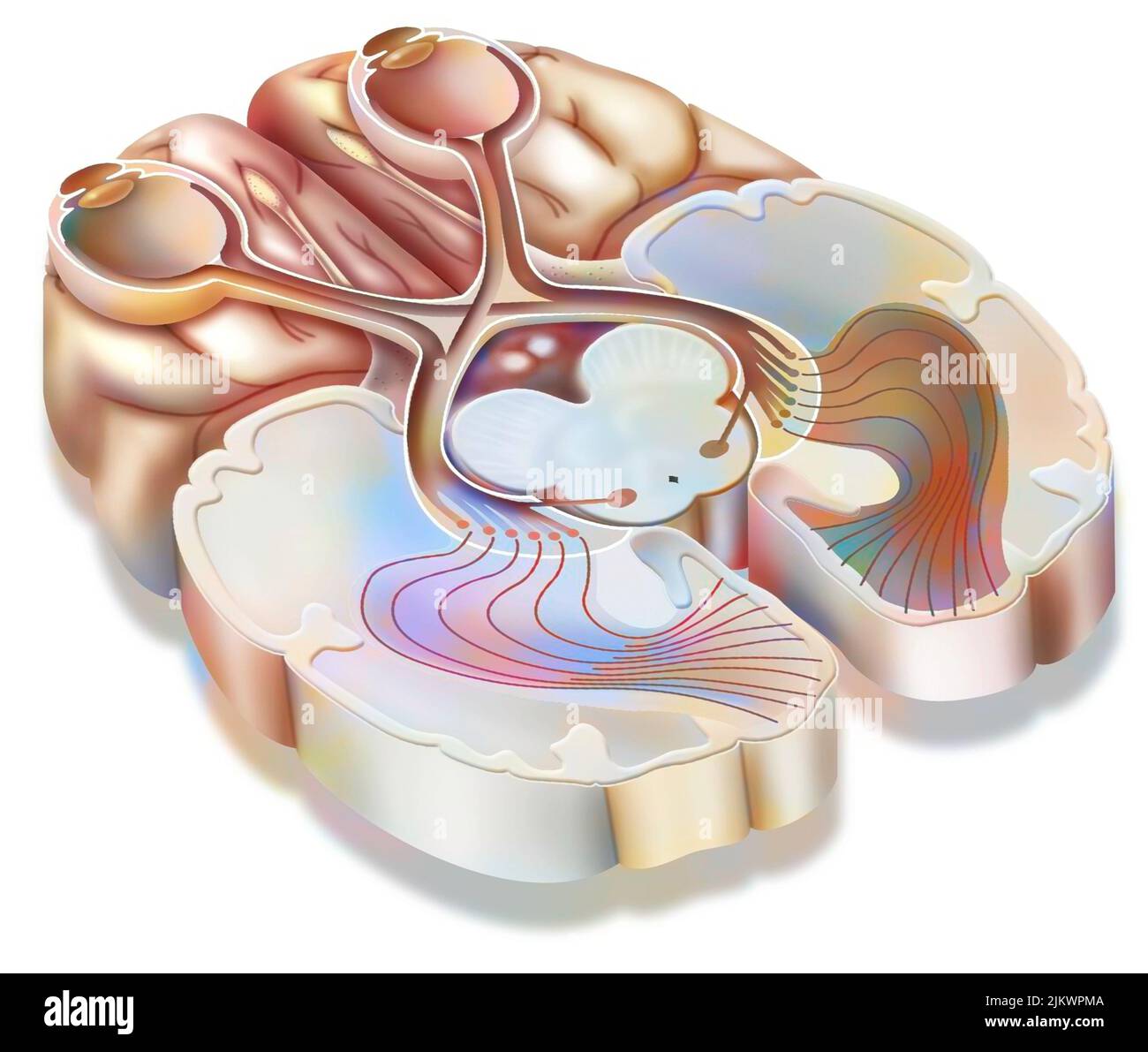 The optic tract: transmission of visual information from the retina to the visual cortex. Stock Photohttps://www.alamy.com/image-license-details/?v=1https://www.alamy.com/the-optic-tract-transmission-of-visual-information-from-the-retina-to-the-visual-cortex-image476925018.html
RF2JKWPMA–The optic tract: transmission of visual information from the retina to the visual cortex.
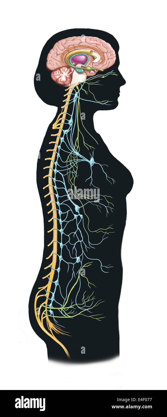 Side view of human body showing autonomic nervous system and limbic system within the brain. Green are parasympathetic nerves. B Stock Photohttps://www.alamy.com/image-license-details/?v=1https://www.alamy.com/stock-photo-side-view-of-human-body-showing-autonomic-nervous-system-and-limbic-71629579.html
RME4F077–Side view of human body showing autonomic nervous system and limbic system within the brain. Green are parasympathetic nerves. B
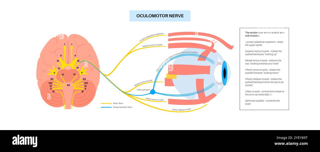 Illustration of the oculomotor nerve anatomy in the human brain. The oculomotor nerve divides into superior and inferior branches in the anterior part of the cavernous sinus. Stock Photohttps://www.alamy.com/image-license-details/?v=1https://www.alamy.com/illustration-of-the-oculomotor-nerve-anatomy-in-the-human-brain-the-oculomotor-nerve-divides-into-superior-and-inferior-branches-in-the-anterior-part-of-the-cavernous-sinus-image628777684.html
RF2YEY89T–Illustration of the oculomotor nerve anatomy in the human brain. The oculomotor nerve divides into superior and inferior branches in the anterior part of the cavernous sinus.
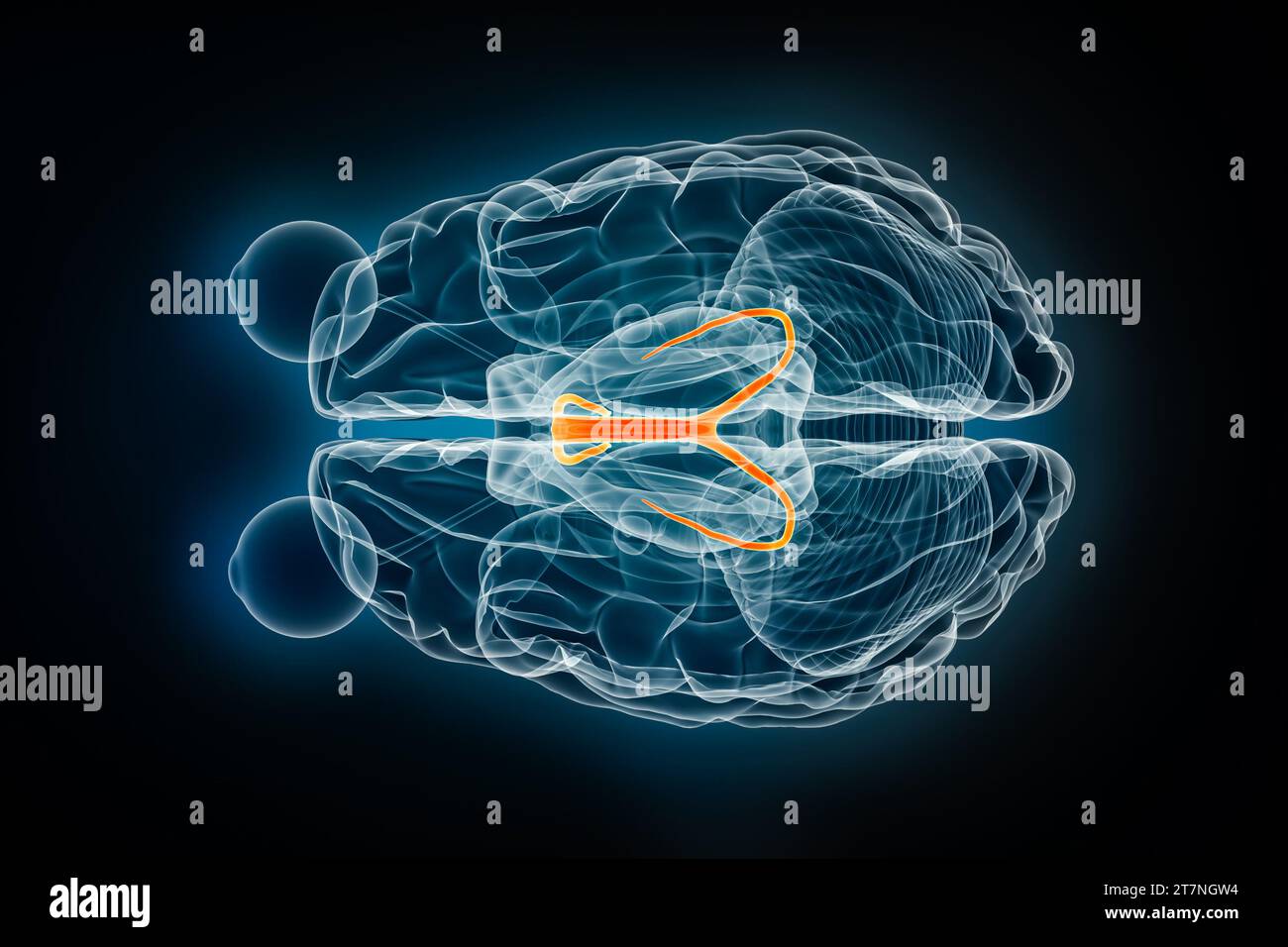 Fornix x-ray superior or top view 3D rendering illustration. Human brain, limbic and nervous system anatomy, medical, healthcare, biology, science, ne Stock Photohttps://www.alamy.com/image-license-details/?v=1https://www.alamy.com/fornix-x-ray-superior-or-top-view-3d-rendering-illustration-human-brain-limbic-and-nervous-system-anatomy-medical-healthcare-biology-science-ne-image572718976.html
RF2T7NGW4–Fornix x-ray superior or top view 3D rendering illustration. Human brain, limbic and nervous system anatomy, medical, healthcare, biology, science, ne
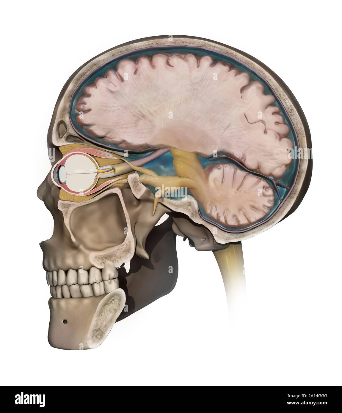 Medical illustration depicting the anatomy of a sagittal section of the human cranium. Stock Photohttps://www.alamy.com/image-license-details/?v=1https://www.alamy.com/medical-illustration-depicting-the-anatomy-of-a-sagittal-section-of-the-human-cranium-image327712464.html
RF2A14GGG–Medical illustration depicting the anatomy of a sagittal section of the human cranium.
 Illustration of the oculomotor nerve anatomy in the human brain. The oculomotor nerve divides into superior and inferior branches in the anterior part of the cavernous sinus. Stock Photohttps://www.alamy.com/image-license-details/?v=1https://www.alamy.com/illustration-of-the-oculomotor-nerve-anatomy-in-the-human-brain-the-oculomotor-nerve-divides-into-superior-and-inferior-branches-in-the-anterior-part-of-the-cavernous-sinus-image628777680.html
RF2YEY89M–Illustration of the oculomotor nerve anatomy in the human brain. The oculomotor nerve divides into superior and inferior branches in the anterior part of the cavernous sinus.
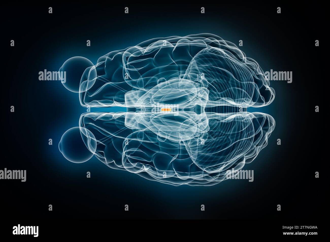 Interthalamic adhesion x-ray superior or top view 3D rendering illustration. Human brain and nervous system anatomy, medical, healthcare, biology, sci Stock Photohttps://www.alamy.com/image-license-details/?v=1https://www.alamy.com/interthalamic-adhesion-x-ray-superior-or-top-view-3d-rendering-illustration-human-brain-and-nervous-system-anatomy-medical-healthcare-biology-sci-image572718982.html
RF2T7NGWA–Interthalamic adhesion x-ray superior or top view 3D rendering illustration. Human brain and nervous system anatomy, medical, healthcare, biology, sci
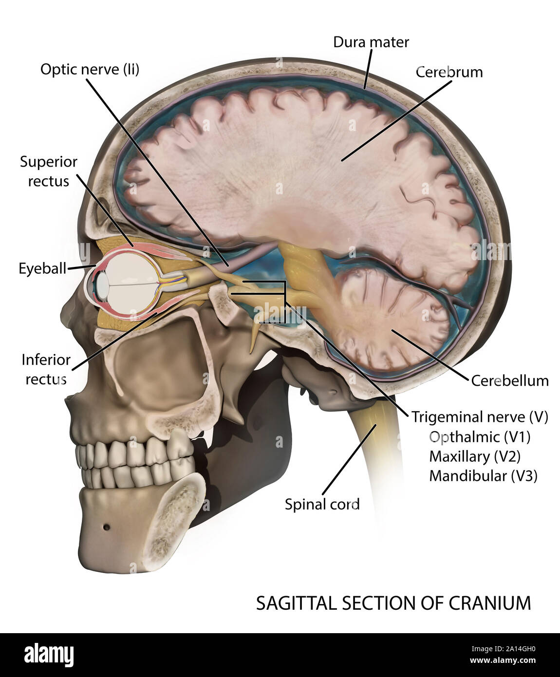 Medical illustration depicting the anatomy of a sagittal section of the human cranium. Stock Photohttps://www.alamy.com/image-license-details/?v=1https://www.alamy.com/medical-illustration-depicting-the-anatomy-of-a-sagittal-section-of-the-human-cranium-image327712476.html
RF2A14GH0–Medical illustration depicting the anatomy of a sagittal section of the human cranium.
 Serotonin released in the brain travels down the spinal cord to close the pain gates and block pain messages. Stock Photohttps://www.alamy.com/image-license-details/?v=1https://www.alamy.com/stock-photo-serotonin-released-in-the-brain-travels-down-the-spinal-cord-to-close-71629390.html
RME4F00E–Serotonin released in the brain travels down the spinal cord to close the pain gates and block pain messages.
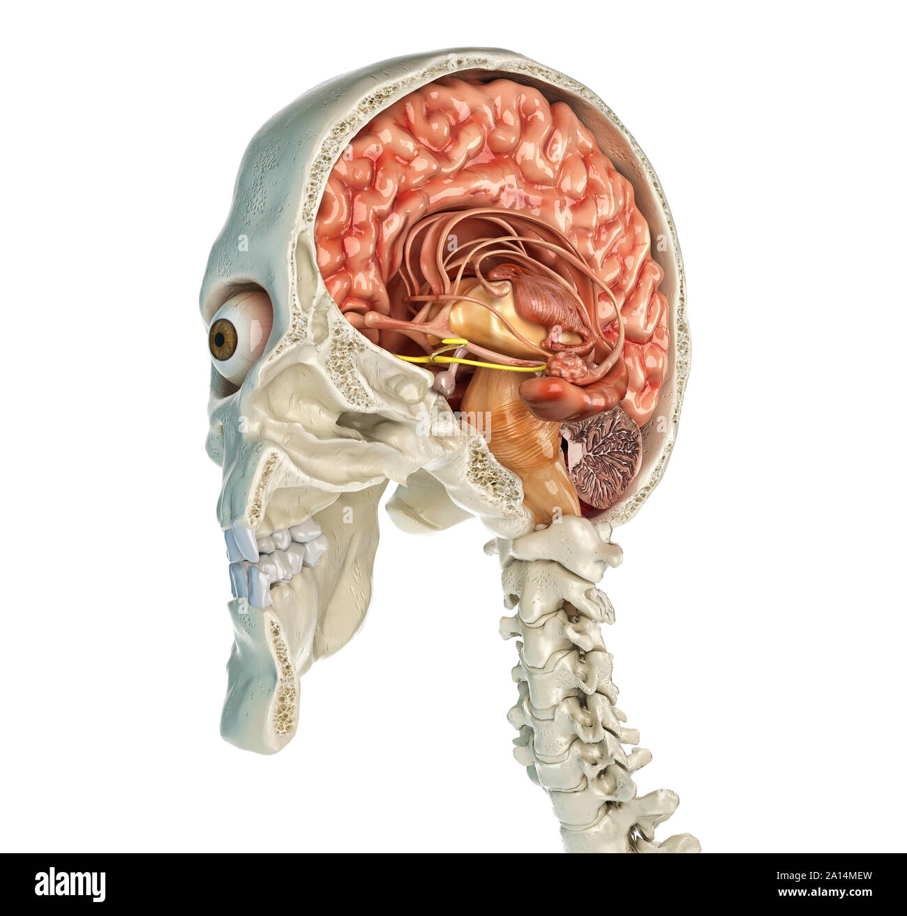
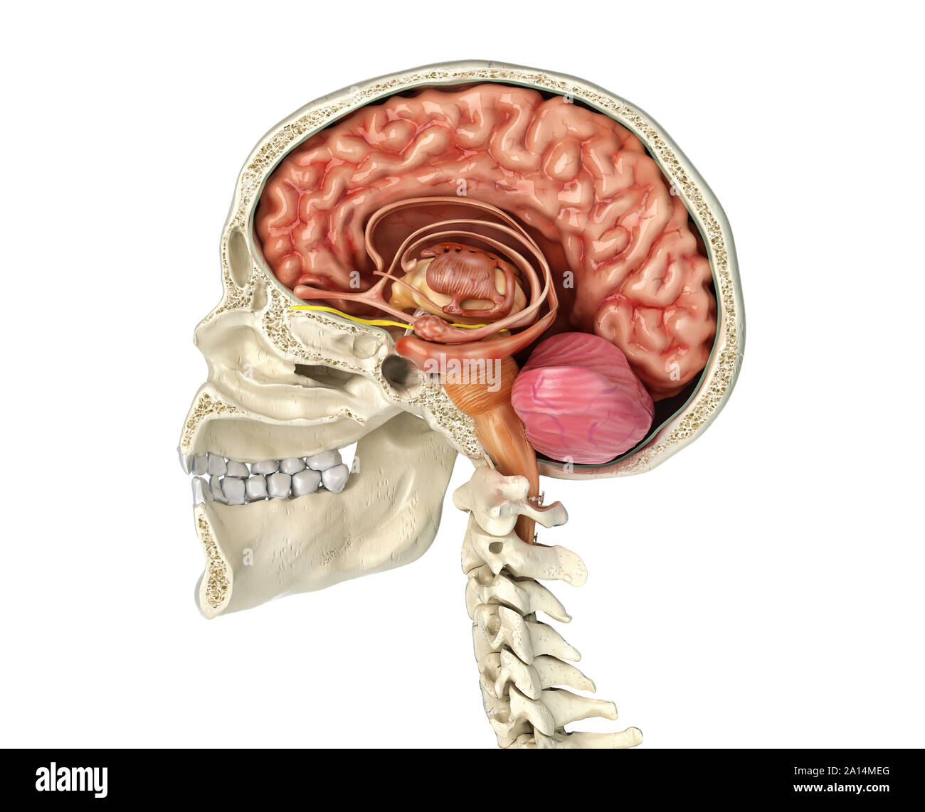 Human skull mid sagittal cross-section with brain. Stock Photohttps://www.alamy.com/image-license-details/?v=1https://www.alamy.com/human-skull-mid-sagittal-cross-section-with-brain-image327715544.html
RF2A14MEG–Human skull mid sagittal cross-section with brain.
 Illustration of the motor nerves of the eye, including the abducens, trochlear and oculomotor nerves in the human brain. These nerves innervate motor, sensory, and autonomic structures in the eyes. Stock Photohttps://www.alamy.com/image-license-details/?v=1https://www.alamy.com/illustration-of-the-motor-nerves-of-the-eye-including-the-abducens-trochlear-and-oculomotor-nerves-in-the-human-brain-these-nerves-innervate-motor-sensory-and-autonomic-structures-in-the-eyes-image628777693.html
RF2YEY8A5–Illustration of the motor nerves of the eye, including the abducens, trochlear and oculomotor nerves in the human brain. These nerves innervate motor, sensory, and autonomic structures in the eyes.
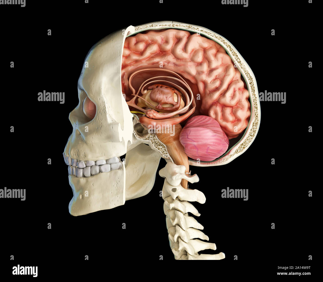 Human skull mid sagittal cross-section with brain. Stock Photohttps://www.alamy.com/image-license-details/?v=1https://www.alamy.com/human-skull-mid-sagittal-cross-section-with-brain-image327715412.html
RF2A14M9T–Human skull mid sagittal cross-section with brain.
 Illustration of the motor nerves of the eye, including the abducens, trochlear and oculomotor nerves in the human brain. These nerves innervate motor, sensory, and autonomic structures in the eyes. Stock Photohttps://www.alamy.com/image-license-details/?v=1https://www.alamy.com/illustration-of-the-motor-nerves-of-the-eye-including-the-abducens-trochlear-and-oculomotor-nerves-in-the-human-brain-these-nerves-innervate-motor-sensory-and-autonomic-structures-in-the-eyes-image628777690.html
RF2YEY8A2–Illustration of the motor nerves of the eye, including the abducens, trochlear and oculomotor nerves in the human brain. These nerves innervate motor, sensory, and autonomic structures in the eyes.
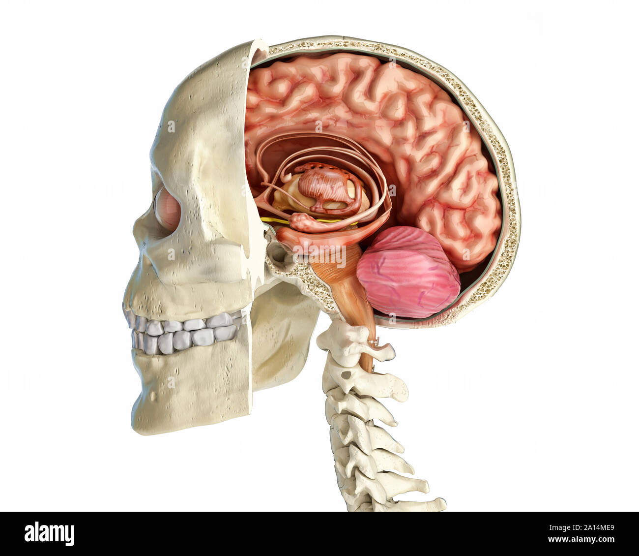 Human skull mid sagittal cross-section with brain. Stock Photohttps://www.alamy.com/image-license-details/?v=1https://www.alamy.com/human-skull-mid-sagittal-cross-section-with-brain-image327715537.html
RF2A14ME9–Human skull mid sagittal cross-section with brain.
 Human skull mid sagittal cross-section with brain. Stock Photohttps://www.alamy.com/image-license-details/?v=1https://www.alamy.com/human-skull-mid-sagittal-cross-section-with-brain-image327715547.html
RF2A14MEK–Human skull mid sagittal cross-section with brain.
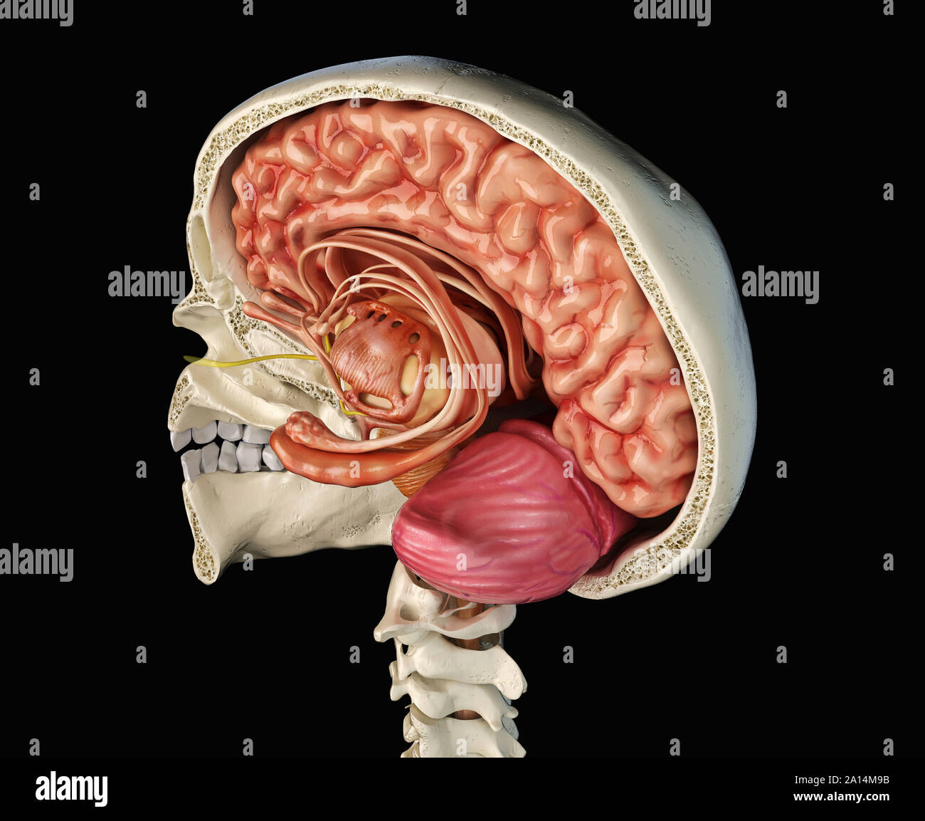 Human skull mid sagittal cross-section with brain. Stock Photohttps://www.alamy.com/image-license-details/?v=1https://www.alamy.com/human-skull-mid-sagittal-cross-section-with-brain-image327715399.html
RF2A14M9B–Human skull mid sagittal cross-section with brain.
